pilus production, hemagglutination, and adhesion by porcine
TRANSCRIPT

Vol. 35, No. 1INFECTION AND IMMUNITY, Jan. 1982, p. 305-3130019-9567/82/010305-09$02.00/0
Pilus Production, Hemagglutination, and Adhesion by PorcineStrains of Enterotoxigenic Escherichia coli Lacking K88, K99,
and 987P AntigensM. AWAD-MASALMEH,t H. W. MOON,* P. L. RUNNELS, AND R. A. SCHNEIDER
National Animal Disease Center, U.S. Department of Agriculture, Science and Education Administration,Ames, Iowa 50010
Received 20 July 1981/Accepted 21 August 1981
Three strains of enterotoxigenic Escherichia coli which adhered, colonizedintensively, and caused disease in pig intestine, but which did not produce pili ofthe K88, K99, or 987P antigen types were designated 3P- ETEC. The 3P- ETECcaused mannose-resistant hemagglutination, adhered to porcine intestinal epithe-lial cells in vitro, and produced pili. However, most bacteria taken directly fromthe intestine of pigs infected with 3P- ETEC appeared to be nonpiliated. Twopreparations were isolated from the 3P- ETEC. One (material A) contained pili,caused mannose-sensitive hemagglutination, and did not inhibit adhesion of wholebacteria to epithelial cells in vitro. The other (material B) had no demonstrablepili, caused mannose-resistant hemagglutination, and blocked ahesion of bacteriato epithelial cells in vitro. Antiserum against an acapsular mutant (K-) of one 3P-ETEC strain was absorbed to remove antibodies directed against somatic (0)antigen. The absorbed antiserum agglutinated all three 3P- ETEC strains grownin the K- form at 37°C, but not when they were grown at 18°C. The absorbedantiserum blocked the hemagglutinating activity of material B, but not of materialA. It also reacted (via indirect immunofluorescence) with all of the 3P- ETECwhen they were grown in pig intestine. The results were interpreted to indicatethat: (i) the epithelial adhesive and mannose-resistant hemagglutinating activitiesof the 3P- ETEC strains may be mediated by an antigen contained in material B;(ii) this antigen either is not pilus associated or is associated with pili that are notdemonstrable by the methods used here; (iii) the 3P- ETEC strains produce type 1pili which do not mediate their adhesion to intestinal epithelium of pigs.
Pilus-mediated adhesion to epithelium ap-pears to be essential for colonization of the smallintestine by enterotoxigenic Escherichia coli(ETEC). However, pili apparently are only oneof a constellation of attributes which facilitatecolonization by ETEC. For example, both cap-sule and pili are required for colonization bysome strains of ETEC (8, 13, 15, 16). Further-more, some ETEC that do not produce any ofthe pili that have been implicated in intestinalcolonization are enteropathogenic, i.e., they col-onize intestine and cause diarrhea in pigs (11) orpeople (9). Three antigenic types of pili (K88,K99, and 987P) have been implicated in coloni-zation of pig intestine by ETEC. ETEC thatadhered, colonized, and caused disease in pigsmall intestine, without producing any of thesethree types of pili, were designated as 3P-ETEC (11).The hypothesis of this study was that 3P-
t Visiting scientist from the Institute of Bacteriology andVeterinary Hygiene, University of Veterinary Medicine, Vi-enna, Austria.
ETEC produce previously unrecognized anti-genic types of pili which mediate their intestinaladhesion and colonization. We found that the3P- ETEC do produce pili, but the pili demon-strated apparently do not mediate intestinal col-onization and adhesion. We present evidencethat adhesion of 3P- ETEC to intestinal epitheli-um is mediated by material which does notcontain pili demonstrable by electron micro-scopic examination of negatively stained prepa-rations.
MATERIALS AND METHODSE. coli strains and growth conditions. The 10 E. coli
strains used and their characteristics are listed in Table1. Previous tests demonstrated that the 3P- strains didnot produce K88, K99, or 987P antigens (11). DoyleEvans (personal communication) determined that theydid not produce pilus antigens CFAII or CFAJII (4).Acapsular (K-) mutants were selected by methodsreported previously (13). Unless stated otherwise, allresults were from bacteria grown on Minca agar plusIsoVitaleX (BBL Microbiology Systems; Minca Is) (5)at 37°C for 24 h. For some studies, bacteria were
305
Dow
nloa
ded
from
http
s://j
ourn
als.
asm
.org
/jour
nal/i
ai o
n 21
Feb
ruar
y 20
22 b
y 11
2.11
8.22
3.14
3.

306 AWAD-MASALMEH ET AL.
TABLE 1. Characteristics of the E. coli strains usedStrain no. Source Serotype Enterotoxinsa Pilus antigen
VAC-1676 Swine 0101:K30:H - b STa 3P-KATI-1706 Swine 0101:K30:H- b STa 3P-VC-1751 Swine 0101:K27:H- b STa 3P-431 Swine 0101:K30:H- STa K99637 Swine 064:K+:H- STa K99987 Swine 09:K103:H- STa 987P263 Swine 08:K87:H19 LT K88123 Swine 043:K-:H28 None ?1793 Laboratoryd 09:K30:H1 None ?1788 Laboratory 0101:K-:H- None ?
a STa is enterotoxin demonstrable in infant mice, and LT is enterotoxin demonstrable in adrenal cells inculture.
b Determined by Fritz and Ida 0rskov.c Does not produce K88, K99, or 987P, but does colonize pig intestine.d Standard laboratory strain, original source not known.
grown on blood agar base (Difco) plus 5% sheep bloodor on Trypticase soy agar (BBL) at 37°C. Whendesired, pellicle formation was encouraged by growthat 37°C for up to a week in Trypticase soy broth(BBL).
Electron microscopy. Cultures and fluids examinedfor pili by electron microscopy were prepared, usingthe potassium phosphotungstate method for negativestaining, and examined as reported previously (8).Sections of ileum from neonatal colostrum-deprivedpigs were obtained 16 to 18 h after the pigs wereinfected with ETEC. The sections were prepared andexamined by electron microscopy (12).
Hemagglutination. Hemagglutination tests were con-ducted as described by Evans et al. (4).Adhesion to intestinal epithelial cells in vitro. Adhe-
sion determinations were conducted by a microscopicmethod (14). The epithelial cells used were isolatedfrom the small intestine of hysterectomy-derived, co-lostrum-deprived pigs that were less than 12 h old.Antiserum production. A K- mutant of strain KATI-
1706 was grown on Minca Is and then inoculated intobroth for antigen production. The antigen producedwas used to raise antiserum in rabbits. Antigen pro-duction and immunization were done according to theprocedure cited as method 1 for the production of E.coli 0: K antiserum (3). The antiserum produced wasabsorbed with the K- mutant of strain KATI-1706,grown at 18°C. The serum was first absorbed with thisantigen after the antigen had been heated (100°C, 1 h),and the serum was then absorbed with the livingbacteria. These absorptions were to remove 0 anti-bodies and antibodies against surface antigens pro-duced when the strain was grown at 18°C. Serumabsorption procedures and agglutination tests (slidemethod) were done by standard techniques (3). Theabsorbed serum did not agglutinate appropriately pre-pared 0101 or K30 antigens but did agglutinate (titer of1:200, slide agglutination) the autologous living acap-sular mutant grown on Minca Is agar at 37°C. Theagglutinins that remained in the absorbed antiserumwere assumed to be directed against surface antigens,and the absorbed antiserum will be referred to as 1706-SA (surface antigens).Immunofluorescence. Sections of ileum from new-
born colostrum-deprived pigs were obtained 16 to 18 h
after the pigs were infected with ETEC (13). Thesections were prepared and stained by using the 1706-SA antiserum and an indirect immunofluorescent tech-nique (11).
Isolation of materials A and B. Acapsular mutants ofstrains VAC-1676, KATI-1706, and VC-1751 grown onMinca Is agar for 24 h at 37°C were suspended inphosphate-buffered saline at pH 8.5 (PBS). The sus-pensions were frozen and thawed twice, agitated vig-orously in a Vortex mixer for 15 min, and centrifuged(12,000 x g and 4°C for 15 min). The supernatantswere harvested, and the centrifugations were repeatedto remove debris. Acetic acid was added to the clearsupernatants to pH 5.5, and the supernatants werecentrifuged (27,000 x g, 4°C, 10 min). The resultingpellets were separated from the supernatants and wereredissolved in PBS, labeled as material A, and frozenat -70°C. The supernatants were mixed with ammoni-um sulfate (70% saturation), held for 2 h at 4°C, andthen centrifuged (27,000 x g, 4°C, 15 min). Theresulting pellets were dissolved in PBS, labeled asmaterial B, frozen, and stored at -70°C.
Purification of material A. Material A was thawed,dialyzed in 0.05 M sodium phosphate at pH 7.0 (P04buffer), and centrifuged (48,000 x g, 40C, 15 min). Thepellet was discarded, and the supermatant was harvest-ed, brought to 10% saturation with ammonium sulfate,and recentrifuged. The resulting pellet was suspendedin P04 buffer and applied to a Sephadex G25 columnequilibrated with P04 buffer. Fractions were collectedand assayed for absorbance at 280 nm. The fractionscoinciding with the first absorbance peak eluted werepooled and brought to 10% ammonium sulfate satura-tion. The resulting precipitate was separated by cen-trifugation, dissolved in P04 buffer, dialyzed againstP04 buffer, and labeled as purified material A.
Purification of material B. Material B was thawed,dialyzed, and centrifuged by the same methods asmaterial A. The resulting supernatant was brought to60% saturation with ammonium sulfate and centri-fuged (48,000 x g, 4°C, 15 min). The pellet washarvested, suspended in P04 buffer, and applied to aSephadex G50 column equilibrated with P04. Frac-tions were collected, and those coinciding with thefirst peak of absorbance at 280 nm eluted were pooled,brought to 20% ammonium sulfate saturation, and
INFECT. IMMUN.
Dow
nloa
ded
from
http
s://j
ourn
als.
asm
.org
/jour
nal/i
ai o
n 21
Feb
ruar
y 20
22 b
y 11
2.11
8.22
3.14
3.

NONPILIATED PORCINE ETEC 307
TABLE 2. MRHA' caused by enterotoxigenic E. coli of different pilus antigen typesE. coli Hemagglutination of erythrocytes fromb:
Strain Growth medium Pilus type Pigs Sheep Rabbits Chickens Guinea-pigs Cattle Horses
VAC-1676 Minca Is agar 3P- c 4+ 4+ 4+ 4+ 4+ 0 1+KATI-1706 Minca Is agar 3P- 4+ 4+ 4+ 4+ 4+ 0 1+VC-1751 Minca Is agar 3P- 4+ 4+ 4+ 4+ 4+ 0 1 +431 MincaIs agar K99 4+ 4+ 4+ 4+ 4+ 2+ 4+263 Trypticase soy agar K88 0 1+ 2+ 2+ 2+ 0 0987 Blood agar 987P 0 0 0 1+ 0 0 0
a Test solutions contained 0.5% mannose.b 4+, Instantaneous and complete hemagglutination; 2+, delayed and incomplete hemagglutination; 1 +, weak
or questionable hemagglutination.c Does not produce K88, K99, or 987P.
centrifuged (48,000 x g, 4°C, 15 min). The supernatantwas harvested, brought to 30% saturation with ammo-nium sulfate, and recentrifuged. The pellet was har-vested, suspended in P04 buffer, dialyzed against P04buffer, and labeled as purified material B.
Immunodiffusion and electrophoresis. Agar gel dou-ble-diffusion (Ouchterlony) and polyacrylamide gelelectrophoresis tests were conducted as reported pre-viously (6).
RESULTSBacterial hemagglutination. Cultures of the
3P- strains (VAC-1676, KATI-1706, and VC-1751) grown on Minca Is agar in the K+ parentalforms and as K- mutants all strongly hemagglu-tinated porcine erythrocytes in a D-mannose-resistant fashion (mannose-resistant hemaggluti-nation; MRHA). This activity was not presentwhen the cultures were grown on Trypticase soyagar or blood agar. Therefore, subsequent hem-agglutination tests with the 3P- strains weredone using K+ bacteria grown on Minca Is agar.The 3P- strains also caused instantaneous andcomplete MRHA of erythrocytes from sheep,rabbits, chickens, and guinea pigs, but not oferythrocytes from cattle and horses (Table 2).This MRHA pattern of the 3P- strains wasdifferent from the MRHA patterns of K99+,K88+, and 987P+ strains of E. coli that weregrown to facilitate production of K99, K88, or987P pili (Table 2).The MRHA activity of the 3P- strains for pig
erythrocytes was not enhanced by cooling (4°C)and was not reversed when the agglutinatedmixtures were warmed to 40, 50, or 60°C. Whenthe 3P- strains were grown at 18°C instead of37°C they did not cause MRHA of erythrocytesfrom pigs.
Pellicle and pilus formation. The 3P- strainsdeveloped pellicles on the surfaces of brothcultures after several days of incubation at 37°C.About 10% of the bacteria from the pellicles andfrom Minca Is agar were piliated when examinedby negative staining and electron microscopy.The pili were straight or gently curved and
regular and had a mean diameter of 5.1 nm (Fig.1).
Serology. The 1706-SA antiserum did not ag-glutinate appropriately prepared 0101, K30,K88, K99, or 987P antigens (prepared fromstrains 1788, 1793, 263, 637, and 987, respective-ly). It did agglutinate strains VAC-1676, KATI-1706, and VC-1751 (titer, 1: 200 by slide aggluti-nation) when they were grown in the K- form onMinca Is agar at 37°C. This agglutination did notoccur when the bacteria were grown at 18°C onMinca Is agar, or at 37°C on blood or Trypticasesoy agar. The K+ forms of VAC-1676, KATI-1706, and VC-1751 were not usually agglutinatedby the serum; however, undiluted serum did
FIG. 1. Electron micrograph of negatively stainedE. coli VAC-1676. The pili have a mean diameter of 5.1nm.
VOL. 35, 1982
Dow
nloa
ded
from
http
s://j
ourn
als.
asm
.org
/jour
nal/i
ai o
n 21
Feb
ruar
y 20
22 b
y 11
2.11
8.22
3.14
3.

308 AWAD-MASALMEH ET AL.
sometimes agglutinate them weakly. The agglu-tinins for KATI-1706 (grown in the K- form onMinca Is agar at 37°C) were removed by absorb-ing the serum with either strain VAC-1676 orstrain VC-1751 (grown in the K- form on MincaIs agar at 37°C).
Adhesion to intestinal epithelial cells in vitro.Acapsular mutants of the 3P- strains (Minca Isgrown) all adhered to isolated epithelial cells.The number of adhering bacteria per epithelialcell with 3P- strains was somewhat less thanthat with the K88+ positive control strain 263but more than that with the negative controlstrain 123 (Table 3). The adhesion of the 3P-strains was markedly reduced (K, 2.3 ± 2.8bacteria per epithelial cell) when the bacteriawere grown at 18°C.
Immunofluorescent staining and electron mi-croscopy of bacteria grown in pig ileum. Thebacterial layers adherent to ileal villi of pigsinfected with each of the 3P- strains (four pigsper strain for each of the three strains) reactedwith the 1706-SA antiserum in indirect immuno-fluorescence (Fig. 2). The serum did not reactwith the adherent bacterial layer in the ileum of apig infected with the 987P+ strain 987 nor theilea from any of eight pigs exposed to thenonenterotoxigenic strain 123.
Ileal fluids from the pigs infected with the 3P-strains were prepared (by negative staining) fordirect electron microscopic examination. Theproportion of the bacteria in these samples thatcarried demonstrable pili varied from 0 to 2%.The pili observed resembled those produced invitro (Fig. 1).
Bacterial layers and associated ileal villous
TABLE 3. Effects of material A and material B' onadhesion of autogenous bacteria to isolated intestinal
epithelial cells from pigs
No. of adherent bacteria per epithelial cell'E. coli after treatmentc:strain
None Material Ad Material B
VAC-1676 17.9 ± 10.1 16.8 ± 8.5 5.4 ± 4.2KATI-1706 14.9 ± 10.2 15.2 ± 11.2 3.4 ± 4.2VC-1751 20.2 ± 9.7 19.3 ± 10.2 5.2 ± 4.3263 27.2 ± 6.5 NT NT123 7.9 ± 3.2 NT NT
a Materials A and B were extracted from acapsularmutants of E. coli strains VAC-1676, KATI-1706, andVC-1751.
b Mean ± standard error, microscopic count, 20epithelial cells evaluated per test and each test repli-cated five times. NT, Not tested.
c Epithelial cells were pretreated with material A orB and then exposed to bacteria of the strain used toprepare the material.
d Contains pili demonstrable by negative stain andelectron microscopy.
FIG. 2. Immunofluorescent photomicrograph ofvilli with fluorescent layers of adherent E. coli strainKATI-1706. Section from pig ileum stained (indirectmethod) with antiserum containing antibodies againstsurface antigens of strain KATI-1706.
epithelium from pigs infected with each of the3P- strains were examined by electron microsco-py (two pigs per strain). The ultrastructuralappearance of the layers and epithelium was thesame in all six of the pigs. Some, but not all, ofthe bacteria were surrounded by electron-lucentregions separating the bacteria from each other,as well as from epithelial cell microvilli andluminal debris (Fig. 3 and 4). Much of thevesicular and membranous luminal debris ap-peared to be from degenerate epithelial cells.The cytoplasm of some epithelial cells was elec-tron lucent, and these cells had patches ofdistorted or effaced microvilli (Fig. 3). Someepithelial cells had blebs of cytoplasm whichappeared to have been extruded through themicrovillus border into the intestinal lumen (Fig.4). The bacterial surfaces had a poorly defined,irregular coat of fuzzy material (Fig. 3), but nodiscrete attached filaments or pili were demon-strable. There were clumps of unidentified cell-free filaments scattered throughout the bacteriallayers (Fig. 3).
Bacterial layers associated with villi from apig infected with the K99+ strain 431 were alsoexamined by electron microscopy. This strainwas chosen for comparison because it has thesame 0 antigen as the 3P- strains and it also hasthe same capsular (K30) antigen as two of the3P- strains (Table 1). The appearance of the
INFECT. IMMUN.
Dow
nloa
ded
from
http
s://j
ourn
als.
asm
.org
/jour
nal/i
ai o
n 21
Feb
ruar
y 20
22 b
y 11
2.11
8.22
3.14
3.

NONPILIATED PORCINE ETEC 309
FIG. 3. Electron micrograph of the 3P- E. coli strain KATI-1706 adherent to epithelium on a villus in theileum of a pig. (A) The bacterial layer contains cell-free filamentous material as well as membranous andvesicular debris. Some of the epithelial cells have patches of irregular or effaced microvilli. Epithelial cells havepale cytoplasm, and the limiting membranes of their large cytoplasmic vacuoles are disrupted. (B) Highermagnification from the area outlined in (A). Cell-free filamentous material at the bottom; bacteria with a poorlydefined, irregular coat of fuzzy material above.
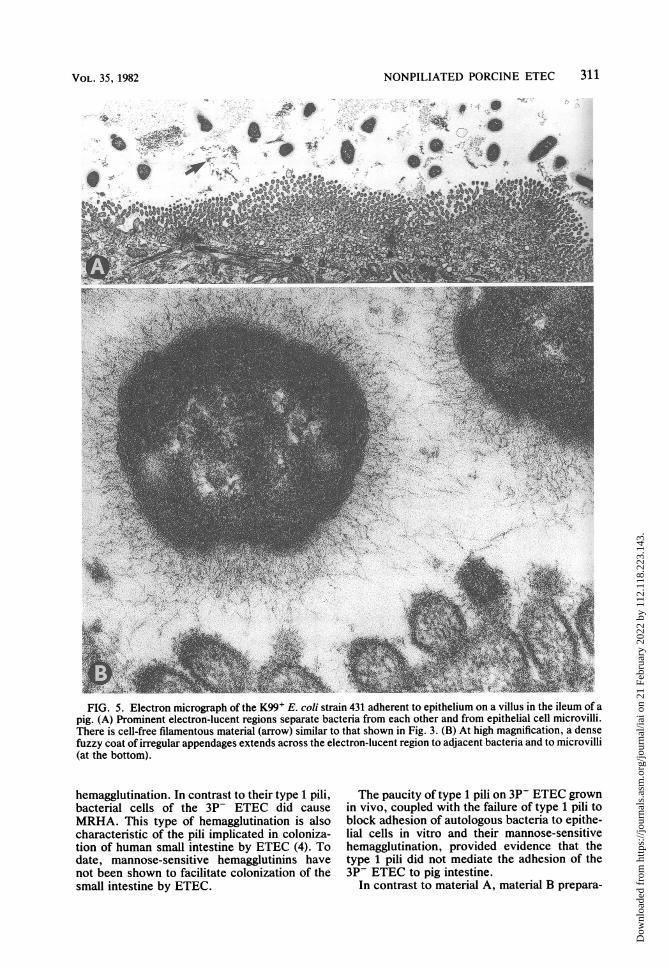
layers formed by this strain was as reportedpreviously for this and other K99+ ETEC (1, 10,12). The layers formed by strain 431 differedfrom those formed by the 3P- strains in thatelectron-lucent regions surrounding bacteriawere broader and were associated with almostevery bacterium (Fig. 5). Furthermore, nearlyall bacteria of strain 431 had a dense fuzzy coatof irregular appendages which at high magnifica-tion could be shown to extend across the elec-tron-lucent regions to adjacent bacteria and mi-crovilli (Fig. 5).
Characteristics of material A. Material A fromeach of the 3P- strains contained numerous pili(Fig. 6) resembling those demonstrated on the
bacterial cells. Material A from each of the threestrains hemagglutinated guinea pig erythrocytes.However, in contrast to the MRHA activities ofthe bacteria (Table 2), the hemagglutinating ac-tivities of all three material A preparations weremannose sensitive. The hemagglutinating activi-ty of the material A preparations was not inhibit-ed when the preparations were mixed with 1706-SA antiserum before the tests were done. Noneof the material A preparations formed precipitinlines when immunodiffusion tests were doneusing 1706-SA antiserum.
Characteristics of material B. In contrast tomaterial A, we were unable to demonstrate piliby electron microscopic examination of nega-
VOL. 35, 1982
Dow
nloa
ded
from
http
s://j
ourn
als.
asm
.org
/jour
nal/i
ai o
n 21
Feb
ruar
y 20
22 b
y 11
2.11
8.22
3.14
3.

310 AWAD-MASALMEH ET AL.
w.-m
4,
FIG. 4. Electron micrograph of the 3P- E. colistrain VC-1751 adherent to epithelium on a villus in theileum of a pig. A bleb of epithelial cell cytoplasmextrudes through the microvillus border into the lu-men. Bacteria with electron-lucent regions betweenthem at the top; intact microvilli towards lower right;epithelial cell junctional complex at the lower left.
tively stained material B from any of the 3P-strains. Furthermore, material B caused MRHAof guinea pig erythrocytes. The MRHA activityof material B was destroyed by heating to 85°Cfor 1 min or by treatment with 1706-SA antise-rum. The activity was not destroyed by antise-rum directed against K88, K99, or 987P anti-gens.The hemagglutination patterns of purified ma-
terials A and B from strain VAC-1676 werecompared (Table 4). The pattern for purifiedmaterial B was the same as that for intactbacterial cells (Table 2).
Material B from each of the three strains and1706-SA antiserum formed single precipitin lines
and lines of identity in immunodiffusion tests.However, precipitin lines did not form in thissystem when material B preparations and anti-sera directed against K99, K88, or 987P antigenswere used. The mobilities of purified material Band purified K99 were compared in sodiumdodecyl sulfate-polyacrylamide gels. Purifiedmaterial B and K99 both produced single bands.The band produced by purified material B wasslightly closer to the origin than that producedby K99, indicating an apparent molecular weightfor purified material B slightly greater than thatfor K99.
Effects of materials A and B on bacterial adhe-sion to epithelium. Isolated intestinal epithelialcells that were pretreated with material A (0.5mg of protein per ml at 37°C for 1 h), centri-fuged, and washed with PBS remained suscepti-ble to adhesion by bacteria of the 3P- strainused to prepare the material (Table 3). In con-trast, such pretreatment with material B mark-edly inhibited adhesion of bacteria of the autolo-gous strain (Table 3).
DISCUSSIONAll of thle 3P- ETEC strains produced a cross-
reacting surface antigen(s) which was distinctfrom their somatic 0 and capsular K antigensand from any of the ETEC pili previously impli-cated in colonization of pig intestine. This rela-tionship was demonstrated by agglutination of37°C Minca Is-grown bacteria in 1706-SA antise-rum. The relationship was also demonstrated(via indirect immunofluorescence with 1706-SAantiserum) with bacteria grown in pig intestine invivo. All of these strains also produced large,straight, regular pili which (as material A)caused mannose-sensitive hemagglutination andthus were type 1 pili (common pili or commonfimbriae; 4, 18). The 1706-SA antiserum mayhave contained antibody against these pili. Theantigen used to prepare the serum probablycontained piliated cells. We don't know whetherthe 18°C antigen used to absorb the serumcontained piliated cells. If 1706-SA antiserumcontained antibodies against the type 1 pili theywere not in high enough titer or appropriate formto block hemagglutination by material A. Thetype 1 pili demonstrated were morphologicallysimilar to 987P pili, which have been implicatedin intestinal colonization by porcine ETEC (8).However, in contrast to the pili reported here,987P does not hemagglutinate (8), shifts dramati-cally to the piliated phase in pig intestine in vivo(11), and blocks adhesion of homologous bacte-ria to porcine intestinal epithelial cells in vitro(7). The other pili implicated in colonization ofpig intestine (K88 and K99) are more irregular(2, 17) than the type 1 pili reported here and theycause MRHA rather than mannose-sensitive
INFECT. IMMUN.
ft
Dow
nloa
ded
from
http
s://j
ourn
als.
asm
.org
/jour
nal/i
ai o
n 21
Feb
ruar
y 20
22 b
y 11
2.11
8.22
3.14
3.
NONPILIATED PORCINE ETEC
e
-.. 0
FIG. 5. Electron micrograph of the K99+ E. coli strain 431 adherent to epithelium on a villus in the ileum of apig. (A) Prominent electron-lucent regions separate bacteria from each other and from epithelial cell microvilli.There is cell-free filamentous material (arrow) similar to that shown in Fig. 3. (B) At high magnification, a densefuzzy coat of irregular appendages extends across the electron-lucent region to adjacent bacteria and to microvilli(at the bottom).
hemagglutination. In contrast to their type 1 pili,bacterial cells of the 3P- ETEC did causeMRHA. This type of hemagglutination is alsocharacteristic of the pili implicated in coloniza-tion of human small intestine by ETEC (4). Todate, mannose-sensitive hemagglutinins havenot been shown to facilitate colonization of thesmall intestine by ETEC.
The paucity of type 1 pili on 3P- ETEC grownin vivo, coupled with the failure of type 1 pili toblock adhesion of autologous bacteria to epithe-lial cells in vitro and their mannose-sensitivehemagglutination, provided evidence that thetype 1 pili did not mediate the adhesion of the3P- ETEC to pig intestine.
In contrast to material A, material B prepara-
311VOL. 35, 1982
Dow
nloa
ded
from
http
s://j
ourn
als.
asm
.org
/jour
nal/i
ai o
n 21
Feb
ruar
y 20
22 b
y 11
2.11
8.22
3.14
3.

312 AWAD-MASALMEH ET AL.
FIG. 6. Electron micrograph of negatively stainedpili isolated (material A) from E. coli strain VC-1751(1.5 mg of protein per ml).
tions: (i) did not contain pili, (ii) caused the sameMRHA pattern as the autologous bacteria, (iii)did not cause hemagglutination in the presenceof 1706-SA antiserum, (iv) formed precipitin
lines in immunodiffusion with 1706-SA anti-serum, and (v) blocked adhesion of autologousbacteria to intestinal epithelial cells in vitro.Material B preparations contained a substancewith an apparent molecular weight comparableto that of K99 in polyacrylamide gel electropho-resis. Two of the 3P- ETEC strains belong toserogroup 0101: K30, which frequently containsK99+ ETEC. K99+ ETEC consistently producea dense coat of irregular filaments (Fig. 5) whichpresumably contain K99 and which are demon-strable on positively stained bacteria in ultrathinsections, through layers of bacteria adherent tointestinal vili (1, 10, 12). However, the 3P-ETEC did not produce such filaments whenexamined by this technique. Piliated bacteriafrom some K88+ and 987P+ strains are notdemonstrated by this technique (12). On theother hand, 987P pili from some strains tend toaggregate and to be readily demonstrated in suchpreparations (8, 12). Material B may have con-tained pili which were not demonstrable by themethods used here.
In conclusion, we were unable to demonstratepili in material B; however, material B probablycontains an antigen which mediates the MRHAand adhesive activities of 3P- ETEC.
TABLE 4. Effects of erythrocyte source and mannose on hemagglutination caused by purified materials Aand B from E. coli VAC-1676
Hemagglutinationa of erythrocytes from:Purified material D-Mannose
(l mg of protein per ml) concn (%) Pigs Sheep Rabbits Chickens Guinea Cattle Horses(1mg ~~~~~~~~~~~~~~~~~~~~~~~pigsCate Hrs
Ab 0 1+ 0 2+ 3+ 4+ 0 00.5 0 0 2+ 1+ 1+ 0 0
B 0 4+ 4+ 4+ 4+ 4+ 0 1 +0.5 4+ 4+ 4+ 4+ 4+ 0 1 +
a Hemagglutination evaluated in a range from 1+ (weak or questionable hemagglutination) through 4+(instantaneous and complete hemagglutination).
b Contains pili demonstrable by negative stain and electron microscopy.
ACKNOWLEDGMENTSThis work was conducted with the technical assistance of S.
M. Skartvedt and K. 0. Schlueter. We are grateful to Fritzand Ida 0rskov for serotyping the 3P- ETEC and to Doyle J.Evans for testing them for CFA/I and II.
LITERATURE CITED1. Bellamy, J. E. C., and S. D. Acres. 1979. Enterotoxigenic
colibacillosis in colostrum-fed calves: pathologic changes.Am. J. Vet. Res. 40:1391-1397.
2. Burrows, M. R., R. Sellwood, and R. A. Gibbons. 1976.Haemagglutinating and adhesive properties associatedwith the K99 antigen of bovine strains of Escherichia coli.J. Gen. Microbiol. 96:269-275.
3. Edwards, P. R., and W. H. Ewing. 1972. Identification ofEnterobacteriaceae. Burgess Publishing Co., Minneapo-lis.
4. Evans, D. J., Jr., D. G. Evans, and H. L. DuPont. 1979.Hemagglutination patterns of enterotoxigenic and entero-pathogenic Escherichia coli determined with human, bo-
vine, chicken, and guinea pig erythrocytes in the presenceand absence of mannose. Infect. Immun. 23:336-346.
5. Guinee, P. A. M., J. Veldkamp, and W. H. Jansen. 1977.Improved Minca medium for the detection of K99 antigenin calf enterotoxigenic strains of Escherichia coli. Infect.Immun. 15:676-678.
6. Isaacson, R. E. 1977. K99 surface antigen of Escherichiacoli: purification and partial characterization. Infect. Im-mun. 15:272-279.
7. Isaacson, R. E., P. C. Fusco, C. C. Brinton, and H. W.Moon. 1978. In vitro adhesion of Escherichia coli toporcine small intestinal epithelial cells: pili as adhesivefactors. Infect. Immun. 21:392-397.
8. Isaacson, R. E., B. Nagy, and H. W. Moon. 1977. Coloni-zation of porcine small intestine by Escherichia coli:colonization and adhesion factors of pig enteropathogensthat lack K88. J. Infect. Dis. 135:531-539.
9. Levine, M. M., M. B. Rennels, V. Daya, and T. P. Hughes.1980. Hemagglutination and colonization factors in en-terotoxigenic and enteropathogenic Escherichia coli that
INFECT. IMMUN.
8
Dow
nloa
ded
from
http
s://j
ourn
als.
asm
.org
/jour
nal/i
ai o
n 21
Feb
ruar
y 20
22 b
y 11
2.11
8.22
3.14
3.

NONPILIATED PORCINE E1EC 313
cause diarrhea. J. Infect. Dis. 141:733-737.10. Moon, H. W. 1978. Pili as protective antigens in vaccines
for the control of enterotoxigenic Escherichia coli infec-tions, p. 393-410. In S. D. Acres (ed.), Proceedings of the2nd Symposium on Neonatal Diarrhea. Veterinary Infec-tious Disease Organization, University of Saskatchewan,Saskatoon.
11. Moon, H. W., E. M. Kohler, R. A. Schneider, and S. C.Whipp. 1980. Prevalence of pilus antigens, enterotoxintypes, and enteropathogenicity among K88-negative en-terotoxigenic Escherichia coli from neonatal pigs. Infect.Immun. 27:222-230.
12. Moon, H. W., B. Nagy, and R. E. Isaacson. 1977. Intesti-nal colonization and adhesion by enterotoxigenic Esche-richia coli: ultrastructural observations on adherence toileal epithelium of the pig. J. Infect. Dis. 136:S124-S129.
13. Nagy, B., H. W. Moon, and R. E. Isaacson. 1976. Coloni-zation of porcine small intestine by Escherichia coli: ilealcolonization and adhesion by pig enteropathogens thatlack K88 antigen and by some acapsular mutants. Infect.Immun. 13:1214-1220.
14. Runnels, P. L., H. W. Moon, and R. A. Schneider. 1980.Development of resistance with host age to adhesion ofK99+ Escherichia coli to isolated intestinal epithelialcells. Infect. Immun. 28:298-300.
15. Smith, H. W., and M. B. Huggins. 1978. The influence ofplasmid-determined and other characteristics of entero-pathogenic Escherichia coli on their ability to proliferatein the alimentary tracts of piglets, calves, and lambs. J.Med. Microbiol. 11:471-492.
16. Sojka, W. J., C. Wray, and J. B. Morris. 1978. Passiveprotection of lambs against experimental enteric colibacil-losis by colostral transfer of antibodies from K99-vacci-nated ewes. J. Med. Microbiol. 11:493-499.
17. Stirm, S., F. 0rskov, I. 0rskov, and A. Birch-Andersen.1967. Episome-carried surface antigen K88 of Escherichiacoli. III. Morphology. J. Bacteriol. 93:740-748.
18. Swaney, L. M., Y. P Liu, C. M. To, C. C. To, K. Ippen-Ihler, and C. C. Brinton, Jr. 1977. Isolation and character-ization of Escherichia coli phase variants and mutantsdeficient in type 1 pilus production. J. Bacteriol. 130:495-505.
VOL. 35, 1982
Dow
nloa
ded
from
http
s://j
ourn
als.
asm
.org
/jour
nal/i
ai o
n 21
Feb
ruar
y 20
22 b
y 11
2.11
8.22
3.14
3.