uredinia and further morphological and molecular ... · uredinia and further morphological and...
TRANSCRIPT
Österr. Z. Pilzk. 24 (2015) – Austrian J. Mycol. 24 (2015) 47
Uredinia and further morphological and molecular confirmation of the rust fungus Uromyces hawksworthii on Loranthaceae from the Brazilian Cerrado DENISE VILELA DE REZENDE1 ANDRÉ FREIRE CRUZ1,2 GENTO TSUJI2 BRUNO FERREIRA DE OLIVEIRA1 ANABELE AZEVEDO LIMA3 LUIZ EDUARDO BASSAY BLUM1
Email: [email protected] 1Departamento de Fitopatologia Instituto de Ciências Biológicas Universidade de Brasília 70910-900, Brasília, DF, Brazil 2Kyoto Prefectural University Graduate School of Life and Environmental Sciences 1-5 Shimagamohangi-cho Sakyo-Ku, Kyoto, 606-8522, Japan 3Departamento de Biologia Molecular Instituto de Ciências Biológicas Universidade de Brasília CEP 70910-900, Brasília, DF, Brazil Accepted 21. July 2015 Key words: Loranthaceae, Uromyces. – Neotropical rust fungi, molecular sequencing. Abstract: This study describes Uromyces hawksworthii (Basidiomycota: Pucciniales) on morphologi-cal and molecular level. This rust fungus often infects Phthirusa stelis (Loranthaceae), a common hemiparasite on trees in the Brazilian cerrado. The morphology of the rust was evaluated by light and scanning electron microscopy. Partial sequences of the ITS region of rDNA were obtained from both aecial and telial stages using cloning techniques. Morphological features of the aecial stage match the description of Aecidium goyazense, while analysis of urediniospores and teliospores indicates that the correct name for this fungus is Uromyces hawksworthii. Molecular identification and maximum likeli-hood analysis revealed that this species belongs to the Pucciniaceae, which contains the Puccin-ia/Uromyces genus complex. Furthermore, four sequences of aeciospores and two of the teliospores showed more than 99% of similarity compared with each other. Morphological and genetic studies confirmed that Uromyces hawksworthii is a macrocyclic autoecious rust fungus, whose aecial stage is A. goyazense, as has already been found by SOUZA & al. 2015. Zusammenfassung: Die vorliegende Untersuchung beschreibt Uromyces hawksworthii (Basidiomy-cota: Pucciniales) morphologisch und molekular. Dieser Rostpilz infiziert häufig Phthirusa stelis (Lo-ranthaceae), einen verbreiteten Hemiparasiten auf Bäumen im brasilianischen Cerrado. Die Morpho-logie des Rostpilzes wurde licht- und rasterelektronenmikroskopisch untersucht. Aus Aecien und Telien wurden Teile der ITS-Region der rDNA sequenziert unter Verwendung von Klonierungstech-niken. Die morphologischen Merkmale der Aecien entsprechen der Beschreibung von Aecidium goya-
48 D. VILELA DE REZENDE & al. 2015: Uromyces hawksworthii on Loranthaceae
zense, während die Analyse der Urediniosporen und Teliosporen zeigt, dass der korrekte Name für diesen Pilz Uromyces hawksworthii ist. Die molekulare Identifikation und Maximum-Likelihood-Analyse ergab, dass diese Art zur Familie Pucciniaceae gehört, die den Puccinia/Uromyces Gattungs-komplex enthält. Miteinander verglichen zeigten darüber hinaus vier Sequenzen der Aeeciosporen und zwei der Teliosporen mehr als 99% Ähnlichkeit. Sowohl die morphologische als auch die genetische Analyse bestätigen somit, dass Uromyces hawksworthii ein makrozyklischer autözischer Rostpilz ist, dessen Aecien als Aecidium goyazense beschrieben wurden, wie bereits von SOUZA & al. 2015 festge-stellt wurde.
Loranthaceae is a ubiquitous family of flowering parasitic plants that can be found in the majority of tropical and subtropical areas worldwide. These plants are epiphytic hemiparasites that adhere to the branches and twigs of trees (WALLY & al. 2012). There are 73 genera, which contain more than 900 species of hemiparasitic shrubs (VIDAL-RUSSEL & NICKRENT 2008).
Nine Uromyces species have been identified on Loranthaceae, namely Uromyces ornatipes ARTHUR, U. euphlebius SYD. & P. SYD., U. socius ARTHUR & HOLW., U. loranthi H. S. JACKS. & HOLW., U. phtirusae MAYOR, U. circumscriptus NEGER, U. urbanianus HENN., U. evastigatus CUMMINS, and U. hawksworthii E. S. C. SOUZA, Z. M. CHAVES, W. R. O. SOARES, D. B. PINHO & DIANESE (SYDOW & SYDOW 1910, CUMMINS 1939, SOUZA & al. 2015). Uromyces loranthi was originally described as Puccinia loranthi by the early Italian-Argentinian botanist, SPEGAZZINI, in 1886 (SACCARDO 1888). CUMMINS (1939) reported U. evastigatus as a new species after it had previously been described as U. urbanianus P. HENN. and Pucciniola urbaniana ARTHUR. Thus, the characterization of several species using finely reticulate or verru-cose teliospores needs to be re-defined based on morphology.
Except U. hawksworthii, the eight Uromyces species listed above are in the CUM-
MINS’ key (HENNEN & al. 2005). This includes U. neophtirusae JACKSON as the new (invalidly published) name for U. phtirusae, which often occurs in Loranthus in Bra-zil. In Pucciniales occurring in Colombia, the name U. neophtirusae was maintained, synonymous with U. phtirusae, which is commonly found on Phthirusa pyrifolia (BURITICÁ & PARDO-CARDONA 1996). SÁNCHEZ & PIEPENBRING (2014) reported two new species in Loranthaceae based on herbarium material. These were U. bahiensis PERD-SANCH. found on undetermined Loranthaceae and U. struthanti PERD-SANCH. found on Struthanthus (Loranthaceae). In addition, they published a key of Uromyces species within this family. The eight species originally in the CUMMINS’ key were main-tained and U. milagiricus T. S. RAMAKR. & K. RAMAKR, which was identified in 1950 on Loranthus sp. in India, was included, in addition to the two new Uromyces species. Spermogonia, uredinia, basidia or basidiospores were not found in either of the new species (BERNDT 2002). The description of these species was based on morphological features of different rust fungi stages using optical and scanning electron microscopes (SEM). There was no evidence of a connection between the aecial and uredinial/telial stages by molecular analysis.
A comparative study of all Aecidium and Uromyces species found on Loranthace-ae is necessary because in several fungal species, the urediniospores were wrongly identified as teliospores. For example, for U. phtirusae on P. pyrifolia, the fact that teliospores in this species were minutely verrucose was overlooked, whereas the striate urediniospores had four distinct equatorial pores (JACKSON 1927). Aecial stages of bulbifaciens NEGER, A. cookeanum DE TONI., A. luculentum SYD. & P. SYD., A.

Österr. Z. Pilzk. 24 (2015) – Austrian J. Mycol. 24 (2015) 49
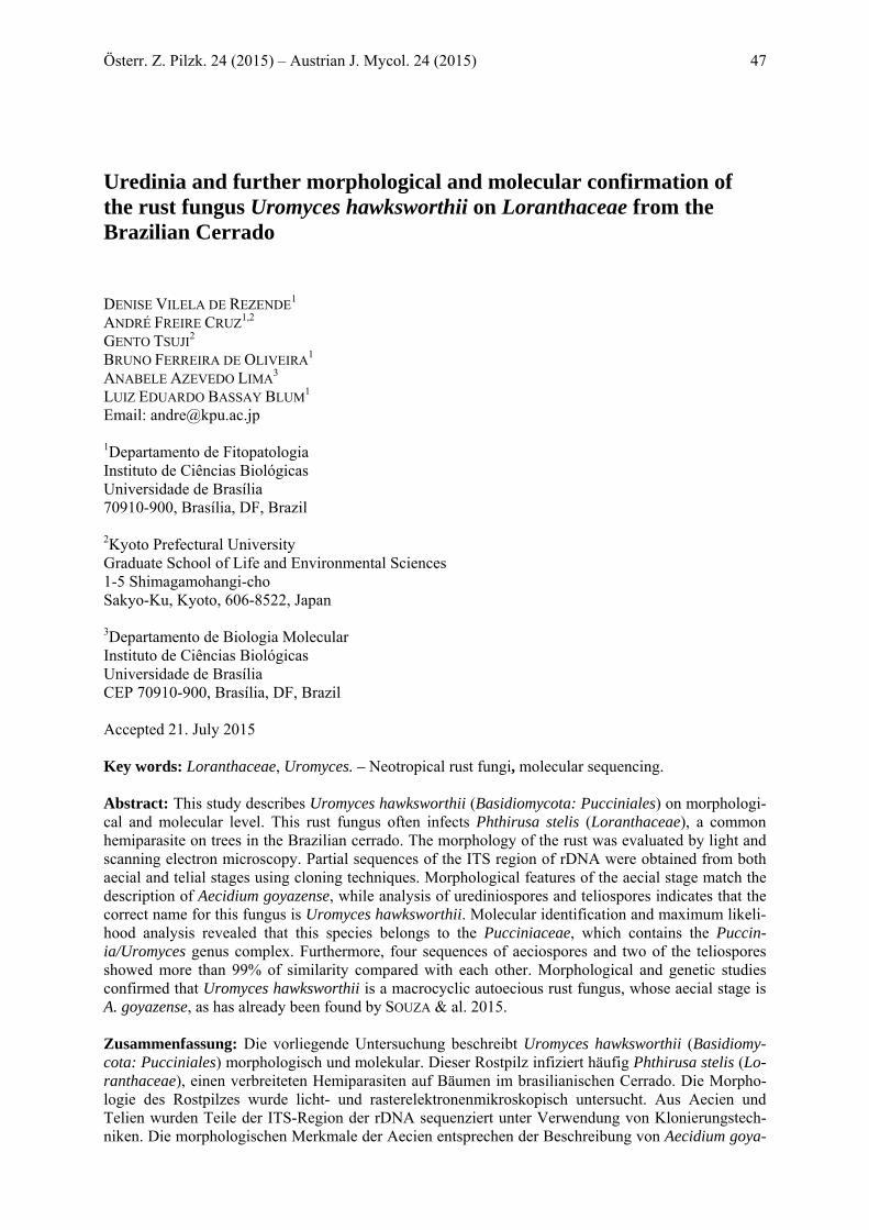
Fig. 1. Aecial stage of U. hawksworthii under the scanning electron (SEM) and light microscope (LM). A Aecium in one gall under SEM. B Cut of aecium showing column of aeciospores and peridi-um under the LM. C Open aecium showing internal wall, peridial cells and aeciospores under SEM. D, E Rhomboid and verrucose peridial cells of aecium under LM and SEM respectively. F Germinated aeciospores showing germ tubes (GT) (arrow). G One aeciospore (AEC) over peridial cells under SEM. Bars: A = 180 µm, B = 120 µm, C = 40 µm, D =150 µm, E, G = 50 µm, F = 70 µm.

50 D. VILELA DE REZENDE & al. 2015: Uromyces hawksworthii on Loranthaceae
phrygilanthi H. S. JACKS & HOLW., A. struthanthi H. S. JACKS. & HOLW. (JACKSON 1927), and A. goyazense HENN. on Phthirusa stelis in Goiás, Brazil (SACCARDO 1899, SYDOW & SYDOW 1924). Recently, SOUZA & al. (2015) reported Uromyces hawks-worthii SOUZA, E. S. C, CHAVES, Z. M. C., SOARES, W. R. O., PINHO, D. B. & DI-
ANESE, J. C. as replacement name for A. goyazense on Phthirusa stelis (Loranthace-ae), based on DNA sequences generated from the ITS and 28S rRNA (LSU) regions of DNA recovered from aeciospores and teliospores.
The current study aims to describe and confirm the morphological characteristics of aecial and uredinial/telial stages of the rust fungus on P. stelis by using microscopy (optical and SEM), and to compare both stages by conducting molecular analyses of this rust species on Loranthaceae.
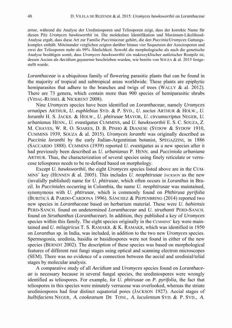
Fig. 2. Uredinial stage of U. hawksworthii. A, B Substomatal uredinia showing globose, echinulate and pedicellate urediniospores getting out one by one from stomata under SEM. C Urediniospores showing details of echinulations under the SEM. Bars: A = 10 μm, B, C = 5 μm. Materials and methods Specimens investigated
Specimens were collected from various regions in the federal district of Brazil. They were isolated from the living leaves of P. stelis at different times, between 1992 and 2003. The specific regions and
Österr. Z. Pilzk. 24 (2015) – Austrian J. Mycol. 24 (2015) 51
collection dates are as follows: Brasília, (Embrapa), CPAC Planaltina, DF, August 21, 1997, D. V. REZENDE 39 (UB col. micol. 15138). Brasília, Península Norte, QI 04, Cj 06, lt 22, May 5, 1992, J. C. DIANESE 400 (UB col. micol. 1482, 1483, 1484, 1485, 1486, 1487, 1488, 1489). Brasília, Península Norte, Cj 06, lt 22, August 9, 1992, J. C. DIANESE 406 (UB col. micol. 1513, 1514, 1515, 1516, 1517, 1518, 1519, 1520, 1521, 1522, 1523, 1524, 1525, 1526, 1527, 1528, 1529, 1530). Brasília, Península Norte, November 11, 1992; J. C. DIANESE (UB col. micol. 2761). Brasília, Fazenda Àgua Limpa, UnB, February 19, 1993, R. B. MEDEIROS (UB col. micol. 3151). Planaltina 166, Estação Ecológica Águas Emendadas, February 19, 1994, C. A. INÁCIO (UB col. micol. 5955). Brasília, Embrapa, CPAC, Planaltina, DF, August 21 1997. D. V. REZENDE (UB col. micol. 15139). Brasília, DF, SQN 410, Bloco N, August 18, 2003, R. C. PEREIRA (UB col. micol. 19398).
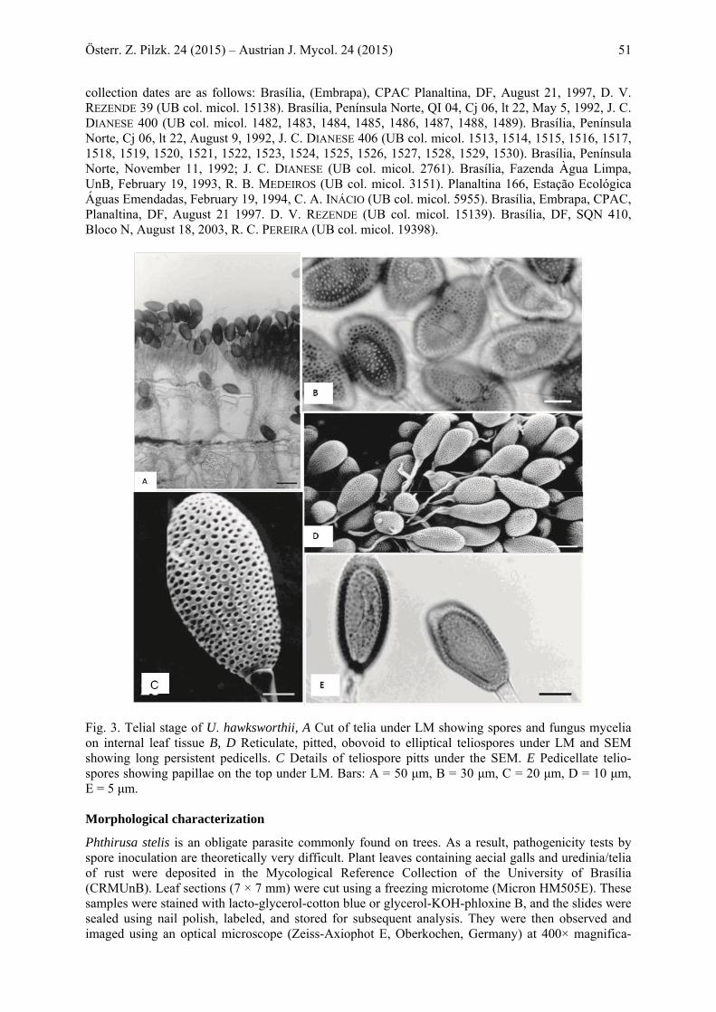
Fig. 3. Telial stage of U. hawksworthii, A Cut of telia under LM showing spores and fungus mycelia on internal leaf tissue B, D Reticulate, pitted, obovoid to elliptical teliospores under LM and SEM showing long persistent pedicells. C Details of teliospore pitts under the SEM. E Pedicellate telio-spores showing papillae on the top under LM. Bars: A = 50 μm, B = 30 μm, C = 20 μm, D = 10 μm, E = 5 μm. Morphological characterization
Phthirusa stelis is an obligate parasite commonly found on trees. As a result, pathogenicity tests by spore inoculation are theoretically very difficult. Plant leaves containing aecial galls and uredinia/telia of rust were deposited in the Mycological Reference Collection of the University of Brasília (CRMUnB). Leaf sections (7 × 7 mm) were cut using a freezing microtome (Micron HM505E). These samples were stained with lacto-glycerol-cotton blue or glycerol-KOH-phloxine B, and the slides were sealed using nail polish, labeled, and stored for subsequent analysis. They were then observed and imaged using an optical microscope (Zeiss-Axiophot E, Oberkochen, Germany) at 400× magnifica-
52 D. VILELA DE REZENDE & al. 2015: Uromyces hawksworthii on Loranthaceae
tion. Size measurements were performed in at least 20 aeciospores and 50 teliospores. Leaf sections containing uredinia/telia and aecial galls were fixed with 0.1 M sodium cacodylate buffer, pH 7.4, and 2% glutaraldehyde (v/v) for 24 h. They were then washed five times with the same buffer solution before being treated with 1% osmium tetroxide (OsO4) in cacodylate buffer at 4°C for 4 h. The sam-ples were washed as described with the buffer solution and dehydrated for 15 min in an aqueous series with increasing concentrations of acetone (15, 30, 50, 75 and 100%). The material was critical-point-dried using a Balzers CPD-030 (Balzers, Fürstentum Liechtenstein) with liquid CO2. Dried tissue fragments were placed on metal cubes coated with silver conductor paste. The samples were sputter coated with gold using Balzers SCD50. Subsequently, the samples were observed and imaged using a SEM (Jeol -JSM 840-AE, Japan), with 5 and 10 KV of acceleration and a working distance of 20 mm. The fungal structures were measured using the scale bars on each image.
Molecular identification (DNA extraction, Polymerase Chain Reaction, Cloning, and sequenc-ing)
DNA was isolated from frozen spores of the aecial (1 template) and telial (2 templates) stages of the fungi using the DNAeasy Plant Mini Kit (Qiagen, Hilden, Germany), according to the manufacturer’s instructions. The DNA template was checked by electrophoresis before polymerase chain reaction (PCR) was performed. A part of the internal transcribed spacer (ITS) region was amplified using the oligonucleotide primers ITS5-u (5′-caaggtttctgtaggtg-3′) and ITS4-u (5′-ggcttttccctcttcat-3′) (PFUNDER & al. 2001), which are more suitable for amplifying the genomic DNA of Pucciniaceae than other known primers. PCR mixtures (25 μl) contained 0.5 μl template DNA, 12.5 μl of 2X KAPA2G Robust HotStart Ready Mix (Nippon Genetics, Tokyo, Japan), and 6 μl of each oligonucleotide, forward and reverse (1 μM). The PCR conditions were as follows: pre-denaturation for 2 min at 94°C, followed by 35 cycles of 1 min at 94°C, 10 s at 55°C, and 15 s at 72°C, with final extension for 10 min at 72°C. Amplification products were separated on a 1.0% agarose gel and detected by ethidium bromide stain-ing and UV illumination.
The PCR products obtained were gel-purified using the Wizard SV Gel and PCR Clean-up Sys-tem (Promega, Tokyo, Japan), and then ligated with the TA-cloning vector pMD20 (Takara, Tokyo, Japan) using the Mighty Mix DNA Ligation Kit (Takara, Tokyo, Japan). Subsequently, the reaction mixture was transformed into competent Escherichia coli DH5α Electro-Cells (Takara, Kyoto, Japan) by electroporation.
From the transformants, the plasmid DNA was purified with the FastGene Plasmid Mini kit (Nip-pon Genetics, Tokyo, Japan) and checked for the insert by PCR using SP6 and M13-20 primer pairs. A total of six plasmid samples from two clones of each template were sequenced by Fasmac (Japan) using the SP6 oligonucleotide.
The six analyzed clones, four from the aecial and two from telial stages, were sequenced, and the results were compared to homologous sequences registered in GenBank (http://www.ncbi.nlm.nihgov) using the standard nucleotide BLAST protocol. Subsequently, the sequences were scanned for similar-ity with those in the databank. These sequences were then manually aligned with groups representing fungal lineages to obtain the approximate identification of the pathogenic fungi. The reference se-quence and the out-group (Melampsoridium betulinum) from the GeneBank are presented in FASTA format. Alignment of DNA sequences and the phylogeny tree were performed using the Phylogeny feature of the MEGA 6.06 program with 1000 bootstrap replications, and Maximum Parsimony analy-sis of taxa and evolutionary distance grouping were performed according to the Kimura model. The sequences from both aecial and telial stages were aligned for comparison.
All sequences from the six clones were registered in the DNA Data Bank of Japan (DDBJ) (http://www.ddbj.nig.ac.jp) with the following registration numbers: Clone 1 - LC050417; Clone 2 - LC050418; Clone 3 - LC050419; Clone 4 - LC050416; Clone 5 - LC050415; Clone 6 - LC050414. All were registered as U. hawksworthii.
Österr. Z. Pilzk. 24 (2015) – Austrian J. Mycol. 24 (2015) 53
Results Uromyces hawksworthii E. S. C. SOUZA, Z. M. CHAVES, W. R. O. SOARES, D. B. PINHO & DIANESE (SOUZA & al. 2015) (Figs. 1–4)
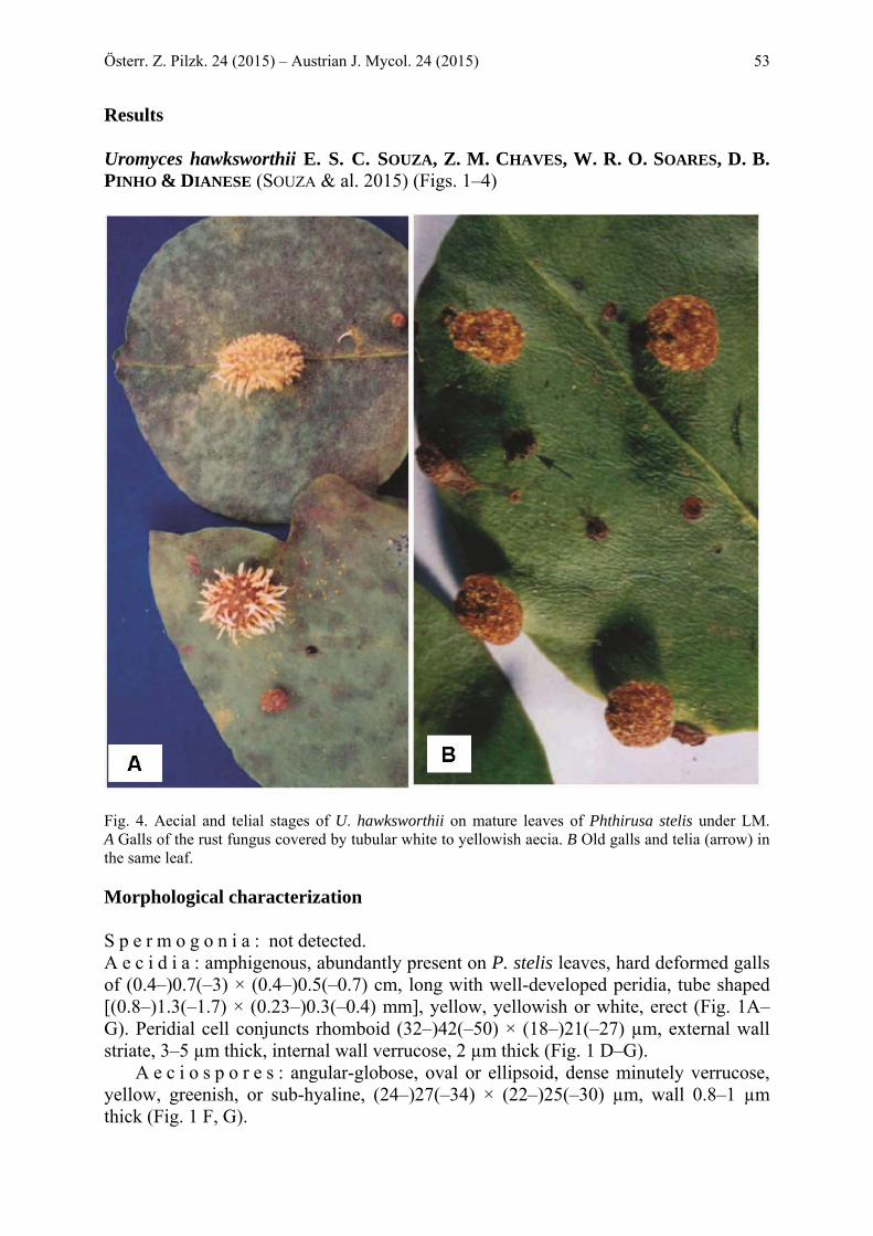
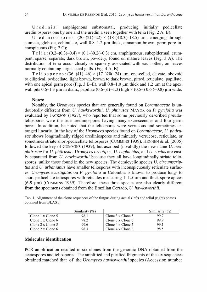
Fig. 4. Aecial and telial stages of U. hawksworthii on mature leaves of Phthirusa stelis under LM. A Galls of the rust fungus covered by tubular white to yellowish aecia. B Old galls and telia (arrow) in the same leaf. Morphological characterization S p e r m o g o n i a : not detected. A e c i d i a : amphigenous, abundantly present on P. stelis leaves, hard deformed galls of (0.4–)0.7(–3) × (0.4–)0.5(–0.7) cm, long with well-developed peridia, tube shaped [(0.8–)1.3(–1.7) × (0.23–)0.3(–0.4) mm], yellow, yellowish or white, erect (Fig. 1A–G). Peridial cell conjuncts rhomboid (32–)42(–50) × (18–)21(–27) µm, external wall striate, 3–5 µm thick, internal wall verrucose, 2 µm thick (Fig. 1 D–G).
A e c i o s p o r e s : angular-globose, oval or ellipsoid, dense minutely verrucose, yellow, greenish, or sub-hyaline, (24–)27(–34) × (22–)25(–30) µm, wall 0.8–1 µm thick (Fig. 1 F, G).
54 D. VILELA DE REZENDE & al. 2015: Uromyces hawksworthii on Loranthaceae
U r e d i n i a : amphigenous substomatal, producing initially pedicellate urediniospores one by one and the uredinia seen together with telia (Fig. 2 A, B).
U r e d i n i o s p o r e s : (20–)21(–22) × (18–)18.3(–18.5) µm, emerging through stomata, globose, echinulate, wall 0.8–1.2 µm thick, cinnamon brown, germ pore in-conspicuous (Fig. 2 C);
T e l i a : (0.2–)0.3(–0.4) × (0.1–)0.2(–0.3) cm, amphigenous, subepidermal, erum-pent, sparse, separate, dark brown, powdery, found on mature leaves (Fig. 3 A). The distribution of telia occur closely or sparsely associated with each other, on leaves normally containing large aecial galls. (Fig. 4 A, B).
T e l i o s p o r e s : (36–)41(–46) × (17–)20(–24) µm, one-celled, clavate, obovoid to elliptical, pedicellate, light brown, brown to dark brown, pitted, reticulate, papillate, with one apical germ pore (Fig. 3 B–E), wall 0.8–1.0 µm thick and 1.2 µm at the apex, wall pits 0.6–1.3 µm in diam., papillae (0.6–)1(–1.3) high × (0.5–) 0.6 (–0.8) µm wide.
Notes: Notably, the Uromyces species that are generally found on Loranthaceae is un-
doubtedly different from U. hawksworthii. U. phtirusae MAYOR on P. pyrifolia was evaluated by JACKSON (1927), who reported that some previously described pseudo-teliospores were the true urediniospores having many excrescencies and four germ pores. In addition, he noted that the teliospores were verrucous and sometimes ar-ranged linearly. In the key of the Uromyces species found on Loranthaceae, U. phtiru-sae shows longitudinally ridged urediniospores and minutely verrucose, reticulate, or sometimes striate short-pedicellate teliospores (CUMMINS 1939). HENNEN & al. (2005) followed the key of CUMMINS (1939), but ascribed (invalidly) the new name U. neo-phtirusae for U. phtirusae. Uromyces ornatipes, U. euphlebius, and U. socius are easi-ly separated from U. hawksworthii because they all have longitudinally striate telio-spores, unlike those found in the new species. The demicyclic species U. circumscrip-tus and U. urbanianus have smaller teliospores with inconspicuously reticulate surfac-es. Uromyces evastigatus on P. pyrifolia in Colombia is known to produce long- to short-pedicellate teliospores with reticules measuring 1–1.5 µm and thick spore apices (6-9 µm) (CUMMINS 1939). Therefore, these three species are also clearly different from the specimens obtained from the Brazilian Cerrado, U. hawksworthii.
Tab. 1. Alignment of the clone sequences of the fungus during aecial (left) and telial (right) phases obtained from BLAST.
Similarity (%) Similarity (%) Clone 1 x Clone 5 98.1 Clone 3 x Clone 5 99.7 Clone 1 x Clone 6 98.2 Clone 3 x Clone 6 99.9 Clone 2 x Clone 5 99.6 Clone 4 x Clone 5 99.1 Clone 2 x Clone 6 98.3 Clone 4 x Clone 6 98.5
Molecular identification PCR amplification resulted in six clones from the genomic DNA obtained from the aeciospores and teliospores. The amplified and purified fragments of the six sequences obtained matched that of the Uromyces hawksworthii species (Accession number
Österr. Z. Pilzk. 24 (2015) – Austrian J. Mycol. 24 (2015) 55
Fig. 5. Phylogenetic analysis of the clones 1–6 of the ITS sequence, showing the relationship between these clones and other rust fungi. NR_132920, E-value = 0.0) with 100% similarity, according to a BLAST GenBank search. The sequences were aligned with each other, and more than 99% similaritywas observed between both stages, reaching to a maximum of 99.9% (Tab. 1). Based on the analysis, it is known that the six clones belong to the same species. The results of DNA sequences show that the all clones shared 99% similarity with the refereed spe-cie in the databank, which belongs to the Uromyces genus. These results revealed that the spores from the aecial (Clones 1–4) and telial stages (Clones 5 and 6) had a high similarity and clustered within the rust family Pucciniaceae, containing the genus Uromyces (Fig. 5). Since the sequences presented high identity, alignment was per-formed with the sequences deposited in the DDBJ and a phylogenetic tree was con-structed based on the ITS region.
56 D. VILELA DE REZENDE & al. 2015: Uromyces hawksworthii on Loranthaceae
Discussion
Morphology Uromyces hawksworthii is a macrocyclic, autoecious rust fungus, whose aecial stage was described as A. goyazense on the same host P. stelis in several articles (SACCAR-
DO 1897, SYDOW & SYDOW 1924, HENNEN & al. 2005, SOUZA & al. 2015). Other species of Uromyces found on Loranthaceae were already described based on the telial phase (CUMMINS 1939, HENNEN & al. 2005). In addition, the identity of the ae-cial/uredinial and telial stages in the life cycle of this fungus was confirmed by molec-ular analysis. Considering the morphological features, this species produces one-celled pedicellate teliospores with an apical germ pore, and shows a small portion of basidia when germinating. Other species, such as U. bahiensis and U. circumscriptus are mor-phologically similar in aecidiospore size. However, peridial cells are absent in the ae-cidia of U. circumscriptus (PERDOMO-SANCHEZ & PIEPENBRING 2014). Uromyces hawksworth is characterized by reticulate pitted teliospores that are smaller than those of all other known species of Uromyces found on Loranthaceae.
Identification by DNA blasting The molecular blasting of the rust fungus with GenBank data, matched that of U. hawksworthii previously deposited in GenBank and registered in Mycobank by SOUZA & al. (2015),, found on Loranthaceae in the Brazilian cerrado. The current research confirms the results found by these authors and furthermore adds the description of the uredinia and urediniospores morphologically and molecularly.
DNA sequencing of Uromyces spp. has revealed the presence of distinct clades: the isolates causing rust on chickpea, fenugreek, and alfalfa; those found on field clo-ver and pea plants; isolates causing rust on bean and cowpea; and those infecting faba beans (BARILLI & al. 2011). Therefore, it was concluded that the Uromyces species could be genetically grouped according to their host plant. STEADMAN & al. (2002) proposed that there is a wide adoption of differential series and nomenclature systems for the physiological races of U. appendiculatus, which contribute to the elaboration of an internationally standardized classification methodology based on host infection. Furthermore, amplified fragment length polymorphic (AFLP) DNA analysis indicated that Eurasian isolates of Puccinia jaceae from different hosts exhibited unique finger-print patterns primarily based on host/pathogenicity preference (YOURMAN & LUSTER 2004). Molecular markers were used to unravel the genomic organization of crown rust resistance caused by P. coronata in a segregating Lolium perenne population (MUYLLE & al. 2005).
Among the rust fungi (Pucciniomycetes), Puccinia and Uromyces are the two larg-est genera, comprising 4000 and 600 species, respectively (CUMMINS & HIRATSUKA 2003). These genera are morphologically distinguished by the cell number of telio-spores, number of germ pores per cell, teliospore germination, presence or absence of paraphyses in sorus, the persistence of pedicels, ornamentation and staining of spores, and the presence or absence of uredinia. However, Puccinia and Uromyces cannot be
Österr. Z. Pilzk. 24 (2015) – Austrian J. Mycol. 24 (2015) 57
distinguished by their spermogonia, aecia, uredinia, or spores produced in these sori (MAIER & al. 2007). There are species having one- and two-celled teliospores or up to three or four celled ones. Based on the characteristics of the life cycles of Puccinia and Uromyces, some taxonomists have indicated that phylogenetically, they could be a large group of genera (MAIER & al. 2007). This group is called the Puccinia/Uromyces complex (BURITICA & al. 2014; MAIER & al. 2003, 2007; WINGFIELD & al. 2004). De-pending on the host biomes and the evolution of rust fungi, Puccinia/Uromyces can be separated by their hosts (VAN DER MERWE & al. 2007, MAIER & al. 2007). According to these authors, Puccinia s. l. and Uromyces s. l. are polyphyletic, but both Uromyces s. str. (type U. appendiculatus), and Puccinia s. str. (type P. graminis) have distinct clades. Phylogenetically, the Uromyces species are usually more spread, suggesting that they are older than the Puccinia species, and are separated into two major clades (VAN DER MERWE & al. 2007). The one-celled teliospores found among two-celled telio-spores in the Puccinia species are mesospores. However, two-celled teliospores in Uromyces are rarer. Therefore, the molecular data support the hypothesis that the small number of teliospore cells in Uromyces represent a more advanced developmental stage (VAN DER MERWE & al. 2007). The molecular data show that there is a monophy-letic group of Uromyces spp. on Fabaceae, whose species type is U. appendiculatus, since few Puccinia species are reported on Fabaceae. Other data have demonstrated a strong relationship between Uromyces pisi (heteroecious) with the alternate host be-longing to Euphorbiaceae and U. viciae-fabae (autoecious). These two species are in a separate clade (VAN DER MERWE & al. 2007). VAN DER MERWE & al. (2007) noted a clear separation of different molecular characteristics within the clade, specifically the two groups of the Uromyces and Puccinia species, and indicated that the two genera are strictly distinguished. Considering this, a simple teliospore septum separates Puccinia and Uromyces. AIME & al. (2006) showed that Puccinia and Uromyces are polyphylet-ic.
This research was partly supported by The National Council for Scientific and Technological De-
velopment (CNPq) – Brazil. The authors wish to thank the phytopathology undergraduate students for technical assistance, Professors HELSON MÁRIO M. DO VALE and RENATO DE OLIVEIRA RESENDE for allowing the use of their laboratory for the molecular analysis, and Professor SÔNIA NAIR BÁO for use of the scanning electron microscope.
References AIME, M. C., MATHENY, P. B., HENK, D. A., FRIEDERS, E. M. NILSSON, R. H., PIEPENBRING, M.,
MCLAUGHLIN, D. J., SZABO, L. J., BEGEROW, D., SAMPAIO, J. P., BAUER, R., WEISS, M., OBERWIN-
KLER, F., HIBBETT., D.S., 2006: An overview of the higher-level classification of Pucciniomy-cotina based on combined analyses of nuclear large and small subunit rDNA sequences. – Mycol. 98: 896–905.
BARILLI, E., SATOVIC, Z., SILLERO, J. C., RUBIALES, D., TORRES, A. M., 2011: Phylogenetic analysis of Uromyces species infecting grain and forage legumes by sequence analysis of nuclear riboso-mal internal transcribed spacer region. – J. Phytopathol. 159: 137–145.
BERNDT, R., 2002: New species, reports and observation on rust fungi. – Nova Hedwigia 75: 415–431. BURITICÁ, P., PARDO-CARDONA, V. M., 1996: Flora Uredineana Colombiana. – Rev. Acad. Colomb.
de Cienc. 20: 183–236. BURITICÁ, P. C., YEPES, M. S., PARDO-CARDONA, V. M., 2014: Pucciniales (Fungi), Royas de
Colombia. – Facul. Nac. de Agron. Suplemento I. 67: 1–93. CUMMINS, G. B., 1939: New species of Uredinales. – Mycologia 31: 169–174.

58 D. VILELA DE REZENDE & al. 2015: Uromyces hawksworthii on Loranthaceae
CUMMINS, G. B., HIRATSUKA, Y., 2003: Illustrated genera of Rust Fungi. 3rd edn. – St. Paul, Minneso-ta: APS.
HENNEN, J. F., FIGUEREDO, M. B., CARVALHO A. A. Jr., HENNEN, P. G., 2005: Catalogue of the spe-cies of plant rust fungi (Uredinales) of Brazil. – Available from [http://www.jbrj.gov.br/publica/ uredinales/Brazil_Catalogue1drevisado.pdf.] [Accessed 20 October 2013].
HENNINGS, V. P., 1895: Fungi goyazenses. – Hedwigia 34: 101. [http://www.biodiversitylibrary. org/item/13877] [Accessed 9 December 2013].
JACKSON, H. S., 1927: The rusts of South America based on the Holway Collections. – Mycologia 19: 53–55.
MAIER, W., BEGEROW, D., WEIΒ, OBERWINKLER, F., 2003: Molecular phylogeny of rust fungi: an approach using nuclear large subunit ribosomal DNA sequences. – Canad. J. Bot. 81: 12–23.
MAIER, W., WINGFIELD, B. D., MENNICKEN, M., WINGFIELD, M. J., 2007: Polyphyly and two emerg-ing lineages in the rust genera Puccinia and Uromyces. – Mycol. Res. 3: 176–185.
MUYLLE, H., BAERT, J., VAN BOCKSTAELE, E., MOERKERKE, B., GOETGHEBEUR, E., ROLDAN-RUIZ, I., 2005: Identification of molecular markers linked with crown rust (Puccinia coronata f. sp. lo-lii) resistance in perennial ryegrass (Lolium perenne) using AFLP markers and a bulked segre-gant approach. – Euphytica 143: 135–144.
PFUNDER, M., SCHÜRCH, S., ROY, B. A., 2001: Sequence variation and geographic distribution of pseudoflower-forming rust fungi (Uromyces pisi s. lat.) on Euphorbia cyparissias. – Mycol. Res. 105: 57–66.
SACCARDO, P. A., 1897: Sylloge fungorum 12. SACCARDO, P. A., 1888: Gasteromyceteae, Phycomyceteae, Myxomyceteae, Ustilagineae et Uredi-
neae. – Syll. Fung. 7. SACCARDO, P. A., 1899: Hymenomycetae, Hyphomycetae. – Syll. Fung. 14. PERDOMO-SÁNCHEZ, O. & PIEPENBRING, M., 2014: Species of Uromyces (Pucciniales, Basidiomyco-
ta) on Loranthaceae. – Trop. Pl. Pathol. 39: 141–153. SOUZA, E. S. C., CHAVES, Z. M., SOARES, W. R. O., PINHO, D. B., DIANESE, J. C., 2015: Uromyces
hawksworthii nom. nov. for Aecidium goyazense, on Phthirusa stelis (Loranthaceae) from the Brazilian Cerrado. – IMA Fungus 6: 155–162.
STEADMAN, J. R., PASTOR-CORRALES, M. A., BEAVER, J. S. 2002: An overview of the 3rd Bean Rust and 2nd Bean Bacterial Blight international workshops. Pietermaritzburg, South Africa. – Ann. Rep. Bean Improv. Coop. 45: 120–125.
SYDOW, P., SYDOW, H., 1910: Monografia Uredinearum: 244–245. – Leipzig: Gebrüder Borntraeger. SYDOW, P., SYDOW, H., 1924: Monografia Uredinearum: 268–270. – Leipzig: Gebrüder Borntraeger. VAN DER MERWE, M., ERICSON, L., WALKER, J., THRALL, P. H., BURDON, J., 2007: Evolutionary rela-
tionships among species of Puccinia and Uromyces (Pucciniaceae, Uredinales) inferred from partial protein coding gene phylogenies. – Mycol. Res. 3: 163–175.
VIDAL-RUSSEL, R., NICKRENT, D. L., 2008: Evolutionary relationships in the showy mistletoe family (Loranthaceae). – Amer. J. Bot. 95: 1015–1029.
WALLY, N. M., EL DIN ALI, A. E., JRAIS, R. N., 2012: Botanical and biological studies of six parasitic species of family Loranthaceae growing in Kingdom of Saudi Arabia. – Int. J. Env. Sci. 1: 196–205.
WINGFIELD, B. D., ERICSON, L., SZARO, T., BURDON, J. J., 2004: Phylogenetic patterns in the Uredi-nales. – Australasian Pl. Path. 33: 327–335.
YOURMAN, L. F., LUSTER, D. G., 2004: Generation of molecular markers for the identification of iso-lates of Puccinia jaceae, a potential biological control agent of yellow starthistle. – Biol. Cont. 29: 73–80.