upper gi bleeding - sothep.comsothep.com/thaiep/files/core_lecture_2010-01/chitlada-upper gi...upper...
TRANSCRIPT
Upper GI bleeding
Chitlada Limjindaporn, MDDiplomate of American Board of Emergency Medicine
Faculty of Medicine, Thammasat University
Upper GI bleeding
Variceal hemorrhage
Non-variceal upper GI bleeding (NVUGIB)
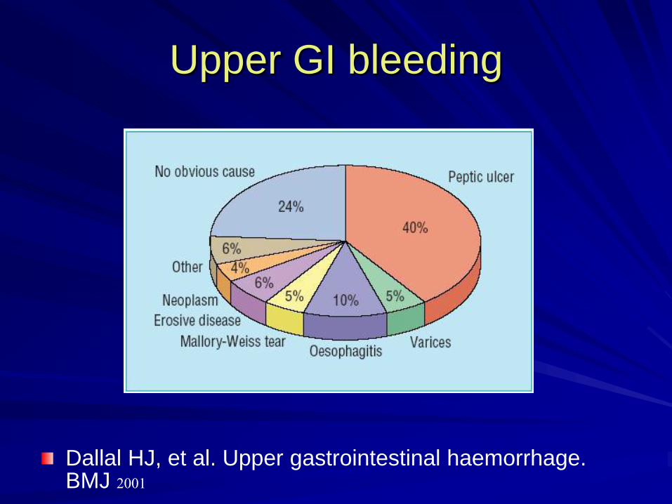
Upper GI bleeding
Dallal HJ, et al. Upper gastrointestinal haemorrhage. BMJ 2001
Nonvariceal bleeding
Approximately 80% of ulcers stop
bleeding.
The overall mortality rate is approximately 10% Elderly with significant comorbidity : increase mortality.

Mortality/Morbidity
Retrospective chart review
73.2% of deaths occurred in patients older than 60 years.One or more comorbid illnesses were noted in 98.3% of patients who died.
Yavorski RT, Wong RK, Maydonovitch C, Battin LS, Furnia A, AmundsonDE. Analysis of 3,294 cases of upper gastrointestinal bleeding in military medical facilities. Am J Gastroenterol. Apr 1995;90(4):568-73.

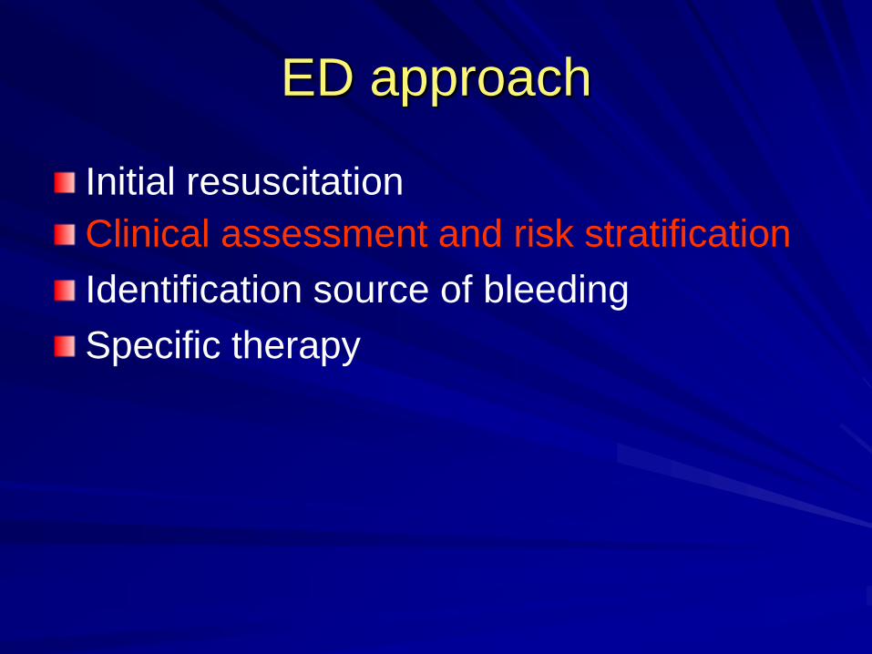
ED approach
Initial resuscitation
Clinical assessment and risk stratification
Identification source of bleeding
Specific therapy

Early resuscitation
Early intensive resuscitation of patients with upper gastrointestinal bleeding decreases mortality.
Aggressive hemodynamic resuscitation, correction of hematocrit ( >28%)and coagulopathy (INR>1.8)
Mortality significantly decrease in intensive resuscitation group
No difference in rebleeding, surgical intervention
Baradarian R, Ramdhaney S, Chapalamadugu R, Skoczylas L, Wang K, Rivilis S, et al. Am J Gastroenterol. Apr 2004;99(4):619-22.

Initial resuscitation
ABC, IV,Oxygen
Intubation
NPO
Fluid resuscitation
PRC, FFP transfusion
Foley catheter
Nasogastric Aspiration
– Severity assessment
– Remove clot
ED approach
Initial resuscitation
Clinical assessment and risk stratification
Identification source of bleeding
Specific therapy
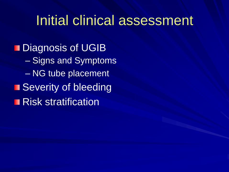
Initial clinical assessment
Diagnosis of UGIB
– Signs and Symptoms
– NG tube placement
Severity of bleeding Risk stratification

Signs and Symptoms
Age, Sex
Symptoms: weakness, dizziness, syncope– melena (50-100ml)
– Hematemesis (1000ml)
– Coffee ground
– Hematochezia (>1000ml/hr)
Associated symptoms: pale, jaundice, ascites
History of previous bleeding
History of severe vomiting
Medication: NSAID, ASA

Signs and Symptoms
Underlying diseases
– Peptic ulcer disease/ gastritis – Liver disease : cirrhosis, hepatitis, alcoholism
– Malignancy
– Hematologic disease: coagulopathy, chronic anemia
– Cardiovascular disease
– Pulmonary disease
– Renal disease
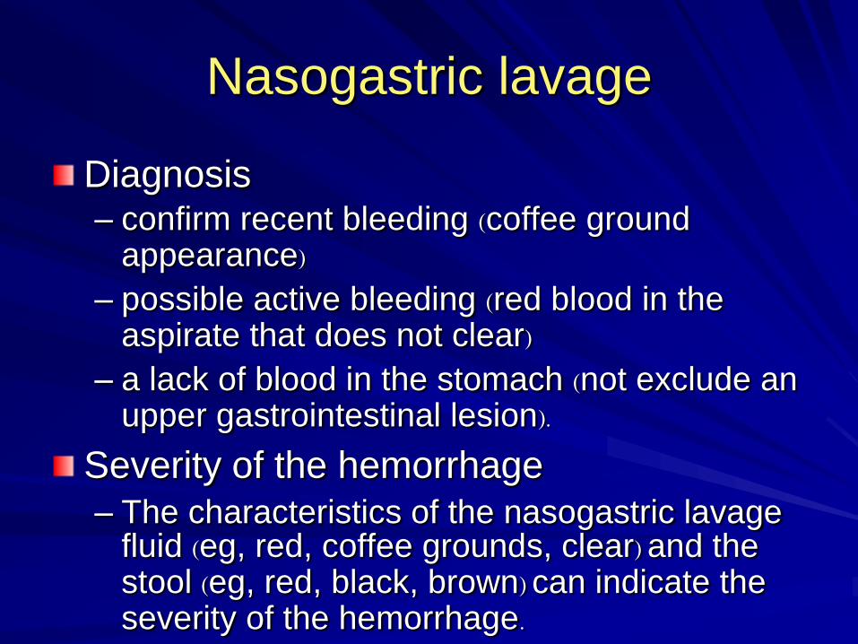
Nasogastric lavage
Diagnosis– confirm recent bleeding (coffee ground
appearance)– possible active bleeding (red blood in the
aspirate that does not clear)– a lack of blood in the stomach (not exclude an
upper gastrointestinal lesion).
Severity of the hemorrhage
– The characteristics of the nasogastric lavage fluid (eg, red, coffee grounds, clear) and the stool (eg, red, black, brown) can indicate the severity of the hemorrhage.
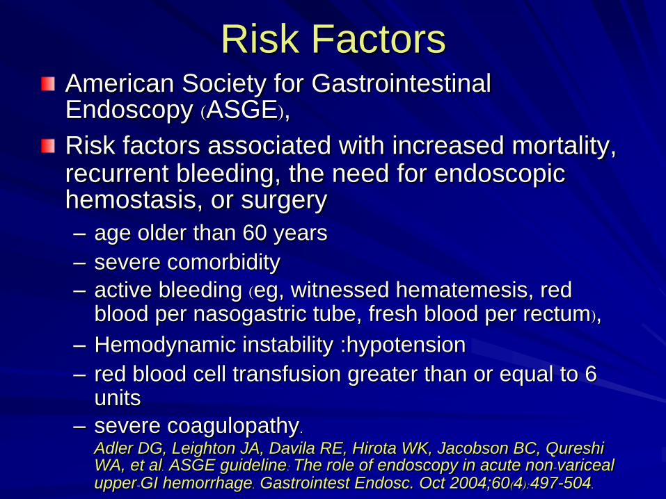
Risk FactorsAmerican Society for Gastrointestinal Endoscopy (ASGE), Risk factors associated with increased mortality, recurrent bleeding, the need for endoscopic hemostasis, or surgery – age older than 60 years
– severe comorbidity
– active bleeding (eg, witnessed hematemesis, red blood per nasogastric tube, fresh blood per rectum),
– Hemodynamic instability :hypotension
– red blood cell transfusion greater than or equal to 6 units
– severe coagulopathy.Adler DG, Leighton JA, Davila RE, Hirota WK, Jacobson BC, Qureshi WA, et al. ASGE guideline: The role of endoscopy in acute non-variceal upper-GI hemorrhage. Gastrointest Endosc. Oct 2004;60(4):497-504.
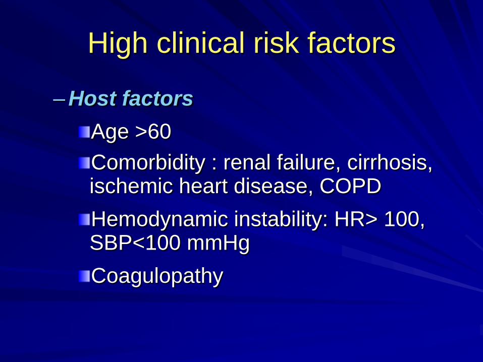
High clinical risk factors
–Host factors
Age >60
Comorbidity : renal failure, cirrhosis, ischemic heart disease, COPD
Hemodynamic instability: HR> 100, SBP<100 mmHg
Coagulopathy

High clinical risk factors
–Bleeding character
Continuous red blood from NG after
irrigation
Red blood per rectum
Need blood transfusion
Rebleeding Inpatient hemodynamic instability
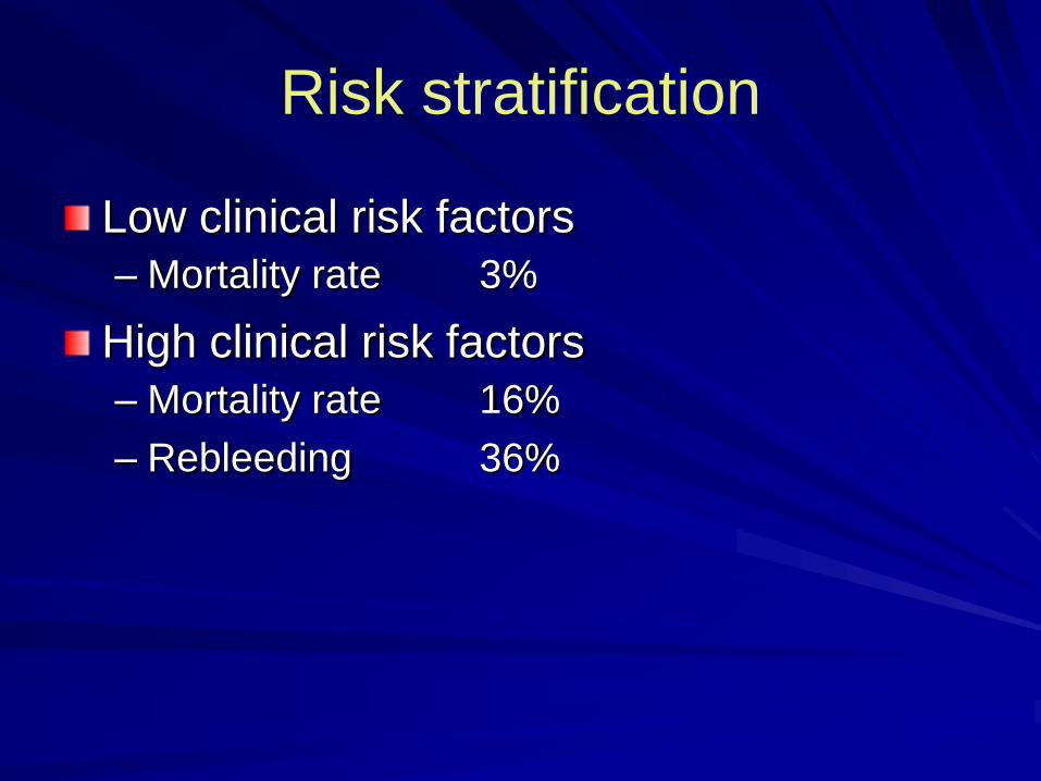
Risk stratification
Low clinical risk factors
– Mortality rate 3%
High clinical risk factors
– Mortality rate 16%
– Rebleeding 36%
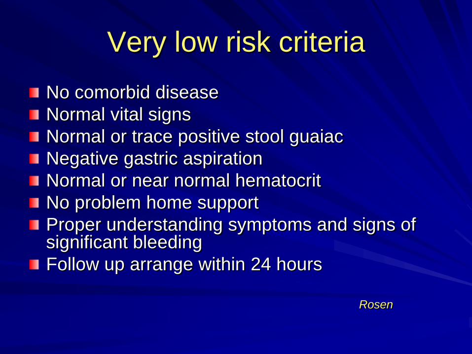
Very low risk criteria
No comorbid disease
Normal vital signs
Normal or trace positive stool guaiac
Negative gastric aspiration
Normal or near normal hematocrit
No problem home support
Proper understanding symptoms and signs of significant bleeding
Follow up arrange within 24 hours
Rosen

ED approach
Initial resuscitation
Clinical assessment and risk stratification
Identification source of bleeding
Specific therapy
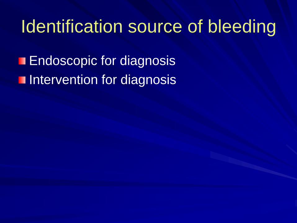
Identification source of bleeding
Endoscopic for diagnosis
Intervention for diagnosis

Early endoscopy
Early endoscopy in upper gastrointestinal hemorrhage: associations with recurrent bleeding, surgery, and length of hospital stay.
Demonstrated a lower rate of rebleeding and shorter length of stay when endoscopy is performed within 24 hours of admission
Cooper GS, Chak A, Way LE, Hammar PJ, Harper DL, Rosenthal
GE. Gastrointest Endosc. Feb 1999;49(2):145-52.
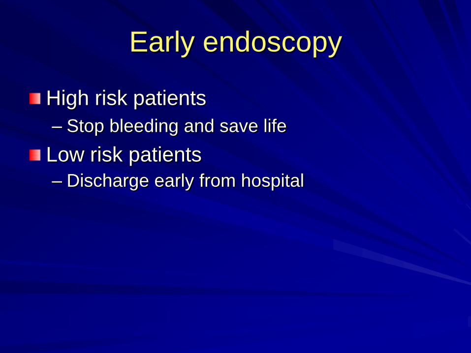
Early endoscopy
High risk patients
– Stop bleeding and save life
Low risk patients
– Discharge early from hospital

Stigmata of recent hemorrhageSRH Prevalence Rebleeding Surgery Mortality
Clean base
42% 5% 0.5% 2%
Flat spot 20% 10% 6% 3%
Adherent clot
17% 22% 10% 7%
Visible vessel
17% 43% 34% 11%
Active bleeding
18% 70% 35% 11%
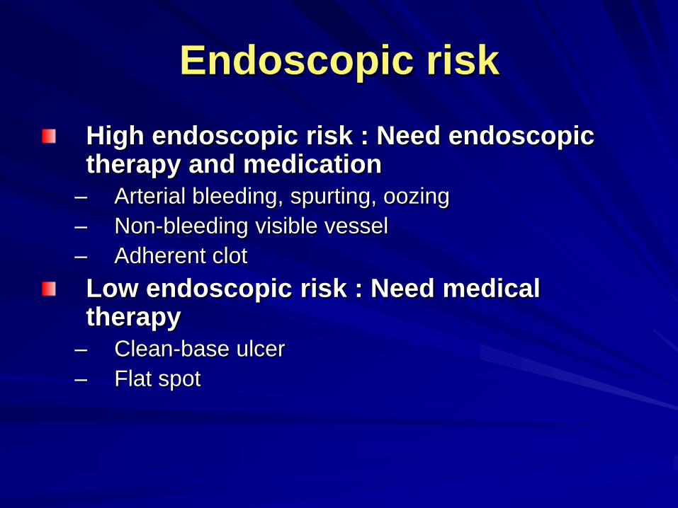
Endoscopic risk
High endoscopic risk : Need endoscopic therapy and medication
– Arterial bleeding, spurting, oozing
– Non-bleeding visible vessel
– Adherent clot
Low endoscopic risk : Need medical therapy
– Clean-base ulcer
– Flat spot
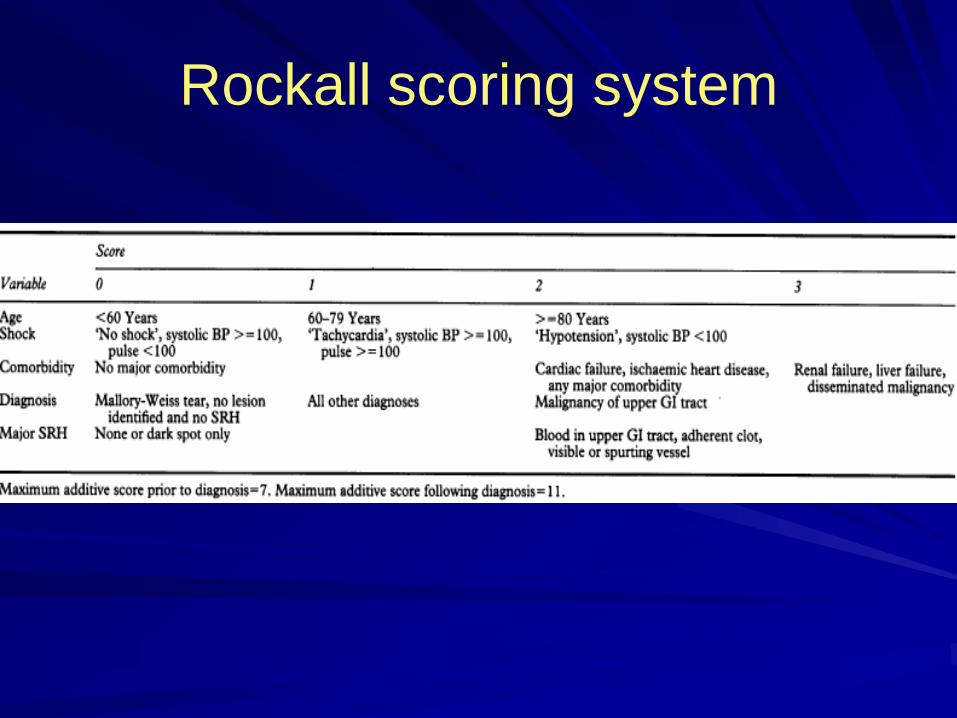
Rockall scoring system
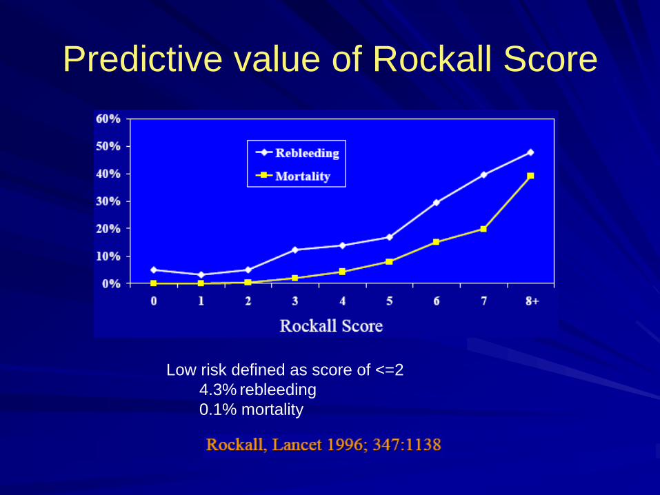
Predictive value of Rockall Score
Low risk defined as score of <=2
4.3% rebleeding
0.1% mortality

Intervention for diagnosis
Angiography : bleeding at least 0.5-1
mL/min
– Bleeding persists and endoscopy fails to identify a bleeding site.
– As salvage therapy, embolization of the
bleeding vessel can be as successful as
emergent surgery in patients who have failed a second attempt of endoscopic therapy

ED approach
Initial resuscitation
Clinical assessment and risk stratification
Identification source of bleeding
Specific therapy
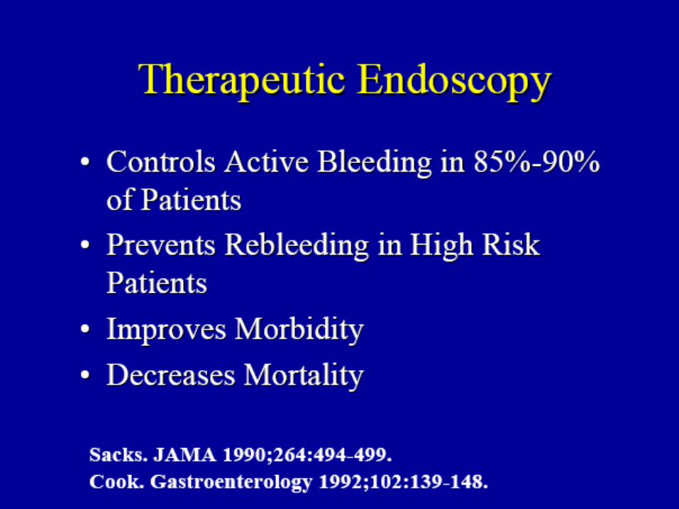
Specific therapy
Medical therapy
Endoscopic therapy
Radiologic intervention
Surgery
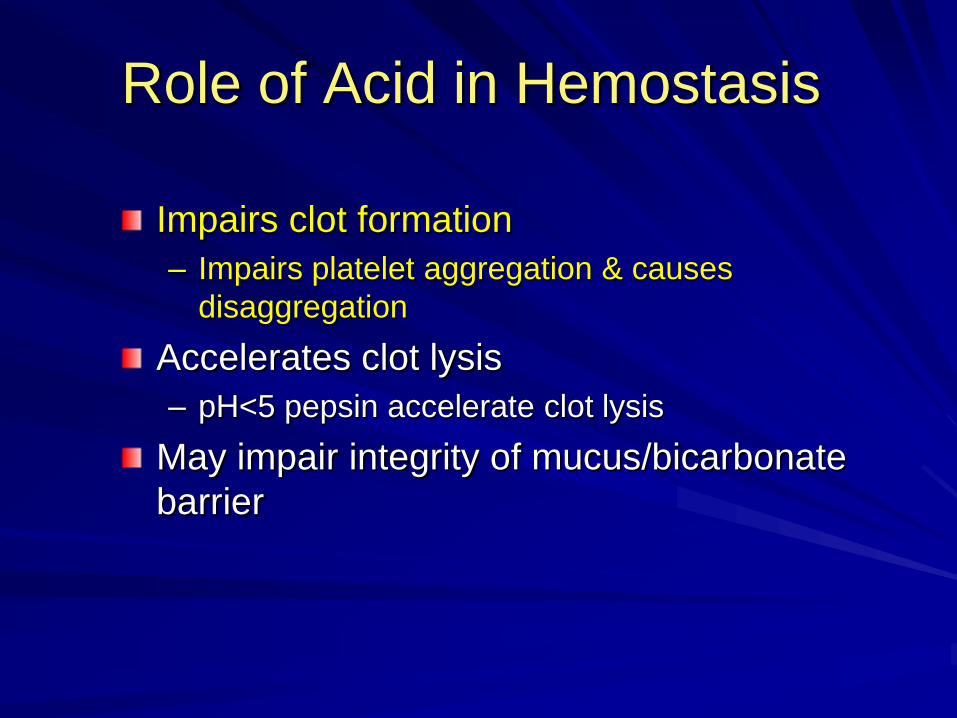
Role of Acid in Hemostasis
Impairs clot formation
– Impairs platelet aggregation & causes
disaggregation
Accelerates clot lysis
– pH<5 pepsin accelerate clot lysis
May impair integrity of mucus/bicarbonate
barrier

Role of Acid in Hemostasis
pH>6
– Effective platelet aggregation
– Irriversible inactivation of pepsin
– Optimal maintenance of hemostasis

H2-receptor antagonists
Intra gastric pH >4 in 65-85% of day
Tolerance in 72 hours

Meta-analysis: the efficacy of intravenous
H2-receptor antagonists in bleeding peptic
ulcer.
30 RCT,3786 bleeding GU and DU
The use of H2-receptor antagonists has not been shown to be effective in altering the course of UGIB.Conclusion : There was a possible minor benefit with intravenous H2 antagonists in bleeding gastric ulcers but no benefit in duodenal ulcers
Levine JE, Leontiadis GI, Sharma VK, Howden CW. Aliment Pharmacol Ther. Jun 2002;16(6):1137-42.

Proton pump inhibitor therapy for peptic ulcer bleeding: Cochrane collaboration meta-analysis of
randomized controlled trials.
A meta-analysis of 24 randomized controlled
trials that evaluated PPIs for bleeding ulcers (with or without endoscopic therapy)
Significant reduction in the risk of rebleeding, the
need for repeat endoscopic hemostasis, and surgery.
Not effect overall mortality, but reduced mortality
in Asian trials and in patients with active bleeding or nonbleeding visible vessels.Leontiadis GI, Sharma VK, Howden CW. Mayo Clin
Proc. Mar 2007;82(3):286-96.

Comparison of intravenous pantoprazole with intravenous ranitidine in prevention of rebleeding from gastroduodenal ulcers
Result :
• During 72 hours rebled was
3.2% in Pantoprazole group
versus 12.9% in Ranitidine
group
Duvnjak M, et.al. Gut /Suppl. III 49 (2001): 2379
Conclusion: Intravenous Pantoprazole is significantly superior
to intravenous ranitidine in the prevention of rebleeding from
gastroduodenal ulcer after initial endoscopic haemostasis.
Rebleeding during 72h
12.9%
3.2%
0%
2%
4%
6%
8%
10%
12%
14%
Pantoprazole i.v. Ranitidine i.v
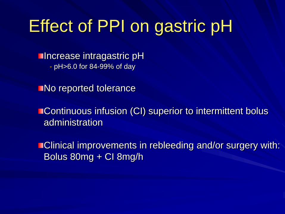
Effect of PPI on gastric pH
Increase intragastric pH pH>6.0 for 84-99% of day
No reported tolerance
Continuous infusion (CI) superior to intermittent bolus
administration
Clinical improvements in rebleeding and/or surgery with:
Bolus 80mg + CI 8mg/h

PPI dose and administration
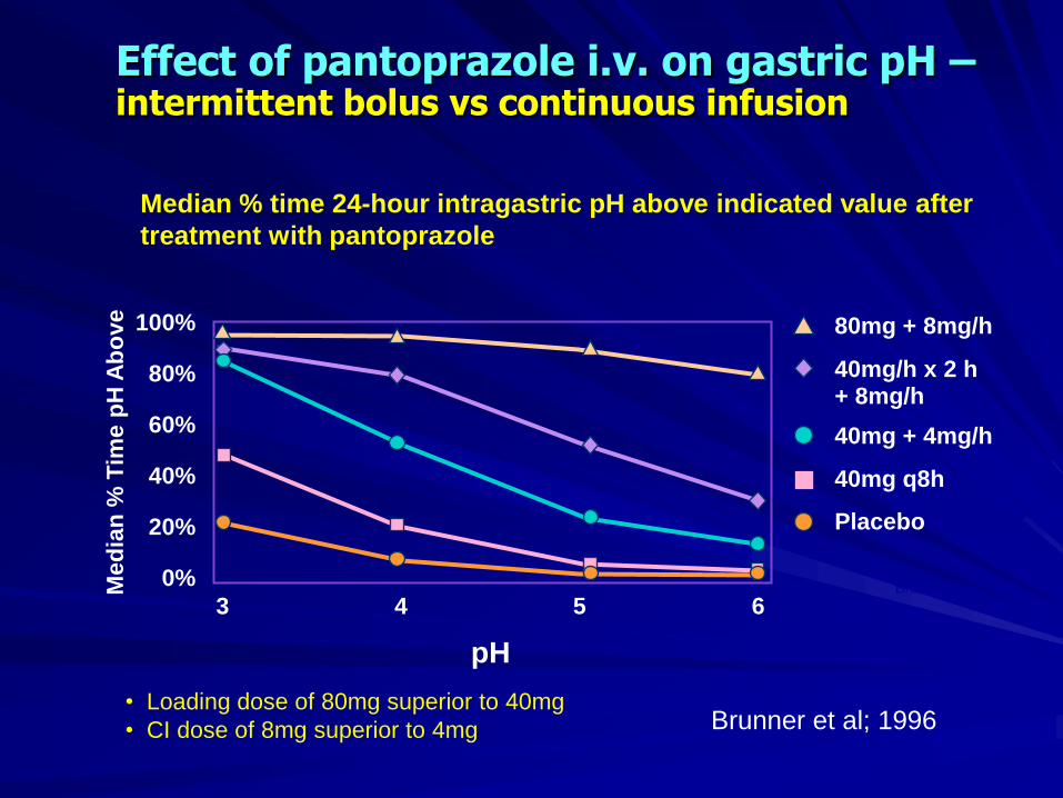
Median % time 24-hour intragastric pH above indicated value after
treatment with pantoprazole
Effect of pantoprazole i.v. on gastric pH –intermittent bolus vs continuous infusion
Brunner et al; 1996Med
ian
% T
ime p
H A
bo
ve 100%
80%
60%
40%
20%
0%
pH
3 4 5 6
80mg + 8mg/h
Placebo
40mg/h x 2 h + 8mg/h
40mg + 4mg/h
40mg q8h
• Loading dose of 80mg superior to 40mg
• CI dose of 8mg superior to 4mg Brunner et al; 1996
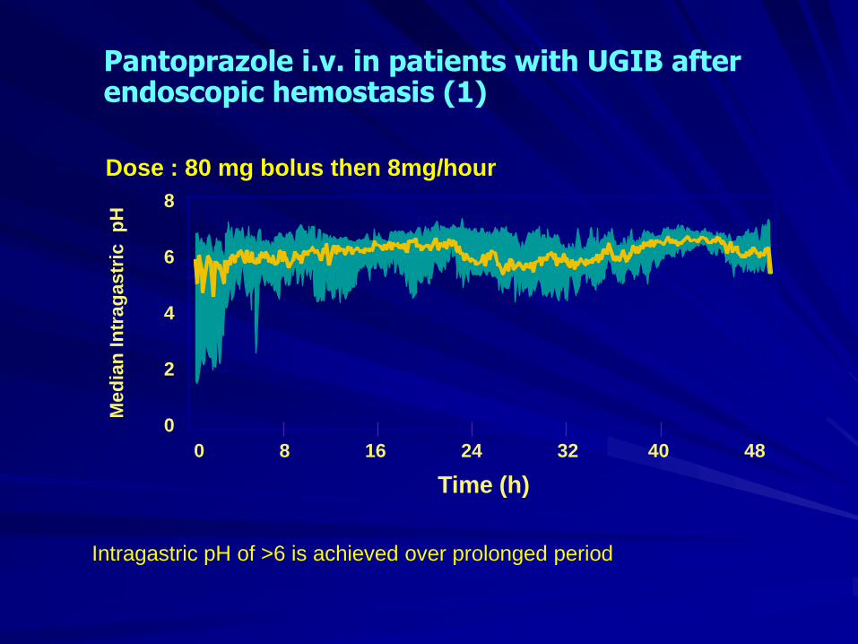
Pantoprazole i.v. in patients with UGIB after endoscopic hemostasis (1)
van Rensburg et al; 1997
Dose : 80 mg bolus then 8mg/hour
Med
ian
In
trag
astr
ic
pH
8
6
4
2
0
Time (h)
0 8 16 24 32 40 48
Intragastric pH of >6 is achieved over prolonged period
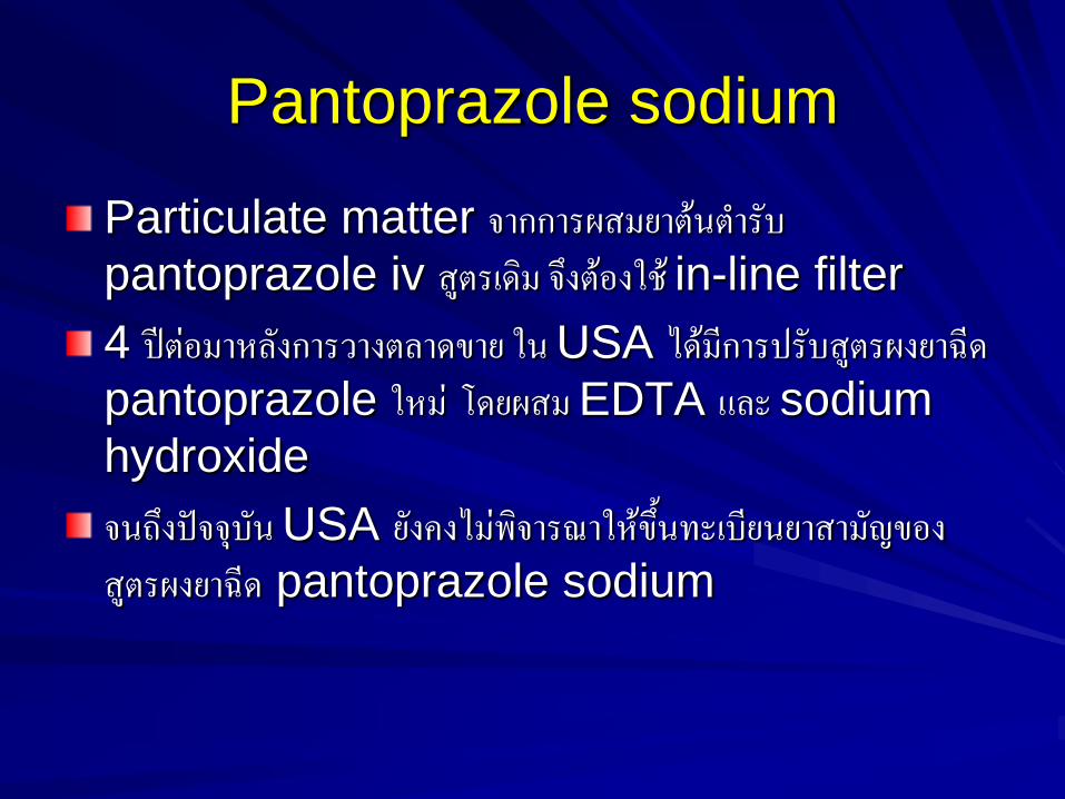
Pantoprazole sodium
Particulate matter จากการผสมยาต้นต ารับ pantoprazole iv สูตรเดิม จึงต้องใช้ in-line filter
4 ปีต่อมาหลังการวางตลาดขาย ใน USA ได้มีการปรับสูตรผงยาฉีดpantoprazole ใหม่ โดยผสม EDTA และ sodium
hydroxide
จนถึงปัจจุบัน USA ยังคงไม่พิจารณาให้ขึ้นทะเบียนยาสามัญของสูตรผงยาฉีด pantoprazole sodium
PPI after endoscopic therapy
An increasing amount of evidence in the literature states that therapy with high-dose PPIs (IV bolus followed by
continuous infusion) may decrease the rate
of rebleeding after endoscopic therapy. By
increasing the gastric pH above 6, the clot is stabilized.
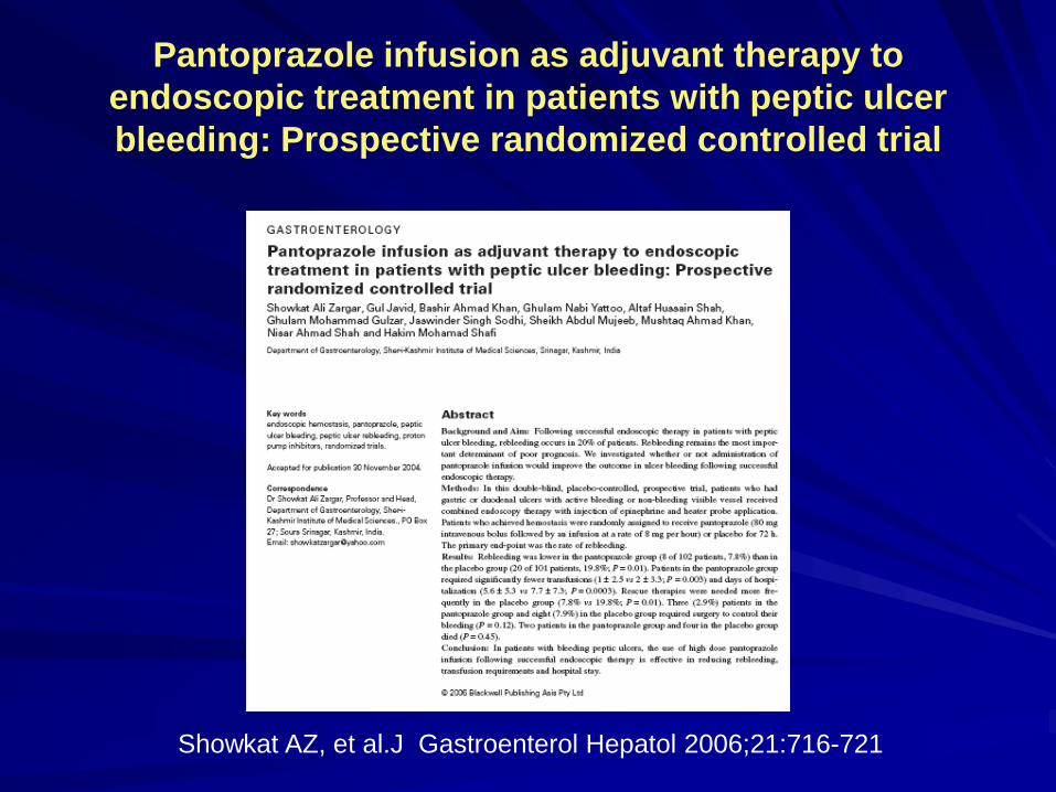
Pantoprazole infusion as adjuvant therapy to
endoscopic treatment in patients with peptic ulcer
bleeding: Prospective randomized controlled trial
Showkat AZ, et al.J Gastroenterol Hepatol 2006;21:716-721

Method• Setting: double-blind, placebo-controlled, prospective trial
• Patients: above 18 years of age with peptic ulcer bleeding
and undertaken successful endoscopic therapy
Endoscopy to
confirm peptic ulcer
bleeding
Endoscopic Tx
(epinephrine inj
& heat probe
Pantoprazole
IV (n=102)
Placebo
IV (n=101)
Random IV 80mg + 8
mg/hr for 72
hrs
Pantoprazole
40 mg tab for
6 wks
• Efficacy measurement
- Primary: rate of rebleeding
- Secondary: need for rescue therapy, need for surgery,
mortality, duration of hospital stay, and
blood transfusion requirement

ResultRebleeding, surgery, and mortality
– Pantoprazole therapy was associated with significant
reductions in rates of rebleeding
7 . 8 %
2 . 9 %
2 . 0 %
1 9 . 8 %
7 . 9 %
4 . 0 %
0 . 0 % 5 . 0 % 1 0 . 0 % 1 5 . 0 % 2 0 . 0 % 2 5 . 0 % 3 0 . 0 %
P a nt opr a z ol e
P l a c e bo
Not sig
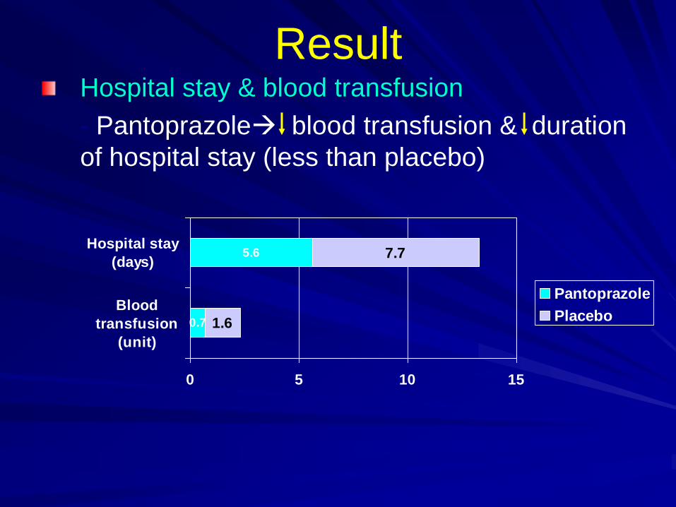
ResultHospital stay & blood transfusion
- Pantoprazole blood transfusion & duration
of hospital stay (less than placebo)
0.7
5.6
1.6
7.7
0 5 10 15
Blood
transfusion
(unit)
Hospital stay
(days)
Pantoprazole
Placebo

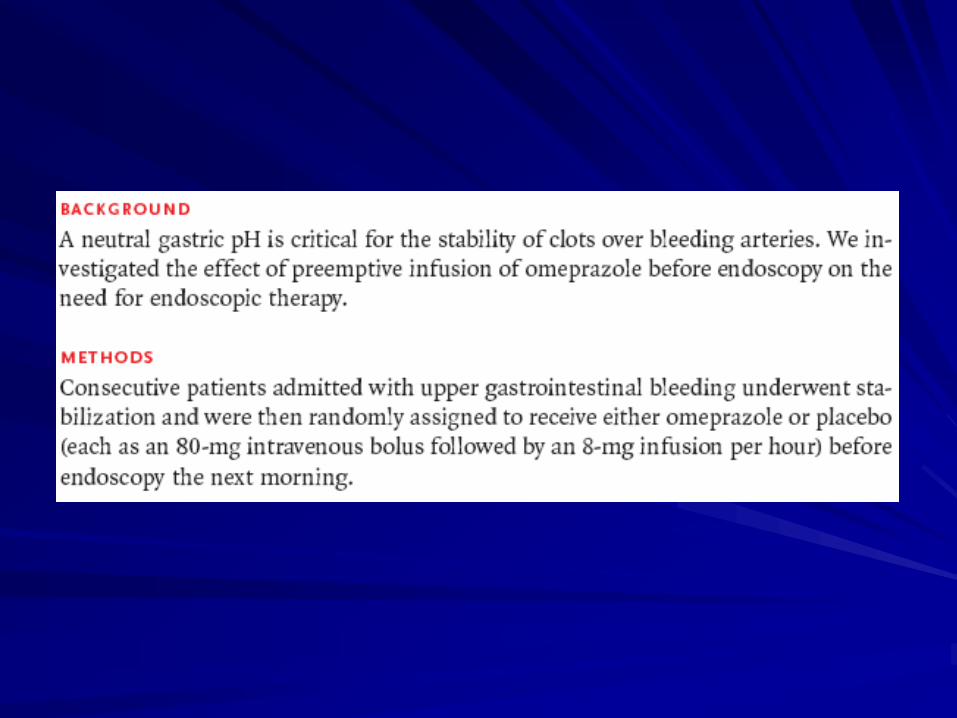
High-dose intravenous proton pump inhibition
following endoscopic therapy in the acute
management of patients with bleeding peptic ulcers in the USA and Canada: a cost-effectiveness
analysis.
The suggested dose of intravenous pantoprazole is 80-mg bolus followed by 8-mg/h
infusion. The infusion is continued for 48-72
hours. This therapy has been shown to be cost-effective
Barkun AN, Herba K, Adam V, Kennedy W, Fallone CA, Bardou M. Aliment Pharmacol Ther. Mar 2004

PPI before endoscopy
N Engl J Med 2007;356:1631-40

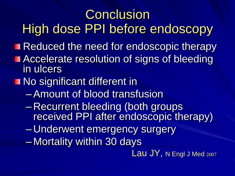
ConclusionHigh dose PPI before endoscopy
Reduced the need for endoscopic therapy
Accelerate resolution of signs of bleeding in ulcers
No significant different in
–Amount of blood transfusion
–Recurrent bleeding (both groups received PPI after endoscopic therapy)
–Underwent emergency surgery
–Mortality within 30 daysLau JY, N Engl J Med 2007
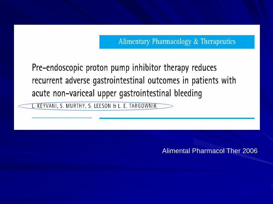
Alimental Pharmacol Ther 2006

Objective
To determine whether using PPI therapy
prior to the performance of endoscopy is
associated with improved clinical
outcomes in patients presenting with signsof ANVUGIB.

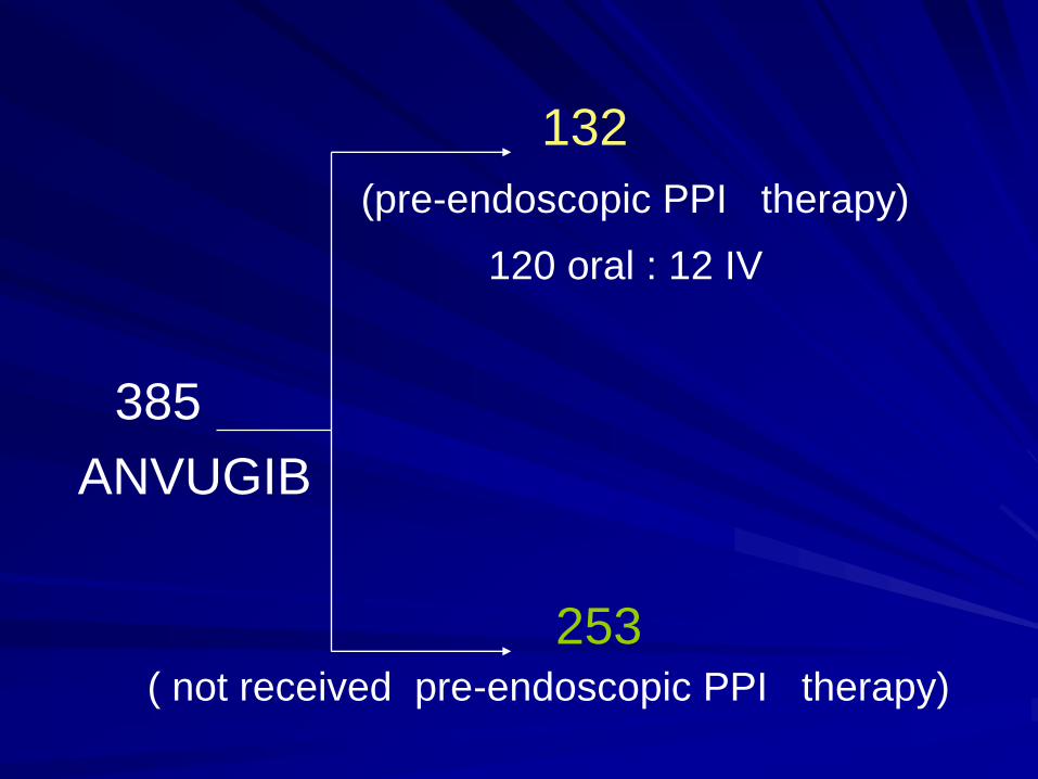
132
(pre-endoscopic PPI therapy)
120 oral : 12 IV
385
ANVUGIB
253( not received pre-endoscopic PPI therapy)
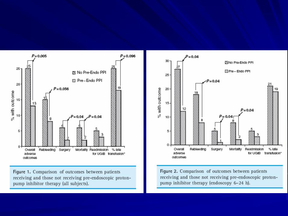

Conclusion
Rebleeding, surgery, mortality, length of
hospital stay decrease in pre-endoscopic PPI group
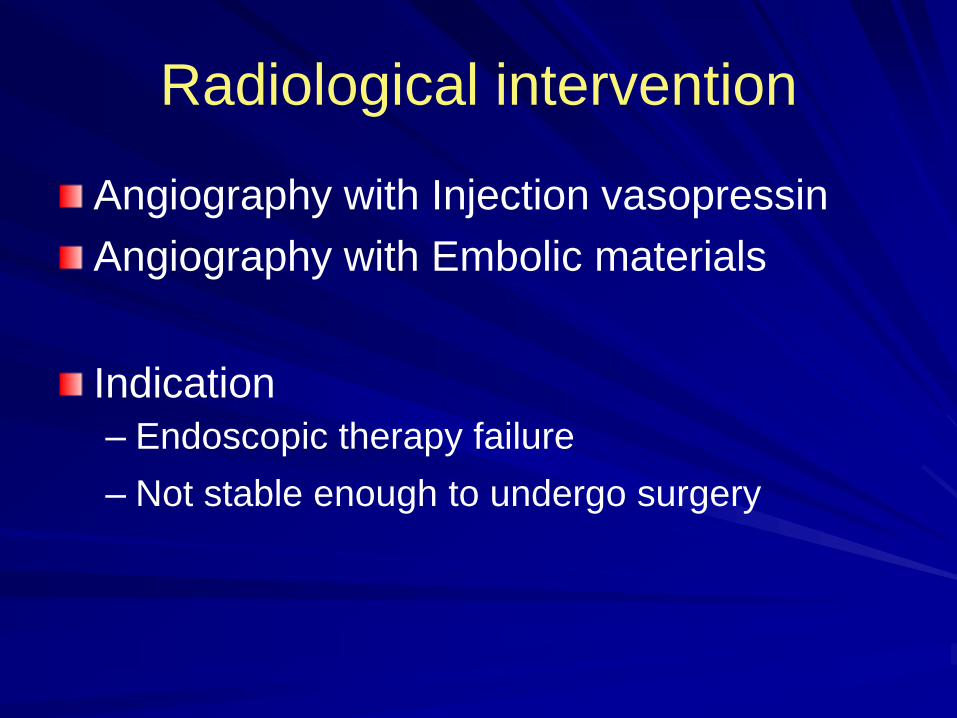
Radiological intervention
Angiography with Injection vasopressin
Angiography with Embolic materials
Indication
– Endoscopic therapy failure
– Not stable enough to undergo surgery

Surgery
Indications
– Severe bleeding, not response to
resuscitation
– Endoscopic therapy failure
– Rebleeding after endoscopic therapy
– Surgical condition: perforation, obstruction, malignancy
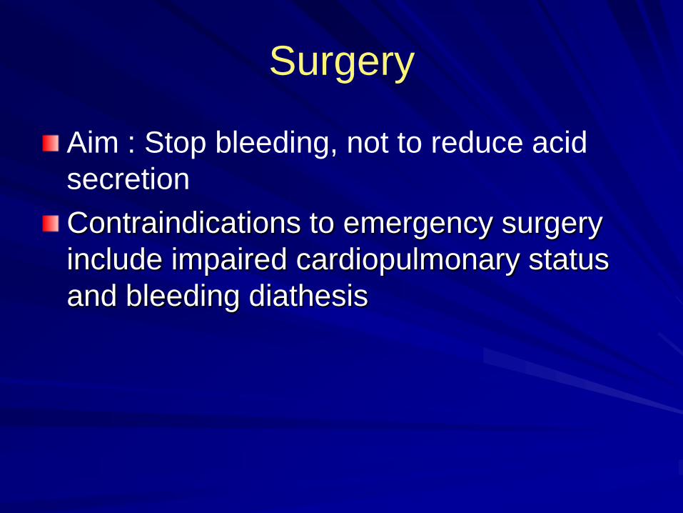
Surgery
Aim : Stop bleeding, not to reduce acid
secretion
Contraindications to emergency surgery
include impaired cardiopulmonary status
and bleeding diathesis

แนวทางการดูแลรักษาผู้ป่วยที่มาด้วยภาวะเลือดออกในทางเดินหายใจส่วนต้นใน
ประเทศไทย
สมาคมแพทย์ทางเดินอาหารแห่งประเทศไทย
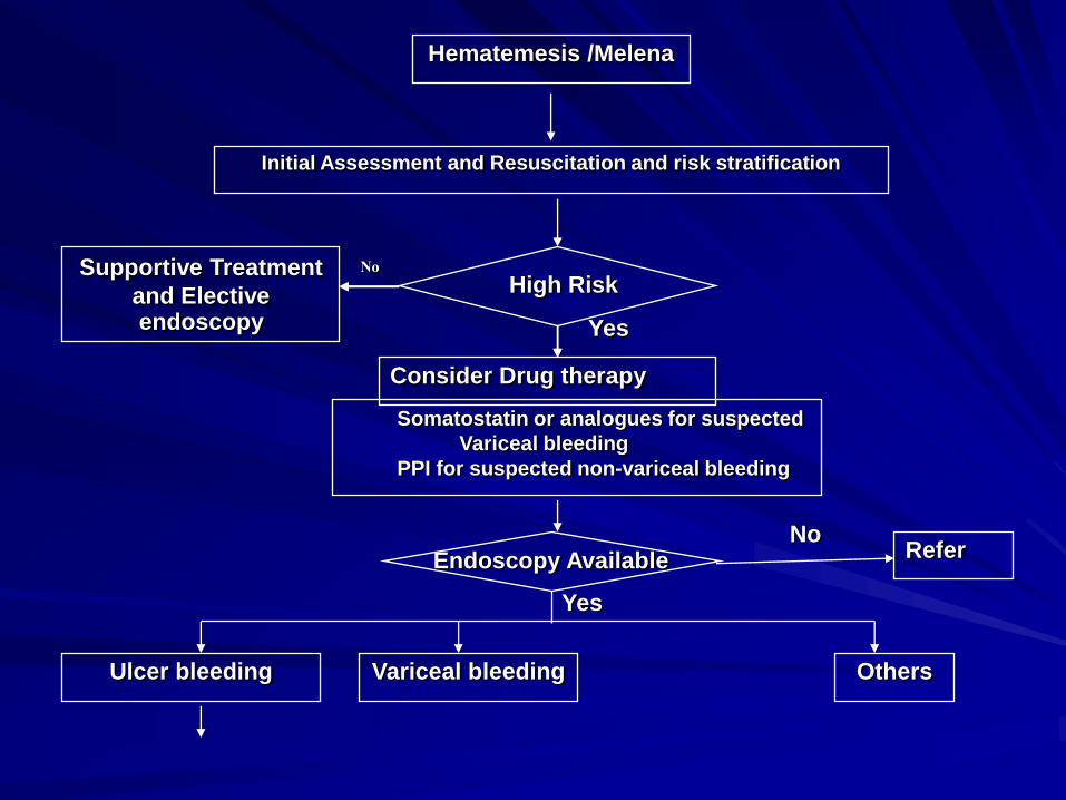
Hematemesis /Melena
Initial Assessment and Resuscitation and risk stratification
Supportive Treatment
and Elective endoscopy
No
Yes
Consider Drug therapy
Somatostatin or analogues for suspected
Variceal bleeding
PPI for suspected non-variceal bleeding
High Risk
NoReferEndoscopy Available
Yes
Ulcer bleeding Variceal bleeding Others
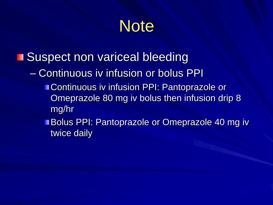
Note
Suspect non variceal bleeding
– Continuous iv infusion or bolus PPI
Continuous iv infusion PPI: Pantoprazole or
Omeprazole 80 mg iv bolus then infusion drip 8
mg/hr
Bolus PPI: Pantoprazole or Omeprazole 40 mg iv
twice daily

Yes
No
Antisecretor
y TherapyNo
Consider surgical
interventions or refer
No
Success
Reendoscopy
No
Continue Drug
and monitoring
Yes
Yes
Rebleeding
Yes
High risk of Rebleeding
Endoscopic Hemostasis
Variceal bleeding
Others Ulcer bleeding