upper and lower gi bleeding - nysge.org upper and lower gi bleeding... · upper and lower gi...
TRANSCRIPT

Upper and Lower GI Bleeding
John Poneros MD, FASGE, NYSGEF
Associate Professor
Columbia University College of Physicians and Surgeons
Acting Director, Endoscopy Unit
New York Presbyterian/Columbia

UGIB
• What is appropriate peri-procedural management of UGIB?
• Which lesions deserve endoscopic therapy and when?
• Should we drop an NGT before EGD?
• Which endoscopic therapies are most efficacious?
• What is the optimal timing for EGD in UGIB?

UGI Bleeding
• 5% of all ER visits
• 300,000 hospitalizations/year
• Gender: 2:1 M:F
• 90% Non-variceal UGIB
• 68% > 60 years old; 27% > 80 years old
• > 3 billion dollars per year
• Mortality 2-14% (10x > for rebleeds)

GI Bleed Mortality

Causes of UGIB

Rare causes of UGIB

Prior to Endoscopy: Resuscitation
•Adequate resuscitation and stabilization is essential prior to endoscopy to
minimize procedure associated complications
•500 ccs of NS over 30 minutes while being type and crossed
•Amount of blood transfusion should be carefully considered – more is not
always better (see next slide)
•Assess cardiac and respiratory status, risk
•Erythromycin (to empty stomach) improves visualization, shortens LOS,
decreases need for repeat EGD and transfusion but does NOT decrease
re-bleeding or mortality
Villaneuva et al NEJM 2013
Gralnek et al Endoscopy 2015

How much to transfuse?
– 921 subjects with severe acute UGIB
– Restrictive (transfuse when Hgb<7; target 7-9)
– Liberal (transfuse when Hgb<9; target 9-11)
– Primary outcome: all cause mortality rate within 45 days NEJM 2013;368;11-21

Restrictive Strategy Superior
Restrictive Liberal P value
Mortality rate 5% 9% 0.02
Rate of further
bleeding
10% 16% 0.01
NEJM 2013;368;11-21
Benefit seen primarily
in PUD and Child A/B
cirrhotics
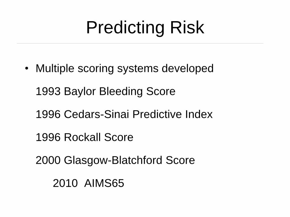
Predicting Risk
• Multiple scoring systems developed
1993 Baylor Bleeding Score
1996 Cedars-Sinai Predictive Index
1996 Rockall Score
2000 Glasgow-Blatchford Score
2010 AIMS65

Rockall Scoring System
• 2 components: clinical + endoscopic
Variable 0 1 2 3
Age <60 60-79 ≥ 80
Shock No
SBP ≥ 100
P<100
Tachy-
SBP ≥ 100
P>100
Hypotension-
SBP <100
Comorbidity No major Cardiac failure,
CAD, other
major
Renal failure,
liver failure,
malignancy
Diagnosis MWT NON Malignant GI cancer
Recent Hemorrhage
None Blood in lumenGut 1996;38:316

Clinical Rockall Score – Mortality Rates
0%
10%
20%
30%
40%
50%
60%
0 1 2 3 4 5 6 7

Glasgow Blatchford Risk Score

AIMS65
lbumin <3.0
NR > 1.5
ental status altered
ystolic BP <90
+ years old
Gastrointest Endosc 2011;74:1215
Robertson et al GIE 2016
AIMS65 better than GBS and pre-EGD Rockall in predicting in
hospital mortality and need for ICU

AIMS65
Gastrointest Endosc 2011;74:1215

• Vital signs in the bleeding patient
Mild to moderate hypovolemia: Resting tachycardia
Blood loss > 15%: Orthostatic hypotension (Decrease in systolic of > 20 mm Hg and/or
increase in HR of 20 when standing)
Blood loss > 40%: Supine hypotension
• Stool color not reliable indicator of location of bleeding
In 80 patients with hematochezia (74% colonic, 11% upper, 9% small bowel and
6% unidentified
• BUN:Cr ratio of > 30 (LR 7.5 that upper source)
• Presence of blood clots in the stool make UGI source less likely
• Ask if prior episode of UGIB: 60% with hx of UGIB bleeding from same lesion
• Take a good history: alarm signs, retching, NSAID use
H&P in bleeding patient

Predicting Risk: Role of NG
lavage
Active Bleeding Death
Clear 10% 6%
Coffee Grounds 13% 10%
Red Blood 23% 18%

Etiology of bleeding with clear
NGT lavage

NGT Lavage Prior to
Endoscopy
Role of nasogastric lavage:
• Assessment for location of bleeding
• Utility in “cleaning”
• Studies have failed to demonstrate a benefit in clinical
outcome

Do I Need to Get Up in the
Middle of the Night?
• Endoscopy within 12h leads to increased use of endotherapy for
stigmata of bleeding
• Two studies evaluated the value of immediate EGD and found no
improvement in clinically important outcomes
• No evidence exists for any clinical benefit of endoscopy
performed within 12h vs 24h (re-bleeding rate, LOS, surgery or
mortality)
• Consensus guidelines recommend endoscopy within 24h
Lee et al GIE 1999
Schacher et al Endoscopy 2005
Kumar et al GIE 2016


Pre-endoscopy PPI
• Reduces the proportion of patients with high risk endoscopic stigmata (“downstages” lesion)
• Decreases need for endoscopic therapy
• Has not been shown to reduce rebleeding, surgery, or mortality rates
N Engl J Med 2007;356:1631
Endoscopic treatment required:Omeprazole – 19% (23% of PUD)Placebo – 28% (37% of PUD)
High risk Low risk

Forrest III
Clean base
Forrrest IIb
Adherent ClotForrest IIa
Visible vessel
Forrest Ib
Oozing without
visible vessle
Forrest Ia
Active bleeding
Gastric Ulcers: Endoscopic
Findings


Endoscopic Stigmata
010-36No stigmata
5-912-18Other stigmata
24-4118-26Adherent clot
(no visible vessel)
18-5517-50Non-bleeding visible vessel
85-1008Spurting arterial bleeding
Re-bleeding(%)
Incidence (%)
Stigmata of hemorrhage

Endoscopic Findings with
GU/DU

Endoscopic Treatment Options
• Injection (Epinephrine1:10,000 or Saline)
• Thermal
– Heater probe (leads to edema, tissue protein coagulation,
contraction of vessels, activation of coagulation cascade)
– Multi/Bipolar probes (no grounding pad)
– APC (Argon Plasma Coagulation)
• Mechanical
– Clips
– Bands
– “Other devices”
Endoscopic hemostatic devices GIE 2009


Endoscopic Clips

Endoscopic Treatment


Argon Plasma Coagulation
• 6000 peak volt energy delivered from
ERBE electrosurgical generator
• Tungsten electrode within probe
ignites gas jet
• Ionized argon “plasma” seeks nearest
ground
• Tissue coagulated with depth of 2-3
mm

Newer WeaponsOvesco © Over the Scope Clips OTSC

Newer weapons
Cook© HemosprayProprietary inorganic powder delivered with CO2
Mechanical barrier and absorption

OUTCOMES FROM AN INTERNATIONAL
MULTICENTRE REGISTRY OF PATIENTS WITH
ACUTE GASTROINTESTINAL BLEEDING
UNDERGOING ENDOSCOPIC TREATMENT WITH
HEMOSPRAY
Presentation Number: 402
View Presentation Add to Schedule
AuthorBlock: Durayd Alzoubaidi1, Radu Rusu11, Jason Mark Dunn11, Johannes Wilhelm Rey4,
Shraddha Gulati6, Bu Hayee6, Selena Dixon9, Sulleman Moreea9, Duncan Napier12, John Anderson12,
Martin Dahan2, Max Hu10, Patricia Duarte10, Phil Boger10, Alberto Murino7, Sina Jameie-Oskooei7,
Edward Despott7, Cora Steinheber3, Martin Goetz3, Sharmila Subramaniam8, Pradeep Bhandari8,
Cormac Magee5, Martin Anthony Everson5, Omer Ahmad1, Matthew Banks5, Laurence Lovat1,
Emmanuel Coron2, Ralf Kiesslich4, Rehan Haidry1,5

OUTCOMES FROM AN INTERNATIONAL MULTICENTRE REGISTRY OF
PATIENTS WITH ACUTE GASTROINTESTINAL BLEEDING UNDERGOING
ENDOSCOPIC TREATMENT WITH HEMOSPRAY
Introduction:
•Hemospray is a novel proprietary mineral blend that forms a mechanical
barrier over the bleeding site when applied endoscopically
•Primary aim of this international prospective multicentre registry is to
collect data on the outcomes of patients with AGIB after endoscopic
application of Hemospray
•Secondary outcomes of rebleeding, 30 day mortality, disease and
procedure specific outcomes were collected


OUTCOMES FROM AN INTERNATIONAL MULTICENTRE REGISTRY OF
PATIENTS WITH ACUTE GASTROINTESTINAL BLEEDING UNDERGOING
ENDOSCOPIC TREATMENT WITH HEMOSPRAY
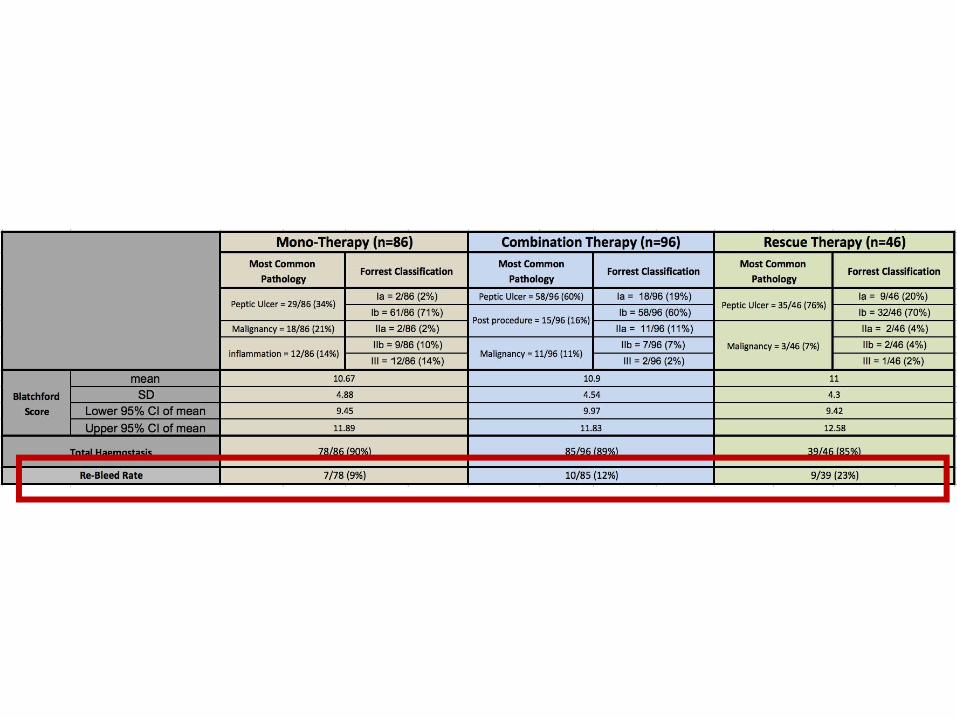
Method:
•Prospective data from 11 centers: UK, France and Germany
•Hemospray use was at endoscopist’s discretion
•Hemospray was either mono therapy, dual-therapy with standard
haemostatic endoscopic techniques or as rescue therapy once standard
methods had failed
•Immediate haemostasis defined as cessation of bleeding within 5 mins
after Hemospray application
•Rebleeding: sustained drop in Hb (>2g/l) OR haematemesis OR
persistent melaena with on going haemodynamic compromise after EGD
OUTCOMES FROM AN INTERNATIONAL MULTICENTRE REGISTRY OF
PATIENTS WITH ACUTE GASTROINTESTINAL BLEEDING UNDERGOING
ENDOSCOPIC TREATMENT WITH HEMOSPRAY
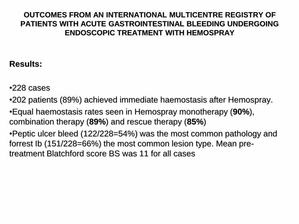
Results:
•228 cases
•202 patients (89%) achieved immediate haemostasis after Hemospray.
•Equal haemostasis rates seen in Hemospray monotherapy (90%),
combination therapy (89%) and rescue therapy (85%)
•Peptic ulcer bleed (122/228=54%) was the most common pathology and
forrest Ib (151/228=66%) the most common lesion type. Mean pre-
treatment Blatchford score BS was 11 for all cases

OUTCOMES FROM AN INTERNATIONAL MULTICENTRE REGISTRY OF
PATIENTS WITH ACUTE GASTROINTESTINAL BLEEDING UNDERGOING
ENDOSCOPIC TREATMENT WITH HEMOSPRAY
Results:
•26 patients did not achieve immediate haemostasis. Mean BS was higher
in this group at 13.35 (p<0.05). Forrest Ib was the most common lesion
type in this group [Ib=20/26 (77%), p<0.05]
•26 cases of rebleeding reported after successful haemostasis. The mean
BS was higher at 12.26 (p< 0.05). Forrest Ib was the most common bleed
in this group [Ib=15/26 (58%), p<0.05]
•55 (24%) patients were anticoagulated at the time of emergency
endoscopy. Haemostasis was achieved in 49/55 (89%) patients

Managing Acid Suppression
after EGD
• Does IV acid suppression matter after endoscopic therapy?
• 767 patients with PUD, randomized to either high-dose IV
PPI (80 mg IV Eso + 8/hr drip) or placebo after successful
endoscopic hemostasis obtained
• Significant difference in 72 hr re-bleeding and all cause
mortality
-Sung Ann Intern Med 2009; 150:455-64

Role of Repeat Endoscopy in
UGI Bleeding
• Therapeutic endoscopy
– At least 2 attempts indicated prior to surgery
• Surgery for failed endoscopic Rx
– Consider early (6-8 units transferred)
• Therapeutic angiography
– High operative risk patients
– Difficult bleeding sources

Variceal bleeding
• Cirrhosis affects 2 million in the USA
• 50% develop varices
• 25-35% bleed within 2 years
• Mortality 30-50%
• Highest mortality first bleed
• Variceal size predicts risk of bleeding
• Severity of underlying liver disease determines survival

asoconstrictor therapy
ntibiotics
esuscitation
U level care
ndoscopy
ternative/Rescue therapies
eta blockade

asoconstrictor therapy
• Goal: Reduce splanchnic blood flow
• Terlipressin – only agent shown to improve control of bleeding and survival in RCTs and meta-analysis– Not available in US
• Somatostatin – not available in US
• Octreotide (somatostatin analogue)• Decreases splanchnic blood flow (variably)
• Efficacy is controversial; no proven mortality benefit
• Standard dose: 50 mcg bolus, then 50 mcg/hr drip for 3-5 days
Gastro 2001;120:946
Cochrane Database Syst Rev 2008
NEJM 1995;333:555
AJGl 2009;104:617

ntibiotics
• Bacterial infection occurs in up to 66% of
patients with cirrhosis and variceal bleed
• Prophylactic antibiotics reduces
incidence of bacterial infection,
significantly reduces early rebleeding
Hepatology 2004;39:746
J Korean Med Sci 2006;21:883
Hepatogastroenterology 2004;51:541

esuscitation
Restrictive Resuscitation
Goal = maintain hemodynamic stability, Hgb ~7-
8, CVP 4-8 mmHg
NEJM paper demonstrated restrictive
transfusion therapy of benefit in Child’s A and B
but not Child’s C

ndoscopy

ndoscopic Banding

Related In: Results (gridquery.php?simResults=3017746_jaan0022-0640-
f1&rFormat=json&query=&req=2&m=1&n=100) - Collection (gridquery.php?
simCollection=3017746_jaan0022-0640-
f1&rFormat=json&query=&req=3&m=1&n=100)
Show All Figures
0
0
Use of ß-blocker therapy to prevent primary bleeding of esophageal
varices
Tursi T - (2010)
© Copyright Policy - open-access (/faq.php#copyright)
License (http://creativecommons.org/licenses/by/2.5/)
Related In: Results (gridquery.php?simResults=3134063_crg0005-0386-
f01&rFormat=json&query=white nipple&req=2&m=1&n=100) - Collection
(gridquery.php?simCollection=3134063_crg0005-0386-f01&rFormat=json&query=white
nipple&req=3&m=1&n=100)
Show All Figures
TweetTweet 0
TweetTweet 0
0
0
The White Nipple Sign: Please Do Not Disturb
Khan NM, Shapiro AB - (2011)
© Copyright Policy - open-access (/faq.php#copyright)
License 1 (http://creativecommons.org/licenses/by-nc-nd/3.0/) - License 2
(http://www.karger.com)
Endoscopic Findings:
Red Wales and White Nipples

ternative/Rescue therapies
• TIPS – Transjugular Intrahepatic Portosystemic Shunt
• Early placement of shunt (within 24-72hrs) associated with improved survival among high-risk patients
• Preferred treatment for gastric variceal bleeding (rule out splenic vein thrombosis first)
Fan, C. (Apr 25 2006). Vascular Interventions in the
Abdomen: New Devices and Applications. The
DAVE Project. Retrieved Aug, 2, 2010, from
http://daveproject.org/viewfilms.cfm?film_id=497Hepatology 2004;40:793
Hepatology 2008;48:Suppl:373A
N Engl J Med. 2010 Jun 24;362:2370

ternative/Rescue therapies
Sengstaken-Blakemore Tube • Very effective for immediate, temporary control
• High complication rate –aspiration, migration, necrosis + perforation of esophagus
• Use as bridge to TIPS within 24 hours
• Airway protection strongly recommended

ternative/Rescue therapies
• Specially designed covered metal stent
• Tamponades distal esophageal varices
• Removable; does not require airway protection
• Very limited data
Self-Expanding Metal Stent
Gastrointest Endosc 2010;71:71

LGIB
• Overall mortality is low
• About 4% in one large series
• Mortality higher in older adults, those with intestinal
ischemia and those with comorbid illness
• 13% of hematochezia patients bleeding from an
upper source (proximal to ligament of Treitz)

Acute Hematochezia: 1559 Pts
• Diverticulosis: 5-42%
• Ischemia: 6-18%
• Anorectal (Hemorrhoids, Anal fissures, Rectal ulcers): 6-16%
• Neoplasia (Polyps and cancer): 3-11%
• Angiodysplasia: 0-3%
• Postpolypectomy: 0-13%
• IBD: 2-4%
• Radiation colitis: 1-3%
• Other colitis (Infectious, antibiotic associated): 3-29%
• Small bowel/UGIB: 3-13%
• Other causes: 1-9%
• Unknown cause: 6-23%
Strate et al Gastro Clinc NA 2005

Colonoscopy in Lower GI
Bleeding
• Advantages:
– Potential to precisely localize the site of bleeding regardless of
the etiology or rate of bleeding,
– Ability to collect specimens and
– Potential for therapeutic intervention
• Disadvantages:
– Need for bowel preparation,
– Poor visualization in unprepped or poorly prepped colon
– Risks of sedation in an acutely bleeding patient
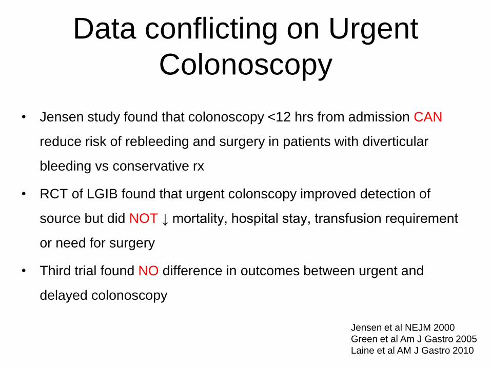
Data conflicting on Urgent
Colonoscopy
• Jensen study found that colonoscopy <12 hrs from admission CAN
reduce risk of rebleeding and surgery in patients with diverticular
bleeding vs conservative rx
• RCT of LGIB found that urgent colonscopy improved detection of
source but did NOT ↓ mortality, hospital stay, transfusion requirement
or need for surgery
• Third trial found NO difference in outcomes between urgent and
delayed colonoscopy
Jensen et al NEJM 2000
Green et al Am J Gastro 2005
Laine et al AM J Gastro 2010


Colonoscopy in LGIB• Patients unable to take the prep may
require NGT
• Metoclopramide can be used
• A definitive or potential bleeding source
visualized in 45-90% of patients
undergoing colonoscopy for LGIB

Other tests in LGIB
• Tagged red blood cell scan
(radionuclide imaging with
technetium (99mTc) sulfur
colloid is most sensitive
radiographic test
• Can detect bleeding at a rate
of 0.1-0.5 mL/min
• Requires ACTIVE bleeding to
detect a source
• Not very accurate as to site

Angiography in LGIB
• Requires active blood loss of 1 to 1.5 mL/min
• SMA first, then IMA and celiac
• Success varies from 25-70%
• Therapeutic and diagnostic (vasopressin has been
replaced by embolization)
• Intestinal infarction is a risk

Conclusions
• Upper GI Bleed– Attempt to risk stratify before procedure
– Don’t cave to pressure on doing something but don’t delay too
much either
– Assess hemodynamic and respiratory status very carefully in
advance
– Be prepared for anything
– Know your equipment
– You CAN save a life
• Lower GI Bleed– Uncertain if you can save a life in the acute setting
– Involve other services (surgery, IR) early

Quotes to remember when
dealing with GIB
“The Enemy of Good is Better”
“Good judgment comes from
experience. Experience comes from
bad judgment.”