update on cerebral small vessel disease: a dynamic whole...
TRANSCRIPT

Update on cerebral small vessel disease:a dynamic whole-brain disease
Yulu Shi,1,2 Joanna M Wardlaw1
To cite: Shi Y, Wardlaw JM.Update on cerebral smallvessel disease: a dynamicwhole-brain disease. Strokeand Vascular Neurology2016;1:e000035.doi:10.1136/svn-2016-000035
Received 26 July 2016Revised 5 September 2016Accepted 7 September 2016
1Centre for Clinical BrainSciences, University ofEdinburgh, Edinburgh, UK2Department of Neurology,Zhongnan Hospital, WuhanUniversity, Wuhan, China
Correspondence toProfessor Joanna MWardlaw;[email protected]
ABSTRACTCerebral small vessel disease (CSVD) is a verycommon neurological disease in older people. Itcauses stroke and dementia, mood disturbance andgait problems. Since it is difficult to visualise CSVDpathologies in vivo, the diagnosis of CSVD has reliedon imaging findings including white matterhyperintensities, lacunar ischaemic stroke, lacunes,microbleeds, visible perivascular spaces and manyhaemorrhagic strokes. However, variations in the useof definition and terms of these features have probablycaused confusion and difficulties in interpreting resultsof previous studies. A standardised use of termsshould be encouraged in CSVD research. These CSVDfeatures have long been regarded as different lesions,but emerging evidence has indicated that they mightshare some common intrinsic microvascularpathologies and therefore, owing to its diffuse nature,CSVD should be regarded as a ‘whole-brain disease’.Single antiplatelet (for acute lacunar ischaemic stroke)and management of traditional risk factors still remainthe most important therapeutic and preventiveapproach, due to limited understanding ofpathophysiology in CSVD. Increasing evidencesuggests that new studies should consider drugs thattarget endothelium and blood–brain barrier to preventand treat CSVD. Epidemiology of CSVD might differ inAsian compared with Western populations (where mostresults and guidelines about CSVD and strokeoriginate), but more community-based data andclear stratification of stroke types are required toaddress this.
INTRODUCTIONThe term ‘cerebral small vessel disease(CSVD)’ refers to a syndrome of clinical andimaging findings that are thought to resultfrom pathologies in perforating cerebralarterioles, capillaries and venules. CSVDcauses up to 45% of dementia, and accountsfor about 20% of all stroke worldwide, 25%of ischaemic (or lacunar strokes), of whomabout 20% are left disabled.1 Cognitiveimpairment, depression and gait problemsare also frequently seen in patients withCSVD. The prevalence of lacunar stroke maybe higher in patients in China where recentstudies have suggested that lacunar infarctionaccounts for 38–46% of ischaemic stroke.2 3
Generally, including in this review, CSVD isused to describe a series of imaging changesin the white matter and subcortical greymatter, including recent small subcorticalinfarct, lacunes, white matter hyperintensities(WMHs), prominent perivascular spaces(PVS), cerebral microbleeds (CMBs) andatrophy.4 Usually, recent small subcorticalinfarcts cause acute stroke symptoms,whereas other CSVD lesions are clinicallymore insidious and thus referred to as‘silent’ lesions. However, the definitions andterms of these lesions have varied greatlyamong studies. For example, a recent reviewidentified 159 different names for recentsmall subcortical infarcts, but these nameslike ‘lacunar infarct’ were also frequentlyused to describe lacunes4 5 that were notnecessarily related to symptoms and mighthave been due to haemorrhage. The substan-tial variation in the use of these terms hasprobably contributed to confusion and diffi-culties in interpreting previous research.Therefore, in 2013, an expert workgroup onCSVD proposed a list of standard terms tohelp avoid confusion and suggests that CSVDresearchers should be encouraged to applythese terms in future studies.4 We will alsouse these terms in this review.The different features of CSVD have long
been regarded as different types of tissuechanges. However, recent studies show thatthese features are correlated, are more likelyto share common diffuse intrinsic smallvessel pathologies, and are probably alsomore ‘dynamic’ than previously thought.Advances in imaging techniques havebrought new insights into mechanisms ofCSVD. In this review, we will summarise find-ings in recent clinical studies on CSVD,discuss CSVD mechanisms and explore emer-ging prevention and treatment options.
Clinical lacunar strokeA lacunar clinical syndrome could be due toeither ischaemia or a small haemorrhage.6
Many haemorrhagic strokes in older peopleare also due to CSVD pathology.1 In this
Shi Y, Wardlaw JM. Stroke and Vascular Neurology 2016;1:e000035. doi:10.1136/svn-2016-000035 1
Open Access Review
Copyright 2016 by BMJ Publishing Group Ltd.
on 28 May 2018 by guest. P
rotected by copyright.http://svn.bm
j.com/
Stroke V
asc Neurol: first published as 10.1136/svn-2016-000035 on 30 S
eptember 2016. D
ownloaded from

review, we will focus mainly on ischaemic CSVD.Lacunar ischaemic stroke is defined as a stroke that isattributable to a recent small infarct <1.5 (or some say2) cm diameter in the white matter, basal ganglia, ponsor brainstem, and is consistent with a lacunar clinicalsyndrome.7 It is commonly attributed to an abnormalityin a single small deep perforating (or lenticulostriate)artery. On MRI, an acute lacunar infarct is shown ashyperintense on diffusion-weighted imaging (DWI),hypointense on an apparent diffusion coefficient map,hyperintense on T2-weighted and fluid-attenuated inver-sion recovery (FLAIR), hypointense on T1 and hypoatte-nuated on CT (figure 1). It can be rounded, ovoid ortubular.4 Generally, the Oxfordshire Community StrokeProject (OCSP) classification, which uses only clinicalfeatures to diagnose the stroke subtype, can predict cor-rectly the size and location of a recent brain infarct onimaging in 75–80% of patients with stroke.8 However, upto 20% of acute lacunar infarcts can present with cor-tical symptoms, and conversely cortical infarcts canpresent with lacunar syndromes.9 One explanation isthat lacunar infarcts closer to the cortex are more likelyto cause cortical symptoms.9 Therefore, in studies wherestroke diagnosis relied mainly on the clinical presenta-tions, this ‘mismatch’ may have added ‘noise’. Thus, inepidemiology, mechanistic studies or clinical trials, it isimportant to verify stroke lesions using sensitive imagingwherever possible.
However, even with sensitive imaging like DWI, about30% of patients with clinically definite stroke did notshow any recent ischaemic change on MRI;10 when fol-lowed up for a year, the DWI-negative patients had justas much recurrent stroke, dependency and cognitiveimpairment as the DWI-positive patients. Therefore,negative DWI/MRI cannot exclude stroke diagnosis.Rapid access to scanning after stroke onset can increasethe chance of positive findings.11 It is also noteworthythat DWI-positive lesions can be clinically ‘silent’, forexample, (1) as a second silent acute infarct in patientspresenting with stroke due to another acute symptomaticinfarct, or (2) in patients with acute haemorrhagicstroke, and (3) in patients with severe WMHs who didnot have any overt stroke symptoms.12
In some clinical stroke classifications such as the Trialof Org 10172 in Acute Stroke Treatment (TOAST) orthe ASCO (A: atherosclerosis; S: small-vessel disease; C:cardiac pathology; O: other causes), another term ‘smallvessel/artery disease’ rather than ‘lacunar stroke’ is usedto represent a stroke that is supposed to be due to asmall artery occlusion. However, these classifications userisk factors to decide the stroke subtype, not just theclinical presentation, so as to distinguish ‘small vessel/artery disease’ from strokes caused by large artery ath-erosclerosis, cardiac emboli or other unknown reasons.However, a small embolus, or atheroma in the middlecerebral artery (MCA) or perforating arterioles can all
Figure 1 STRIVE, STandards for Reporting and Imaging of Small Vessel Disease: example findings (upper), schematic
representation (middle) and a summary of imaging characteristics (lower) of MRI features for changes related to small vessel
disease.4 DWI, diffusion-weighted imaging; FLAIR, fluid-attenuated inversion recovery; SWI, susceptibility-weighted imaging;
GRE, gradient-recalled echo.
2 Shi Y, Wardlaw JM. Stroke and Vascular Neurology 2016;1:e000035. doi:10.1136/svn-2016-000035
Open Access
on 28 May 2018 by guest. P
rotected by copyright.http://svn.bm
j.com/
Stroke V
asc Neurol: first published as 10.1136/svn-2016-000035 on 30 S
eptember 2016. D
ownloaded from

block the perforating arteriole, and any of these cancause a lacunar ischaemic stroke (see figure 2).Therefore, it might be better to focus on the clinicalpresentation to assign the stroke syndrome and separ-ately focus on the risk factors for patient management.
Risk factors and causes of lacunar infarctsFour possible main aetiologies for lacunar ischaemicstroke have been proposed (figure 2): atheroma ofparent arteries (usually MCA) or perforating arterioles,embolism from the heart or carotid arteries, and intrin-sic small vessel disease (lipohyalinosis or fibrinoid necro-sis). Atheroma in MCA appears to cause <20% oflacunar ischaemic stroke. In the Warfarin AspirinSymptomatic Intracranial Disease (WASID) trial, only11% (38/347) of all patients with stroke were lacunartype,13 which is surprising if MCA stenosis is supposed tobe a common cause of lacunar stroke. A recent studyalso did not find any association between lacunar strokeand MCA stenosis.14 A systematic review of Asian studiesshowed that parent artery atherosclerosis accounted for20% of single lacunar infarcts in anterior circulation ter-ritory; however, these hospital-based studies were rathersmall (n=71–118) and some were even retrospective.15
Larger and tubular lacunar infarcts might be more likelyto be caused by proximal artery diseases.16 However, theresults of both our study and the Secondary Preventionof Small Subcortical Stokes Trial (SPS3) suggest that it isnot possible to identify the cause of a particular recent
lacunar ischaemic stroke based on its size, shape orlocation.17 18
Evidence for embolism as a common cause forlacunar ischaemic stroke is limited. Presence of cardi-oembolic sources was found significantly less often inlacunar than in non-lacunar ischaemic stroke.19 20 Few ifany associations were found between ipsilateral carotidstenosis and lacunar ischaemic stroke or other featuresof CSVD.21 22 In primate models, <6% of emboliinjected into carotid arteries entered the lenticulostriatearteries, while the majority entered the cortical arter-ies.23 Lacunar ischaemic strokes in the basal gangliawere marginally more often associated with embolismthan those in the centrum semiovale (11% vs 3%,respectively), but the overall rate of known embolicsources in symptomatic lacunar ischaemic stroke wasvery low (11%).18
Intrinsic small vessel pathologies remain the mostcommon cause of lacunar ischaemic stroke, althoughthe underlying mechanism is unclear. Fisher attributedthe lipohyalinosis in small arteries to hypertension.However, the diagnosis and treatment of hypertensionwere less good when Fisher was working in the 1950sand 1960s and he may have seen some particularlysevere cases of hypertension. Now, epidemiology datashow that hypertension is equally common in non-lacunar as in lacunar ischaemic stroke;19 and manypatients with lacunar stroke are normotensive. Similarly,other traditional risk factors like diabetes mellitus,
Figure 2 Four possible mechanisms that cause a lacunar infarct (from bottom to top): (A) an embolus from the big arteries or
cardiac sources goes up to MCA and ends up entering and occluding lenticulostriate arteries, resulting in a lacunar lesion in
basal ganglia; (B) if the atheroma in the parent artery (ie, MCA) is positioned at the opening of its penetrating branches, it could
lead to an acute occlusion of one or several penetrating arteries, hence causing a lacunar infarct; (C) a lacunar infarct could also
be due to atheroma in the perforating artery if an acute occlusion happens; (D) intrinsic small vessel disease may lead to diffused
disrupted blood–brain barrier. If this happens at an arteriolar level, plasma fluid components would enter and deposit in the
vessel wall, resulting in narrowing of the arteriolar lumen, vessel wall thickening and eventually a secondary luminal occlusion
and traditional infarct. MCA, middle cerebral arteries.
Shi Y, Wardlaw JM. Stroke and Vascular Neurology 2016;1:e000035. doi:10.1136/svn-2016-000035 3
Open Access
on 28 May 2018 by guest. P
rotected by copyright.http://svn.bm
j.com/
Stroke V
asc Neurol: first published as 10.1136/svn-2016-000035 on 30 S
eptember 2016. D
ownloaded from

hypercholesterolaemia and smoking were as frequent inlacunar stroke as in other ischaemic strokes.24 Riskfactor profiles of lacunar stroke seemed different inChina, but it might be too early to say so. The Beijingstroke registry (n=1184) showed a higher proportion ofhypertension in lacunar (acute stroke symptoms+subcor-tical lesion <2 cm diameter on acute CT/MRI) than innon-lacunar stroke after adjusting for age and gender.3
Some other studies had similar findings, but the strokediagnosis varied: in some studies, the differentiationbetween lacunar stroke and ‘large artery atherosclerosis’stroke relied only on lesion size, and clinical classifica-tion included risk factors.25 26 Additionally, most studieswere hospital-based. Hence, population scale data onlacunar stroke are lacking. It is important to distinguishlacunar stroke from other subtypes because of the mech-anism, hence prevention and treatment might differ.More data and careful separation of lacunar stroke fromother subtypes are required in future studies.
Clinically ‘Silent’ CSVDWhite matter hyperintensitiesWMH of presumed vascular origin are very common inolder individuals and regarded as typical signs of CSVD.Symptoms of WMH develop insidiously, such as cognitiveimpairment, dementia and depression,1 but it almosttriples the risk of stroke, doubles the risk of dementiaand increases the risk of death.27
WMHs are usually symmetrically and bilaterally distrib-uted in the white matter including the pons and brainstem, and also occur in deep grey matter. They appearhyperintense to the normal brain on T2 or FLAIR MRI(figure 1), and can be patchy or confluent dependingon their stage in development and severity.Owing to limited pathology studies, the underlying
pathology of WMH remains imprecise. Demyelination,loss of oligodendrocytes and axonal damage were oftenreported. Diffusion tensor imaging studies providedindirect evidence for axonal damage and impairedwhite matter integrity in WMH.28 Indeed, recent evi-dence indicates that WMHs are rather heterogeneous,perhaps reflecting different disease stages. Reduceddensity of glia and vacuolation were observed in severeWMH suggesting end stage disease.29 Autopsy MRIstudies also found oedema that suggests leakage of fluidfrom an impaired blood–brain barrier (BBB) in andaround WMH.30 31 Although these ‘white’ lesions haveuntil now been treated as if they were all the same, dif-ferent degrees of ‘whiteness’ might indicate different‘stages of formation’—some very white WMHs are prob-ably at the end stage of disease and irreversible oncedemyelination or axonal damage has happened; someperhaps less white lesions might be reversible if they aremainly interstitial fluid (ISF) imbalances before perman-ent tissue damage has occurred. These observationsremain to be confirmed in larger studies. These micro-structural changes happen in WMH, and are alsopresent in normal appearing white matter (NAWM).32 33
The white matter integrity in NAWM declines withincreasing closeness to the edge of WMH32 and withmore severe WMH.34
Multiple mechanisms underlying WMH such asincomplete infarct, chronic hypoperfusion and venouscollagenous have been proposed, but evidence for eachis limited. In a pathology study (n=15), no incompleteinfarct was found in WMH.29 Though many cross-sectional studies have found low cerebral blood flow(CBF) to be associated with higher WMH burden, thecausality between low CBF and WMH is unclear.35 A lon-gitudinal study (n=575) showed that more severe base-line WMH predated CBF decline over time rather thanfalling CBF predating WMH progression.36 In a post-mortem study, some non-inflammatory, periventricularvenulopathy was observed in periventricular WMH, sug-gesting that venous collagenosis might cause tissuedamage by vasogenic oedema and impede ISF circula-tion.31 However, this theory remains to be confirmed inin vivo studies. Impaired BBB was noted in WMH areasin autopsies,29 30 which was corroborated by studiesusing cerebrospinal fluid (CSF)/plasma albumin ratio37
and MRI.38–41 It is hypothesised that the disrupted BBBwould result in leakage of fluid, plasma components andcells and eventually lead to perivascular inflammation,demyelination and gliosis. Indeed, the formation ofWMH is likely to be multifactorial. Hypoperfusion,venous pathologies and BBB impairment might all playcritical roles in WMH initiation or progression and inter-act with each other, but which one is the key initialfactor remains unknown.
LacunesThe term ‘lacune’ was used by Fisher to describe a smallfluid cavity in the brain which he thought was a healedlacunar infarct. Therefore, in CSVD research, it is verycommon that terms like ‘lacunar infarction’, ‘lacunarstroke’ and ‘silent brain infarct’ were used to refer tothe CSF-filled cavities on brain MRI or autopsy.42 In fact,lacunes are not always ‘ischaemic’. They can also be theresidual lesion of a small haemorrhage43 (figure 3).Also, it is common that many non-cavitated lacunarischaemic strokes were not counted as ‘lacunar infarcts’.Therefore, in order to avoid more confusion, the term‘lacune of presumed vascular origin’ was proposed toreplace ‘lacune’ and the term ‘lacunar infarct’ shouldNOT be used to describe ‘lacunes’ any more.Lacunes of presumed vascular origin are round or
ovoid, subcortical, fluid-filled cavities with a diameter of3–15 mm. These can occur without any prior symptoms,but can also result from a previous acute small subcor-tical infarct or haemorrhage4 (figure 1). PVS could alsomimic lacunes when they are more than 3 mm in diam-eter.44 Large PVS might have also been miscounted aslacunes in many studies.42 Lacunes usually present as ahypointense ‘hole’ on FLAIR surrounded by a hyperin-tense rim which can help its differentiation from PVS.However, the rim can be absent in some cases and PVS
4 Shi Y, Wardlaw JM. Stroke and Vascular Neurology 2016;1:e000035. doi:10.1136/svn-2016-000035
Open Access
on 28 May 2018 by guest. P
rotected by copyright.http://svn.bm
j.com/
Stroke V
asc Neurol: first published as 10.1136/svn-2016-000035 on 30 S
eptember 2016. D
ownloaded from

within extensive WMH areas may appear as if sur-rounded by hyperintensities, so the insistence on a rimto differentiate lacunes from PVS is not helpful in prac-tice. Nonetheless, it is important to distinguish betweenlacunes and PVS if possible, on size at least, becausethey represent different pathologies as well as differ inclinical associations and implications.Although many lacunes might have lacked acute symp-
toms, when present in larger numbers they are associatedwith dementia, cognitive impairment, gait disturbance andincreased risk of stroke.5 45 46 In the general elderly popu-lation, the prevalence of lacunes ranges from 8% to 28%(mean age=50–75 years).5 A systematic review suggests thatsilent brain infarcts (another term sometimes used forlacune) are more prevalent in the Asian than in thenon-Asian population.47 However, it is noteworthy thatmost of these Asian studies were hospital-based, whereasall non-Asian studies were community-based; therefore,more relevant comparisons are needed to determine ifthe prevalence of lacunes and other CSVD features doesdiffer between world regions and ethnic groups.
Perivascular spacesPVS are the extension of subarachnoid spaces that sur-round cerebral microvessels.48 They are fluid-filledspaces that follow the course of a vessel through thebrain parenchyma.48 PVS are usually microscopic andnot detected on CT or conventional MRIs. Whenenlarged, PVS are commonly seen as hyperintense onT2 MRI, either punctuate with a diameter <3 mm ifimaged perpendicular to the course of the vessel, orlinear if imaged parallel to the course of the vessel49
(figure 1). PVS are most frequent in the inferior partsof the basal ganglia and centrum semiovale but can alsooccur in the brainstem. Though 3 mm has generallybeen considered as the cut-off diameter for distinguish-ing PVS from lacunes,44 occasional PVS could be largerand even cause a mass effect.4 PVS usually do not have ahyperintense rim on T2-weighted or FLAIR unlesspassing through a WMH area, which can help the dis-crimination between PVS and lacunes.Whether PVS should be regarded as ‘lesions’ is still
controversial, as their clinical significance remains
Figure 3 Example of MRIs of a lacune from a haemorrhagic source (A,B), and from a lacunar infarct (C, D). D (the DWI) is from
the acute presentation (i.e. within a few days of the stroke), and C (the FlAIR) is weeks to months later when the lesion has
cavitated. DWI, diffusion-weighted imaging; FLAIR, fluid-attenuated inversion recovery; SWI, susceptibility-weighted imaging.
Shi Y, Wardlaw JM. Stroke and Vascular Neurology 2016;1:e000035. doi:10.1136/svn-2016-000035 5
Open Access
on 28 May 2018 by guest. P
rotected by copyright.http://svn.bm
j.com/
Stroke V
asc Neurol: first published as 10.1136/svn-2016-000035 on 30 S
eptember 2016. D
ownloaded from

unclear. Although a few PVS can be normal,50 numbersof PVS increased with advancing age and other featuresof CSVD.51–54 In some studies, more PVS were associatedwith increased risk of dementia or worse cognitive func-tion or hypertension.44 55 56 The mechanisms under-lying enlarged PVS are not well understood. In normalageing and other neurological diseases like multiplesclerosis, PVS are associated with inflammatorymarkers.57 In CSVD, it might be a sign of impairedBBB.39 There is also a hypothesis that visible PVS areassociated with a blockage of drainage of ISF,58 whichmight be attributed to increased vessel stiffness, as arter-ial pulsatility is thought to be a key driver of ISF drain-age.59 They may also be a key conduit for drainage ofbrain interstitial metabolic products that occurs duringsleep.60
Cerebral microbleedsCMBs are regarded as small round and homogeneousfoci of hypointensity on T2-weighted (gradient echo)MRI and susceptibility-weighted imaging (figure 1). Inthe very few studies of radiological–pathological correl-ation, perivascular hemosiderin-laden macrophages werefound to underlie most of the CMBs shown on MRI.Other possible pathologies include old haematomas,intact erythrocytes and, very rarely, vascular pseudocalci-fication, microaneurysm and distended dissectedvessels.61 Lipofibrohyalinosis and amyloid angiopathy arethe most common vascular findings in relation to CMB.These two vasculopathies are thought to have differentpatterns of CMB distribution: CMBs in the basal ganglia,thalamus, brainstem and cerebellum are typically attribu-ted to lipofibrohyalinosis, whereas amyloid angiopathy ismore associated with lobar CMBs.62 However, somestudies suggest that there may be more overlap andlarger studies are awaited to confirm the specificity ofCMB distribution for particular pathologies.Most CMBs are asymptomatic; they can be found in
healthy adults but are more often a marker of vascularrisk factor exposure or amyloid deposition.63 In additionto its potential association with stroke, CMBs also con-tribute to cognitive impairment and dementia, and totransient neurological deficits.64 The prevalence ofCMBs detected in community-dwelling participants inthe Rotterdam Scan study (n=3979, mean age=60.3years) and AGES-Reykjavik study (n=1962, mean age=76years) was 11.1–15.3%65 66 and increased with age.66 Inpatients with ischaemic stroke and non-traumatic intra-cerebral haemorrhage, the prevalence of CMBs couldbe as high as 33.5–67.5%.63 It seems that CMBs may bemore common in the Asian than in the non-Asian popu-lation. However, the differences might be due to ahigher proportion of hypertensive patients recruited inthese Asian studies or more hospital-based than commu-nity studies.It is unclear whether CMBs increase the risk of haem-
orrhage in patients receiving antiplatelet or anticoagu-lant or thrombolytic therapy and further discussion is
outside the ischaemic focus of this review. We refer thereader to recent reviews on this topic63 67 and note thatrandomised trials are needed to answer these questions.
Risk factors and causes of ‘silent’ CSVDIncreasing age is significantly associated with CSVD fea-tures; thus, age has to be controlled for while interpret-ing relevant studies. Modifiable risk factors includinghypertension, hypercholesterolaemia, smoking and dia-betes mellitus are also thought to be key risk factors inthe pathogenesis of CSVD, particularly hypertension.However, the relationship between these risk factors andCSVD is complex. Lipohyalinosis, the typical vascularchanges of CSVD, has long been thought to result fromhypertension. The theory is supported by clinical evi-dence showing that hypertension is more prevalent inpatients with WMH and that higher blood pressure wasassociated with more severe WMH.68 A recent studyshows that vascular risk factors and large artery diseaseexplained only 2% of the variance in WMH, leaving98% of the variance unexplained, providing further evi-dence that WMHs are mostly non-atheromatous.69 Thisfinding may give a clue as to why risk factor modifica-tions so far have very limited effects on preventingWMH progression. Other important risk factors forCSVD include other high-risk lifestyles: lack of exercise,poor diet and smoking. High salt intake is associatedwith more severe WMH through causing high bloodpressure, as well as by having direct effects on the endo-thelium.70 Current smoking is also an independent pre-dictor of WMH progression71 and is associated with ahigh burden of combined CSVD features.72 Lack ofexercise is a risk factor for having more WMH in laterlife, although it is not clear if active exercise pro-grammes reduce WMH risk.73
CSVD as a ‘whole-brain disease’Common small vessel pathologies and BBB impairmentwere found in clinically evident and covert CSVD fea-tures, suggesting that CSVD should be regarded as awhole-brain disease rather than be treated separately asindividual conditions. Small penetrating vessels and theendothelium, which forms the BBB, are diffuse in thebrain. Various studies also demonstrate that all theseCSVD features were associated with each other: patientswith small vessel stroke (TOAST classification) orlacunar stroke (OCSP classification) had more WMHthan those who had other stroke subtypes;74 75 morethan 90% of incident lacunes appeared at the edge ofWMH or had a partial overlap with WMH in 365 patientswith Cerebral Autosomal-Dominant Arteriopathy withSubcortical Infarcts and Leukoencephalopathy(CADASIL);76 visible PVS were frequently seen inpatients with lacunar stroke, WMH and lacunes; CMBswere also associated with WMH and lacunar stroke.63
When counting the presence of any CSVD as the totalCSVD score, patients with lacunar stroke had a
6 Shi Y, Wardlaw JM. Stroke and Vascular Neurology 2016;1:e000035. doi:10.1136/svn-2016-000035
Open Access
on 28 May 2018 by guest. P
rotected by copyright.http://svn.bm
j.com/
Stroke V
asc Neurol: first published as 10.1136/svn-2016-000035 on 30 S
eptember 2016. D
ownloaded from
significantly higher CSVD burden than those with cor-tical stroke.72
Why do some CSVD lesions cause stroke while othersare ‘silent’? One explanation is the locations of lesions.A study using probability mapping shows that lesionspresenting with stroke were predominantly located in ornear the primary motor and sensory tracts, whereassilent lesions were mostly in the basal ganglia andcentrum semiovale away from these main tracts.77
Another explanation could be the levels of vessels wherethe vascular pathologies happened. In general, dis-rupted BBB would enable plasma fluid components andblood cells to enter the vessel wall, leading to disintegra-tion of the vessel wall and fibrin deposition. If thishappens at arterioles where there is smooth muscle, thecomponents deposited in the arteriolar wall could resultin dilation and narrowing of the vessel lumen and vesselwall thickening, which would eventually precipitateinflammation, platelet adhesion, luminal occlusion andthus traditional infarct. However, at the capillary levelwhere there is no smooth muscle between the epithe-lium and brain tissue, the leaky BBB would cause directdamage in the tissue, such as oedema and demyelin-ation in white matter tracts. Further studies to assess
changes over time in lesion development and symptomsare required to find out the reasons.
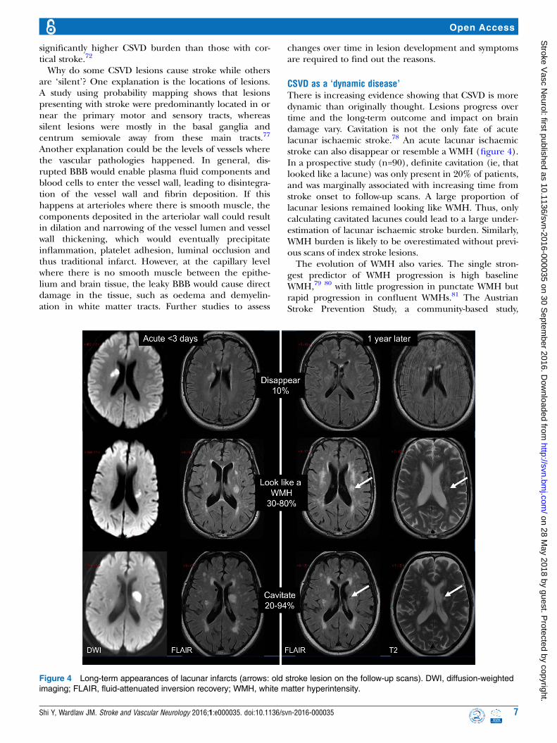
CSVD as a ‘dynamic disease’There is increasing evidence showing that CSVD is moredynamic than originally thought. Lesions progress overtime and the long-term outcome and impact on braindamage vary. Cavitation is not the only fate of acutelacunar ischaemic stroke.78 An acute lacunar ischaemicstroke can also disappear or resemble a WMH (figure 4).In a prospective study (n=90), definite cavitation (ie, thatlooked like a lacune) was only present in 20% of patients,and was marginally associated with increasing time fromstroke onset to follow-up scans. A large proportion oflacunar lesions remained looking like WMH. Thus, onlycalculating cavitated lacunes could lead to a large under-estimation of lacunar ischaemic stroke burden. Similarly,WMH burden is likely to be overestimated without previ-ous scans of index stroke lesions.The evolution of WMH also varies. The single stron-
gest predictor of WMH progression is high baselineWMH,79 80 with little progression in punctate WMH butrapid progression in confluent WMHs.81 The AustrianStroke Prevention Study, a community-based study,
Figure 4 Long-term appearances of lacunar infarcts (arrows: old stroke lesion on the follow-up scans). DWI, diffusion-weighted
imaging; FLAIR, fluid-attenuated inversion recovery; WMH, white matter hyperintensity.
Shi Y, Wardlaw JM. Stroke and Vascular Neurology 2016;1:e000035. doi:10.1136/svn-2016-000035 7
Open Access
on 28 May 2018 by guest. P
rotected by copyright.http://svn.bm
j.com/
Stroke V
asc Neurol: first published as 10.1136/svn-2016-000035 on 30 S
eptember 2016. D
ownloaded from

reported WMH progression in about 18% of participantswith vascular risk factors.79 WMH can also cavitate totake on the appearance of lacunes and they can alsodisappear—these dynamic features are only now beingrealised. Though early microstructural impairmentcould be detected in NAWM contouring WMH, not allNAWM will eventually develop into WMH.82 The level ofNAWM deterioration was also strongly associated withWMH severity, regardless of distance from the WMH.32
The variance in long-term changes of CSVD lesionsmight reflect different pathologies underlying thesimilar appearance on imaging, for example, reversiblelacunar ischaemic stroke lesions versus those that cavi-tated, or NAWM in patients with mild WMH versus inextensive WMH. Serial imaging studies using advancedtechniques like cerebral vascular reactivity, BBB and CBFimaging and use of higher fields, for example, 7 teslaMRI might help differentiate these changes.83
Treatments for CSVDManagement of traditional risk factors is still the mainapproach for treating or preventing CSVD, despite thefact that most of these treatments have not yet shownideal effects on long-term outcome. Antihypertensivetreatment produced contradictory results: it reducedWMH progression in some observational studies84 butshowed little or no effects in randomised controlledtrials.85 86 Although hypertension has been reported tobe highly associated with CSVD, other factors may beinvolved or be influenced by genetic factors,87 yet moreevidences are required. Likewise, most lipid-loweringtreatment had neutral results in preventing WMH, likepravastatin.88 Post hoc analysis of a 2-year follow-up studyfrom Hong Kong showed that statins might be able todelay WMH progression in patients with severe baselineWMH.89 Statins might also have other therapeuticeffects including anti-inflammatory and proendothelialactivities.90 Likewise, subgroup analysis of the VITAminsTO Prevent Stroke (VITATOPS) MRI substudy showsthat vitamin B supplementation may reduce WMH pro-gression in patients with severe baseline CSVD.91
Studies of treatment specifically targeting lacunarstroke are limited.90 Apart from the SPS3 trial, there arevery few clinical trials of antiplatelets where the resultswere reported by stroke subtype, and, except trials ofcilostazol92 93 which has weak antiplatelet effects,94 areespecially scarce in Asian populations. Although sometrials reported the proportion of lacunar stroke in theirstudy population, the diagnostic criteria varied consider-ably and the results were not always reported by sub-group. A systematic review of randomised trials foundthat any single antiplatelet appeared beneficial for sec-ondary prevention of lacunar stroke,95 but the SPS3 trialshowed that long-term dual antiplatelet treatmentdoubled the risk of bleeding without reducing the risk ofstroke recurrence in patients with recent lacunar stroke.Also, blood pressure lowering did not show significant
reduction in recurrent lacunar stroke in the SPS3 trial,although it was consistent with a modest benefit.96
Prevention and treatment of CSVD in the futureshould consider targeting the BBB, brain endotheliumand microvascular function. There are multiple poten-tial endothelial targets, such as the nitric oxide/cyclicguanylate monophosphate (cGMP) system and prosta-cyclin/cyclic AMP (cAMP) system.90 Therefore, interven-tions that could induce cAMP or cGMP or reduce theirdegradation appear promising. There are severallicensed drugs that have these properties like somenitric oxide donors and phosphodiesterases-5 inhibi-tors,90 while others are still in development. Moreexperimental studies should be encouraged. However, inthe meantime, management of these traditional riskfactors according to guidelines should still be encour-aged except to avoid long-term dual antiplatelet drugs.In conclusion, CSVD is not just a collection of individual
brain lesions, but is both a ‘dynamic’ and ‘whole-brain’disease. All CSVD subtypes might share some commonintrinsic CSVD aetiologies. Some pathological changes atthe early stage of the disease could be reversible, but willgradually worsen and become irreversible as the damagein vessels and tissues accumulates. Modification of trad-itional risk factors and a healthy lifestyle are currently themost important prophylactic and therapeutic approachesfor CSVD indefinitely and until more specific treatmentsare available. Apart from the trials of cilostazol which havemostly been conducted in China or Japan, in general,large clinical trials of CSVD treatments targeting the Asianpopulation are lacking, especially in lacunar stroke.Community-based studies of CSVD prevalence and pro-gression are also needed to determine if prevalence genu-inely differs in different world regions or ethnic groups.Future studies in CSVDs should stratify by stroke subtypeand by MRI diagnosis and measure risk factors carefully.Clinical trials and experimental studies targeting endothe-lium and BBB integrity should be pursued.
Contributors This paper is based on a lecture given by JMW at the ChineseStroke Association Inaugural Conference in 2015, Beijing. YS drafted thereview which was then amended and approved by JMW.
Funding YS is supported by the China Scholarships Council. The workdescribed in this paper was supported by the Wellcome Trust (WT088134/Z/09/A), the MRC, the Scottish Chief Scientist Office (CZB/4/281), Chest HeartStroke Scotland, the UK HTA, etc.
Competing interests None declared.
Provenance and peer review Commissioned; externally peer reviewed.
Data sharing statement No additional data are available.
Open Access This is an Open Access article distributed in accordance withthe terms of the Creative Commons Attribution (CC BY 4.0) license, whichpermits others to distribute, remix, adapt and build upon this work, forcommercial use, provided the original work is properly cited. See: http://creativecommons.org/licenses/by/4.0/
REFERENCES1. Pantoni L. Cerebral small vessel disease: from pathogenesis and
clinical characteristics to therapeutic challenges. Lancet Neurol2010;9:689–701.
8 Shi Y, Wardlaw JM. Stroke and Vascular Neurology 2016;1:e000035. doi:10.1136/svn-2016-000035
Open Access
on 28 May 2018 by guest. P
rotected by copyright.http://svn.bm
j.com/
Stroke V
asc Neurol: first published as 10.1136/svn-2016-000035 on 30 S
eptember 2016. D
ownloaded from

2. Tsai CF, Thomas B, Sudlow CL. Epidemiology of stroke and itssubtypes in Chinese vs white populations: a systematic review.Neurology 2013;81:264–72.
3. Fang XH, Wang WH, Zhang XQ, et al. Incidence and survival ofsymptomatic lacunar infarction in a Beijing population: a 6-yearprospective study. Eur J Neurol 2012;19:1114–20.
4. Wardlaw JM, Smith EE, Biessels GJ, et al. Neuroimaging standardsfor research into small vessel disease and its contribution to ageingand neurodegeneration. Lancet Neurol 2013;12:822–38.
5. Vermeer SE, Longstreth WT Jr, Koudstaal PJ. Silent brain infarcts: asystematic review. Lancet Neurol 2007;6:611–19.
6. Mori E, Tabuchi M, Yamadori A. Lacunar syndrome due tointracerebral hemorrhage. Stroke 1985;16:454–9.
7. Wardlaw JM, Smith C, Dichgans M. Mechanisms of sporadiccerebral small vessel disease: insights from neuroimaging. LancetNeurol 2013;12:483–97.
8. Mead GE, Lewis SC, Wardlaw JM, et al. How well does theOxfordshire community stroke project classification predict the siteand size of the infarct on brain imaging? J Neurol NeurosurgPsychiatr 2000;68:558–62.
9. Potter G, Doubal F, Jackson C, et al. Associations of clinical strokemisclassification (‘clinical-imaging dissociation’) in acute ischemicstroke. Cerebrovasc Dis 2010;29:395–402.
10. Makin SD, Doubal FN, Dennis MS, et al. Clinically confirmed strokewith negative diffusion-weighted imaging magnetic resonanceimaging: longitudinal study of clinical outcomes, stroke recurrence,and systematic review. Stroke 2015;46:3142–8.
11. Doubal FN, Dennis MS, Wardlaw JM. Characteristics of patients withminor ischaemic strokes and negative MRI: a cross-sectional study.J Neurol Neurosurg Psychiatr 2011;82:540–2.
12. Kimberly WT, Gilson A, Rost NS, et al. Silent ischemic infarcts areassociated with hemorrhage burden in cerebral amyloid angiopathy.Neurology 2009;72:1230–5.
13. Khan A, Kasner SE, Lynn MJ, et al. Risk factors and outcome ofpatients with symptomatic intracranial stenosis presenting withlacunar stroke. Stroke 2012;43:1230–3.
14. Wardlaw JM, Doubal FN, Eadie E, et al. Little association betweenintracranial arterial stenosis and lacunar stroke. Cerebrovasc Dis2011;31:12–18.
15. Kim JS, Yoon Y. Single subcortical infarction associated withparental arterial disease: important yet neglected sub-type ofatherothrombotic stroke. Int J Stroke 2013;8:197–203.
16. de Jong G, Kessels F, Lodder J. Two types of lacunar infarcts: furtherarguments from a study on prognosis. Stroke 2002;33:2072–6.
17. Asdaghi N, Pearce LA, Nakajima M, et al. Clinical correlates ofinfarct shape and volume in lacunar strokes: the SecondaryPrevention of Small Subcortical Strokes trial. Stroke2014;45:2952–8.
18. Del Bene A, Makin SD, Doubal FN, et al. Variation in risk factors forrecent small subcortical infarcts with infarct size, shape, andlocation. Stroke 2013;44:3000–6.
19. Jackson CA, Hutchison A, Dennis MS, et al. Differing risk factorprofiles of ischemic stroke subtypes: evidence for a distinct lacunararteriopathy? Stroke 2010;41:624–9.
20. Lodder J, Bamford JM, Sandercock PA, et al. Are hypertension orcardiac embolism likely causes of lacunar infarction? Stroke1990;21:375–81.
21. Potter GM, Doubal FN, Jackson CA, et al. Lack of association ofwhite matter lesions with ipsilateral carotid artery stenosis.Cerebrovasc Dis 2012;33:378–84.
22. Kwon HM, Lynn MJ, Turan TN, et al. Frequency, risk factors, andoutcome of coexistent small vessel disease and intracranial arterialstenosis: results from the Stenting and Aggressive MedicalManagement for Preventing Recurrent Stroke in Intracranial Stenosis(SAMMPRIS) Trial. JAMA Neurol 2016;73:36–42.
23. Macdonald RL, Kowalczuk A, Johns L. Emboli enter penetratingarteries of monkey brain in relation to their size. Stroke1995;26:1247–50; discussion 1250–1.
24. Jackson C, Sudlow C. Are lacunar strokes really different? Asystematic review of differences in risk factor profiles betweenlacunar and nonlacunar infarcts. Stroke 2005;36:891–901.
25. Zhang B, Zhang W, Li X, et al. Admission markers predict lacunarand non-lacunar stroke in young patients. Thromb Res2011;128:14–17.
26. Lv P, Jin H, Liu Y, et al. Comparison of risk factor between lacunarstroke and large artery atherosclerosis stroke: a cross-sectionalstudy in China. PLoS ONE 2016;11:e0149605.
27. Debette S, Markus HS. The clinical importance of white matterhyperintensities on brain magnetic resonance imaging: systematicreview and meta-analysis. BMJ 2010;341:c3666.
28. Madden DJ, Bennett IJ, Burzynska A, et al. Diffusion tensor imagingof cerebral white matter integrity in cognitive aging. Biochim BiophysActa 2012;1822:386–400.
29. Munoz DG, Hastak SM, Harper B, et al. Pathologic correlates ofincreased signals of the centrum ovale on magnetic resonanceimaging. Arch Neurol 1993;50:492–7.
30. Feigin I, Popoff N. Neuropathological changes late in cerebral edema:the relationship to trauma, hypertensive disease and Binswanger’sencephalopathy. J Neuropathol Exp Neurol 1963;22:500–11.
31. Black S, Gao F, Bilbao J. Understanding white matter disease:imaging-pathological correlations in vascular cognitive impairment.Stroke 2009;40:S48–52.
32. Maniega SM, Valdes Hernandez MC, Clayden JD, et al. Whitematter hyperintensities and normal-appearing white matter integrityin the aging brain. Neurobiol Aging 2015;36:909–18.
33. Bastin ME, Clayden JD, Pattie A, et al. Diffusion tensor andmagnetization transfer MRI measurements of periventricular whitematter hyperintensities in old age. Neurobiol Aging 2009;30:125–36.
34. Maillard P, Fletcher E, Lockhart SN, et al. White matterhyperintensities and their penumbra lie along a continuum of injuryin the aging brain. Stroke 2014;45:1721–6.
35. Shi Y, Thrippleton MJ, Makin SD, et al. Cerebral blood flow in smallvessel disease: a systematic review and meta-analysis. J CerebBlood Flow Metab 2016. Published online first: 5 Aug 2016 doi:10.1177/0271678X16662891.
36. van der Veen PH, Muller M, Vincken KL, et al. Longitudinalrelationship between cerebral small-vessel disease and cerebralblood flow: the second manifestations of arterial disease-magneticresonance study. Stroke 2015;46:1233–8.
37. Farrall AJ, Wardlaw JM. Blood-brain barrier: ageing andmicrovascular disease–systematic review and meta-analysis.Neurobiol Aging 2009;30:337–52.
38. Wardlaw JM, Doubal FN, Valdes-Hernandez M, et al. Blood-brainbarrier permeability and long-term clinical and imaging outcomes incerebral small vessel disease. Stroke 2013;44:525–7.
39. Wardlaw JM, Doubal F, Armitage P, et al. Lacunar stroke isassociated with diffuse blood-brain barrier dysfunction. Ann Neurol2009;65:194–202.
40. Topakian R, Barrick TR, Howe FA, et al. Blood-brain barrierpermeability is increased in normal-appearing white matter inpatients with lacunar stroke and leucoaraiosis. J Neurol NeurosurgPsychiatr 2010;81:192–7.
41. Taheri S, Gasparovic C, Huisa BN, et al. Blood-brain barrierpermeability abnormalities in vascular cognitive impairment. Stroke2011;42:2158–63.
42. Potter GM, Marlborough FJ, Wardlaw JM. Wide variation indefinition, detection, and description of lacunar lesions on imaging.Stroke 2011;42:359–66.
43. Franke CL, van Swieten JC, van Gijn J. Residual lesions oncomputed tomography after intracerebral hemorrhage. Stroke1991;22:1530–3.
44. Hernandez Mdel C, Piper RJ, Wang X, et al. Towards the automaticcomputational assessment of enlarged perivascular spaces on brainmagnetic resonance images: a systematic review. J Magn ResonImaging 2013;38:774–85.
45. Snowdon DA, Greiner LH, Mortimer JA, et al. Brain infarction andthe clinical expression of Alzheimer disease. The Nun Study. JAMA1997;277:813–17.
46. Vermeer SE, Prins ND, den Heijer T, et al. Silent brain infarcts andthe risk of dementia and cognitive decline. N Engl J Med2003;348:1215–22.
47. Fanning JP, Wong AA, Fraser JF. The epidemiology of silent braininfarction: a systematic review of population-based cohorts. BMCMed 2014;12:119.
48. Braffman BH, Zimmerman RA, Trojanowski JQ, et al. Brain MR.pathologic correlation with gross and histopathology. 1. Lacunarinfarction and Virchow-Robin spaces. AJR Am J Roentgenol1988;151:551–8.
49. Potter GM, Chappell FM, Morris Z, et al. Cerebral perivascularspaces visible on magnetic resonance imaging: development of aqualitative rating scale and its observer reliability. Cerebrovasc Dis2015;39:224–31.
50. Groeschel S, Chong WK, Surtees R, et al. Virchow-Robin spaces onmagnetic resonance images: normative data, their dilatation, and areview of the literature. Neuroradiology 2006;48:745–54.
51. Zhu YC, Tzourio C, Soumare A, et al. Severity of dilatedVirchow-Robin spaces is associated with age, blood pressure, andMRI markers of small vessel disease: a population-based study.Stroke 2010;41:2483–90.
52. Heier LA, Bauer CJ, Schwartz L, et al. Large Virchow-Robin spaces:MR-clinical correlation. AJNR Am J Neuroradiol 1989;10:929–36.
Shi Y, Wardlaw JM. Stroke and Vascular Neurology 2016;1:e000035. doi:10.1136/svn-2016-000035 9
Open Access
on 28 May 2018 by guest. P
rotected by copyright.http://svn.bm
j.com/
Stroke V
asc Neurol: first published as 10.1136/svn-2016-000035 on 30 S
eptember 2016. D
ownloaded from

53. Potter GM, Doubal FN, Jackson CA, et al. Enlarged perivascularspaces and cerebral small vessel disease. Int J Stroke2015;10:376–81.
54. Doubal FN, MacLullich AM, Ferguson KJ, et al. Enlargedperivascular spaces on MRI are a feature of cerebral small vesseldisease. Stroke 2010;41:450–4.
55. Zhu YC, Dufouil C, Soumare A, et al. High degree of dilatedVirchow-Robin spaces on MRI is associated with increased risk ofdementia. J Alzheimers Dis 2010;22:663–72.
56. Maclullich AM, Wardlaw JM, Ferguson KJ, et al. Enlargedperivascular spaces are associated with cognitive function in healthyelderly men. J Neurol Neurosurg Psychiatr 2004;75:1519–23.
57. Wuerfel J, Haertle M, Waiczies H, et al. Perivascular spaces—MRImarker of inflammatory activity in the brain? Brain2008;131:2332–40.
58. Weller RO, Djuanda E, Yow HY, et al. Lymphatic drainage of thebrain and the pathophysiology of neurological disease. ActaNeuropathol 2009;117:1–14.
59. Iliff JJ, Wang M, Zeppenfeld DM, et al. Cerebral arterial pulsationdrives paravascular CSF-interstitial fluid exchange in the murinebrain. J Neurosci 2013;33:18190–9.
60. Xie L, Kang H, Xu Q, et al. Sleep drives metabolite clearance fromthe adult brain. Science 2013;342:373–7.
61. Shoamanesh A, Kwok CS, Benavente O. Cerebral microbleeds:histopathological correlation of neuroimaging. Cerebrovasc Dis2011;32:528–34.
62. Greenberg SM, Vernooij MW, Cordonnier C, et al. Cerebralmicrobleeds: a guide to detection and interpretation. Lancet Neurol2009;8:165–74.
63. Cordonnier C, Al-Shahi Salman R, Wardlaw J. Spontaneous brainmicrobleeds: systematic review, subgroup analyses and standardsfor study design and reporting. Brain 2007;130:1988–2003.
64. Martinez-Ramirez S, Greenberg SM, Viswanathan A. Cerebralmicrobleeds: overview and implications in cognitive impairment.Alzheimers Res Ther 2014;6:33.
65. Sveinbjornsdottir S, Sigurdsson S, Aspelund T, et al. Cerebralmicrobleeds in the population based AGES-Reykjavik study:prevalence and location. J Neurol Neurosurg Psychiatr2008;79:1002–6.
66. Poels MM, Vernooij MW, Ikram MA, et al. Prevalence and riskfactors of cerebral microbleeds: an update of the Rotterdam scanstudy. Stroke 2010;41:S103–6.
67. Kakar P, Charidimou A, Werring DJ. Cerebral microbleeds: a newdilemma in stroke medicine. JRSM Cardiovasc Dis 2012;1:22.
68. van Dijk EJ, Breteler MM, Schmidt R, et al. The association betweenblood pressure, hypertension, and cerebral white matter lesions:cardiovascular determinants of dementia study. Hypertension2004;44:625–30.
69. Wardlaw JM, Allerhand M, Doubal FN, et al. Vascular risk factors,large-artery atheroma, and brain white matter hyperintensities.Neurology 2014;82:1331–8.
70. Ihara M, Yamamoto Y. Emerging evidence for pathogenesis ofsporadic cerebral small vessel disease. Stroke 2016;47:554–60.
71. van Dijk EJ, Prins ND, Vrooman HA, et al. Progression of cerebralsmall vessel disease in relation to risk factors and cognitiveconsequences: Rotterdam Scan study. Stroke 2008;39:2712–19.
72. Staals J, Makin SD, Doubal FN, et al. Stroke subtype, vascular riskfactors, and total MRI brain small-vessel disease burden. Neurology2014;83:1228–34.
73. Gow AJ, Bastin ME, Munoz Maniega S, et al. Neuroprotectivelifestyles and the aging brain: activity, atrophy, and white matterintegrity. Neurology 2012;79:1802–8.
74. Rost NS, Rahman RM, Biffi A, et al. White matter hyperintensityvolume is increased in small vessel stroke subtypes. Neurology2010;75:1670–7.
75. Wardlaw JM, Lewis SC, Keir SL, et al. Cerebral microbleeds areassociated with lacunar stroke defined clinically and radiologically,independently of white matter lesions. Stroke 2006;37:2633–6.
76. Duering M, Csanadi E, Gesierich B, et al. Incident lacunespreferentially localized to the edge of white matter hyperintensities:
insights into the pathophysiology of cerebral small vessel disease.Brain 2013;136:2717–26.
77. Valdes Hernandez Mdel C, Maconick LC, Munoz Maniega S, et al. Acomparison of location of acute symptomatic vs. ‘silent’ small vessellesions. Int J Stroke 2015;10:1044–50.
78. Potter GM, Doubal FN, Jackson CA, et al. Counting cavitatinglacunes underestimates the burden of lacunar infarction. Stroke2010;41:267–72.
79. Schmidt R, Enzinger C, Ropele S, et al. Progression of cerebralwhite matter lesions: 6-year results of the Austrian Stroke PreventionStudy. Lancet 2003;361:2046–8.
80. Gouw AA, van der Flier WM, Fazekas F, et al. Progression of whitematter hyperintensities and incidence of new lacunes over a 3-yearperiod: the Leukoaraiosis and Disability study. Stroke2008;39:1414–20.
81. Schmidt R, Seiler S, Loitfelder M. Longitudinal change ofsmall-vessel disease-related brain abnormalities. J Cereb BloodFlow Metab 2016;36:26–39.
82. Munoz Maniega S, Chappell FM, Valdes Hernandez MC, et al.Integrity of normal-appearing white matter: influence of age, visiblelesion burden and hypertension in patients with small-vesseldisease. J Cereb Blood Flow Metab 2016. Published Online First: 1Mar 2016 doi: 10.1177/0271678X16635657.
83. Bouvy WH, Biessels GJ, Kuijf HJ, et al. Visualization of perivascularspaces and perforating arteries with 7 T magnetic resonanceimaging. Invest Radiol 2014;49:307–13.
84. Dufouil C, de Kersaint-Gilly A, Besancon V, et al. Longitudinal studyof blood pressure and white matter hyperintensities: the EVA MRICohort. Neurology 2001;56:921–6.
85. Dufouil C, Chalmers J, Coskun O, et al. Effects of blood pressurelowering on cerebral white matter hyperintensities in patients withstroke: the PROGRESS (Perindopril Protection Against RecurrentStroke Study) Magnetic Resonance Imaging Substudy. Circulation2005;112:1644–50.
86. Weber R, Weimar C, Blatchford J, et al. Telmisartan on top ofantihypertensive treatment does not prevent progression of cerebralwhite matter lesions in the prevention regimen for effectively avoidingsecond strokes (PRoFESS) MRI substudy. Stroke 2012;43:2336–42.
87. Turner ST, Fornage M, Jack CR Jr, et al. Genomic susceptibility locifor brain atrophy in hypertensive sibships from the GENOA study.Hypertension 2005;45:793–8.
88. ten Dam VH, van den Heuvel DM, van Buchem MA, et al. Effect ofpravastatin on cerebral infarcts and white matter lesions. Neurology2005;64:1807–9.
89. Mok VC, Lam WW, Fan YH, et al. Effects of statins on theprogression of cerebral white matter lesion: post hoc analysis of theROCAS (Regression of Cerebral Artery Stenosis) study. J Neurol2009;256:750–7.
90. Bath PM, Wardlaw JM. Pharmacological treatment and prevention ofcerebral small vessel disease: a review of potential interventions. IntJ Stroke 2015;10:469–78.
91. Cavalieri M, Schmidt R, Chen C, et al. B vitamins and magneticresonance imaging-detected ischemic brain lesions in patients withrecent transient ischemic attack or stroke: the VITAmins TO PreventStroke (VITATOPS) MRI-substudy. Stroke 2012;43:3266–70.
92. Shinohara Y, Katayama Y, Uchiyama S, et al. Cilostazol for preventionof secondary stroke (CSPS 2): an aspirin-controlled, double-blind,randomised non-inferiority trial. Lancet Neurol 2010;9:959–68.
93. Huang Y, Cheng Y, Wu J, et al. Cilostazol as an alternative toaspirin after ischaemic stroke: a randomised, double-blind, pilotstudy. Lancet Neurol 2008;7:494–9.
94. Comerota AJ. Effect on platelet function of cilostazol, clopidogrel,and aspirin, each alone or in combination. Atheroscler Suppl2005;6:13–19.
95. Kwok CS, Shoamanesh A, Copley HC, et al. Efficacy of antiplatelettherapy in secondary prevention following lacunar stroke: pooledanalysis of randomized trials. Stroke 2015;46:1014–23.
96. Group SPSS, Benavente OR, Coffey CS, et al. Blood-pressuretargets in patients with recent lacunar stroke: the SPS3 randomisedtrial. Lancet 2013;382:507–15.
10 Shi Y, Wardlaw JM. Stroke and Vascular Neurology 2016;1:e000035. doi:10.1136/svn-2016-000035
Open Access
on 28 May 2018 by guest. P
rotected by copyright.http://svn.bm
j.com/
Stroke V
asc Neurol: first published as 10.1136/svn-2016-000035 on 30 S
eptember 2016. D
ownloaded from