unveiling the membrane-binding properties of n terminal
TRANSCRIPT
Unveiling the Membrane-Binding Properties of N‑Terminal andC‑Terminal Regions of G Protein-Coupled Receptor Kinase 5 byCombined Optical SpectroscopiesBei Ding,† Alisa Glukhova,‡ Katarzyna Sobczyk-Kojiro,§ Henry I. Mosberg,§ John J. G. Tesmer,*,‡
and Zhan Chen*,†
†Department of Chemistry, University of Michigan, Ann Arbor, Michigan 48109, United States‡Life Sciences Institute and the Department of Pharmacology, University of Michigan, Ann Arbor, Michigan 48109-2216, UnitedStates§College of Pharmacy, Department of Medicinal Chemistry, University of Michigan, Ann Arbor, Michigan 48109-1065, United States
*S Supporting Information
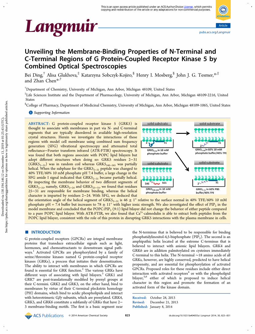
ABSTRACT: G protein-coupled receptor kinase 5 (GRK5) isthought to associate with membranes in part via N- and C-terminalsegments that are typically disordered in available high-resolutioncrystal structures. Herein we investigate the interactions of theseregions with model cell membrane using combined sum frequencygeneration (SFG) vibrational spectroscopy and attenuated totalreflectance−Fourier transform infrared (ATR-FTIR) spectroscopy. Itwas found that both regions associate with POPC lipid bilayers butadopt different structures when doing so: GRK5 residues 2−31(GRK52−31) was in random coil whereas GRK5546−565 was partiallyhelical. When the subphase for the GRK52−31 peptide was changed to40% TFE/60% 10 mM phosphate pH 7.4 buffer, a large change in theSFG amide I signal indicated that GRK52−31 became partially helical.By inspecting the membrane behavior of two different segments ofGRK52−31, namely, GRK52−24 and GRK525−31, we found that residues25−31 are responsible for membrane binding, whereas the helicalcharacter is imparted by residues 2−24. With SFG, we deduced thatthe orientation angle of the helical segment of GRK52−31 is 46 ± 1° relative to the surface normal in 40% TFE/60% 10 mMphosphate pH = 7.4 buffer but increases to 78 ± 11° with higher ionic strength. We also investigated the effect of PIP2 in themodel membrane and concluded that the POPC:PIP2 (9:1) lipid bilayer did not change the behavior of either peptide comparedto a pure POPC lipid bilayer. With ATR-FTIR, we also found that Ca2+·calmodulin is able to extract both peptides from thePOPC lipid bilayer, consistent with the role of this protein in disrupting GRK5 interactions with the plasma membrane in cells.
■ INTRODUCTION
G protein-coupled receptors (GPCRs) are integral membraneproteins that transduce extracellular signals such as light,hormones, and chemoattractants to downstream signal path-ways.1 Activated GPCRs are phosphorylated by a family ofserine/threonine kinases named G protein-coupled receptorkinases (GRKs), a process that initiates their desensitization.The ability to interact with membranes in which GPCRs arefound is essential for GRK function.2 The various GRKs havedifferent ways of associating with lipid bilayers.3 GRK1 andGRK7 are post-translationally modified by prenyl groups attheir C-termini. GRK2 and GRK3, on the other hand, bind tomembranes by virtue of their C-terminal pleckstrin homology(PH) domains, which bind to acidic phospholipids and interactwith heterotrimeric Gβγ subunits, which are prenylated. GRK4,GRK5, and GRK6 constitute a subfamily of GRKs that have 2−3 membrane-binding motifs. The first is a basic segment near
the N-terminus that is believed to be responsible for bindingphosphatidylinositol-4,5-bisphosphate (PIP2). The second is anamphipathic helix located at the extreme C-terminus that isbelieved to interact with anionic lipid bilayers. GRK4 andGRK6 are in addition palmitoylated on cysteines immediatelyC-terminal to this helix. The N-terminal ∼18 amino acids of allGRKs, however, are highly conserved, predicted to have helicalpropensity, and are essential for phosphorylation of activatedGPCRs. Proposed roles for these residues include either directinteraction with activated receptors4 or with the phospholipidbilayer,5,6 either of which is proposed to induce helicalcharacter in this region and promote the formation of anactivated form of the kinase domain.
Received: October 28, 2013Revised: December 21, 2013Published: January 8, 2014
Article
pubs.acs.org/Langmuir
© 2014 American Chemical Society 823 dx.doi.org/10.1021/la404055a | Langmuir 2014, 30, 823−831
This is an open access article published under an ACS AuthorChoice License, which permitscopying and redistribution of the article or any adaptations for non-commercial purposes.
Dow
nloa
ded
via
130.
194.
146.
12 o
n D
ecem
ber
13, 2
019
at 0
1:25
:43
(UT
C).
See
http
s://p
ubs.
acs.
org/
shar
ingg
uide
lines
for
opt
ions
on
how
to le
gitim
atel
y sh
are
publ
ishe
d ar
ticle
s.

Over the past several years, crystallographic studies haveyielded new insights into the molecular mechanism forregulation of GRKs by their interactions with receptors andmembranes.4 However, crystallographic analysis requires theremoval of protein complexes from their native membraneenvironment and cannot provide direct information on howthese molecules are arranged on the membrane surface in situ.Sum frequency generation (SFG) vibrational spectroscopy is apowerful tool to examine peptides and proteins at bio-interfaces7−14 such as lipid bilayers.15−21 For example,orientations of peptides with different secondary structures,such as linear α-helices,22,23 bent α-helices,24,25 β-sheets,26 and310-helices
27 associated with solid substrate supported lipidbilayers have been deduced using polarized SFG studies. SFGhas also been applied to investigate the membrane orientationsof Gβγ, the Gβγ−GRK2 complex, and Gαβγ heterotrimers insitu.28,29 Recently, we showed that the use of both SFG andattenuated total reflectance−Fourier transform infrared (ATR-FTIR) spectroscopy can determine orientations of complexproteins with greater certainty.30 In this research, we used SFGand ATR-FTIR to study the membrane interactions of the N-and C-terminal segments of GRK5 to gain insight into whichregions were most important for membrane binding and whatstructure and orientation they adopt while interacting withmembranes.GRK5 residues 2−31 (GRK52−31) are highly conserved in
the GRK4 subfamily of GRKs (Figure 1), which includes
GRK4, GRK5, and GRK6. In previous research, it wassuggested that residues 22−29, which include basic aminoacids Lys22, Arg23, Lys24, Lys26, Lys28, and Lys29, bind toPIP2.
31 An overlapping region (residues 20−39) has also beenimplicated in binding to calmodulin·Ca2+ (CaM·Ca2+).32 Thestructure of GRK6 (a close homologue of GRK5) determinedby X-ray crystallography suggests that the N-terminal portion ofthe peptide (residues 2−23) is disordered when the enzyme isin an inactive state,33 but residues 2−18 become ordered whenthe enzyme assumes a more active, presumably receptor-boundconformation.4 However, it is not known if this region forms aplatform for binding to lipid membranes or activated GPCRs.Therefore, elucidating the ability of different segments of theGRK5 N-terminus to interact with the membrane is key tounderstanding how the membrane influences GRK5 function.The C-terminal residues 552−562 of GRK5 are believed to
be another region that interacts with phospholipids. Deletion ofthese residues results in a 100-fold loss in membrane bindingaffinity.34 Residues 549−557 are predicted to form anamphipathic helix when bound to membranes.35 In the activeconformation of the GRK6 crystal structure, residues 548−557form an amphipathic helix that docks to the core of the enzymebut is far removed from the predicted membrane surface andthe N-terminal segment believed to bind PIP2.
4 Thus, eitherthis structural element does not bind to membranes, or it onlybinds to membranes when GRK6 is in a more inactive state, or
the structure represents a soluble form of the enzyme, such aswhen it translocates to the nucleus to phosphorylate tran-scription factors.36
By combining data from two complementary opticalspectroscopic techniques, SFG and ATR-FTIR, we are seekingto answer the following questions. First, do peptidesrepresenting the N-terminal (GRK52−31) and C-terminal(GRK5546−565) regions bind to membranes on their own, and,if so, what structure do they adopt? Second, does PIP2 affect thebinding properties of these two peptides? Finally, is CaM·Ca2+
able to dissociate these GRK5 peptides from the membrane, asproposed to be required for nuclear translocation?
■ EXPERIMENTAL SECTIONMaterials. Peptides GRK52−31, GRK52−24, and GRK5546−565
(Figure 1) were synthesized by the following procedure. Protectedamino acids and N-methylpyrrolidone (NMP), 1-hydroxybenzotria-zole (HOBt), and O-benzotriazole-N,N,N′,N′-tetramethyluroniumhexafluorophosphate (HBTU) were purchased from Creosalus.Acetonitrile, HPLC grade water, trifluoroacetic acid (TFA), diethylether, and phenol were from Fisher Scientific. Piperidine, N,N-diisopropylethylamine (DIPEA), dimethylformamide (DMF), thio-anisole, triisopropylsilane (TIPS), and calmodulin were from Sigma-Aldrich. Solid-phase synthesis resin NovaPEG Rink Amide Resin (0.5mmol/g) was purchased from Novabiochem. Analytical HPLCanalysis was done using an Alliance system with 250 × 5 mm C18 3μm column (Vydac). Mass spectrometry analysis was done using a6130 Quadrupole LC/MS (Agilent Technologies). SemipreparativeHPLC purification was performed using a Delta 600 system (Waters)with 150 × 19 mm XBridge Prep C18 10 μm OBD column (Waters).HPLC analysis and purification were done using solvent system 0.1%TFA in water and 0.1% TFA in acetonitrile. Peptides GRK52−24 andGRK52−31 were synthesized using 9-fluorenylmethoxycarbonyl(FMOC) chemistry. The syntheses of C-terminal sequences up toAla15 were carried out on a CS336X automated synthesizer (C.S. BioCo.), and the syntheses were then continued on a Discover SPS singlemode manual microwave synthesizer (CEM Corp.) (power = 20 W, 5min per coupling and power 20 W, 1.5 min per deprotection;temperature 70−75 °C). The synthesis scale was 0.2 mmol. Thegeneral protocol included double coupling and double deprotection aswell as acetylation of the unreacted amino groups. Coupling cycleswere performed using 4 equiv of incoming amino acid, HOBt/HBTUin DMF and DIPEA in NMP. Fmoc deprotection was accomplishedusing 20% piperidine solution in NMP. Cleavage of the peptide fromthe resin and side-chain deprotection was performed using 10 mL ofthe mixture DI water:phenol:thioanisole:TIPS:TFA (0.5 mL:0.7 g:0.5mL:0.25 mL:8.75 mL). The reaction was left running at roomtemperature for 2 h. After filtration of the resin, crude peptide wasprecipitated with cold ethyl ether. The resulting crude peptides werepurified by semipreparative HPLC, as described above. The purity ofthe final peptide was analyzed using HPLC and molecular weightconfirmed by MS. Peptide GRK525−31 was synthesized by Peptide 2.0Inc. by a similar approach. POPC (1-palmitoyl-2-oleoyl-sn-glycero-3-phosphocholine) and PIP2 were purchased from Avanti Polar Lipids.
Bilayer Construction. Supported POPC/POPC lipid bilayerswere constructed on CaF2 prisms by the Langmuir−Blodgett/Langmuir−Schaefer method, as reported in detail previously.37,38
The first POPC layer is deposited on one of the square faces of theright-angle CaF2 prism with A KSV2000 LB system: The plasma-cleaned prism was first immersed in the LB trough. Then a certainamount of POPC chloroform solution, typically 5 drops of 10 mg/mL,was spread on the water surface until the surface tension reaches ∼5mN/m. Two barrier arms were suppressed so that the surface tensionremained 34 mN/m, while the CaF2 prism was lifted from thesubphase. A layer of POPC lipids was deposited on the faceperpendicular to the water surface in this way. After aligning thelaser beams to find the monolayer signal, a 2 mL reservoir filled withwater was placed beneath the prism. POPC lipids were added to the
Figure 1. Sequences of the human GRK5 N-terminal and C-terminalpeptides used in this study. Residues highlighted in gray adopt an α-helical conformation in the structure of the GRK6·sangivamycincomplex.5
Langmuir Article
dx.doi.org/10.1021/la404055a | Langmuir 2014, 30, 823−831824

surface of the water in the reservoir so that the surface tension wasaround 34 mN/m. The reservoir was elevated so that the the lipidmonolayer on the water surface contacted with the first layer depositedon the prism to form a lipid bilayer.SFG Experiments. SFG theory,39−43 our experimental design, and
data analysis method23,44 have been reported before. The concen-tration of each of the four peptides was 3.8 μM, and the peptides weredissolved in 10 mM potassium phosphate buffer (pH 7.4). BecauseCaF2 prisms were used as substrates to prepare the lipid bilayers, small
amounts of Ca2+ may be dissolved in the subphase. 2 mM EDTA wasadded to the above buffer solution to minimize any influence of theCa2+ released from the CaF2 substrates. For each of the three N-terminal peptides studied here, we added the peptide into thesubphase in contact with the substrate supported bilayer and afterequilibration recorded the SFG signal in the water O−H stretchingfrequency range as well as in the peptide amide I frequency region. Forall peptides we studied, the adsorption time on the POPC lipid bilayerin either 10 mM phosphate buffer or PBS buffer was less than 200 s
Figure 2. SFG signals from GRK52−31 indicate strong association with model membranes and helical character in a more hydrophobic environment.(a) Spectra in the C−H and O−H stretching frequency region detected from the interface between the POPC/POPC bilayer and buffer alone(black), GRK52−31 in 10 mM phosphate buffer pH 7.4 (red), after washing (blue), and in a mixture of 60% buffer/40% TFE (dark cyan). (b) SFGspectra of GRK52−31 associated with a POPC/POPC bilayer in contact with peptide solution in 60% 10 mM phosphate buffer pH 7.4/40% TFE inthe amide I frequency region. (c) SFG spectra of GRK52−31 associated with a POPC/POPC bilayer in contact with 60% PBS/40% TFE.
Figure 3. SFG ppp signals detected from the GRK525−31 and GRK52−24 peptides indicate that the latter peptide only weakly associates with modelmembranes. (a) SFG spectra in C−H and O−H stretching frequency region from the interface between the POPC/POPC bilayer and buffer alone(black), GRK525−31 in 10 mM phosphate buffer pH 7.4 (red), after washing (blue), and in a mixture of 60% buffer/40% TFE (dark cyan). (b) SFGspectra in the amide I frequency region from GRK525−31 associated with a POPC/POPC bilayer in 60% 10 mM phosphate buffer pH 7.4/40% TFE.(c) SFG spectra in C−H and O−H stretching frequency region from the interface between the POPC/POPC bilayer and buffer alone (black),GRK52−24 in 10 mM phosphate buffer pH 7.4 (red), after washing (blue), and in a mixture of 60% buffer/40% TFE (dark cyan). (d) SFG spectra inthe amide I frequency region from GRK52−24 associated with a POPC/POPC bilayer in contact with peptide solution in 60% 10 mM phosphatebuffer pH 7.4/40% TFE.
Langmuir Article
dx.doi.org/10.1021/la404055a | Langmuir 2014, 30, 823−831825

(see the time-dependent SFG signals detected in the CH and OHstretching frequency range in the Supporting Information). For thesecond step, we substituted the peptide solution subphase withpotassium phosphate buffer (∼6 mL in total) to wash off looselyassociated peptides and recorded the SFG signal in the water O−Hstretching frequency range again. For the last step, we substituted thephosphate buffer subphase with a solution of buffer containing 40%TFE and again collected SFG spectra in the water O−H stretchingfrequency range and the amide I frequency range. For the C-terminalpeptide, we only performed the first two steps of the above procedure.PIP2 experiments were performed in the same way as POPCexperiments except that when making bilayers, lipids with a 9:1molar ratio of POPC:PIP2 were used. Because peptides were used atthe same concentration in these experiments yet likely have differentaffinities and because water signals are also strongly affected by netcharge as well as charge distribution in the peptides (and othereffects), we defined peptides as weakly membrane associated if thewater signal recovered after the buffer wash, as opposed to directcomparison of changes in the SFG signals from water O−H stretchingafter addition of peptide.ATR-FTIR Spectroscopy. ATR-FTIR experiments were performed
on a Nicolet Magna 550 FTIR spectrometer. Lipid bilayers weredeposited on a ZnSe crystal (Specac Ltd. RI, U.K.) using the vesiclefusion method. 1 mL of POPC toluene solution (5 mg/mL) was driedwith nitrogen flow and then in vacuum for 2 h. The POPC power wasthen dissolved in 10 mM phosphate D2O buffer pH 7.4, and themixture was vortexed for 5 min before addition to the surface of thedetachable ZnSe crystal to form bilayers. After 30 min, the vesiclesfloating in the subphase were washed away using excess buffer. GRK5peptides were then injected into the subphase (1.6 mL) to achieve aconcentration of 11.4 μM. After the system reached equilibrium,spectra before and after extensive wash with D2O buffer wererecorded. For GRK5546−565, s- and p-polarized spectra were taken sothat data analysis on the peptide orientation could be performed. Inthe CaM·Ca2+ experiments, after peptides were associated with thelipid bilayers equimolar CaM (11.4 μM) and 50 μM CaCl2 solutionwere added to the subphase.
■ RESULTS
SFG Studies on N-Terminal Peptides. We firstinvestigated molecular interactions between the GRK5 N-terminal peptides GRK52−31, GRK52−24, and GRK525−31 and aPOPC/POPC lipid bilayer. The POPC/POPC bilayer iszwitterionic, and the electrostatic potential across the bilayerinduces the water dipoles to orient near the bilayer surface.45,46
The water region (detected between 2700 and 3700 cm−1)47,48
monitored by SFG spectroscopy can be used to determine thebinding properties of ions49,50 or peptides.25 In our experi-ments, we observed two broad water O−H stretching peakscentered at ∼3200 and 3400 cm−1 in the SFG spectrum fromthe lipid bilayer/potassium phosphate buffer interface (Figure2a). Peptides were then added into the subphase, and thesystem was allowed to reach equilibrium. The water OHstretching signal decreased upon addition of the GRK52−31 orGRK525−31 peptides to the subphase, consistent with theirinteraction with the POPC/POPC bilayers (Figures 2a and 3a).SFG spectra were also collected after extensive washing, but nosubstantial changes were observed, suggesting that bothGRK52−31 and GRK525−31 peptides are strongly associatedwith the bilayer. However, the SFG water O−H stretchingsignal only decreased slightly after the addition of the GRK52−24peptide to the subphase, and the SFG water signal recoveredafter washing the interface with buffer (Figure 3c), consistentwith GRK52−24 only being weakly associated with the POPC/POPC bilayer. Thus, the highly charged residues spanningresidues 25−31 are primarily responsible for membrane bindingin these peptides.For all three GRK5 N-terminal peptides, no discernible SFG
amide I signal could be detected from the lipid bilayerinterfaces after their addition. This suggests that the membraneassociated peptides form either ordered structures but withrandom orientations or essentially random structures. Afterreplacing the subphase with a 40% TFE solution, a strong SFGamide I signal was detected from the GRK52−31 peptide (Figure2b), but not from GRK525−31 or GRK52−24 (Figures 3b and3d), consistent with only GRK52−31 forming α-helical structurewhen the subphase becomes more hydrophobic. Thisconclusion is also consistent with spectral features detected inthe water O−H stretching frequency range after the subphasebuffer was replaced by the TFE mixture. Figure 2a shows thatonly for GRK52−31, a negative peak at ∼3300 cm −1 appeared,originating from the interference between the N−H stretchingsignals of well-ordered α-helices and the broad waterbackground. This N−H stretch signal can be attributed tothe backbone N−H stretch or/and the side chains such as LysNH3
+.51 Although the predicted helical propensity of GRK52−24is the same as that of GRK52−31, no changes in the spectra upon
Figure 4. SFG ppp signals detected from GRK5546−565 indicate strong binding to model membranes and helical character. (a) SFG spectra in the C−H and O−H stretching frequency region from the interface between the POPC/POPC bilayer and buffer alone (black) and GRK5546−565 associatedin 10 mM phosphate buffer pH 7.4 (red). (b) SFG spectra in the amide I frequency region from GRK5546−565 associated with a POPC/POPC bilayerin 10 mM phosphate buffer pH 7.4.
Langmuir Article
dx.doi.org/10.1021/la404055a | Langmuir 2014, 30, 823−831826

addition of TFE were detected likely because the peptide wasnot strongly associated with the membrane and washed off inthe previous step.Orientation Analysis of the α-Helical Segment in
GRK52−31. After substituting the subphase with 40% TFE, aprominent α-helical signal centered at ∼1655 cm−1 arises fromGRK52−31. This could be interpreted as residues 2−18 adoptingan α-helical conformation, consistent with a prior crystalstructure of GRK64 and secondary structure predictions. Thisphenomenon also highlights that SFG, as a second-ordernonlinear spectroscopy, is much more sensitive to orderedstructure (such as α-helices) than disordered molecules (suchas random coils), which is not the case for linear vibrationalspectroscopy such as ATR-FTIR.SFG spectra collected from amide I modes of peptides and
proteins using different polarization combinations can be usedto determine membrane orientations of peptides and proteins,as shown in a previous publication.22 Using the measured signalstrength ratio of the α-helical contribution in the ppp and sspspectra, we deduced that in 40% TFE, the orientation angle ofthe helical segment (presumed to be residues 2−18) ofGRK52−31 is ∼46 ± 1° relative to the membrane surface normal(with χppp/χssp = 2.08 ± 0.01) if we assume the peptides adopt asingle orientation distribution. Interestingly, this orientationangle increases to ∼78 ± 11° (with χppp/χssp = 2.43 ± 0.06)when the ionic strength of the subphase is increased by use ofPBS instead of phosphate buffer (Figure 2c). Details of theorientation analysis can be found in the SupportingInformation. This result suggests that the increase in ionicstrength does not change the conformation of the GRK52−31but rather changes the charge distribution on the peptidesurface and thus facilitates the interaction of helical elements ofthe peptide with the lipid head groups.SFG Studies on the C-Terminal Peptide. The SFG
spectrum of GRK5546−565 (Figure 4) is similar to that ofGRK52−31, in that the two broad peaks at 3200 and 3400 cm−1
decreased and remained so even after extensive washing,indicating strong interaction of GRK5546−565 with the lipidbilayer. However, two new peaks centered at 2876 and 2940cm−1 appeared. These were also observed for GRK52−31 butwere not as significant. These two peaks could be attributed toamino acid side chains,51 disruption of the lipid bilayer,52 orboth. The SFG amide I spectra of GRK5546−565, however, isvery different from those of the N-terminal peptides. Withoutchanging the subphase into 40% TFE, an amide I signal wasreadily detected. In the spectra, the peak at 1655 cm−1 is
attributed to α-helical structure and the shoulder at ∼1600cm−1 is likely from amide groups of side chains.53 The peak at1720 cm−1 is from carbonyl groups in the disrupted lipidbilayer. This agrees with the CH stretching signal changementioned above, supporting the hypothesis that the lipidbilayer is disrupted. Because the intensity is not as high as thatof GRK52−31 in 40% TFE with 10 mM phosphate buffer, nodiscernible NH peak (∼3300 cm−1) in the water range (3000−4000 cm−1) was detected. Orientation analysis was notperformed here due to the low signal-to-noise ratio of theSFG spectra and because there are multiple contributions to thespectra. In summary, the main difference between GRK5546−565and GRK52−31 is that the former is partially α-helical whenassociated with lipid bilayers without need for TFE to inducehelical structure.
SFG Studies on the Effect of PIP2. PIP2 is known toenhance the GRK5-mediated phosphorylation of GPCRs.31 Inorder to test whether this enhancement is related to themembrane binding of the peptides we are studying herein, weconstructed (9:1) POPC:PIP2 lipid bilayers and studied itsinteraction with GRK52−31, GRK52−24, and GRK5546−565. Theseresults (see Supporting Information Figure S2) were thencompared to those obtained when using a pure POPC lipidbilayer. The SFG intensities and signal strength ratios of theamide I signals detected in the amide I frequency range usingdifferent polarization combinations of the GRK52−31 associatedwith the two types of bilayers exposed to the solution with 40%TFE were observed to be similar, indicating that PIP2 did notenhance the adsorption of GRK52−31 to the lipid bilayer. Theinteractions of GRK52−24 and GRK5546−565 with (9:1)POPC:PIP2 bilayers were also similar to those with the purePOPC system. This is reminiscent of protein MARCKS:neither the native protein nor a peptide representing its positivecharged cluster requires PIP2 for binding to the membrane.However, PIP2 is laterally sequestered in the presence ofMARCKS and the peptide.54 How PIP2 can increase theautophosphorylation of GRK5 and phosphorylation ofactivated GPCRs calls for further investigation. However, itshould be noted that residues 24−31 are well ordered in bothavailable crystal structures of GRK64,33 and that formation of ahigh affinity site for PIP2 may require the assumption of tertiarystructure by this polypeptide, as mandated by the fold of theenzyme. The study on the effect of PIP2 suggests that theconclusions on peptide affinity drawn from Figure 2−4 do notrequire the existence of PIP2.
Figure 5. ATR-FTIR spectra of GRK5 N-terminal peptides confirm weak binding of GRK52−24. Spectra of (a) GRK52−31, (b) GRK52−24, and (c)GKR525−31 associated with a POPC/POPC lipid bilayer in the presence of 10 mM phosphate buffer pH 7.4 before (black) and after (red) bufferwash.
Langmuir Article
dx.doi.org/10.1021/la404055a | Langmuir 2014, 30, 823−831827

ATR-FTIR Studies. Because SFG is sensitive to orderedstructures, the signals generated from ordered α-helices arenormally much stronger than those detected from random coil.On the other hand, ATR-FTIR spectroscopy detects amide I(1600−1700 cm−1) signals with similar sensitivities fromdifferent secondary structural motifs, such as α-helices, randomcoils, and β-sheets from peptides and proteins.55−59 We usedATR-FTIR spectra to confirm the peptide adsorption behaviordetected by SFG. For the ATR-FTIR experiments, theconcentrations of all peptides used were 11.4 μM in 10 mMphosphate D2O buffer (pD 7.1). For all the N-terminalpeptides, the amide I peak center is around 1642 cm−1 (Figure5), indicating that the peptides are most likely random coils.For GRK52−31 and GRK525−31, the amide I peak intensities didnot change after washing with buffer, but for GRK52−24 theamide I signal decreased to about half, again suggesting aweaker interaction between GRK52−24 and the lipid bilayer, assuggested by the SFG studies. The reason that membraneassociated GRK52−24 did not completely disappear afterwashing, as observed in SFG, is likely because the peptideconcentration is 3 times higher than that used in SFGmeasurements.By taking secondary derivatives of the ATR-FTIR spectra for
GRK5546−565 (Figure 6), we found two peaks centered at 1646
and 1653 cm−1, respectively. The average band position in D2Ois reported to be ∼1652 cm−1 for α-helix and ∼1645 cm−1 fordisordered secondary structure.57 Therefore, the peak centeredat 1646 cm−1 is attributed by random coil and the other at 1653cm−1 is attributed to α-helices, consistent with SFG resultsindicating that the GRK5 C-terminal peptide forms an α-helicalstructure. After extensive washing, the ATR-FTIR signalremained, suggesting a strong interaction with the lipid bilayer,also compatible with the SFG data.
ATR-FTIR Studies of CaM·Ca2+ Interactions with N-Terminal and C-Terminal Peptides. ATR-FTIR was furtherused to investigate the molecular interactions of GRK52−31 withcalmodulin. CaM·Ca2+ itself has very weak binding with themembrane (Supporting Information). As shown in Figure 7a,the addition of equimolar amounts of CaM·Ca2+ and GRK52−31to the subphase decreased the ATR-FTIR amide I signal byabout 50%. Further extensive washing with buffer led to a moresubstantial decline of the random coil ATR-FTIR signal. Thisclearly shows that CaM·Ca2+ facilitates the extraction ofGRK52−31 from the lipid bilayer. However, CaM·Ca2+ couldnot extract GRK525−31 from the membrane (SupportingInformation), suggesting that the helix formed by residues 2−24 is important for high affinity binding to CaM·Ca2+.60 CaM·Ca2+ also was able to extract GRK5546−565 from our modelmembranes (Figure 7b). The initial increase in the signal afteraddition of CaM·Ca2+ to GRK5546−565 was unexpected.However, this may simply reflect that when CaM·Ca2+ formsa complex with this peptide, it remains associated with themembrane to a greater extent than when in complex with theGRK52−31 peptide. Notably, in either case, the subsequentbuffer wash eliminates binding, indicating weak binding.
■ DISCUSSION
Our study is a clear example of how SFG and ATR-FTIRspectroscopies complement each other as methods forinterrogating the structure of proteins/peptides at membranesurfaces. Because SFG is a second-order nonlinear opticaltechnique, under the electric dipole approximation, it onlydetects signal where inversion symmetry is broken. Thus, SFGcan minimize the interfering effects of proteins in bulk solution.For example, in our studies, we measured well-defined amidesignals using SFG from the GRK52−31 peptide associated withlipid bilayers in contact with solutions with 40% TFE, which
Figure 6. ATR-FTIR spectra of GRK5546−565 associated with a POPC/POPC lipid bilayer in contact with 10 mM phosphate buffer pH 7.4.
Figure 7. CaM·Ca2+ decreases the association of GRK5 N and C-terminal peptides. ATR-FTIR signals detected before and after the addition ofequimolar CaM·Ca2+ to the subphase for peptides (a) GRK52−31 and (b) GRK5546−565. The spectra correspond to before (black), and after (red)washing, to the addition of CaM·Ca2+ to the subphase (blue), and after subsequent washing (dark cyan).
Langmuir Article
dx.doi.org/10.1021/la404055a | Langmuir 2014, 30, 823−831828

generates a high background signal in ATR-FTIR spectroscopy(Supporting Information). Another advantage of SFG is thatmeasurements do not require D2O, which is used in ATR-FTIRto minimize interference by H2O absorption at ∼1650 cm−1.SFG is also more sensitive to ordered secondary structures thandisordered ones. We observed a drastic change of the amide Isignal of GRK52−31 when its subphase was altered to contain40% TFE. This change was more subtle in the ATR-FTIRspectra because random coils and α-helices have peak centersclose to each other (∼1647 and ∼1653 cm−1, respectively) andusually make similar contributions to the spectra. For largeproteins (e.g., GRK5), sometimes the switch from the activestate to the inactive state is accompanied by conformationalchanges. The unique ability of SFG to distinguish random coilsfrom α-helices might shed light on the mechanisms of theseprocesses, which may not be easily distinguishable using ATR-FTIR spectra. On the other hand, ATR-FTIR can directlymonitor the adsorption of unstructured peptides and proteinssimply by inspecting the amide I signals. Because unstructureddomains (e.g., random coils) cannot be readily detected by SFGspectroscopy, the adsorption of such molecules cannot bedirectly assessed using the SFG amide I signal. However, thisgoal can be achieved indirectly by monitoring the ordered watersignal change in SFG spectra.In this work we combined SFG and ATR-FTIR spectros-
copies to study the in situ membrane binding potential of tworegions of GRK5 previously implicated in binding tophospholipid bilayers. The uniform orientation of watermolecules near the bilayer surface was exploited first, as thedisappearance of the SFG water signal suggests their displace-ment by peptide molecules. Whether or not the water signalwould resume after washing the system with buffer was used todetermine if the peptide molecules are weakly or stronglyadsorbed. It was shown that of the three N-terminal peptides,only GRK52−24 binds weakly to the lipid bilayer, suggesting thatGRK52−24 alone does not play a significant role in GRK5membrane binding and that residues 25−31 of the GRK52−31peptide, which are exceptionally basic and include a tryptophanresidue, are primarily responsible for membrane binding in thisregion. This conclusion is also supported by monitoring thechanges in the amide I signal from the peptides before and afterwashing with buffer using ATR-FTIR. From the amide I SFGsignals we found that the segment containing amino acidresidues 2−24 of peptide GRK52−31 undergoes a conforma-tional change from a random coil into a well-ordered α-helixwhen the hydrophobicity of the environment increases (in ourexperiment by substituting the buffer subphase with a solutioncontaining 40% TFE). It is possible that TFE emulates whathappens when this region encounters either an activated GPCRor its own activated kinase domain. Furthermore, theinteraction of this region, or of an adjacent region (i.e.,residues 25−31), with membranes is not enough to induceorder in this segment. Our results are consistent with thosereported previously that residues 2−18 play an important role
in protein−protein interactions, such as those with activatedGPCRs or with the catalytic core of the enzyme to stabilize amore active state.4,2 Polarization-dependent SFG measure-ments were used to determine the angle of the helical segmentof this peptide to the surface normal. This angle was found toincrease substantially upon an increase in ionic strength of thesurrounding buffer solution. With a similar approach, both SFGand ATR-FTIR results showed that GRK5546−565 was partiallyhelical on POPC lipid bilayers, even in the absence of a helix-inducing agent such as TFE. A model summarizing themembrane interactions of the peptides is shown in Figure 8.
Therefore, both N- and C-terminal peptide segments ofGRK5 contribute to bilayer binding and likely account for theconstitutive localization of GRK5 on cell membranes, eventhough it lacks the palmitoylation found in the closely relatedenzymes GRK4 and GRK6. Both residues 25−31 and 546−565bind strongly to membranes, as evidenced by their persistenceeven after exhaustive washing. However, residues 2−24 at theextreme N-terminus do not represent a strong membranebinding determinant. Instead, our results are most consistentwith this highly conserved region only becoming ordered whenit forms protein−protein interfaces, such as when in complexwith an activated GPCR or when it interacts with the small lobeof the GRK kinase domain. Unexpectedly, PIP2 does not affectthe binding properties of the peptides we studied. It is possiblethat the N-terminal peptide does not fully recapitulate thebinding site for this lipid because the peptide is unstructuredwhen bound to membranes, as opposed to the analogouspeptide in the context of the full-length enzyme, where itsstructure is imposed by the fold of the enzyme. The membraneinteraction mechanisms of the N-terminal and C-terminalpeptides are, however, different. Previous biochemical studies
Table 1. Fitting Results for SFG Spectra Shown in Figure 2a
subphase polarization peak center (cm−1) peak width (cm−1) χeff ratio tilt angle (deg)
60% phosphate buffer/40% TFE ssp 1657 14.2 14.8 2.09 ± 0.01 46 ± 1ppp 1657 14.7 51.1
PBS buffer/40% TFE ssp 1650 11.0 8.8 2.43 ± 0.06 78 ± 11ppp 1652 14.0 17.6
aErrors represent standard deviations of four replicates obtained in each of two individual experiments.
Figure 8. Schematic showing proposed membrane interactionmechanisms of the GRK5 N-terminal peptide GRK52−31 and theGRK5 C-terminal peptide GRK5546−565.
Langmuir Article
dx.doi.org/10.1021/la404055a | Langmuir 2014, 30, 823−831829

showed that the C-terminal peptide likely forms an amphipathichelix that enhances GRK5 membrane binding.35 The analogousC-terminal region has only been observed in one structure ofGRK6 in a relatively active state,4 wherein it forms the expectedamphipathic helix. However, the hydrophobic residues bind tothe catalytic core of the enzyme, and the helix seems too farfrom the predicted membrane surface to directly engage lipids.As phospholipids are not present in this crystal structure, the C-terminal structure could represent a crystallographic artifact.Alternatively, because the interactions of the C-terminus ofGRK6 with the core of the enzyme are extensive (buriedaccessible surface area of 2400 Å2), the packing of this helixcould represent the situation when GRK6 is in a cytoplasmicand/or autoinhibited state. Because PIP2 is believed to bind tothe 25−31 region in the N-terminal region and this site is astructured part of the catalytic core in prior crystal structures,this interaction may be more important for achieving properorientation of the enzyme at the membrane, whereas the C-terminal amphipathic helix, which is connected to the rest ofthe enzyme by a 21-amino acid linker, is merely important formaintaining its association at the membrane. CaM·Ca2+ is ableto dissociate GRK52−31 and GRK5546−565 peptides from themembrane, consistent with the ability of this protein to driveGRK5 off the membrane of cells and consequently to thenucleus, where it is believed to phosphorylate transcriptionfactors controlling hypertrophic genes.36
■ ASSOCIATED CONTENT*S Supporting InformationSFG experimental setup, PIP2 related spectra, time-dependentspectra, and SFG data analysis. This material is available free ofcharge via the Internet at http://pubs.acs.org.
■ AUTHOR INFORMATIONCorresponding Author*E-mail: [email protected] (Z.D.), [email protected](J.J.G.T.).NotesThe authors declare no competing financial interest.
■ ACKNOWLEDGMENTSThe work is supported by the National Institute of Healthgrants GM081655 (to Z.C.) and HL071818 (to J.J.G.T.). B.D.acknowledges the financial support of Barbour Fellowship fromthe University of Michigan. We genuinely thank Dr. Pei Yangfor technical support.
■ REFERENCES(1) Neves, S. R.; Ram, P. T.; Iyengar, R. G protein pathways. Science2002, 296, 1636−1639.(2) Homan, K. T.; Glukhova, A.; Tesmer, J. J. G. Regulation of Gprotein-coupled receptor kinases by phospholipids. Curr. Med. Chem.2013, 20, 39−46.(3) Gurevich, E. V.; Tesmer, J. J. G.; Mushegian, A.; Gurevich, V. V.G protein-coupled receptor kinases: more than just kinases and notonly for GPCRs. Pharmacol. Therapeut. 2012, 133, 40−69.(4) Boguth, C. A.; Singh, P.; Huang, C.; Tesmer, J. J. G. Molecularbasis for activation of G protein-coupled receptor kinases. EMBO J.2010, 29, 3249−3259.(5) Noble, B.; Kallal, L. A.; Pausch, M. H.; Benovic, J. L.Development of a yeast bioassay to characterize G protein-coupledreceptor kinases. Identification of an NH2-terminal region essential forreceptor phosphorylation. J. Biol. Chem. 2003, 278, 47466−47476.
(6) Pao, C. S.; Barker, B. L.; Benovic, J. L. Role of the aminoterminus of G protein-coupled receptor kinase 2 in receptorphosphorylation. Biochemistry 2009, 48, 7325−7333.(7) Phillips, D. C.; York, R. L.; Mermut, O.; McCrea, K. R.; Ward, R.S.; Somorjai, G. A. Side chain, chain length, and sequence effects onamphiphilic peptide adsorption at hydrophobic and hydrophilicsurfaces studied by sum-frequency generation vibrational spectroscopyand quartz crystal microbalance. J. Phys. Chem. C 2007, 111, 255−261.(8) York, R. L.; Holinga, G. J.; Somorjai, G. A. An investigation of theinfluence of chain length on the interfacial ordering of L-lysine and L-proline and their homopeptides at hydrophobic and hydrophilicinterfaces studied by sum frequency generation and quartz crystalmicrobalance. Langmuir 2009, 25, 9369−9374.(9) Weidner, T.; Apte, J. S.; Gamble, L. J.; Castner, D. G. Probing theorientation and conformation of α-helix and β-strand model peptideson self-assembled monolayers using sum frequency generation andNEXAFS Spectroscopy. Langmuir 2010, 26, 3433−3440.(10) Weidner, T.; Castner, D. G. SFG analysis of surface boundproteins: a route towards structure determination. Phys. Chem. Chem.Phys. 2013, 15, 12516−12524.(11) Breen, N. F.; Weidner, T.; Li, K.; Castner, D. G.; Drobny, G. P.Direct observation of phenylalanine orientations in statherin bound tohydroxyapatite surfaces. J. Am. Chem. Soc. 2009, 131, 14148−14189.(12) Weidner, T.; Breen, N. F.; Li, K.; Drobny, G. P.; Castner, D. G.Sum frequency generation and solid-state NMR study of the structure,orientation, and dynamics of polystyrene-adsorbed peptides. Proc.Natl. Acad. Sci. U. S. A. 2010, 107, 13288−13293.(13) Jung, S.-Y.; Lim, S.-M.; Albertorio, F.; Kim, G.; Gurau, M. C.;Yang, R. D.; Holden, M. A.; Cremer, P. S. The Vroman effect: amolecular level description of fibrinogen displacement. J. Am. Chem.Soc. 2003, 125, 12782−12786.(14) Campen, R. K.; Ngo, T. T. M.; Sovago, M.; Ruysschaert, J. M.;Bonn, M. Molecular restructuring of water and lipids upon theinteraction of DNA with lipid monolayers. J. Am. Chem. Soc. 2010,132, 8037−8047.(15) Engel, M. F. M.; vandenAkker, C. C.; Schleeger, M.; Velikov, K.P.; Koenderink, G. H.; Bonn, M. The polyphenol EGCG inhibitsamyloid formation less efficiently at phospholipid interfaces than inbulk solution. J. Am. Chem. Soc. 2012, 134, 14781−14788.(16) Tong, Y.; Li, N.; Liu, H.; Ge, A.; Osawa, M.; Ye, S. Mechanisticstudies by sum-frequency generation spectroscopy: hydrolysis of asupported phospholipid bilayer by phospholipase A2. Angew. Chem.,Int. Ed. 2010, 49, 2319−2323.(17) Fu, L.; Liu, J.; Yan, E. C. Y. Chiral sum frequency generationspectroscopy for characterizing protein secondary structures atinterfaces. J. Am. Chem. Soc. 2011, 133, 8094−8097.(18) Fu, L.; Ma, G.; Yan, E. C. Y. In situ misfolding of human isletamyloid polypeptide at interfaces probed by vibrational sum frequencygeneration. J. Am. Chem. Soc. 2010, 132, 5405−5412.(19) Diaz, A. J.; Albertorio, F.; Daniel, S.; Cremer, P. S. Doublecushions preserve transmembrane protein mobility in supportedbilayer systems. Langmuir 2008, 24, 6820−6826.(20) Volkov, V.; Bonn, M. Structural Properties of gp41 FusionPeptide at a Model Membrane Interface. J. Phys. Chem. B 2013, 117,15527−15535.(21) Roeters, S. J.; Dijk, C. N.; van Torres-Knoop, A.; Backus, E. H.G.; Campen, R. K.; Bonn, M.; Woutersen, S. Determining in situprotein conformation and orientation from the amide-I sum-frequencygeneration spectrum: theory and experiment. J. Phys. Chem. A 2013,117, 6311−6322.(22) Nguyen, K. T.; Le Clair, S. V.; Ye, S.; Chen, Z. In situ molecularlevel studies on membrane related peptides and proteins in real timeusing sum frequency generation vibrational spectroscopy. J. Phys.Chem. B 2009, 113, 12169−12180.(23) Ding, B.; Chen, Z. Molecular interactions between cellpenetrating peptide Pep-1 and model cell membranes. J. Phys. Chem.B 2012, 116, 2545−2552.(24) Chen, X.; Wang, J.; Boughton, A. P.; Kristalyn, C. B.; Chen, Z.Multiple orientation of melittin inside a single lipid bilayer determined
Langmuir Article
dx.doi.org/10.1021/la404055a | Langmuir 2014, 30, 823−831830

by combined vibrational spectroscopic studies. J. Am. Chem. Soc. 2007,129, 1420−1427.(25) Ding, B.; Soblosky, L.; Nguyen, K.; Geng, J.; Yu, X.;Ramamoorthy, A.; Chen, Z. Physiologically-relevant modes ofmembrane interactions by the human antimicrobial peptide, LL-37,revealed by SFG experiments. Sci. Rep. 2013, 3, 1854.(26) Xiao, D.; Fu, L.; Liu, J.; Batista, V. S.; Yan, E. C. Y. Amphiphilicadsorption of human islet amyloid polypeptide aggregates to lipid/aqueous interfaces. J. Mol. Biol. 2012, 421, 537−547.(27) Ye, S.; Nguyen, K. T.; Chen, Z. Interactions of alamethicin withmodel cell membranes investigated using sum frequency generationvibrational spectroscopy in real time in situ. J. Phys. Chem. B 2010, 114,3334−3340.(28) Boughton, A. P.; Yang, P.; Tesmer, V. M.; Ding, B.; Tesmer, J. J.G.; Chen, Z. Membrane Orientation of G Gαiβ1γ2 and Gβ1γ2determined via combined vibrational spectroscopic studies. Proc.Natl. Acad. Sci. U. S. A. 2011, 108, E667−E673.(29) Chen, X.; Boughton, A. P.; Tesmer, J. J. G.; Chen, Z. In situinvestigation of heterotrimeric G protein γ subunit binding andorientation on membrane bilayers. J. Am. Chem. Soc. 2007, 129,12658−12659.(30) Yang, P.; Boughton, A.; Homan, K. T.; Tesmer, J. J. G.; Chen, Z.Membrane orientation of G Gαiβ1γ2 and Gβ1γ2 determined viacombined vibrational spectroscopic studies. J. Am. Chem. Soc. 2013,135, 5044−5051.(31) Pitcher, J. A.; Fredericks, Z. L.; Stone, W. C.; Premont, R. T.;Stoffel, R. H.; Koch, W. J.; Lefkowitz, R. J. Phosphatidylinositol 4,5-bisphosphate (PIP2)-enhanced G protein-coupled receptor kinase(GRK) activity. J. Biol. Chem. 1996, 271, 24907−24913.(32) Pronin, A. N.; Satpaev, D. K.; Slepak, V. Z.; Benovic, J. L.Regulation of G protein-coupled receptor kinases by calmodulin andlocalization of the calmodulin binding domain. J. Biol. Chem. 1997,272, 18273−18280.(33) Lodowski, D. T.; Tesmer, V. M.; Benovic, J. L.; Tesmer, J. J. G.The structure of G protein-coupled receptor kinase (GRK)-6 defines asecond lineage of GRKs. J. Biol. Chem. 2006, 281, 16785−16793.(34) Pronin, A. N.; Carman, C. V.; Benovic, J. L. Structure-functionanalysis of G protein-coupled receptor kinase-5. Biochemistry 1998,273, 31510−31518.(35) Thiyagarajan, M. M.; Stracquatanio, R. P.; Pronin, A. N.;Evanko, D. S.; Benovic, J. L.; Wedegaertner, P. B. A predictedamphipathic helix mediates plasma membrane localization of GRK5. J.Biol. Chem. 2004, 279, 17989−17995.(36) Gold, J. I.; Martini, J. S.; Hullmann, J.; Gao, E.; Chuprun, J. K.;Lee, L.; Tilley, D. G.; Rabinowitz, J. E.; Bossuyt, J.; Bers, D. M.; Koch,W. J. Nuclear translocation of cardiac G protein-Coupled Receptorkinase 5 downstream of select Gq-activating hypertrophic ligands is acalmodulin-dependent process. PLoS One 2013, 8, e57324.(37) Ye, S.; Nguyen, K. T.; Le Clair, S. V.; Chen, Z. In situ molecularlevel studies on membrane related peptides and proteins in real timeusing sum frequency generation vibrational spectroscopy. J. Struct. Biol.2009, 168, 61−77.(38) Liu, J.; Conboy, J. C. Direct measurement of the transbilayermovement of phospholipids by sum-frequency vibrational spectrosco-py. J. Am. Chem. Soc. 2004, 126, 8376−8377.(39) Gan, W.; Wu, D.; Zhang, Z.; Feng, R.; Wang, H. Polarizationand experimental configuration analyses of sum frequency generationvibrational spectra, structure, and orientational motion of the air/waterinterface. J. Chem. Phys. 2006, 124, 114705.(40) Chen, Z.; Shen, Y. R.; Somorjai, G. A. Studies of polymersurfaces by sum frequency generation vibrational spectroscopy. Annu.Rev. Phys. Chem. 2002, 53, 437−465.(41) Zhuang, X.; Miranda, P.; Kim, D.; Shen, Y. Mapping molecularorientation and conformation at interfaces by surface nonlinear optics.Phys. Rev. B 1999, 59, 12632−12640.(42) Moad, A. J.; Simpson, G. J. A unified treatment of selection rulesand symmetry relations for sum-frequency and second harmonicspectroscopies. J. Phys. Chem. B 2004, 108, 3548−3562.
(43) Moad, A. J.; Moad, C. W.; Perry, J. M.; Wampler, R. D.; Goeken,G. S.; Begue, N. J.; Shen, T.; Heiland, R.; Simpson, G. J. NLOPredict:Visualization and Data Analysis Software for Nonlinear Optics. J.Comput. Chem. 2007, 28, 1996−2002.(44) Nguyen, K. T.; Le Clair, S. V.; Ye, S.; Chen, Z. Molecularinteractions between magainin 2 and model membranes in situ. J. Phys.Chem. B 2009, 113, 12358−12363.(45) Bockmann, R. A.; Hac, A.; Heimburg, T.; Grubmuller, H. Effectof sodium chloride on a lipid bilayer. Biophys. J. 2003, 85, 1647−1655.(46) Gurtovenko, A. A.; Vattulainen, I. Membrane potential andelectrostatics of phospholipid bilayers with asymmetric transmembranedistribution of anionic lipids. J. Phys. Chem. B 2008, 112, 1953−1962.(47) Jena, K. C.; Hore, D. K. Variation of ionic strength reveals theinterfacial water structure at a charged mineral surface. J. Phys. Chem. C2009, 113, 15364−15372.(48) Jena, K. C.; Hore, D. K. Water structure at solid surfaces and itsimplications for biomolecule adsorption. Phys. Chem. Chem. Phys.2010, 12, 14383−14404.(49) Okur, H. I.; Kherb, J.; Cremer, P. S. Cations bind only weakly toamides in aqueous solutions. J. Am. Chem. Soc. 2013, 135, 5062−5067.(50) Yang, Z.; Li, Q.; Chou, K. C. Structures of water molecules atthe interfaces of aqueous salt solutions and silica: cation effects. J. Phys.Chem. C 2009, 8201−8205.(51) Mermut, O.; Phillips, D. C.; York, R. L.; McCrea, K. R.; Ward,R. S.; Somorjai, G. A. In situ adsorption studies of a 14-amino acidleucine-lysine peptide onto hydrophobic polystyrene and hydrophilicsilica surfaces using quartz crystal microbalance, atomic forcemicroscopy, and sum frequency generation vibrational spectroscopy.J. Am. Chem. Soc. 2006, 128, 3598−3607.(52) Ge, A.; Wu, H.; Darwish, T. A.; James, M.; Osawa, M.; Ye, S.Structure and lateral interaction in mixed monolayers of dioctade-cyldimethylammonium chloride (DOAC) and stearyl alcohol.Langmuir 2013, 29, 5407−5417.(53) Tamm, L. K.; Tatulian, S. A. Secondary structure, orientation,oligomerization, and lipid interactions of the transmembrane domainof influenza hemagglutinin. Q. Rev. Biophys. 1997, 30, 365−429.(54) McLaughlin, S.; Murray, D. Plasma membrane phosphoinositideorganization by protein electrostatics. Nature 2005, 438, 605−611.(55) Frey, S.; Tamm, L. K. Orientation of melittin in phospholipidbilayers. Biophys. J. 1991, 60, 922−930.(56) Tamm, L. K.; Tatulian, S. A. Infrared spectroscopy of proteinsand peptides in lipid bilayers. Q. Rev. Biophys. 1997, 30, 365−429.(57) Barth, A.; Zscherp, C. What vibrations tell us about proteins. Q.Rev. Biophys. 2002, 35, 369−430.(58) Heimburg, T.; Schunemann, J.; Weber, K.; Geisler, N. FTIR-Spectroscopy of multistranded coiled coil proteins. Biochemistry 1999,38, 12727−12734.(59) Wang, J.; Chen, J.; Hochstrasser, R. M. Local structure of β-hairpin isotopomers by FTIR, 2D IR, and Ab initio theory. J. Phys.Chem. B 2006, 110, 7545−7555.(60) Rhoads, R.; Friedburg, F. Sequence motifs for calmodulinrecognition. FASEB J. 1997, 11, 331−340.
Langmuir Article
dx.doi.org/10.1021/la404055a | Langmuir 2014, 30, 823−831831