bag-1 is a novel cytoplasmic binding partner of the membrane
TRANSCRIPT

BAG-1 is a Novel Cytoplasmic Binding Partner of the Membrane Form
of Heparin-binding EGF-like Growth Factor:
A Unique Role for proHB-EGF in Cell Survival Regulation
Jianqing Lin+, Lloyd Hutchinson#, Sandra M. Gaston*, Gerhard Raab#,
and Michael R. Freeman+
+The Urologic Laboratory and the #Laboratory for Surgical Research, Children’s
Hospital; *Division of Urology and Dept. of Surgery, Beth Israel Deaconess
Medical Center; and the Department of Surgery, Harvard Medical School, Boston,
MA
¶Author for correspondence:
Michael R. FreemanEnders Research Laboratories, room 1161300 Longwood Ave.Boston, MA 02115tel: 617-355-6054fax: [email protected]
Running Title: Interaction between proHB-EGF and BAG-1
Key Words: HB-EGF, cytoplasmic tail, apoptosis, ErbB1/EGF receptor
Supported by grants to M.R.F. from the National Institutes of Health (RO1
DK47556, RO1 CA77386, and RO1 DK57691); and to S.M.G from CaPCURE. J.L.
is an American Foundation for Urologic Disease Research Fellow.
Copyright 2001 by The American Society for Biochemistry and Molecular Biology, Inc.
JBC Papers in Press. Published on May 4, 2001 as Manuscript M010237200 by guest on M
arch 29, 2018http://w
ww
.jbc.org/D
ownloaded from

2
Abbreviations
EGFR: epidermal growth factor receptor
HB-EGF: heparin-binding EGF-like growth factor
hsp70/hsc70: 70 kDa heat shock protein/heat shock chaperone
TGFα: transforming growth factor-α
GST: glutathione-S-transferase
H2O2: hydroxyl peroxide
AP: alkaline phosphatase
PAGE: polyacrylamide gel electrophoresis
CHO: Chinese hamster ovary
MTT: 3-[4,5-dimethylthiazol-2-yl]-2,5-diphenyl-tetrazolium bromide
PBS: phosphate buffer saline
BSA: bovine serum albumin
by guest on March 29, 2018
http://ww
w.jbc.org/
Dow
nloaded from

3
Abstract
Several cell functions related to growth and survival regulation have been
attributed specifically to the membrane form of heparin-binding EGF-like growth
factor (proHB-EGF), rather than to the diffusible, processed HB-EGF isoform.
These findings suggest the existence of a functional binding partner specifically
for the membrane form of the growth factor. In this study we have identified the
prosurvival cochaperone, BAG-1, as a protein that interacts with the cytoplasmic
tail domain of proHB-EGF. Interaction between BAG-1 and the 24-amino acid
proHB-EGF cytoplasmic tail was initially identified in a yeast two-hybrid screen
and was confirmed in mammalian cells. The proHB-EGF tail bound BAG-1 in an
hsp70-independent manner and within a 97 amino acid segment that includes
the ubiquitin homology domain in BAG-1 but does not include the hsp70 binding
site. Effects of BAG-1 and proHB-EGF co-expression were demonstrated in cell
adhesion and cell survival assays and in quantitative assays of regulated
secretion of soluble HB-EGF. Because the BAG-1 binding site is not present on
the mature, diffusible form of the growth factor, these findings suggest a new
mechanism by which proHB-EGF, in isolation from the diffusible form, can
mediate cell signaling events. In addition, because effects of BAG-1 on regulated
secretion of soluble HB-EGF were also identified, this interaction has the
potential to alter the signaling capabilities of both the membrane-anchored and
the diffusible forms of the growth factor.
by guest on March 29, 2018
http://ww
w.jbc.org/
Dow
nloaded from
4
Introduction
Soluble ligands for the ErbB family of receptor tyrosine kinases, the EGF-
like growth factors, are initially expressed as membrane-anchored precursors,
which undergo regulated proteolysis to release the mature mitogens into the
extracellular space. An interesting property of the membrane-anchored forms of
the EGF-like factors is that they can be biologically active while still tethered to
the membrane (1-3). This property has been termed juxtacrine signaling, as
distinct from autocrine or paracrine signaling, which requires processed forms of
the growth factors to diffuse from the cell and mediate receptor activation.
Heparin-binding EGF-like growth factor (HB-EGF) is a direct activating
ligand for the EGF receptor (EGFR/ErbB1) and the related tyrosine kinase, ErbB4
(reviewed in (4)). HB-EGF gene expression and protein synthesis are upregulated
in response to cell stress, consistent with a cytoprotective function for the
molecule. The secreted form of HB-EGF is proteolytically processed from a
membrane-anchored precursor, proHB-EGF, expressed by many epithelial,
fibromuscular and other cell types. Membrane proHB-EGF has been
demonstrated to exhibit a variety of biological activities, including stimulation of
DNA synthesis, enhanced intercellular adhesion (2), regulation of cell survival
(5,6), and binding and internalization of diphtheria toxin (7). Some of these
activities are known to result from proHB-EGF binding to its cognate receptor on
adjacent cells or with accessory proteins resident in the cell membrane (3,8).
Several reports have provided evidence that proHB-EGF-dependent signaling
cannot always be replicated by the soluble forms of the molecule (5,9,10),
suggesting that the membrane-bound forms are involved in signaling events
distinct from those mediated by the diffusible forms.
by guest on March 29, 2018
http://ww
w.jbc.org/
Dow
nloaded from

5
In this study we report that the cytoplasmic tail of proHB-EGF interacts
with BAG-1, a multifunctional protein first identified as a binding partner of the
anti-apoptotic protein Bcl-2 (11). BAG-1 associates with several signaling
molecules and is capable of suppressing apoptosis. Our findings suggest a novel
mechanism through which proHB-EGF might mediate physiological processes
related to growth, adhesion and cell survival.
by guest on March 29, 2018
http://ww
w.jbc.org/
Dow
nloaded from

6
Materials and Methods
Yeast two hybrid analysis. LexA-based yeast two-hybrid screening was
performed as described (12), using a constitutively expressed proHB-EGF-tail
bait fusion and a galactose inducible prey-fusion library. A DNA fragment
encoding the intracellular domain of human HB-EGF (amino acids 185-208) was
generated by polymerase chain reaction (PCR) using the primer pairs: 5’-
CTCGAATTCAGGTACCATAGGAGAGGAGGT-3’ and 5’-
TCTCTCGAGGTGGGAATTAGTCATGCCCAA-3’. The fragment was subcloned into
the EcoRI and XhoI sites of the vector pEG202, such that the HB-EGF
cytoplasmic tail fragment was in frame with the LexA DNA binding domain.
Intrinsic transcription activation activity of the bait plasmid pLexA-HB-EGF-tail
was negligible. The cDNA library used for screening, a gift from Dr. Russell Finley,
Jr, was generated from poly(A)+ RNA isolated from a human prostate carcinoma
(LNCaP) xenograft grown in an athymic mouse host. Potential interactors were
screened by auxotrophic selection on plates supplemented with galactose or
glucose, but lacking histidine, uracil, tryptophan and leucine (Gal/-HUTL or Glu/-
HUTL), and for the ability to metabolize X-Gal on Gal/X-gal/-HUT or Glu/X-gal/-
HUT plates. Positive colonies grew on Gal/-HUTL plates and also appeared blue
on Gal/X-gal/-HUT plates.
GST-fusion protein construction and pull-down assay. The GST-HB-EGF
tail fusion construct was generated by ligating an EcoRI-XhoI HB-EGF tail PCR
product into pGEX-4T1(Pharmacia). The GST-BAG-1 fusion construct was
generated by excising a DNA fragment encoding the 219 amino acid form of
mouse BAG-1 from the prey plasmid pJG45-mBAG-1 (clone#B11), using EcoRI
and XhoI, and cloned into pGEX-4T1. N- and C-terminally truncated variants of
the GST-BAG-1 fusion construct were generated by PCR from pJG45-mBAG-1
using the following primers: GST-BAG1 (∆C): 5’-CAG ACC GAA TTC ATG GCC
by guest on March 29, 2018
http://ww
w.jbc.org/
Dow
nloaded from
7
AAG ACC G-3’ and 5’- CTTCCTCGAGATTGCTCTTTT-3’; GST-BAG-1 (∆N): 5’- TCC
AGA ATT CGA GGT TGA GTT-3’ and 5’- GAC AAG CCG ACA ACC TTG ATT GGA
G-3’. PCR was performed using a high fidelity Taq polymerase (Gibco/BRL,
Gaithersburg, MD) and products were subcloned into pGEX-4T1 and sequenced.
All GST fusion proteins were purified as described (11).
The GST fusion proteins pull-down assay was employed as described (11). BAG-
1-enriched lysates were generated from COS7 cells overexpressing recombinant
BAG-1, or from the human prostate carcinoma cell lines, LNCaP and PC-3, in
phosphate-buffered saline (PBS) containing protease inhibitor cocktail (Roche,
Indianapolis, IN). HB-EGF-enriched lysates from transfected cells were prepared
in 50 mM Tris pH 7.4, 150 mM NaCl, 1% NP-40 supplemented with protease
inhibitor cocktail. Cell lysates (~500 µg total protein) were incubated with 5µg
GST fusion proteins. BAG-1 was detected with the following antibodies: anti-
human BAG-1 (Oncogene Research Products, Cambridge, MA), anti-BAG-
1/RAP46/HAP1 (Neomarkers, Union City, CA) and/or anti-BAG-1 (clone 4A2,
MBL, Nagoya, Japan). Anti-alkaline phosphatase (AP) antibody (Ab) (anti-human
placental AP) was from Zymed (San Francisco, CA) and anti-hsp70/hsc70 was
from Santa Cruz Biotechnology (Santa Cruz, CA).
Far-Western blot analysis. Far-Western blotting was modified from the filter
binding assay as described (11). Briefly, 1 µg each GST and GST-HB-EGF(185-208)
were size-fractionated by 10% SDS-polyacrylamide gel electrophoresis (PAGE)
and transferred to Immobilon-P membrane. Membranes were preblocked by 20
mM HEPES (pH 7.4), 75 mM KCl, 2.5 mM MgCl2, 2 mM EDTA, 1 mM dithiothreitol
(DTT), 0.1% Triton X-100 containing 3% BSA for 1 h and then incubated
overnight at 4°C in the same solution containing 1 mM AEBSF, 3% BSA, 1%
(v/v) lysate from pcDNA3.1/BAG-1 transfected COS7 cells. Bound BAG-1 on the
by guest on March 29, 2018
http://ww
w.jbc.org/
Dow
nloaded from
8
membrane was detected with anti-BAG-1 monoclonal Ab (MBL) immunoblotting
and visualized by ECL (Chemicon, Temecula, CA).
Cell culture and transfection. LNCaP and PC-3 cells were grown in RPMI-
1640/10% FBS. CHO-K1 cells were cultured in F12K/10%FBS. NRK52E and
COS7 cells were grown in DMEM/10% FBS. MC2 cells were cultured in T medium
as described (13). All cells were maintained in a humidified atmosphere of 95%
air/5% CO2 at 37oC.
The proHB-EGF-AP fusion construct (pRc/CMV-proHB-EGF-AP) and the
tail-less form of this construct (pRc/CMV-proHB-EGF (∆tail)-AP) have been
described (13). A cDNA fragment encoding BAG-1 was excised from pJG45-
mBAG-1 (clone #B11) and subcloned into the EcoRI and XhoI sites of either
pcDNA3.1/Myc-His (+) or pcDNA6/His [Invitrogen, Carlsbad, CA] using standard
protocols. NRK52E cells expressing proHB-EGF or the tail-less form of proHB-EGF
were created by transfecting cells with the plasmids described above using
FuGENE6 transfection reagent (Roche). Double-stable (HB-EGF+BAG-1) CHO
transfectants were generated as in (10). Initially cells were transfected with
pRc/CMV-proHB-EGF-AP or with the empty vector and selected in G418-
containing medium. Each population was then transfected with either
pcDNA6/His-BAG-1 or the control vector, pcDNA6/His-LacZ and stable
transfectants were selected by blasticidin (Invitrogen). MC2-proHB-EGF-AP and
MC2-proHB-EGF(∆tail)-AP cloned cells have been described (13) and were double
transfected by pcDNA6/His-BAG-1 or pcDNA6/His-LacZ as above. Cell
populations were selected and maintained in medium supplemented with 300
µg/ml G418 and/or 5 µg/ml blasticidin.
MTT and DNA fragmentation assays were performed as described
(14,15).
by guest on March 29, 2018
http://ww
w.jbc.org/
Dow
nloaded from
9
Immunofluorescence Confocal Microscopy. BAG-1-transfected MC2-
proHB-EGF-AP cells were either treated or untreated by 0.4 mM etoposide for
24 h and cells were fixed with incubation with 2% paraformaldehyde for 1 h at
room temperature. Cell permeabilization was performed with 0.1% Triton-X-100
in PBS for 3 min on ice. Cells were washed with PBS, blocked for 30 min with
PBS/0.1% BSA/0.075% glycine (blocking buffer) and incubated with anti-AP
monoclonal Ab (8B6, Sigma) (1:250) and rabbit polyclonal anti-BAG-1 antibody
(1:1000) (N20, Santa Cruz) diluted in blocking buffer for 1 h at room
temperature. After washing with blocking buffer, cells were incubated with
Texas-red-conjugated donkey anti-mouse IgG and fluorescein (FITC)-conjugated
donkey anti-rabbit IgG (Jackson ImmunoResearch, Inc. West Grove, PA) for 45
min. Slides were washed extensively with blocking buffer prior to mounting and
were viewed using a BioRad 1024 Laser Scanning Confocal Imaging System. Up
to forty serial optical sections (approximately 0.5µm section thickness) were
collected on informative cells. Individual channels of double labeled cells were
collected as two separate series and merged in Confocal Assistant (written by
Todd Breljie).
HB-EGF secretion assay. Secretion of HB-EGF was measured by determining
levels of alkaline phosphatase (AP) in the medium using cells expressing proHB-
EGF-AP fusion proteins as described previously (13). Briefly, 40,000 cells/well
were seeded in 24-well plates and 24 h later, cells were stimulated by etoposide
at the different concentrations under serum free conditions. Medium was
collected 24 h later and AP activity was measured spectrophotometrically.
Statistical Analysis. Data were compared using a paired Student t-test. P
values less than 0.05 were considered significant.
by guest on March 29, 2018
http://ww
w.jbc.org/
Dow
nloaded from

10
Results
The HB-EGF tail domain interacts with the prosurvival protein, BAG-
1
The 24-residue cytoplasmic tail of proHB-EGF exhibits a high degree of
inter-species sequence conservation (95% amino acid identity between mouse
and human), suggesting an important functional role for this region of the
protein. In a previous study, proHB-EGF was shown to protect NRK52E renal
epithelial cells from apoptosis induced by H2O2 or etoposide treatment (9).
Soluble HB-EGF was not able to replicate this cytoprotective effect. In order to
determine if the proHB-EGF tail is involved in this process, NRK52E cells were
transfected with intact proHB-EGF or proHB-EGF tail-deleted expression
constructs. Cells expressing the two forms of the protein were then challenged
with etoposide or H2O2. NRK52E cells expressing the proHB-EGF construct
exhibited less apoptosis than vector-only control cells (Fig. 1), consistent with
findings reported by Takemura et al. (9). In contrast, cells expressing the tail-
deleted construct exhibited a similar level of apoptosis to the vector-only cells,
suggesting a role for the tail domain in cytoprotection from apoptosis inducers.
These observations led us to search for proteins that interact with the
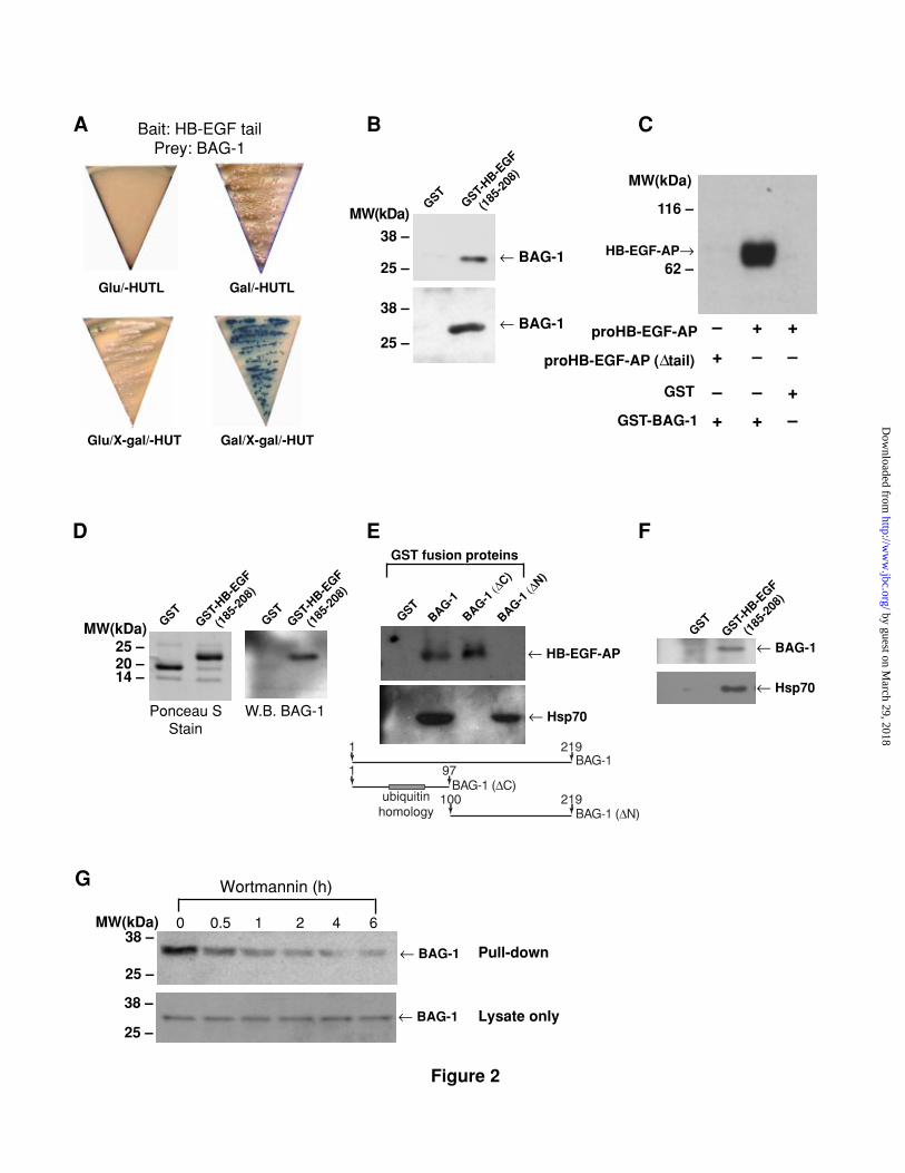
proHB-EGF cytoplasmic domain. We screened a yeast two-hybrid expression
library, constructed from a human prostate (LNCaP) xenograft tumor growing in
a mouse host, with the proHB-EGF tail domain (a.a. 185-208), using a LexA-
based system. Approximately 106 independent clones were screened. From the
6 strongest potential interactors identified in the screen, two clones, pJG45-B11
and pJG45-E68 (Fig. 2A), each contained the entire open reading frame of the
short form of the mouse protein, BAG-1 (219 amino acids, expected MW 24.5
kDa) (11). These two clones are identical but were derived from two
by guest on March 29, 2018
http://ww
w.jbc.org/
Dow
nloaded from
11
independent yeast transformations. In follow-up experiments, BAG-1 and the HB-
EGF tail also interacted in the yeast two-hybrid system when the cDNAs were
switched into the opposite (prey<-->bait) plasmid vectors.
To confirm the association between BAG-1 and HB-EGF, a GST fusion
protein containing the HB-EGF cytoplasmic tail (GST-HB-EGF(185-208)) was
constructed and used in pull-down assays. COS7 cells were transiently
transfected with expression plasmids encoding mouse BAG-1. Lysates from
these cells or from LNCaP cells (to test for binding with the native/endogenous,
human form of BAG-1) were incubated with purified GST-HB-EGF(185-208). A
complex was formed between GST-HB-EGF(185-208) and BAG-1 but not between
BAG-1 and GST alone (Fig. 2B). The converse experiment was also performed
with a GST-BAG-1 fusion protein, using lysates from cells expressing AP-tagged
proHB-EGF. In these experiments, a complex was formed between proHB-EGF
and GST-BAG-1, but not between proHB-EGF and GST, or between GST-BAG-1
and proHB-EGF in which the tail domain was deleted (Fig. 2C). Complex
formation between BAG-1 and the proHB-EGF tail was also demonstrated by Far-
Western blot, in which the HB-EGF tail was immobilized and the interaction
occurred on blotting membranes instead of in solution (Fig. 2D).
The 219 amino acid form of BAG-1 identified in the screen contains a
ubiquitin homology domain (residues 37-73) and a central region (residues 90-
172) that binds to Bcl-2 (11). Its carboxyl-terminal domain is required for direct
interaction with the ATPase domain of hsp70 heat shock protein (16). We
generated a GST-BAG-1(∆C) construct (residues 1-97), containing the ubiquitin
homology region, and GST-BAG-1 (∆N) (residues 100-219), which carries
binding sites for most of the known BAG-1 interactors. Complex formation with
proHB-EGF was observed with GST-BAG-1 and GST-BAG-1(∆C), but not with
GST-BAG-1(∆N). Complex formation did occur, however, between GST-BAG-
by guest on March 29, 2018
http://ww
w.jbc.org/
Dow
nloaded from

12
1(∆N) and hsp70 (Fig. 2E), demonstrating the capability of GST-BAG-1(∆N) to
bind to a known BAG-1 binding protein despite its failure to bind to proHB-EGF.
This result also indicates that the BAG-1 interaction with proHB-EGF is not
mediated by hsp70 and it rules out the possibility that aberrant folding of GST-
BAG-1(∆N) is the reason for the absence of binding to HB-EGF. In a reciprocal
experiment, GST-HB-EGF(185-208) was also able to form a complex with
endogenous BAG-1 (MW 33-35 kDa) and endogenous hsp70 from human
(LNCaP) cells (Fig. 2F).
We also investigated the dynamics of the BAG-1/HB-EGF tail interaction in
cells induced to undergo apoptosis. LNCaP cells were treated with wortmannin, a
PI3-kinase inhibitor that rapidly induces apoptosis in this cell line (15), and
lysates were used in pull-down experiments. Interestingly, complex formation
between the HB-EGF tail and endogenous BAG-1 diminished in a time-dependent
manner following wortmannin treatment (Fig. 2G). Similar results were obtained
when wortmannin-insensitive PC-3 cells were induced to undergo apoptosis by
treatment with staurosporine, a protein kinase inhibitor (data not shown). These
results suggest that the BAG-1/proHB-EGF interaction is not favored when cells
undergo programmed cell death.
Taken together, these experiments 1) demonstrated a direct interaction
between BAG-1 and the proHB-EGF cytoplasmic domain, 2) localized the HB-EGF
interacting domain to within residues 1-97 of BAG-1 and 3) also revealed that
BAG-1 can form a ternary complex with both proHB-EGF and hsp70 through
interactions with these proteins at distinct binding sites.
by guest on March 29, 2018
http://ww
w.jbc.org/
Dow
nloaded from

13
proHB-EGF and BAG-1 functionally cooperate in vivo
To explore the possibility of a functional interaction between proHB-EGF
and BAG-1, CHO cell populations were engineered sequentially to stably express
either BAG-1, proHB-EGF, or both proteins. BAG-1+proHB-EGF-expressing cells
exhibited a more epithelial-like cellular morphology, in comparison to cells
expressing either BAG-1 or proHB-EGF alone or control vectors, or the parent
cell, all of which exhibited a more fibroblastic appearance (Fig. 3). These data
suggest that coexpression of both proteins confers functional properties on
transfected cells that are not seen when each protein is expressed separately.
A similar requirement for proHB-EGF and BAG-1 co-expression to change a
cellular phenotype was observed in other assays. BAG-1+proHB-EGF-expressing
cells exhibited quantitatively reduced cell adhesion, as measured by sensitivity
to trypsin/EDTA treatment, in comparison to cells expressing either BAG-1 or
proHB-EGF or control plasmids (Fig. 4A). The presence of BAG-1 with proHB-EGF
in CHO cell transformants also affected the sensitivity of these cells to certain
apoptotic stimuli. BAG-1+proHB-EGF cells demonstrated increased resistance to
apoptosis induced by etoposide, a topoisomerase inhibitor, in comparison to
cells expressing either BAG-1 or proHB-EGF alone (Fig.4B). This resistance to
apoptosis induction appeared to be confined to specific survival pathways,
however, because BAG-1+proHB-EGF cells did not show synergistic protective
effects when apoptosis was induced by staurosporine (data not shown).
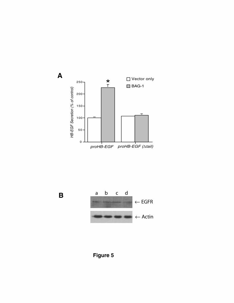
We also compared secretion of soluble HB-EGF in response to apoptotic
stimuli in proHB-EGF- and proHB-EGF(∆tail)-expressing MC2 cells transfected with
BAG-1 and vector only. Etoposide treatment induced rapid secretion of soluble
HB-EGF in cells expressing transfected BAG-1 but not in cells transfected with an
empty vector (Fig. 5A). In contrast, in cells expressing a tail-deleted form of
proHB-EGF, transfection with BAG-1 did not alter the secretion response to
by guest on March 29, 2018
http://ww
w.jbc.org/
Dow
nloaded from

14
etoposide. EGFR levels in proHB-EGF and proHB-EGF(∆tail) expressing cells were
equivalent (Fig. 5B), indicating that differential capture of the soluble HB-EGF
ligand by the EGFR cannot account for the observed differences in the secretion
response. Similar results were observed in the CHO cell background (data not
shown). These data suggest that the proHB-EGF tail is involved in regulated
processing of the cell-associated form of the growth factor to the soluble form
and that BAG-1 is capable of modulating this process in a manner that is
dependent on the presence of the tail domain.
Immunofluorescence confocal microscopy indicated that proHB-EGF and
BAG-1 co-localized within cytoplasm vesicles and at the plasma membrane,
consistent with the possibility that BAG-1 can affect trafficking and maturation
of soluble HB-EGF (Fig. 6). BAG-1 and proHB-EGF colocalized sites diminished in
frequency when cells were treated with etoposide.
by guest on March 29, 2018
http://ww
w.jbc.org/
Dow
nloaded from

15
Discussion
The results of this study demonstrate that the membrane form of HB-EGF
interacts with the anti-apoptotic protein, BAG-1, and that this interaction is
likely to have functional significance. The BAG-1/HB-EGF tail interaction was
demonstrated in a number of independent assays, including yeast two-hybrid,
GST-pull-down, and Far-Western blot methods. Evidence for colocalization of
BAG-1 and proHB-EGF was also obtained with immunofluorescence confocal
microscopy. In addition, cooperative effects of BAG-1 and proHB-EGF expression
were observed in assays of cell adhesion, apoptosis, and growth factor
secretion. Consistent with these findings, we found that deletion of the proHB-
EGF tail diminished the growth factor’s cytoprotective function and resulted in
the loss of the ability of etoposide to induce HB-EGF secretion, results that point
to an important role for the tail region. The diverse effects of proHB-EGF and
BAG-1 coexpression indicate that the BAG-1/HB-EGF interaction may impinge on
a number of discrete signaling pathways. Further, because we were able to
demonstrate effects of BAG-1 expression on secretion of soluble HB-EGF, this
interaction may regulate physiological activities of both the processed as well as
the membrane-anchored forms of the growth factor.
Importantly, our results suggest a novel mechanism whereby the
membrane-anchored form of HB-EGF might alter cell function independently of
the soluble form of the molecule. BAG-1 was originally identified as a Bcl-2-
binding protein but is now known to interact with and regulate a number of
signaling proteins. Because the proHB-EGF tail and membrane-anchoring domains
are removed from the mature growth factor by proteolytic cleavage, these
results provide the first unambiguous mechanism whereby the HB-EGF pro- form
could mediate processes distinct from those conferred by processed HB-EGF.
by guest on March 29, 2018
http://ww
w.jbc.org/
Dow
nloaded from
16
Several previous studies have identified bioactivities solely attributable to
proHB-EGF, although the reason for the distinction between the precursor and
secreted forms is not clear because both proteins presumably function
principally by activating identical high-affinity receptor tyrosine kinases.
Interactions between proHB-EGF and several cell surface molecules have been
identified previously, including interactions with CD9/DRAP27, a tetraspanin
membrane protein, the α3ß1 integrin, and heparan-sulfated glycosaminoglycans,
by mechanisms that are dependent on the proHB-EGF ectodomain (3,8,17).
However, BAG-1 is the first protein to be identified that interacts with the
proHB-EGF cytoplasmic domain in an ectodomain-independent manner. Because
CHO cells express low or negligible EGFR levels (18), our observations also
suggest the interesting possibility--although this was not formally tested in the
present study--that the cooperative effects we observed between proHB-EGF
and BAG-1 do not require the EGFR. Receptor-independent signaling by proHB-
EGF may be related to cell-cell adhesive functions previously noted for this
growth factor (2,8).
BAG-1 is an important regulatory protein that has been shown to have a
variety of binding partners and a range of bioactivities, including protection from
apoptotic signals (11,19), enhancement of cell motility (20) and regulation of
transcription (21,22). BAG-1 has been reported to bind the cytoplasmic domain
of the HGF receptor (14), the Raf-1 kinase (23) and several members of the
steroid hormone receptor superfamily (21,24), and to be capable of altering the
activity of these molecules. Multiple BAG-1 isoforms, with differing patterns of
cellular localization, arise by alternate translation initiation. Human BAG-1L
(MW~57 kDa), for example, is a nuclear as well as a cytosolic protein, while the
“small” BAG-1 isoforms studied here (MW 29-36 kDa) are predominantly
cytosolic (25). Like HB-EGF, the BAG-1 proteins appear to have an important
function in stress-regulated signaling. Enforced BAG-1 expression can promote
by guest on March 29, 2018
http://ww
w.jbc.org/
Dow
nloaded from
17
cell survival independently of effects on cell proliferation, motility and invasive
properties (19), suggesting that effects on cell survival signaling are distinct
from mechanisms of cell growth or cell cycle transit. In a similar fashion to our
results, antiapoptotic effects of BAG-1 can be enhanced by coexpression of
BAG-1 binding proteins, such as Bcl-2 (11,26). From these and other data, a
critical role for BAG-1 in cell signaling related to cell survival mechanisms, but
also to other processes, can be inferred despite uncertainty as to its precise
mechanism of action.
The heat shock protein, hsp70, has been reported to be a favored BAG-1
interactor. In this case, BAG-1 appears to function as a regulator of protein
folding and/or trafficking by acting as a competitive antagonist of the
cochaperone, Hip (27). Although a number of proteins are thought to associate
with BAG-1 because of its ability to bind hsp70 (16), we demonstrate in this
study that proHB-EGF binds to a C-terminal deletion mutant of BAG-1 which
does not bind hsp70 and that the HB-EGF interaction site on BAG-1 is distinct
from the hsp70 interaction site. Furthermore, the HB-EGF tail-BAG-1 interaction
occurs in yeast and yeast hsp70 is not a BAG-1 binding partner (16). Taken
together, these data indicate that proHB-EGF and BAG-1 interact directly and in
an hsp70-independent manner. This finding suggests that interaction of BAG-1
with proHB-EGF, and possibly with other regulatory proteins, may be functionally
distinct from its role as a cochaperone in mechanisms of protein folding and/or
stabilization.
BAG-1 is one of only a handful of proteins demonstrated to interact with
the tail domains of membrane-bound EGF-like growth factors. TACIP18/syntenin
and αA1 syntrophin were recently identified as specific interactors with the tail
domain of proTGFα (28). TACIP18/syntenin appears to be involved in
intracellular trafficking of TGFα. LIM kinase 1 (LIMK1) was identified as an
by guest on March 29, 2018
http://ww
w.jbc.org/
Dow
nloaded from

18
interactor with the tail domain of neuregulins and to co-localize with neuregulins
at the neuromuscular synapse (29). LIMK1 does not interact with the proTGFα
tail, suggesting that the cytoplasmic partners of membrane EGF-like growth
factors are likely to play specialized roles. This hypothesis is consistent with the
fact that ErbB1 ligands, despite similar receptor binding affinity to their primary
receptor(s), do not show identical patterns of receptor transactivation or similar
patterns of intracellular localization of their membrane forms and thus are
functionally specialized.
In conclusion, our findings provide evidence that the prosurvival protein,
BAG-1, is a functional binding partner of the membrane form of the receptor
tyrosine kinase ligand, HB-EGF. This interaction could credibly alter aspects of
cell signaling relating to unique functions of both the membrane as well as the
soluble HB-EGF isoforms.
by guest on March 29, 2018
http://ww
w.jbc.org/
Dow
nloaded from
19
Acknowledgments
We thank Drs. R. Adam and J. Kim for helpful discussions, P. Guthrie for
making yeast two-hybrid bait constructs and for help with preparing the
manuscript, D. Brown for help with confocal microscopy, and N. Kamei, M.
Ranasinghe, G. Lin, C. Riordan, A. Butler and D. Rice for technical assistance.
by guest on March 29, 2018
http://ww
w.jbc.org/
Dow
nloaded from
20
Figure Legends
Figure 1. Deletion of the proHB-EGF cytoplasmic tail abolishes the
cytoprotective effects of proHB-EGF expression in NRK52E cells.
Transfected cells were stimulated by 0.5 mM H2O2 or 44.5 µM etoposide for 24
h. DNA fragments released into the cytoplasm were collected for agarose gel
electrophoresis and visualized by ethidium bromide staining.
Figure 2. Interaction between proHB-EGF and BAG-1. (A) BAG-1 is a
binding partner for the proHB-EGF tail in yeast. Transformed yeast colony
(clone# 11) was streaked onto medium lacking histidine, uracil, tryptophan,
leucine, and containing galactose (Gal/-HUTL) or glucose (Glu/-HUTL) as carbon
source and growth was monitored 3 d later. The LacZ reporter gene was also
monitored in medium containing X-gal (Gal/X-gal/-HUT or Glu/X-gal/-HUT). (B)
GST-HB-EGF(185-208) pull-down assay: GST fusion proteins were incubated with cell
lysates and the binding proteins were co-precipitated with glutathione-agarose
and subjected to SDS-PAGE followed by anti-BAG-1 Western blot. Cell lysates
were from COS7 cells transfected with pcDNA3.1/Myc-BAG-1 (upper panel) or
human LNCaP cell lines (lower panel). (C) GST-BAG-1 pull-down experiment with
proHB-EGF-AP-enriched cell lysates followed by detection of proHB-EGF-AP
fusion protein with anti-AP Ab. (D) Far-Western blot: GST-HB-EGF(185-208) fusion
proteins (1 µg) were subjected to SDS-PAGE and transferred to Immobilon-P
membrane (left panel). The membrane was incubated with cell lysates from
COS7 cells overexpressing BAG-1, followed by anti-BAG-1 immunodetection
(right panel). (E) Identification of the BAG-1 domain responsible for proHB-EGF
binding. GST-BAG-1 pull-down assay followed by anti-AP Western blot as in C
(top). The same membrane was stripped and re-probed with anti-hsp70/hsc70
by guest on March 29, 2018
http://ww
w.jbc.org/
Dow
nloaded from
21
monoclonal Ab (middle). The structure of the GST-BAG-1 fusion proteins used
are indicated in the diagram. (F) GST-HB-EGF(185-208) pull-down experiment with
LNCaP cell extracts followed by anti-BAG-1 Western blot (upper panel). The
membrane was stripped and re-probed with anti-hsp70/hsc70 monoclonal Ab
(lower panel). (G) Changes in the association between proHB-EGF and BAG-1.
LNCaP cells were treated with 100 nM wortmannin and cell lysates were
collected at the indicated time points. The GST-HB-EGF(185-208) binding proteins
were detected by anti-BAG-1 Ab (upper panel). Levels of BAG-1 in cell lysates
are shown by Western blot in the lower panel.
Figure 3. Phase contrast micrographs of BAG-1 and proHB-EGF-
expressing CHO cells. CHO cells were stably transfected with (A) empty
vector + LacZ vector; (B) proHB-EGF + LacZ vector; (C) empty vector + BAG-1
and (D) proHB-EGF + BAG-1. Western blots of the cell lysates from the four cell
populations (A to D) are shown in the lower panel.
Figure 4. Synergistic effects of proHB-EGF and BAG-1 expression in
CHO cells. (A) Adhesion assay. Cell populations (50,000/well) were seeded in
24 well plates. 48 h later cells were treated by trypsin-EDTA (1:15 dilution) for
the times indicated. The cells remaining on the plate were quantified by crystal
violet staining. (B) Cell survival assay. The four transfected cell populations
were seeded in 96 well plate (10,000/well) and 24 h later cells were treated
with etoposide for 24 h. Cells were exposed to MTT for 4 h at 37oC prior to
harvest. In comparison to the other 3 groups, *: P<0.0005, **: P<0.01.
Figure 5. Requirement of the proHB-EGF tail in BAG-1 regulated
secretion of HB-EGF. (A) MC2/proHB-EGF and MC2/proHB-EGF(∆tail) cells
were transfected with a BAG-1 expression construct or empty vector. Stable
double-transfected cell populations were stimulated with 0.22 mM etoposide for
by guest on March 29, 2018
http://ww
w.jbc.org/
Dow
nloaded from

22
24 h and HB-EGF levels in the conditioned medium were measured. *: P<0.0001.
(B) Western blot of the total cell lysates from the above MC2 cells stably
transfected with (a) proHB-EGF + empty vector; (b) proHB-EGF + BAG-1; (c)
proHB-EGF(∆tail) + empty vector; (d) proHB-EGF(∆tail) + BAG-1.
Figure 6. Co-localization of BAG-1 and proHB-EGF. Confocal
immunofluorescence microscopic evaluation of proHB-EGF and BAG-1 in BAG-1
transfected MC2/proHB-EGF cells. Representative patterns of proHB-EGF (A and
D) and BAG-1 (B and E) staining in unstimulated cells are shown. Representative
examples of etoposide stimulated cells are shown in G and H. Merged images are
shown in C, F and I. Etoposide untreated (-) and treated (+).
by guest on March 29, 2018
http://ww
w.jbc.org/
Dow
nloaded from
23
References
1. Anklesaria, P., Teixido, J., Laiho, M., Pierce, J. H., Greenberger, J. S.,
and Massague, J. (1990) Proc Natl Acad Sci U S A 87(9), 3289-93
2. Raab, G., Kover, K., Paria, B. C., Dey, S. K., Ezzell, R. M., and
Klagsbrun, M. (1996) Development 122(2), 637-45
3. Higashiyama, S., Iwamoto, R., Goishi, K., Raab, G., Taniguchi, N.,
Klagsbrun, M., and Mekada, E. (1995) J Cell Biol 128(5), 929-38
4. Raab, G., and Klagsbrun, M. (1997) Biochim Biophys Acta
1333(3), F179-99
5. Takemura, T., Hino, S., Murata, Y., Yanagida, H., Okada, M.,
Yoshioka, K., and Harris, R. C. (1999) Kidney Int 55(1), 71-81
6. Iwamoto, R., Handa, K., and Mekada, E. (1999) J Biol Chem
274(36), 25906-12
7. Naglich, J. G., Metherall, J. E., Russell, D. W., and Eidels, L. (1992)
Cell 69(6), 1051-61
8. Nakamura, K., Iwamoto, R., and Mekada, E. (1995) J Cell Biol
129(6), 1691-705
9. Takemura, T., Kondo, S., Homma, T., Sakai, M., and Harris, R. C.
(1997) J Biol Chem 272(49), 31036-42
10. Miyoshi, E., Higashiyama, S., Nakagawa, T., Hayashi, N., and
Taniguchi, N. (1997) J Biol Chem 272(22), 14349-55
11. Takayama, S., Sato, T., Krajewski, S., Kochel, K., Irie, S., Millan, J.
A., and Reed, J. C. (1995) Cell 80(2), 279-84
12. Finley, R. L., Jr., and Brent, R. (1994) Proc Natl Acad Sci U S A
91(26), 12980-4
13. Dethlefsen, S. M., Raab, G., Moses, M. A., Adam, R. M., Klagsbrun,
M., and Freeman, M. R. (1998) J Cell Biochem 69(2), 143-53
by guest on March 29, 2018
http://ww
w.jbc.org/
Dow
nloaded from

24
14. Bardelli, A., Longati, P., Albero, D., Goruppi, S., Schneider, C.,
Ponzetto, C., and Comoglio, P. M. (1996) Embo J 15(22), 6205-12
15. Lin, J., Adam, R. M., Santiestevan, E., and Freeman, M. R. (1999)
Cancer Res 59(12), 2891-7
16. Zeiner, M., Gebauer, M., and Gehring, U. (1997) Embo J 16(18),
5483-90
17. Shi, W., Fan, H., Shum, L., and Derynck, R. (2000) J Cell Biol
148(3), 591-602
18. Livneh, E., Prywes, R., Kashles, O., Reiss, N., Sasson, I., Mory, Y.,
Ullrich, A., and Schlessinger, J. (1986) J Biol Chem 261(27), 12490-7
19. Takaoka, A., Adachi, M., Okuda, H., Sato, S., Yawata, A., Hinoda, Y.,
Takayama, S., Reed, J. C., and Imai, K. (1997) Oncogene 14(24), 2971-7
20. Naishiro, Y., Adachi, M., Okuda, H., Yawata, A., Mitaka, T.,
Takayama, S., Reed, J. C., Hinoda, Y., and Imai, K. (1999) Oncogene 18(21),
3244-51
21. Froesch, B. A., Takayama, S., and Reed, J. C. (1998) J Biol Chem
273(19), 11660-6
22. Zeiner, M., Niyaz, Y., and Gehring, U. (1999) Proc Natl Acad Sci U S
A 96(18), 10194-9
23. Wang, H. G., Takayama, S., Rapp, U. R., and Reed, J. C. (1996) Proc
Natl Acad Sci U S A 93(14), 7063-8
24. Schneikert, J., Hubner, S., Martin, E., and Cato, A. C. (1999) J Cell
Biol 146(5), 929-40
25. Takayama, S., Krajewski, S., Krajewska, M., Kitada, S., Zapata, J. M.,
Kochel, K., Knee, D., Scudiero, D., Tudor, G., Miller, G. J., Miyashita, T., Yamada,
M., and Reed, J. C. (1998) Cancer Res 58(14), 3116-31
26. Schulz, J. B., Bremen, D., Reed, J. C., Lommatzsch, J., Takayama,
S., Wullner, U., Loschmann, P. A., Klockgether, T., and Weller, M. (1997) J
Neurochem 69(5), 2075-86
by guest on March 29, 2018
http://ww
w.jbc.org/
Dow
nloaded from
25
27. Takayama, S., Xie, Z., and Reed, J. C. (1999) J Biol Chem 274(2),
781-6
28. Fernandez-Larrea, J., Merlos-Suarez, A., Urena, J. M., Baselga, J.,
and Arribas, J. (1999) Mol Cell 3(4), 423-33
29. Wang, J. Y., Frenzel, K. E., Wen, D., and Falls, D. L. (1998) J Biol
Chem 273(32), 20525-34
by guest on March 29, 2018
http://ww
w.jbc.org/
Dow
nloaded from

H2O
2 Etoposide
vector onlyproH
B-EGF(∆tail)
proHB-EG
Fvector onlyproH
B-EGF(∆tail)
proHB-EG
F
Figure 1
by guest on March 29, 2018
http://ww
w.jbc.org/
Dow
nloaded from
Gal/X-gal/-HUT
A
Glu/-HUTL Gal/-HUTL
Glu/X-gal/-HUT
Bait: HB-EGF tailPrey: BAG-1
MW(kDa)GST
GST-HB-E
GF
(185
-208
)
← BAG-1
← BAG-1
B
38 –
25 –
38 –
25 –
C
proHB-EGF-AP (∆tail)
116 –
62 – HB-EGF-AP→
MW(kDa)
proHB-EGF-AP
D
14 –
GSTGST-
HB-EGF
(1
85-2
08)
GSTGST-
HB-EGF
(1
85-2
08)
MW(kDa)25 –
Ponceau SStain
W.B. BAG-1
20 –
E
GSTBAG-1
BAG-1 (∆
C)
BAG-1 (∆
N)
← HB-EGF-AP
← Hsp70
219
ubiquitinhomology
97
100 219
1
1BAG-1 (∆C)
BAG-1 (∆N)
BAG-1
GST fusion proteins
F
← BAG-1
← Hsp70
GSTGST-
HB-EGF
(1
85-2
08)
38 –← BAG-1
25 –
0 0.5 1 2 4 6
Pull-down
Lysate only← BAG-138 –
25 –
Wortmannin (h)G
MW(kDa)
+ +–
+ ––
GST +––
GST-BAG-1 + + –
Figure 2
by guest on March 29, 2018
http://ww
w.jbc.org/
Dow
nloaded from

A B C D
← proHB-EGF
← BAG-1
Figure 3
by guest on March 29, 2018
http://ww
w.jbc.org/
Dow
nloaded from

B
0
20
40
60
80
Cel
l Sur
viva
l (%
of
cont
rol)
0.22 0.44 0.89
Etoposide (mM)
proHB-EGF + BAG-1
empty vector + BAG-1proHB-EGF + LacZ vector
empty vector + LacZ vector
*
**
A
0
25
50
75
100
125
0 5 10 15 20 25
Time (min)
Cel
l Adh
esio
n (%
of c
ontr
ol)
proHB-EGF + BAG-1
empty vector + BAG-1
proHB-EGF + LacZ vector
empty vector + LacZ vector
Figure 4
by guest on March 29, 2018
http://ww
w.jbc.org/
Dow
nloaded from
0
50
100
150
200
250
HB
-EG
F S
ecre
tion
(% o
f con
trol) BAG-1
Vector only
*
proHB-EGF proHB-EGF (∆tail)
*
Figure 5
A
B a b c d
← EGFR
← Actin
by guest on March 29, 2018
http://ww
w.jbc.org/
Dow
nloaded from

(–)
(+)
(–)
proHB-EGF BAG-1 proHB-EGF/BAG-1
Figure 6
by guest on March 29, 2018
http://ww
w.jbc.org/
Dow
nloaded from

Jianqing Lin, Lloyd Hutchinson, Sandra M. Gaston, Gerhard Raab and Michael R. FreemanSurvival Regulation
Heparin-binding EGF-like Growth Factor: A Unique Role for proHB-EGF in Cell BAG-1 is a Novel Cytoplasmic Binding Partner of the Membrane Form of
published online May 4, 2001J. Biol. Chem.
10.1074/jbc.M010237200Access the most updated version of this article at doi:
Alerts:
When a correction for this article is posted•
When this article is cited•
to choose from all of JBC's e-mail alertsClick here
by guest on March 29, 2018
http://ww
w.jbc.org/
Dow
nloaded from