understanding and interpreting oct - optometry's meeting€¦ · · 2017-08-21understanding...
TRANSCRIPT
Understanding and Interpreting OCT
Mark T. Dunbar, O.D., F.A.A.O.Bascom Palmer Eye Institute
University of Miami, School of Medicine
The Swiss Army Pocket Knife of Eye Care
Mark Dunbar: Disclosure
Consultant for Allergan
Optometry Advisory Board and Speaker bureau for: Allergan
Carl Zeiss Meditec
Artic Dx
Sucampo
Mark Dunbar does not own stock in any of the above companies
Optical Coherence Tomography (OCT)
Non-contact, non-invasive imaging device
Produces high-resolution images of the posterior segmentOptical biopsy
Quickly emerged as the standard of care for imaging in retina and glaucoma
Revolutionized ocular disease management in all of eye care
Advances in SD-OCTImproving software
Noise reduction/over sampling technology that provides higher resolution imaging
Wider and deeper scans
Greater density in the scans
Improvements in 3D imaging
Enhanced depth imaging – imaging choroid
Progression analysis software
Main Clinical Utilities of OCT
High resolution evaluation of retinal anatomy Diagnosis of macular conditions difficult to
establish with biomicroscopy Quantitative assessment of retinal and
vitreoretinal anatomic alterations Objective means for monitoring disease
progression and/or therapeutic response
Spectral Domain OCTThe Competition
Carl Zeiss: Cirrus
OptiVue: Avantis and the iVue
Heidelberg: Spectralis
Topcon – Maestro coming out
Optos
SOCT Copernicus (Reichert) Now owned by Cannon
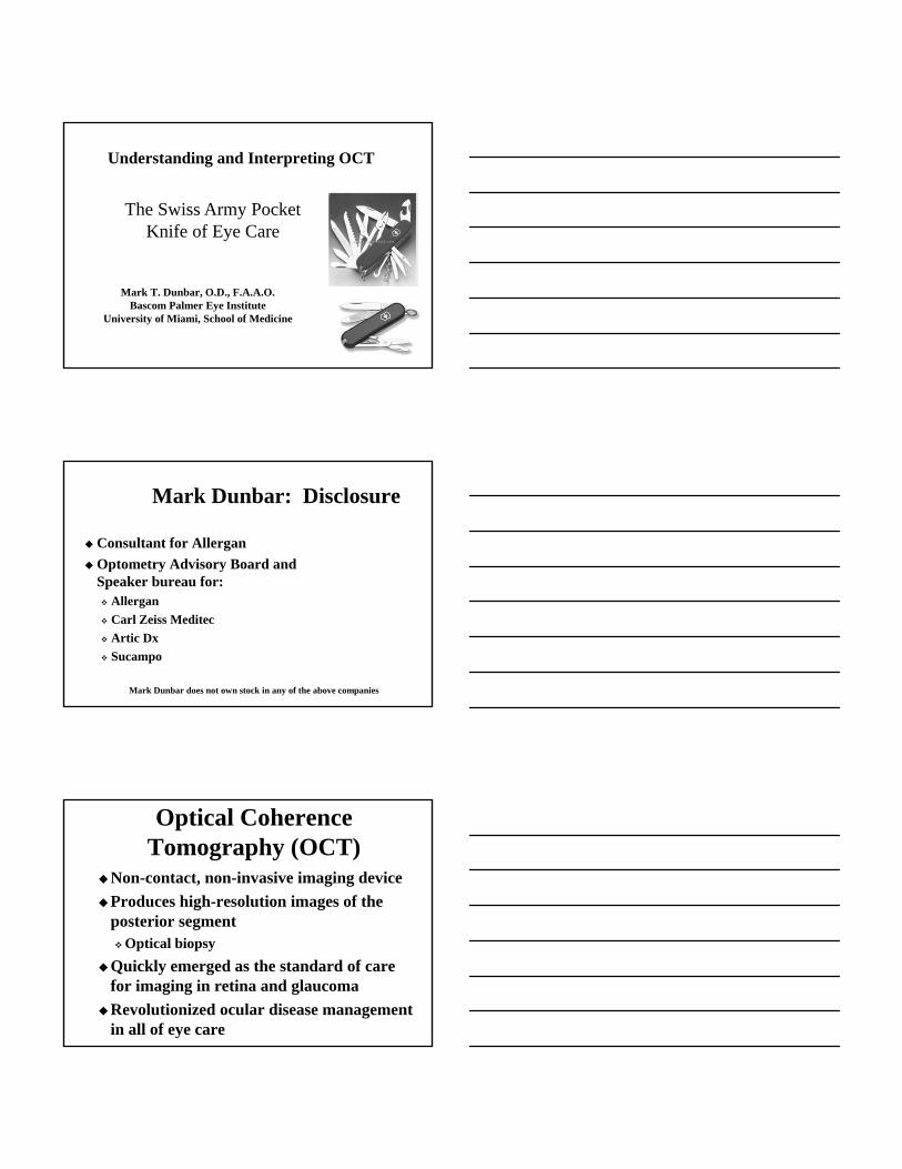
The Anatomy
General PrinciplesDon’t make it more complicated then it
needs to be
Don’t get caught up in the minutia!
Pay attention to the IS/OS junctionAka – PIL (photoreceptor integrity line)
Provides great anatomic perspective – but it won’t “tell” you the diagnosis
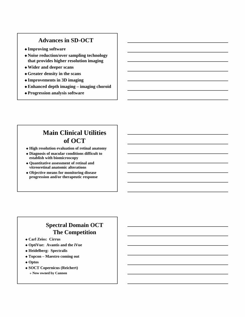
Central Serous and Neurosensory Retinal
Detachment
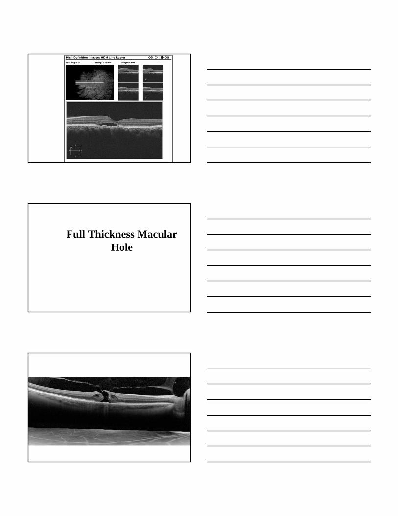
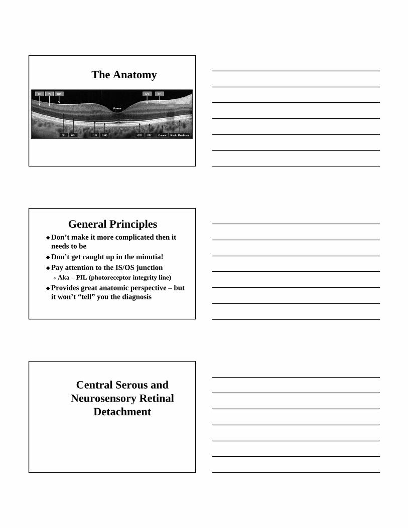
Full Thickness Macular Hole
When is a hole….a hole?
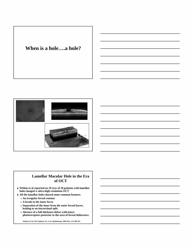
Lamellar Macular Hole in the Era of OCT
Witkin et al reported on 19 eyes of 18 patients with lamellar holes imaged w ultra-high resolution OCT
All the lamellar holes shared some common features An irregular foveal contour A break in the inner fovea Separation of the inner from the outer foveal layers,
leading to an intraretinal split Absence of a full thickness defect with intact
photoreceptors posterior to the area of foveal dehiscence.
Witkin AJ, Ko TH, Fujimoto JG, et al. Ophthalmogy. 2006 Mar; 113:388-397.
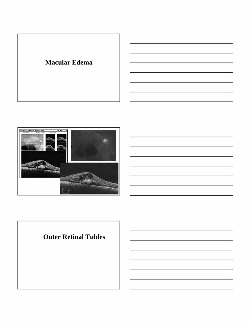
Macular Edema
Outer Retinal Tubles
Plaquenil Screening: Traditionally
Baseline macula photos
Color vision testing
Amsler grid
10-2 Visual fields
Yearly exams
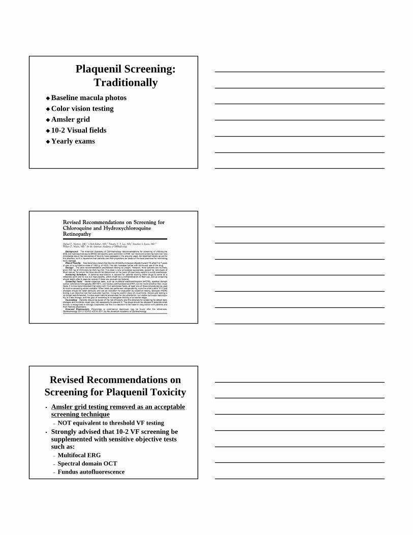
Revised Recommendations on Screening for Plaquenil Toxicity• Amsler grid testing removed as an acceptable
screening technique– NOT equivalent to threshold VF testing
• Strongly advised that 10-2 VF screening be supplemented with sensitive objective tests such as:– Multifocal ERG– Spectral domain OCT– Fundus autofluorescence

Revised Recommendations on Screening for Plaquenil ToxicityTests Not Recommended for Screening:
Fundus photographyTime domain OCTFluorescein angiographyFull-field ERGAmsler gridColor vision screeningEOG
Leonardo57 y/o Hispanic Male
“Routine” exam
Has had poor vision for ~ 25 yrs or so
VA: 20/70 RE; 20/60 LE
CVF: FTFC OU
Pupils: ERRL – No APD
SLE – Tr NS
Advanced RPE AnalysisGain new insights on your AMD patients
• RPE Elevations: If the RPE is raised, a new proprietary algorithm for Cirrus maps and measures the area and volume of the elevations.
• Sub-RPE Illumination.If the RPE is absent or has lost integrity a new proprietary algorithm for Cirrus can map and measure the affected area.
RPE Elevations Sub-RPE Illumination
Cirrus 6.0

OCT in Glaucoma
Traditional Methods of Assessing Glaucoma
IOP monitoringMajor risk factor
Subjective evaluation of the optic nerve
Visual field testing
SD OCT Stop Light Display of RNFL Normative Range
100%
95%
1%
0%
5%
1%
4%
90%
5%
95% of normal population falls in or below green band; 90% falls within green band
5% of normal population falls within or below yellow band: 4% falls within the yellow band
1% falls within red band; considered outside normal limits
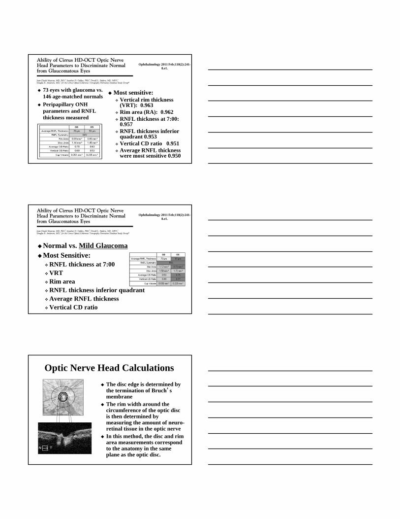
73 eyes with glaucoma vs. 146 age-matched normals
Peripapillary ONH parameters and RNFL thickness measured
Most sensitive: Vertical rim thickness
(VRT): 0.963 Rim area (RA): 0.962 RNFL thickness at 7:00:
0.957 RNFL thickness inferior
quadrant 0.953 Vertical CD ratio 0.951 Average RNFL thickness
were most sensitive 0.950
Ophthalmology 2011 Feb;118(2):241-8.e1.
Normal vs. Mild GlaucomaMost Sensitive:
RNFL thickness at 7:00VRTRim areaRNFL thickness inferior quadrantAverage RNFL thicknessVertical CD ratio
Ophthalmology 2011 Feb;118(2):241-8.e1.
The disc edge is determined by the termination of Bruch’s membrane
The rim width around the circumference of the optic disc is then determined by measuring the amount of neuro-retinal tissue in the optic nerve
In this method, the disc and rim area measurements correspond to the anatomy in the same plane as the optic disc.
Optic Nerve Head Calculations
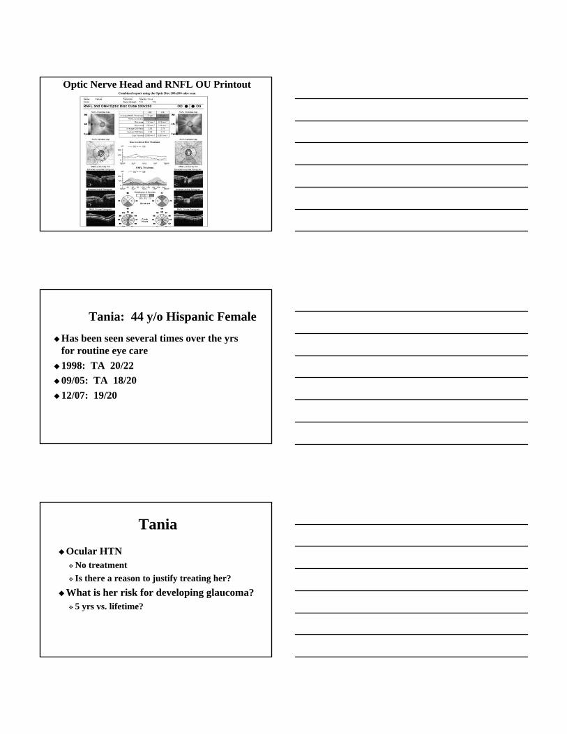
Optic Nerve Head and RNFL OU PrintoutCombined report using the Optic Disc 200x200 cube scan
Tania: 44 y/o Hispanic Female
Has been seen several times over the yrs for routine eye care
1998: TA 20/22
09/05: TA 18/20
12/07: 19/20
Tania
Ocular HTNNo treatment
Is there a reason to justify treating her?
What is her risk for developing glaucoma? 5 yrs vs. lifetime?
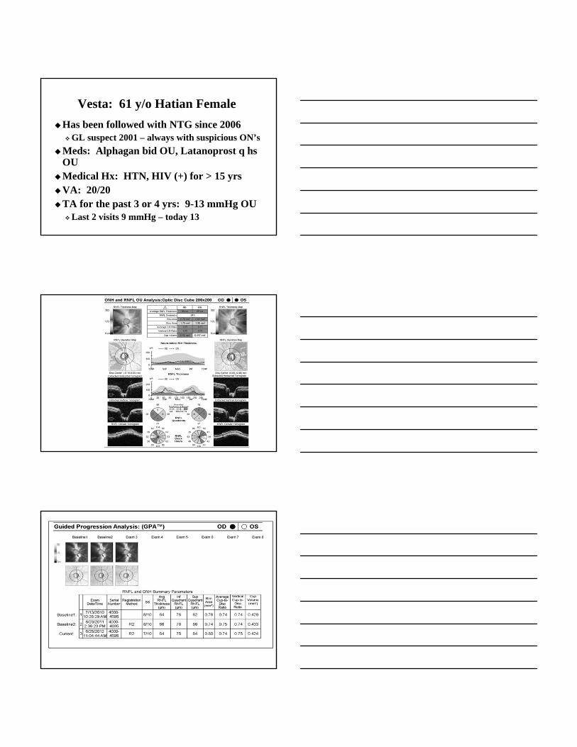
Vesta: 61 y/o Hatian Female
Has been followed with NTG since 2006GL suspect 2001 – always with suspicious ON’s
Meds: Alphagan bid OU, Latanoprost q hs OU
Medical Hx: HTN, HIV (+) for > 15 yrsVA: 20/20TA for the past 3 or 4 yrs: 9-13 mmHg OU
Last 2 visits 9 mmHg – today 13
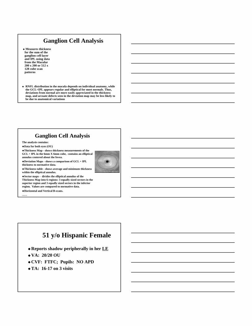
Ganglion Cell Analysis Measures thickness
for the sum of the ganglion cell layer and IPL using data from the Macular 200 x 200 or 512 x 128 cube scan patterns
RNFL distribution in the macula depends on individual anatomy, while the GCL+IPL appears regular and elliptical for most normals. Thus, deviations from normal are more easily appreciated in the thickness map, and arcuate defects seen in the deviation map may be less likely to be due to anatomical variations
Ganglion Cell AnalysisThe analysis contains:
Data for both eyes (OU)
Thickness Map - shows thickness measurements of the GCL + IPL in the 6mm X 6mm cube, contains an elliptical annulus centered about the fovea.
Deviation Maps - shows a comparison of GCL + IPL thickness to normative data.
Thickness table - shows average and minimum thickness within the elliptical annulus.
Sector maps - divides the elliptical annulus of the Thickness Map into 6 regions: 3 equally sized sectors in the superior region and 3 equally sized sectors in the inferior region. Values are compared to normative data.
Horizontal and Vertical B-scans. Cirrus 6.0
51 y/o Hispanic Female
Reports shadow peripherally in her LE
VA: 20/20 OU
CVF: FTFC; Pupils: NO APD
TA: 16-17 on 3 visits
Glaucoma Package Heidelberg Spectralis
RTVue Glaucoma Package
Summary: OCT and Glaucoma
OCT is able to accurately detected early glaucoma with good reliability
Also very good with already established glaucoma
Determining same day reliability is critical Corroborate your findings
To be to accurately utilize serial analysis in future scans
OCT is as good as other ON imaging devices