ultraviolet and visible spectrometry
TRANSCRIPT

Advanced strategies in food analysis UV/VIS spectrometry
Richard Koplík
Ultraviolet and visible spectrometry
Theoretical overview
Molecular absorption of electromagnetic radiation
changes of energy state of the molecule include
– electronic state ∆Ee =150-600 kJ/mol
(electron transitions between orbitals)
– vibrational state ∆Ev =2-60 kJ/mol
– rotational state ∆Er ≈ 3 kJ/mol
relation to the absorbed radiation wavelength
∆E = ∆Ee + ∆Ev + ∆Er = h . ν = h . c / λ
h = 6.626 . 10-34
J s (Planck’s constant)
Spectral regions
Region λ Absorbing compounds
Far ultraviolet (vacuum UV region) 190 nm saturated and mono-unsaturated
(Near) ultraviolet 190-380 nm poly-unsaturated and aromatic
Visible light region 380-780 nm coloured
Visible light absorption
Table of complementary colours:
λ (nm) Colour of light Colour of absorbing body
400–435 violet yellow-green
435–480 blue yellow
480–490 green-blue orange
490–500 blue-green red-orange
500–560 green red
560–580 green-yellow violet
580–595 yellow-orange blue
595–620 red-orange green-blue
620–760 red blue-green

Advanced strategies in food analysis UV/VIS spectrometry
Richard Koplík
Labert-Beer law
transmittance T = I/I0
in a diluted solution the value of absorbance A measured at the specific wavelength is
proportional to the concentration of absorbing compound
Aλ = - log T = log (I0/I) = ελ . b . c
Energy changes of electronic transitions
Probability of transition influences the value of absorption coefficient
relation to spin state of excited electron
1) transition S0 (ground singlet) →S1 (upper singlet) is allowed
εmax ≈ 103–10
5 l.mol
-1.cm
-1
2) transition S0 → T1 (triplet) is forbidden
εmax ≈ 100 l.mol
-1.cm
-1
E σ*
π*
n
π
σ
σ→σ*
n→σ*
π→π*
n→π*
Advanced strategies in food analysis UV/VIS spectrometry
Richard Koplík
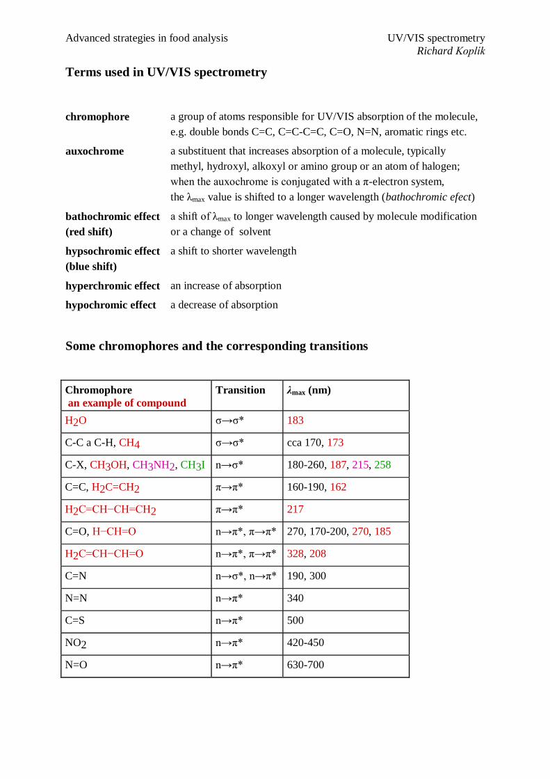
Terms used in UV/VIS spectrometry
chromophore a group of atoms responsible for UV/VIS absorption of the molecule,
e.g. double bonds C=C, C=C-C=C, C=O, N=N, aromatic rings etc.
auxochrome a substituent that increases absorption of a molecule, typically
methyl, hydroxyl, alkoxyl or amino group or an atom of halogen;
when the auxochrome is conjugated with a π-electron system,
the λmax value is shifted to a longer wavelength (bathochromic efect)
bathochromic effect
(red shift)
a shift of λmax to longer wavelength caused by molecule modification
or a change of solvent
hypsochromic effect
(blue shift)
a shift to shorter wavelength
hyperchromic effect an increase of absorption
hypochromic effect a decrease of absorption
Some chromophores and the corresponding transitions
Chromophore
an example of compound
Transition λmax (nm)
H2O σ→σ* 183
C-C a C-H, CH4 σ→σ* cca 170, 173
C-X, CH3OH, CH3NH2, CH3I n→σ* 180-260, 187, 215, 258
C=C, H2C=CH2 π→π* 160-190, 162
H2C=CH−CH=CH2 π→π* 217
C=O, H−CH=O n→π*, π→π* 270, 170-200, 270, 185
H2C=CH−CH=O n→π*, π→π* 328, 208
C=N n→σ*, n→π* 190, 300
N=N n→π* 340
C=S n→π* 500
NO2 n→π* 420-450
N=O n→π* 630-700
Advanced strategies in food analysis UV/VIS spectrometry
Richard Koplík
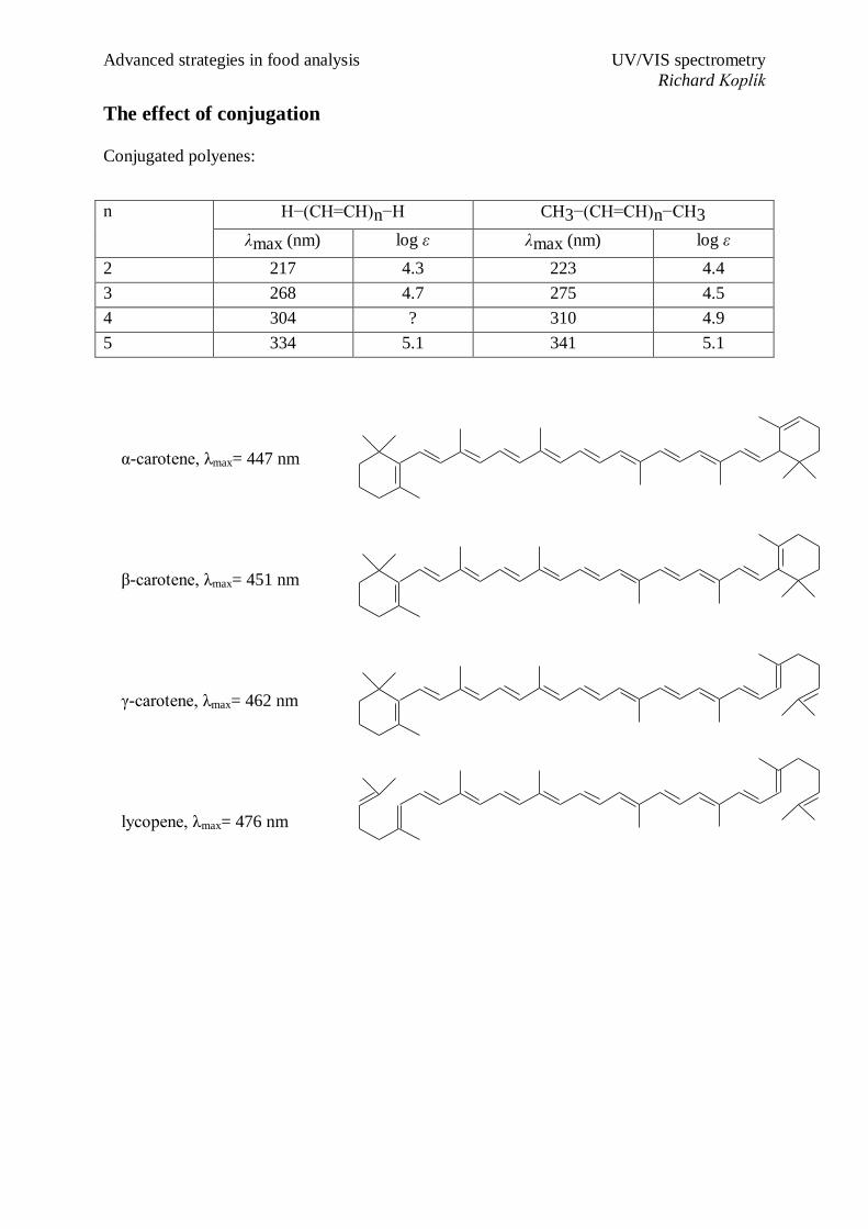
The effect of conjugation
Conjugated polyenes:
n H−(CH=CH)n−H CH3−(CH=CH)n−CH3
λmax (nm) log ε λmax (nm) log ε
2 217 4.3 223 4.4
3 268 4.7 275 4.5
4 304 ? 310 4.9
5 334 5.1 341 5.1
α-carotene, λmax= 447 nm
β-carotene, λmax= 451 nm
γ-carotene, λmax= 462 nm
lycopene, λmax= 476 nm

Advanced strategies in food analysis UV/VIS spectrometry
Richard Koplík
Benzene and its derivatives
Compound λmax (nm) log ε λmax (nm) log ε λmax (nm) log ε
benzene 204 3.9 254 2.0 - -
toluene 207 3.8 261 2.4 - -
brombenzene 210 3.9 261 2.3 - -
phenol 211 3.8 270 3.2 - -
benzaldehyde 250 4.1 280 3.0 320 1.7
acetophenone 246 4.0 280 3.0 320 1.7
benzoic acid 230 4.1 273 3.0 - -
aniline 230 3.9 280 3.5 - -
styrene 247 4.0 281 2.0 - -
cinnamaldehyde 285 4.4 - - - -
cinnamic acid 273 4.3 - - - -
biphenyl 248 4.2 - - - -
Heterocyclic compounds
5-membered
Compound λmax (nm) log ε λmax (nm) log ε
furan 200 4.0 - -
2-furaldehyde 227 3.3 272 4.1
2-acetylfuran 225 3.4 269 4.1
pyrrole 210 4.2 240 2.5
2-acetylpyrrole 250 3.6 287 4.2
thiophene - - 235 3.7
2-acetylthiophene 260 3.9 285 3.7
thiazole - - 240 3.6

Advanced strategies in food analysis UV/VIS spectrometry
Richard Koplík
6-membered
Compound λmax (nm) log ε λmax (nm) log ε λmax (nm) log ε
Pyridine 195 - 250 3.3 - -
2-Picoline - - 262 3.4 - -
Pyrazine - - 260 3.7 - -
Quinoline 227 4.6 275 3.7 313 3.4
Isoquinoline 218 4.9 262 3.6 317 3.5
Pyrimidine - - - - 343 3.3
Polycyclic aromatic hydrocarbons

Advanced strategies in food analysis UV/VIS spectrometry
Richard Koplík
Practical rules for spectrophotometric measurement
choice of a measuring cell
quartz: for UV
glass: for VIS
plastic: for some routine measurement in VIS
length of a cell: most commonly 0.1–5 cm optimum absorbance 0.1–2
choice of a solvent
the kind of solvent may influence the position of spectral band and the maximum
absorbance
spectrum recording
scan rate
very fast scan higher noise of the spectrum
spectral band-width
narrow SBW (0.2–0.5 nm) better resolution and higher noise of the spectrum
wide SBW (2–4 nm) low resolution, low noise; suitable for the recording of wide
bands (VIS region) and the highly precise measurement of a single absorbance value
sample dilution
allowed only for stable species
Solvents for UV spectrometry
Table the lowest wavelengths of measurement with the solvent
Solvent λ (nm) Solvent λ (nm)
acetonitrile, water 190 chloroform 240
isooctane, cyclohexane 195 ethylacetate 260
hexane 201 dimethylformamide 270
methanol, ethanol 205 acetic acid. 270
1,4-dioxane 215 benzene 280
diethylether 220 toluene 285
glycerol 230 pyridine 300
dichloromethane 233 acetone 330

Advanced strategies in food analysis UV/VIS spectrometry
Richard Koplík
Effect of solvent on the absorption spectrum
The kind of solvent slightly affects
values of λmax, ε
shape of the spectrum
Spectra of biologically important compounds
Compound λmax (nm) ε (l.mol
-1.cm
-1)
NAD, NADP 260 15 000
NADH, NADPH
260 15 000
340 6 200
FMN, FAD
260 15 000
375 10 000 (FMN)
9 000 (FAD)
445 12 500 (FMN)
450 11 000 (FAD)
pyridoxal
250 3 000
320 6 000
spectra of phenol measured in
isooctane and ethanol

Advanced strategies in food analysis UV/VIS spectrometry
Richard Koplík
Compound λmax (nm) ε (l.mol-1.cm-1)
cholesterol 235 20 000
calciferols 265 18 300
β-carotene 450 120 000
retinol 330 45 000
trans, trans-9,12-
octadecenoic acid.
231 35 000
adenosine 267 12 300
guanosine 248 11 000
cytidine 271 9 100
thymidine 267 9 650
uridine 262 8 500

Advanced strategies in food analysis UV/VIS spectrometry
Richard Koplík
Two-component analysis
Rule of absorbance additivity:

Advanced strategies in food analysis UV/VIS spectrometry
Richard Koplík
Derivative spectrometry
T = /0
A = - log10T = - 2,303 . ln T = . b . c
dA/d = -2.303 . (1/T) . dT/d = b . c . d /d
the first (and also the second) derivative of absorbance is proportional to the concentration
of the absorbing compound
original spectrum
A vs. λ
1st derivative
dA/dλ vs. λ
2nd
derivative
d2A/dλ
2 vs. λ
Advanced strategies in food analysis UV/VIS spectrometry
Richard Koplík
Flow injection analysis – FIA
an optional arrangement of a (spectrophotometric) measurement
instead of the batch-preparation of the measured solution the sample is injected into the
flow of the carrier solution or the reagent solution and then measured (usually using a
spectrophotometer)
FIA is much faster than traditional batch analysis and can be easily automated
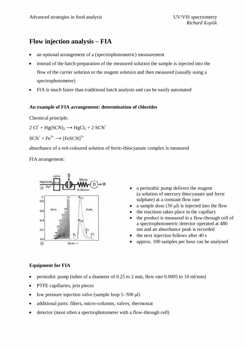
An example of FIA arrangement: determination of chlorides
Chemical principle:
2 Cl- + Hg(SCN)2 → HgCl2 + 2 SCN
-
SCN- + Fe
3+ → [FeSCN]
2+
absorbance of a red-coloured solution of ferric-thiocyanate complex is measured
FIA arrangement:
Equipment for FIA
peristaltic pump (tubes of a diameter of 0.25 to 2 mm, flow rate 0.0005 to 10 ml/min)
PTFE capillaries, join pieces
low pressure injection valve (sample loop 5–500 μl)
additional parts: filters, micro-columns, valves, thermostat
detector (most often a spectrophotometer with a flow-through cell)
a peristaltic pump delivers the reagent
(a solution of mercury thiocyanate and ferric
sulphate) at a constant flow rate
a sample dose (30 μl) is injected into the flow
the reactions takes place in the capillary
the product is measured in a flow-through cell of
a spectrophotometric detector operated at 480
nm and an absorbance peak is recorded
the next injection follows after 40 s
approx. 100 samples per hour can be analysed