ultrasound guided distal peripheral nerve block of the upper limb
DESCRIPTION
Jurnal anestesiTRANSCRIPT
296 Journal of Anaesthesiology Clinical Pharmacology | July-September 2015 | Vol 31 | Issue 3
Upper extremity surgery is commonly performed under regional anesthesia. The advent of ultrasonography has made performing upper extremity nerve blocks relatively easy with a high degree of reliability. The proximal approaches to brachial plexus block such as supraclavicular plexus block, infraclavicular plexus block, or the axillary block are favored for the most surgical procedures of distal upper extremity. Ultrasound guidance has however made distal nerve blocks of the upper limb a technically feasible, safe and efficacious option. In recent years, there has thus been a resurgence of distal peripheral nerve blocks to facilitate hand and wrist surgery. In this article, we review the technical aspects of performing the distal blocks of the upper extremity and highlight some of the clinical aspects of their usage.
Key words: Distal, median, musculocutaneous, peripheral nerve blocks, radial, ulnar, ultrasound
Ultrasound guided distal peripheral nerve block of the upper limb: A technical review
Herman Sehmbi, Caveh Madjdpour, Ushma Jitendra Shah, Ki Jinn ChinDepartment of Anesthesia, Toronto Western Hospital, Toronto, Canada
Introduction
Wrist and hand surgery are commonly performed under regional anesthesia.[1,2] The advent of ultrasonography has made performing upper extremity ner ve blocks relatively easier and increased their efficacy.[3] More proximal approaches such as supraclavicular plexus block, infraclavicular plexus block, or the axillary block are usually favored for surgical procedures of distal upper extremity.[4-6] Ultrasound guidance has however made distal nerve blocks of the upper limb a technically feasible, safe, and efficacious option.[7-9] In recent years, there has thus been a resurgence of distal peripheral nerve blocks to facilitate hand and wrist surgery.[10] In this article, we review the technical aspects of performing the distal blocks of the upper extremity and highlight some of the clinical aspects of their usage.
Potential Benefits of Distal Nerve Blocks of Upper Extremity
A more distal approach involving the peripheral nerves of the upper extremity (radial, median, ulnar and musculocutaneous nerves) may offer several benefits. These include the following:1. Distal approaches to upper extremity block in general
lie away from critical, more central structures such as the pleura, subclavian or axillary artery and the phrenic nerve and thus avoid the risk of inadvertent needle trauma to these structures.
2. Distal peripheral nerve blocks allow preservation of proximal muscle function of the upper limb. The inability to use the affected limb due to the motor block of proximal and distal musculature has been shown to reduce patient satisfaction.[11] A recent randomized controlled trial comparing ultrasound guided supraclavicular plexus block with distal peripheral nerve blocks for outpatient hand surgery showed better strength preservation and greater patient satisfaction with distal blocks.[12]
3. In conjunction with a proximal brachial plexus block (infraclavicular), distal nerve blocks of the upper limb have been shown to hasten block onset times and improve block consistency.[10]
4. Combining a proximal brachial plexus block using a short acting local anesthetic and distal nerve blocks using a longer acting local anesthetic agent may prolong the analgesic component of the block while minimizing proximal muscle dysfunction.[13]
Abstract
Address for correspondence: Dr. Herman Sehmbi, APT 2006, 10 Yonge Street, Toronto, Ontario M5E1R4, Canada. E-mail: [email protected]
Access this article onlineQuick Response Code:
Website: www.joacp.org
DOI: 10.4103/0970-9185.161654
Review Article
[Downloaded free from http://www.joacp.org on Monday, August 03, 2015, IP: 180.250.197.98]
Sehmbi, et al.: Distal blocks upper limb
Journal of Anaesthesiology Clinical Pharmacology | July-September 2015 | Vol 31 | Issue 3 297
5. Preservation of motor strength may also allow the patients to move affected digits when instructed to do so during the surgery. This may be vital during certain types of hand surgeries.
Potential Limitations of Distal Nerve Blocks of Upper Extremity
1. The cutaneous innervation of the upper arm is provided by the musculocutaneous nerve, medial cutaneous nerve of the arm, posterior cutaneous nerve of the arm, and intercostobrachial nerve. Distal nerve blocks will therefore not prevent tourniquet pain in an unsedated patient.
2. Nerves originating proximally in the axilla such as the medial cutaneous nerve of the forearm and the musculocutaneous nerve (which gives rise to the lateral cutaneous nerve of the forearm) contribute to the cutaneous innervation of the forearm. Thus, distal nerve blocks may not always be sufficient for surgical procedures on the forearm.
3. A distal approach to nerve block for the upper limb requires blockade of multiple nerves. This, therefore, involves multiple injections that may cause more patient discomfort.
4. The peripheral nerves can be anisotropic, and scanning for them can be challenging initially. Experimenting with different degrees of probe tilt and scanning positions to find the best view is helpful.
Recommended Applications of Distal Block
Distal blocks of the upper limb may be useful in the following circumstances:• Primary surgical anesthesia: For superficial orminor
surgery of the distal upper limb that does not require the use of an arm tourniquet or profound muscle relaxation.
• Secondaryanalgesia:Tosupplementageneralanestheticor a brachial plexus block performed using short-acting agents. In this instance, increased vigilance is required during block performance due to the reduced ability of the patient to report paresthesia or pain from neural trauma. Strategies to increase safety include the use of bothperipheralnervestimulator(PNS)andultrasound,[14,15] and injection pressure monitoring.[16,17]
• As rescue techniques for incomplete brachial plexusblocks.
Required Equipment and Ergonomics
In general the following are required to perform the blocks:• Appropriate anesthetic equipment and personnel for
sedation, monitoring, and oxygenation.
• Ultrasoundmachinewith high-frequency linear arraytransducer (10-15 MHz).
• Sterile ultrasound probe cover orTegaderm™ (3M,St.Paul,MN,USA)dressing.
• Sterileultrasoundgel.• Sterile skin preparation (2% chlorhexidine or other
appropriate disinfectant).• Localanesthetic for skin infiltration(usually0.5-1ml2%lignocaine).Itmustbekeptinmindthatthiswouldincrease the number of needle pricks patient receives.
• A22G,50mmshort-bevelregionalanesthesiablockneedle.• Sterilegloves.• Localanesthetic:Usuallyavolumeof3-5mlsufficefor
each nerve. The objective is to achieve a circumferential spread of the local anesthetic around the nerve. While usingalowerconcentration(suchas0.25%bupivacaine)may suffice for analgesia, the duration may also be limited. Usingahigherconcentration(suchas0.5%bupivacaine)provides an anesthetic block, with a longer duration. Lignocainemaybeusedwhenarapidonsetisdesired.
• Ergonomics:Werecommendthattheoperatorstandonthe side being blocked, with the ultrasound machine on the opposite side of the patient. This maintains an in-line orientation between the operator, the injection site, and the ultrasound screen.
Specific Distal Nerve Blocks of the Upper Limb
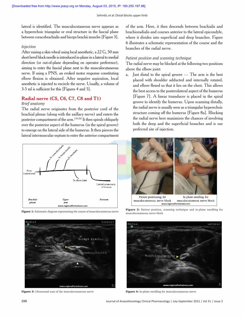
Musculocutaneous nerve (C5, C6, and C7)Brief anatomyThis nerve is a branch of the lateral cord of the brachial plexus, arising opposite the lower border of the pectoralis major.[18,19] After penetrating the coracobrachialis muscle, the nerve passes obliquely between the biceps brachii and the brachialis innervating all three muscles, and supplying the elbow joint. It then emerges on the lateral side of the arm, pierces the deep fascia lateral to the tendon of the biceps brachii, and continues into the forearm as the lateral cutaneous nerve of the forearm. AschematicdiagramisshowninFigure1.
Patient positionThe patient is positioned supine with the arm being blocked abducted away from the body at a right angle, and extended at the elbow. Alternatively, the abducted arm may be flexed attheelbow[Figure2].
ScanningA linear transducer is placed at the axilla to identify the axillary artery, above the teres major muscle. The coracobrachialis muscle lateral to the artery, and the biceps brachii further
[Downloaded free from http://www.joacp.org on Monday, August 03, 2015, IP: 180.250.197.98]
Sehmbi, et al.: Distal blocks upper limb
298 Journal of Anaesthesiology Clinical Pharmacology | July-September 2015 | Vol 31 | Issue 3
lateral is identified. The musculocutaneous nerve appears as a hyperechoic triangular or oval structure in the fascial plane betweencoracobrachialisandbicepsbrachiimuscles[Figure3].
InjectionAfterraisingaskinwhealusinglocalanesthetic,a22G,50mmshort bevel block needle is introduced in-plane in a lateral to medial direction (or out-of-plane depending on operator preference), aiming to enter the fascial plane next to the musculocutaneous nerve.IfusingaPNS,anevokedmotorresponseconstitutingelbow flexion is obtained. After negative aspiration, local anesthetic is injected to encircle the nerve. Usually, a volume of 3-5mlissufficientforthis[Figures4and5].
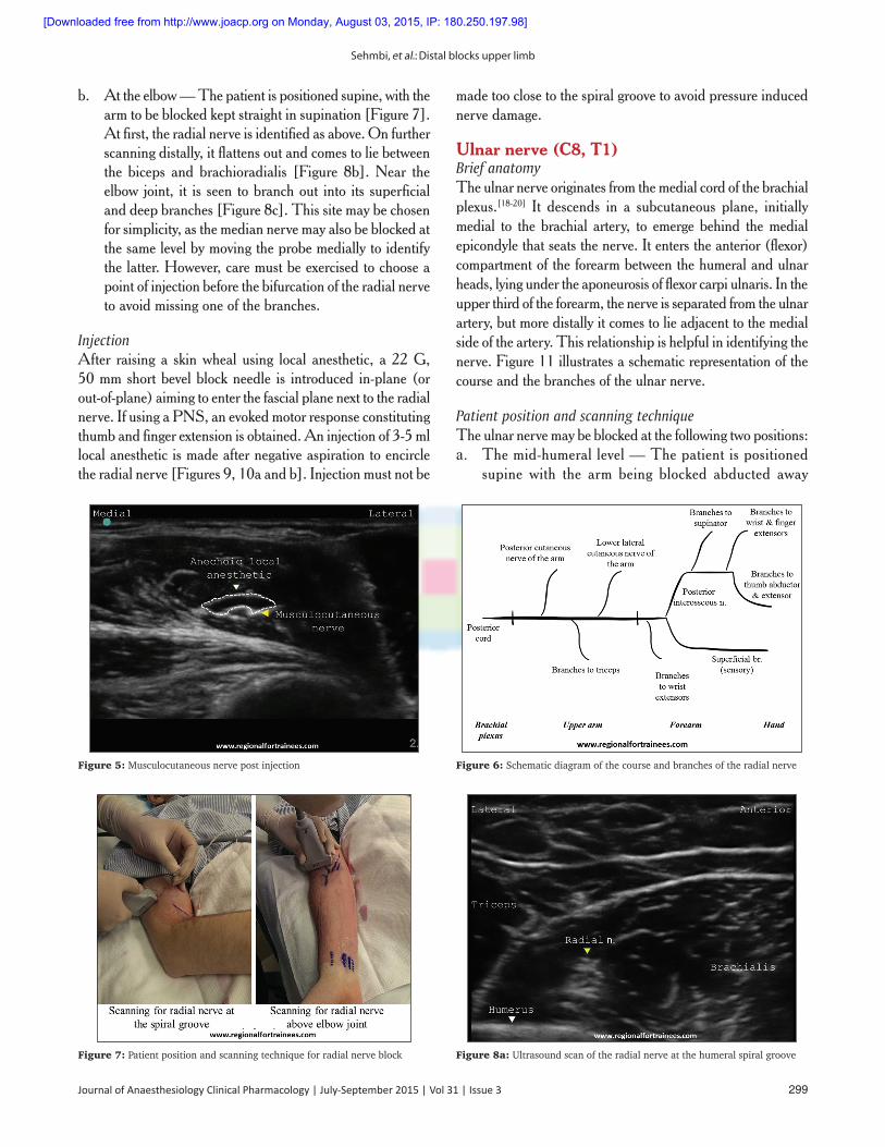
Radial nerve (C5, C6, C7, C8 and T1)Brief anatomyThe radial nerve originates from the posterior cord of the brachial plexus (along with the axillary nerve) and enters the posterior compartment of the arm.[18-20] It then spirals obliquely over the posterior aspect of the humerus (in the spiral groove) to emerge on the lateral side of the humerus. It then pierces the lateral intermuscular septum to enter the anterior compartment
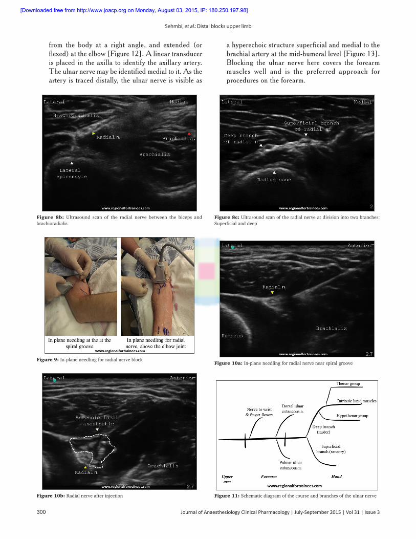
of the arm. Here, it then descends between brachialis and brachioradialis and courses anterior to the lateral epicondyle, whereitdividesintosuperficialanddeepbranches.Figure6 illustrates a schematic representation of the course and the branches of the radial nerve.
Patient position and scanning techniqueThe radial nerve may be blocked at the following two positions above the elbow joint:a. Just distal to the spiral groove — The arm is the best
placed with shoulder adducted and internally rotated, and elbow flexed so that it lies on the chest. This allows the best access to the posterolateral aspect of the humerus [Figure7].Alineartransducer isplacedinthespiralgroove to identify the humerus. Upon scanning distally, the radial nerve is usually seen as a triangular hyperechoic structurecomingoffthehumerus[Figure8a].Blockingthe radial nerve here maximizes the chances of involving both the deep and the superficial branches and is our preferred site of injection.
Figure 1: Schematic diagram representing the course of musculocutaneous nerveFigure 2: Patient position, scanning technique and in-plane needling for musculocutaneous nerve block
Figure 3: Ultrasound scan of the musculocutaneous nerve Figure 4: In-plane needling for musculocutaneous nerve
[Downloaded free from http://www.joacp.org on Monday, August 03, 2015, IP: 180.250.197.98]
Sehmbi, et al.: Distal blocks upper limb
Journal of Anaesthesiology Clinical Pharmacology | July-September 2015 | Vol 31 | Issue 3 299
b. At the elbow — The patient is positioned supine, with the armtobeblockedkeptstraightinsupination[Figure7].At first, the radial nerve is identified as above. On further scanning distally, it flattens out and comes to lie between the biceps and brachioradialis [Figure 8b].Near theelbow joint, it is seen to branch out into its superficial anddeepbranches[Figure8c].Thissitemaybechosenfor simplicity, as the median nerve may also be blocked at the same level by moving the probe medially to identify the latter. However, care must be exercised to choose a point of injection before the bifurcation of the radial nerve to avoid missing one of the branches.
InjectionAfter raisinga skinwhealusing local anesthetic, a22G,50 mm short bevel block needle is introduced in-plane (or out-of-plane) aiming to enter the fascial plane next to the radial nerve.IfusingaPNS,anevokedmotorresponseconstitutingthumb and finger extension is obtained. An injection of 3-5 ml local anesthetic is made after negative aspiration to encircle theradialnerve[Figures9,10aandb].Injectionmustnotbe
made too close to the spiral groove to avoid pressure induced nerve damage.
Ulnar nerve (C8, T1)Brief anatomyThe ulnar nerve originates from the medial cord of the brachial plexus.[18-20] It descends in a subcutaneous plane, initially medial to the brachial artery, to emerge behind the medial epicondyle that seats the nerve. It enters the anterior (flexor) compartment of the forearm between the humeral and ulnar heads, lying under the aponeurosis of flexor carpi ulnaris. In the upper third of the forearm, the nerve is separated from the ulnar artery, but more distally it comes to lie adjacent to the medial side of the artery. This relationship is helpful in identifying the nerve.Figure11illustratesaschematicrepresentationofthecourse and the branches of the ulnar nerve.
Patient position and scanning techniqueThe ulnar nerve may be blocked at the following two positions:a. The mid-humeral level — The patient is positioned
supine with the arm being blocked abducted away
Figure 5: Musculocutaneous nerve post injection Figure 6: Schematic diagram of the course and branches of the radial nerve
Figure 7: Patient position and scanning technique for radial nerve block Figure 8a: Ultrasound scan of the radial nerve at the humeral spiral groove
[Downloaded free from http://www.joacp.org on Monday, August 03, 2015, IP: 180.250.197.98]
Sehmbi, et al.: Distal blocks upper limb
300 Journal of Anaesthesiology Clinical Pharmacology | July-September 2015 | Vol 31 | Issue 3
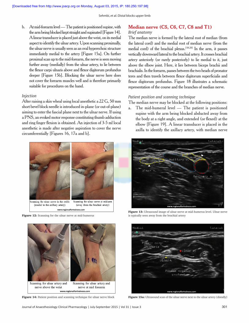
from the body at a right angle, and extended (or flexed)attheelbow[Figure12].Alineartransduceris placed in the axilla to identify the axillary artery. The ulnar nerve may be identified medial to it. As the artery is traced distally, the ulnar nerve is visible as
a hyperechoic structure superficial and medial to the brachialarteryatthemid-humerallevel[Figure13].Blocking the ulnar nerve here covers the forearmmuscles well and is the preferred approach for procedures on the forearm.
Figure 10a: In-plane needling for radial nerve near spiral groove
Figure 8b: Ultrasound scan of the radial nerve between the biceps and brachioradialis
Figure 8c: Ultrasound scan of the radial nerve at division into two branches: Superficial and deep
Figure 9: In-plane needling for radial nerve block
Figure 10b: Radial nerve after injection Figure 11: Schematic diagram of the course and branches of the ulnar nerve
[Downloaded free from http://www.joacp.org on Monday, August 03, 2015, IP: 180.250.197.98]
Sehmbi, et al.: Distal blocks upper limb
Journal of Anaesthesiology Clinical Pharmacology | July-September 2015 | Vol 31 | Issue 3 301
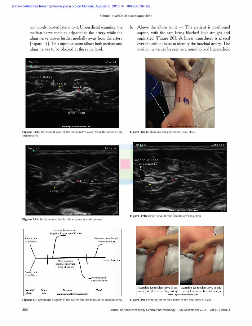
b. At mid-forearm level — The patient is positioned supine, with thearmbeingblockedkeptstraightandsupinated[Figure14].A linear transducer is placed just above the wrist, on its medial aspect to identify the ulnar artery. Upon scanning proximally, the ulnar nerve is usually seen as an oval hyperechoic structure immediatelymedialtotheartery[Figure15a].Onfurtherproximal scan up to the mid-forearm, the nerve is seen moving further away (medially) from the ulnar artery, to lie between the flexor carpi ulnaris above and flexor digitorum profundus deeper[Figure15b].Blocking theulnarnerveheredoesnot cover the forearm muscles well and is therefore primarily suitable for procedures on the hand.
InjectionAfterraisingaskinwhealusinglocalanesthetica22G,50mmshort bevel block needle is introduced in-plane (or out-of-plane) aiming to enter the fascial plane next to the ulnar nerve. If using aPNS,anevokedmotorresponseconstitutingthumbadductionand ring finger flexion is obtained. An injection of 3-5 ml local anesthetic is made after negative aspiration to cover the nerve circumferentially[Figures16,17aandb].
Median nerve (C5, C6, C7, C8 and T1)Brief anatomyThe median nerve is formed by the lateral root of median (from the lateral cord) and the medial root of median nerve (from the medial cord) of the brachial plexus.[18-20] In the arm, it passes vertically downward lateral to the brachial artery. It crosses brachial artery anteriorly (or rarely posteriorly) to lie medial to it, just above the elbow joint. Here, it lies between biceps brachii and brachialis. In the forearm, passes between the two heads of pronator teres and then travels between flexor digitorum superficialis and flexordigitorumprofundus.Figure18 illustrates a schematicrepresentation of the course and the branches of median nerve.
Patient position and scanning techniqueThe median nerve may be blocked at the following positions:a. The mid-humeral level — The patient is positioned
supine with the arm being blocked abducted away from the body at a right angle, and extended (or flexed) at the elbow[Figure19].Alineartransducerisplacedintheaxilla to identify the axillary artery, with median nerve
Figure 12: Scanning for the ulnar nerve at mid-humerusFigure 13: Ultrasound image of ulnar nerve at mid-humerus level. Ulnar nerve is typically seen away from the brachial artery
Figure 14: Patient position and scanning technique for ulnar nerve block Figure 15a: Ultrasound scan of the ulnar nerve next to the ulnar artery (distally)
[Downloaded free from http://www.joacp.org on Monday, August 03, 2015, IP: 180.250.197.98]
Sehmbi, et al.: Distal blocks upper limb
302 Journal of Anaesthesiology Clinical Pharmacology | July-September 2015 | Vol 31 | Issue 3
commonly located lateral to it. Upon distal scanning, the median nerve remains adjacent to the artery while the ulnar nerve moves further medially away from the artery [Figure13].Thisinjectionpointallowsbothmedianandulnar nerves to be blocked at the same level.
b. Above the elbow joint — The patient is positioned supine, with the arm being blocked kept straight and supinated [Figure 20].A linear transducer is placedover the cubital fossa to identify the brachial artery. The median nerve can be seen as a round to oval hyperechoic
Figure 17a: In-plane needling for ulnar nerve at mid-forearmFigure 17b: Ulnar nerve at mid-forearm after injection
Figure 18: Schematic diagram of the course and branches of the median nerve Figure 19: Scanning for median nerve at the mid-humerus level
Figure 15b: Ultrasound scan of the ulnar nerve away from the ulnar artery (proximally)
Figure 16: In-plane needling for ulnar nerve block
[Downloaded free from http://www.joacp.org on Monday, August 03, 2015, IP: 180.250.197.98]
Sehmbi, et al.: Distal blocks upper limb
Journal of Anaesthesiology Clinical Pharmacology | July-September 2015 | Vol 31 | Issue 3 303
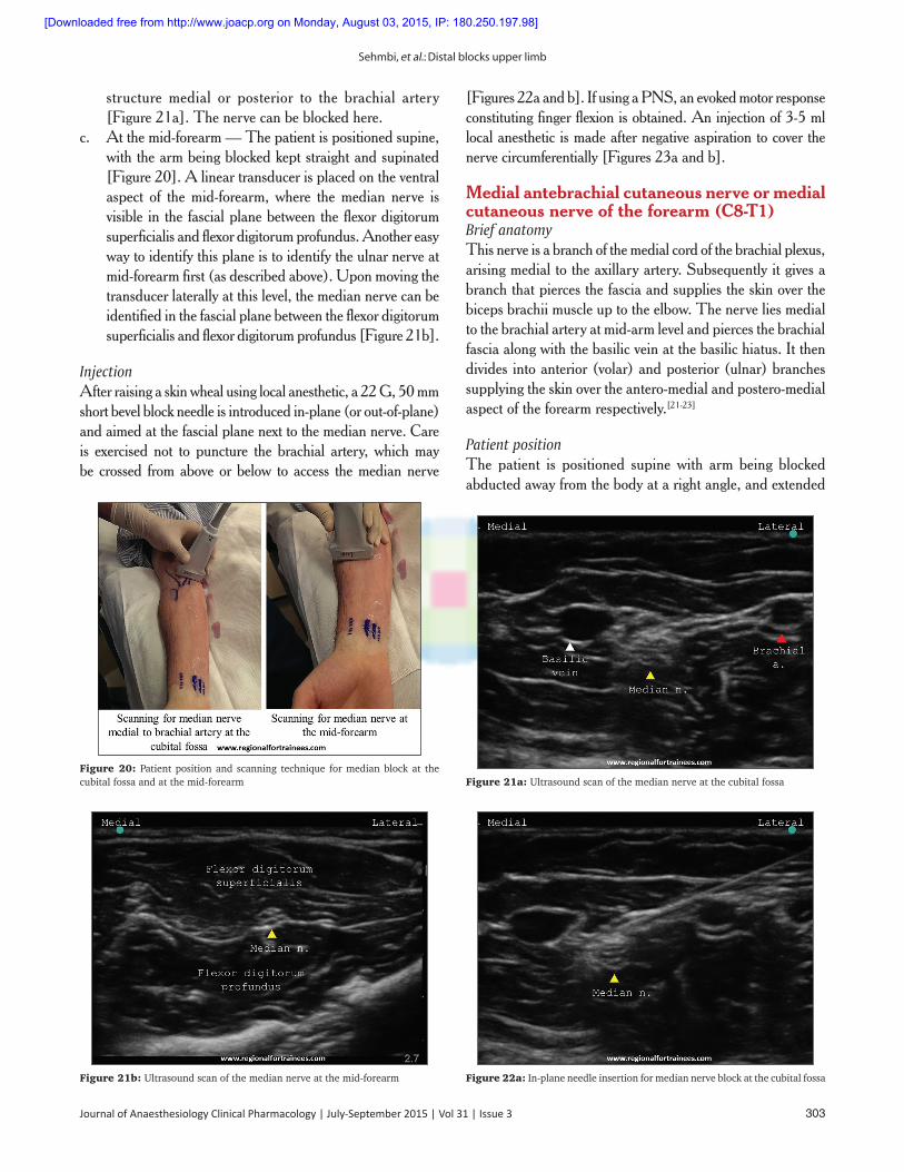
structure medial or posterior to the brachial artery [Figure21a].Thenervecanbeblockedhere.
c. At the mid-forearm — The patient is positioned supine, with the arm being blocked kept straight and supinated [Figure20].Alineartransducerisplacedontheventralaspect of the mid-forearm, where the median nerve is visible in the fascial plane between the flexor digitorum superficialis and flexor digitorum profundus. Another easy way to identify this plane is to identify the ulnar nerve at mid-forearm first (as described above). Upon moving the transducer laterally at this level, the median nerve can be identified in the fascial plane between the flexor digitorum superficialisandflexordigitorumprofundus[Figure21b].
InjectionAfterraisingaskinwhealusinglocalanesthetic,a22G,50mmshort bevel block needle is introduced in-plane (or out-of-plane) and aimed at the fascial plane next to the median nerve. Care is exercised not to puncture the brachial artery, which may be crossed from above or below to access the median nerve
[Figures22aandb].IfusingaPNS,anevokedmotorresponseconstituting finger flexion is obtained. An injection of 3-5 ml local anesthetic is made after negative aspiration to cover the nervecircumferentially[Figures23aandb].
Medial antebrachial cutaneous nerve or medial cutaneous nerve of the forearm (C8-T1)Brief anatomyThis nerve is a branch of the medial cord of the brachial plexus, arisingmedialtotheaxillaryartery.Subsequentlyitgivesabranch that pierces the fascia and supplies the skin over the biceps brachii muscle up to the elbow. The nerve lies medial to the brachial artery at mid-arm level and pierces the brachial fascia along with the basilic vein at the basilic hiatus. It then divides into anterior (volar) and posterior (ulnar) branches supplying the skin over the antero-medial and postero-medial aspect of the forearm respectively.[21-23]
Patient positionThe patient is positioned supine with arm being blocked abducted away from the body at a right angle, and extended
Figure 20: Patient position and scanning technique for median block at the cubital fossa and at the mid-forearm Figure 21a: Ultrasound scan of the median nerve at the cubital fossa
Figure 21b: Ultrasound scan of the median nerve at the mid-forearm Figure 22a: In-plane needle insertion for median nerve block at the cubital fossa
[Downloaded free from http://www.joacp.org on Monday, August 03, 2015, IP: 180.250.197.98]

Sehmbi, et al.: Distal blocks upper limb
304 Journal of Anaesthesiology Clinical Pharmacology | July-September 2015 | Vol 31 | Issue 3
at the elbow. Alternatively, the abducted arm may be flexed attheelbow[Figure24].
ScanningA linear transducer is placed at the mid-arm level (in short axis) to identify the biceps brachii muscle, brachial artery, median nerve
(medial to the brachial a.), ulnar nerves (subcutaneous medially) and basilic vein (in the most superficial subcutaneous plane). The medial antebrachial cutaneous nerve (MACN) can be visualized as a hyperechoic oval immediately lateral to the basilic vein[Figure25a].Uponadistalscan,itcanbeseendividingintoitstwobranches,anteriorandposterior[Figure25b].While
Figure 22b: Ultrasound image of the median nerve post injectionFigure 23a: In-plane needle insertion for median nerve block at mid-forearm
Figure 23b: Ultrasound image of the median nerve at mid-forearm postinjection
Figure 24: Patient position for medial antebrachial cutaneous nerve, lateral antebrachial cutaneous nerve and posterior antebrachial cutaneous nerve
Figure 25a: Ultrasound scan of the medial antebrachial cutaneous nerve at the mid-arm level before its division
Figure 25b: Ultrasound scan of the medial antebrachial cutaneous nerve (MACN) at the mid-arm level after its division (MACN (A) = MACN anterior branch, MACN (P) = MACN posterior branch)
[Downloaded free from http://www.joacp.org on Monday, August 03, 2015, IP: 180.250.197.98]
Sehmbi, et al.: Distal blocks upper limb
Journal of Anaesthesiology Clinical Pharmacology | July-September 2015 | Vol 31 | Issue 3 305
the anterior branch moves laterally, the posterior branch passes under the vein to lie on its ulnar aspect.[24,25]
InjectionA25-27Ghypodermicneedleisintroducedin-planeinalateral to medial direction aiming to enter the fascial plane next to the basilic vein. After negative aspiration, 2-3 ml local anesthetic is injected to fill the fascial plane containing the MACN.
Lateral antebrachial cutaneous nerve or lateral cutaneous nerve of the forearm (C5, C6, C7)Brief anatomyIn the arm, the musculocutaneous nerve pierces the coracobrachialis to descend in the plane between the brachialis and the biceps brachii muscles. After providing motor branches to these muscles, the nerve emerges lateral to the tendon of the biceps brachii muscle at the elbow, pierces the deep fascia distal to the interepicondylar line and continues as the lateral antebrachialcutaneousnerve(LACN).Itusuallyliesmedialto the cephalic vein and may divide into anterior (volar) and posterior (ulnar) branches supplying the skin over the antero-lateral and postero-lateral aspect of the forearm respectively.[26,27]
Patient positionThe patient is positioned supine with arm being blocked abducted away from the body at a right angle, and extended attheelbow[Figure24].
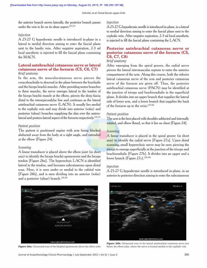
ScanningA linear transducer is placed above the elbow joint (in short axis) to identify the biceps brachii aponeurosis and the biceps tendon[Figure26a].ThehyperechoicLACNisidentifiedlateral to the tendon, and becomes subcutaneous upon distal scan. Here, it is seen under or medial to the cubital vein [Figure26b],andisseendividingintoananterior(volar)and a posterior (ulnar) branch.[28,29]
InjectionA25-27Ghypodermicneedleisintroducedin-plane,inalateralto medial direction aiming to enter the fascial plane next to the cephalic vein. After negative aspiration, 2-3 ml local anesthetic isinjectedtofillthefascialplanecontainingtheLACN.
Posterior antebrachial cutaneous nerve or posterior cutaneous nerve of the forearm (C5, C6, C7, C8)Brief anatomyAfter emerging from the spiral groove, the radial nerve pierces the lateral intermuscular septum to enter the anterior compartment of the arm. Along this course, both the inferior lateral cutaneous nerve of the arm and posterior cutaneous nerve of the forearm are given off. Thus, the posterior antebrachial cutaneous nerve (PACN) may be identified at the junction of triceps and brachioradialis in the superficial plane. It divides into an upper branch that supplies the lateral side of lower arm, and a lower branch that supplies the back of the forearm up to the wrist.[18,30]
Patient positionThe arm is the best placed with shoulder adducted and internally rotated,andelbowflexed,sothatitliesonchest[Figure24].
ScanningA linear transducer is placed in the spiral groove (in short axis)toidentifytheradialnerve[Figure27a].Upondistalscanning, small hyperechoic nerve may be seen piercing the triceps to emerge superficially at the junction of the triceps and brachioradialis[Figure27b].Itdividesintoanupperandalowerbranch[Figure27c].[28,30]
InjectionA25-27Ghypodermicneedleisintroducedin-plane,inananterior to posterior direction aiming to enter the subcutaneous
Figure 26a: Ultrasound scan of the bicipital aponeurosis above the elbow jointFigure 26b: Ultrasound scan of the lateral antebrachial cutaneous nerve just below the elbow joint, where the nerve is located medial to the cephalic vein
[Downloaded free from http://www.joacp.org on Monday, August 03, 2015, IP: 180.250.197.98]

Sehmbi, et al.: Distal blocks upper limb
306 Journal of Anaesthesiology Clinical Pharmacology | July-September 2015 | Vol 31 | Issue 3
plane containing the nerve. After negative aspiration, 2-3 ml local anesthetic is injected to fill the fascial plane containing the PACN.
Conclusion
The distal peripheral nerve blocks are relatively easy to perform using ultrasound guidance. These blocks are very useful as rescue blocks in the event of an incomplete anesthesia following brachial plexus blocks, or as standalone anesthetic oranalgesictechniques.Goodanatomicalknowledgeoftheircourse and branches helps in their identification and selection for appropriate surgical procedures.
Acknowledgments
All images in this manuscript have been taken from www.regionalfortrainees.com (courtesy of www.regionalfortrainees.com).
References
1. Maga JM, Cooper L, Gebhard RE. Outpatient regional anesthesia for upper extremity surgery update (2005 to present) distal to shoulder. Int Anesthesiol Clin 2012;50:47-55.
2. Lin E, Choi J, Hadzic A. Peripheral nerve blocks for outpatient surgery: Evidence-based indications. Curr Opin Anaesthesiol 2013;26:467-74.
3. Tran DQ, Pham K, Dugani S, Finlayson RJ. A prospective, randomized comparison between double-, triple-, and quadruple-injection ultrasound-guided axillary brachial plexus block. Reg Anesth Pain Med 2012;37:248-53.
4. Tsui BC, Doyle K, Chu K, Pillay J, Dillane D. Case series: Ultrasound-guided supraclavicular block using a curvilinear probe in 104 day-case hand surgery patients. Can J Anaesth 2009;56:46-51.
5. Chin KJ, Singh M, Velayutham V, Chee V. Infraclavicular brachial plexus block for regional anaesthesia of the lower arm. Anesth Analg 2010;111:1072.
6. Handoll HH, Koscielniak-Nielsen ZJ. Single, double or multiple injection techniques for axillary brachial plexus block for hand, wrist or forearm surgery. Cochrane Database Syst Rev 2006 Jan 25;(1):CD003842.
7. McCartney CJ, Xu D, Constantinescu C, Abbas S, Chan VW. Ultrasound examination of peripheral nerves in the forearm. Reg Anesth Pain Med 2007;32:434-9.
8. Foxall GL, Skinner D, Hardman JG, Bedforth NM. Ultrasound anatomy of the radial nerve in the distal upper arm. Reg Anesth Pain Med 2007;32:217-20.
9. Kathirgamanathan A, French J, Foxall GL, Hardman JG, Bedforth NM. Delineation of distal ulnar nerve anatomy using ultrasound in volunteers to identify an optimum approach for neural blockade. Eur J Anaesthesiol 2009;26:43-6.
10. Fredrickson MJ, Ting FS, Chinchanwala S, Boland MR. Concomitant infraclavicular plus distal median, radial, and ulnar nerve blockade accelerates upper extremity anaesthesia and improves block consistency compared with infraclavicular block alone. Br J Anaesth 2011;107:236-42.
11. Fredrickson MJ, Price DJ. Analgesic effectiveness of ropivacaine 0.2% vs 0.4% via an ultrasound-guided C5-6 root/superior trunk perineural ambulatory catheter. Br J Anaesth 2009;103:434-9.
12. Lam NC, Charles M, Mercer D, Soneru C, Dillow J, Jaime F, et al. A triple-masked, randomized controlled trial comparing ultrasound-guided brachial plexus and distal peripheral nerve block anesthesia for outpatient hand surgery. Anesthesiol Res Pract 2014;2014:324083.
13. Smith BE, Challands JF, Suchak M, Siggins D. Regional anaesthesia for surgery of the forearm and hand. A technique of combined supraclavicular and discrete blocks. Anaesthesia 1989;44:747-9.
Figure 27c: Ultrasound scan of the branches of posterior antebrachial cutaneous nerve (PACN) (PACN (U) = PACN upper branch, PACN (L) = PACN lower branch). Inferior lateral brachial cutaneous nerve (ILBCN) can also be seen
Figure 27a: Ultrasound scan of the radial nerve at the spiral groove
Figure 27b: Ultrasound scan of the posterior antebrachial cutaneous nerve just below the spiral groove
[Downloaded free from http://www.joacp.org on Monday, August 03, 2015, IP: 180.250.197.98]

Sehmbi, et al.: Distal blocks upper limb
Journal of Anaesthesiology Clinical Pharmacology | July-September 2015 | Vol 31 | Issue 3 307
14. Tsui BC, Pillay JJ, Chu KT, Dillane D. Electrical impedance to distinguish intraneural from extraneural needle placement in porcine nerves during direct exposure and ultrasound guidance. Anesthesiology 2008;109:479-83.
15. Dillane D, Tsui BC. Is there still a place for the use of nerve stimulation? Paediatr Anaesth 2012;22:102-8.
16. Gadsden J, McCally C, Hadzic A. Monitoring during peripheral nerve blockade. Curr Opin Anaesthesiol 2010;23:656-61.
17. Gadsden JC, Choi JJ, Lin E, Robinson A. Opening injection pressure consistently detects needle-nerve contact during ultrasound-guided interscalene brachial plexus block. Anesthesiology 2014;120:1246-53.
18. Stranding S, editor. Upper arm. Gray’s Anatomy: The Anatomical Basis of Clinical Practice. 40th ed. London: Churchill Livingstone; 2008. p. 823-30.
19. Stranding S, editor. Forearm. Gray’s Anatomy: The Anatomical Basis of Clinical Practice. 40th ed. London: Churchill Livingstone; 2008. p. 839-56.
20. Stranding S, editor. Wrist and hand. Gray’s Anatomy: The Anatomical Basis of Clinical Practice. 40th ed. London: Churchill Livingstone; 2008. p. 857-98.
21. Viscomi CM, Reese J, Rathmell JP. Medial and lateral antebrachial cutaneous nerve blocks: An easily learned regional anesthetic for forearm arteriovenous fistula surgery. Reg Anesth 1996;21:2-5.
22. Masear VR, Meyer RD, Pichora DR. Surgical anatomy of the medial antebrachial cutaneous nerve. J Hand Surg Am 1989;14:267-71.
23. Thallaj A, Marhofer P, Kettner SC, Al-Majed M, Al-Ahaideb A, Moriggl B. High-resolution ultrasound accurately identifies the medial antebrachial cutaneous nerve at the midarm level: A clinical anatomic study. Reg Anesth Pain Med 2011;36:499-501.
24. Moritz T, Prosch H, Pivec CH, Sachs A, Pretterklieber ML, Kriechbaumer L, et al. High-resolution ultrasound visualization of the subcutaneous nerves of the forearm: A feasibility study in anatomic specimens. Muscle Nerve 2014;49:676-9.
25. Thallaj A. Ultrasound guidance of uncommon nerve blocks. Saudi J Anaesth 2011;5:392-4.
26. Beldner S, Zlotolow DA, Melone CP Jr, Agnes AM, Jones MH. Anatomy of the lateral antebrachial cutaneous and superficial radial nerves in the forearm: A cadaveric and clinical study. J Hand Surg Am 2005;30:1226-30.
27. Wongkerdsook W, Agthong S, Amarase C, Yotnuengnit P, Huanmanop T, Chentanez V. Anatomy of the lateral antebrachial cutaneous nerve in relation to the lateral epicondyle and cephalic vein. Clin Anat 2011;24:56-61.
28. Blanco R, Gómez BM, González JM. Ultrasound appearance of the cutaneous nerves of the upper limb: A novel description in pain management. J Pain Relief 2012;1:e109.
29. Chiavaras MM, Jacobson JA, Billone L, Lawton JM, Lawton J. Sonography of the lateral antebrachial cutaneous nerve with magnetic resonance imaging and anatomic correlation. J Ultrasound Med 2014;33:1475-83.
30. Egeler C. Cutaneous Nerves of the Arm and Forearm-How Small Can We Go? Abstracts and Highlight Papers of the 32nd Annual European Society of Regional Anaesthesia and Pain Therapy (ESRA) Congress 2013: Invited Speaker Highlight Papers; 2013. p. E1-259.
How to cite this article: Sehmbi H, Madjdpour C, Shah UJ, Chin KJ. Ultrasound guided distal peripheral nerve block of the upper limb: A technical review. J Anaesthesiol Clin Pharmacol 2015;31:296-307.Source of Support: Nil, Conflicts of Interest: None declared.
Name of conference Dates Venue Name of organising secretary with contact details25th National Conference of Research Society of Anaesthesiology Clinical Pharmacology RSACPCON 2015
October 2nd, 3rd & 4th, 2015
SGRD Amritsar Organising SecretaryDr. Ruchi GuptaTelephone: +91 9814320805Email Id: [email protected]: http://www.rsacpcon2015.com/
16th North Zone Annual Conference of Indian Society of Anaesthesiologists 2015 NZ-ISACON 2015
October 16-18th, 2015
Department of Anaesthesiology, Dr.Rajendra Prasad Govt. Medical College Kangra at Tanda (HP)
Dr. Sudarshan Kumar (Organizing Secretary), Deptt. of Anaesthesia, Dr. RPGMC Kangra, TandaPhone No. +91 9418086604Email: [email protected]: www.nzisacon2015.com
3rd Annual Conference of the Ophthalmic Forum of Indian Society of Anaesthesiologists OFISACON 2015
October 17-18, October 2015
JLN Auditorium, AIIMS, New Delhi
Prof. Dilip ShendeTelephone: +919868397820Email Id: [email protected]: www.3rdofisacon2015.in
Conference Calendar 2015
[Downloaded free from http://www.joacp.org on Monday, August 03, 2015, IP: 180.250.197.98]