uk standards for microbiology investigations · the genus shigella belongs to the family...
TRANSCRIPT
Issued by the Standards Unit, Microbiology Services, PHE Bacteriology – Identification | ID 20 | Issue no: 3 | Issue date: 15.04.15 | Page: 1 of 22
© Crown copyright 2015
UK Standards for Microbiology Investigations
Identification of Shigella species

Identification of Shigella species
Bacteriology – Identification | ID 20 | Issue no: 3 | Issue date: 15.04.15 | Page: 2 of 22 UK Standards for Microbiology Investigations | Issued by the Standards Unit, Public Health England
Acknowledgments UK Standards for Microbiology Investigations (SMIs) are developed under the auspices of Public Health England (PHE) working in partnership with the National Health Service (NHS), Public Health Wales and with the professional organisations whose logos are displayed below and listed on the website https://www.gov.uk/uk-standards-for-microbiology-investigations-smi-quality-and-consistency-in-clinical-laboratories. SMIs are developed, reviewed and revised by various working groups which are overseen by a steering committee (see https://www.gov.uk/government/groups/standards-for-microbiology-investigations-steering-committee). The contributions of many individuals in clinical, specialist and reference laboratories who have provided information and comments during the development of this document are acknowledged. We are grateful to the Medical Editors for editing the medical content. For further information please contact us at: Standards Unit Microbiology Services Public Health England 61 Colindale Avenue London NW9 5EQ E-mail: [email protected] Website: https://www.gov.uk/uk-standards-for-microbiology-investigations-smi-quality-and-consistency-in-clinical-laboratories PHE Publications gateway number: 2015013 UK Standards for Microbiology Investigations are produced in association with:
Logos correct at time of publishing.

Identification of Shigella species
Bacteriology – Identification | ID 20 | Issue no: 3 | Issue date: 15.04.15 | Page: 3 of 22 UK Standards for Microbiology Investigations | Issued by the Standards Unit, Public Health England
Contents ACKNOWLEDGMENTS .......................................................................................................... 2
AMENDMENT TABLE ............................................................................................................. 4
UK STANDARDS FOR MICROBIOLOGY INVESTIGATIONS: SCOPE AND PURPOSE ....... 6
SCOPE OF DOCUMENT ......................................................................................................... 9
INTRODUCTION ..................................................................................................................... 9
TECHNICAL INFORMATION/LIMITATIONS ......................................................................... 10
1 SAFETY CONSIDERATIONS .................................................................................... 12
2 TARGET ORGANISMS .............................................................................................. 12
3 IDENTIFICATION ....................................................................................................... 12
4 IDENTIFICATION OF SHIGELLA SPECIES .............................................................. 17
5 REPORTING .............................................................................................................. 18
6 REFERRALS .............................................................................................................. 18
7 NOTIFICATION TO PHE OR EQUIVALENT IN THE DEVOLVED ADMINISTRATIONS .................................................................................................. 19
REFERENCES ...................................................................................................................... 20

Identification of Shigella species
Bacteriology – Identification | ID 20 | Issue no: 3 | Issue date: 15.04.15 | Page: 4 of 22 UK Standards for Microbiology Investigations | Issued by the Standards Unit, Public Health England
Amendment Table Each SMI method has an individual record of amendments. The current amendments are listed on this page. The amendment history is available from [email protected]. New or revised documents should be controlled within the laboratory in accordance with the local quality management system.
Amendment No/Date. 5/15.04.15
Issue no. discarded. 2.2
Insert Issue no. 3
Section(s) involved Amendment
Whole document. Hyperlinks updated to gov.uk.
Page 2. Updated logos added.
Introduction.
The taxonomy of Shigella species has been updated. More information has been added to the Characteristics section. The medically important species of Shigella are mentioned. Section on Principles of Identification has been updated to include the MALDI-TOF.
Technical Information/Limitations.
Addition of information regarding serotyping, oxidase test, quality control and MALDI-TOF MS.
Safety considerations. This section has been updated on the handling of Shigella species as well as laboratory acquired infections.
Target Organisms. The section on the Target organisms has been updated and presented clearly.
Identification.
Updates have been done on 3.2, 3.3 and 3.4 to reflect standards in practice. Section 3.4.3 and 3.4.4 has been updated to include MALDI-TOF MS and NAATs with references. Subsection 3.5 has been updated to include the Rapid Molecular Methods.
Identification Flowchart. Modification of flowchart for identification of Shigella species has been done for easy guidance.
Identification of Shigella species
Bacteriology – Identification | ID 20 | Issue no: 3 | Issue date: 15.04.15 | Page: 5 of 22 UK Standards for Microbiology Investigations | Issued by the Standards Unit, Public Health England
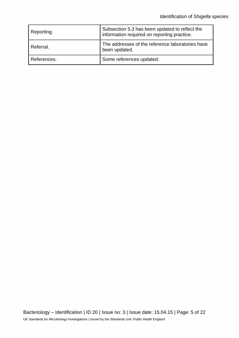
Reporting. Subsection 5.3 has been updated to reflect the information required on reporting practice.
Referral. The addresses of the reference laboratories have been updated.
References. Some references updated.
Identification of Shigella species
Bacteriology – Identification | ID 20 | Issue no: 3 | Issue date: 15.04.15 | Page: 6 of 22 UK Standards for Microbiology Investigations | Issued by the Standards Unit, Public Health England
UK Standards for Microbiology Investigations#: Scope and Purpose Users of SMIs
• SMIs are primarily intended as a general resource for practising professionals operating in the field of laboratory medicine and infection specialties in the UK
• SMIs provide clinicians with information about the available test repertoire and the standard of laboratory services they should expect for the investigation of infection in their patients, as well as providing information that aids the electronic ordering of appropriate tests
• SMIs provide commissioners of healthcare services with the appropriateness and standard of microbiology investigations they should be seeking as part of the clinical and public health care package for their population
Background to SMIs SMIs comprise a collection of recommended algorithms and procedures covering all stages of the investigative process in microbiology from the pre-analytical (clinical syndrome) stage to the analytical (laboratory testing) and post analytical (result interpretation and reporting) stages. Syndromic algorithms are supported by more detailed documents containing advice on the investigation of specific diseases and infections. Guidance notes cover the clinical background, differential diagnosis, and appropriate investigation of particular clinical conditions. Quality guidance notes describe laboratory processes which underpin quality, for example assay validation. Standardisation of the diagnostic process through the application of SMIs helps to assure the equivalence of investigation strategies in different laboratories across the UK and is essential for public health surveillance, research and development activities.
Equal Partnership Working SMIs are developed in equal partnership with PHE, NHS, Royal College of Pathologists and professional societies. The list of participating societies may be found at https://www.gov.uk/uk-standards-for-microbiology-investigations-smi-quality-and-consistency-in-clinical-laboratories. Inclusion of a logo in an SMI indicates participation of the society in equal partnership and support for the objectives and process of preparing SMIs. Nominees of professional societies are members of the Steering Committee and Working Groups which develop SMIs. The views of nominees cannot be rigorously representative of the members of their nominating organisations nor the corporate views of their organisations. Nominees act as a conduit for two way reporting and dialogue. Representative views are sought through the consultation process. SMIs are developed, reviewed and updated through a wide consultation process.
#Microbiology is used as a generic term to include the two GMC-recognised specialties of Medical Microbiology (which includes Bacteriology, Mycology and Parasitology) and Medical Virology.
Identification of Shigella species
Bacteriology – Identification | ID 20 | Issue no: 3 | Issue date: 15.04.15 | Page: 7 of 22 UK Standards for Microbiology Investigations | Issued by the Standards Unit, Public Health England
Quality Assurance NICE has accredited the process used by the SMI Working Groups to produce SMIs. The accreditation is applicable to all guidance produced since October 2009. The process for the development of SMIs is certified to ISO 9001:2008. SMIs represent a good standard of practice to which all clinical and public health microbiology laboratories in the UK are expected to work. SMIs are NICE accredited and represent neither minimum standards of practice nor the highest level of complex laboratory investigation possible. In using SMIs, laboratories should take account of local requirements and undertake additional investigations where appropriate. SMIs help laboratories to meet accreditation requirements by promoting high quality practices which are auditable. SMIs also provide a reference point for method development. The performance of SMIs depends on competent staff and appropriate quality reagents and equipment. Laboratories should ensure that all commercial and in-house tests have been validated and shown to be fit for purpose. Laboratories should participate in external quality assessment schemes and undertake relevant internal quality control procedures.
Patient and Public Involvement The SMI Working Groups are committed to patient and public involvement in the development of SMIs. By involving the public, health professionals, scientists and voluntary organisations the resulting SMI will be robust and meet the needs of the user. An opportunity is given to members of the public to contribute to consultations through our open access website.
Information Governance and Equality PHE is a Caldicott compliant organisation. It seeks to take every possible precaution to prevent unauthorised disclosure of patient details and to ensure that patient-related records are kept under secure conditions. The development of SMIs are subject to PHE Equality objectives https://www.gov.uk/government/organisations/public-health-england/about/equality-and-diversity. The SMI Working Groups are committed to achieving the equality objectives by effective consultation with members of the public, partners, stakeholders and specialist interest groups.
Legal Statement Whilst every care has been taken in the preparation of SMIs, PHE and any supporting organisation, shall, to the greatest extent possible under any applicable law, exclude liability for all losses, costs, claims, damages or expenses arising out of or connected with the use of an SMI or any information contained therein. If alterations are made to an SMI, it must be made clear where and by whom such changes have been made. The evidence base and microbial taxonomy for the SMI is as complete as possible at the time of issue. Any omissions and new material will be considered at the next review. These standards can only be superseded by revisions of the standard, legislative action, or by NICE accredited guidance. SMIs are Crown copyright which should be acknowledged where appropriate.
Identification of Shigella species
Bacteriology – Identification | ID 20 | Issue no: 3 | Issue date: 15.04.15 | Page: 8 of 22 UK Standards for Microbiology Investigations | Issued by the Standards Unit, Public Health England
Suggested Citation for this Document Public Health England. (2015). Identification of Shigella species. UK Standards for Microbiology Investigations. ID 20 Issue 3. https://www.gov.uk/uk-standards-for-microbiology-investigations-smi-quality-and-consistency-in-clinical-laboratories
Identification of Shigella species
Bacteriology – Identification | ID 20 | Issue no: 3 | Issue date: 15.04.15 | Page: 9 of 22 UK Standards for Microbiology Investigations | Issued by the Standards Unit, Public Health England
Scope of Document This SMI describes the identification of Shigella species with particular reference to isolation from faeces. This SMI should be used in conjunction with other SMIs.
Introduction Taxonomy The genus Shigella belongs to the family Enterobacteriaceae and consists of four species; Shigella dysenteriae, Shigella flexneri, Shigella boydii, and Shigella sonnei1. Each of the species, with the exception of S. sonnei, is subdivided by serotype.
Characteristics Shigella species are small Gram negative rods, 0.3 - 1µm in diameter and 1 - 6μm in length, appearing singly, in pairs and in chains. Shigella species are facultative anaerobes and are non-spore formers. Unlike Salmonella, Shigella species do not possess flagella and hence are non-motile. Shigellae are differentiated into four subgroups on the basis of their O (somatic) antigens and further differentiated into serotypes; - S. dysenteriae (Group A) contains 15 distinct antigenic serotypes. - S. flexneri (Group B) contains 6 serotypes (1-6) that can be further divided into sub-serotypes based on their possession of group factors designated 3,4; 4; 6; 7; and 7,8 - S. boydii (Group C) contains 20 distinct antigenic serotypes. - S. sonnei (Group D) contains only 1 serotype that may occur in two forms, form I (smooth) and form II (rough). On XLD agar, Shigella species produce 1-2mm diameter pink red colonies with no black centre. Some strains may have a pink or yellow periphery on XLD agar. Colonies on DCA agar are colourless (S. sonnei may form pale pink colonies because of late lactose fermentation). They are non- lactose fermenters (however S. sonnei can ferment lactose after prolonged incubation) and do not produce gas from carbohydrates. They all ferment glucose. They also tend to be overall biochemically inert. They are negative for urease, oxidase, do not decarboxylate lysine and give variable results for indole (with the exception of S. sonnei that is always indole negative) and positive for catalase with the exception of S. dysenteriae Type 12,3. Differentiation of Shigella strains from E. coli is one of the problems faced by diagnostic microbiology laboratories and may reflect the fact that E. coli and all four Shigella species are very closely related on the basis of the DNA-DNA relationship. However, biochemical reactions are of considerable value in the differentiation of members of the genus Shigella from Escherichia3. The Shigella species may be further differentiated by biochemical tests and serology of their lipopolysaccharides3. The majority of Shigella species, except S. flexneri 6, and S. boydii 13 and 14, ferment sugars without gas production. S. boydii, S. flexneri and S. sonnei, with a few exceptions, ferment mannitol; S. dysenteriae does not. S. sonnei, and S. dysenteriae type 1 are the only species that are ONPG positive. S. boydii 13 are ornithine positive, and some may be ONPG positive.

Identification of Shigella species
Bacteriology – Identification | ID 20 | Issue no: 3 | Issue date: 15.04.15 | Page: 10 of 22 UK Standards for Microbiology Investigations | Issued by the Standards Unit, Public Health England
They have been isolated from faeces and rarely in blood samples4. The type species is Shigella dysenteriae.
Principles of Identification Isolates from primary culture are identified by colonial appearance, biochemical tests and serology (agglutination with specific antisera). Plesiomonas shigelloides cross reacts with S. sonnei antisera. If confirmation of identification is required, isolates should be sent to the Reference Laboratory. All identification tests should ideally be performed from non-selective agar. Full molecular identification using for example, MALDI-TOF MS can be used to identify Shigella isolates to species level. Typing and differentiation between strains of Shigella species can be achieved using a range of molecular techniques eg Real-time Polymerase Chain Reaction (PCR), Multilocus Sequence Typing, Pulsed Field Gel Electrophoresis (PFGE) and Whole Generation sequencing. For more information, see section 3.5 on further identification.
Technical Information/Limitations Quality control Each new lot or shipment of antisera/commercial identification systems should be tested and validated for positive and negative reactivity using known control strains; ensuring it is fit for purpose. Laboratories should follow manufacturer’s instructions when using these products. Serotyping Rough strains: Serotyping should not be attempted on Shigella strains which autoagglutinate saline. Rough isolates seldom revert to smooth forms. However, if autoagglutination in saline is observed, an attempt to recover smooth colonies may be made by performing 2- 4 serial sub-cultures on enriched media, such as blood agar3. Capsular antigens: Occasionally, the presence of capsular antigens may prevent some isolates of Shigella species from reacting with polyvalent antisera. The presence of capsular antigens should be considered when isolates which are biochemically typical of Shigella sp., fail to agglutinate (or agglutinate poorly) with Shigella polyvalent antisera. In these situations, an attempt should be made to remove the capsular antigen (by boiling at 100°C for 45 minutes) and then repeating the procedure. Shigella sonnei polyvalent antisera can produce cross-reactions with some cultures of Shigella boydii type 6 due to the presence of conserved antigens. In these situations, biochemical profile and the use of Shigella boydii type 6 monovalent antiserum will be necessary to confirm the identification3. Some Plesiomonas shigelloides strains share antigens with Shigella sonnei, and cross-reactions with Shigella antisera occur5.

Identification of Shigella species
Bacteriology – Identification | ID 20 | Issue no: 3 | Issue date: 15.04.15 | Page: 11 of 22 UK Standards for Microbiology Investigations | Issued by the Standards Unit, Public Health England
Oxidase Test The test should not be performed on cultures from media containing tellurite and fermentable carbohydrates as these may prevent the reaction from occurring6. Organisms grown on media containing dyes may give aberrant results7. MALDI-TOF MS One of the limitations of MALDI-TOF MS is its current inability to reliably discriminate pathogenic E. coli from Shigella species because of the close genetic relatedness of the organisms which is challenging8.

Identification of Shigella species
Bacteriology – Identification | ID 20 | Issue no: 3 | Issue date: 15.04.15 | Page: 12 of 22 UK Standards for Microbiology Investigations | Issued by the Standards Unit, Public Health England
1 Safety Considerations9-25 All Shigella dysenteriae Type 1 are Hazard Group 3 organisms and suspected isolates must be handled in a containment level 3 room. Most Shigella species are in Hazard Group 2. An important exception is Shigella dysenteriae Type 1. All work on Shigella dysenteriae type 1 must be performed under Containment level 3 conditions. Shigella species are highly infective, particularly S. dysenteriae considered the most virulent, and can produce a potent cytotoxin known as “Shigatoxin”15,16. Shigella dysenteriae type 1 causes severe and sometimes fatal disease. Shigella species have been recently identified to be the most frequently identified agent of laboratory-acquired infections because of their high virulence and low infectious dose26. A large number of laboratory acquired infections have been reported. Infection may be acquired through ingestion or accidental parenteral inoculation27,28. Refer to current guidance on the safe handling of all Hazard Group 2 organisms documented in this SMI. Appropriate personal protective equipment (PPE) and techniques designed to minimise exposure of the laboratory workers should be worn and adhered to at all times. The most effective method for preventing laboratory-acquired infections is the adoption of safe working practices. Laboratory procedures that give rise to infectious aerosols must be conducted in a microbiological safety cabinet. The above guidance should be supplemented with local COSHH and risk assessments. Compliance with postal and transport regulations is essential.
2 Target Organisms Shigella species reported to have caused human infections4,29 All species cause human infections. Shigella dysenteriae (15 serotypes), Shigella boydii (20 serotypes), Shigella flexneri (6 serotypes which can be sub-divided into sub-serotypes), Shigella sonnei (1 serotype, 2 variants - rough and smooth).
3 Identification 3.1 Microscopic Appearance Gram negative rods, 0.3 - 1µm in diameter and 1 - 6µm in length, arranged singly, in pairs and in chains.
3.2 Primary Isolation Media Xylose-lysine-desoxycholate agar (XLD) agar incubated in air at 35-37°C for 18 - 24hr. Desoxycholate citrate agar (DCA) agar incubated in air at 35-37°C for 18 - 24hr.
Identification of Shigella species
Bacteriology – Identification | ID 20 | Issue no: 3 | Issue date: 15.04.15 | Page: 13 of 22 UK Standards for Microbiology Investigations | Issued by the Standards Unit, Public Health England
3.3 Colonial Appearance XLD – Colonies are pink red, 1- 2mm diameter, and with no black centre. Some strains may have a pink or yellow periphery on XLD agar. DCA - Colonies are colourless (S. sonnei may form pale pink colonies because of late lactose fermentation).
3.4 Test Procedures
3.4.1 Serotyping Serotyping is a subtyping method based on the immuno-reactivity of various antigens. Shigella species are by definition non-motile, as such, only the somatic (O) antigens are utilized for the determination of serotype. Flagellar (H) antigens are not expressed. Serological identification is performed by slide agglutination with polyvalent, somatic (O) antigen grouping sera, followed by testing with monovalent antisera for specific serotype identification30. NOTE: The commercially available antisera may not cover all possible epitopes of the O antigen of Shigella species. There are probably a multitude of epitopes not covered by the typing scheme currently in use. New serotypes or sub-serotypes are being isolated from different parts of the world eg Shigella boydii serovar 20 isolated in Canada29.
3.4.2 Biochemical tests Urease Test (TP 36 - Urease Test) Shigella species do not produce urease. Oxidase Test (optional) (TP 26 - Oxidase Test) Shigella species are oxidase negative. Carbohydrates Fermentation Tests All Shigella species ferment mannitol except Shigella dysenteriae and some serotypes of Shigella flexneri. Commercial Identification Systems Laboratories should follow manufacturer’s instructions and rapid tests and kits should be validated and be shown to be fit for purpose prior to use.
3.4.3 Matrix-Assisted Laser Desorption/Ionisation Time of Flight Mass Spectrometry (MALDI-TOF MS) Matrix-assisted laser desorption ionization time of flight mass spectrometry (MALDI-TOF MS), which can be used to analyse the protein composition of a bacterial cell, has emerged as a new technology for species identification. This has been shown to be a rapid and powerful tool because of its reproducibility, speed and sensitivity of analysis. The advantage of MALDI-TOF as compared with other identification methods is that the results of the analysis are available within a few hours rather than several days. The speed and the simplicity of sample preparation and result acquisition associated with minimal consumable costs make this method well suited for routine and high-throughput use31.

Identification of Shigella species
Bacteriology – Identification | ID 20 | Issue no: 3 | Issue date: 15.04.15 | Page: 14 of 22 UK Standards for Microbiology Investigations | Issued by the Standards Unit, Public Health England
This technique has not been particularly useful for identifying Shigella species. One of the limitations is the current inability of MALDI-TOF MS to reliably discriminate pathogenic E. coli from Shigella species because of the close genetic relatedness of the organisms which is challenging8.
3.4.4 Nucleic Acid Amplification Tests (NAATs) PCR is usually considered to be a good method for bacterial detection as it is simple, rapid, sensitive and specific. The basis for PCR diagnostic applications in microbiology is the detection of infectious agents and the discrimination of non-pathogenic from pathogenic strains by virtue of specific genes. However, it does have limitations. Although the 16S rRNA gene is generally targeted for the design of species-specific PCR primers for identification, designing primers is difficult when the sequences of the homologous genes have high similarity. A PCR assay based on the amplification of the invasion plasmid antigen H (ipaH) gene sequence is used for the diagnosis of Shigellosis (bacillary dysentery). IpaH is carried by all four Shigella species as well as by enteroinvasive Escherichia coli (EIEC). The ipaH PCR assay has been used to detect Shigella species and EIEC, indicating high specificity32,33.
3.5 Further Identification Rapid Molecular Methods Molecular methods have had an enormous impact on the taxonomy of Shigella. Analysis of gene sequences has increased understanding of the phylogenetic relationships of Shigella and related organisms; and has resulted in the recognition of numerous new species. Molecular techniques have made identification of many species more rapid and precise than is possible with phenotypic techniques. A variety of rapid typing methods have been developed for isolates from clinical samples; these include molecular techniques such as Pulsed Field Gel Electrophoresis (PFGE), Multilocus Sequence Typing (MLST), Multiple-Locus Variable Number Tandem Repeat Analysis (MVLA), SNP assays and Whole Genome Sequencing (WGS). All of these approaches enable subtyping of unrelated strains, but do so with different accuracy, discriminatory power, and reproducibility. However, some of these methods remain accessible to reference laboratories only and are difficult to implement for routine bacterial identification in a clinical laboratory. Pulsed Field Gel Electrophoresis (PFGE) PFGE detects genetic variation between strains using rare-cutting restriction endonucleases, followed by separation of the resulting large genomic fragments on an agarose gel. PFGE is known to be highly discriminatory and a frequently used technique for outbreak investigations and has gained broad application in characterizing epidemiologically related isolates. However, the stability of PFGE may be insufficient for reliable application in long-term epidemiological studies. However, due to its time-consuming nature (30hr or longer to perform) and its requirement for special equipment, PFGE is not used widely outside the reference laboratories34,35. PFGE has been employed successfully for strain discrimination of a variety of bacteria, including S. sonnei and S. dysenteriae type 1 isolates using restriction endonuclease Not1. This technique has been and is very useful for discriminating Shigella strains for investigation of outbreaks36. However, for S. sonnei isolates that

Identification of Shigella species
Bacteriology – Identification | ID 20 | Issue no: 3 | Issue date: 15.04.15 | Page: 15 of 22 UK Standards for Microbiology Investigations | Issued by the Standards Unit, Public Health England
are indistinguishable, inter-IS1 spacer typing (IST) provides greater subtyping information than PFGE and could investigate the genetic relationships among S. sonnei strains circulating over a longer time span37. Multilocus Sequence Typing (MLST) MLST measures the DNA sequence variations in a set of housekeeping genes directly and characterizes strains by their unique allelic profiles. The principle of MLST is simple: the technique involves PCR amplification followed by DNA sequencing. Nucleotide differences between strains can be checked at a variable number of genes depending on the degree of discrimination desired. One of the advantages of MLST over other molecular typing methods is that sequence data are portable between laboratories and have led to the creation of global databases that allow for exchange of molecular typing data via the Internet38.This has been used to identify Shiga-toxin producing E. coli and Shigella species39. The drawbacks of MLST are the substantial cost and laboratory work required to amplify, determine, and proof read the nucleotide sequence of the target DNA fragments, making the method hardly suitable for routine laboratory testing. Multiple-Locus Variable Number Tandem Repeat Analysis (MVLA) Multiple-Locus Variable number tandem repeat Analysis (MLVA) is a method used to perform molecular typing of particular microorganisms. It utilizes the naturally occurring variation in the number of tandem repeated DNA sequences found in many different loci in the genome of a variety of organisms. The molecular typing profiles are used to study transmission routes, to assess sources of infection and also to assess the impact of human intervention such as vaccination and use of antibiotics on the composition of bacterial populations. This has been used successfully to identify Shigella and Escherichia strains, suggesting that it could significantly contribute to epidemiological trace-back analysis of Shigella infections and pathogenic Escherichia outbreaks40. Whole Genome Sequencing (WGS) This is also known as “full genome sequencing, complete genome sequencing, or entire genome sequencing”. It is a laboratory process that determines the complete DNA sequence of an organism's genome at a single time. There are several high-throughput techniques that are available and used to sequence an entire genome such as pyrosequencing, nanopore technology, IIIumina sequencing, Ion Torrent sequencing, etc. This sequencing method holds great promise for rapid, accurate, and comprehensive identification of bacterial transmission pathways in hospital and community settings, with concomitant reductions in infections, morbidity, and costs. WGS has been used successfully to explore the genome of Shigella species and E. coli O157:H7 to identify candidate genes responsible for pathogenesis, and to develop better methods of strain detection and to advance the understanding of the evolution of E. coli26,41. Other Specialised Tests Phage Typing Phage typing has proven to be an important tool for strain characterisation and the results obtained have been used in surveillance, source attribution and outbreak investigations. Phage typing is, however, also a phenotypical method that depends

Identification of Shigella species
Bacteriology – Identification | ID 20 | Issue no: 3 | Issue date: 15.04.15 | Page: 16 of 22 UK Standards for Microbiology Investigations | Issued by the Standards Unit, Public Health England
very much on the experience of the individual laboratory and on support from the reference centre that coordinates the maintenance of phages and the updating of the system. Only when the phage typing method is harmonised and the performance in different laboratories is controlled, can the results be regarded as definitive and comparable between laboratories42. However, it has been used successfully in the characterisation of Shigella species – S. sonnei, S. boydii29.
3.6 Storage and Referral If required, save the pure isolate on a nutrient agar slope for referral to the Reference Laboratory.

Identification of Shigella species
Bacteriology – Identification | ID 20 | Issue no: 3 | Issue date: 15.04.15 | Page: 17 of 22 UK Standards for Microbiology Investigations | Issued by the Standards Unit, Public Health England
4 Identification of Shigella species
Clinical specimenPrimary isolation plate
XLD and / or DCA agar incubated at 35-37°C for 18-24hr in air. Colony appearance and morphology varies. See 3.3.
Purity plate on a non-selective plate
Gram stain on pure culture Gram negative rods arranged singly in pairs and in chains
Oxidase test(TP 26)
Urease test(TP 36)
Positive Negative
Non-Shigella species
Shigella species
PositiveNegative
Shigella species
Non-Shigella species
Carbohydrate fermentation test (Mannitol)
Positive Negative
Shigella flexneriShigella boydiiShigella sonnei
Shigella dysenteriae
Positive Negative Positive Negative
Pure culture specific agglutination with
monovalent antisera
Shigella sonnei (1)Shigella flexneri (1-6, x,y)
Shigella boydii (1-6, 7-11, 12-15)Note: Not all serotypes are
contained in polyvalent antisera
Further identification, if clinically indicated, send to
Reference Laboratory
Agglutination with polyvalent antiserum for each species
Agglutination with polyvalent antiserum S. dysenteriae
Not S. dysenteriae
The flow chart is for guidance only.
Identification of Shigella species
Bacteriology – Identification | ID 20 | Issue no: 3 | Issue date: 15.04.15 | Page: 18 of 22 UK Standards for Microbiology Investigations | Issued by the Standards Unit, Public Health England
5 Reporting 5.1 Presumptive Identification If appropriate growth characteristics, colonial appearance, urease and serology results are demonstrated.
5.2 Confirmation of Identification Further biochemical tests and/or molecular methods and/or reference laboratory report.
5.3 Medical Microbiologist According to local protocols, inform the medical microbiologist of presumptive or confirmed S. boydii, S. flexneri and S. dysenteriae O1 isolates. The medical microbiologist should also be informed of a presumptive or confirmed Shigella species (S. sonnei, S. dysenteriae) if the request card bears relevant information eg:
• enterocolitis, dysentery (especially if complicated by haemolytic-uraemic syndrome)
• history of laboratory work
• investigations of outbreak situations Follow local protocols for reporting to clinician.
5.4 CCDC Refer to local Memorandum of Understanding.
5.5 Public Health England43 Refer to current guidelines on CIDSC and COSURV reporting.
5.6 Infection Prevention and Control Team Inform the infection prevention and control team of presumptive or confirmed isolates of Shigella species.
6 Referrals 6.1 Reference Laboratory Contact appropriate devolved nation reference laboratory for information on the tests available, turnaround times, transport procedure and any other requirements for sample submission: Gastrointestinal Bacteria Reference Unit Bacteriology Reference Department Public Health England 61 Colindale Avenue London NW9 5EQ

Identification of Shigella species
Bacteriology – Identification | ID 20 | Issue no: 3 | Issue date: 15.04.15 | Page: 19 of 22 UK Standards for Microbiology Investigations | Issued by the Standards Unit, Public Health England
Contact PHE’s main switchboard: Tel. +44 (0) 20 8200 4400 England and Wales https://www.gov.uk/specialist-and-reference-microbiology-laboratory-tests-and-services Scotland http://www.hps.scot.nhs.uk/reflab/index.aspx Northern Ireland http://www.belfasttrust.hscni.net/Laboratory-MortuaryServices.htm
7 Notification to PHE43,44 or Equivalent in the Devolved Administrations45-48 The Health Protection (Notification) regulations 2010 require diagnostic laboratories to notify Public Health England (PHE) when they identify the causative agents that are listed in Schedule 2 of the Regulations. Notifications must be provided in writing, on paper or electronically, within seven days. Urgent cases should be notified orally and as soon as possible, recommended within 24 hours. These should be followed up by written notification within seven days. For the purposes of the Notification Regulations, the recipient of laboratory notifications is the local PHE Health Protection Team. If a case has already been notified by a registered medical practitioner, the diagnostic laboratory is still required to notify the case if they identify any evidence of an infection caused by a notifiable causative agent. Notification under the Health Protection (Notification) Regulations 2010 does not replace voluntary reporting to PHE. The vast majority of NHS laboratories voluntarily report a wide range of laboratory diagnoses of causative agents to PHE and many PHE Health protection Teams have agreements with local laboratories for urgent reporting of some infections. This should continue. Note: The Health Protection Legislation Guidance (2010) includes reporting of Human Immunodeficiency Virus (HIV) & Sexually Transmitted Infections (STIs), Healthcare Associated Infections (HCAIs) and Creutzfeldt–Jakob disease (CJD) under ‘Notification Duties of Registered Medical Practitioners’: it is not noted under ‘Notification Duties of Diagnostic Laboratories’. https://www.gov.uk/government/organisations/public-health-england/about/our-governance#health-protection-regulations-2010 Other arrangements exist in Scotland45,46, Wales47 and Northern Ireland48.

Identification of Shigella species
Bacteriology – Identification | ID 20 | Issue no: 3 | Issue date: 15.04.15 | Page: 20 of 22 UK Standards for Microbiology Investigations | Issued by the Standards Unit, Public Health England
References 1. Euzeby,JP. List of prokaryotic names with standing in nomenclature - Genus Shigella. 2013.
2. Rowe B, Gross RJ. Genus II Shigella. In: Krieg NR, Holt JG, editors. Bergey's Manual of Systematic Bacteriology.Vol 1. Baltimore: Williams and Wilkins; 1984. p. 423-7.
3. Ploeg van der,CA, Vinas,MR, erragno,R et al . Laboratory protocol: "Serotyping of Shigella spp.". 2010. p. 1-24.
4. Erqou S, Teferra E, Mulu A, Kassu A. A case of shigellosis with intractable septic shock and convulsions. Jpn J Infect Dis 2007;60:314-6.
5. Niedziela T, Lukasiewicz J, Jachymek W, Dzieciatkowska M, Lugowski C, Kenne L. Core oligosaccharides of Plesiomonas shigelloides O54:H2 (strain CNCTC 113/92): structural and serological analysis of the lipopolysaccharide core region, the O-antigen biological repeating unit, and the linkage between them. J Biol Chem 2002;277:11653-63.
6. Collins CH, Lyne PM, Grange JM, Falkinham JO. Identification methods. In: Collins CH, Lyne PM, Grange JM, Falkinham JO, editors. Collins and Lyne's Microbiological Methods. 8th ed. Arnold; 2004. p. 89-109.
7. Shields P, Cathcart,L. Oxidase Test Protocol. ASM peer-reviewed. 2013. p. 1-10.
8. Clark AE, Kaleta EJ, Arora A, Wolk DM. Matrix-assisted laser desorption ionization-time of flight mass spectrometry: a fundamental shift in the routine practice of clinical microbiology. Clin Microbiol Rev 2013;26:547-603.
9. European Parliament. UK Standards for Microbiology Investigations (SMIs) use the term "CE marked leak proof container" to describe containers bearing the CE marking used for the collection and transport of clinical specimens. The requirements for specimen containers are given in the EU in vitro Diagnostic Medical Devices Directive (98/79/EC Annex 1 B 2.1) which states: "The design must allow easy handling and, where necessary, reduce as far as possible contamination of, and leakage from, the device during use and, in the case of specimen receptacles, the risk of contamination of the specimen. The manufacturing processes must be appropriate for these purposes".
10. Official Journal of the European Communities. Directive 98/79/EC of the European Parliament and of the Council of 27 October 1998 on in vitro diagnostic medical devices. 7-12-1998. p. 1-37.
11. Health and Safety Executive. Safe use of pneumatic air tube transport systems for pathology specimens. 9/99.
12. Department for transport. Transport of Infectious Substances, 2011 Revision 5. 2011.
13. World Health Organization. Guidance on regulations for the Transport of Infectious Substances 2013-2014. 2012.
14. Home Office. Anti-terrorism, Crime and Security Act. 2001 (as amended).
15. Advisory Committee on Dangerous Pathogens. The Approved List of Biological Agents. Health and Safety Executive. 2013. p. 1-32
16. Advisory Committee on Dangerous Pathogens. Infections at work: Controlling the risks. Her Majesty's Stationery Office. 2003.
17. Advisory Committee on Dangerous Pathogens. Biological agents: Managing the risks in laboratories and healthcare premises. Health and Safety Executive. 2005.

Identification of Shigella species
Bacteriology – Identification | ID 20 | Issue no: 3 | Issue date: 15.04.15 | Page: 21 of 22 UK Standards for Microbiology Investigations | Issued by the Standards Unit, Public Health England
18. Advisory Committee on Dangerous Pathogens. Biological Agents: Managing the Risks in Laboratories and Healthcare Premises. Appendix 1.2 Transport of Infectious Substances - Revision. Health and Safety Executive. 2008.
19. Centers for Disease Control and Prevention. Guidelines for Safe Work Practices in Human and Animal Medical Diagnostic Laboratories. MMWR Surveill Summ 2012;61:1-102.
20. Health and Safety Executive. Control of Substances Hazardous to Health Regulations. The Control of Substances Hazardous to Health Regulations 2002. 5th ed. HSE Books; 2002.
21. Health and Safety Executive. Five Steps to Risk Assessment: A Step by Step Guide to a Safer and Healthier Workplace. HSE Books. 2002.
22. Health and Safety Executive. A Guide to Risk Assessment Requirements: Common Provisions in Health and Safety Law. HSE Books. 2002.
23. Health Services Advisory Committee. Safe Working and the Prevention of Infection in Clinical Laboratories and Similar Facilities. HSE Books. 2003.
24. British Standards Institution (BSI). BS EN12469 - Biotechnology - performance criteria for microbiological safety cabinets. 2000.
25. British Standards Institution (BSI). BS 5726:2005 - Microbiological safety cabinets. Information to be supplied by the purchaser and to the vendor and to the installer, and siting and use of cabinets. Recommendations and guidance. 24-3-2005. p. 1-14
26. Peng J, Yang J, Jin Q. The molecular evolutionary history of Shigella spp. and enteroinvasive Escherichia coli. Infection, Genetics and Evolution 2009;9:147-52.
27. Singh K. Laboratory-acquired infections. Clin Infect Dis 2009;49:142-7.
28. Baron EJ, Miller JM. Bacterial and fungal infections among diagnostic laboratory workers: evaluating the risks. 3 2008;60:241-6.
29. Woodward DL, Clark CG, Caldeira RA, Ahmed R, Soule G, Bryden L, et al. Identification and characterization of Shigella boydii 20 serovar nov., a new and emerging Shigella serotype. J Med Microbiol 2005;54:741-8.
30. Bopp CA, Brenner FW, Fields PI, et al. Escherichia, Shigella, and Salmonella. Manual of Clinical Microbiology. 8th ed. Washington DC: ASM press; 2003. p. 645-71.
31. Barbuddhe SB, Maier T, Schwarz G, Kostrzewa M, Hof H, Domann E, et al. Rapid identification and typing of listeria species by matrix-assisted laser desorption ionization-time of flight mass spectrometry. Appl Environ Microbiol 2008;74:5402-7.
32. Vu DT, Sethabutr O, von SL, Tran VT, Do GC, Bui TC, et al. Detection of Shigella by a PCR assay targeting the ipaH gene suggests increased prevalence of shigellosis in Nha Trang, Vietnam. J Clin Microbiol 2004;42:2031-5.
33. Dutta S, Chatterjee A, Dutta P, Rajendran K, Roy S, Pramanik KC, et al. Sensitivity and performance characteristics of a direct PCR with stool samples in comparison to conventional techniques for diagnosis of Shigella and enteroinvasive Escherichia coli infection in children with acute diarrhoea in Calcutta, India. J Med Microbiol 2001;50:667-74.
34. Liu D. Identification, subtyping and virulence determination of Listeria monocytogenes, an important foodborne pathogen. J Med Microbiol 2006;55:645-59.
Identification of Shigella species
Bacteriology – Identification | ID 20 | Issue no: 3 | Issue date: 15.04.15 | Page: 22 of 22 UK Standards for Microbiology Investigations | Issued by the Standards Unit, Public Health England
35. Brosch R, Brett M, Catimel B, Luchansky JB, Ojeniyi B, Rocourt J. Genomic fingerprinting of 80 strains from the WHO multicenter international typing study of listeria monocytogenes via pulsed-field gel electrophoresis (PFGE). Int J Food Microbiol 1996;32:343-55.
36. Talukder KA, Dutta DK, Albert MJ. Evaluation of pulsed-field gel electrophoresis for typing of Shigella dysenteriae type 1. J Med Microbiol 1999;48:781-4.
37. Chiou CS, Wei HL, Wang YW, Liao JC, Li CC. Usefulness of inter-IS1 spacer polymorphisms for subtyping of Shigella sonnei isolates. J Clin Microbiol 2006;44:3928-33.
38. Feil EJ, Spratt BG. Recombination and the population structures of bacterial pathogens. Annu Rev Microbiol 2001;55:561-90.
39. Choi SY, Jeon YS, Lee JH, Choi B, Moon SH, von SL, et al. Multilocus sequence typing analysis of Shigella flexneri isolates collected in Asian countries. J Med Microbiol 2007;56:1460-6.
40. Gorge O, Lopez S, Hilaire V, Lisanti O, Ramisse V, Vergnaud G. Selection and validation of a multilocus variable-number tandem-repeat analysis panel for typing Shigella spp. J Clin Microbiol 2008;46:1026-36.
41. Perna NT, Plunkett G, III, Burland V, Mau B, Glasner JD, Rose DJ, et al. Genome sequence of enterohaemorrhagic Escherichia coli O157:H7. Nature 2001;409:529-33.
42. Baggesen DL, Sorensen G, Nielsen EM, Wegener HC. Phage typing of Salmonella Typhimurium - is it still a useful tool for surveillance and outbreak investigation? Euro Surveill 2010;15:19471.
43. Public Health England. Laboratory Reporting to Public Health England: A Guide for Diagnostic Laboratories. 2013. p. 1-37.
44. Department of Health. Health Protection Legislation (England) Guidance. 2010. p. 1-112.
45. Scottish Government. Public Health (Scotland) Act. 2008 (as amended).
46. Scottish Government. Public Health etc. (Scotland) Act 2008. Implementation of Part 2: Notifiable Diseases, Organisms and Health Risk States. 2009.
47. The Welsh Assembly Government. Health Protection Legislation (Wales) Guidance. 2010.
48. Home Office. Public Health Act (Northern Ireland) 1967 Chapter 36. 1967 (as amended).