ucsf family medicine board review - emergency medicine ...fcm.ucsf.edu/sites/fcm.ucsf.edu/files/07...
TRANSCRIPT

UCSF Family Medicine Board Review Course Emergency Medicine Topics March 19, 2018 Jahan Fahimi, MD MPH Assistant Professor Department of Emergency Medicine University of California, San Francisco [email protected]
Objectives:
1) Review emergency medicine-related board review questions on ACLS, trauma, burns, pediatric orthopedics, toxicology, and bites.


1. Which of the following conditions is most likely to be a precipitating factor for pneumothorax?
a) COPD
b) cigarette smoking
c) Marfan syndrome
d) exertion
e) Pneumocystis pneumonia
answer: b
Pneumothoraces are subcategorized as primary spontaneous, secondary spontaneous, and traumatic. Two-thirds of spontaneous pneumothoraces are primary. Of those, the most important risk factor is cigarette smoking, with a 20:1 increased risk compared to non-smokers. Mitral valve prolapse and Marfan syndrome are other important risks, but exertion is not. COPD and Pneumocystis pneumonia are important risks of secondary pneumothorax, but secondary pneumothorax is not as common as primary. PCP is the most common cause of pneumothorax in HIV patients.
Pneumothorax (PTX) PRIMARY SPONTANEOUS PTX
• Occurring in patients without underlying lung disease. • More common in tall, thin men (20-40 years of age) and SMOKERS
due to rupture of a small, unrecognized bleb.
SECONDARY SPONTANEOUS PTX
• Occurring in patients with existing underlying lung disease (esp. COPD).

TRAUMATIC PTX (includes iatrogenic)
• More common in penetrating trauma (stab wound, gun shot wound) than blunt trauma (usually due to rib fractures)
• Iatrogenic causes include transthoracic needle procedures (thoracentesis, transthoracic needle biopsy) accounts for half of iatrogenic PTX, while subclavian central lines account for one-fourth.
TENSION PTX
• True emergency complication of all types of pneumothoraces. • Positive pressure from PTX dramatically reduces venous return to the
right heart and thus cardiac output • Physical Exam: distended neck veins, tracheal deviation (away from
side of PTX), hypotension • Immediate decompression is required, use a 14 gauge needle if
possible.
CXR DIAGNOSIS
• CXR has 83% sensitive. No difference between expiratory and inspiratory films.
• Must differentiate from pseudo-pneumothorax (skin folds, scapular border, tubes) by identifying vascular markings outside these lines and also blending into the chest wall
• Obtain a chest CT in equivocal cases
2. The nurse reports that this patient has become very dyspneic and his blood pressure dropped to 80/50. His neck veins are distended. What is the next best course of action?
a) 100% oxygen via non-rebreather
b) immediate placement of a chest tube
c) intubation
d) needle thoracostomy
e) normal saline 1000 cc IV bolus

answer: d
This simple pneumothorax has evolved into a tension pneumothorax. Immediate needle decompression is required. Chest tube placement would not be timely enough for this emergency.
PTX Treatments HIGH-FLOW OXYGEN
• Lower partial pressures of nitrogen in supplemental oxygen therapy will promote resorption of relatively nitrogen-rich air in the pneumothorax
• May be appropriate as sole therapy in small, atraumatic, stable PTX (<15%)
• Small, primary spontaneous PTX may be observed for 6 hours prior to discharge if there is no change in PTX on repeat CXR, follow-up is ensured, and there is a reassuring social situation. A repeat CXR is recommended in 12-48 hours.
• 23-40% of patient treated by observation eventually require tube thoracostomy.
NEEDLE THORACOSTOMY
• For tension PTX • Insert 14 gauge IV in 2nd intercostal space, mid-clavicular line
TUBE THORACOSTOMY
• For larger spontaneous PTX (>15%) and all traumatic PTX • Chest tube placed at 4th or 5th intercostal space, mid- or anterior-
axillary line
RECURRENCE
• About 30% for spontaneous pneumothorax • Only reduced by pleurodesis, video-assisted thorascopy, and
thoracotomy - NOT tube thoracostomy

3. The BEST treatment option is:
a) carbonic anhydrase inhibitor
b) gentle pressure to reduce the eye
c) lateral canthotomy
d) ophthalmology outpatient follow-up next day
e) decongestant nasal sprays, oral antibiotics, pain medications
answer: c
When there is significant ocular trauma with decreased visual acuity, a retrobulbar hematoma should be considered. As vision loss can be irreversible and occur within an hour, the best definitive treatment is a lateral canthotomy to release the pressure of the hematoma. Carbonic anhydrase inhibitors, mannitol, and topical beta-blockers may be used to decrease intraocular pressure, but these are not definitive treatment options. Though gentle pressure to reduce an eye is the treatment option for a ocular globe subluxation without globe rupture, this would cause more injury through increasing the pressure in the case of a retrobulbar hematoma. Ophthalmology consultation is necessary, but since vision loss can quickly ensure, next day follow-up is inappropriate. Decongestant nasal sprays, antibiotics, and pain management is appropriate for orbital wall fractures without retrobulbar hematoma.
Retrobulbar Hematoma
PATHOPHYSIOLOGY
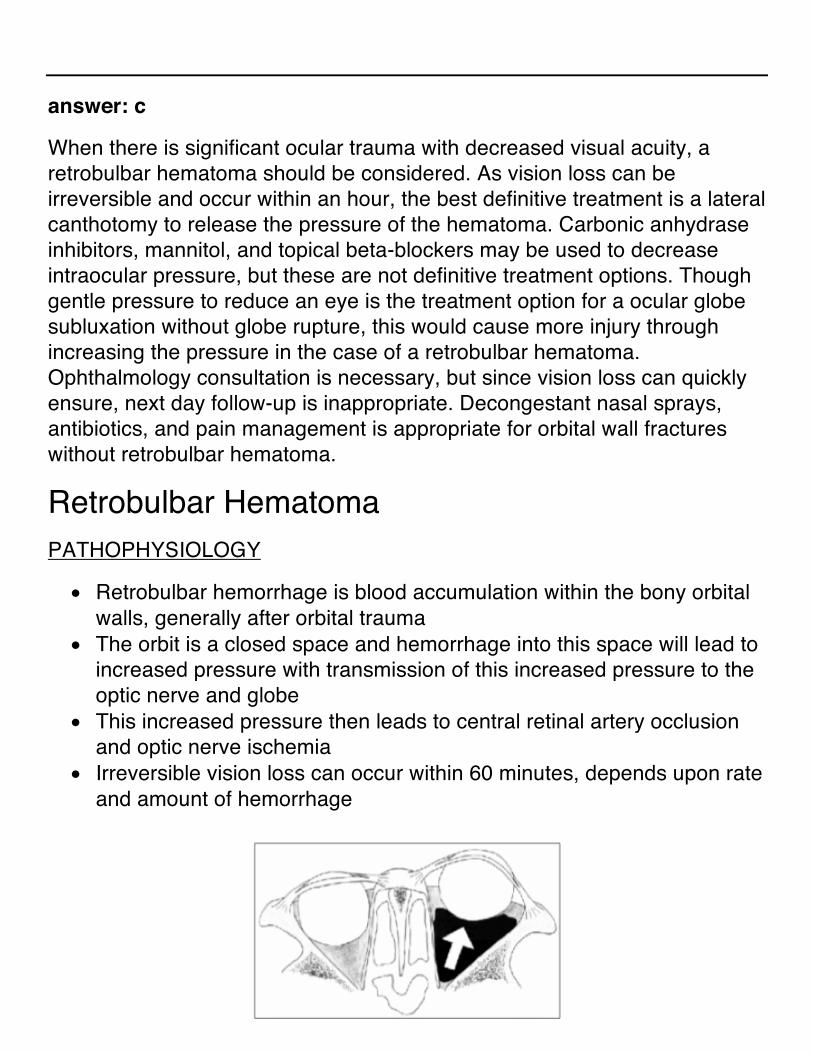
• Retrobulbar hemorrhage is blood accumulation within the bony orbital walls, generally after orbital trauma
• The orbit is a closed space and hemorrhage into this space will lead to increased pressure with transmission of this increased pressure to the optic nerve and globe
• This increased pressure then leads to central retinal artery occlusion and optic nerve ischemia
• Irreversible vision loss can occur within 60 minutes, depends upon rate and amount of hemorrhage
SYMPTOMS AND SIGNS
• Symptoms: eye pain and swelling, decreased vision, diplopia • Signs: acute proptosis, decreased visual acuity, decrease in ocular
movement, increased intraocular pressure
DIAGNOSIS
• Mainly clinical, based on history, symptoms, and physical exam • Head CT with orbital cuts is the study of choice for equivocal cases • Imaging should not delay definitive treatment if there is clinical
suspicion, as vision loss may ensue
TEMPORIZING MEASURES
• Temporizing medical treatments are controversial and should NOT delay definitive treatment
• O2 - theoretically decreases ischemic insult • Mannitol and acetazolamide decrease intraocular pressure • Topical beta-blockers decrease aqueous humor production
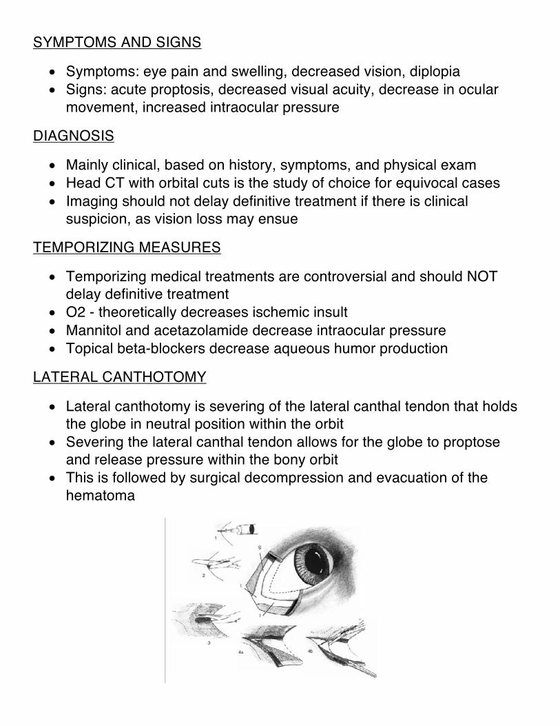
LATERAL CANTHOTOMY
• Lateral canthotomy is severing of the lateral canthal tendon that holds the globe in neutral position within the orbit
• Severing the lateral canthal tendon allows for the globe to proptose and release pressure within the bony orbit
• This is followed by surgical decompression and evacuation of the hematoma
OTHER CONSIDERATIONS
• Most black eyes do not require immediate treatment as long as there are no visual symptoms
• Orbital wall fractures without retrobulbar hemorrhage are more common
• Ophthalmology consultation is required for orbital wall fractures with diplopia (sign of extraocular muscle entrapment), enophthalmos or hypo-ophthalmos, or significant wall fracture (>1/3 of the orbital wall)
• Treatment for orbital wall fractures is avoidance of nose blowing, nasal decongestants, +/- oral antibiotics, pain management, and ophthalmology referral

4. The most appropriate next steps in the management of this case include:
a) aspirin 325 mg, morphine 2 mg IV, and admit to CCU
b) aspirin 325 mg, nitroglycerin SL x3, heparin bolus, cardiac catheterization
c) ketorolac 30 mg IV, followed by ibuprofen 800 mg PO TID for 1 week as outpatient
d) lidocaine 75 mg bolus then 2 mg/min infusion, labetalol 20 mg IV, and admit to a monitored bed
e) metoprolol 5 mg IV, nitroglycerin IV infusion titrated to pain, and cardiology consultation
answer: c
Treatment of acute pericarditis includes NSAIDs, hydration, and reassurance. Patients with idiopathic pericarditis who achieve reasonable pain control in outpatient setting may be sent home with instructions for outpatient follow-up care. In acute MI, ST segments correspond to ischemic portions of the heart and typically have ST-segment depression in reciprocal leads. Also, PR depression is not a feature of acute MI. Although aspirin can be an effective treatment in pericarditis, there is no indication for nitrates, anticoagulation, beta- blockers, or admission to a monitored setting. The tachycardia and hypertension are most likely secondary to pain.
Acute Pericarditis
PRESENTATION
• Chest pain - most common symptom, usually worse in supine position and relieved by sitting up. Can be sudden or gradual in onset and may radiate
• Dyspnea, low-grade fevers, or dysphagia may be present. • Pericardial friction rub - best heard over the left lower sternal border or

apex, with the patient in sitting position. Classically is 3-phase friction rub (ventricular systole, ventricular diastole, atrial systole).
EKG
• Stage 1 (hours to days): diffuse ST-segment elevation with a “concave up” pattern (except aVR and V1), PR-interval depression (except aVR and V1)
• Stage 2: ST and PR segments normalize, but T waves flatten • Stage 3: Deep, symmetric T wave inversion • Stage 4: Normalization, though T wave inversions may be permanent
• Presence of PR-segment depression, and absence of reciprocal ST-
segment changes suggest acute pericarditis

5. What is the next appropriate step in the management of this patient?
a) intubation
b) epinephrine 1 mg IV
c) magnesium 1-2 g IV
d) cardioversion at 100 J monophasic
e) defibrillation at 360 J monophasic
answer: e
This patient has assumed unstable ventricular tachycardia as no pulse can be found on this symptomatic patient and there is a wide complex on the cardiac monitor. Defibrillation at is the most appropriate next step, even above managing the airway or giving pressors. CPR and defibrillation are the only modalities in cardiac arrest proven to improve mortality and morbidity. Synchronized cardioversion is not appropriate for an unstable patient. This is not torsades de pointes, so magnesium is not warranted.

6. After the initial defibrillation, what is the next most appropriate step in the management of this patient?
a) start CPR, 2 minutes
b) check for a pulse
c) check the monitor, and administer another shock if needed
d) administer epinephrine 1 mg IV
answer: a
In the AHA/ACLS 2010 guidelines, uninterrupted chest compressions are emphasized and all shocks should be followed by immediate CPR. Only after CPR should a pulse and monitor checks be performed. Administering epinephrine should not delay resumption of CPR.
ACLS PEARLS • Good CPR is critically important • May substitute 1st or 2nd dose of epi with vasopressin 40U IV/IO • Atropine REMOVED from PEA/asystole algorithm • No shocks or pacing for PEA/asystole • Always be thinking of the H’s and T’s to identify a potential reversible
cause of the cardiac arrest “H’s AND T’s”
• Hypovolemia, Hypoxia, H+ ions (acidosis), Hyperkalemia/Hypokalemia, Hypoglycemia, Hypothermia
• Toxins/Tablets, Tamponade (cardiac), Tension Pneumothorax , Thrombosis (ACS or PE), Trauma

7. Which of the following oral overdoses will NOT benefit from activated charcoal administration?
a) acetaminophen
b) iron
c) lorazepam
d) levothyroxine
answer: b
Activated charcoal is a highly adsorbant powdered material made from distilled wood pulp. It is inert, nontoxic, and has a very large surface area that nonspecifically binds to many substances.
If given within 1 hour of ingestion, it can reduce absorption by 75%. Do not give if the patient cannot protect their airway (altered mental status)
Does not absorb: Alcohols, hydrocarbons, acids and alkalis, heavy metals, inorganic mineral (potassium, arsenic), lithium , organophosphates

8. Activated charcoal is absolutely contraindicated in the treatment of toxic ingestions of:
a) caustics (acids/alkalis)
b) any substance if milk was co-ingested or administered
c) lithium
d) heavy metals
e) digitalis
answer: a
The only absolute contraindication to the use of activated charcoal is ingestion of acids/alkalis. AC does not absorb these and is ineffective in preventing tissue damage. Furthermore, it interferes with subsequent endoscopy and may cause vomiting, leading to further caustic injury.
9. If this patient adamantly refused to comply with drinking the activated charcoal, which of the following is true regarding your ability to administer charcoal?
a) you cannot force the patient to take charcoal
b) you must wait to get parental permission prior to treating her
c) a court injunction is needed to force her to drink the charcoal
d) if repeated attempts to get the patient to take charcoal, a nasogastric tube may be placed to facilitate treatment
e) refusal to take charcoal orally is an indication for IV charcoal

answer: d
Suicidal patients do not have the right to refuse potentially life-saving care. Physicians may do what they need to do in an effort to save the patient. In emergencies, parental permission for treatment is not necessary. Charcoal is not used in IV form.
10. Which of the following statements about acetaminophen ingestion is TRUE?
a) serial LFTs are indicated in all acetaminophen ingestions
b) renal sequelae are expected
c) IV N-acetylcysteine (NAC) is safer than oral NAC
d) an acetaminophen level drawn 4 hours post-ingestion dictates need for antidotal therapy
answer: d
An acetaminophen level drawn at hours 4-20 can be plotted on the Rumack-Matthew nomogram to guide therapy based on hepatic (not renal) toxicity. Liver function tests are not indicated for trivial acetaminophen ingestions, but may be useful in severe ingestions. One potential side effect of IV NAC is an anaphylactoid reaction.
Acetaminophen Toxicity
PATHOPHYSIOLOGY
• Acetaminophen is hepatically metabolized to NAPQI, the toxic free radical metabolite which causes local hepatic injury. At non-toxic doses, NAPQI is detoxified by glutathione in the liver.
• 7.5 g may be toxic in adults; 150 mg/kg may be toxic in children • Four phases of toxicity:
o PHASE 1 (first 24 h): GI symptoms (nausea, vomiting, malaise) without lab abnormalities
o PHASE 2 (24-48 h): abdominal pain with LFT abnormalities o PHASE 3 (48-96 h): severe hepatotoxicity (encephalopathy,
coagulopathy, hypoglycemia), jaundice, renal failure, worsening lab abnormalities.
o PHASE 4 (>96 h): if pathologic effects of Phase III are reversible, complete resolution of hepatic dysfunction will occur.
LABORATORY
• Use the Rumack-Matthew nomogram for predicting acetaminophen toxicity.
• An acetaminophen level earlier than 4 hours post-ingestion is not helpful.
TREATMENT
• Activated charcoal within first 1-2 hours ideally • N-acetylcysteine should be started within 8 hours of ingestion. IV form
available. NAC increases synthesis of glutathione and serves as a free-radical scavenger
• Hemodialysis is not used • Alkalinization has no role in treatment, unlike TCA (serum), salicylate
(urinary), or phenobarbital (urinary) overdoses.

11. A 32 year old man presents 30 minutes after getting a tooth knocked out in a fight. On examination, a small clot in the socket is noted. The next step in management is:
a) call the patient’s dentist
b) clean the tooth with a brush
c) gently irrigate the socket
d) immediately replace the tooth
e) tell the patient the tooth cannot be reimplanted
answer: c
The patient has an avulsion injury and the tooth should be reimplanted. Although little preparation is needed to ready the socket for reimplantation, clots should be removed with gentle irrigation with normal saline, though there should be as little manipulation of the socket as possible.

Tooth Subluxations and Avulsions
TOOTH SUBLUXATION
• Partial avulsion or dislodgment of a tooth from the alveolar bone • May be some laxity and tenderness to percussion of the tooth • Does not require splinting or emergent follow-up, unless there is
significant laxity • Usually managed with NSAIDs, soft diet and referral to a dentist to
confirm diagnosis and exclude more severe injury • Watch out for associated alveolar bone fractures secondary to
intrusion of the tooth deep into the socket
TOOTH AVULSION
• Total displacement of a tooth from its socket • Reimplantation is possible if performed within 2-3 hours • Acceptable transport media to preserve periodontal ligament fibers:
Hank’s balanced solution, sterile NS, milk (Ca and Mg), and saliva • Rinse tooth with normal saline or tap water to remove debris • Only handle crown (tooth root has periodontal ligaments) • Gently remove clots in the socket by irrigation with normal saline • Splint tooth
PEARLS
• Do not handle the tooth root or scrub the tooth • Only reimplant secondary teeth • Consider dental blocks or local anesthesia for better reimplantation
success • Even early improper reimplantation holds a higher success rate for
tooth salvage than delayed reimplantation resulting from waiting for an oral surgeon

12. Which of the following medications would be most efficacious to treat this condition?
a) cetirizine
b) diphenhydramine
c) epinephrine
d) methylprednisolone
e) dexamethasone
answer: c
Severe allergic reactions range from urticaria to angioedema to anaphylaxis. The presentations of these reactions may overlap each other. In this patient with airway compromise from angioedema, intramuscular epinephrine is indicated. Epinephrine-induced catecholamine release causes vasoconstriction to reduce periglottic edema and improve airway patency. It also increases systemic vascular resistance to maintain blood pressure in those in anaphylactic shock.

Allergic Reactions URTICARIA
• Pruritic, erythematous, raised wheals of the skin • Reaction involves the epidermis • Treatment is mainly supportive • Removal of offending agent, cold compresses • Antihistamines: H1 blockade, H2 blockers may be added for severe or
refractory symptoms • Corticosteroids for severe or refractory symptoms; use short course
ANGIOEDEMA
• Nonpruritic edema of submucosa, usually involving tongue, lips, and face
• Reaction involves the dermis (deeper than in urticaria) • Have high degree of suspicion for airway compromise, consider
laryngoscopic evaluation • Treatment is same as for urticaria, but also consider:
o Intramuscular epinephrine (0.3-0.5 cc of 1:1000) for impending airway compromise
o Early airway protection with intubation with intraoral, pharyngeal, and lingual involvement
ANAPHYLAXIS
• Severe systemic hypersensitivity reaction characterized by multisystem involvement, manifested by hypotension or airway compromise
• Look for bronchospasm, voice changes and stridor from laryngeal edema, drooling
• Treatment is same for urticaria and angioedema, but also consider intubation for unstable patients, pressors, and usual ACLS resuscitation

13. Which of the following spiders can cause this presentation?
a) tarantula
b) hobo spider
c) brown recluse spider
d) wolf spider
e) black widow spider
answer: e
Black widow spiders are characterized by an initial pinprick sensation followed by a mild local inflammatory response. Within an hour, crampy myalgias develop at the bite site and spread up the extremity, eventually involving the entire body. Patients may present with a rigid abdomen that is impossible to differentiate clinically from peritonitis. A brown recluse spider bite classically causes a necrotic ulcer. The remaining spider bites are generally unremarkable.

Spider Bites
BLACK WIDOW BITES
• Distinct orange-red hourglass marking • Latroxin is the toxin that is thought to
cause a massive neurotransmitter release • Treatment is supportive as the condition is
generally self-limiting • Opioids and benzodiazepines for pain and
muscle spasms • Antivenom rarely used, but an option for
severe cases
BROWN RECLUSE BITES
• Distinct violin-shaped marking • Classically causes local dermal
necrosis, and rarely black eschars • Also rare is DIC and hemolysis • Usually, the bite-induced necrosis is
self-limited and self-healing without long-term consequences
• Treatment is supportive
14. Which of the following statements regarding bite wounds is correct?
a) Cat bites are most commonly polymicrobial
b) Cat bites do not require antibiotics unless there is a foreign body in the wound
c) mammalian bites are not tetanus-prone wounds
d) only 5-6% of dog bites ultimately become infected without treatment
answer: d
Only 5-6% of dog bites become infected without treatment, which is about the infection rate of lacerations from non-bite mechanisms. Prophylactic antibiotics are not indicated for routine dog bite wounds, but are recommended for high-risk patients or wounds involving the hand. Most dog bites are polymicrobial, and though P. multocida has been isolated in dog

bites, the Pasturella usually found are not virulent. This is in contrast to cat bites, which become infected 60-80% of the time and thought to be related to the virulent strains of P. multocida. Mammal bites produce tetanus-prone wounds.
Mammalian Bites
GENERAL PRINCIPLES
• Attention to life-threatening bites (vascular, neck) • Meticulous examination (esp. joints and tendons) and wound cleansing • Obtain radiographs if there is suspicion of a foreign body • Puncture-type bite wounds, wounds older than 6 hrs, contaminated
wounds, and most hand and foot wounds should not be primarily closed
DOG AND CAT BITES
• Uncomplicated dog bites do not routinely need prophylactic antibiotics, unless it is a hand wound or already infected.
• EXCEPTION: Give prophylactic antibiotics for dog bites in immunocompromised patients (alcoholic liver disease, functional asplenia, lung disease, chronic corticosteroid use). These patients are at risk for sepsis due to Capnocytophaga canimorsus, causing cutaneous gangrene, purpura, and petechiae.
• All cat bites should receive prophylactic antibiotics amoxicillin/clavulanate for 5-7 days
• PCN, amoxicillin, cefuroxime, and doxycycline will also cover P. multocida
HUMAN BITES
• Treat all human bites as contaminated puncture wounds • Usually polymicrobial (Staph/Strep species, Eikenella), treat with
amoxicillin/clavulanate

15. What is the total body surface area (TBSA) of a man who presents with blistered burns of the anterior halves of both his thighs and legs?
a) 9%
b) 18%
c) 27%
d) 36%
answer: b
The Rule of 9’s can be used to estimate TBSA burned (see below). This patient likely has a partial thickness (or 2nd degree burn).

Burns
RULE OF 9’S - to estimate total body surface area (TBSA) involvement (only count 2nd and 3rd degree burns)

INDICATIONS FOR ADMISSION TO A BURN UNIT
• Burns involving the face, hand, feet, genitalia, perineum, or major joints • Electrical burns • Chemical burns with potential for cosmetic/functional impairment • Significant inhalation injury • Co-morbid disease • Partial thickness (2nd degree) burns > 10% TBSA • Full thickness (3rd degree) in any age group
PARKLAND FORMULA
• Isotonic solution (usually lactated ringers solution) to give over first 24 hours = 4 cc x %TBSA burn x weight (kg)
• Give half of total volume in first 8 hours after injury, rest in next 16 hours
PEARLS FOR TREATMENT
• Consider carbon monoxide poisoning • Look for singed nasal hairs, carbonaceous sputum, and facial burns as
markers for potential airway edema and obstruction • Look for circumferential burns • Prophylactic antibiotics are not indicated in burns

16. Which of the following is the most common adolescent hip disorder?
a) Osgood-Schlatter
b) hip fracture
c) septic arthritis
d) growing pains
e) slipped capital femoral epiphysis
answer: e
Slipped capital femoral epiphysis the femoral epiphysis slips posteriorly resulting in a limp and impaired internal rotation. Osgood-Schlatter disease is characterized by pain and swelling at the tibial tubercle where the patellar tendon inserts. It generally presents as knee pain. Hip fractures and septic arthritis should not be missed, and careful history (injury, trauma, infectious symptoms), examination, and laboratory/radiographic testing are important.
SCFE
RISK FACTORS
• Major risk factor is obesity • Occurs in early adolescence • Lesser risk factors are male gender, black/Hispanic ethnicity • Some children have associated endocrinopathy
CLASSIFICATION
• Practical classification is unstable vs stable • STABLE = able to bear weight • UNSTABLE = unable to bear weight
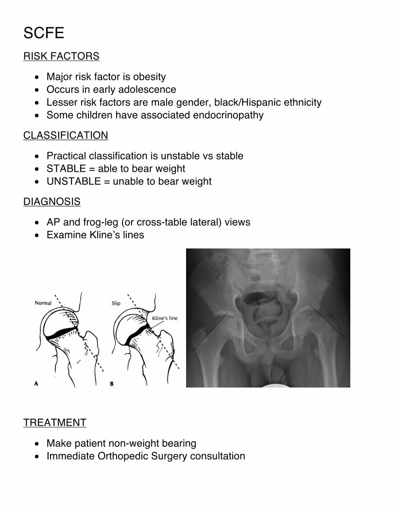
DIAGNOSIS
• AP and frog-leg (or cross-table lateral) views • Examine Kline’s lines
TREATMENT
• Make patient non-weight bearing • Immediate Orthopedic Surgery consultation