two case examples of pelvic fractures in medieval ... case examples of pelvic fractures in medieval...
TRANSCRIPT

JASs ReportsJournal of Anthropological Sciences
the JASs is published by the Istituto Italiano di Antropologia www.isita-org.com
Vol. 88 (2010), pp. 179-188
Two case examples of pelvic fractures in medieval populations from central Europe
Maria Ines Hofmann1*, Christina Papageorgopoulou1*, Thomas Böni2,3 & Frank J. Rühli1,3
1) Institute of Anatomy, University of Zurich, Winterthurerstr. 190, 8057 Zürich, Switzerlande-mail: [email protected]
2) Orthopedic University Clinic Balgrist, Forchstr. 340, 8032 Zürich, Switzerland
3) Institute and Museum for the History of Medicine, University of Zurich, Hirschengraben 82, 8001 Zürich, Switzerland
* These authors contributed equally to this work
Summary - Pelvic fractures are considered to be uncommon and difficult to treat, even in the modern medical literature. Serious and eventually life-threatening associated injuries may occur, requiring emergency abdominal, vascular or neurologic surgery. Pelvic fractures can also be managed non-operatively; however, a considerable dispute exists on the suitable management strategy. The treatment and healing of such injuries in the bioarchaeological record, is therefore of great interest for anthropological and medico-historical studies. Fractures of the pelvis are rarely reported in the anthropological literature either due to poor preservation of the innominate bone or lack of adequate examination. Here we present two cases of pelvic fractures observed on two adult male individuals from two European medieval sites. They differ in severity and in the pattern of healing. They are both adequately healed and probably had no acute life-threatening consequences, which gives us some insight into the medical knowledge and means of management of past populations.
Keywords - Computed tomography, Iliac wing fracture, Femur fracture, Paleopathology.
Introduction
Traumatic lesions are common abnormalities observed in skeletal remains and in many cases the diagnosis of trauma in archaeological skeletons is easily made. Trauma affects the skeleton in vari-ous ways, with fractures being the most common one (Ortner, 2003). Important information, such as the occurrence of domestic accidents, inter-personal violence, occupation related traumas, subsistence strategy, availability of treatment and even nutritional status, can be obtained from the bone healing process (Brothwell, 1961; Larsen, 2002). Therefore, paleopathological studies have the potential to provide insight into the life of past populations (Grauer & Roberts 1996).
Specifically pelvic fractures (PF) are consid-ered even in the modern medicine quite rare and complicated to treat. A literature series regard-ing management protocols, long-term functional prognosis and retrospective studies exists (Dalal et al., 1989; Heetveld et al., 2004; Geeraerts et al., 2007). Although a rather uncommon injury, the mortality associated with these injuries can be profound. Studies of traumatized patients with PF refer to a mortality rate of 15% to 20% (Young & Resnik, 1990; Ali et al., 2009; Ioannidis et al., 2009) and the estimated risk of death is higher (1,71) than in other bone fractures, e.g. forearm (1,01), ribs (1,27) (Ioannidis et al., 2009). Serious and eventually life-threatening associated inju-ries may occur, requiring emergency abdominal,
180 Medieval Pelvis Fracture
vascular or neurologic surgery (Mucha & Welch 1988; Geeraerts et al., 2007). Specific PF can be managed non-operatively, however, a certain medical treatment dispute exists specific to the
proper strategy for nonunion or malunion of PF (Dujardin et al., 1998; Switzer et al., 2000; Burkhardt et al., 2005; Abrassart et al., 2009; Ali et al., 2009, Taller et al., 2009).
The presence and management of such inju-ries in the bioarchaeological record is of interest for anthropologists, medical historians and physicians. However, PF have received little attention in the anthropological literature. There is no published data about this kind of trauma in paleopathology review papers (Lovell, 1997), specialized textbooks (Ortner, 2003; Mann & Hunt, 2005) nor in frac-ture-pattern research papers (Lovejoy & Heiple, 1981; Alvrus, 1999; Walter et al., 1999; Judd & Roberts, 1999; Djuric et al., 2006; Dommet & Tayles, 2006; Paine et al., 2009). This low per-cent of paleopathological data probably derives from the difficulties in identification based on the taphonomic condition of the pelvis in compari-son to other bones of the skeleton, i.e. as a conse-quence of postmortem fragility. The “fundamental conceptual problems” such as selective mortality and hidden heterogeneity in risks described by Wood and colleagues (1992) should be also con-sidered as biased when interpretating this lack of data concerning traumas to particular parts of the skeleton. In the present study, we present two cases of PF from two European medieval sites. They offer valuable insight on the treatment and clinical management of such complicated traumas in this specific temporo-spatial setting.
Material and Methods
The first case (grave # 34a) concerns a partially preserved skeleton of an adult male, between 40 and 60 years old. Most of the lower limbs were recovered whereas parts of the axial skeleton and the upper limbs were not preserved. The skeletal material (54 individuals) comes from a historic monastery in Dalheim, Germany most likely dated to the mid 11th century AD (radiocarbon dat-ing by Institute for Particle Physics Swiss Federal Institute of Technology, Zurich). The excavation area and burial were described in previous studies (Hofmann et al., 2008). The demographic profile
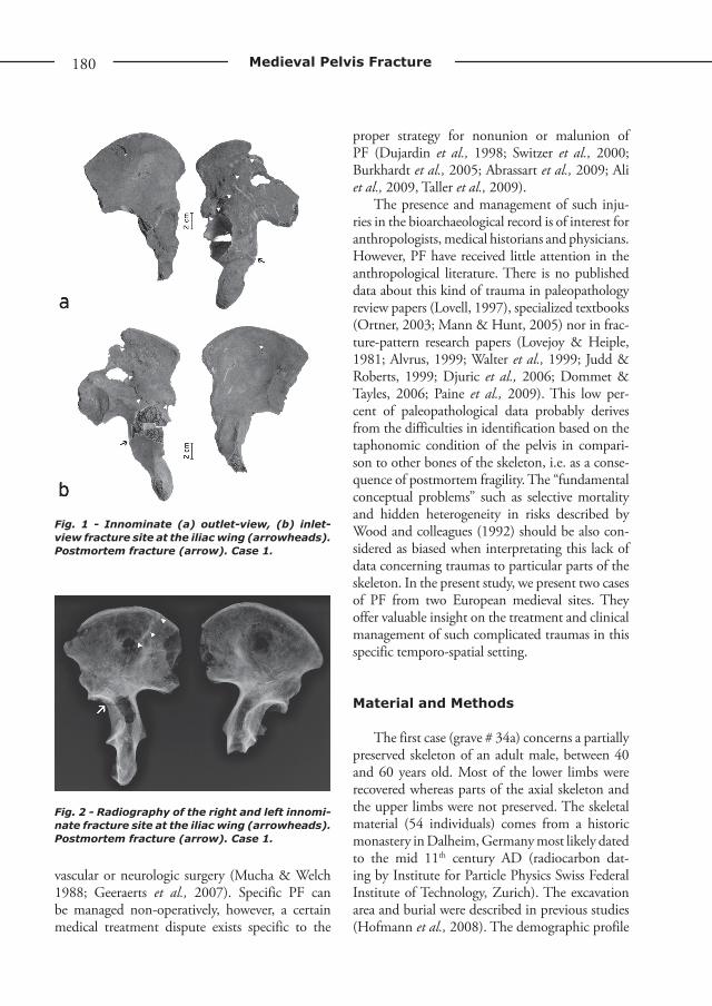
Fig. 1 - Innominate (a) outlet-view, (b) inlet-view fracture site at the iliac wing (arrowheads). Postmortem fracture (arrow). Case 1.
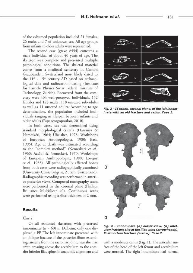
Fig. 2 - Radiography of the right and left innomi-nate fracture site at the iliac wing (arrowheads). Postmortem fracture (arrow). Case 1.
www.isita-org.com
181M.I. Hofmann et al.
of the exhumed population included 21 females, 26 males and 7 of unknown sex. All age groups from infants to older adults were represented.
The second case (grave #454) concerns a male individual of about 40 years of age. The skeleton was complete and presented multiple pathological conditions. The skeletal material comes from a medieval cemetery in Canton Graubünden, Switzerland most likely dated to the 11th - 15th century AD based on archaeo-logical data and radiocarbon dating (Institute for Particle Physics Swiss Federal Institute of Technology, Zurich). Recovered from the cem-etery were 404 well-preserved individuals, 152 females and 123 males, 118 unsexed sub-adults as well as 11 unsexed adults. According to age determination, the population included indi-viduals ranging in lifespan between infants and older adults (Papageorgopoulou, 2010).
In both cases, sex was determined using standard morphological criteria (Harsányi & Nemeskéri, 1964; Ubelaker, 1978; Workshops of European Anthropologist, 1980; Bass, 1995). Age at death was estimated according to the “complex method” (Nemeskéri et al., 1960; Acsàdi & Nemeskéri, 1970; Workshops of European Anthropologist, 1980; Lovejoy et al., 1985). All pathologically affected bones from both cases were radiographically examined (University Clinic Balgrist, Zurich, Switzerland). Radiographic recording was performed in anteri-or-posterior views. Computed tomography scans were performed in the coronal plane (Phillips Brilliance Multislicer 40). Continuous scans were performed using a slice thickness of 2 mm.
Results
Case 1 Of all exhumed skeletons with preserved
innominates (n = 60) in Dalheim, only one dis-played a PF. The left innominate presented with an oblique fracture of the posterior ilium extend-ing laterally from the sacroiliac joint, near the iliac crest, crossing above the acetabulum to the ante-rior inferior iliac spine, in anatomic alignment and
with a moderate callus (Fig. 1). The articular sur-face of the head of the left femur and acetabulum were normal. The right innominate had normal
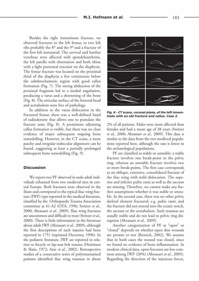
Fig. 3 - CT scans, coronal plane, of the left innom-inate with an old fracture and callus. Case 1.
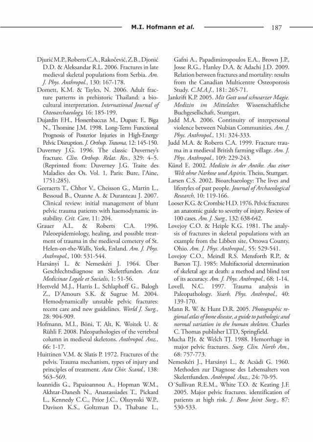
Fig. 4 - Innominate (a) outlet-view, (b) inlet-view fracture site at the iliac wing (arrowheads). Postmortem fracture (arrow). Case 2.

182 Medieval Pelvis Fracture
morphology. The femurs were in a very good con-dition with no visible pathology. The preserved tibiae and fibulae were also free from pathology.
Conventional X-ray and CT images mainly confirmed the macroscopic appearance. Adjacent to the fracture line, (which does not appear to be fully consolidated and still exhibits a mild dislo-cation, Figs. 2 and 3) one finds cloudy patches of increased sclerosis consistent with callus for-mation. Bone remodelling took place and ragged fracture edges are no longer visible. There are no obvious signs of major bone remodelling specific to infection or reactive osteopenia. Some areas of largely compact bone such as the iliac crest and acetabular region, are only partially preserved. Additionally, there is clear diagenesis-related trauma present on the bone.
Case 2Of the 404 exhumed skeletons with preserved
innominates (n = 336) from Tomils / Sogn Murezi, only one displayed a PF. The right ilium presented an oblique fracture of the posterior ilium extend-ing laterally from the sacroiliac joint, near the iliac crest, crossing obliquely up to the middle part of the bone. The PF was in anatomic alignment and had a moderate callus (Fig. 4).
Radiological examination revealed that the fracture line in this individual (Figs. 5,6) appeared to be completely consolidated in comparison to case 1 and was represented by a well-defined band of radiodensity. No adjoining zones of radi-olucency nor ongoing bone remodelling was vis-ible. Also, partial postmortem damage including a fracture and missing bone areas were visible.
Fig. 5 - Radiography of the right and left innominate fracture site at the iliac wing (arrowheads). Postmortem fracture (arrow). Case 2.
www.isita-org.com
183M.I. Hofmann et al.
Besides the right innominate fracture, we observed fractures to the left femur, to two left ribs probably the 8th and the 9th and a fracture of the first left metatarsal. The cervical and lumbar vertebrae were affected with spondyloarthritis, the left patella with eburnation and both tibiae with a light periosteal reaction on the diaphysis. The femur fracture was located on the proximal third of the diaphysis a few centimetres below the subthrochanteric region with good callus formation (Fig. 7). The strong abduction of the proximal fragment led to a medial angulation, producing a varus and a shortening of the bone (Fig. 8). The articular surface of the femoral head and acetabulum were free of pathology.
In addition to the varus dislocation in the fractured femur, there was a well-defined band of radiodensity that allows one to postulate the fracture zone (Fig. 8). A prominent adjoining callus formation is visible, but there was no clear evidence of major subsequent ongoing bone remodelling. However, in the CT scans, a more patchy and irregular trabecular alignment can be found, suggesting at least a partially prolonged subsequent bone remodelling (Fig. 9).
Discussion
We report two PF observed in male adult indi-viduals exhumed from two medieval sites in cen-tral Europe. Both fractures were observed in the ilium and correspond to the typical iliac wing frac-ture (IWF) type reported in the medical literature, classified by the Orthopaedic Trauma Association committee as 61-A2 (OTA, 1996; Switzer et al., 2000; Abrassart et al., 2009). Iliac wing fractures are uncommon and difficult to treat (Switzer et al., 2000). There is little information in the literature about adult IWF (Abrassart et al., 2009), although the first descriptions of such injuries had been reported in 1751 (reprinted Duverney, 1996). In the pediatric literature, IWF are reported in rela-tion to bicycle or lap-seat belt trauma (Huittinen & Slatis, 1972; Amr et al., 2002). Retrospective studies of a consecutive series of polytraumatised patients identified iliac wing traumas in about
2% of all patients. Males were more affected than females and had a mean age of 38 years (Switzer et al., 2000; Abrassart et al., 2009). This data is similar to the data from the two medieval popula-tions reported here, although the rate is lower in the archaeological populations.
PF are classified as stable or unstable: a stable fracture involves one break-point in the pelvic ring, whereas an unstable fracture involves two or more break-points. The first case corresponds to an oblique, extensive, consolidated fracture of the iliac wing with mild dislocation. The supe-rior and inferior pubic rami as well as the sacrum are missing. Therefore, we cannot make any fur-ther assumptions whether it was stable or unsta-ble. In the second case, there was no other pelvic skeletal element fractured, e.g. pubic rami, and the fracture did not extend into the sciatic notch, the sacrum or the acetabulum. Such traumas are usually stable and do not lead to pelvic ring dis-ruption (Abrassart et al., 2009).
Another categorization of PF as “open” or “closed” depends on whether open skin wounds are present or not (Resnick, 2002). We assume that in both cases the wound was closed, since we found no evidence of bone inflammation. In modern clinical data, open fractures are less com-mon among IWF (20%) (Abrassart et al., 2009). Regarding the direction of the injurious forces,
Fig. 6 - CT scans, coronal plane, of the left innom-inate with an old fracture and callus. Case 2.

184 Medieval Pelvis Fracture
we would classify the fractures as a less severe type in the lateral compression group, where oblique iliac wing fractures can be observed in isolation (Young et al., 1986; Young & Resnik, 1990). Nonetheless, such categorizations on complex bone elements such as the pelvic ring, based on fragmentary material and lacking soft tissue, should be viewed with caution. We tried to reconstruct these cases based on modern clini-cal data, but we also have to consider the una-voidable limitations of such reconstructions.
Healing of IWF occurs without difficulty due to the excellent vascularity, supplied by good mus-cle coverage, and because there are no rotations or vertical instability of the pelvis (Abrassart et al., 2009). The potential risks arising from such traumas relates mainly to the non-skeletal injuries involving the visceral, vascular, urologic and gyne-cologic systems. Major trauma with associated
injuries to the hip and other bones, predisposes to complications such as malalignment (rotational or angular deformity and femoral shortening), vascu-lar injury, infection, thrombophlebitis, fat emboli-zation, and chest and abdominal injuries (Poole et al., 1991; O´Sullivan et al., 2005; Vásquez et al., 2008). Nerve lesions are rare, but have been also reported in one case (Switzer et al., 2009). Based on modern data, it is quite probable that the two medieval “patients” suffered from one or more soft tissue injuries.
Treatment methods of fractures in ancient times were very similar to those of modern times not only in principle, but also in practice (Clark, 1937; Künzl, 2002). There is historical evidence for fracture treatment in medieval Europe (Clark, 1937; Grauer & Roberts, 1996; Jankrift, 2005). Orthopaedic surgeons, known as bone-setters, and barber surgeons manipulated, splinted, and
Fig. 7 - Right and left (a) femur ventral view, (b) dorsal view. Case 2.

www.isita-org.com
185M.I. Hofmann et al.
even used extension methods by windlasses, levers, ratchets, and pulleys. A mild fracture may heal in several weeks without surgery (Schultz, 1961). IWF in particular exhibit a high rate of successful non-operative treatment (Abrassart et al., 2009). We can assume that the two PF were treated to a certain degree. The fractures were well healed indicating that they had occurred years prior to the individuals´ death. In the sec-ond case, the characteristic femoral shortening and varus suggests that fracture reduction, e.g. by extension was not attempted or had failed. It is probable that the individual was treated in recumbence, staying in bed for a long time until the pain had subsided. That could explain the good consolidation of the IWF.
The etiology of IWF in modern populations is associated with high energy trauma, including road traffic crashes and falls from great heights (Vásquez et al., 2008) or spontaneously after minor falls in people with bone-weakening dis-eases. It is difficult to reconstruct the exact etiol-ogy of the medieval fractures, but they may also have been caused by similar high intensity forces. Both IWF were oblique suggesting that indirect forces were responsible, e.g. a blunt blow such as an animal kick (Huittinen & Slätis, 1972) may be applicable in the first case, in which no other bone was fractured and a fall on the left side of the body e.g. fall from a building or a cart in movement on the second case with the multiple fractures. Documentation on daily activities of these two medieval populations include many risk factors that may lead to such traumas, e.g. ploughing fields, carting goods, felling trees, herd-ing animals (Segin, 1935; Rüthing, 1980; Pieper, 2003; Papageorgopoulou, 2009). Both individu-als exhibited no signs of bone-weakening diseases, so a fracture due to osteoporosis is unlikely.
Conclusion
In these two historic cases, we could peer into the wide spectrum of IWF. It has been shown that PF, although rare, are present in archaeologi-cal populations and could heal successfully. The
interpretation of the two cases entails limitations due to the fragmentary nature of the first case, but it is clear that both fractures were not fatal. Regarding the available means and the times, it is quite aston-ishing how well these traumas were managed, since both individuals survived the injuries. This reveals the high level of medieval medical knowledge and is
Fig. 8 - Radiography of the left femur with the varus angle (135◦). Case 2.

186 Medieval Pelvis Fracture
specifically of interest for medico-historic research. The present paper may enlighten modern discus-sions about treatment strategies, operative versus non-operative management, by offering a differ-ent dataset to modern clinical research. It also gives some information on domestic and/or occupation related traumas, convalescence and survival chances, which is significant within a historic context.
Acknowledgments
We thank K. W. Alt, DMD (Institute of Anthropol-ogy, Johannes Gutenberg University, Mainz, Ger-many) and the Archaeological Service of the Canton Graubünden, Switzerland for providing the skeletal material, Heinz Sonderegger (Institute of Anatomy,
Zurich) for the photographs, the Division of Radiology (University Hospital Balgrist, Zurich) for providing the Radiographs and CT scanning and two anony-mous reviewers for useful comments and suggestions.
References
Abrassart S., Stern, R. & Peter R. 2009. Morbidity associated with isolated iliac wing fractures. J. Trauma, 66: 200-3.
Acsàdi G. & Nemeskéri J. 1970. History of human life span and mortality. Hungarian Academic Society, Budapest.
Ali J., Ahmadi K.A. & Williams J.I. 2009. Predictors of laparotomy and mortality in poly-trauma patients with pelvic fractures. Can. J. Surg., 52: 271-276.
Alvrus A. 1999. Fracture patterns among the Nubians of Semna South, Sudanese Nubia. International Journal of Osteoarchaeology, 9: 417-429.
Amr S.M., Abdel-Meguid K.M. & Kholeif A.M. 2002: Neurologic injury caused by fracture of the iliac wing (Duverney’s fracture): case report. J. Trauma, 52: 370-376.
Bass W.M. 1995. Human osteology: a labora-tory and field manual. Missouri Archaeological Society, Missouri.
Brothwell D. (1961). The Paleopathology of early British man: An essay on the problems of di-agnosis and analysis. The Journal of the Royal Anthropological Institute of Great Britain and Ireland, 91: 318-344.
Burkhardt M., Culemann U., Seekamp A. & Poh-lemann T. 2005. Operative Versorgungsstrat-egien beim Polytrauma mit Beckenfraktur. Unfallchirurg., 108, 814-20.
Clark W.A. 1937. History of fracture treatment up to the sixteenth century. J. Bone Joint Surg., XIX: 47-63.
Dalal, S.A., Burgess, A.R., Siegel, J.H., Young, J.W., Brumback, R.J., Poka, A., Dunham, C.M., Gens, D. & Bathon, H. 1989. Pelvic fracture in multiple trauma: classification by mechanism is key to pattern of organ injury, resuscitative requirements, and outcome. J. Trauma, 29: 981-1000.
Fig. 9 - CT scans, coronal plane of the left femur. Case 2.
www.isita-org.com
187M.I. Hofmann et al.
Djurić M.P., Roberts C.A., Rakočević, Z.B., Djonić D.D. & Aleksandar R.L. 2006. Fractures in late medieval skeletal populations from Serbia. Am. J. Phys. Anthropol., 130: 167-178.
Domett, K.M. & Tayles, N. 2006. Adult frac-ture patterns in prehistoric Thailand: a bio-cultural interpretation. International Journal of Osteoarchaeology, 16: 185-199.
Dujardin F.H., Hossenbaccus M., Duparc F., Biga N., Thomine J.M. 1998. Long-Term Functional Prognosis of Posterior Injuries in High-Energy Pelvic Disruption. J. Orthop. Trauma, 12: 145-150.
Duverney J.G. 1996. The classic Duverney’s fracture. Clin. Orthop. Relat. Res., 329: 4–5. (Reprinted from: Duverney J.G. Traite des Maladies des Os. Vol. 1, Paris: Bure, l’Aine, 1751:285).
Geeraerts T., Chhor V., Cheisson G., Martin L., Bessoud B., Ozanne A. & Duranteau J. 2007. Clinical review: initial management of blunt pelvic trauma patients with haemodynamic in-stability. Crit. Care, 11: 204.
Grauer A.L. & Roberts C.A. 1996. Paleoepidemiology, healing, and possible treat-ment of trauma in the medieval cemetery of St. Helen-on-the-Walls, York, Enland. Am. J. Phys. Anthropol., 100: 531-544.
Harsányi L. & Nemeskéri J. 1964. Über Geschlechtsdiagnose an Skelettfunden. Acta Medicinae Legale et Socialis, 1: 51-56.
Heetveld M.J., Harris I., Schlaphoff G., Balogh Z., D’Amours S.K. & Sugrue M. 2004. Hemodynamically unstable pelvic fractures: recent care and new guidelines. World J. Surg., 28: 904-909.
Hofmann, M.I., Böni, T. Alt, K. Woitek U. & Rühli F. 2008. Paleopathologies of the vertebral column in medieval skeletons. Anthropol. Anz., 66: 1-17.
Huittinen V.M. & Slatis P. 1972. Fractures of the pelvis. Trauma mechanism, types of injury and principles of treatment. Acta Chir. Scand., 138: 563–569.
Ioannidis G., Papaioannou A., Hopman W.M., Akhtar-Danesh N., Anastassiades T., Pickard L., Kennedy C.C., Prior J.C., Olszynski W.P., Davison K.S., Goltzman D., Thabane L.,
Gafni A., Papadimitropoulos E.A., Brown J.P., Josse R.G., Hanley D.A. & Adachi J.D. 2009. Relation between fractures and mortality: results from the Canadian Multicentre Osteoporosis Study. C.M.A.J., 181: 265-71.
Jankrift K.P. 2005. Mit Gott und schwarzer Magie. Medizin im Mittelalter. Wissenschaftliche Buchgesellschaft, Stuttgart.
Judd M.A. 2006. Continuity of interpersonal violence between Nubian Communities. Am. J. Phys. Anthropol., 131: 324-333.
Judd M.A. & Roberts C.A. 1999. Fracture trau-ma in a medieval British farming village. Am. J. Phys. Anthropol., 109: 229-243.
Künzl E. 2002. Medizin in der Antike. Aus einer Welt ohne Narkose und Aspirin. Theiss, Stuttgart.
Larsen C.S. 2002. Bioarchaeology: The lives and lifestyles of past people. Journal of Archaeological Research, 10: 119-166.
Looser K.G. & Crombie H.D. 1976. Pelvic fractures: an anatomic guide to severity of injury. Review of 100 cases. Am. J. Surg., 132: 638-642.
Lovejoy C.O. & Heiple K.G. 1981. The analy-sis of fractures in skeletal populations with an example from the Libben site, Ottowa County, Ohio. Am. J. Phys. Anthropol., 55: 529-541.
Lovejoy C.O., Meindl R.S. Mensforth R.P., & Barton T.J. 1985: Multifactorial determination of skeletal age at death: a method and blind test of its accuracy. Am. J. Phys. Anthropol., 68: 1-14.
Lovell, N.C. 1997. Trauma analysis in Paleopathology. Yearb. Phys. Anthropol., 40: 139-170.
Mann R. W. & Hunt D.R. 2005. Photographic re-gional atlas of bone disease, a guide to pathologic and normal variation in the human skeleton. Charles C. Thomas publisher LTD, Springfield.
Mucha P.Jr. & Welch TJ. 1988. Hemorrhage in major pelvic fractures. Surg. Clin. North Am., 68: 757-773.
Nemeskéri J., Harsányi L., & Acsàdi G. 1960. Methoden zur Diagnose des Lebensalters von Skelettfunden. Anthropol. Anz., 24: 70-95.
O´Sullivan R.E.M., White T.O. & Keating J.F. 2005. Major pelvic fractures. identification of patients at high risk. J. Bone Joint Surg., 87: 530-533.

Orthopaedic Trauma Association committee for cod-ing and classification. 1996. Fracture and disloca-tion compendium. J. Orthop. Trauma, 10: 66-70.
Ortner D.J. 2003. Identification of pathological conditions in human skeletal remains. Academic Press, San Diego.
Paine R.R., Vargiu R., Signoretti C., Coppa A. 2009. A health assessment for Imperial Roman burials recovered from the necropolis of San Donato and Bivio CH, Urbino, Italy. J. Anthropol. Sci., 87: 193-210.
Papageorgopoulou C. 2010. The medieval popula-tion of Tomils and archaeo-anthropological ap-proach. B.A.R. - International Series (Accepted).
Pieper R. 2003. Dalheim Pfarrort-Kloster- Staats-domäne. Ardey Verlag, Münster.
Poole G.V., Ward E.F., Muakkassa F.F., Hsu H.S.H., Griswold J.A. & Rhodes R.S. 1991. Pelvic fracture from major blunt trauma. Ann. Surg., 213: 532-538.
Resnick D. 2002. Bone and joint disorders. W.B. Saunders Company, Philadelphia.
Rothenberger D.A., Fischer R.P., Strate R.G., Velasco R. & Perry J.F.Jr. 1978. The mortality associated with pelvic fractures. Surgery, 84: 356-361.
Rüthing H. 1980. Zur Wirtschaftsgeschichte des Klosters Böddeken vom 14. bis 16. Jahrhundert. Westfälische Zeitschrift, 130: 150-166.
Schultz A.H. 1961. Some factors influencing the social life of primates in general and of early man in particular. Viking Fund Publications in Anthropology, 31: 58-90.
Segin W. 1935. Kloster Dalheim im Sintfelde bei Paderborn. Westfälische Zeitschrift, 91: 130-205.
Switzer J.A., Nork S.E. & Routt M.L.Jr. 2000. Comminuted fractures of the iliac wing. J. Orthop. Trauma, 14: 270 –276.
Taller S., Srám J., Lukás R. & Krivohlávek M. 2009. Nonunions or malunions of pelvic fractures. Acta Chir. Orthop. Traumatol. Cech., 76: 121-7.
Vásquez V., Bello González A. & Caballero E.A. 2008. Control de daños en fracturas de huesos largos y pelvis en el Centro Trauma Cruz Roja Mexicana. Acta Ortop. Mex., 22: 45-49.
Ubelaker D.H. 1978. Human skeletal remains. Aldine Publishing Company, Chicago.
Walter A., Neves A.M., Barros & Costa M.A. 1999. Incidence and Distribution of Postcranial Fractures in the Prehistoric Population of San Pedro de Atacama, Northern Chile. Am. J. Phys. Anthropol., 109: 253-258.
Wood J.W., Milner G.R., Harpending H.C., Weiss K.M. 1992. The osteological paradox. Curr. Anthropol., 33: 343–370.
Workshops of European Anthropologist. 1980. Recommendations’ for age and sex diagnoses of skeletons. J. Hum. Evol., 9: 517-549.
Young J.W.R., Burgess A.R., Brumback R.J.& Poka A. 1986. Lateral compression of the pel-vis: The importance of plain radiographs in the diagnosis and surgical management. Skeletal Radiol., 15: 103-109.
Young J.W.R. & Resnik C.S. 1990. Fracture of the pelvis: current concepts of classification. Am. J. Roentgenol., 155: 1169-1175.
Associate Editor, Robert R. Paine