026 classification, management and outcomes of severe pelvic ring fractures
DESCRIPTION
ISCRR ReportTRANSCRIPT

7/18/2019 026 Classification, management and outcomes of severe pelvic ring fractures
http://slidepdf.com/reader/full/026-classification-management-and-outcomes-of-severe-pelvic-ring-fractures 1/72
Accompanying documents to th is report (12pt)
Title Report number
Summary: Classification,management and outcomes ofsevere pelvic ring fractures
0712-026-R1B
Classification, management andoutcomes of severe pelvic ring
fractures
School of Public Health and Preventive Medicine,Monash University
The Alfred Hospital, Royal Melbourne Hospital,Epworth Health
Associate Professor Belinda Gabbe
Dr Dirk-Jan HofsteeProf Richard de Steiger
Associate Professor Andrew Bucknill Associate Professor Max Esser
Dr Matthias RussProf Peter Cameron
2
nd
July 2012
Research report #: 0712-026-R1C

7/18/2019 026 Classification, management and outcomes of severe pelvic ring fractures
http://slidepdf.com/reader/full/026-classification-management-and-outcomes-of-severe-pelvic-ring-fractures 2/72
Research Report # 0712-026-R1C Page 2 of 72
Table of Contents
Project Background ........................................................................................................... 4
Study 1: Reliabil ity of pelvic r ing f racture classification ................................................ 6
Introduction ...................................................................................................................... 6Project aims ..................................................................................................................... 6Methods ........................................................................................................................... 7
Setting .......................................................................................................................... 7Fracture classification systems ..................................................................................... 7Participants and sample size ........................................................................................ 8Procedures ................................................................................................................... 9
Results ........................................................................................................................... 10Profile of cases ........................................................................................................... 10Inter-rater reliability ..................................................................................................... 11
Discussion ...................................................................................................................... 14Conclusions .................................................................................................................... 21
Study 2: Mortality following severe pelvic ring fracture: Does hospi tal of definitivecare matter? ..................................................................................................................... 22
Introduction .................................................................................................................... 22
Project aims ................................................................................................................... 25Methods ......................................................................................................................... 25
Setting ........................................................................................................................ 25Participants ................................................................................................................. 25VSTR and VOTOR data ............................................................................................. 26Medical record review ................................................................................................. 26Data analysis .............................................................................................................. 27
Results ........................................................................................................................... 28Comparison of cases managed at The Alfred and RMH ............................................. 28
Discussion ...................................................................................................................... 34Conclusions .................................................................................................................... 39
Study 3: Factors associated with long term patient-reported outcomes after severepelvic ring fracture........................................................................................................... 40
Introduction .................................................................................................................... 40Project Aims ................................................................................................................... 40Methods ......................................................................................................................... 41
Participants ................................................................................................................. 41Procedures ................................................................................................................. 41Data analysis .............................................................................................................. 42
Results ........................................................................................................................... 43Profile of participants .................................................................................................. 43
Severity, management and complications .................................................................. 44Functional outcomes (GOS-E) .................................................................................... 47

7/18/2019 026 Classification, management and outcomes of severe pelvic ring fractures
http://slidepdf.com/reader/full/026-classification-management-and-outcomes-of-severe-pelvic-ring-fractures 3/72
Research Report # 0712-026-R1C Page 3 of 72
Return to work ............................................................................................................ 50Pain ............................................................................................................................ 52Health-related quality of life ........................................................................................ 54
Discussion ...................................................................................................................... 59
Conclusions .................................................................................................................... 63Summary of key findings ................................................................................................ 64
Recommendations ........................................................................................................... 65
References ....................................................................................................................... 66

7/18/2019 026 Classification, management and outcomes of severe pelvic ring fractures
http://slidepdf.com/reader/full/026-classification-management-and-outcomes-of-severe-pelvic-ring-fractures 4/72
Research Report # 0712-026-R1C Page 4 of 72
Project Background
Unstable pelvic ring fractures are associated with a high risk of mortality (1, 2). Where
patients survive these injuries, the potential for long term morbidity such as lost health-
related quality of life (HRQoL), pain and functional loss is significant.
Because of the high risk of fracture-related haemorrhage associated with severe pelvic
ring fracture, most studies have focused on establishing predictors of mortality. The risk of
haemorrhage, combined with the propensity for severe associated injuries, highlight the
clinical challenge of managing unstable pelvic fractures. While guidelines exist, there is no
clear consensus about the optimal management approach for haemodynamically unstable
patient with pelvic fracture (3-8). In Victoria, the two adult major trauma services (MTS)
favour different protocols for the management of these patients. A preliminary study, using
data only from the Victorian State Trauma Registry, found no difference in risk-adjusted
mortality between the MTS hospitals (9). However, limitations were noted and the need
for additional data regarding resuscitation practices, and fracture classification, were
acknowledged as necessary for a full evaluation of the MTS approaches to management.
Studies assessing the long term outcomes of patients are few and largely limited to small
samples (n<50), single centre studies (10-12), and specific sub-sets of the population (e.g.
surgically managed, female patients). These studies confirm the poor long term outcomes
but have not investigated the relationship between management, fracture classification
and outcome. The most effective method of fixation for maximising long term outcomes
remains unknown.
Optimal fixation requires high quality imaging to guide surgical management and decision
making, while research studies of the pelvic ring require accurate and reliable classification
of fractures to enable valid and appropriate comparisons of study populations and patient
outcomes. The major classifications for pelvic ring fracture are the Young-Burgess and
Tile/AO. Both systems were developed in the 1980s for fracture classification based on
plain radiographs of the pelvis, but computed tomography (CT) is at the forefront of
modern imaging. Two studies have investigated the reliability of classification of pelvic
ring fractures and have found fair to substantial agreement using plain radiographs and 2D
axial CT scans, which is not sufficient for research and clinical purposes.
This project comprised three distinct studies:

7/18/2019 026 Classification, management and outcomes of severe pelvic ring fractures
http://slidepdf.com/reader/full/026-classification-management-and-outcomes-of-severe-pelvic-ring-fractures 5/72
Research Report # 0712-026-R1C Page 5 of 72
i. A study to establish the inter- and intra-tester reliability of the classification of
severe pelvic ring fractures using plain radiographs and 3D CT reconstruction
imaging of the pelvis.
ii. A detailed comparison of the early acute management of severe pelvic ring
fractures between the MTS hospitals to further explore the association between
protocols and risk-adjusted mortality.
iii. A prospective cohort study to establish the relationship between approaches to
pelvic fracture fixation, fracture classification and long term functional, return to
work, pain and health-related quality of life outcomes.

7/18/2019 026 Classification, management and outcomes of severe pelvic ring fractures
http://slidepdf.com/reader/full/026-classification-management-and-outcomes-of-severe-pelvic-ring-fractures 6/72
Research Report # 0712-026-R1C Page 6 of 72
Study 1: Reliabili ty of pelvic ring fracture classification
Introduction
Optimal fixation of fractures requires high quality imaging to guide surgical management
and decision making, while research studies of the pelvic ring require accurate and reliable
classification of fractures to enable valid and appropriate comparisons of study populations
and patient outcomes. The two commonly used classification systems for pelvic ring
fractures are the Tile/AO and the Young-Burgess systems (13-15). Both systems were
developed in the 1980s for fracture classification based on plain radiographs of the pelvis.
Since this time, there have been rapid and significant advancements in imaging of the
pelvis based on computed tomography (CT), including the development of three
dimensional (3D) CT reconstructions of the pelvis. For many institutions, 3D CT
reconstructions have become a standard imaging tool for pelvis fractures, providing
valuable additional information not available from plain radiographs and 2D axial CT scans
(13, 16, 17).
Two studies have investigated the reliability of pelvic ring fracture classification. One study
investigated the inter-rater and intra-rater reliability using five raters and 89 pelvic fracturepatients (14). The second study investigated the inter-rater reliability of the Young-
Burgess and Tile/AO classifications using six raters and 30 pelvic ring fracture patients
(15). Both studies used plain radiographs and 2D axial CT scans, and demonstrated
moderate agreement, but there were limitations to these studies with respect to
inadequate sample size and sampling approaches. The use of 3D CT reconstructions for
pelvic ring fracture classification was not studied, despite the recognised benefit of this
technique for the complex skeletal anatomy of the pelvis, and the fact that these views are
widely used in the evaluation planning and management of pelvic ring fractures.
Project aims
The aim of this project was to establish the inter- and intra-tester reliability of the
classification of severe pelvic ring fractures using plain radiographs and 3D CT
reconstruction imaging of the pelvis.

7/18/2019 026 Classification, management and outcomes of severe pelvic ring fractures
http://slidepdf.com/reader/full/026-classification-management-and-outcomes-of-severe-pelvic-ring-fractures 7/72
Research Report # 0712-026-R1C Page 7 of 72
Methods
Setting
Cases were drawn from the two adult major trauma service (MTS) hospitals in Victoria;
The Alfred, and the RMH. The project was approved by the following Human Research
Ethics Committees: Monash University (Project number CF11/3004 – 2011001687); Alfred
Health (Project number 68/11); and Melbourne Health (Project number 2011.089).
Fracture classification systems
The Tile/AO system classifies pelvic ring fractures into three classes of injury based on
mechanism of injury and fracture stability (13-15). The Tile/AO system has three maincategories, each with three sub-categories:
i. Type A fractures are the most common. These fractures are considered stable, and
the entire pelvic rim remains intact anteriorly and posteriorly. Type A1 fracture
involves apophyseal avulsion, type A2 represents a stable iliac wing fracture, and
type A3 involves a sacro-coccygeal fracture.
ii. Type B fractures involve complete fracture of the anterior pelvic structures (the
anterior arch) but incomplete disruption of the posterior elements or arch, and there
is partial stability. Type B1 fractures relates to “open book” injuries generally
resulting from violent external rotation of the femur, or an anterior-posterior
compression force. Type B2 fractures represent internal rotation injuries from a
lateral compression force, and type B3 involves bilateral external rotation (bilateral
“open book”) injuries with widening of the sacroiliac joints through an anterior
compression mechanism.
iii. Type C fractures involve complete disruption of the sacroiliac complex and anterior
pelvic structures, and the pelvis is unstable, usually from vertical shearing forces.
For C1 fractures, there is complete unilateral rupture. A C2 fracture involves
bilateral injury of B and C types, while a C3 fracture represents a bilateral complete
rupture.
The Young-Burgess classification system for pelvis fractures divides the injuries into four
major types: anterior-posterior compression (APC); lateral compression (LC); vertical
shear (VS) and; combined mechanism injury (CMI) (14, 15).

7/18/2019 026 Classification, management and outcomes of severe pelvic ring fractures
http://slidepdf.com/reader/full/026-classification-management-and-outcomes-of-severe-pelvic-ring-fractures 8/72
Research Report # 0712-026-R1C Page 8 of 72
i. The APC class involves symphyseal diastasis or longitudinal rami fractures and can
be further divided into the sub-types APC I, APC II, and APC III depending on the
structures involved.
ii. The LC class includes transverse fractures of the pubic rami, ipsilateral or
contralateral to the posterior injury. These injuries can also be further subdivided
into LC I, LC II and LC III.
iii. The VS class involves symphyseal diastasis or vertical displacement in the anterior
or posterior direction, typically through the sacroiliac joint, but occasionally through
the sacrum or iliac wing.
iv. The CMI class of injuries includes those with a combined injury pattern, usually the
LC/VS combination.
Participants and sample size
Patients meeting the following criteria were included:
i. A date of injury from July 2007 to June 2011 (inclusive), captured by the VSTR and
definitively managed at the state’s adult major trauma services (The Alfred and
Royal Melbourne Hospital)
ii. An Abbreviated Injury Scale (AIS) pelvis fracture coded as 852606.4, 852608.4, or
852610.5 were selected for this study.
iii. Plain radiographs and 3D CT reconstruction images available.
The date of injury inclusion criterion was extended to include an additional year for this
study to obtain sufficient cases with both imaging modalities available.
The Young-Burgess classification system has eight categories while the Tile/AO
classification has three major categories (and 9 sub-categories). The focus on severe
pelvic ring fractures for this study was likely to exclude Type A Tile/AO fractures, limiting
the Tile/AO classification to six categories. The minimum sample size requirement for a
study with a classification system with more than two categories is 2k2, where k is the
number of categories (18). Based on the categories of the Young-Burgess system, the
target sample size for this study was 128.

7/18/2019 026 Classification, management and outcomes of severe pelvic ring fractures
http://slidepdf.com/reader/full/026-classification-management-and-outcomes-of-severe-pelvic-ring-fractures 9/72
Research Report # 0712-026-R1C Page 9 of 72
Procedures
Profile of eligible cases
Data for eligible cases were extracted from the VSTR/VOTOR database and the medical
record review to describe the profile of cases included in this study. Data extracted
included; patient demographics, injury event details, injury severity and early management.
Imaging
For each case, the relevant plain radiographs and 3D CT reconstruction images were
extracted from the imaging databases at The Alfred and Royal Melbourne Hospitals by a
member of the research team who was not a rater (CH). Identifying information was
removed from the images. The sets of images were randomly ordered and distributed to
the raters. The raters were provided with an information pack outlining the classification
systems prior to receiving the images, and were asked to independently classify each
fracture using both the Tile/AO and Young-Burgess systems using the plain radiographs
and 3D CT images provided. Rater classifications for each case were recorded in an
Excel spreadsheet which was returned to the principal investigator (BG) when complete.
Raters
Three orthopaedic specialists with extensive experience in the management of severe
pelvic ring fracture reviewed and classified the images. The years of experience of the
specialists rating the images ranged from 17 to 27 years.
Analysis
Kappa (Κ) statistics wer e used to describe absolute agreement between raters. Reliability
was assessed for:
(i) All nine categories of the AO/Tile classification;
(ii) AO/Tile collapsed to the commonly used three category system of “A”, “B”, and “C”
type fractures;
(iii) All eight categories of the Young-Burgess;
(iv) The commonly used four category version of the Young-Burgess classification
(APC, LC, Vertical Shear, CMI), and;

7/18/2019 026 Classification, management and outcomes of severe pelvic ring fractures
http://slidepdf.com/reader/full/026-classification-management-and-outcomes-of-severe-pelvic-ring-fractures 10/72
Research Report # 0712-026-R1C Page 10 of 72
(v) The Young-Burgess categorised as “stable” (APC1 and LC1) or “unstable” (all other
categories) (19).
Ninety-five percent confidence intervals (95% CI) were calculated using the 95th
percentileinterval from 1,000 bootstrap replications. For the purposes of this study, the guidelines for
interpretation recommended by Landis and Koch were used (20). Values less than zero
suggest poor agreement, 0 to 0.20 represent slight agreement, 0.21 to 0.40 fair
agreement, 0.41 to 0.60 moderate agreement, 0.61 to 0.80 substantial, and 0.81 to 1.00
reflect almost perfect agreement (20). A Stuart-Maxwell test of marginal homogeneity was
performed to establish if there was evidence of unidirectional response shift (i.e. if one
rater consistently classified fractures more or less severe than the other rater). All
analyses were performed using Stata Version 11.2 (StataCorp Inc., College Station, TX)
and a p-value <0.05 was considered significant.
Results
Profile of cases
From July 2007 to June 2011, there were 187 severe pelvic ring fracture cases registered
by the VSTR. There were 115 with a plain radiograph and 3D CT reconstruction (61.5%),
52 with a plain radiograph only (27.8%), 12 with 3D CT reconstructions only (6.4%), and 8
cases had no images on file (4.3%). Fifteen cases with both 3D CT reconstruction and
plain radiograph images were distributed too late to the raters for use in this classification
study. A brief profile of cases in each imaging group is shown in Table 1. Cases with only
plain radiographs available were more severely injured, as measured by the ISS and in-
hospital death rate, than cases with both plain radiographs and 3D CT reconstructions
available (Table 1). The in-hospital mortality rate for cases without pre-intervention (i.e.
pelvic binders applied in the prehospital or ED setting) imaging available was 63%. Cases
with imaging, and particularly 3D CT reconstructions, represent the less critically injured
end of the severe pelvic ring fracture spectrum.

7/18/2019 026 Classification, management and outcomes of severe pelvic ring fractures
http://slidepdf.com/reader/full/026-classification-management-and-outcomes-of-severe-pelvic-ring-fractures 11/72
Research Report # 0712-026-R1C Page 11 of 72
Table 1: Profile of severe pelvic r ing fracture cases according to availability of imaging
Plain XR and 3D
CT (n=115)
Plain XR
only (n=52)
3D CT only
(n=12)
No imaging
(n=8)
Age Mean (SD) years 43.8 (20.6) 37.6 (19.8) 45.6 (16.9) 57.8 (29.4)
Gender n (%) male 84 (73.0) 39 (76.5) 11 (91.7) 5 (62.5)
Cause n (%) transport-related 89 (77.4) 41 (80.4) 10 (83.3) 3 (37.5)
Outcome n (%) in-hospital death 11 (9.6) 14 (27.4) 0 (0.0) 5 (62.5)
ISS Median (IQR) 29 (24-41) 34 (29-43) 24 (20-32) 42 (29-57)
Length of
stay
Median (IQR) days 14.8 (9.0-27.7) 19.2 (1.1-39.3) 14.8 (8.8-20.6) 1.5 (0.2-27.7)
Inter-rater reliability
Young-Burgess classification
The distribution of rater classifications of the 100 cases using the Young-Burgess system
differed considerably (Table 2). More than half of the cases were classified as Vertical
Shear or CMI by Rater 3, but accounted for only 3 per cent of Rater 1 and 10 per cent of
Rater 2 classifications. Rater 2 was able to classify all cases, while Rater 1, and Rater 3
did not classify five, and 13, cases respectively.
Table 2: Distribution of rater classifications using the Young-Burgess classifi cation
Category Rater 1
n (%)
Rater 2
n (%)
Rater 3
n (%)
APC APC 1
APC 2
APC 3
All
26 (27.4)
10 (10.5)
7 (7.4)
43 (45.3)
4 (4.0)
16 (16.0)
9 (9.0)
29 (29.0)
5 (5.7)
8 (9.2)
3 (3.4)
16 (18.4)
LC LC 1
LC 2
LC 3
All
26 (27.4)
18 (18.9)
5 (5.3)
49 (51.6)
18 (18.0)
17 (17.0)
26 (26.0)
61 (61.0)
9 (10.3)
8 (9.2)
7 (8.1)
24 (27.6)
Vertical Shear 1 (1.0) 8 (8.0) 26 (29.9)
CMI 2 (2.1) 2 (2.0) 21 (24.1)
Total 95 (100.0) 100 (100.0) 87 (100.0)

7/18/2019 026 Classification, management and outcomes of severe pelvic ring fractures
http://slidepdf.com/reader/full/026-classification-management-and-outcomes-of-severe-pelvic-ring-fractures 12/72
Research Report # 0712-026-R1C Page 12 of 72
The level of agreement between raters was only slight for the complete Young-Burgess
classification and the abbreviated, four category version. Consistent with Table 2, the test
of symmetry showed unidirectional bias in classification for each rater pair. When the
Young-Burgess was dichotomised into “stable” and “unstable”, there was no demonstrable
improvement in agreement, although unidirectional bias was absent for one rater pair
(Table 3).
Table 3: Level of Agreement between Raters – Young-Burgess c lassification
Young-Burgess Raters % Agreement Kappa (95% CI) Test of symmetry
(p-value)
Complete
classification
1 vs. 2
1 vs. 3
2 vs. 3
20.0
14.0
23.0
0.09 (0.01, 0.17)
0.06 (-0.01, 0.14)
0.14 (0.06, 0.24)
Χ 7=40.8 (<0.0001)
Χ27=52.5 (<0.0001)
Χ27=38.3 (<0.0001)
Four category 1 vs. 2
1 vs. 3
2 vs. 3
53.7
37.2
39.1
0.17 (0.01, 0.33)
0.18 (0.09, 0.29)
0.19 (0.09, 0.31)
Χ23=9.5 (0.024)
Χ23=43.8 (<0.0001)
Χ23=36.2 (<0.0001)
Stable vs. unstable 1 vs. 2
1 vs. 3
2 vs. 3
57.9
55.8
75.9
0.21 (0.06, 0.35)
0.16 (0.01, 0.30)
0.17 (-0.07, 0.42)
Χ 1=28.9 (<0.0001)
Χ21=26.9 (<0.0001)
Χ21=0.4 (0.513)
AO/Tile Classification
Consistent with the Young-Burgess classification, the distribution of ratings based on the
AO/Tile classification differed between the raters (Table 4). The majority of cases were
classified as B-type fractures by Rater 1 and Rater 2, while Rater 3 classified the majority
of cases as C-type fractures. No rater used all categories of the Tile/AO classification
(Table 4).
7/18/2019 026 Classification, management and outcomes of severe pelvic ring fractures
http://slidepdf.com/reader/full/026-classification-management-and-outcomes-of-severe-pelvic-ring-fractures 13/72
Research Report # 0712-026-R1C Page 13 of 72
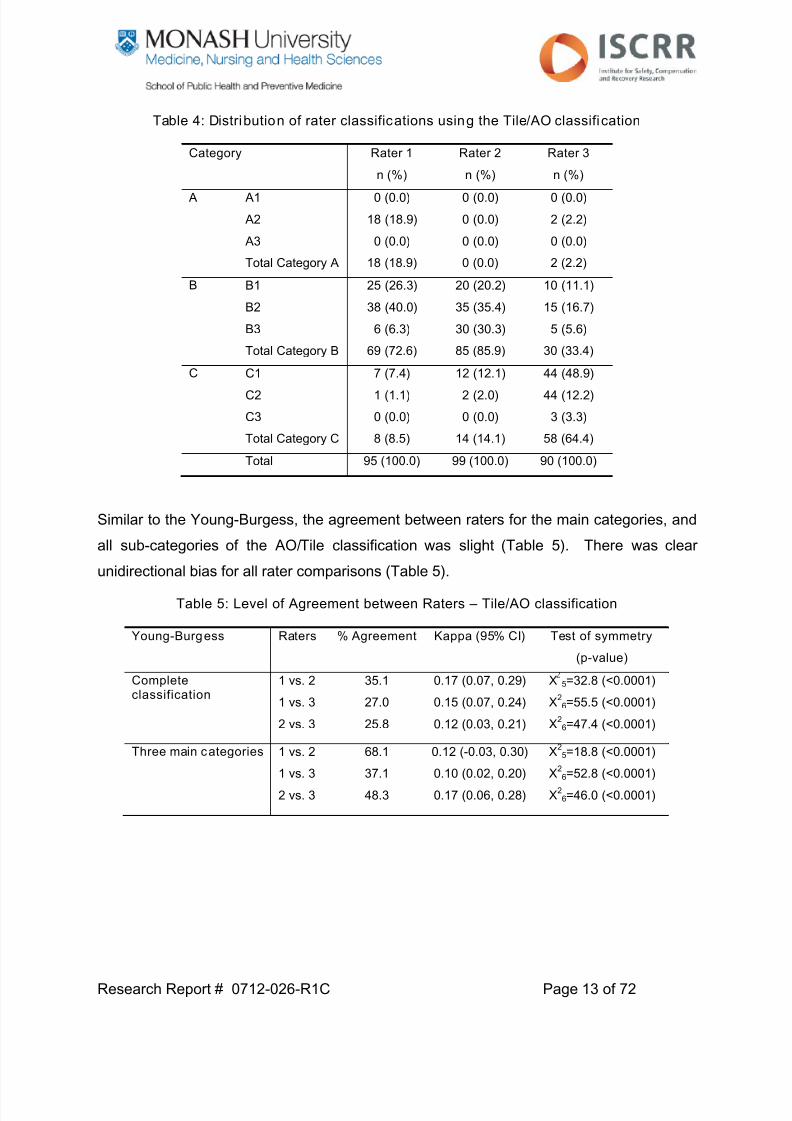
Table 4: Distribution of rater classifications using the Tile/AO classification
Category Rater 1
n (%)
Rater 2
n (%)
Rater 3
n (%)
A A1
A2
A3
Total Category A
0 (0.0)
18 (18.9)
0 (0.0)
18 (18.9)
0 (0.0)
0 (0.0)
0 (0.0)
0 (0.0)
0 (0.0)
2 (2.2)
0 (0.0)
2 (2.2)
B B1
B2
B3
Total Category B
25 (26.3)
38 (40.0)
6 (6.3)
69 (72.6)
20 (20.2)
35 (35.4)
30 (30.3)
85 (85.9)
10 (11.1)
15 (16.7)
5 (5.6)
30 (33.4)
C C1
C2
C3
Total Category C
7 (7.4)
1 (1.1)
0 (0.0)
8 (8.5)
12 (12.1)
2 (2.0)
0 (0.0)
14 (14.1)
44 (48.9)
44 (12.2)
3 (3.3)
58 (64.4)
Total 95 (100.0) 99 (100.0) 90 (100.0)
Similar to the Young-Burgess, the agreement between raters for the main categories, and
all sub-categories of the AO/Tile classification was slight (Table 5). There was clear
unidirectional bias for all rater comparisons (Table 5).
Table 5: Level of Agreement between Raters – Tile/AO classification
Young-Burgess Raters % Agreement Kappa (95% CI) Test of symmetry
(p-value)
Completeclassification
1 vs. 2
1 vs. 3
2 vs. 3
35.1
27.0
25.8
0.17 (0.07, 0.29)
0.15 (0.07, 0.24)
0.12 (0.03, 0.21)
Χ 5=32.8 (<0.0001)
Χ26=55.5 (<0.0001)
Χ26=47.4 (<0.0001)
Three main categories 1 vs. 2
1 vs. 3
2 vs. 3
68.1
37.1
48.3
0.12 (-0.03, 0.30)
0.10 (0.02, 0.20)
0.17 (0.06, 0.28)
Χ25=18.8 (<0.0001)
Χ26=52.8 (<0.0001)
Χ26=46.0 (<0.0001)

7/18/2019 026 Classification, management and outcomes of severe pelvic ring fractures
http://slidepdf.com/reader/full/026-classification-management-and-outcomes-of-severe-pelvic-ring-fractures 14/72
Research Report # 0712-026-R1C Page 14 of 72
Discussion
The accurate classification of pelvic ring fractures requires quality imaging obtained prior
to intervention to treat the fracture. This study assessed the inter-rater agreement of three
consultant orthopaedic surgeons with specific expertise in the management of severe
pelvic ring fracture, using the Young-Burgess and Tile/AO classifications. In particular, the
study focused on the inter-rater reliability using plain radiographs and 3D CT
reconstructions of 100 severe pelvic ring fracture patients. Inter-rater agreement was
slight for both the Young-Burgess and Tile/AO classifications, and the findings highlight
particular challenges for classification of this serious sub-group of pelvic ring fractures.
Only two studies have investigated the reliability of pelvic ring fracture classification. Koo
et al studied the inter-rater reliability of six reviewers (of varying experience) of 30 pelvic
fracture cases and found fair agreement for the Tile classification using plain radiographs
alone and when 2D axial CT scans were provided, while the agreement for the Young-
Burgess classification was substantial for plain radiographs alone, and moderate when 2D
axial CT scans were also used (15). Furey et al studied the inter- and intra-rater reliability
of the Tile and Young-Burgess classification systems using five orthopaedic surgeon
reviewers and the combined plain radiographs and 2D CT scans of 89 pelvic fracture
cases. These authors found moderate agreement for the Tile classification and substantial
agreement for the Young-Burgess system (14). In our study, the inter-rater agreement
between three experienced pelvic and acetabular surgeons was much lower than the
results of Furey et al and Koo et al although there are a number of plausible explanations
for this.
For both previous studies, cases were selected based on the availability and quality of
films, potentially excluding cases where imaging of the pelvis was less clear. Furey et al
specifically excluded cases without “adequate” plain radiographs and CT scans. In our
study, we included all cases with both imaging modalities available, irrespective of the
quality of images, as this represents the clinical conditions under which classification of
these cases occur. It is likely that this approach results in reduced inter-rater reliability but
the findings are more representative of everyday clinical practice.
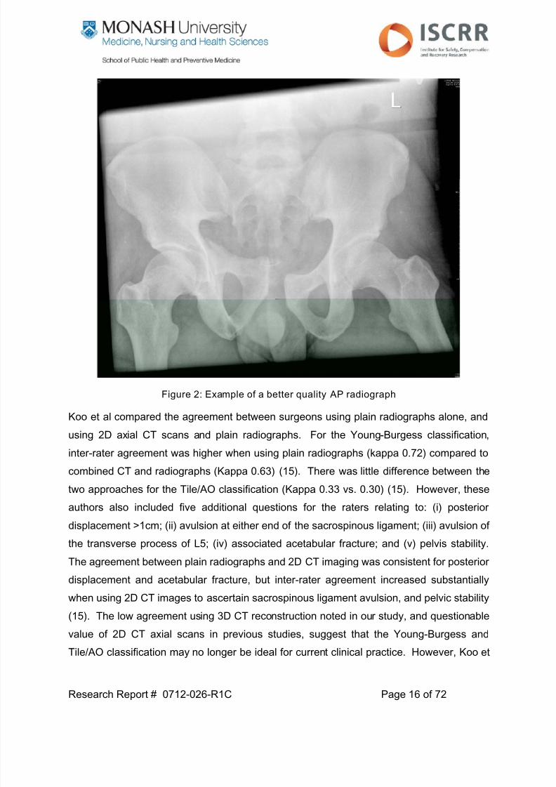
The Young-Burgess and Tile/AO classifications were developed for use with AP, inlet and
outlet view plain radiographs. The inclusion for both previous studies required three plain

7/18/2019 026 Classification, management and outcomes of severe pelvic ring fractures
http://slidepdf.com/reader/full/026-classification-management-and-outcomes-of-severe-pelvic-ring-fractures 15/72
7/18/2019 026 Classification, management and outcomes of severe pelvic ring fractures
http://slidepdf.com/reader/full/026-classification-management-and-outcomes-of-severe-pelvic-ring-fractures 16/72
Research Report # 0712-026-R1C Page 16 of 72
Figure 2: Example of a better quality AP radiograph
Koo et al compared the agreement between surgeons using plain radiographs alone, and
using 2D axial CT scans and plain radiographs. For the Young-Burgess classification,
inter-rater agreement was higher when using plain radiographs (kappa 0.72) compared to
combined CT and radiographs (Kappa 0.63) (15). There was little difference between the
two approaches for the Tile/AO classification (Kappa 0.33 vs. 0.30) (15). However, these
authors also included five additional questions for the raters relating to: (i) posterior
displacement >1cm; (ii) avulsion at either end of the sacrospinous ligament; (iii) avulsion of
the transverse process of L5; (iv) associated acetabular fracture; and (v) pelvis stability.
The agreement between plain radiographs and 2D CT imaging was consistent for posterior
displacement and acetabular fracture, but inter-rater agreement increased substantially
when using 2D CT images to ascertain sacrospinous ligament avulsion, and pelvic stability
(15). The low agreement using 3D CT reconstruction noted in our study, and questionable
value of 2D CT axial scans in previous studies, suggest that the Young-Burgess and
Tile/AO classification may no longer be ideal for current clinical practice. However, Koo et

7/18/2019 026 Classification, management and outcomes of severe pelvic ring fractures
http://slidepdf.com/reader/full/026-classification-management-and-outcomes-of-severe-pelvic-ring-fractures 17/72
Research Report # 0712-026-R1C Page 17 of 72
al’s findings suggest that CT scans may assist with an overall rating of pelvic ring stability
and classification of specific features of pelvic ring disruption. Classification using a
combination of 3D CD reconstructions and 2D axial CT scans may warrant future
investigation, as these modalities are usually available together, and the 2D axial CT
scans do have advantages when assessing specific features such as sacral fractures. In
addition, the use of 2D axial CT scans may be needed where the quality of the 3D
reconstruction is poor (see Figure 3 and Figure 4 for contrasting quality of images).
Figure 3: Example of poor quality 3D CT reconstruction

7/18/2019 026 Classification, management and outcomes of severe pelvic ring fractures
http://slidepdf.com/reader/full/026-classification-management-and-outcomes-of-severe-pelvic-ring-fractures 18/72
Research Report # 0712-026-R1C Page 18 of 72
Figure 4: Example of better quality 3D CT reconstruc tion
The group of patients in our study represent the most severe pelvic ring fractures, who
demonstrate a high risk of mortality and long term disability. Many of these patients arrive
in the emergency department clinically unstable, requiring rapid treatment and decision
making. More than a third of cases arrive in the ED with a binder in situ, which is usually
not removed for imaging purposes, and limits the capacity to accurately classify the extent
of the pelvic ring fracture (21) (see Figure 5).

7/18/2019 026 Classification, management and outcomes of severe pelvic ring fractures
http://slidepdf.com/reader/full/026-classification-management-and-outcomes-of-severe-pelvic-ring-fractures 19/72
Research Report # 0712-026-R1C Page 19 of 72
Figure 5: Plain radiograph of patient with binder in situ
Similarly, the decision to intervene (e.g. emergency placement of an external fixateur) can
be made prior to routine CT scanning in the ED due to urgent need to controlhaemorrhage, again impacting on the capacity to classify the pelvic ring fracture. Previous
classification studies have not focused specifically on severe fractures. Koo et al randomly
selected 30 cases from a Level 1 trauma facility database (15). No attempt was made to
include a representative sample of fractures. Given the small sample size and the low
prevalence of severe pelvic ring fractures in trauma patients, it is highly likely that Koo et
al’s sample included very few severe fractures. As the distribution of each rater’s
classifications was not provided, it is difficult to ascertain the patient group studied by Koo
et al. Furey et al aimed to include a wide range of pelvic ring fractures in their
classification study, with 28% classified as Tile A, 54% as Tile B, and 18% as Tile C. Our
study included almost exclusively Tile B and C-type fractures. It is reasonable to assume
that simple pelvic ring fractures are more amenable to reliable classification, potentially
contributing to the higher reliability of previous studies.
While Koo et al did not specify the software or screen resolution used for classification,
Furey et al ensured that all images were viewed with the specialist picture archiving andcommunication system (PACS) software. In our study, images were downloaded from

7/18/2019 026 Classification, management and outcomes of severe pelvic ring fractures
http://slidepdf.com/reader/full/026-classification-management-and-outcomes-of-severe-pelvic-ring-fractures 20/72
Research Report # 0712-026-R1C Page 20 of 72
PACS at the major trauma services as jpeg files for viewing on personal computers
consistent with the standard computers available in ED, operating theatres and outpatient
consulting rooms. Radiologists at the major trauma services view the images on specialist
high resolution equipment when reporting. Whether the use of improved resolution would
have resulted in better inter-rater reliability is not known. However, the methods used in
this study are consistent with what is available to orthopaedic surgeons, and therefore
more representative of the clinical environment in which fracture classification would occur.
The original sample size for the study was 128 which assumed that all categories of the
Young-Burgess classification would be utilised. While the number of pelvic fractures
cases far exceeded the proposed sample size, obtaining cases with pre-intervention plain
radiographs and 3D CT reconstructions was difficult for many of the reasons discussed.
The study was more than adequately powered to assess the reliability of the Tile/AO
classification, but was under-powered for the Young-Burgess classification. Nevertheless,
this study still represents the largest inter-rater reliability study of pelvic ring fractures
undertaken to date. The low kappa scores and the resulting 95% CI suggest that even
with an additional 28 cases, the findings would be consistent – slight to fair agreement.
The participating surgeons were from three separate institutions, but were all consultantorthopaedic surgeons with extensive experience in managing pelvic fractures, and
received the same documentation describing the classification systems. There was
strong, unidirectional bias between the raters, suggesting that each rater’s application of
the classification guidelines differed. Furey et al studied the reliability of surgeons from a
single institution, and the institution where the Young-Burgess classification was
developed and is used exclusively, but still showed substantial differences between raters
in the distribution of cases to categories of the Young-Burgess and AO/Tile. For example,
in their study, Rater 2 classified 18% of cases as CMI and 27% of cases as C-type
fractures, compared to Rater 4 who classified no cases as CMI and 4% of cases as C-type
fractures (14). Furey et al acknowledged that their results could represent a “best case
scenario” for reliability of rating, further explaining the much lower inter-rater agreement
observed in our study.
Finally, the initial study plan was to include a component assessing intra-rater reliability.
We chose not to complete this aspect of the study given the clear issues with theclassification systems in this group of patients, the low inter-rater reliability, and the

7/18/2019 026 Classification, management and outcomes of severe pelvic ring fractures
http://slidepdf.com/reader/full/026-classification-management-and-outcomes-of-severe-pelvic-ring-fractures 21/72
Research Report # 0712-026-R1C Page 21 of 72
amount of time required from consultant orthopaedic surgeons to complete the
classifications. Pelvic ring fracture classification is a time consuming process, with
consultants providing their time “in kind” to the project. The 3D CT reconstruction can
produce up to 42 single images, resulting in more than 4000 images requiring review from
100 cases. The results from the inter-rater reliability study were sufficient to suggest that
the classifications have limited clinical and research relevance for severe pelvic ring
fractures. Therefore, the need to assess intra-rater reliability was not considered
necessary for this project.
Conclusions
Using plain radiographs and 3D CT reconstructions of the pelvis, inter-rater agreement of
the Young-Burgess and Tile/AO classifications was fair at best for severe pelvic ring
fractures. Current clinical practice at the MTS hospitals does not routinely provide the
plain radiograph views for which the Young-Burgess and Tile/AO classifications were
developed. Severe pelvic ring fracture patients represent a difficult classification prospect
for existing classification systems due to the prevalence of interventions pre-hospital (e.g.
binders) and in the emergency department. The results of this study, and previous
reliability studies, confirm that the Young-Burgess and Tile/AO classifications are
insufficient for clinical and research purposes. Without a reliable classification system,
valid comparison of interventions and patient outcomes is extremely limited. The
development of a classification system specifically for CT images is needed to support
current clinical practice.

7/18/2019 026 Classification, management and outcomes of severe pelvic ring fractures
http://slidepdf.com/reader/full/026-classification-management-and-outcomes-of-severe-pelvic-ring-fractures 22/72
Research Report # 0712-026-R1C Page 22 of 72
Study 2: Mortality following severe pelvic ring fracture:
Does hospital of definit ive care matter?
Introduction
The primary reason for the elevated risk of death following pelvic ring disruption is the
potential for fracture-related haemorrhage through direct injury to the adjacent vasculature
(venous and arterial) from bony fragments, disruption of vessels by shear forces, and
bleeding from the bone surfaces (3-5, 22). In the majority of cases, bleeding is venous in
nature. The risk of haemorrhage, combined with the propensity for severe associated
injuries, increases the risk of mortality in these patients and highlights the need for quality,
evidence-based early management.
While guidelines exist, there is no clear consensus about the optimal management
approach for haemodynamically unstable patient with pelvic fracture (3-8). Protocols tend
to favour either early interventional radiography (i.e. angiography to identify the source of
bleeding and embolization to stop the bleeding) or immediate laparotomy for surgical
control of bleeding and pelvic packing with large sponges.
In Victoria, the major trauma service (MTS) hospitals manage more than 90% of severe
pelvic ring fractures in the state (9). While both hospitals have implemented similar
massive transfusion protocols, each has implemented a different protocol for the
management of the haemodynamically unstable pelvic fracture patient. Each MTS
hospital uses a different indication for angiographic embolisation, with The Alfred
embolising patients who demonstrate an arterial “blush” on CT scan as a priority (Figure
6). In contrast, the RMH uses a protocol that advocates angiography ± embolisation if
haemodynamically unstable and in the presence of a pelvic haematoma (Figure 7).
A preliminary study, using data only from the Victorian State Trauma Registry, found no
difference in risk-adjusted mortality between the MTS hospitals (9). However, limitations
were noted and the need for additional data regarding resuscitation practices, and fracture
classification, were acknowledged as necessary for a full evaluation of the MTS
approaches to management.

7/18/2019 026 Classification, management and outcomes of severe pelvic ring fractures
http://slidepdf.com/reader/full/026-classification-management-and-outcomes-of-severe-pelvic-ring-fractures 23/72
Research Report # 0712-026-R1C Page 23 of 72
Figure 6: Protocol for management of the haemodynamically unstable patient with pelvic
fracture (The Alfred)

7/18/2019 026 Classification, management and outcomes of severe pelvic ring fractures
http://slidepdf.com/reader/full/026-classification-management-and-outcomes-of-severe-pelvic-ring-fractures 24/72
Research Report # 0712-026-R1C Page 24 of 72
Figure 7: Protocol for management of the haemodynamically unstable patient with pelvic
fracture (RMH)

7/18/2019 026 Classification, management and outcomes of severe pelvic ring fractures
http://slidepdf.com/reader/full/026-classification-management-and-outcomes-of-severe-pelvic-ring-fractures 25/72
Research Report # 0712-026-R1C Page 25 of 72
Project aims
The aims of this study were to:
i. Describe the profile of severe pelvic ring fracture cases definitively managed at The
Alfred and RMH
ii. Describe the early management practices of the MTS hospitals for severe pelvic
ring fracture
iii. Establish the association between definitive hospital of management and in-hospital
mortality.
Methods
Setting
The state of Victoria, Australia has a population of approximately 5.4 million and operates
a regionalised, inclusive trauma system. The trauma system is monitored by the Victorian
State Trauma Registry (VSTR) and the Victorian Orthopaedic Trauma Outcomes Registry
(VOTOR). The VSTR is a population-based registry collecting data about all major trauma
patients in Victoria (23). Since 2007, the VSTR has followed-up all adult survivors to
discharge at 6, 12 and 24-months post-injury by telephone interview (24). VOTOR is a
sentinel site registry, integrated within the VSTR, collecting data about all adult
orthopaedic trauma patients with a length of stay >24 hours, and admitted to The Alfred,
Royal Melbourne, Geelong and Northern Hospitals. All VOTOR patients are followed-up
by telephone interview at 6 and 12-months post-injury using the same methodology as the
VSTR patients.
Participants
Patients with a date of injury from July 2007 to June 2010 (inclusive), captured by the
VSTR and definitively managed at the state’s adult major trauma services (The Alfred and
Royal Melbourne Hospital), were included. All cases with an Abbreviated Injury Scale
(AIS) pelvis fracture coded as 852606.4, 852608.4, or 852610.5 were selected for this
study. The selected AIS codes related to pelvic fractures with substantial “deformation or
displacement” and a severity score of “4” (severe) or “5” (critical).

7/18/2019 026 Classification, management and outcomes of severe pelvic ring fractures
http://slidepdf.com/reader/full/026-classification-management-and-outcomes-of-severe-pelvic-ring-fractures 26/72
Research Report # 0712-026-R1C Page 26 of 72
VSTR and VOTOR data
Data for all eligible cases were extracted from the VSTR and VOTOR databases and
included:
i. Patient demographics
ii. Comorbid status
iii. Injury event details
iv. Pre-hospital management observations
v. Inter-hospital transfer and transfer times
vi. Status on arrival in the emergency department (ED)
vii. Injury diagnoses and severity
viii. Admission to ICU, ICU length of stay, hospital length of stay, discharge destination
and in-hospital mortality.
Medical record review
Individual patient medical records and hospital surgical systems were reviewed to collect
more detailed data about the haemodynamic status of the patient, resuscitation data,
operative management, complications and readmission. The full data collection form is
attached as Appendix A, but key data items abstracted from the medical record and
hospital systems included:
i. Markers of haemodynamic instability such as the base excess (BE) on arrival,
lowest international normalised ratio (INR) and fibrinogen levels. Low BE,
measured in mEq/L, suggests metabolic acidosis resulting from a shortage of
circulating oxygen. The INR is a measure of clotting time with higher values
representing longer time to clotting of the blood, and is therefore a marker of
coagulopathy. Fibrinogen (measured in g/L) is also a measure of coagulopathy with
low fibrinogen levels suggesting systemic activation of the clotting system and
consumption of clotting factors faster than they can be reproduced.
ii. Use of blood products including the type and units used. In particular, units used of
packed red blood cells (PRBC), fresh frozen platelets (FFP) and platelets were
recorded.
7/18/2019 026 Classification, management and outcomes of severe pelvic ring fractures
http://slidepdf.com/reader/full/026-classification-management-and-outcomes-of-severe-pelvic-ring-fractures 27/72
Research Report # 0712-026-R1C Page 27 of 72
iii. Use of fluid products including the type and volume given
iv. Use and timing of binders, C-clamps, and external fixateurs. Binders are a non-
invasive method of applying strong compressive force to the pelvis to assist incontrolling bleeding and reduction of the fracture. External fixation is a form of
surgical management where holes are drilled into the bone from outside the body
and a metal frame inserted to reduce the fracture. The C-clamp is a metal clamp
applied to rapidly reduce and stabilise the posterior pelvic ring.
v. Use and timing of angiography and embolisation. Angiography provides a method
of identifying arterial bleeding sources and embolisation is employed to slow or stop
bleeding from the source.
vi. Use and timing of surgical fixation of the pelvis fracture
vii. Documented complications and readmission to hospital.
Fractures were classified using the Young-Burgess and Tile/AO systems. Given the low
inter-tester reliability established in Study 1, classification by consensus was used. A
consensus of two orthopaedic surgeons was required to record a classification for the
pelvic fracture.
Data analysis
Data were summarised using percentages for categorical, and mean and standard
deviations (SD) or median and interquartile range (IQR) for continuous variables.
Comparison of groups (e.g. in-hospital deaths vs. survivors, RMH vs. The Alfred) was
conducted using chi-square tests for categorical variables, and independent t-tests or
Mann-Whitney U-tests for continuous variables.
Hospital of definitive care was the primary exposure of interest. Therefore, establishing an
accurate estimate of the association between hospital of definitive care and in-hospital
mortality requires adjustment for key confounders. Variables were considered to be
confounders of the association between hospital of definitive management and in-hospital
mortality if they differed between hospital, and were also associated with in-hospital
mortality. Univariate logistic regression was used to establish the unadjusted odds of
mortality at one hospital relative to the other. Multivariate logistic regression was used to
establish the association between hospital of definitive care and in-hospital mortality,

7/18/2019 026 Classification, management and outcomes of severe pelvic ring fractures
http://slidepdf.com/reader/full/026-classification-management-and-outcomes-of-severe-pelvic-ring-fractures 28/72
Research Report # 0712-026-R1C Page 28 of 72
adjusted for established confounders. Variables with a p-value <0.10 were considered to
be potential confounders and included in the multivariate model. Adjusted odds ratios
(AOR) and 95% confidence intervals (CI) were calculated, and a p-value <0.05 was
considered significant for all statistical tests. All analyses were performed using Stata
Version 11.2 (StataCorp, College Station, TX).
Results
Comparison of cases managed at The Alfred and RMH
There were 145 severe pelvic ring fracture patients definitively managed at The Alfred and
RMH from July 2007 to June 2010 (inclusive). Ninety cases were managed at The Alfred
and 55 cases at RMH. Almost half of severe pelvic ring cases were aged 15-34 years,
and the vast majority were male (Table 6). Eighty per cent were the result of transport
crashes, predominantly motor vehicle, motorcycle and pedestrian incidents (Table 6).
The demographic and injury event profile of cases definitively managed at each MTS
hospital was similar (Table 6). There was no difference between the hospitals with respect
to age group (Χ22=0.23 p=0.89), gender (Χ2
1=0.20 p=0.66), intent of injury (Χ21=0.52
p=0.47), or compensable status (Χ21=0.85 p=0.36). The proportion of transport-related
cases across the two hospitals was similar (Χ21=0.49 p=0.48), but the proportion of motor
vehicle related cases was higher at The Alfred (48% vs. 29%) and the proportion of
pedestrian cases was higher at RMH (26% vs. 8.9%). The proportion of cases with a
documented comorbidity was higher for The Alfred (Χ21=4.5 p=0.03).

7/18/2019 026 Classification, management and outcomes of severe pelvic ring fractures
http://slidepdf.com/reader/full/026-classification-management-and-outcomes-of-severe-pelvic-ring-fractures 29/72
Research Report # 0712-026-R1C Page 29 of 72
Table 6: Characteristics of patients
Descrip tor The Alfred
(n=90)
RMH
(n=55)
Age N (%)
15-34 years
35-64 years
≥ 65 years
44 (48.9)
32 (35.6)
14 (15.5)
25 (45.4)
20 (36.4)
10 (18.2)
Gender N (%)
Male
Female
70 (77.8)
20 (22.2)
41 (74.6)
14 (25.4)
CCI comorbid condition N (%)
No
Yes
55 (61.1)
35 (38.9)
43 (78.2)
12 (21.8)
Transport related? N (%)
No
Yes
19 (21.1)
71 (78.9)
9 (16.4)
46 (83.6)
Mechanism N (%)
Motor vehicle
Motorcycle
Pedestrian
High fall
Other
43 (47.8)
18 (20.0)
8 (8.9)
7 (7.8)
14 (15.6)
16 (29.1)
14 (25.5)
14 (25.5)
4 (7.3)
7 (12.7)
Intent N (%)
Unintentional
Intentional self-harm
86 (95.5)
4 (4.5)
51 (92.7)
4 (7.3)
Compensable status N (%)
Compensable
Non-compensable
67 (74.4)
23 (25.6)
45 (83.3)
9 (16.7)
Almost 40 per cent of cases were hypotensive (SBP <90 mmHg) on arrival of the
paramedics at the scene, over a third were fitted with a pelvic binder, and the majority
were given IV fluids in the pre-hospital setting. Only eight cases (6.5%) received blood
products prior to arrival at hospital (Table 7).

7/18/2019 026 Classification, management and outcomes of severe pelvic ring fractures
http://slidepdf.com/reader/full/026-classification-management-and-outcomes-of-severe-pelvic-ring-fractures 30/72
Research Report # 0712-026-R1C Page 30 of 72
Table 7: Pre-hospital management, transport to hospital, and ED care
Descrip tor The Alfred
(n=90)
RMH
(n=55)
Hypotensive on arrivalat the scene of injury
aN (%)
No
Yes
42 (58.3)
30 (41.7)
35 (67.3)
17 (32.7)
Binder used pre-hospital
N (%)
No
Yes
54 (61.4)
34 (38.6)
32 (61.5)
20 (38.5)
Blood given pre-hospitalc
N (%)
No
Yes
80 (91.9)
7 (8.1)
43 (97.7)
1 (2.3)
Fluid given pre-hospital
N (%)
NoYes
13 (16.1)73 (83.9)
4 (9.1)40 (90.9)
Inter-hospital transfer N (%)
No
Yes
65 (72.2)
25 (27.8)
53 (96.4)
2 (3.6)
Hypotensive on arrival in EDe
N (%)
No
Yes
74 (86.1)
12 (13.9)
38 (73.1)
14 (26.9)
Binder in ED
N (%)
No
Yes
23 (25.6)
67 (74.4)
14 (27.5)
37 (72.5)Blood given in ED
gN (%)
No
Yes
35 (49.8)
53 (60.2)
12 (26.7)
33 (73.3)
Base Excess Median (IQR) mEq/L -4.0 (-7.0 to -1.0) -6.7 (-12.4 to -3.2)
Fibrinogen Median (IQR) g/L 1.6 (1.1 to 2.3) 1.8 (1.0 to 2.7)
INR Median (IQR) 1.3 (1.2 to 1.6) 1.2 (1.1 to 1.5)
24-hour PRBC requirements Median (IQR) units 4 (0 to 8) 5 (1 to 9)a Data missing for n=21 cases;
b Data missing for n=5 cases;
c Data missing for n=14 cases;
d Data missing
for n=15 cases;e Data missing for n=7 cases;
f Data missing for n=4 missing;
g Data missing for n=12 cases
There was no difference in the proportion of cases hypotensive (SBP <90 mmHg) at the
scene of injury (Χ21=0.01 p=0.95), administered blood products (Χ2
1=1.32 p=0.25), given
IV fluids (Χ21=2.51 p=0.12), or fitted with a pelvic binder by paramedics (Χ2
1=1.03 p=0.31),
between the hospitals (Table 7). The proportion of cases experiencing an inter-hospital
transfer was significantly higher at The Alfred (Χ21=0.13.13 p<0.001). The proportion of
cases arriving at The Alfred ED in hypotension was lower (Χ21=3.56 p=0.06), and the first
arterial base excess taken in the ED was significantly lower for RMH patients (z=2.88
p=0.004), suggesting greater haemodynamic instability (Table 7).

7/18/2019 026 Classification, management and outcomes of severe pelvic ring fractures
http://slidepdf.com/reader/full/026-classification-management-and-outcomes-of-severe-pelvic-ring-fractures 31/72
Research Report # 0712-026-R1C Page 31 of 72
Table 8: Fracture severity and associated injuries
Descriptor The Alfred
(n=90)
RMH
(n=55)
Injury Severity Score Median (IQR) 29 (24-41) 34 (26-45)
Associated head injury (AIS severity >3) N (%)
No
Yes
78 (86.7)
12 (13.3)
43 (78.2)
12 (21.8)
Associated thoracic injury (AIS severity>3)
N (%)
No
Yes
80 (88.9)
10 (11.1)
51 (92.7)
4 (7.3)
Associated abdominal injury (AISseverity >3)
N (%)
No
Yes
77 (85.6)
13 (14.4)
46 (83.6)
9 (16.4)
Associated femoral shaft fracture N (%)No
Yes
75 (83.3)
15 (16.7)
43 (78.2)
12 (21.8)
GCS on arrival in ED N (%)
9-15
3-8
71 (85.5)
12 (14.5)
42 (80.8)
10 (19.2)
Young and Burgess classification N (%)
APC1
APC2
APC3
LC1LC2
LC3
Vertical Shear
CMI
4 (4.7)
11 (12.8)
7 (8.1)
9 (10.5)13 (15.1)
33 (38.4)
5 (5.8)
4 (4.7)
2 (3.9)
8 (15.7)
4 (7.8)
14 (27.5)3 (5.9)
13 (25.5)
4 (7.8)
3 (5.9)
Tile/AO classification N (%)
A1
B1
B2
B3
C1
C2
C3
1 (1.2)
12 (14.3)
30 (35.7)
24 (28.6)
15 (14.9)
1 (1.2)
1 (1.2)
1 (1.9)
11 (21.1)
17 (32.7)
11 (21.2)
6 (11.5)
4 (7.7)
2 (3.9)
The INR was higher for cases at The Alfred (z=1.86, p=0.06), but there was no difference
in the 24-hour packed red blood cell requirements (z=-0.95, p=0.35), or fibrinogen levels
(z=-0.26, p=0.80), between the hospitals. The ISS was higher for cases definitively
managed at RMH (z=-2.02, p=0.04). However, the proportion of cases with severe
associated head (Χ21=1.78, p=0.18), thoracic (Χ21=0.58, p=0.45) or abdominal (Χ21=0.10,
p=0.76) injuries did not differ between the hospitals (Table 8). The proportion of B-type

7/18/2019 026 Classification, management and outcomes of severe pelvic ring fractures
http://slidepdf.com/reader/full/026-classification-management-and-outcomes-of-severe-pelvic-ring-fractures 32/72
Research Report # 0712-026-R1C Page 32 of 72
and C-type fractures definitively managed at The Alfred (78.6% and 20.2%) was similar to
the proportion managed at RMH (75.0% and 23.1%). Similarly, there was no difference in
the profile of fractures classified using the Young-Burgess system between the hospitals
(Χ23=0.95, p=0.81).
Table 9: Fracture management
Descrip tor The Alfred
(n=90)
RMH
(n=55)
Angiography? N (%)
No
Yes
70 (77.8)
20 (22.2)
26 (47.3)
29 (52.7)
Embolisation?** N (%)
No
Yes
8 (40.0)
12 (60.0)
1 (3.5)
28 (96.5)
Time to angiography Median (IQR) hours 5.1 (3.7-10.5) 2.8 (1.6-4.1)
C-clamp used? N (%)
No
Yes
85 (94.4)
5 (5.6)
54 (98.2)
1 (1.8)
External fixateur? N (%)
No
Yes
48 (53.3)
42 (46.7)
38 (69.1)
17 (30.9)
Time to external fixation Median (IQR) hours 2.9 (1.9-5.2) 4.8 (3.7-24.0)
Pelvic packing used? N (%)No
Yes
81 (90.0)
9 (10.0)
51 (92.7)
4 (7.3)
ORIF? N (%)
No
Yes
20 (22.2)
70 (77.8)
31 (56.4)
24 (43.6)
Laparotomy? N (%)
No
Yes
62 (68.9)
28 (31.1)
42 (76.4)
13 (23.6)
Time to ORIF Median (IQR) days 1.5 (1.0-2.5) 4.0 (2.5-8.0)
** Patients referred for angiography only
Consistent with the different protocols implemented, there were significant differences
between the hospitals with respect to early fracture management. The proportion of cases
referred for angiography was significantly higher (Χ21=14.20, p<0.001), and the time to
angiography was much lower (z=3.49, p<0.001), at RMH (Table 9). The rate of
embolisation was higher at RMH compared to The Alfred (Χ21=10.55, p=0.001). The one
case at RMH where embolisation was not performed was due to calcification of the
femoral artery, preventing access for embolisation.
7/18/2019 026 Classification, management and outcomes of severe pelvic ring fractures
http://slidepdf.com/reader/full/026-classification-management-and-outcomes-of-severe-pelvic-ring-fractures 33/72
Research Report # 0712-026-R1C Page 33 of 72
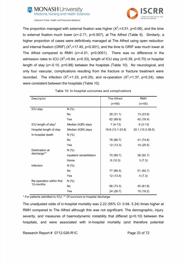
The proportion managed with external fixation was higher (Χ21=3.51, p=0.06), and the time
to external fixation much lower (z=-2.71, p=0.007), at The Alfred (Table 9). Similarly, a
higher proportion of cases were definitively managed at The Alfred using open reduction
and internal fixation (ORIF) (Χ21=17.45, p<0.001), and the time to ORIF was much lower at
The Alfred compared to RMH (z=-4.01, p=0.0001). There was no difference in the
admission rates to ICU (Χ21=0.94, p=0.33), length of ICU stay (z=0.39, p=0.70) or hospital
length of stay (z=-0.15, p=0.88) between the hospitals (Table 10). No neurological, and
only four vascular, complications resulting from the fracture or fracture treatment were
recorded. The infection (Χ21=1.33, p=0.25), and re-operation (Χ2
1=1.37, p=0.24), rates
were consistent between the hospitals (Table 10).
Table 10: In-hospital outcomes and complications
Descrip tor The Alfred
(n=90)
RMH
(n=55)
ICU stay N (%)
No
Yes
28 (31.1)
62 (68.9)
13 (23.6)
42 (76.4)
ICU length of stay* Median (IQR) days 7 (4-13) 6 (3-13)
Hospital length of stay Median (IQR) days 19.8 (12.1-33.8) 20.1 (10.3-39.5)In-hospital death N (%)
No
Yes
78 (86.7)
12 (13.3)
41 (74.6)
14 (25.5)
Destination atdischarge**
N (%)
Inpatient rehabilitation
Home
70 (89.7)
8 (10.3)
38 (92.7)
3 (7.3)
Infection N (%)
No
Yes
77 (86.5)
12 (13.5)
51 (92.7)
4 (7.3)
Re-operation within first12-months
N (%)
No
Yes
66 (73.3)
24 (26.7)
45 (81.8)
10 (18.2)
* For patients admitted to ICU; ** Of survivors to hospital discharge
The unadjusted odds of in-hospital mortality was 2.22 (95% CI: 0.94, 5.24) times higher at
RMH compared to The Alfred although this was not significant. The demographic, injury
severity, and measures of haemodynamic instability that differed (p<0.10) between thehospitals, and were associated with in-hospital mortality (and therefore potential
7/18/2019 026 Classification, management and outcomes of severe pelvic ring fractures
http://slidepdf.com/reader/full/026-classification-management-and-outcomes-of-severe-pelvic-ring-fractures 34/72
Research Report # 0712-026-R1C Page 34 of 72
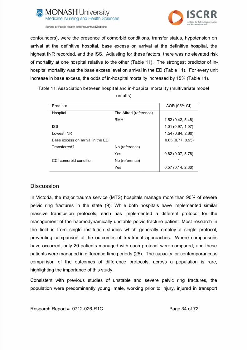
confounders), were the presence of comorbid conditions, transfer status, hypotension on
arrival at the definitive hospital, base excess on arrival at the definitive hospital, the
highest INR recorded, and the ISS. Adjusting for these factors, there was no elevated risk
of mortality at one hospital relative to the other (Table 11). The strongest predictor of in-
hospital mortality was the base excess level on arrival in the ED (Table 11). For every unit
increase in base excess, the odds of in-hospital mortality increased by 15% (Table 11).
Table 11: Association between hospital and in-hospi tal mortality (multivariate model
results)
Predicto AOR (95% CI)
Hospital The Alfred (reference)
RMH
1
1.52 (0.42, 5.48)
ISS 1.01 (0.97, 1.07)
Lowest INR 1.54 (0.84, 2.80)
Base excess on arrival in the ED 0.85 (0.77, 0.95)
Transferred? No (reference)
Yes
1
0.62 (0.07, 5.78)
CCI comorbid condition No (reference)
Yes
1
0.57 (0.14, 2.30)
Discussion
In Victoria, the major trauma service (MTS) hospitals manage more than 90% of severe
pelvic ring fractures in the state (9). While both hospitals have implemented similar
massive transfusion protocols, each has implemented a different protocol for the
management of the haemodynamically unstable pelvic fracture patient. Most research in
the field is from single institution studies which generally employ a single protocol,preventing comparison of the outcomes of treatment approaches. Where comparisons
have occurred, only 20 patients managed with each protocol were compared, and these
patients were managed in difference time periods (25). The capacity for contemporaneous
comparison of the outcomes of difference protocols, across a population is rare,
highlighting the importance of this study.
Consistent with previous studies of unstable and severe pelvic ring fractures, the
population were predominantly young, male, working prior to injury, injured in transport

7/18/2019 026 Classification, management and outcomes of severe pelvic ring fractures
http://slidepdf.com/reader/full/026-classification-management-and-outcomes-of-severe-pelvic-ring-fractures 35/72
Research Report # 0712-026-R1C Page 35 of 72
accidents, and involving other associated injuries (11, 26, 27). All had sustained other
associated injuries and the mean ISS (31.8) was consistent with other studies (22, 28, 29).
The results of this study highlight substantial differences in the early management ofsevere pelvic ring fractures between the hospitals despite relatively few differences in
patient case-mix. The key differences between severe pelvic ring fracture patients
managed at The Alfred and RMH were a higher prevalence of pre-existing comorbidity in
The Alfred cohort, and almost all cases directly transported from the scene of injury to
RMH compared to 28% of The Alfred patients arriving via inter-hospital transfer. A higher
proportion of RMH cases arrived in the ED exhibiting signs of haemodynamic shock (e.g.
decreased base excess, hypotensive, etc.), potentially reflecting the higher proportion of
direct transports from the scene. However, in data no shown, restricting analysis to
patients transported directly from the scene (n=65 Alfred vs. n=53 RMH), the differences in
highest INR and first base excess levels persisted.
More than half of RMH cases were referred for angiography (and almost all cases
subsequently embolised) compared to 22% of The Alfred cases referred for angiography
(of which 60% were embolised). Despite the differences in protocols, the prevalence of
pelvic packing and laparotomy were consistent between the hospitals, but markeddifferences in pelvic fracture fixation noted. External and internal fixation use was more
prevalent at The Alfred, and the time to fixation much shorter, when compared to RMH.
Consistent with the previous preliminary study (9), unadjusted in-hospital mortality rates
were higher at RMH but there was no difference in the risk of mortality between The Alfred
and RMH when adjusted for potential confounders. Again, when restricting the analysis to
direct transports to the MTS hospitals from the scene, the unadjusted odds of mortality at
RMH compared to The Alfred was 1.76 (95% CI: 0.72, 2.49). Similarly, adjusting for the
differences in INR and BE levels, the overall finding was consistent (AOR 1.53, 95% CI:
0.48, 4.88).
The only previous study to directly compare a pelvic packing approach with early
angiography involved 40 patients (20 in each group) from a single institution at different
times (25). Osborn et al found no difference in mortality between the treatment
approaches, but reported a decrease in the proportion of cases requiring transfusion in the
pelvic packing group in the 24 hours after the intervention and less early deaths (25). The24-hour packed red blood cell requirements were lower at The Alfred, the institution
7/18/2019 026 Classification, management and outcomes of severe pelvic ring fractures
http://slidepdf.com/reader/full/026-classification-management-and-outcomes-of-severe-pelvic-ring-fractures 36/72
Research Report # 0712-026-R1C Page 36 of 72
favouring pelvic packing, but the number of patients receiving pelvic packing was low (and
consistent) at both institutions, and the reduced PRBC requirements for The Alfred cases
could reflect greater haemodynamic stability on arrival in the ED.
Accounting for differences in the prevalence of comorbidity, hypotension on arrival in the
ED, transfer status, clotting times (INR), and base excess (BE) on arrival in the ED, there
was no difference in the risk of mortality between the hospitals, suggesting much of the
noted difference in mortality was explained by differences in the condition of patients on
arrival at hospital, rather than the protocol employed. The base excess on arrival was the
strongest predictor of in-hospital mortality in this group of severe pelvic ring fractures, with
each unit decrease resulting in a 15% increase in the odds of mortality. Jeske et al, in
their study of 45 haemodynamically unstable patients with pelvic fractures, also found that
that BE and ISS were associated with survival (30). Similarly, Siegel et al found that BE
was a highly significant early predictor of outcome in pelvic fracture (31), while multiple
studies have identified BE as a predictor of mortality in trauma patients (32, 33). The BE
reflects the disturbances in the physiologic status of the patient. A low value indicates
metabolic acidosis, which is an imbalance between oxygen delivery and consumption
resulting in anaerobic metabolism. The base deficit (i.e. low BE) represents the nett result
of the oxygen demand and delivery to the cells, thus representing the respiratory
combined with the hemodynamic pathophysiologic findings of the patient. A highly
negative base deficit indicates a patient in whom one or both systems are failing to deliver
with consequences for morbidity and mortality, explaining the high risk of mortality in
patients with a low BE.
The difference in the proportion of patients undergoing angiography and subsequent
embolisation, and external fixation, reflects the different protocols employed by the
hospitals. Many papers have described the role of angiography in haemodynamically
unstable patients with pelvic fractures. The prevalence of patients requiring embolisation is
reported to be less than 10% (34-36). However, in a select group of patients (e.g.
persistent hypotension, high revised trauma score and older age), the proportion requiring
embolisation ranges from 57-75% (19, 37). At the RMH, were the protocol describes early
angiography, 53% of patients were referred for angiography and 98% were subsequently
embolised. In contrast, only 22% of cases at The Alfred were referred to angiography and
60% were subsequently embolised. The higher rate of embolisation could reflect the

7/18/2019 026 Classification, management and outcomes of severe pelvic ring fractures
http://slidepdf.com/reader/full/026-classification-management-and-outcomes-of-severe-pelvic-ring-fractures 37/72
Research Report # 0712-026-R1C Page 37 of 72
earlier referral to angiography of RMH patients, with embolisation of bleeding sources that
may or may not have resolved without embolisation.
Early angiography and subsequent embolisation has been advocated by many authors toreduce mortality in this challenging patient population (30, 34, 38, 39). A key point to
make about angiography is that it can only address arterial sources of bleeding. Where
bleeding occurs from the surface of the fracture fragments or venous vessels, angiography
will have no impact on control of bleeding. Another disadvantage of angiography is that it
is not easy accessible, and Gansslen et al highlighted that even in a Level-I trauma
centres, there can give considerable delays (6). Angiography can be time-consuming and
delayed treatment of associated injuries can occur. At the RMH the median time to
angiography was 2.8 hours, which is consistent with the literature (6, 34, 40), but whether
this was the time to arrival at the angiography suite or the time to embolisation was difficult
to ascertain from the patient record. Aside from the potential for delays in treatment,
angiography and embolisation has been associated with cases of gluteal necrosis (41).
However, others have suggested that this complication is related to trauma to the gluteal
region along with protracted hypotension rather than a direct complication of embolisation
(40).
Less has been published about the effectiveness, and limitations, of external fixation and
pelvic packing of severe pelvic ring fractures. Advocates of early external fixation of pelvic
fractures highlight the capacity of this approach to reduce the bleeding occurring from
fracture surfaces, and associated pain relief, as the key benefits. While these results can
be achieved with binders, binders cannot be left in situ for prolonged periods due to
compromise of soft tissue structures around the pelvis. Where time to operating theatre
for definitive fracture fixation is unknown, external fixation may be a sensible choice.
However, as with all invasive procedures, there are disadvantages. Pin site infections are
relatively common.
Historically, pelvic packing was performed as a trans-peritoneal procedure after an
exploratory laparotomy late in the resuscitation phase with poor results (42). Opening of
the peritoneum can potentially alter the tamponade effect of the retroperitoneal space.
The tamponade effect occurs where pressure from bleeding within the confined space of
the retroperitoneal cavity acts to compress the vessels and slow bleeding naturally.Opening the peritoneum releases the pressure, allowing more bleeding to occur. The

7/18/2019 026 Classification, management and outcomes of severe pelvic ring fractures
http://slidepdf.com/reader/full/026-classification-management-and-outcomes-of-severe-pelvic-ring-fractures 38/72
Research Report # 0712-026-R1C Page 38 of 72
more recent development of a controlled retroperitoneal approach is considered quicker,
easier to perform and less invasive (43), which is potentially less likely to result in an
altered tamponade effect, and also considered to have similar mortality rates to
emergency angiography (44). However, there is a risk of infection with this invasive
approach and additional surgery is always required to remove the sponges used for pelvic
packing.
Papakostidis et al explored the efficacy of pelvic packing and arterial embolisation for
managing pelvic fractures using a systematic review approach (45, 46). These authors
concluded that an emergency-treatment protocol based on prompt pelvic ring stabilisation,
pelvic tamponade through a minimal extraperitoneal approach followed, when necessary,
by pelvic angiography and selective embolisation could offer the best chances for survival
(46). However, there were no randomised controlled trials to review, and the systematic
review focused exclusively on cases series, limiting the conclusions that could be drawn.
Suzuki et al, in their review of the literature, concluded that the two interventions are
complementary but that the haemodynamically unstable patients should first go to theatre
for pelvic stabilization and packing and other procedures required for associated injuries
(47). The findings of our study found no clear superiority of one protocol over the other,
and the incidence of complications such as reoperation and infection were consistent
between the hospitals. However, the use of pelvic packing was rare, accounting for 10%
of cases managed at The Alfred and 7% of cases at RMH, with the greater diversity in
management related to external fixation and angiography.
There were a number of strengths to this study. The study included more than 90% of
eligible cases within the defined population, and involved contemporaneous comparison of
outcomes between the hospitals. Pre-hospital data were able to be included, providing
information about the pelvic fracture care provided prior to arrival at hospital.
Nevertheless, limitations to the study were evident. The collection of fluid management,
resuscitation and detailed clinical management data directly from the medical record is
challenging. Seven records from the RMH could not be obtained for coding, despite
repeated requests. Whilst this is not likely to have introduced substantial bias as there
were no clear patient-related details that defined the missing medical records, it did reduce
the completeness of the data, and the power of the study, particularly with respect toassessing the resuscitation details of these patients. From Study 1, there were a number

7/18/2019 026 Classification, management and outcomes of severe pelvic ring fractures
http://slidepdf.com/reader/full/026-classification-management-and-outcomes-of-severe-pelvic-ring-fractures 39/72
Research Report # 0712-026-R1C Page 39 of 72
of patients with insufficient imaging for classification, and the reliability of the classification
was low. To address this, a consensus approach was used for classification. There was
difficulty ascertaining the incidence of complications due to inconsistent reporting in the
medical record. In particular, separating treatment (i.e. iatrogenic) complications from
complications resulting from the injury itself was challenging. The study was observational
in design. While a randomised controlled trial (RCT) would be the design of choice for
assessing the effectiveness and efficacy of treatment approaches, this would not be
feasible due to the need to staff, train and equip both hospitals to implement both
protocols simultaneously.
Conclusions
The MTS hospitals have implemented different protocols for managing the
haemodynamically unstable pelvic fracture patient. Key differences between the hospitals
were clear with RMH favouring early referral to angiography and embolisation of virtually
all cases referred to angiography. In contrast, early external fixation was a feature of
cases managed at The Alfred. The in-hospital mortality rate was slightly higher at RMH,
but adjusted for key confounders, there was no difference in the risk-adjusted odds of
mortality following severe pelvic ring fracture between the MTS hospitals. There was no
clearly superior treatment approach, continuing the controversy that exists regarding best
practice management of these cases. Ongoing monitoring and review is required.

7/18/2019 026 Classification, management and outcomes of severe pelvic ring fractures
http://slidepdf.com/reader/full/026-classification-management-and-outcomes-of-severe-pelvic-ring-fractures 40/72
Research Report # 0712-026-R1C Page 40 of 72
Study 3: Factors associated with long term patient-
reported outcomes after severe pelvic ring fracture
Introduction
Severe pelvic ring fractures are high-energy injuries, resulting from significant force such
as motor vehicle and motorcycle crashes, pedestrian struck by a car, and falls from height.
The primary aim of fracture management is to restore the anatomical integrity of the pelvic
ring (11), while the overall goal of treatment and rehabilitation is the return to pre-injury
levels of functioning for the patient. In the past, most pelvic fractures were managed
conservatively, but advances in surgical fixation and understanding of the anatomy of the
pelvic ring have led to the development of operative approaches (48).
Historically, pelvic fracture outcomes have been limited to assessment of radiological and
selected clinical outcome measures (11), which are generally poorly correlated with patient
function and quality of life. Studies assessing the long term patient-reported outcomes of
patients are few and have been largely limited to very small samples, often from single
centre studies (10-12), limiting the capacity to assess the contribution of pelvic fracture
severity and treatment to patient outcome in this group of often multiply injured patients.
Other pelvic fracture outcome studies have focused on patients only managed surgically or
managed using a specific technique, or have focused on specific patient sub-sets such as
unstable sacral fractures and female patients (11, 27, 49-53). Overall, there is insufficient
evidence in the literature to describe the long term outcomes of severe pelvic ring
fractures, the factors that predict outcome, and the contribution of fracture management to
patient-reported outcomes.
Project Aims
i. Establish the relationship between approaches to pelvic fracture fixation, fracture
classification and long term functional, return to work, pain and HRQL outcomes.
ii. Independent of fixation type, identify predictors of poor long term patient outcomes
following severe pelvic ring fracture.

7/18/2019 026 Classification, management and outcomes of severe pelvic ring fractures
http://slidepdf.com/reader/full/026-classification-management-and-outcomes-of-severe-pelvic-ring-fractures 41/72
Research Report # 0712-026-R1C Page 41 of 72
Methods
Participants
Patients meeting the following inclusion criteria were included in this study:
i. Date of injury from July 2007 to June 2010 (inclusive) and captured by the VSTR
ii. Definitively managed at the state’s adult major trauma services (The Alfred and
Royal Melbourne Hospital)
iii. An Abbreviated Injury Scale (AIS) pelvis fracture coded as 852606.4, 852608.4, or
852610.5, relating to pelvic fractures with substantial “deformation or displacement”
and a high severity code (4 or 5).
iv. Survived to hospital discharge.
Procedures
Data were extracted from the VSTR/VOTOR database, medical records, hospital
information systems, and imaging databases.
VSTR/VOTOR Data
Patient demographics, injury event details; pre-hospital management, inter-hospital
transfer information, and in-hospital management and outcomes were extracted from the
VSTR and VOTOR databases. Data from the 6, 12 and 24-month post-injury follow-up
interviews were also extracted for analysis and included the 12-item Short Form Health
Survey (SF-12), pain scores, extended Glasgow Outcome Scale (GOSE-E), return to work
and work disability questions.
The GOS-E classifies patients into eight levels of function, from death to upper good
recovery. The GOS-E can be administered by proxy if the patient is not able to participate
in the interview. The SF-12 captures information about health-related quality of life and
has both a mental component summary score (MCS-12) and a physical component
summary score (PCS-12). A numerical rating scale (NRS) asks the patient to rate their
level of pain from zero (no pain at all) to 10 (worst possible pain), with a score of five or
greater considered moderate to severe pain. Information about whether the patient was
working prior to injury is collected at each time point. Both the SF-12 and the NRS are
administered to the patient only.

7/18/2019 026 Classification, management and outcomes of severe pelvic ring fractures
http://slidepdf.com/reader/full/026-classification-management-and-outcomes-of-severe-pelvic-ring-fractures 42/72
Research Report # 0712-026-R1C Page 42 of 72
Definitive fracture management and fracture classification
Individual patient medical records and hospital surgical systems were reviewed to collect
more detailed data about the management of the pelvic fracture, readmission to hospitaland complications experienced by the patient. Definitive management of the pelvic ring
fracture was categorised as: (i) conservative (no operative management); (ii) external
fixation only; (iii) internal fixation of the anterior elements only; (iv) internal fixation of the
posterior elements with or without external fixation; and (v) Internal fixation of the anterior
and posterior pelvic elements.
Images of the pelvic ring fracture were extracted from the hospital electronic databases for
imaging. Plain radiographs and 3D CT reconstructions of the pelvic ring were reviewed
where available (see Study 1). The images were reviewed by experienced orthopaedic
surgeons and classified using the Young-Burgess and Tile/AO classification systems using
a consensus approach. Agreement by two orthopaedic surgeons was taken as
consensus.
Data analysis
Due to small numbers in some sub-categories, the Young-Burgess classification was
collapsed into three groups (APC, LC and CMI/VS) for analysis, while the AO/Tile
classification was analysed according to major sub-type only (Type B vs. Type C). The
definitive management of the fracture was categorised as “conservative” (no internal or
external fixation), “anterior element fixation” (either external or internal fixation), and
“posterior element fixation” (internal fixation of the posterior elements ± external or internal
fixation of the anterior elements). The number of intentional self-harm cases was too low
to include as a potential predictor of outcome.
Data were summarised using percentages for categorical, and mean and standard
deviations (SD) or median and interquartile range (IQR) for continuous variables.
Comparison of groups (e.g. RMH vs. The Alfred, good outcome vs. poor outcome) was
conducted using chi-square tests for categorical variables, and independent t-tests or
Mann-Whitney U-tests for continuous variables. SF-12 summary scores were also
compared with population norms using independent t-tests. Generalised Estimating
Equations (GEE) analysis was used to model long term outcomes, providing a “population-
average” (or marginal) approach to modelling data with repeated measures. An

7/18/2019 026 Classification, management and outcomes of severe pelvic ring fractures
http://slidepdf.com/reader/full/026-classification-management-and-outcomes-of-severe-pelvic-ring-fractures 43/72
Research Report # 0712-026-R1C Page 43 of 72
exchangeable working correlation was used. Variables demonstrating an association with
a p-value <0.20 on univariate testing were included in the multivariate model. Multivariate
GEE models were used to identify important predictors of outcome in this patient group.
Linear models were applied to the PCS-12 and MCS-12 summary scores of the SF-12,
and logistic models for used for return to work, persistent pain and the GOS-E. Odds
ratios (OR) and adjusted odds ratios (AOR), with corresponding 95% confidence intervals
(CI), were reported. For linear models, coefficients and 95% CI of the coefficients were
reported. A p-value <0.05 was considered significant for all statistical tests. All analyses
were performed using Stata Version 11.2 (StataCorp, College Station, TX).
Results
Profile of participants
There were 119 patients who survived to hospital discharge, of which 114 (95.8%) were
successfully followed-up for at least one time point; 94 at all time points, 16 at two time
points, and 4 at one time point.
Table 12: Profi le of participants
Descrip tor n (%)
Age group 15-34 years
35-64 years
≥ 65 years
51 (44.7)
46 (40.4)
17 (14.9)
Gender Male
Female
86 (75.8)
28 (24.6)
Intent of injury Unintentional
Intentional self-harm
109 (95.6)
5 (4.4)
Transport-related? No
Yes
22 (19.3)
92 (80.7)
Compensation status Not compensable
Compensable
16 (14.2)
97 (85.8)
Working prior to injury? No
Yes
33 (30.0)
77 (70.0)
Comorbid condition? No
Yes
73 (64.0)
41 (36.0)
Pre-injury disability? No
Yes
95 (85.6)
16 (14.4)
Highest level ofeducation
University degree
Advanced diploma, diploma or certificate
Completed high school
Did not complete high school
16 (15.4)
24 (23.1)
19 (18.3)
45 (43.3)

7/18/2019 026 Classification, management and outcomes of severe pelvic ring fractures
http://slidepdf.com/reader/full/026-classification-management-and-outcomes-of-severe-pelvic-ring-fractures 44/72
Research Report # 0712-026-R1C Page 44 of 72
Table 12 shows the profile of cases followed-up. Consistent with study, patients were
predominantly young, male, injured in transport incidents, and healthy prior to injury (Table
12). Seventy per cent of patients were working prior to injury.
Severity, management and complications
The median ISS was consistent with Study 1 despite the exclusion of in-hospital deaths.
However, the proportion with a severe head injury (GCS 3-8) was lower (Table 13). More
than 80 per cent of cases were Tile/AO Type B, and 61 per cent were lateral compression
fractures according to the Young-Burgess classification (Table 13). The pelvic ring
fracture was treated conservatively in 11% of cases; 40% were managed using external
fixation or internal fixation of the anterior arch; and 49% were managed using internal
fixation of the posterior pelvic arch (with or without fixation of the anterior pelvic arch).
Ninety-one per cent (n=68) of cases at The Alfred were managed with open reducation
and internal fixation, compared to 59% (n=23) at RMH. Twenty-two cases required a
return to the operating theatre in the first 12-months; 13 due to infection, and nine due to a
loss of fracture position or fixation.
As expected, the type of fracture was associated with fracture management. Eighty-two
per cent of fractures classified as vertical shear (VS) or combined mechanism (CMI) using
the Young-Burgess system required internal fixation of posterior ± anterior elements, and
none were managed conservatively. Sixteen per cent of Young-Burgess classified lateral
compression fractures were managed conservatively, compared to 3 per cent of anterior-
posterior compression (APC) type fractures, although the proportion requiring internal
fixation of the posterior elements ± anterior elements was similar across the categories
(45% for LC and 48% for APC).

7/18/2019 026 Classification, management and outcomes of severe pelvic ring fractures
http://slidepdf.com/reader/full/026-classification-management-and-outcomes-of-severe-pelvic-ring-fractures 45/72
Research Report # 0712-026-R1C Page 45 of 72
Table 13: Severity and management of injuries
Descrip tor n (%)
Hospital Alfred
RMH
75 (65.8)
39 (34.2)
Head injury severitya
GCS 9-15
GCS 3-8
99 (89.2)
12 (10.8)
Injury Severity Score Median (IQR) 29 (24-41)
AO/Tile classification
Type B1
Type B2
Type B3
Type C1
Type C2
Type C3
21 (19.3)
39 (35.8)
28 (25.7)
17 (15.6)
3 (2.8)
1 (0.9)
Young-Burgessclassification
c APC1 APC2
APC3
LC1
LC2
LC3
Vertical Shear
CMI
6 (5.4)18 (16.2)
8 (7.2)
17 (15.3)
14 (12.6)
37 (33.3)
6 (5.4)
5 (4.5)
Definitive fracturemanagement
Conservative
External fixation
Internal fixation of anterior elementsInternal fixation of posterior elements
Internal fixation of anterior and posterior elements
12 (10.7)
12 (10.7)
33 (29.5)10 (8.9)
45 (40.2)
Re-operation in first 12-months?
No
Yes
92 (80.7)
22 (19.3)a Data missing for 3 cases;
b Data missing for 5 cases;
c Data missing for 3 cases;
No AO/Tile Type C fractures were managed conservatively, and 85% required internal
fixation of the posterior elements ± anterior elements. The surgical management of Type
B fractures was variables with 13% managed conservatively, 45% through external fixation
or internal fixation of the anterior elements, and 43% requiring internal fixation of the
posterior elements ± anterior elements.
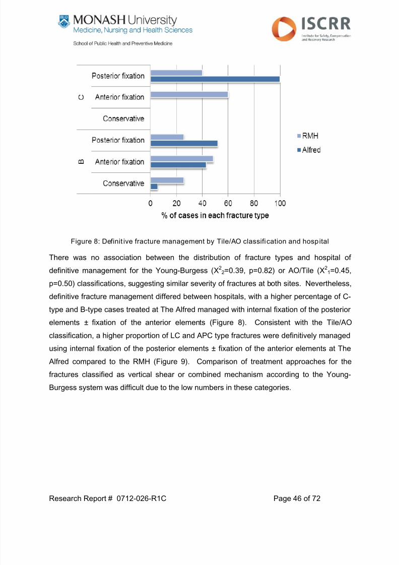
7/18/2019 026 Classification, management and outcomes of severe pelvic ring fractures
http://slidepdf.com/reader/full/026-classification-management-and-outcomes-of-severe-pelvic-ring-fractures 46/72
Research Report # 0712-026-R1C Page 46 of 72
Figure 8: Definit ive fracture management by Tile/AO classification and hospital
There was no association between the distribution of fracture types and hospital of
definitive management for the Young-Burgess (Χ22=0.39, p=0.82) or AO/Tile (Χ2
1=0.45,
p=0.50) classifications, suggesting similar severity of fractures at both sites. Nevertheless,
definitive fracture management differed between hospitals, with a higher percentage of C-
type and B-type cases treated at The Alfred managed with internal fixation of the posterior
elements ± fixation of the anterior elements (Figure 8). Consistent with the Tile/AO
classification, a higher proportion of LC and APC type fractures were definitively managed
using internal fixation of the posterior elements ± fixation of the anterior elements at The
Alfred compared to the RMH (Figure 9). Comparison of treatment approaches for the
fractures classified as vertical shear or combined mechanism according to the Young-
Burgess system was difficult due to the low numbers in these categories.

7/18/2019 026 Classification, management and outcomes of severe pelvic ring fractures
http://slidepdf.com/reader/full/026-classification-management-and-outcomes-of-severe-pelvic-ring-fractures 47/72
Research Report # 0712-026-R1C Page 47 of 72
Figure 9: Definitive fracture management by Young-Burgess classification and hospital
There was no association between compensation status (Χ21=0.30, p=0.59), transport
status (Χ21=0.29, p=0.59), comorbid status (Χ2
1=0.37, p=0.54), ISS, head injury severity
(Χ21=1.72, p=0.19), sex (Χ2
1=2.79, p=0.10), and whether the pelvic ring fracture was
managed operatively or not. The percentage of patients aged 65 years and over receiving
operative management of their pelvic ring fracture was 71%, compared to 94% of 35-64
year olds, and 89% of 15-34 year olds (Χ22=6.24, p=0.04). The percentage of RMH cases
where the pelvic ring was fixed operatively was 77% compared to 94% at The Alfred
(Χ21=6.86, p=0<0.001).
Functional outcomes (GOS-E)
There were three deaths in the cohort between hospital discharge and 24-months post-
injury. Improvement in functional outcome was evident over time, particularly from 6 to 12-
months post-injury (Table 14 and Figure 10), although only 10 per cent of patients hadfully recovered, according to the GOS-E scale (upper good recovery), at 24-months.

7/18/2019 026 Classification, management and outcomes of severe pelvic ring fractures
http://slidepdf.com/reader/full/026-classification-management-and-outcomes-of-severe-pelvic-ring-fractures 48/72
Research Report # 0712-026-R1C Page 48 of 72
Table 14: Severity and management of injuries
GOS-E score 6-months
(n=109)
12-months
(n=106)
24-months
(n=106)
Death 2 (1.8) 3 (2.8) 3 (2.8)
Lower severe disability 22 (20.2) 11 (10.4) 12 (11.3)
Upper severe disability 16 (14.7) 13 (12.3) 9 (8.5)
Lower moderate disability 26 (23.9) 24 (22.6) 22 (20.8)
Upper moderate disability 31 (28.4) 36 (34.0) 38 (35.8)
Lower good recovery 6 (5.5) 8 (7.6) 11 (10.4)
Upper good recovery 6 (5.5) 11 (10.4) 11 (10.4)
The proportion of patients living independently according to the GOS-E scale (lower
moderate disability or above) was 63% at 6-months, 75% at 12-months, and 77% at 24-
months. The proportion who had experienced a good recovery was much lower – 11% at
6-months, 18% at 12-months, and 21% at 24-months (Figure 10).
Figure 10: Longer term functional outcomes of severe pelvic r ing fractures
The results of univariate analyses to identify predictors of a good recovery (GOS-E score
of lower or upper good recovery) are shown in Table 15. Compared to 6-months post-
injury, the odds of a good recovery increased at 12-months (OR 1.65, 95% CI: 0.90, 3.01),
and 24-months (OR 1.97, 95% CI: 1.08, 3.57), post-injury. Factors associated (p<0.20)
with a good recovery at any time post-injury were the hospital of management, mechanism
of injury, compensable status, GCS group, and the ISS (Table 15). The unadjusted odds
of a good recovery were lower for cases definitively managed at RMH, compensable

7/18/2019 026 Classification, management and outcomes of severe pelvic ring fractures
http://slidepdf.com/reader/full/026-classification-management-and-outcomes-of-severe-pelvic-ring-fractures 49/72
Research Report # 0712-026-R1C Page 49 of 72
patients, transport-related cases, those with a higher ISS, and patients with a low GCS on
arrival at hospital (Table 15). Fracture type and management were not associated with a
good recovery.
Table 15: Factors associated with a good funct ional recovery over t ime (univariate)
Descriptor OR (95% CI) p-value
Gender Male (reference)
Female
1
1.48 (0.63, 3.47)
0.37
Age group 15-34 years (reference)
35-64 years
≥ 65 years
1
0.58 (0.25, 1.36)
0.40 (0.10, 1.54)
0.27
Comorbidity? No (reference)
Yes
1
0.60 (0.25, 1.41)
0.24
Compensable? No (reference)
Yes
1
0.24 (0.10, 0.59)
0.002
Transport-related? No (reference)
Yes
1
0.41 (0.17, 0.95)
0.04
Working prior toinjury?
No (reference)
Yes
1
0.89 (0.39, 2.07)
0.79
Highest level ofeducation
University degree (reference)
Advanced diploma, diploma or certificate
Completed high school
Did not complete high school
1
2.50 (0.57, 11.03)
1.61 (0.33, 8.02)
1.85 (0.45, 7.62)
0.66
Hospital Alfred (reference)
RMH
1
0.36 (0.13, 0.95)
0.04
Head injury severity GCS 9-15 (reference)
GCS 3-8
1
0.13 (0.01, 1.77)
0.13
Injury Severity Score 0.95 (0.91, 0.99) 0.01
AO/Tile classification Type B (reference)
Type C
1
1.09 (0.41, 2.89)
0.86
Young-Burgessclassification
APC (reference)
LC
Vertical shear/CMI
1
1.13 (0.46, 2.75)
0.78 (0.16, 3.89)
0.88
Definitive pelvicfracture management
Conservative (reference)
Fixation of anterior elements
Fixation of posterior elements (± anterior)
1
2.51 (0.46, 13.55)
2.20 (0.41, 11.73)
0.57
Hospital of definitive management, ISS, compensation and transport status, and GCS
group were entered into the multivariate GEE model for predicting a good recovery.
Because of the strong association between compensable and transport status, an
interaction term was included to determine if the rate of recovery of transport and non-
transport cases differed according to compensable status. The results of this multivariate
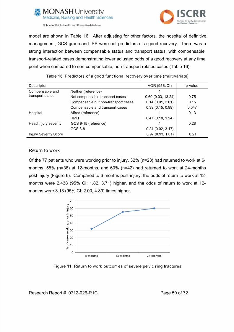
7/18/2019 026 Classification, management and outcomes of severe pelvic ring fractures
http://slidepdf.com/reader/full/026-classification-management-and-outcomes-of-severe-pelvic-ring-fractures 50/72
Research Report # 0712-026-R1C Page 50 of 72
model are shown in Table 16. After adjusting for other factors, the hospital of definitive
management, GCS group and ISS were not predictors of a good recovery. There was a
strong interaction between compensable status and transport status, with compensable,
transport-related cases demonstrating lower adjusted odds of a good recovery at any time
point when compared to non-compensable, non-transport related cases (Table 16).
Table 16: Predictors of a good functional recovery over time (multivariate)
Descriptor AOR (95% CI) p-value
Compensable andtransport status
Neither (reference)
Not compensable transport cases
Compensable but non-transport cases
Compensable and transport cases
1
0.60 (0.03, 13.24)
0.14 (0.01, 2.01)
0.39 (0.15, 0.99)
0.75
0.15
0.047
Hospital Alfred (reference)
RMH
1
0.47 (0.18, 1.24)
0.13
Head injury severity GCS 9-15 (reference)
GCS 3-8
1
0.24 (0.02, 3.17)
0.28
Injury Severity Score 0.97 (0.93, 1.01) 0.21
Return to work
Of the 77 patients who were working prior to injury, 32% (n=23) had returned to work at 6-
months, 55% (n=38) at 12-months, and 60% (n=42) had returned to work at 24-months
post-injury (Figure 6). Compared to 6-months post-injury, the odds of return to work at 12-
months were 2.438 (95% CI: 1.82, 3.71) higher, and the odds of return to work at 12-
months were 3.13 (95% CI: 2.00, 4.89) times higher.
Figure 11: Return to work outcomes of severe pelvic r ing fractures

7/18/2019 026 Classification, management and outcomes of severe pelvic ring fractures
http://slidepdf.com/reader/full/026-classification-management-and-outcomes-of-severe-pelvic-ring-fractures 51/72
Research Report # 0712-026-R1C Page 51 of 72
Table 17: Factors associated with return to work over time (univariate)
Descriptor OR (95% CI) p-value
Gender Male (reference)
Female
1
0.80 (0.33, 1.89)
0.60
Age group 15-34 years (reference)
35-64 years
≥ 65 years
1
1.06 (0.48, 2.34)
0.43 (0.09, 2.10)
0.53
Comorbidity? No (reference)
Yes
1
0.53 (0.23, 1.23)
0.14
Compensable? No (reference)
Yes
1
0.42 (0.12, 1.51)
0.18
Transport-related? No (reference)Yes
10.65 (0.23, 1.82)
0.42
Highest level ofeducation
University degree (reference)
Advanced diploma, diploma or certificate
Completed high school
Did not complete high school
1
1.09 (0.33, 3.63)
0.58 (0.17, 1.92)
0.42 (0.14, 1.27)
0.24
Hospital Alfred (reference)
RMH
1
1.06 (0.48, 2.37)
0.88
Head injury severity GCS 9-15 (reference)
GCS 3-8
1
0.11 (0.02, 0.63)
0.01
Injury Severity Score 0.91 (0.87, 0.96)<0.001
AO/Tile classification Type B (reference)
Type C
1
0.38 (0.12, 1.25)
0.11
Young-Burgessclassification
APC (reference)
LC
Vertical shear/CMI
1
0.65 (0.30, 1.44)
0.10 (0.01, 1.41)
0.17
Definitive pelvicfracture management
Conservative (reference)
Fixation of anterior elements
Fixation of posterior elements (± anterior)
1
1.72 (0.36, 8.17)
0.74 (0.16, 3.53)
0.12
Factors associated with return to work at any time point post-injury, were injury severity,
head injury severity (GCS), comorbid status, compensation status, pelvic fracture severity
and the definitive management of the pelvic fracture (Table 17).
The multivariate analysis identified injury severity (ISS) as the most important predictor of
return to work. Adjusted for comorbid status, compensable status, head injury severity,
pelvic fracture management, and pelvic fracture severity, the odds of return to work at any
time point decreased 5% for every point increase in the ISS (Table 18).

7/18/2019 026 Classification, management and outcomes of severe pelvic ring fractures
http://slidepdf.com/reader/full/026-classification-management-and-outcomes-of-severe-pelvic-ring-fractures 52/72
Research Report # 0712-026-R1C Page 52 of 72
Table 18: Predictors of return to work over time (multivariate)
Descriptor AOR (95% CI) p-value
Injury Severity Score 0.94 (0.89, 0.99) 0.04
Head injury severity GCS 9-15 (reference)
GCS 3-8
1
0.27 (0.04, 1.79)
0.18
Compensable? No (reference)
Yes
1
0.38 (0.08, 1.90)
0.24
Definitive pelvicfracture management
Conservative (reference)
Fixation of anterior elements
Fixation of posterior elements (± anterior)
1
1.43 (0.26, 7.91)
0.63 (0.11, 3.64)
0.25
AO/Tile classification Type B (reference)
Type C
1
0.53 (0.14, 1.96)
0.34
Comorbidity? No (reference)
Yes
1
1.03 (0.36, 2.95)
0.96
Pain
A pain scale score is collected when direct interview of the patient is possible, and
therefore is limited to patients without pre-existing or post-injury cognitive deficit or other
disability affecting completion of the interview. Pain scores were obtained from 67 patients
at 6-months, 74 at 12-months and 68 patients at 24-months. The increase in pain score
completion at 12-months is likely to reflect overall improvement of the patient’s condition,
allowing direct interview.
0
2
4
6
8
1 0
P a i n
s c o r e
6-months 12-months 24-months
Figure 12: Distribution of pain scores at follow-up

7/18/2019 026 Classification, management and outcomes of severe pelvic ring fractures
http://slidepdf.com/reader/full/026-classification-management-and-outcomes-of-severe-pelvic-ring-fractures 53/72
Research Report # 0712-026-R1C Page 53 of 72
The distribution of pain scores shown in Figure 12 suggests little improvement in pain over
time. The proportion of patients reporting no pain (pain score 0) at follow-up was 43% at
6-months, 42% at 12-months and 43% at 24-months. The proportion of patients reporting
persistent moderate to severe pain (pain score 5-10) was 31%, 24%, and 29% at 6-, 12-,
and 24-months, respectively. The odds or reporting moderate to persistent pain were not
significantly lower at 12-months (OR 0.72, 95% CI: 0.39, 1.33), or 24-months (OR 0.86,
95% CI: 0.46, 1.59), compared to 6-months post-injury.
Table 19: Factors associated with persistent moderate to severe pain (univariate)
Descriptor OR (95% CI) p-value
Gender Male (reference)
Female
1
1.54 (0.66, 3.59)
0.32
Age group 15-34 years (reference)
35-64 years
≥ 65 years
1
1.18 (0.53, 2.64)
0.62 (0.18, 2.11)
0.61
Comorbidity? No (reference)
Yes
1
1.39 (0.63, 3.05)
0.42
Working prior toinjury?
No (reference)
Yes
1
1.22 (0.57, 2.92)
0.66
Compensable? No (reference)
Yes
1
2.69 (0.77, 9.42)
0.12
Transport-related? No (reference)
Yes
1
2.50 (0.86, 7.29)
0.09
Highest level ofeducation
University degree (reference)
Advanced diploma, diploma or certificate
Completed high school
Did not complete high school
1
0.71 (0.21, 2.40)
0.93 (0.25, 3.43)
0.67 (0.21, 2.14)
0.88
Hospital Alfred (reference)
RMH
1
0.65 (0.29, 1.49)
0.31
Injury Severity Score 1.01 (0.98, 1.05) 0.47
AO/Tile classification Type B (reference)
Type C
1
1.42 (0.57, 3.58)
0.45
Young-Burgessclassification
APC (reference)
LC
Vertical shear/CMI
1
1.45 (0.61, 3.47)
4.74 (1.22, 18.46)
0.08
Definitive pelvicfracture management
Conservative (reference)
Fixation of anterior elements
Fixation of posterior elements (± anterior)
1
0.47 (0.14, 1.54)
0.82 (0.28, 2.46)
0.34
* Head injury severity excluded as pain score not administered to severe head injury patients
Factors associated (p<0.20) with persistent moderate to severe pain at any time point
were fracture type (measured using the Young-Burgess system), cause of injury, and
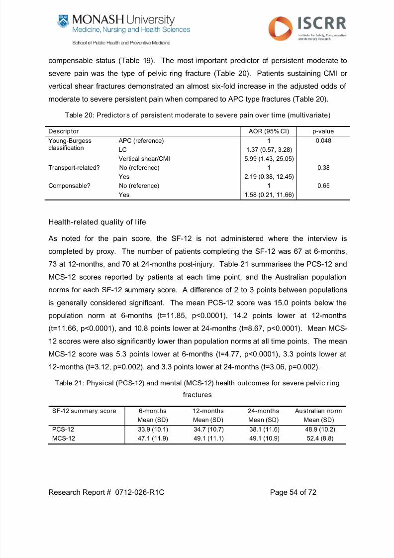
7/18/2019 026 Classification, management and outcomes of severe pelvic ring fractures
http://slidepdf.com/reader/full/026-classification-management-and-outcomes-of-severe-pelvic-ring-fractures 54/72
Research Report # 0712-026-R1C Page 54 of 72
compensable status (Table 19). The most important predictor of persistent moderate to
severe pain was the type of pelvic ring fracture (Table 20). Patients sustaining CMI or
vertical shear fractures demonstrated an almost six-fold increase in the adjusted odds of
moderate to severe persistent pain when compared to APC type fractures (Table 20).
Table 20: Predictors of persistent moderate to severe pain over time (multivariate)
Descriptor AOR (95% CI) p-value
Young-Burgessclassification
APC (reference)
LC
Vertical shear/CMI
1
1.37 (0.57, 3.28)
5.99 (1.43, 25.05)
0.048
Transport-related? No (reference)
Yes
1
2.19 (0.38, 12.45)
0.38
Compensable? No (reference)
Yes
1
1.58 (0.21, 11.66)
0.65
Health-related quality of l ife
As noted for the pain score, the SF-12 is not administered where the interview is
completed by proxy. The number of patients completing the SF-12 was 67 at 6-months,
73 at 12-months, and 70 at 24-months post-injury. Table 21 summarises the PCS-12 and
MCS-12 scores reported by patients at each time point, and the Australian population
norms for each SF-12 summary score. A difference of 2 to 3 points between populations
is generally considered significant. The mean PCS-12 score was 15.0 points below the
population norm at 6-months (t=11.85, p<0.0001), 14.2 points lower at 12-months
(t=11.66, p<0.0001), and 10.8 points lower at 24-months (t=8.67, p<0.0001). Mean MCS-
12 scores were also significantly lower than population norms at all time points. The mean
MCS-12 score was 5.3 points lower at 6-months (t=4.77, p<0.0001), 3.3 points lower at
12-months (t=3.12, p=0.002), and 3.3 points lower at 24-months (t=3.06, p=0.002).
Table 21: Physical (PCS-12) and mental (MCS-12) health outcomes for severe pelvic ring
fractures
SF-12 summary score 6-months
Mean (SD)
12-months
Mean (SD)
24-months
Mean (SD)
Austral ian norm
Mean (SD)
PCS-12 33.9 (10.1) 34.7 (10.7) 38.1 (11.6) 48.9 (10.2)
MCS-12 47.1 (11.9) 49.1 (11.1) 49.1 (10.9) 52.4 (8.8)

7/18/2019 026 Classification, management and outcomes of severe pelvic ring fractures
http://slidepdf.com/reader/full/026-classification-management-and-outcomes-of-severe-pelvic-ring-fractures 55/72
Research Report # 0712-026-R1C Page 55 of 72
Figure 13 shows the standardised mean difference (SMD) of the PCS-12 and MCS-12
scores at 6, 12 and 24-months post-injury. The SMD provides a method of showing the
degree of deviation from the population norm by standardising individual scores by age
and gender. The SMD is effectively an effect size where an SMD of zero suggests no
difference to population norms and a score above zero suggests health-related quality of
life of severe pelvic ring fracture patients above population norms. The size of the SMD
represents the magnitude of difference between the SF-12 scores of the Australian
population and severe pelvic ring fracture patients patients, with values >0.8 considered to
be large.
Figure 13: Age and gender adjusted standardised mean dif ferences (95% CI) for PCS-12 and
MCS-12 scores over time
Predictors of physical health scores (PCS-12)
There was no significant increase in mean PCS-12 scores at 12-months compared to 6-
months (mean difference 0.84, 95% CI: -1.60, 3.27), but clear evidence of improvedphysical health at 24-months compared to 6-months (mean difference 4.23, 95% CI: 1.35,
SMD (95% CI)
-3.0 -2.5 -2.0 -1.5 -1.0 -0.5 0.0 0.5
PCS-12
MCS-12
6-months
12-months
24-monthsZero reference line

7/18/2019 026 Classification, management and outcomes of severe pelvic ring fractures
http://slidepdf.com/reader/full/026-classification-management-and-outcomes-of-severe-pelvic-ring-fractures 56/72
Research Report # 0712-026-R1C Page 56 of 72
7.10). Without adjusting for other factors, fracture type was not associated with physical
health at any time point, but type of definitive fracture management was (Table 22). Other
factors univariately associated (p<0.20) with PCS-12 scores were age, compensable
status and the ISS (Table 22), and all were included in the multivariate model (Table 23).
Table 22: Factors associated with physical health (PCS-12 scores) over time (univariate)
Descriptor Mean dif ference (95%CI)
p-value
Gender Male (reference)
Female
-
0.02 (-4.59, 4.63)
0.99
Age group 15-34 years (reference)
35-64 years
≥ 65 years
-
-3.45 (-7.54, 0.64)
-4.58 (-9.54, 0.39)
0.12
Comorbidity? No (reference)
Yes
-
-1.22 (-5.18, 2.74)
0.55
Working prior toinjury?
No (reference)
Yes
-
1.16 (-2.77, 5.09)
0.56
Compensable? No (reference)
Yes
-
-5.46 (-10.78, -1.14)
0.04
Transport-related? No (reference)
Yes
-
-1.69 (-6.22, 2.84)
0.46
Highest level ofeducation
University degree (reference)
Advanced diploma, diploma or certificateCompleted high school
Did not complete high school
-
2.44 (-3.16, 8.04)4.96 (-0.73, 10.65)
2.14 (-3.17, 7.46)
0.40
Hospital Alfred (reference)
RMH
-
-2.04 (-4.59, 4.63)
0.28
Injury Severity Score -0.14 (-0.32, 0.04) 0.13
AO/Tile classification Type B (reference)
Type C
-
-2.07 (-6.57, 2.43)
0.37
Young-Burgessclassification
APC (reference)
LC
Vertical shear/CMI
-
-0.62 (-4.89, 3.65)
-5.10 (-11.03, 0.82)
0.21
Definitive pelvicfracture management
Conservative (reference)
Fixation of anterior elements
Fixation of posterior elements (± anterior)
-
5.83 (-0.55, 12.21)
-2.37 (-3.61, 8.35)
0.12
* Head injury severity excluded as SF-12 not administered to severe head injury patients
After adjusting for other potential confounders, compensable patients demonstrated
adjusted mean PCS-12 scores more than 7.5 points below non-compensable patients
(Table 23), while operatively managed severe pelvic ring fractures reported adjusted mean
PCS-12 scores above the scores reported by patients managed conservatively (Table 23).

7/18/2019 026 Classification, management and outcomes of severe pelvic ring fractures
http://slidepdf.com/reader/full/026-classification-management-and-outcomes-of-severe-pelvic-ring-fractures 57/72
Research Report # 0712-026-R1C Page 57 of 72
Table 23: Predictors of physical health (PCS-12 scores) over time (multivariate)
Descrip tor Adjus ted meandif ference (95% CI)
p-value
Compensable? No (reference)Yes
--7.50 (-12.64, -2.37)
0.004
Definitive pelvicfracture management
Conservative (reference)
Fixation of anterior elements
Fixation of posterior elements (± anterior)
-
6.44 (-0.78, 13.66)
1.57 (-4.88, 8.02)
0.04
Age group 15-34 years (reference)
35-64 years
≥ 65 years
-
-4.04 (-7.91, -0.16)
-4.15 (-9.22, 0.91)
0.08
Injury Severity Score -0.04 (-0.21, 0.14) 0.66
Predictors of mental health outcomes (MCS-12)
There was no improvement in MCS-12 scores at 12-months (mean difference 1.00, 95%
CI: -1.63, 3.63) or 24-months (mean difference 1.13, 95% CI: -1.48, 3.75), compared to 6-
months. This is consistent with Figure 13. Factors associated with mental health
outcomes at any time point were compensable status, cause of injury (transport vs. non-
transport related), whether the person was working prior to injury, and the definitive
management of the pelvic ring fracture.
Definitive fracture management, compensable status and transport status were included
the multivariate model to identify important predictors of MCS-12 scores over time. An
interaction term was included between transport status and compensable status to see if
mental health outcomes differed for compensable patients, according to transport status
Table 25).

7/18/2019 026 Classification, management and outcomes of severe pelvic ring fractures
http://slidepdf.com/reader/full/026-classification-management-and-outcomes-of-severe-pelvic-ring-fractures 58/72
Research Report # 0712-026-R1C Page 58 of 72
Table 24: Factors associated with mental health (MCS-12 scores) over time (univariate)
Descriptor Mean dif ference (95%CI)
p-value
Gender Male (reference)Female
--2.29 (-7.21, 2.64)
0.36
Age group 15-34 years (reference)
35-64 years
≥ 65 years
-
-1.72 (-6.8, 2.74)
-2.39 (-9.00, 4.23)
0.65
Comorbidity? No (reference)
Yes
-
-2.52 (-6.92, 1.87)
0.26
Working prior toinjury?
No (reference)
Yes
-
3.33 (-1.38, 8.03)
0.17
Compensable? No (reference)
Yes
-
-6.33 (-10.85, -1.82)
0.006
Transport-related? No (reference)
Yes
-7.31 (-11.17, -3.45) <0.001
Highest level ofeducation
University degree (reference)
Advanced diploma, diploma or certificate
Completed high school
Did not complete high school
-
5.74 (-1.49, 13.00)
1.57 (-6.91, 10.05)
1.35 (-5.91, 8.60)
0.20
Hospital Alfred (reference)
RMH
-
0.80 (-3.51, 5.12)
0.72
Injury Severity Score -0.07 (-0.30, 0.15) 0.53
AO/Tile classification Type B (reference)
Type C
-
0.55 (-5.67, 6.78)
0.86
Young-Burgessclassification
APC (reference)
LC
Vertical shear/CMI
-
-2.20 (-6.47, 2.07)
-5.03 (-15.80, 5.75)
0.47
Definitive pelvicfracture management
Conservative (reference)
Fixation of anterior elements
Fixation of posterior elements (± anterior)
-
9.30 (2.40, 16.21)
8.10 (1.06, 15.11)
0.03
* Head injury severity excluded as SF-12 not administered to severe head injury patients
Participants who were compensable, and transport-related, reported adjusted mean MCS-
12 scores 8 points lower than non-compensable patients injured in via other mechanisms
at any time point post-injury. Any type of surgical fixation of the pelvic ring fracture
resulted in improved mental health outcomes compared to patients managed
conservatively (Table 25).

7/18/2019 026 Classification, management and outcomes of severe pelvic ring fractures
http://slidepdf.com/reader/full/026-classification-management-and-outcomes-of-severe-pelvic-ring-fractures 59/72
Research Report # 0712-026-R1C Page 59 of 72
Table 25: Predictors of mental health (MCS-12 scores) over time (multivariate)
Descrip tor Adjus ted meandif ference (95% CI)
p-value
Compensable andtransport status
Neither (reference)Not compensable transport cases
Compensable but non-transport cases
Compensable and transport cases
-2.69 (-0.93, 6.32)
-0.32 (-5.97, 5.33)
-8.01 (-12.46, -3.57)
0.15
0.91
<0.001
Definitive pelvicfracture management
Conservative (reference)
Fixation of anterior elements
Fixation of posterior elements (± anterior)
-
9.16 (1.90, 16.42)
6.69 (-0.35, 13.74)
0.007
0.06
Working prior toinjury?
No (reference)
Yes
-
1.96 (-2.62, 6.55)
0.40
Discussion
This multi-centre study described the predictors of long term outcomes of 114 severe
pelvic ring fracture patients, and represents the largest study of severe pelvic ring fractures
to date. The results highlight the variation in definitive fracture management between the
major trauma service hospitals, and identified a number of patient, injury and treatment
factors predictive of patient outcome. Compensable status was a significant independent
predictor of functional and health-related quality of life outcome, with compensablepatients demonstrating poorer outcomes. The overall injury severity of the patient was the
most important predictor of return to work, while fracture type was a significant
independent predictor of persistent pain. Definitive pelvic fracture management was an
independent predictor of health-related quality of life, with patients surgically managed
demonstrating higher long term physical and mental health outcomes than patients
managed conservatively.
Consistent with previous studies of unstable and severe pelvic ring fractures, the
population were predominantly young, male, working prior to injury, injured in transport
accidents, and involving other associated injuries (11, 26, 27, 54). All had sustained other
associated injuries and the mean ISS (31.8) was higher than other studies (11, 53-55).
The proportion of cases surgically managed with internal fixation was higher than
previously reported. In our study, 11% were managed conservatively, 11% with external
fixation only, and 79% with open reduction and internal fixation of the pelvic ring fracture.
Suzuki et al studied the outcomes of 57 unstable, B- and C-type, fractures from a singlecentre in Japan and 40% were treated conservatively, 39% with external fixation, and 21%
7/18/2019 026 Classification, management and outcomes of severe pelvic ring fractures
http://slidepdf.com/reader/full/026-classification-management-and-outcomes-of-severe-pelvic-ring-fractures 60/72
Research Report # 0712-026-R1C Page 60 of 72
with internal fixation (43). Comparison of rates of surgery with other studies is difficult as
many have focused only on operatively treated patients (11, 27, 50, 54, 56, 57), or have
included less severe fractures in their cohort (26, 58).
Disability was prevalent even 24-months post-injury. There was considerable
improvement in outcome to 24-months post-injury for return to work, functional outcome,
and physical health scores. However, there was no evidence of improvement in pain or
mental health scores over time. Comparison of patient outcomes from our cohort requires
studies with common outcome measures. The GOS-E has not been used by other severe
pelvic ring fractures, despite being used widely in multi-trauma populations, but return to
work, health-related quality of life and pain have been used previously by studies
investigating the outcomes of severe pelvic ring fractures.
In our study, health-related quality of life, measured using the PCS-12 and MCS-12 of the
SF-12 instrument, was significantly below age and gender adjusted population norms, with
the PCS-12 11 points lower, and the MCS-12 3 points lower, at 24-months post-injury.
Other studies of severe pelvic ring fractures have largely used the SF-36, but also found
physical health scores well below population norms (11, 43, 54, 55). Suzuki et al, in their
study of 57 unstable pelvic ring fractures patients, found PCS scores 13.4 points lowerthan population norms more than 2 years after injury (43). Oliver et al studied 46 B- and
C-type pelvic ring fractures, and found PCS-36 scores 11 points, and MCS-36 scores 4.1
points, below US population norms. Together, these studies highlight the large and
significant reductions in health-related quality of life experienced by patients sustaining
severe pelvic ring fractures even 2-years following injury.
The 60% return to work rate at 24-months for severe pelvic ring fracture patients who were
working prior to injury is relatively consistent with the published literature. Suzuki et al studied 57 unstable pelvic ring fractures and found only 9 (16%) were unable to return to
work more than 2-years after injury (43). However, most other studies have found lower
return to work rates for severe pelvic ring fracture patients. Madhu et al studied 30
patients with combined pelvic and spinal fracture and reported that 57% had returned to
work at a mean follow-up of 57 months (10). Van den Bosch et al and Tornetta et al
reported return to work rates of 68% and 67% in their studies of 37 and 46 operatively
treated pelvic ring fractures, respectively (53, 59).

7/18/2019 026 Classification, management and outcomes of severe pelvic ring fractures
http://slidepdf.com/reader/full/026-classification-management-and-outcomes-of-severe-pelvic-ring-fractures 61/72
Research Report # 0712-026-R1C Page 61 of 72
In our study, we found that the prevalence of moderate to severe persistent pain was 30%
at 24-months post-injury. Gerbershagen et al studied 69 patients with pelvic and
acetabular fractures using the Mainz Pain Staging System, and found the prevalence of
pain was high (64%) and many patients experience long term pain chronification and
associated biopsychosocial implications (60). While compensable status has been
identified as a predictor of persistent pain in trauma patients previously (61, 62), the
strongest predictor of persistent moderate to severe pain in our study was the type of
fracture sustained. Patients with vertical shear or combined mechanism fractures,
classified according to the Young-Burgess system, demonstrated a six-fold increased risk
of reporting moderate to severe pain at any follow-up time point.
Factors such as age, gender, associated severe head injury, the presence of comorbidity,
the Tile/AO classification of fractures, and the level of education of patients were largely
not important predictors of outcome in this cohort. The type of fracture sustained was only
a significant independent predictor of persistent pain; compensable status was an
important predictor of function and quality of life; the overall injury severity (ISS) of patients
predicted return to work; and fracture management was a strong predictor of health-related
quality of life.
A key aim of our study was to explore the relationship between type of fracture, fracture
management and long term patient outcomes. Fracture type, as measured using the
Young-Burgess and Tile/AO classifications, was not associated with many of the outcomes
measured. Similarly, the type of fracture management did not impact on functional, return
to work or pain outcomes. However, patients managed operatively demonstrated
improved mental health scores, as measured by the MCS-12, than patients managed
conservatively. Fixation of the anterior elements of the pelvic ring also resulted in adjusted
mean PCS-12 scores 6.6 points higher than conservatively managed patients, while
patients managed with external fixation or internal fixation of the anterior elements
reported adjusted mean PCS-12 scores 4.8 points higher than patients with fixation of the
posterior ± anterior ring. Van Loon et al compared the functional outcomes of 38 “open
book” (Type B1) pelvic fractures according to surgical approaches and concluded that
outcomes tended to be worse for patients receiving additional posterior stabilisation but
the number of cases receiving additional posterior stabilisation was low (n=6) (55). Our
study provides some support for the findings of Van Loon et al, but caution in interpretation

7/18/2019 026 Classification, management and outcomes of severe pelvic ring fractures
http://slidepdf.com/reader/full/026-classification-management-and-outcomes-of-severe-pelvic-ring-fractures 62/72
Research Report # 0712-026-R1C Page 62 of 72
is needed. It is possible that the requirement to fix the posterior ring elements suggests a
more serious fracture which could go on to worse outcomes anyway, and that other patient
factors could impact on the decision to operate or manage conservatively. There are no
clear clinical indications for particular surgical approaches, with the surgeon generally
applying judgement based on the patient’s general condition, local soft tissue condition,
and factors such as the degree of fracture displacement. Fracture displacement is not
measured in either the AO/Tile or Young-Burgess systems for classifying fracture type.
Papakostidis et al, published a systematic review of treatment modalities and outcomes of
pelvic ring fractures, and concluded that fixation of all injured elements of the pelvic ring
led to better radiological outcomes but there was no clear benefit with respect to patient
function (48). However, the quality of studies included was poor, outcome measures used
were limited, and the studies were insufficient to provide clear guidance about optimal
treatment for unstable pelvic ring fractures (48).
Eighty-six per cent of severe pelvic ring fracture cases in this study were compensable
(predominantly TAC) patients. Compensable patients demonstrated significantly higher
odds of poorer functional outcome and markedly lower health-related quality of life scores.
This is consistent with data from other studies of serious injury (61-67). Observationalstudies, such as this prospective cohort study, cannot identify the reasons for the poorer
outcomes of compensable patients. However, results from two qualitative studies provide
insight into the experiences of patients with compensable agencies that could contribute to
the poorer outcomes noted (68, 69). Factors such as a lack of trust, difficulty accessing
post-acute treatment services, delays in approval of treatment services, and pre-existing
conditions such as anxiety and depression, could all contribute to poorer outcomes of
compensable cases (68, 69). The potential for patients to overstate their limitations for
secondary gain cannot be excluded as a possible reason for poorer outcomes in this
patient group (63).
There were a number of strengths to this study. Previous studies have almost exclusively
focused on pelvic fractures managed at a single institution. We studied a large number of
severe pelvic ring fracture patients from two major trauma services, representing more
than 90% of all hospitalised, severe pelvic fractures in Victoria, and were able to show
variation in practice between the hospitals. Cases were prospectively followed-up atmultiple, standardised time points. Published studies have retrospectively identified cases
7/18/2019 026 Classification, management and outcomes of severe pelvic ring fractures
http://slidepdf.com/reader/full/026-classification-management-and-outcomes-of-severe-pelvic-ring-fractures 63/72
Research Report # 0712-026-R1C Page 63 of 72
and followed up cases at inconsistent time points post-injury. The follow-up rate of 96%
was much higher than previously reported studies (10, 11, 26, 43, 54), minimising bias in
reporting of patient outcomes.
Nevertheless, there were limitations to the study. Compared to previous studies, the
sample size (n=114) was large, but was insufficient to identify all but the largest effects. In
Study 1, we showed that the inter-rater reliability of fracture classification using the Young-
Burgess and AO/Tile systems was low, and due the relatively low number of cases in the
study, these classification systems needed to be collapsed into broad categories for
analysis. In doing so, there is a loss of information and subtle differences in outcomes
between sub-categories of fracture types could not be shown. Radiological outcomes
were not included in this study despite previous studies showing an association between
radiological outcomes (i.e. displacement) and functional outcome (57, 59, 70), and should
be considered for future studies. Only a limited range of complications could be collected
retrospectively from the medical record and standardised reporting is not used. Finally,
pelvic-specific outcome measures were not collected in this study. While a number of
pelvic-specific outcome measures are available, none have been sufficiently validated
(71). In addition, all patients were multi-trauma patients, sustaining significant associated
injuries. Disentangling disability associated with the pelvic ring fracture from the disability
of other sustained injuries is challenging, with studies previously showing that region-
specific measures do not discriminate disability related to other injuries (72).
Conclusions
This study of 114 severe pelvic ring fractures shows that disability is prevalent even at 2-
years post-injury. There was marked variation in practice between the major trauma
service hospitals. Fracture classification impacted little on patient outcome, but the results
provide some evidence for improved patient quality of life following surgical fixation of
fractures. The complexity of these patients, the prevalence of disability, and the variation
in practice noted highlight the need for ongoing monitoring and benchmarking of these
patients if care and outcomes are to be optimised.

7/18/2019 026 Classification, management and outcomes of severe pelvic ring fractures
http://slidepdf.com/reader/full/026-classification-management-and-outcomes-of-severe-pelvic-ring-fractures 64/72
Research Report # 0712-026-R1C Page 64 of 72
Summary of key findings
This project comprised three distinct studies to: establish the reliability of the classification
of severe pelvic ring fractures; compare the early clinical management of severe pelvic
ring fractures between the MTS hospitals and the impact on in-hospital mortality, and;
establish the relationship between approaches to pelvic fracture fixation, fracture
classification and long term functional, return to work, pain and health-related quality of life
outcomes. Severe pelvic ring fractures represent a challenging patient group to research
but there were clear findings from this research:
1. Accurate and reliable classification of fractures is necessary to enable valid and
appropriate comparisons of study populations and patient outcomes. Previous
studies of pelvic ring fracture classification have found inter- and intra-tester
reliability too low for clinical and benchmarking purposes. In this project, the use of
3D CT reconstruction images was explored as a potential imaging source that could
result in improved reliability of classification. The study findings identified clear
issues with the reliability of fracture classification in this group of severe pelvic ring
fractures. Inter-tester reliability was low and current clinical practice at the MTS
hospitals does not routinely provide the plain radiograph views for which the Young-
Burgess and Tile/AO classifications were developed. Severe pelvic ring fracture
patients represent a difficult classification prospect for existing classification
systems due to the prevalence of interventions pre-hospital (e.g. binders) and in the
emergency department.
2. There were profound variations in practice between the MTS hospitals with respect
to the early and definitive management of severe pelvic ring fractures. The Alfred
hospital favours rapid stabilisation of the fracture with external fixation, and a higher
prevalence of definitive management using open reduction and internal fixation.
RMH favours early referral to angiography and embolisation to reduce bleeding,
and lower rates of open reduction and internal fixation. . The in-hospital mortality
rate was slightly higher at RMH, but adjusted for key confounders, there was no
difference in the risk-adjusted odds of mortality following severe pelvic ring fracture
between the MTS hospitals. There was no clearly superior treatment approach,
7/18/2019 026 Classification, management and outcomes of severe pelvic ring fractures
http://slidepdf.com/reader/full/026-classification-management-and-outcomes-of-severe-pelvic-ring-fractures 65/72
Research Report # 0712-026-R1C Page 65 of 72
continuing the controversy that exists regarding best practice management of these
cases.
3. This project included the largest cohort study of the long term outcomes of severepelvic ring fractures. Disability was prevalent even 24-months after injury and there
were a number of predictors of poor outcome. There was marked variation in
definitive fracture management between the major trauma service hospitals. While
fracture classification impacted little on patient outcome, the results provide some
evidence for improved patient quality of life following surgical fixation of fractures.
Recommendations
The primary recommendations from this project are:
1. The development of a pelvic ring fracture classification system based on current
imaging practices is needed to support current clinical practice. Without a reliable
classification system, clinical research and benchmarking of patient outcomes will
be limited.
2. There was no difference in mortality or long term outcomes, but substantial
differences in treatment approaches between the MTS hospitals. Fixation of
fractures was associated with better health-related quality of life outcomes for
patients. Consideration should be given to using linked TAC claims and
VSTR/VOTOR data to explore treatment costs as vast majority are TAC
compensable cases.
3. Overall, there is extreme variation in practice at the MTS hospitals, highlighting the
lack of evidence-based care in severe pelvic ring fractures. These hospitals provide
an ideal ecological study of the management of severe pelvic fractures due to the
variation in practice, the comparability of patients managed at both hospitals, and
the relatively complete case capture across the population. While the low incidence
of cases limited the capacity to identify differences in outcomes in this study, the
mortality and disability experienced by patients is high, justifying ongoing
monitoring. Further monitoring and evaluation is critical for establishing best
practice care for these patients.

7/18/2019 026 Classification, management and outcomes of severe pelvic ring fractures
http://slidepdf.com/reader/full/026-classification-management-and-outcomes-of-severe-pelvic-ring-fractures 66/72
Research Report # 0712-026-R1C Page 66 of 72
References
1. Heetveld M, Harris I, Schlaphoff G, Sugrue M. Guidelines for the management of
haemodynamically unstable pelvic fracture patients. Australian and New Zealand Journal
of Surgery. 2004;74:520-9.
2. O'Sullivan R, White T, Keating J. Major pelvic fractures - identification of patients at
high risk. Journal of Bone and Joint Surgery. 2005;87-B:530-3.
3. Durkin A, Sagi H, Durham R, Flint L. Contemporary management of pelvic
fractures. American Journal of Surgery. 2006;192:211-23.
4. Dyer G, Vrahas M. Review of the pathophysiology and acute management of
haemorrhage in pelvic fracture. Injury. 2006;37:602-13.
5. Frevert S, Dahl B, Lonn L. Update on the roles of angiography and embolisation in
pelvic fracture. Injury. 2008;39:1290-4.
6. Gansslen A, Giannoudis P, Pape H. Hemorrhage in pelvic fracture: who needs
angiography? Current Opinion in Critical Care. 2003;9:515-23.
7. Geeraerts T, Chhor V, Cheisson G, Martin L, Bessoud B, Ozanne A, et al. Clinicalreview: initial management of blunt pelvic trauma patients with haemodynamic instability.
Critical Care. 2007;11(1).
8. Giannoudis P, Pape H. Damage control orthopaedics in unstable pelvic ring injuries.
Injury. 2004;35:671-7.
9. Gabbe B, de Steiger R, Esser M, Bucknill A, Russ M, Cameron P. Predictors of
mortality following severe pelvic ring fracture: Results of a population-based study. Injury.
2011;42:985-91.
10. Madhu T, Raman R, Giannoudis P. Long-term outcome in patients with combined
spinal and pelvic fractures. Injury. 2007;38:598-606.
11. Borg T, Berg P, Fugl-Meyer K, Larsson S. Health-related quality of life and life
satisfaction in patients following surgically treated pelvic ring fracture - A prospective
observational study with two years follow-up. Injury. 2010;41(4):400-4.
12. Hakim R, Gruen G, Delitto A. Outcomes of patients with pelvic ring fracturemanaged by open reduction internal fixation. Physical Therapy. 1996;76(3):286-95.
7/18/2019 026 Classification, management and outcomes of severe pelvic ring fractures
http://slidepdf.com/reader/full/026-classification-management-and-outcomes-of-severe-pelvic-ring-fractures 67/72
Research Report # 0712-026-R1C Page 67 of 72
13. Theumann N, Verdon J, Mouhsine E, Denys A, Schnyder P, Portier F. Traumatic
injuries: imaging of pelvic fractures. European Radiology. 2002;12:1312-30.
14. Furey A, O'Toole R, Nascone J, Sciadini M, Copeland C, Turen C. Classification ofpelvic fractures: Analysis of inter- and intraobserver variability using the Young-Burgess
and Tile classification systems. Orthopedics. 2009;32(6):401-6.
15. Koo H, Leveridge M, Thompson C, Zdero R, Bhandari M, Kreder H, et al.
Interobserver reliability of the Young-Burgess and Tile classification systems for fractures
of the pelvic ring. Journal of Orthopaedic Trauma. 2008;22(6):379-84.
16. Jurik A, Jensen L, Hansen J. Total effective radiation dose from spiral CT and
conventional radiography of the pelvis with regard to fracture classification. Acta
Radiologica. 1996;37:651-4.
17. Kung M, Fung K. Three-dimensional CT reconstruction: local experience. Journal of
the Hong Kong Medical Association. 1994;43(1):81-6.
18. Cicchetti D. Testing the normal approximation and minimal sample size
requirements of weighted kappa when the number of categories is large. Applied
Psychological Measurement. 1981;5(1):101-4.
19. Eastridge B, Starr A, Minei J, O'Keefe G. The importance of fracture pattern in
guiding therapeutic decision-making in patients with hemorrhagic shock and pelvic ring
disruptions. Journal of Trauma. 2002;53:446-51.
20. Landis J, Koch G. The measurement of observer agreement for categorical data.
Biometrics. 1977;33:159-74.
21. Chesser T, Cross A, Ward A. The use of pelvic binders in the emergent
management of potential pelvic trauma. Injury. 2012;43:667-9.
22. Croce M, Magnotti L, Savage S, Wood G, Fabian T. Emergent pelvic fixation in
patients with exsanguinating pelvic fractures. Journal of the American College of
Surgeons. 2007;204:935-42.
23. Cameron P, Finch C, Gabbe B, Collins L, Smith K, McNeil J. Developing Australia's
first statewide trauma registry - What are the lessons? Australian and New Zealand
Journal of Surgery. 2004;74(6):424-8.

7/18/2019 026 Classification, management and outcomes of severe pelvic ring fractures
http://slidepdf.com/reader/full/026-classification-management-and-outcomes-of-severe-pelvic-ring-fractures 68/72
Research Report # 0712-026-R1C Page 68 of 72
24. Gabbe B, Sutherland A, Hart M, Cameron P. Population-based capture of long-term
functional and quality of life outcomes after major trauma - the experiences of the Victorian
State Trauma Registry. Journal of Trauma. 2010;69(3):532-6.
25. Osborn P, Smith W, Moore E, Cothren C, Morgan S, Williams A, et al. Direct
retroperitoneal pelvic packing versus pelvic angiography: a comparison of two
management protocols for haemodynamically unstable pelvic fractures. Injury. 2009;40:54-
60.
26. Draijer F, Egbers H, Havemann D. Quality of life after pelvic ring injuries: follow-up
results of a prospective study. Archives of Orthopaedic Trauma Surgery. 1997;116:22-6.
27. Gruen G, Leit M, Gruen R, Garrison H, Auble T, Peitzman A. Functional outcome of
patients with unstable pelvic ring fractures stabilized with open reduction and internal
fixation. Journal of Trauma. 1995;39(5):838-45.
28. Balogh Z, Caldwell E, Heetveld M, D'Amours S, Schlaphoff G, Harris I, et al.
Institutional practice guidelines on management of pelvic fracture-related hemodynamic
instability: Do they make a difference? Journal of Trauma. 2005;58(4):778-82.
29. Miller P, Moore P, Mansell E, Meredith J, Chang M. External fixation or arteriogram
in bleeding pelvic fracture: initial therapy guided by markers of arterial hemorrhage.
Journal of Trauma. 2003;54:437-43.
30. Jeske H, Larndorfer R, Krappinger D, Attal R, Klingensmith M, Lottersberger C, et
al. Management of hemorrhage in severe pelvic injuries. Journal of Trauma.
2010;68(2):415-20.
31. Siegel J, Rivkind A, Dalal S, Goodarzi S. Early physiologic predictors of injury
severity and death in blunt multiple trauma. Archives of Surgery. 1990;125(4):498-508.
32. Kroezen F, Bijlsma T, Liem M, Meeuwis J, Leenen L. Base deficit-based predictive
modeling of outcome in trauma patients admitted to intensive care units in Dutch trauma
centers. Journal of Trauma. 2007;63(4):908-13.
33. Tremblay L, Feliciano D, Rozycki G. Assessment of initial base deficit as a predictor
of outcome: mechanism of injury does make a difference. American Surgeon.
2002;68(8):689-93.
7/18/2019 026 Classification, management and outcomes of severe pelvic ring fractures
http://slidepdf.com/reader/full/026-classification-management-and-outcomes-of-severe-pelvic-ring-fractures 69/72
Research Report # 0712-026-R1C Page 69 of 72
34. Agolini S, Shah K, Jaffe J, Newcomb J, Rhodes M, Reed J. Arterial embolization is
a rapid and effective technique for controlling pelvic fracture hemorrhage. Journal of
Trauma. 1997;43(3):395-9.
35. Demetriades D, Karaiskakis M, Toutouzas K, Alo K, Velmahos G, Chan L. Pelvic
fractures: epidemiology and predictors of associated abdominal injuries and outcomes.
Journal of American College of Surgeons. 2002;195(1):1-10.
36. Starr A, Griffin D, Reinert C, Frawley W, Walker J, Whitlock S. Pelvic ring
disruptions: prediction of associated injuries, transfusion requirement, pelvic arteriography,
complications, and mortality. Journal of Orthopaedic Trauma. 2002;16(8):553-61.
37. Hagiwara A, Minakawa K, Fukushima H, Murata A, Masuda H, Shimazaki S.
Predictors of death in patients with life-threatening pelvic hemorrhage after successful
transcatheter arterial embolization. Journal of Trauma. 2003;55(4):696-703.
38. Barentsz M, Vonken E, van Herwaarden J, Leenen L, Mali W, Van den Bosch E.
Clinical outcome of intra-arterial embolization for treatment of patients with pelvic trauma.
Radiology Research and Practice. 2011;2011(935484).
39. Fu C, Wang Y, Wu S, Chen R, Hsieh C, Huang H. Angioembolization provides
benefits in patients with concomitant unstable pelvic fracture and unstable hemodynamics.
American Journal of Emergency Medicine. 2012;30(1):207-13.
40. Totterman A, Dormagen J, Madsen J, Klow N, Skaga N, Roise O. A protocol for
angiographic embolization in exsanguinating pelvic trauma: a report on 31 patients. Acta
Orthopaedica. 2006;77(3):462-8.
41. Yasumura K, Ikegami K, Kamohara T, Nohara Y. High incidence of ischemic
necrosis of the gluteal muscle after transcatheter angiographic embolization for severe
pelvic fracture. Journal of Trauma. 2005;58(5):985-90.
42. Hawkins L, Pomerantz M, Eiseman B. Laparotomy at the time of pelvic fracture.
Journal of Trauma. 1970;10(8):619-23.
43. Suzuki T, Shindo M, Soma K, Minehara H, Nakamura K, Uchino M, et al. Long-term
functional outcome after unstable pelvic ring fracture. Journal of Trauma. 2007;63(4):884-
8.

7/18/2019 026 Classification, management and outcomes of severe pelvic ring fractures
http://slidepdf.com/reader/full/026-classification-management-and-outcomes-of-severe-pelvic-ring-fractures 70/72
Research Report # 0712-026-R1C Page 70 of 72
44. Cothren C, Osborn P, Moore E, Morgan S, Johnson J, Smith W. Preperitoneal
pelvic packing for hemodynamically unstable pelvic fractures: a paradigm shift. Journal of
Trauma. 2007;62:834-42.
45. Papakostidis C, Giannoudis P. Pelvic ring injuries with haemodynamic instability:
efficacy of pelvic packing, a systematic review. Injury. 2009;40(Suppl 4):S53-S61.
46. Papakostidis C, Kanakaris N, Dimitrou R, Giannoudis P. The role of arterial
emolization in controlling pelvic fracture haemmorhage: a systematic review of the
literature. European Journal of Radiology. 2012;81(5):897-904.
47. Suzuki T, Smith W, Moore E. Pelvic packing or angiography: competitive or
complimentary? Injury. 2009;40(4):343-53.
48. Papakostidis C, Kanakaris N, Kontakis G, Giannoudis P. Pelvic ring disruptions:
treatment modalities and analysis of outcomes. International Orthopaedics. 2009;33:329-
38.
49. Copeland C, Bosse M, McCarthy M, MacKenzie E, Guzinski G, Hash C, et al. Effect
of trauma and pelvic fracture on femal genitourinary, sexual, and reproductive function.
Journal of Orthopaedic Trauma. 1997;11(2):73-81.
50. Kabak S, Halici M, Tuncel M, Avsarogullari L, Baktir A, Basturk M. Functional
outcome of open reduction and internal fixation for completely unstable pelvic ring
fractures (Type C). Journal of Orthopaedic Trauma. 2003;17(8):555-62.
51. Krappinger D, Larndorfer R, Struve P, Rosenberger R, Arora R, Blauth M. Minimally
invasive transiliac plate osteosynthesis for Type C injuries of the pelvic ring: A clinical and
radiological follow-up. Journal of Orthopaedic Trauma. 2007;21(9):595-602.
52. Totterman A, Glott T, Madsen J, Roise O. Unstable sacral fractures: Associated
injuries and morbidity at 1 year. Spine. 2006;31(18):E628-E35.
53. Van den Bosch E, Van der Klyn R, Hogervorst M, Van Vugt A. Functional outcome
of internal fixation for pelvic ring fractures. Journal of Trauma. 1999;47(2):365-71.
54. Oliver C, Twaddle B, Agel J, Routt M. Outcome after pelvic ring fractures:
evaluation using the medical outcomes short form SF-36. Injury. 1996;27(9):635-41.

7/18/2019 026 Classification, management and outcomes of severe pelvic ring fractures
http://slidepdf.com/reader/full/026-classification-management-and-outcomes-of-severe-pelvic-ring-fractures 71/72
Research Report # 0712-026-R1C Page 71 of 72
55. Van Loon P, Kuhn S, Hofmann A, Hessmann M, Rommens P. Radiological
analysis, operative management and functional outcome of open book pelvic lesions: a
13-year cohort study. Injury. 2011;42:1012-9.
56. Meyhoff C, Thomsen C, Rasmussen L, Nielsen P. High incidence of chronic pain
following surgery for pelvic fracture. Clinical Journal of Pain. 2006;22(2):167-72.
57. Matta J, Tornetta P. Internal fixation of unstable pelvic ring fractures. Clinical
Orthopaedics and Related Research. 1996;329:129-40.
58. Miranda M, Riemer B, Butterfield S, Burke C. Pelvic ring injuries: A long term
functional outcome study. Clinical Orthopaedics and Related Research. 1996;329:152-9.
59. Tornetta P, Matta J. Outcome of operatively treated unstable posterior pelvic ring
disruptions. Clinical Orthopaedics and Related Research. 1996;329:186-93.
60. Gerbershagen H, Dagtekin O, Isenberg J, Martens N, Ozgur E, Krep H, et al.
Chronic pain and disability after pelvic and acetabular fractures - assessment with the
Mainz Pain Staging System. Journal of Trauma. 2010;69(1):128-36.
61. Holmes A, Williamson O, Hogg M, Arnold C, Prosser A, Clements J, et al.
Predictors of pain 12 months after serious injury. Pain Medicine. 2010;11:1599-611.
62. Williamson O, Gabbe B, Cameron P, Edwards E, Richardson M. Predictors of
persistent moderate or severe pain following orthopaedic injury. Journal of Orthopaedic
Trauma. 2009;23(2):139-44.
63. Gabbe B, Cameron P, Williamson O, Edwards E, Graves S, Richardson M. The
relationship between compensable status and long-term patient outcomes following
orthopaedic trauma. Medical Journal of Australia. 2007;187(1):14-7.
64. Harris I, Mulford J, Solomon M, van Gelder J, Young J. Association between
compensation status and outcome after surgery. Journal of the American Medical
Association. 2005;293(13):1644-52.
65. Harris I, Young J, Jalaludin B, Solomon M. The effect of compensation on general
health in patients sustaining fractures in motor vehicle trauma. Journal of Orthopaedic
Trauma. 2008;22(4):216-20.
66. Harris I, Young J, Rae H, Jalaludin B, Solomon M. Predictors of general health after
major trauma. Journal of Trauma. 2008;64(4):969-74.

7/18/2019 026 Classification, management and outcomes of severe pelvic ring fractures
http://slidepdf.com/reader/full/026-classification-management-and-outcomes-of-severe-pelvic-ring-fractures 72/72
67. Gabbe B, Simpson P, Sutherland A, Wolfe R, Fitzgerald M, Judson R, et al.
Improved functional outcomes for major trauma patients in a regionalised, inclusive trauma
system. Annals of Surgery. 2012;255(6):1009-15.
68. Murgatroyd D, Cameron I, Harris I. Understanding the effect of compensation on
recovery from severe motor vehicle crash injuries: a qualitative study. Injury Prevention.
2011;17:222-7.
69. Gabbe B, Sleney J, Gosling C, Christie N. Exploring patient perceptions of barriers
and facilitators of recovery following trauma. Institute for Safety, Compensation and
Recovery Research, 2012 2 April 2012. Report No.: Contract No.: 0412-023-R1D.
70. Lindahl J, Hirvensalo E, Lindahl J, Hirvensalo E. Outcome of operatively treated
type-C injuries of the pelvic ring. Acta Orthopaedica. 2005;76(5):667-78.
71. Lefaivre K, Slobogean G, Valeriote J, O'Brien P, Macadam S. Reporting and
interpretation of the functional outcomes after the surgical treatment of disruptions of the
pelvic ring. Journal of Bone and Joint Surgery. 2012;94-B(4):549-55.
72. Dowrick A, Gabbe B, Williamson O. Does the presence of an upper extremity injury
affect outcome following major trauma? Journal of Trauma. 2005;58(1175-1178).