tuning the membrane permeability of polymersome
TRANSCRIPT

Nanoscale
PAPER
Cite this: Nanoscale, 2019, 11, 12643
Received 22nd March 2019,Accepted 13th June 2019
DOI: 10.1039/c9nr02507c
rsc.li/nanoscale
Tuning the membrane permeability ofpolymersome nanoreactors developed by aqueousemulsion polymerization-induced self-assembly†
Spyridon Varlas, a Jeffrey C. Foster, a Panagiotis G. Georgiou, a,b
Robert Keogh,a,b Jonathan T. Husband, a David S. Williamsa,c andRachel K. O’Reilly *a
Polymeric vesicles (or polymersomes) are hollow bilayer structures consisting of an inner aqueous com-
partment enclosed by a hydrophobic membrane. Vesicular constructs are ubiquitous in nature and
perform a variety of functions by compartmentalizing molecules into disparate environments. For
polymer chemists, the synthesis of vesicles can be readily accomplished using polymerization-induced
self-assembly (PISA), whereby pure vesicle morphologies can be easily accessed by tuning initial reaction
parameters. Research into polymersomes is motivated primarily by the fact that hydrophilic cargo such as
drug molecules, DNA, or enzymes can be encapsulated and protected from the often harsh conditions of
the surrounding environment. A key factor governing the capability of vesicles to retain and protect their
cargo is the permeability of their hydrophobic membrane. Herein, we demonstrate that membrane per-
meability of enzyme-loaded epoxy-functionalized polymersomes synthesized by aqueous emulsion PISA
can be modulated via epoxide ring-opening with various diamine cross-linkers and hydrophobic primary
amines. In general, membrane cross-linking or amine conjugation resulted in increased polymersome
membrane thickness. Membrane modification was also found to decrease permeability in all cases, as
measured by enzymatically-catalysed oxidation of an externally administered substrate. Functionalization
with hydrophobic amines resulted in the largest reduction in enzyme activity, suggesting significant
blocking of substrate diffusion into the central aqueous compartment. This procedurally facile strategy
yields meaningful insight into how the chemical structure of the membrane influences permeability and
thus could be generally applied to the formulation of polymeric vesicles for therapeutic applications.
Introduction
Structural organization is an essential feature of nature’stoolbox for maintaining all forms of life. Evolution of compart-mentalized environments on both cellular and subcellularlevel (i.e. organelles) allows for vital biological reactions tooccur selectively in confined spaces that simultaneously separ-ate and protect them from external detrimental agents.1,2
Communication and transport of energy, nutrients and other
signaling molecules between such compartments is achievedvia metabolic pathways that in most cases involve diffusionthrough semi-permeable or stimuli-responsive membranes.2–4
Inspired by nature, researchers have developed method-ologies to design minimal synthetic analogues that mimicthese complex systems.5 Among them, self-assembled bilayernanostructures such as liposomes and amphiphilic blockcopolymer vesicles (also referred to as polymersomes) havebeen studied extensively for their application as functionalartificial organelles and catalytic nanoreactors due to theirability to incorporate both hydrophilic and hydrophobic mole-cules into their domains.6–12 Additionally, polymersomes haveattracted significant research interest owing to their higherchemical versatility, physical stability and more facilefunctionalization in comparison to liposomes.13–15
Until recently, preparation of polymeric vesicles wasachieved by multi-step conventional block copolymer self-assembly strategies in solution, such as solvent-switch or thin-film rehydration, at low polymer concentrations (≤1% w/w)
†Electronic supplementary information (ESI) available: Materials and methods,supplementary NMR, FT-IR and DLS data, additional dry-state and cryo-TEMimages, and HRP control experiment activity results. See DOI: 10.1039/c9nr02507c
aSchool of Chemistry, University of Birmingham, B15 2TT, Birmingham, UK.
E-mail: [email protected] of Chemistry, University of Warwick, Gibbet Hill Road, CV4 7AL,
Coventry, UKcDepartment of Chemistry, College of Science, Swansea University, SA2 8PP,
Swansea, UK
This journal is © The Royal Society of Chemistry 2019 Nanoscale, 2019, 11, 12643–12654 | 12643
Ope
n A
cces
s A
rtic
le. P
ublis
hed
on 1
5 Ju
ne 2
019.
Dow
nloa
ded
on 4
/5/2
022
9:25
:27
AM
. T
his
artic
le is
lice
nsed
und
er a
Cre
ativ
e C
omm
ons
Attr
ibut
ion
3.0
Unp
orte
d L
icen
ce.
View Article OnlineView Journal | View Issue

that in the majority of cases require the use of organicsolvents.16–20 Over recent years, aqueous polymerization-induced self-assembly (PISA) has been established as a power-ful single-step approach for in situ fabrication of block copoly-mer nano-objects at high solids concentrations (typically10–30% w/w) that allows for access to higher-order mor-phologies, such as worm-like micelles and polymersomes, in areproducible manner.21–26
In particular, development of single-phase block copolymervesicles via PISA in dispersed aqueous media has been primar-ily achieved using reversible addition–fragmentation chain-transfer (RAFT) polymerization,22,27,28 as well as non-radicalmethodologies such as ring-opening metathesis polymeriz-ation (ROMP).29,30 However, a limited number of reports cen-tered upon polymersomes prepared under emulsion PISA con-ditions have been introduced in the literature thus far,31–34
while the factors that allow for higher-order morphologiesother than kinetically trapped spheres to be accessed underemulsion polymerization conditions remain currently unclear.
Importantly, different methodologies to conduct visible-light initiated PISA (photo-PISA) for synthesis of nano-objectsat ambient reaction temperatures either by using specialphotoinitiators and photoredox catalysts or via the “photo-iniferter” mechanism of chain transfer agents (CTAs) havebeen recently reported.35–41 Aqueous photo-PISA has enabledthe direct non-disruptive encapsulation of inorganicnanoparticles,36,42 as well as other sensitive (bio)molecules,such as fluorophores,36,43 and proteins/enzymes,43–48 intopolymeric vesicles for the efficient construction of deliveryvehicles, therapeutics, and catalytic nanoreactors with biologi-cally relevant applications.49
To date, studies in the field of cell-mimicking enzyme-loaded polymersome nanoreactors have been pioneered andextensively investigated by the van Hest, Lecommandoux,Meier, Battaglia, and Voit groups.50–54 The membranes of suchnanoconstructs were rendered permeable toward substratemolecules and catalysis products upon incorporation ofchannel-forming transmembrane proteins,52,55 DNA nano-pores,56 or stimuli-responsive moieties51,54 into their hydro-phobic domains. Furthermore, a few reports were based uponthe inherent permeability of the polymersome membranes forpassive diffusion of small molecules between their outer andinner aqueous compartments.57,58
More recently, our group has demonstrated the preparationof enzyme-loaded poly(ethylene glycol)-b-poly(2-hydroxypropylmethacrylate) (PEG-b-PHPMA) nanoreactors via one-potaqueous photo-PISA that were able to communicate throughtheir inherently permeable and relatively hydrated membranes,allowing for catalytic cascade reactions to occur inside separ-ated compartments.43 Moreover, encapsulation of a therapeuticenzyme into the same system resulted in nanoreactorsintended for leukemia treatment, whilst protection of the cargofrom antibody binding and proteolytic degradation owing tothe size-selective permeability of the PHPMA membrane wasalso presented.46 In a different study by our group, incorpor-ation of a channel-forming porin protein into the membrane of
PEG-b-PHPMA vesicles led to a significant permeabilityenhancement.47 Other attempts to enhance the membrane per-meability of PISA polymersomes by incorporating pH-respon-sive units into their core-forming blocks for drug release appli-cations have also been reported recently.59,60
Herein, enzyme-loaded epoxy-functionalized polymersomenanoreactors of well-defined characteristics and inherent per-meability were developed via aqueous RAFT-mediated emul-sion photo-PISA at mild temperature using a mixture of2-hydroxypropyl methacrylate (HPMA) and glycidyl methacry-late (GlyMA) as the core-forming monomers. The pendantepoxide groups of PGlyMA units provided a reactive handle forstraightforward post-PISA functionalization of the membranethrough nucleophilic ring-opening reactions induced by aseries of primary amines and cross-linking diamines.61–63 Inall cases, modification of the chemical composition of thecore-forming block resulted in a distinct increase of vesicularmembrane thickness and as a consequence in less-hydratednanoreactors with tunable permeability toward small moleculesubstrates, as determined by enzymatic assays. Enhancedblocking efficiency was evident upon increasing the hydropho-bicity of the nucleophile employed, allowing for identificationof valuable structure–property relationships. Overall, our find-ings expand the current knowledge on membrane character-istics of semi-permeable nanocompartments and could facili-tate the design of biomembrane-mimicking nanostructuresand artificial “nanofactories” with programmed size-selectivepermeability via one-pot PISA.
Experimental sectionMaterials and methods
Materials and characterization techniques used are includedin the ESI.†
Synthetic procedures
Synthesis of poly(ethylene glycol)113-based macromolecularchain transfer agent (PEG113 macro-CTA). The synthesis ofPEG113 macro-CTA by N,N′-dicyclohexylcarbodiimide (DCC)coupling between poly(ethylene glycol) monomethyl ether(PEG113-OH) and 4-cyano-4-[(ethylsulfanylthiocarbonyl)sulfa-nyl] pentanoic acid (CEPA) was performed according to pre-viously reported experimental protocols by our group andothers.36,40
Synthesis of PEG113-b-P(HPMA320-co-GlyMA80) diblock copo-lymer vesicles by aqueous RAFT-mediated emulsion photoini-tiated polymerization-induced self-assembly (photo-PISA).Photo-PISA reactions were performed in a custom-built photo-reactor setup. This ensured the polymerization solutions wereonly exposed to the light from the 400–410 nm LED sourceplaced underneath the vials. The detailed description of thephotoreactor setup specifications is given in the ESI.†
A typical synthetic procedure to achieve epoxy-functiona-lized PEG113-b-P(HPMA320-co-GlyMA80) diblock copolymer vesi-cles at [solids] = 10% w/w via aqueous RAFT-mediated emul-
Paper Nanoscale
12644 | Nanoscale, 2019, 11, 12643–12654 This journal is © The Royal Society of Chemistry 2019
Ope
n A
cces
s A
rtic
le. P
ublis
hed
on 1
5 Ju
ne 2
019.
Dow
nloa
ded
on 4
/5/2
022
9:25
:27
AM
. T
his
artic
le is
lice
nsed
und
er a
Cre
ativ
e C
omm
ons
Attr
ibut
ion
3.0
Unp
orte
d L
icen
ce.
View Article Online
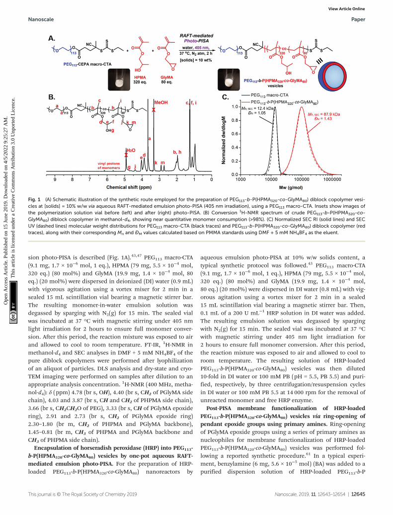
sion photo-PISA is described (Fig. 1A).43,47 PEG113 macro-CTA(9.1 mg, 1.7 × 10⁻⁶ mol, 1 eq.), HPMA (79 mg, 5.5 × 10⁻⁴ mol,320 eq.) (80 mol%) and GlyMA (19.9 mg, 1.4 × 10⁻⁴ mol, 80eq.) (20 mol%) were dispersed in deionized (DI) water (0.9 mL)with vigorous agitation using a vortex mixer for 2 min in asealed 15 mL scintillation vial bearing a magnetic stirrer bar.The resulting monomer-in-water emulsion solution wasdegassed by sparging with N2(g) for 15 min. The sealed vialwas incubated at 37 °C with magnetic stirring under 405 nmlight irradiation for 2 hours to ensure full monomer conver-sion. After this period, the reaction mixture was exposed to airand allowed to cool to room temperature. FT-IR, 1H-NMR inmethanol-d4 and SEC analyses in DMF + 5 mM NH4BF4 of thepure diblock copolymers were performed after lyophilizationof an aliquot of particles. DLS analysis and dry-state and cryo-TEM imaging were performed on samples after dilution to anappropriate analysis concentration. 1H-NMR (400 MHz, metha-nol-d4): δ (ppm) 4.78 (br s, OH), 4.40 (br s, CH2 of PGlyMA sidechain), 4.03 and 3.87 (br s, CH and CH2 of PHPMA side chain),3.66 (br s, CH2CH2O of PEG), 3.33 (br s, CH of PGlyMA epoxidering), 2.91 and 2.73 (br s, CH2 of PGlyMA epoxide ring)2.30–1.80 (br m, CH2 of PHPMA and PGlyMA backbone),1.45–0.81 (br m, CH3 of PHPMA and PGlyMA backbone andCH3 of PHPMA side chain).
Encapsulation of horseradish peroxidase (HRP) into PEG113-b-P(HPMA320-co-GlyMA80) vesicles by one-pot aqueous RAFT-mediated emulsion photo-PISA. For the preparation of HRP-loaded PEG113-b-P(HPMA320-co-GlyMA80) nanoreactors by
aqueous emulsion photo-PISA at 10% w/w solids content, atypical synthetic protocol was followed.43 PEG113 macro-CTA(9.1 mg, 1.7 × 10⁻⁶ mol, 1 eq.), HPMA (79 mg, 5.5 × 10⁻⁴ mol,320 eq.) (80 mol%) and GlyMA (19.9 mg, 1.4 × 10⁻⁴ mol,80 eq.) (20 mol%) were dispersed in DI water (0.8 mL) with vig-orous agitation using a vortex mixer for 2 min in a sealed15 mL scintillation vial bearing a magnetic stirrer bar. Then,0.1 mL of a 200 U mL−1 HRP solution in DI water was added.The resulting emulsion solution was degassed by spargingwith N2(g) for 15 min. The sealed vial was incubated at 37 °Cwith magnetic stirring under 405 nm light irradiation for2 hours to ensure full monomer conversion. After this period,the reaction mixture was exposed to air and allowed to cool toroom temperature. The resulting solution of HRP-loadedPEG113-b-P(HPMA320-co-GlyMA80) vesicles was then diluted10-fold in DI water or 100 mM PB (pH = 5.5, PB 5.5) and puri-fied, respectively, by three centrifugation/resuspension cyclesin DI water or 100 mM PB 5.5 at 14 000 rpm for the removal ofunreacted monomer and free HRP enzyme.
Post-PISA membrane functionalization of HRP-loadedPEG113-b-P(HPMA320-co-GlyMA80) vesicles via ring-opening ofpendant epoxide groups using primary amines. Ring-openingof PGlyMA epoxide groups using a series of primary amines asnucleophiles for membrane functionalization of HRP-loadedPEG113-b-P(HPMA320-co-GlyMA80) vesicles was performed fol-lowing a reported synthetic procedure.61 In a typical experi-ment, benzylamine (6 mg, 5.6 × 10⁻⁵ mol) (BA) was added to apurified dispersion solution of HRP-loaded PEG113-b-P
Fig. 1 (A) Schematic illustration of the synthetic route employed for the preparation of PEG113-b-P(HPMA320-co-GlyMA80) diblock copolymer vesi-cles at [solids] = 10% w/w via aqueous RAFT-mediated emulsion photo-PISA (405 nm irradiation), using a PEG113 macro-CTA. Insets show images ofthe polymerization solution vial before (left) and after (right) photo-PISA. (B) Conversion 1H-NMR spectrum of crude PEG113-b-P(HPMA320-co-GlyMA80) diblock copolymer in methanol-d4, showing near quantitative monomer consumption (>98%). (C) Normalized SEC RI (solid lines) and SECUV (dashed lines) molecular weight distributions for PEG113 macro-CTA (black traces) and PEG113-b-P(HPMA320-co-GlyMA80) diblock copolymer (redtraces), along with their corresponding Mn and ĐM values calculated based on PMMA standards using DMF + 5 mM NH4BF4 as the eluent.
Nanoscale Paper
This journal is © The Royal Society of Chemistry 2019 Nanoscale, 2019, 11, 12643–12654 | 12645
Ope
n A
cces
s A
rtic
le. P
ublis
hed
on 1
5 Ju
ne 2
019.
Dow
nloa
ded
on 4
/5/2
022
9:25
:27
AM
. T
his
artic
le is
lice
nsed
und
er a
Cre
ativ
e C
omm
ons
Attr
ibut
ion
3.0
Unp
orte
d L
icen
ce.
View Article Online

(HPMA320-co-GlyMA80) vesicles at 10× dilution (1% w/w solidscontent) in DI water (2 mL) ([amine]/[epoxide] molar ratio =2.0). The mixture was then stirred at room temperature for18 hours to allow for ring-opening of the epoxide groups andvesicle membrane functionalization. The resulting modifiedvesicles were then purified by one centrifugation/resuspensioncycle in 100 mM PB 5.5 at 14 000 rpm prior to kinetic colori-metric analysis. Successful ring-opening of PGlyMA units wasconfirmed by FT-IR spectroscopy of a lyophilized sample. Theabove protocol was also followed for 1-naphthylmethylamine(NMA) and 1-adamantanemethylamine (AMA). In the case ofcross-linking diamines poly(ethylene glycol)n diamine (n = 23,46) (PEGnDA), ethylenediamine (C2DA), 1,3-diaminopropane(C3DA), 1,4-diaminobutane (C4DA), hexamethylenediamine(C6DA), and p-xylylenediamine (PXDA), a [diamine]/[epoxide]molar ratio = 1.0 was maintained.
Kinetic colorimetric analyses for determination of theactivity of HRP-loaded PEG113-b-P(HPMA320-co-GlyMA80) vesi-cles before and after post-PISA membrane functionalization.Purified HRP-loaded PEG113-b-P(HPMA320-co-GlyMA80) vesiclesbefore and after post-PISA membrane functionalization at 20×dilution (0.5% w/w solids content) in 100 mM PB 5.5 (120 μL)were diluted with 100 mM PB 5.5 (20 μL) in a 96-well platemicrowell. A fixed concentration of 3,3′-dimethoxybenzidine(DMB) (2 mM, 40 μL) was then added. Finally, a 35% w/vaqueous solution of hydrogen peroxide (20 μL) was added, andthe change in absorbance at λ = 492 nm was recorded everyminute for a period of 30 min using a plate reader.Absorbance values were corrected against particle absorbanceat t = 0 min and reported as ΔAbs492 nm.
For Michaelis–Menten kinetics determination, activity ofpurified HRP-loaded PEG113-b-P(HPMA320-co-GlyMA80) vesiclesbefore and after post-PISA membrane functionalization usingBA, NMA, and PXDA was evaluated at different [DMB](0–6 mM) with H2O2 under saturating conditions. Vesicles at20× dilution (0.5% w/w solids content) in 100 mM PB 5.5(120 μL) were diluted with 100 mM PB 5.5 (20 μL) in a 96-wellplate microwell. DMB of appropriate concentration (0–30 mM,40 μL) was then added. Finally, a 35% w/v aqueous solutionof hydrogen peroxide (20 μL) was added, and the changein absorbance was monitored in an identical manner.Absorbance values were corrected against particle absorbanceat t = 0 min and reported as ΔAbs492 nm. In each case, theaverage initial slope of three repeat measurements (V0) for thefirst 10 min of the assay was used for construction ofMichaelis–Menten kinetic plots and was normalized againstVmax (Table S3†). Calculated K*
m values are presented as mean± standard error.
A similar process was followed for activity testing of the freeenzyme after a series of control experiments at appropriate[HRP]. The free HRP solutions at final [HRP] = 1 U mL−1 in100 mM PB 5.5 (20 μL) were diluted with 100 mM PB 5.5(120 μL) in a 96-well plate microwell. DMB (2 mM, 40 μL) wasthen added. Finally, a 35% w/v aqueous solution of hydrogenperoxide (20 μL) was added, and the change in absorbance wasmonitored in an identical manner. In all cases, measurements
were performed in at least triplicate and results are reported astheir average values.
Results and discussionSynthesis of epoxy-functionalized PEG113-b-P(HPMA320-co-GlyMA80) polymersomes via aqueous RAFT-mediated emulsionphoto-PISA
Following related PISA studies previously reported by ourgroup and others,36,40 a water-soluble poly(ethylene glycol)macromolecular chain transfer agent (PEG113 macro-CTA) wasfirst synthesized through an esterification reaction betweenpoly(ethylene glycol) monomethyl ether (PEG113-OH) and anacid-functionalized CTA (esterification efficiency = 93%,Mn, SEC = 12.4 kg mol−1, ĐM = 1.05). Aqueous RAFT-mediatedphotoinitiated PISA (photo-PISA) of a mixture of commerciallyavailable water-miscible 2-hydroxypropyl methacrylate (HPMA)and water-immiscible glycidyl methacrylate (GlyMA) as thecore-forming monomers was achieved under emulsionpolymerization conditions, using PEG113 macro-CTA as boththe steric stabilizer block and the surfactant for stabilizationof the heterogeneous monomer-in-water solution.64 Dispersedmonomer droplets of varying size (5–30 μm) were observedupon optical microscopy imaging of the formed emulsionsolution prior to polymerization (Fig. S2†).
Single-phase epoxy-functionalized PEG113-b-P(HPMA-co-GlyMA) diblock copolymer vesicles were developed uponirradiation of the polymerization solution under 405 nmvisible light in the absence of an externally added photo-initiator or catalyst (“photoiniferter” mechanism oftrithiocarbonates)35,65,66 at 37 °C, targeting DPPHPMA = 320(80 mol%) and DPPGlyMA = 80 (20 mol%) at 10% w/w solidscontent (Fig. 1A). Emulsion photo-PISA was carried out at mildtemperature to ensure quantitative retention of epoxy func-tional groups, as epoxides can undergo partial hydrolysis inaqueous solution at elevated temperatures (60–100 °C).61
Importantly, 99% of PGlyMA pendant epoxy groups remainedintact after photo-PISA, as calculated from 1H-NMR spec-troscopy by comparing the integral ratio of the peaks corres-ponding to epoxy proton signals at 2.73 and 2.91 ppm(I2.73/2.91 ppm = 1.98) to the peak of the methacrylic backbone–CH2– protons at 1.50–2.30 ppm (I1.50–2.30 ppm = 10.00)(Fig. S3†). A kinetic study revealed that near completemonomer conversion (>98%) was achieved after 2 hours ofirradiation time, as calculated by 1H-NMR spectroscopy, whilstthe onset of particle micellization accompanied by an increasein polymerization rate was observed after ∼20 min ofirradiation time (ca. 17% monomer conversion) (Fig. 1B andS4A†). The distribution of PGlyMA units along the growingpolymer chain was also investigated during kinetics monitor-ing and the calculated HPMA/GlyMA molar ratio variedbetween 3 and 4 throughout the whole duration of photo-PISA,suggesting the formation of random copolymers (Fig. S4B†).The prepared PEG113-b-P(HPMA320-co-GlyMA80) diblock copoly-mer possessed monomodal molecular weight distribution with
Paper Nanoscale
12646 | Nanoscale, 2019, 11, 12643–12654 This journal is © The Royal Society of Chemistry 2019
Ope
n A
cces
s A
rtic
le. P
ublis
hed
on 1
5 Ju
ne 2
019.
Dow
nloa
ded
on 4
/5/2
022
9:25
:27
AM
. T
his
artic
le is
lice
nsed
und
er a
Cre
ativ
e C
omm
ons
Attr
ibut
ion
3.0
Unp
orte
d L
icen
ce.
View Article Online

relatively low dispersity value, as determined by SEC analysis(Mn, SEC = 87.9 kg mol−1, ĐM = 1.43) (Fig. 1C).
Dynamic light scattering (DLS) analysis revealed the for-mation of nano-objects with a monomodal size distributionand mean hydrodynamic diameter (Dh) of 188.3 ± 5.6 nm, lowpolydispersity (PD = 0.18 ± 0.02) and a smooth, single expo-nential decay autocorrelation function with optimal signal-to-noise ratio (Y-intercept >0.9) (Fig. 2A and B). The absence ofcharges on the outer surface of the developed particles wasconfirmed by microelectrophoretic analysis at neutral pH(measured zeta-potential = −1.87 ± 0.10 mV). Dry-state trans-mission electron microscopy (TEM) imaging confirmed thesuccessful development of well-defined PEG113-b-P(HPMA320-co-GlyMA80) unilamellar vesicles of spherical shape anduniform size (Ddry-state = 170.3 ± 23.0 nm) (Fig. 2C and S5A†).Additionally, the characteristics of PEG113-b-P(HPMA320-co-GlyMA80) vesicles in solution were studied by cryogenic TEM(cryo-TEM) imaging. Average vesicle size measured from par-ticle counting analysis based on cryo-TEM was in good agree-
ment with values determined by DLS and dry-state TEM ana-lyses (Dcryo = 175.5 ± 24.1 nm), while calculated average mem-brane thickness was Mave = 28.0 ± 3.0 nm (Fig. 2D–F andS5B†).
Preparation of catalytic enzyme-loaded PEG113-b-P(HPMA320-co-GlyMA80) polymersome nanoreactors via one-pot aqueousemulsion photo-PISA
Next, PEG113-b-P(HPMA320-co-GlyMA80) polymersomes wereused as a nanocarrier platform for loading of a model hydro-philic enzyme to prepare catalytically active epoxy-functiona-lized nanoreactors.43,47 Enzyme horseradish peroxidase (HRP)was encapsulated into the inner aqueous compartment of thevesicles by performing a one-pot emulsion photo-PISA processunder the same mild reaction conditions described herein inthe presence of an aqueous HRP solution for the developmentof enzyme-loaded PEG113-b-P(HPMA320-co-GlyMA80) polymer-some nanoreactors. Purification of the vesicle sample for com-plete removal of free HRP was achieved by consecutive cen-trifugation/resuspension cycles in either deionized (DI) wateror 100 mM phosphate buffer (pH = 5.5, PB 5.5).
Importantly, the encapsulation process of HRP into PEG113-b-P(HPMA320-co-GlyMA80) polymersomes did not alter theiroverall characteristics as judged by DLS analysis and TEMimaging (Fig. S6†). Comparable Dh and PD values weremeasured for HRP-loaded vesicles with respect to non-HRP-loaded vesicles (Dh = 182.6 ± 2.5 nm, and PD = 0.14 ± 0.04),whilst average vesicle size and average membrane thicknessmeasured from cryo-TEM images were Dcryo = 172.5 ± 25.5 nmand Mave = 27.9 ± 2.6 nm, respectively.
HRP is known to catalyze the oxidation reaction of colour-less substrate 3,3′-dimethoxybenzidine (DMB) to a coloureddimer product (red-brown), which can be monitored bymeasuring the change in absorbance at λ = 492 nm over timevia kinetic colorimetric analysis. This assay provides a read-outof enzyme activity and consequently of polymersome mem-brane permeability. Notably, PEG113-b-P(HPMA320-co-GlyMA80)vesicles were found to be inherently permeable toward smallmolecule DMB, although they presented reduced enzymeactivity compared to their reported PEG113-b-PHPMA400counterparts presumably due to increased membrane hydro-phobicity in the former case.43 Although nanoreactors withnear identical membrane thickness values were developed inboth cases, the presence of an additional hydrophobic core-block component in the case of PEG113-b-P(HPMA320-co-GlyMA80) vesicles resulted in particles with less hydrated mem-branes and reduced permeability, making the exchange of sub-strates between their outer and inner compartment a moredifficult process.47 Control experiments to assess the activity offree HRP after incubation at 37 °C for 2 hours in 10% w/wHPMA or HPMA/GlyMA (80 : 20 molar ratio) monomer solu-tions in DI water under 405 nm irradiation (photo-PISA con-ditions) revealed quantitative retention of activity in both casescompared to untreated enzyme, showing that the enzyme func-tion was not affected by either monomer (Fig. S7†). Inaddition, high-performance liquid chromatography (HPLC)
Fig. 2 (A) Intensity-weighted size distributions along with average Dh
and PD values (the error shows the standard deviation from 5 repeatmeasurements), and (B) autocorrelation function obtained by DLS forempty PEG113-b-P(HPMA320-co-GlyMA80) vesicles. (C) Representativedry-state TEM image, stained with 1 wt% uranyl acetate (UA) solution,and (D) representative cryo-TEM image of empty PEG113-b-P(HPMA320-co-GlyMA80) vesicles. (E) Histogram of size distribution, and (F) histo-gram of membrane thickness distribution along with calculated averagediameter and membrane thickness values, respectively, measured fromparticle analysis based on cryo-TEM images for empty PEG113-b-P(HPMA320-co-GlyMA80) vesicles. In each case, at least 100 particles wereanalyzed.
Nanoscale Paper
This journal is © The Royal Society of Chemistry 2019 Nanoscale, 2019, 11, 12643–12654 | 12647
Ope
n A
cces
s A
rtic
le. P
ublis
hed
on 1
5 Ju
ne 2
019.
Dow
nloa
ded
on 4
/5/2
022
9:25
:27
AM
. T
his
artic
le is
lice
nsed
und
er a
Cre
ativ
e C
omm
ons
Attr
ibut
ion
3.0
Unp
orte
d L
icen
ce.
View Article Online

and matrix-assisted laser desorption/ionization time-of-flightmass spectrometry (MALDI-ToF MS) were employed to deter-mine potential modification of HRP by ring-opening of theepoxide groups via its lysine residues. Notably, incubation offree HRP with a water-soluble small molecule epoxide (glyci-dol) for 2 hours in DI water showed no evident modificationupon comparison with the untreated enzyme (Fig. S8†).
Membrane functionalization of enzyme-loaded PEG113-b-P(HPMA320-co-GlyMA80) vesicles via ring-opening of epoxidegroups using primary amines
Incorporation of PGlyMA units and thus the presence ofpendant epoxy groups within the hydrophobic membrane ofprepared HRP-loaded nanoreactors provides a reactive handlethat allows for post-PISA functionalization of the polymer-somes with nucleophilic compounds. A commonly utilizedprocedure involves the ring-opening of epoxide groups byprimary amines under mild reaction conditions.61–63 To thisextent, we envisioned that introducing hydrophobic or cross-linking moieties into the polymersome membrane wouldresult in permeability reduction or nanoreactors completelyimpermeable toward small molecules.
A series of commercially available primary amines and dia-mines (cross-linking agents) of varying hydrophobicity werechosen as nucleophiles for ring-opening of PGlyMA epoxygroups and subsequent vesicle membrane modification(Scheme 1). Post-polymerization functionalization wasachieved by mixing purified HRP-loaded PEG113-b-P(HPMA320-co-GlyMA80) vesicle solutions in DI water at [solids] = 1% w/wwith different amines at room temperature for 18 hours. In allcases, the [amine]/[epoxide] molar ratio was constantly main-tained at 2.0 (i.e. [diamine]/[epoxide] = 1.0).61–63 Following thisperiod, membrane-functionalized polymersomes were trans-
ferred into PB 5.5 (optimum pH value of HRP) by one centrifu-gation/resuspension cycle prior to kinetic colorimetric analysisfor enzyme activity and nanoreactor permeability determi-nation. FT-IR spectroscopy was used for the confirmation ofsuccessful ring-opening of PGlyMA units in each case by moni-toring the characteristic asymmetric vibration peaks of epoxidegroups at 849 and 909 cm−1 before and after ring-openingreactions (Fig. S11, S14 and S16†). It should also be noted thatincubation of free HRP in DI water at room temperature for18 hours as a control experiment resulted in no loss of enzymeactivity (>99% retention of activity), compared to incubation ofthe enzyme in PB 5.5 for the same period of time (Fig. S9†).Additionally, a control experiment for comparison of theactivity of non-epoxy-functionalized and purified HRP-loadedPEG113-b-PHPMA400 vesicles before and after incubation withthe utilized primary amine and diamine molecules at roomtemperature for 18 hours in DI water, highlighted that the pro-longed presence of amines in the particle dispersion solutionsdid not affect the catalytic activity of HRP (>95% retention ofactivity in all cases – control vesicle solutions after incubationwith CnDA cross-linkers could not be resuspended in PB 5.5and hence their activity was not assessed) (Fig. S10†).
Ring-opening of PGlyMA units using linear poly(ethyleneglycol) diamines (PEGnDA) as cross-linkers. First, two water-soluble poly(ethylene glycol)-based diamine polymers ofdiffering molecular weight (PEGnDA, n = 23 or 46) wereselected as cross-linking agents for post-PISA membranefunctionalization of HRP-loaded PEG113-b-P(HPMA320-co-GlyMA80) nanoreactors following the above described process(Scheme 1). In both cases, efficient ring-opening of epoxygroups of PGlyMA using polymeric PEG23/46DA as cross-linkerswas confirmed by FT-IR spectroscopy (Fig. S11†) that resultedin a noticeable increase of nanostructure size. Dh values of
Scheme 1 Schematic illustration of the membrane functionalization procedure of HRP-loaded PEG113-b-P(HPMA320-co-GlyMA80) polymersomenanoreactors via ring-opening of pendant PGlyMA epoxide groups using a series of primary amines as nucleophiles to yield polymersomes with con-trolled permeability.
Paper Nanoscale
12648 | Nanoscale, 2019, 11, 12643–12654 This journal is © The Royal Society of Chemistry 2019
Ope
n A
cces
s A
rtic
le. P
ublis
hed
on 1
5 Ju
ne 2
019.
Dow
nloa
ded
on 4
/5/2
022
9:25
:27
AM
. T
his
artic
le is
lice
nsed
und
er a
Cre
ativ
e C
omm
ons
Attr
ibut
ion
3.0
Unp
orte
d L
icen
ce.
View Article Online

PEGnDA-modified particles were approximately 240 nm (PD =0.11–0.14), as measured by DLS analysis (Fig. 3A-I and B-I),whereas the hydrodynamic diameter of the original PEG113-b-P(HPMA320-co-GlyMA80) polymersome platform was ∼180 nm.No evident macroscopic precipitation of the particle solutionswas observed that could imply inter-vesicular cross-linking. Inparticular, dry-state and cryo-TEM imaging revealed the reten-tion of vesicular morphology in both cases with no apparentparticle agglomeration, whilst average vesicle sizes determinedfrom particle counting analysis based on cryo-TEM imageswere in excellent agreement with DLS results (Dcryo, PEG23DA =224.0 ± 21.0 nm, and Dcryo, PEG46DA = 218.5 ± 22.3 nm)(Fig. 3A-II, B-II and S12†). The observed size increase can beexplained by the hydrophilic nature of the PEGnDA cross-linkers that could potentially lead to hydration of the vesiclemembranes and as a result to partial swelling of the assem-blies. Moreover, a measurable increase in average membranethickness to ∼32 nm was noticed in both cases compared tothe non-functionalized vesicles (Fig. 3A-III and B-III).
Surprisingly, activity comparison between encapsulatedHRP into inherently permeable PEG113-b-P(HPMA320-co-GlyMA80) vesicles and PEGnDA cross-linked PEG113-b-P(HPMA320-co-GlyMA80) vesicles showed a significant enzymeactivity decrease for the latter ones. More specifically, an absor-bance decrease of 45 ± 5% was measured from kinetic colori-metric analysis after 30 min in the case of cross-linked PEG113-b-P(HPMA320-co-GlyMA80) + PEG23DA vesicles compared totheir non-functionalized epoxide-containing counterparts,while a further absorbance decrease of 69 ± 4% was identifiedupon increasing the molecular weight of the PEGnDA cross-linker (n = 46) (Fig. 3C and S13†). The observed decrease inHRP activity demonstrates the permeability reduction ofPEGnDA-functionalized nanoreactors toward DMB and hydro-gen peroxide. Contrary to the observed particle swelling thatwould suggest a potential permeability enhancement, this canbe understood in terms of the considerably thicker and moredensely packed vesicular membranes after cross-linking thatcreate an additional diffusive barrier which in turn hinders thepassage of small molecule substrates to reach the activeenzyme site.43
Ring-opening of PGlyMA units using linear aliphatic dia-mines (CnDA) as cross-linkers. Next, a series of linear aliphaticdiamine small molecules with increasing chain length (CnDA,n = 2, 3, 4, 6) were selected as cross-linkers for ring-opening ofepoxy groups located in the hydrophobic domain of HRP-loaded PEG113-b-P(HPMA320-co-GlyMA80) vesicles (Scheme 1).We hypothesized that increasing the hydrophobicity of thecross-linking moiety compared to PEGnDA would result in amore pronounced enzyme activity reduction similar to thebackground activity (∼0%) of empty vesicles. This would allowfor fabrication of nanoreactors that are effectively imperme-able toward substrate molecules, whereby their encapsulatedcargo is isolated from the outer aqueous surrounding environ-ment of the particles.
FT-IR spectroscopy was used to confirm the successfulmodification of the polymersome membrane (i.e. dis-
Fig. 3 (A) For HRP-loaded PEG113-b-P(HPMA320-co-GlyMA80) +PEG23DA vesicles, and (B) for HRP-loaded PEG113-b-P(HPMA320-co-GlyMA80) + PEG46DA vesicles: (I) Intensity-weighted size distributionsalong with average Dh and PD values obtained by DLS (the error showsthe standard deviation from 5 repeat measurements), (II) representativecryo-TEM images, and (III) histograms of membrane thickness distri-bution along with calculated average membrane thickness valuesmeasured from particle analysis based on cryo-TEM images. In eachcase, at least 100 particles were analyzed. (C) Schematic illustration ofHRP-catalyzed oxidation reaction of DMB to a red-brown dimerproduct, detected by kinetic colorimetric assay, taking place insideinherently permeable HRP-loaded PEG113-b-P(HPMA320-co-GlyMA80)vesicles versus cross-linked HRP-loaded PEG113-b-P(HPMA320-co-GlyMA80) + PEGnDA vesicles (I), and enzymatic activity of the purifiedempty (grey line), HRP-loaded (red line), and HRP-loaded PEGnDA-func-tionalized (purple lines) PEG113-b-P(HPMA320-co-GlyMA80) vesicles (endpoint = 30 min, λ = 492 nm) (the insets show the end-point microwellsin each case) (II).
Nanoscale Paper
This journal is © The Royal Society of Chemistry 2019 Nanoscale, 2019, 11, 12643–12654 | 12649
Ope
n A
cces
s A
rtic
le. P
ublis
hed
on 1
5 Ju
ne 2
019.
Dow
nloa
ded
on 4
/5/2
022
9:25
:27
AM
. T
his
artic
le is
lice
nsed
und
er a
Cre
ativ
e C
omm
ons
Attr
ibut
ion
3.0
Unp
orte
d L
icen
ce.
View Article Online

appearance of the characteristic bands of epoxide groups at849 and 909 cm−1) (Fig. S14†). Interestingly, DLS analysis ofthe CnDA-functionalized HRP-loaded polymersomes revealed asize decrease to 125–130 nm (PD = 0.12–0.13) for the shorterC2DA and C3DA cross-linkers compared to the original PEG113-b-P(HPMA320-co-GlyMA80) vesicles which was attributed toshrinkage of the particles upon cross-linking with hydrophobiccompounds that results in exclusion of water molecules fromthe membrane. In the case of longer C4DA and C6DA cross-linkers, a vast Dh increase was measured to 1497 ± 266 nm and1389 ± 149 nm, respectively, accompanied by a noticeable poly-dispersity increase (PD = 0.23–0.24) (Fig. 4A). These findingssuggest the occurrence of particle agglomeration in the lattercases due to the development of inter-vesicular interactionsresulting in the apparent population of particles withincreased size. This was also evident by visual inspection ofthe PISA solutions upon functionalization after one week,where macroscopic precipitation was visible to some extent forPEG113-b-P(HPMA320-co-GlyMA80) + C4/6DA assemblies, imply-ing the occurrence of time-dependent coagulation in aqueousmedia. The morphology retention of HRP-loaded PEG113-b-P(HPMA320-co-GlyMA80) + CnDA polymersomes was also verifiedby dry-state and cryo-TEM imaging after post-PISA functionali-zation (Dcryo = 116–120 nm), for which near identical vesicleformulations were observed in all cases. A negligible popu-lation of aggregated particles could only be observed in thecase of C4DA and C6DA-functionalized polymersomes that sup-ports the DLS findings described above. The average mem-brane thickness of the prepared nanoreactors was also calcu-lated from particle counting analysis in each case. Increasedmembrane thickness values of Mave = 30–32 nm were measuredin all cases, comparable to PEGnDA-functionalized vesicles(Fig. 4B and S15†).
Similar to PEGnDA cross-linked vesicles, assessment of theenzyme activity of HRP-loaded PEG113-b-P(HPMA320-co-GlyMA80) + CnDA nanoreactors through kinetic colorimetricanalysis revealed an absorbance decrease ranging from 50% to61% depending on the cross-linker molecule used as com-pared to their PEG113-b-P(HPMA320-co-GlyMA80) counterparts(Fig. 4C). The absorbance decrease measured was approxi-mately the same in all cases, whilst there was no apparenttrend based on the length of the cross-linker used for ring-opening of PGlyMA units (i.e. increasing the CnDA lengthdidn’t result in an analogous HRP activity decrease). Overall,membrane cross-linking of the vesicles using CnDA moleculesand subsequent introduction of additional hydrophobicityyielded particles with thicker and possibly less hydrated mem-branes with markedly reduced permeability.
Ring-opening of PGlyMA units using other hydrophobicprimary amines as nucleophiles. As a next step, a more hydro-phobic cross-linking xylylene-based diamine (PXDA) wasselected for ring-opening of PGlyMA epoxy groups along withthree other hydrophobic compounds bearing a single aminogroup in their structure (i.e. BA, NMA, and AMA) to prevent for-mation of linkages and agglomeration of the vesicles, asobserved for longer CnDA cross-linkers, and to achieve com-
Fig. 4 (A) Intensity-weighted size distributions along with average Dh
and PD values obtained by DLS (the error shows the standard deviationfrom 5 repeat measurements) for HRP-loaded PEG113-b-P(HPMA320-co-GlyMA80) + CnDA (n = 2, 3, 4, 6) vesicles. (B) Representative cryo-TEMimages of HRP-loaded PEG113-b-P(HPMA320-co-GlyMA80) + CnDA vesi-cles, and corresponding histograms of membrane thickness distributionalong with calculated average membrane thickness values measuredfrom particle analysis based on cryo-TEM images. In each case, at least100 particles were analyzed. (C) Enzymatic activity of the purified empty(grey line), HRP-loaded (red line), and HRP-loaded CnDA-functionalized(purple/pink lines) PEG113-b-P(HPMA320-co-GlyMA80) vesicles, andHRP-loaded PEG113-b-P(HPMA)400 vesicles (black line) (end point =30 min, λ = 492 nm) (the insets show the end-point microwells in eachcase).
Paper Nanoscale
12650 | Nanoscale, 2019, 11, 12643–12654 This journal is © The Royal Society of Chemistry 2019
Ope
n A
cces
s A
rtic
le. P
ublis
hed
on 1
5 Ju
ne 2
019.
Dow
nloa
ded
on 4
/5/2
022
9:25
:27
AM
. T
his
artic
le is
lice
nsed
und
er a
Cre
ativ
e C
omm
ons
Attr
ibut
ion
3.0
Unp
orte
d L
icen
ce.
View Article Online

plete reduction of membrane permeability for enzyme-loadedPEG113-b-P(HPMA320-co-GlyMA80) polymersome nanoreactors(Scheme 1). Notably, the majority of selected amines resultedin fabrication of stable nanoparticle dispersions, except forAMA, where prominent macroscopic precipitation and for-mation of a film at the bottom of the vial was observed afterepoxide ring-opening reaction, implying instability of thenano-objects in this case due to significantly increased hydro-phobicity. In all cases, FT-IR spectroscopic analysis confirmedthe successful ring-opening of PGlyMA epoxide groups(Fig. S16†).
In the case of BA-functionalized vesicles, Dh and PD valuescomparable with those of the original HRP-loaded vesicleswere measured from DLS analysis (Dh = 203.4 ± 2.8 nm andPD = 0.08) (Fig. 5A-I and II). Dry-state TEM imaging was firstused to confirm that post-PISA functionalization did not altertheir morphology and uniform size (Fig. 5A-III and S17A-I†),while size and membrane thickness measurements werecarried out from particle counting analysis based on cryo-TEMimages (Fig. 5A-IV to VI and S17A-II†). Importantly, the averagediameter of BA membrane-modified vesicles was in goodagreement with DLS results (Dcryo, BA = 175.4 ± 27.7 nm),whereas a significant increase in average membrane thicknesswas also observed (Mave, BA = 33.2 ± 2.9 nm) compared toPEG113-b-P(HPMA320-co-GlyMA80) vesicles.
Near identical well-defined vesicle formulations of low PDand similar Dh values were identified by DLS analysis and dry-state TEM imaging in the case of NMA and PXDA amines(Fig. 5B-I to III, C-I to III and S17†). Similar to HRP-loadedPEG113-b-P(HPMA320-co-GlyMA80) + BA nanoreactors, average
Dcryo values were in good agreement with light scatteringresults, whilst an increased average membrane thickness wascalculated for PXDA-functionalized vesicles (Mave, PXDA = 31.1 ±2.7 nm). Additionally, a further membrane thickness increaseto Mave, NMA = 37.6 ± 2.8 nm was calculated upon PGlyMA ring-opening using a more hydrophobic amine (i.e. NMA), showingan evident relationship between the structure/hydrophobicityof the nucleophile and the membrane thickness of the result-ing vesicles (Fig. 5B-IV to VI, C-IV to VI and S17†).
The activity of HRP-loaded PEG113-b-P(HPMA320-co-GlyMA80) + BA/NMA/PXDA polymersomes was assessed bykinetic colorimetric analysis, monitoring the enzyme-catalyzedperoxidation of DMB in line with above described assays(Fig. 6A). The rate of DMB peroxidation was clearly affected bythe physicochemical nature of the polymersome (particularlyMave), with reductions in substrate processing correlating toincreasing thickness of the diffusional barriers provided by thefunctionalized membrane. At a fixed substrate concentration(0.4 mM DMB and 1.1 M H2O2), there was a small reduction inproduct formation for the PXDA cross-linked polymersomes of31 ± 4% upon comparison with the original HRP-loadedPEG113-b-P(HPMA320-co-GlyMA80) vesicles that dropped to>80% in the case of BA and NMA-functionalized nanoreactors(Fig. 6B). Although these values correlate well with measuredMave values (i.e. NMA > BA > PXDA > PGlyMA) and showcasehow post-PISA chemical modification affects the membranecharacter altering its relative permeability, it was important toexpand this data to explore the effect of substrate concen-tration; measuring the observable permeability. No change tothe actual Km of the intact enzyme was taken into consider-
Fig. 5 (A) For HRP-loaded PEG113-b-P(HPMA320-co-GlyMA80) + BA vesicles, (B) for HRP-loaded PEG113-b-P(HPMA320-co-GlyMA80) + NMA vesicles,and (C) for HRP-loaded PEG113-b-P(HPMA320-co-GlyMA80) + PXDA vesicles: (I) Intensity-weighted size distributions along with average Dh and PDvalues (the error shows the standard deviation from 5 repeat measurements), and (II) autocorrelation functions obtained by DLS, (III) representativedry-state TEM images, stained with 1 wt% UA solution, and (IV) representative cryo-TEM images, (V) histograms of size distribution, and (VI) histo-grams of membrane thickness distribution along with calculated average diameter and membrane thickness values, respectively, measured fromparticle analysis based on cryo-TEM images. In each case, at least 100 particles were analyzed.
Nanoscale Paper
This journal is © The Royal Society of Chemistry 2019 Nanoscale, 2019, 11, 12643–12654 | 12651
Ope
n A
cces
s A
rtic
le. P
ublis
hed
on 1
5 Ju
ne 2
019.
Dow
nloa
ded
on 4
/5/2
022
9:25
:27
AM
. T
his
artic
le is
lice
nsed
und
er a
Cre
ativ
e C
omm
ons
Attr
ibut
ion
3.0
Unp
orte
d L
icen
ce.
View Article Online

ation as such fundamental changes to activity are not generallyassociated with encapsulation.67 Normalized kinetic data waspresented in terms of V0/Vmax due to the focus on comparisonsbetween equilibrium substrate dissociation constant (Km) ofthe nanoreactor-loaded enzyme rather than absolute activity;an [enzyme]-dependent parameter.
Systematically varying [DMB] (with [H2O2] under saturatingconditions) gave a more detailed insight into the effect ofmembrane modification on the performance of nanoreactors,where the trend identified previously was confirmed and could
be elucidated with greater detail (Fig. 6C-I and S18†). As com-pared to the Km value of the free enzyme at 0.79 ± 0.06 mM,the K*
m of HRP-loaded PEG113-b-P(HPMA320-co-GlyMA80) nanor-eactors increased by ca. 30% (up to 1.14 ± 0.18 mM) as a resultof enzyme compartmentalization within the semi-permeableP(HPMA320-co-GlyMA80) membrane. This effect could be tunedby introducing cross-linking or chemical modification. PXDAcross-linking of polymersomes further increased K*
m by ca.30% (up to 1.84 ± 0.18 mM), in accordance with earlier dataunder fixed substrate concentration conditions. Interestingly,using such kinetic assays, we were able to resolve the effect ofBA and NMA modification on the permeability of the polymer-some membrane. Indeed, post-PISA membrane modificationand insertion of benzylic (BA) groups gave rise to a 70%increase in K*
m, which increased by a further 230% when usingbulkier naphthyl (NMA) groups (up to 3.14 ± 0.32 & 10.27 ±4.34 mM, respectively). Overall, the observed rate of substrateturnover of the unmodified polymersomes was ca. 90% higherthan that of the NMA-functionalized nanoreactors. Relatingthese values for K*
m to the measured values of average mem-brane thickness (Mave), it is apparent that their relationship isnon-linear, which means that the physicochemical origin ofthis tunability cannot be fully explicated by the influence ofchemical modifier upon the membrane dimensions but is alsorelated to the nature of the new membrane (Fig. 6C-II). In thisinstance, we can understand the increasing K*
m value (anddecreasing permeability) between BA and NMA membranesarising due to the increasing non-covalent aromatic inter-actions between polymer chains (i.e. increasing the densityand reducing the porosity of the bilayer).
Overall, our findings provide a more in depth understand-ing of how relatively simple chemistries, used for thefunctionalization of polymersome membranes with increas-ingly more hydrophobic moieties, can tune the passivediffusion of small molecules into their aqueous inner lumen.This strategy has been utilized to achieve greater control oversubstrate processing by enzymes encapsulated within a poly-mersome nanoreactor.
Conclusions
In summary, we report a facile strategy to modulate the mem-brane permeability of polymeric vesicles. Epoxy-containing,enzyme-loaded vesicle nanoreactors obtained via aqueousemulsion PISA were functionalized with a series of diaminecross-linkers or hydrophobic primary amines using a simpleprocedure. Membrane modification resulted in increasedthickness and reduced permeability relative to the non-functio-nalized particles. Of the compounds tested, the hydrophobicamines exhibited the most dramatic blocking effect on thevesicle membranes, reducing permeability by over 80% asdetermined by a colorimetric assay involving substrate oxi-dation by the encapsulated enzymes. This fundamental studyreveals important insight into the relationship between mem-brane thickness, cross-linking density, or hydrophobicity and
Fig. 6 (A) Schematic illustration of HRP-catalyzed oxidation reaction ofDMB to a red-brown dimer product, detected by kinetic colorimetricassay, taking place inside inherently permeable HRP-loaded PEG113-b-P(HPMA320-co-GlyMA80) vesicles versus impermeable HRP-loadedPEG113-b-P(HPMA320-co-GlyMA80) + BA/NMA vesicles, and (B) enzy-matic activity of the purified empty (grey line), HRP-loaded (red line),HRP-loaded PXDA-functionalized (purple line), and HRP-loaded BA/NMA-functionalized (green lines) PEG113-b-P(HPMA320-co-GlyMA80)vesicles, and HRP-loaded PEG113-b-P(HPMA)400 vesicles (black line) (endpoint = 30 min, λ = 492 nm) (the insets show the end-point microwellsin each case). (C) Normalized Michaelis–Menten kinetic plots of freeHRP and HRP-loaded PEG113-b-P(HPMA320-co-GlyMA80) + BA/NMA/PXDA vesicles (I), and calculated K*
m values (mean ± standard error) foreach sample as a function of average nanoreactor membrane thickness(Mave) (II).
Paper Nanoscale
12652 | Nanoscale, 2019, 11, 12643–12654 This journal is © The Royal Society of Chemistry 2019
Ope
n A
cces
s A
rtic
le. P
ublis
hed
on 1
5 Ju
ne 2
019.
Dow
nloa
ded
on 4
/5/2
022
9:25
:27
AM
. T
his
artic
le is
lice
nsed
und
er a
Cre
ativ
e C
omm
ons
Attr
ibut
ion
3.0
Unp
orte
d L
icen
ce.
View Article Online

permeability. Such fine control over the diffusion of substratesacross inherently permeable vesicular membranes has rarelybeen demonstrated and should inform particle design infuture studies.
Conflicts of interest
There are no conflicts to declare.
Acknowledgements
This work was supported by the ERC (grant number 615142),EPSRC, and the University of Birmingham. D. S. W. thanks theSer Cymru II programme for support; this project receivedfunding from the European Union’s Horizon 2020 researchand innovation under the Marie Skłodowska-Curie grant agree-ment no. 663830. Advanced BioImaging Research TechnologyPlatform, BBSRC ALERT14 award BB/M01228X/1, is thankedfor supporting cryo-TEM characterization and Dr S. Bakker(University of Warwick) is thanked for cryo-TEM assistance.Dr M. C. Arno (University of Birmingham) is thanked foroptical microscopy assistance.
Notes and references
1 J. W. Szostak, D. P. Bartel and P. L. Luisi, Nature, 2001, 409,387–390.
2 W. Martin, Philos. Trans. R. Soc., B, 2010, 365, 847–855.3 K. Sugano, M. Kansy, P. Artursson, A. Avdeef, S. Bendels,
L. Di, G. F. Ecker, B. Faller, H. Fischer, G. Gerebtzoff,H. Lennernaes and F. Senner, Nat. Rev. Drug Discovery,2010, 9, 597–614.
4 N. J. Yang and M. J. Hinner, in Site-Specific Protein Labeling:Methods and Protocols, ed. A. Gautier and M. J. Hinner,Springer New York, New York, NY, 2015, pp. 29–53, DOI:10.1007/978-1-4939-2272-7_3.
5 M. Marguet, C. Bonduelle and S. Lecommandoux, Chem.Soc. Rev., 2013, 42, 512–529.
6 R. Roodbeen and J. C. M. van Hest, BioEssays, 2009, 31,1299–1308.
7 R. J. R. W. Peters, I. Louzao and J. C. M. van Hest, Chem.Sci., 2012, 3, 335–342.
8 J. Xu, F. J. Sigworth and D. A. LaVan, Adv. Mater., 2010, 22,120–127.
9 J. Gaitzsch, X. Huang and B. Voit, Chem. Rev., 2016, 116,1053–1093.
10 B. C. Buddingh’ and J. C. M. van Hest, Acc. Chem. Res.,2017, 50, 769–777.
11 P. A. Gale, J. T. Davis and R. Quesada, Chem. Soc. Rev.,2017, 46, 2497–2519.
12 A. Belluati, I. Craciun, C. E. Meyer, S. Rigo andC. G. Palivan, Curr. Opin. Biotechnol., 2019, 60, 53–62.
13 J. F. Le Meins, O. Sandre and S. Lecommandoux, Eur.Phys. J. E, 2011, 34, 14.
14 C. G. Palivan, R. Goers, A. Najer, X. Zhang, A. Car andW. Meier, Chem. Soc. Rev., 2016, 45, 377–411.
15 E. Rideau, R. Dimova, P. Schwille, F. R. Wurm andK. Landfester, Chem. Soc. Rev., 2018, 47, 8572–8610.
16 B. M. Discher, Y.-Y. Won, D. S. Ege, J. C. M. Lee, F. S. Bates,D. E. Discher and D. A. Hammer, Science, 1999, 284, 1143–1146.
17 D. E. Discher and A. Eisenberg, Science, 2002, 297, 967–973.
18 J. Rodríguez-Hernández, F. Chécot, Y. Gnanou andS. Lecommandoux, Prog. Polym. Sci., 2005, 30, 691–724.
19 S. Sugihara, A. Blanazs, S. P. Armes, A. J. Ryan andA. L. Lewis, J. Am. Chem. Soc., 2011, 133, 15707–15713.
20 Y. Mai and A. Eisenberg, Chem. Soc. Rev., 2012, 41, 5969–5985.
21 A. Blanazs, J. Madsen, G. Battaglia, A. J. Ryan andS. P. Armes, J. Am. Chem. Soc., 2011, 133, 16581–16587.
22 N. J. Warren and S. P. Armes, J. Am. Chem. Soc., 2014, 136,10174–10185.
23 N. J. Warren, O. O. Mykhaylyk, D. Mahmood, A. J. Ryan andS. P. Armes, J. Am. Chem. Soc., 2014, 136, 1023–1033.
24 N. J. Warren, O. O. Mykhaylyk, A. J. Ryan, M. Williams,T. Doussineau, P. Dugourd, R. Antoine, G. Portale andS. P. Armes, J. Am. Chem. Soc., 2015, 137, 1929–1937.
25 W. Zhou, Q. Qu, Y. Xu and Z. An, ACS Macro Lett., 2015, 4,495–499.
26 J. C. Foster, S. Varlas, B. Couturaud, Z. Coe andR. K. O’Reilly, J. Am. Chem. Soc., 2019, 141, 2742–2753.
27 S. L. Canning, G. N. Smith and S. P. Armes,Macromolecules, 2016, 49, 1985–2001.
28 X. Wang and Z. An, Macromol. Rapid Commun., 2019, 40,1800325.
29 D. B. Wright, M. A. Touve, M. P. Thompson andN. C. Gianneschi, ACS Macro Lett., 2018, 7, 401–405.
30 J. C. Foster, S. Varlas, L. D. Blackman, L. A. Arkinstall andR. K. O’Reilly, Angew. Chem., Int. Ed., 2018, 57, 10672–10676.
31 W. Zhang, F. D’Agosto, O. Boyron, J. Rieger andB. Charleux, Macromolecules, 2012, 45, 4075–4084.
32 W. Zhang, F. D’Agosto, P.-Y. Dugas, J. Rieger andB. Charleux, Polymer, 2013, 54, 2011–2019.
33 S. Y. Khor, N. P. Truong, J. F. Quinn, M. R. Whittaker andT. P. Davis, ACS Macro Lett., 2017, 6, 1013–1019.
34 J. Tan, X. Dai, Y. Zhang, L. Yu, H. Sun and L. Zhang, ACSMacro Lett., 2019, 8, 205–212.
35 J. Yeow and C. Boyer, Adv. Sci., 2017, 4, 1700137.36 J. Tan, H. Sun, M. Yu, B. S. Sumerlin and L. Zhang, ACS
Macro Lett., 2015, 4, 1249–1253.37 J. Tan, Y. Bai, X. Zhang, C. Huang, D. Liu and L. Zhang,
Macromol. Rapid Commun., 2016, 37, 1434–1440.38 J. Yeow, O. R. Sugita and C. Boyer, ACS Macro Lett., 2016, 5,
558–564.39 J. Tan, D. Liu, Y. Bai, C. Huang, X. Li, J. He, Q. Xu,
X. Zhang and L. Zhang, Polym. Chem., 2017, 8, 1315–1327.40 L. D. Blackman, K. E. B. Doncom, M. I. Gibson and
R. K. O’Reilly, Polym. Chem., 2017, 8, 2860–2871.
Nanoscale Paper
This journal is © The Royal Society of Chemistry 2019 Nanoscale, 2019, 11, 12643–12654 | 12653
Ope
n A
cces
s A
rtic
le. P
ublis
hed
on 1
5 Ju
ne 2
019.
Dow
nloa
ded
on 4
/5/2
022
9:25
:27
AM
. T
his
artic
le is
lice
nsed
und
er a
Cre
ativ
e C
omm
ons
Attr
ibut
ion
3.0
Unp
orte
d L
icen
ce.
View Article Online

41 B. Couturaud, P. G. Georgiou, S. Varlas, J. R. Jones,M. C. Arno, J. C. Foster and R. K. O’Reilly, Macromol. RapidCommun., 2019, 40, 1800460.
42 J. Tan, D. Liu, X. Zhang, C. Huang, J. He, Q. Xu, X. Li andL. Zhang, RSC Adv., 2017, 7, 23114–23121.
43 L. D. Blackman, S. Varlas, M. C. Arno, A. Fayter,M. I. Gibson and R. K. O’Reilly, ACS Macro Lett., 2017, 6,1263–1267.
44 J. Tan, D. Liu, Y. Bai, C. Huang, X. Li, J. He, Q. Xu andL. Zhang, Macromolecules, 2017, 50, 5798–5806.
45 J. Tan, X. Zhang, D. Liu, Y. Bai, C. Huang, X. Li andL. Zhang, Macromol. Rapid Commun., 2017, 38,1600508.
46 L. D. Blackman, S. Varlas, M. C. Arno, Z. H. Houston,N. L. Fletcher, K. J. Thurecht, M. Hasan, M. I. Gibson andR. K. O’Reilly, ACS Cent. Sci., 2018, 4, 718–723.
47 S. Varlas, L. D. Blackman, H. E. Findlay, E. Reading,P. J. Booth, M. I. Gibson and R. K. O’Reilly, Macromolecules,2018, 51, 6190–6201.
48 S. Varlas, P. G. Georgiou, P. Bilalis, J. R. Jones,N. Hadjichristidis and R. K. O’Reilly, Biomacromolecules,2018, 19, 4453–4462.
49 G. Cheng and J. Pérez-Mercader, Macromol. RapidCommun., 2019, 40, 1800513.
50 R. J. R. W. Peters, M. Marguet, S. Marais, M. W. Fraaije,J. C. M. van Hest and S. Lecommandoux, Angew. Chem., Int.Ed., 2014, 53, 146–150.
51 H. Che, S. Cao and J. C. M. van Hest, J. Am. Chem. Soc.,2018, 140, 5356–5359.
52 C. Nardin, S. Thoeni, J. Widmer, M. Winterhalter andW. Meier, Chem. Commun., 2000, 1433–1434.
53 J. Gaitzsch, D. Appelhans, L. Wang, G. Battaglia andB. Voit, Angew. Chem., Int. Ed., 2012, 51, 4448–4451.
54 D. Gräfe, J. Gaitzsch, D. Appelhans and B. Voit, Nanoscale,2014, 6, 10752–10761.
55 T. Einfalt, R. Goers, I. A. Dinu, A. Najer, M. Spulber,O. Onaca-Fischer and C. G. Palivan, Nano Lett., 2015, 15,7596–7603.
56 L. Messager, J. R. Burns, J. Kim, D. Cecchin, J. Hindley,A. L. B. Pyne, J. Gaitzsch, G. Battaglia and S. Howorka,Angew. Chem., Int. Ed., 2016, 55, 11106–11109.
57 S. F. M. van Dongen, M. Nallani, J. J. L. M. Cornelissen,R. J. M. Nolte and J. C. M. van Hest, Chem. – Eur. J., 2009,15, 1107–1114.
58 I. Louzao and J. C. M. van Hest, Biomacromolecules, 2013,14, 2364–2372.
59 W.-J. Zhang, C.-Y. Hong and C.-Y. Pan, ACS Appl. Mater.Interfaces, 2017, 9, 15086–15095.
60 X.-F. Xu, C.-Y. Pan, W.-J. Zhang and C.-Y. Hong,Macromolecules, 2019, 52, 1965–1975.
61 P. Chambon, A. Blanazs, G. Battaglia and S. P. Armes,Langmuir, 2012, 28, 1196–1205.
62 J. R. Lovett, L. P. D. Ratcliffe, N. J. Warren, S. P. Armes,M. J. Smallridge, R. B. Cracknell and B. R. Saunders,Macromolecules, 2016, 49, 2928–2941.
63 F. L. Hatton, J. R. Lovett and S. P. Armes, Polym. Chem.,2017, 8, 4856–4868.
64 S. Piogé, T. N. Tran, T. G. McKenzie, S. Pascual,M. Ashokkumar, L. Fontaine and G. Qiao, Macromolecules,2018, 51, 8862–8869.
65 S. Muthukrishnan, E. H. Pan, M. H. Stenzel, C. Barner-Kowollik, T. P. Davis, D. Lewis and L. Barner,Macromolecules, 2007, 40, 2978–2980.
66 M. Chen, M. Zhong and J. A. Johnson, Chem. Rev., 2016,116, 10167–10211.
67 L. M. P. E. van Oppen, L. K. E. A. Abdelmohsen, S. E. vanEmst-de Vries, P. L. W. Welzen, D. A. Wilson,J. A. M. Smeitink, W. J. H. Koopman, R. Brock,P. H. G. M. Willems, D. S. Williams and J. C. M. van Hest,ACS Cent. Sci., 2018, 4, 917–928.
Paper Nanoscale
12654 | Nanoscale, 2019, 11, 12643–12654 This journal is © The Royal Society of Chemistry 2019
Ope
n A
cces
s A
rtic
le. P
ublis
hed
on 1
5 Ju
ne 2
019.
Dow
nloa
ded
on 4
/5/2
022
9:25
:27
AM
. T
his
artic
le is
lice
nsed
und
er a
Cre
ativ
e C
omm
ons
Attr
ibut
ion
3.0
Unp
orte
d L
icen
ce.
View Article Online