tumour budding in oral squamous cell carcinoma: a meta
TRANSCRIPT

Tumour budding in oral squamous cellcarcinoma: a meta-analysisAlhadi Almangush*,1,2,3, Matti Pirinen4,5,6, Ilkka Heikkinen1,2, Antti A Makitie7, Tuula Salo2,8,9,10
and Ilmo Leivo11
1Department of Patholosgy, University of Helsinki, Haartmaninkatu 3, PO Box 21, Helsinki FIN-00014, Finland; 2Department of Oraland Maxillofacial Diseases, University of Helsinki, Haartmaninkatu 8, PO Box 63, Helsinki FI-00014, Finland; 3Institute of Dentistry,University of Misurata, PO Box 2478, Misurata, Libya; 4Institute for Molecular Medicine Finland (FIMM), University of Helsinki,Helsinki FIN-00014, Finland; 5Helsinki Institute for Information Technology HIIT and Department of Mathematics and Statistics,University of Helsinki, Helsinki FIN-00014, Finland; 6Department of Public Health, University of Helsinki, Helsinki FIN-00014,Finland; 7Department of Otorhinolaryngology – Head and Neck Surgery, Helsinki University Hospital and University of Helsinki,Kasarminkatu 11-13, Helsinki FI-00130, Finland; 8Helsinki University Hospital, Helsinki FIN-00014, Finland; 9Research Group ofCancer Research and Translational Medicine, Medical Faculty, University of Oulu, PO Box 5281, Oulu 90014, Finland; 10MedicalResearch Center, Oulu University Hospital, Oulu 90220, Finland and 11Department of Pathology, University of Turku,Kiinamyllynkatu 10, Turku 20520, Finland
Background: Tumour budding has been reported as a promising prognostic marker in many cancers. This meta-analysis assessedthe prognostic value of tumour budding in oral squamous cell carcinoma (OSCC).
Methods: We searched OvidMedline, PubMed, Scopus and Web of Science for articles that studied tumour budding in OSCC.We used reporting recommendations for tumour marker (REMARK) criteria to evaluate the quality of studies eligible for meta-analysis.
Results: A total of 16 studies evaluated the prognostic value of tumour budding in OSCC. The meta-analysis showed that tumourbudding was significantly associated with lymph node metastasis (odds ratio¼ 7.08, 95% CI¼ 1.75–28.73), disease-free survival(hazard ratio¼ 1.83, 95% CI¼ 1.34–2.50) and overall survival (hazard ratio¼ 1.88, 95% CI¼ 1.25–2.82).
Conclusions: Tumour budding is a simple and reliable prognostic marker for OSCC. Evaluation of tumour budding could facilitatepersonalised management of OSCC.
Oral squamous cell carcinoma (OSCC) is the most commonmalignancy of the oral cavity and constitutes the majority of headand neck squamous cell carcinomas. According to a recent report,B300 000 new cases of oral cancer were diagnosed worldwide in2012, and with a consequent 145 000 cancer-related deaths (Ferlayet al, 2015). The incidence of OSCC has increased in manycountries and especially in young people (Muller et al, 2008;Korvala et al, 2017). In the Western world, the main aetiologicalfactors for OSCC are tobacco and alcohol consumption. Chewing
of Areca nuts and the use of snuff are the classic risk factors in theIndian population. The 5-year survival rate of OSCC patients isrelatively low, and especially the patients with recurrence havepoor outcomes. Identifying cases at risk for recurrence remainschallenging.
Many histopathologic prognostic parameters (e.g., tumourgrade, depth of invasion, perineural invasion, lymphovascularinvasion, lymphocytic host response and mitotic activity) areusually evaluated in haematoxylin- and eosin- (H–E) stained
*Correspondence: Dr A Almangush; E-mail: [email protected] or [email protected]
Received 29 September 2017; revised 27 October 2017; accepted 30 October 2017; published online 30 November 2017
r 2018 Cancer Research UK. All rights reserved 0007 – 0920/18
FULL PAPER
Keywords: tumour budding; oral squamous cell carcinoma; prognosis; marker; invasive front; treatment; survival
British Journal of Cancer (2018) 118, 577–586 | doi: 10.1038/bjc.2017.425
www.bjcancer.com | DOI:10.1038/bjc.2017.425 577

sections. Such information is included in pathology reports to aidin predicting the behaviour of OSCC. This is paramount forplanning of an appropriate and successful management. However,some of these parameters (e.g., tumour grade and lymphocyticresponse) have not been promising prognosticators, especially inearly stage OSCC (Chen et al, 2013; Almangush et al, 2015a).Moreover, recent research has introduced several biomarkers forOSCC, but they are not yet eligible to be included in the pathologyreport (Soland and Brusevold, 2013; Almangush et al, 2017a). Inaddition, such biomarkers require additional staining procedureswhich are not routinely used. Therefore, it is important to identifynew powerful prognostic markers that are adaptable to conven-tional H–E staining.
Tumour budding, defined as the presence of single cancer cell(s)or cluster(s) of less than five cancer cells at the invasive front (IF),has been reported in many cancers as a promising prognosticfeature (Kadota et al, 2015; Almangush et al, 2016; Rogers et al,2016). Tumour budding at the IF (Figure 1) indicates thedissociation of invasive cancer cells from the main tumour mass.Several recent studies have evaluated the significance of tumourbudding in OSCC. The aim of the current study was tosystematically review the studies on tumour budding in OSCCand to present a meta-analysis of the prognostic value of tumourbudding in OSCC. We also discuss the shortcomings in thepublished studies and provide recommendations for furtherresearch to standardise the evaluation method of tumour buddingin OSCC.
MATERIALS AND METHODS
Search protocol. OvidMedline, PubMed, Scopus and Web ofScience were searched using the following keywords: (‘oral’ or‘mouth’ or ‘tongue’ or ‘floor of mouth’ or ‘lip’ or ‘gingiva’ or‘buccal’ or ‘palate’) and (‘tumour budding’). Our search was limitedto articles in the English language. The end point of the search wasMay 2017. To ensure inclusion of all relevant articles, we manuallysearched the reference lists of all eligible studies. When searchingand screening the studies, we followed the Preferred ReportingItems for Systematic Review and Meta-Analysis (PRISMA) (Moheret al, 2009).
Exclusion criteria. We excluded studies in a language other thanEnglish, studies on animal samples and conference abstracts.
Quality assessment. We used reporting recommendations fortumour marker prognostic studies (REMARK) guidelines (Altmanet al, 2012) to assess the quality of studies that evaluated theprognostic value of tumour budding in OSCC. We summarised themain guidelines in Table 1. Any study that received a score of lessthan 6 was not included in our meta-analysis.
Statistical methods. The meta-analysis was performed by the‘meta’ package (version 4.8-1) in statistical software R (version3.4.0). For each analysis, we carried out an inverse variance-weighted fixed-effects analysis. For completeness, a DerSimonian–Laird random effects analysis (DerSimonian and Laird, 1986) wasalso performed. We considered the random effects analysis as ourmain result to account for heterogeneity between the studies. Inaddition to the meta-analysed effect sizes, our results also includedthe estimated proportion of variation in effect sizes due toheterogeneity (I2) (Higgins and Thompson, 2002) and theDerSimonian–Laird estimate of the variance of the effect sizes(t2) (DerSimonian and Laird, 1986). We first conducted meta-analyses for each survival end point even if tumour stage, oralsubsite or budding cutoff point varied between the studies. Toreduce heterogeneity among the included studies, we thenconducted additional meta-analyses specifically for studies withearly stage cases and for studies from single oral subsite (oraltongue). We also conducted separate meta-analyses for studieswith a similar cutoff point of tumour budding.
RESULTS
Search results. A total of 63 hits were retrieved from searches ofdatabases, and 39 hits were excluded as duplicates. There were 22studies that had evaluated tumour budding in OSCC (Figure 2). Ofthese, 16 studies had reported the prognostic value of tumourbudding in OSCC (Table 2). The other six studies had evaluatedtumour budding in OSCC without providing its prognostic value(Table 3).
Statistical results. A meta-analysis of the prognostic value oftumour budding for lymph node metastasis, disease-free survivaland overall survival is summarised in Figures 3–5. For each end
Figure 1. Tumour budding, defined as single cancer cell or clusters of less than five cells at the invasive front of oral squamous cell carcinoma(OSCC). (A) Low magnification (� 4); and (B) high magnification (�20) of the area inside the circle.
BRITISH JOURNAL OF CANCER Tumour budding in oral squamous cell carcinoma
578 www.bjcancer.com | DOI:10.1038/bjc.2017.425

point, there was at least one meta-analysis of three high-qualitystudies (according to REMARK guidelines; Table 1) that hadreported the necessary statistical values (hazard ratio (HR) or oddsratio (OR) and confidence interval (CI)). According to our
analyses, there was strong evidence for tumour budding to beconsidered as a promising prognostic marker for OSCC.
Our meta-analyses of eligible studies with different buddingcutoff points for risk stratification indicated that high-grade
Table 1. Evaluation criteria that have been used to assess the quality of studies evaluated tumour budding in OSCC (adaptedfrom REMARK)
Checklist CriteriaIntroduction The hypotheses and objectives of the study were clearly explained
Cohort description Retrospective or prospective cohort with a well-defined study populationMedical treatment of the cases was explained
Patient data The basic data such as age, gender, clinical stage and histopathologic grade was provided
Evaluation method Well-described method including the microscopic field/s and the cutoff point. Inter-observer variability was evaluated
Prognostic analysis The survival end point was defined and/or the relationship between the tumour budding and lymph node metastasis wasstudied
Statistical analysis Estimated effect (e.g., hazard ratio, relative risk with their confidence interval), which reveal the relationship between tumourbudding and the survival end point/sThe independence of prognostic value was reported by multivariate analysis
Classical prognostic factors The prognostic value of the classical prognostic factors (e.g., stage and grade) were reportedThe relationship between tumour budding and classical prognostic factors was reported
Interpretation of the prognosticvalue and discussion
Comparison of the current findings with other studies
Strengths and limitations of the current dataRecommendation for further research
Number of records identifiedthrough systematic search
(N= 63)
Number of duplicates(N= 39)
Number of records unrelated to the topic(N= 2)
Number of studies excludedbecause they did not provide
prognostic value(N= 6)
Number of studies excluded duringquality assessment or due to
overlapping of cohorts(N= 7)
Number of records withoutduplicates
(N= 24)
Incl
uded
Elig
ibili
tyS
cree
ning
Iden
tific
atio
n
Number of studies have evaluatedtumour budding in OSCC
(N= 22)
Number of studiesassessed for quality
(N= 16)
Number of studies included in at least one of the meta-analyses(N= 9)
Figure 2. Flow diagram outlining the search strategy and the search results along various steps.
Tumour budding in oral squamous cell carcinoma BRITISH JOURNAL OF CANCER
www.bjcancer.com | DOI:10.1038/bjc.2017.425 579

Table 2. Summary of the studies that examined the prognostic value of tumour budding in OSCC
(Authors,year)Country
Cases Stage LocationFollowup
Primarytreatment
Staining Cutoff % FieldSurvivalanalysis
HR (95% CI)P
valueQuality
(Wanget al, 2011)China
133 I–IV Tongue 65months
Surgery H–E 5 buds 44.4% �20 OS 3.350(1.774�6.323)
3.029(1.535�5.977)
0.0014 8
(Alman-gush et al,2014)Finland
233 cT1–2N0 Tongue 67months
Surgery H–E 5 buds 34.8% �20 DSS 2.00(1.17�3.40)
2.04(1.17�3.55)
0.01 7
(Manjulaet al, 2015)India
33 T1–T4 Gingivobuccalcomplex
15months
Surgery H–E 10buds
63.6% NA DFS 1.32(0.59�2.95)
0.49 6
LNM OR 7.5(1.49�37.66)
0.014
(Alman-gush et al,2015b)Finlandand Brazil
311 cT1–2N0 Tongue 57months
Surgery H–E 5 buds 30.9% �20 DSS 2.59(1.58�4.26)
1.76(1.01�3.06)
o0.0010.044
7
DFS 1.85 (1.21–2.82)1.80
(1.10�2.93)
0.0050.020
OSa 1.40(1.01�1.93)
1.62(1.17�2.25)
0.0420.004
(Angadiet al, 2015)India
75 T1–T4 Oralcavity
NA Surgery H–E 10buds
45.3% �25 LNM OR 6.79(2.28�20.18)
o0.0010.001
7
(Attrama-dal et al,2015)Norway
58 cT1–2N0 Oralcavity
55months
Surgery IHC 5 buds 51.7% �20 DFS NA 0.043 5
(Jensenet al,2015a)Denmark
199 T1–T4 Tongueand floorof mouth
4.6years
Surgery IHC Medianbud
count
50.3% �20(DIA)
LNM AUC of 0.69(95% CI
0.61� 0.76)
NA 8
OS 1.8 (1.3�2.6)1.6 (1.1�2.3)
0.01
DFS 2.1 (1.2�3.6) o0.01
(Xie et al,2015)China
195(106with
followup)
cT1–2N0 Tongue 56months
Surgery IHC 5 buds 52.8% �20 OccultLNM
NA 0.015 7
Localrelapse
NA 0.001
OS 10.44(2.43�44.88)
5.58(1.23�25.38)
0.0020.026
(Nanditaet al, 2016)India
30 NA Oralcavity
NA NA H–E 5 buds NA NA OS NA NA 3
(Seki et al,2016)Japan
91 T1–T4 Tongueand floorof mouth(biopsy)
From 4monthsto 5years
Surgery; 47casesreceivedpreoperativeCT
IHC 3 buds 50.5% �20 LNM Univariate: NAOR 31
(2.6�331.8)
o0.01 6
OS NA o0.05
RFS NA o0.01
(Xie et al,2016)China
100 T1–T4 Tongue 3 years Surgery H–E, IHC 5 buds 49% �20 OS 2.23(0.99�5.01)
0.046
BRITISH JOURNAL OF CANCER Tumour budding in oral squamous cell carcinoma
580 www.bjcancer.com | DOI:10.1038/bjc.2017.425
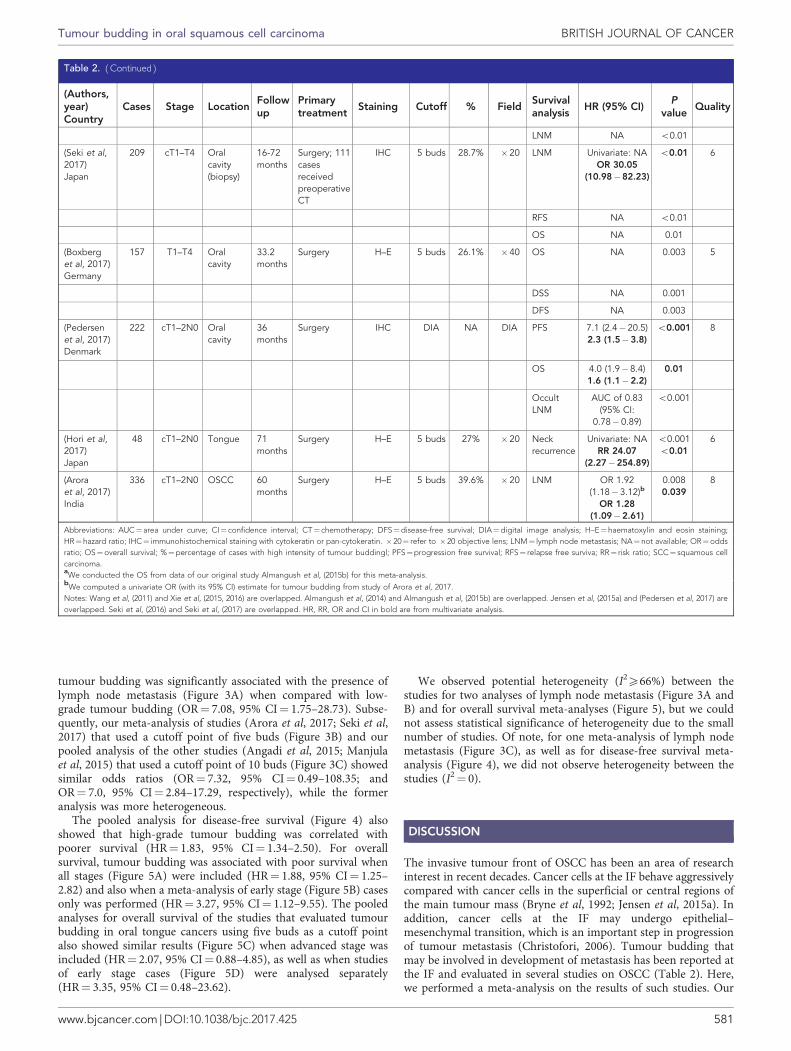
tumour budding was significantly associated with the presence oflymph node metastasis (Figure 3A) when compared with low-grade tumour budding (OR¼ 7.08, 95% CI¼ 1.75–28.73). Subse-quently, our meta-analysis of studies (Arora et al, 2017; Seki et al,2017) that used a cutoff point of five buds (Figure 3B) and ourpooled analysis of the other studies (Angadi et al, 2015; Manjulaet al, 2015) that used a cutoff point of 10 buds (Figure 3C) showedsimilar odds ratios (OR¼ 7.32, 95% CI¼ 0.49–108.35; andOR¼ 7.0, 95% CI¼ 2.84–17.29, respectively), while the formeranalysis was more heterogeneous.
The pooled analysis for disease-free survival (Figure 4) alsoshowed that high-grade tumour budding was correlated withpoorer survival (HR¼ 1.83, 95% CI¼ 1.34–2.50). For overallsurvival, tumour budding was associated with poor survival whenall stages (Figure 5A) were included (HR¼ 1.88, 95% CI¼ 1.25–2.82) and also when a meta-analysis of early stage (Figure 5B) casesonly was performed (HR¼ 3.27, 95% CI¼ 1.12–9.55). The pooledanalyses for overall survival of the studies that evaluated tumourbudding in oral tongue cancers using five buds as a cutoff pointalso showed similar results (Figure 5C) when advanced stage wasincluded (HR¼ 2.07, 95% CI¼ 0.88–4.85), as well as when studiesof early stage cases (Figure 5D) were analysed separately(HR¼ 3.35, 95% CI¼ 0.48–23.62).
We observed potential heterogeneity (I2X66%) between the
studies for two analyses of lymph node metastasis (Figure 3A andB) and for overall survival meta-analyses (Figure 5), but we couldnot assess statistical significance of heterogeneity due to the smallnumber of studies. Of note, for one meta-analysis of lymph nodemetastasis (Figure 3C), as well as for disease-free survival meta-analysis (Figure 4), we did not observe heterogeneity between thestudies (I2¼ 0).
DISCUSSION
The invasive tumour front of OSCC has been an area of researchinterest in recent decades. Cancer cells at the IF behave aggressivelycompared with cancer cells in the superficial or central regions ofthe main tumour mass (Bryne et al, 1992; Jensen et al, 2015a). Inaddition, cancer cells at the IF may undergo epithelial–mesenchymal transition, which is an important step in progressionof tumour metastasis (Christofori, 2006). Tumour budding thatmay be involved in development of metastasis has been reported atthe IF and evaluated in several studies on OSCC (Table 2). Here,we performed a meta-analysis on the results of such studies. Our
Table 2. ( Continued )
(Authors,year)Country
Cases Stage LocationFollowup
Primarytreatment
Staining Cutoff % FieldSurvivalanalysis
HR (95% CI)P
valueQuality
LNM NA o0.01
(Seki et al,2017)Japan
209 cT1–T4 Oralcavity(biopsy)
16-72months
Surgery; 111casesreceivedpreoperativeCT
IHC 5 buds 28.7% �20 LNM Univariate: NAOR 30.05
(10.98�82.23)
o0.01 6
RFS NA o0.01
OS NA 0.01
(Boxberget al, 2017)Germany
157 T1–T4 Oralcavity
33.2months
Surgery H–E 5 buds 26.1% �40 OS NA 0.003 5
DSS NA 0.001
DFS NA 0.003
(Pedersenet al, 2017)Denmark
222 cT1–2N0 Oralcavity
36months
Surgery IHC DIA NA DIA PFS 7.1 (2.4�20.5)2.3 (1.5�3.8)
o0.001 8
OS 4.0 (1.9�8.4)1.6 (1.1�2.2)
0.01
OccultLNM
AUC of 0.83(95% CI:
0.78� 0.89)
o0.001
(Hori et al,2017)Japan
48 cT1–2N0 Tongue 71months
Surgery H–E 5 buds 27% �20 Neckrecurrence
Univariate: NARR 24.07
(2.27�254.89)
o0.001o0.01
6
(Aroraet al, 2017)India
336 cT1–2N0 OSCC 60months
Surgery H–E 5 buds 39.6% �20 LNM OR 1.92(1.18�3.12)b
OR 1.28(1.09�2.61)
0.0080.039
8
Abbreviations: AUC¼ area under curve; CI¼ confidence interval; CT¼ chemotherapy; DFS¼disease-free survival; DIA¼digital image analysis; H–E¼haematoxylin and eosin staining;HR¼hazard ratio; IHC¼ immunohistochemical staining with cytokeratin or pan-cytokeratin. � 20¼ refer to � 20 objective lens; LNM¼ lymph node metastasis; NA¼ not available; OR¼oddsratio; OS¼overall survival; %¼percentage of cases with high intensity of tumour buddingl; PFS¼progression free survival; RFS¼ relapse free surviva; RR¼ risk ratio; SCC¼ squamous cellcarcinoma.aWe conducted the OS from data of our original study Almangush et al, (2015b) for this meta-analysis.bWe computed a univariate OR (with its 95% CI) estimate for tumour budding from study of Arora et al, 2017.Notes: Wang et al, (2011) and Xie et al, (2015, 2016) are overlapped. Almangush et al, (2014) and Almangush et al, (2015b) are overlapped. Jensen et al, (2015a) and (Pedersen et al, 2017) areoverlapped. Seki et al, (2016) and Seki et al, (2017) are overlapped. HR, RR, OR and CI in bold are from multivariate analysis.
Tumour budding in oral squamous cell carcinoma BRITISH JOURNAL OF CANCER
www.bjcancer.com | DOI:10.1038/bjc.2017.425 581

meta-analysis shows that tumour budding is a promisingprognostic marker for OSCC.
The importance of tumour budding in cancer prognosis hasbeen studied widely particularly in colorectal cancer (Rogers et al,2016; Lugli et al, 2017), where it is recognised as an additionalprognostic marker (Koelzer et al, 2014). In oesophageal cancer(Almangush et al, 2016), pancreatic cancer (Karamitopoulou,2012), breast cancer (Gujam et al, 2015) and lung cancer (Kadotaet al, 2014), tumour budding has been reported as a promisingprognostic marker. A significant correlation between high tumourbudding count and the presence of lymph node metastases is oneof the most important findings observed in OSCC (Figure 3) and inmany other cancers (Yamaguchi et al, 2010; Landau et al, 2014;Salhia et al, 2015; Cappellesso et al, 2017). Such a finding mightindicate that tumour budding is an early step en route tometastasis. A correlation between tumour budding and occultlymph node metastasis was reported in early stage OSCC (Xie et al,2015). As occult metastasis is the most common reason for relapseand poor prognosis in early stage cases, it is of great importance tovalidate this correlation in other large multicentre cohorts.
Simplicity, reproducibility and low cost are important char-acteristics when considering a new marker for clinical application.The published studies in OSCC and in other cancers repeatedlyreported these advantages for tumour budding (Wang et al, 2011;Graham et al, 2015; Almangush et al, 2015b). Another advantageof the studies of tumour budding in OSCC is that their results areconsistent with those from the first study that evaluated budding inOSCC (Wang et al, 2011). Conversely, controversial findings werereported for the prognostic biomarkers identified for OSCC(Soland and Brusevold, 2013; Almangush et al, 2017a).
When considering a new prognostic marker for clinicalapplication, the marker should also have a significant prognosticvalue independent from classical markers. Interestingly, for tumourbudding, most of the studies that provided multivariate analysis(Wang et al, 2011; Angadi et al, 2015; Almangush et al, 2015b; Sekiet al, 2016; Hori et al, 2017; Pedersen et al, 2017) reported that
tumour budding has a superior prognostic value compared to otherclassical markers such as TNM stage, depth of invasion or WHOtumour grade. However, in one study, (Manjula et al, 2015),tumour thickness (5-mm cutoff point) showed superior prognosticvalue compared with tumour budding, and the same was observedfor depth of invasion in the study by Arora et al. (2017). In anotherstudy (Jensen et al, 2015a), advanced stage was associated with apoorer prognosis than in cases with high-grade budding. Of note,in the latter two studies (Jensen et al, 2015a; Arora et al, 2017)tumour budding was also reported as an independent prognosticmarker in multivariate analysis. Therefore, multivariate analysis ofpublished studies indicates that high-intensity tumour budding,either independently or in addition to the advanced stage, deeplyinvaded tumour or both, is associated with poor prognosis ofOSCC. Only in the study by Manjula et al. (2015), tumour buddingwas not a prognostic marker in multivariate analysis. However,Manjula et al. used a 10-bud cutoff point to stratify cases into riskscores, and it is possible that some cases with Xfive buds wereincluded in the low-grade budding group, which subsequentlyreduced the prognostic value of tumour budding in this cohort.
Different methods have been introduced for the evaluation oftumour budding (Koelzer et al, 2014). However, a traditionalmethod was widely used in the studies on OSCC. In this method,the IF is scanned under low magnification (� 4), and the field withthe highest budding number is counted under high magnification(� 20) and used for the score (Wang et al, 2011). The evaluation ofintra-tumoural budding was not reported in OSCC. Of note, intra-tumoural budding was shown as a valid method in colorectalcancer (Lugli et al, 2011). In only a few studies, evaluation of theprognostic value of tumour budding at the IF was carried out inbiopsy specimens of OSCC (Seki et al, 2016, 2017; Almangush et al,2017b). However, the IF area might not be included in a biopsyspecimen. In such cases, another form of tumour budding, theintra-tumoural budding (i.e., tumour budding between tumourislands) might be more applicable. The latter approach may be ofgreat importance from a clinical point of view for treatment
Table 3. Summary of the studies evaluated tumour budding in OSCC without analysis of its prognostic value
(Authors,year)Country
Cases Stage LocationFollowup
Primarytreatment
Staining Cutoff % Field Findings related to tumour budding
(MarangonJunior et al,2014)Brazil
57 NA Oralcavity
NA NA IHC 5 buds 75.4% � 20 High intensity tumour budding isassociated with higher density of stromalmyofibroblasts and higher expression oflaminin-5 gamma 2 chain
(Sawazaki-Caloneet al, 2015)Brazil
113 T1–T4 Oralcavity
5 years Surgery H–E 5 buds NA � 20 Tumour budding is a parameter of thebudding-depth (BD) prognostic model. BDshowed a superior prognostic valuecompared to other histopathologicgrading systems
(Jensenet al,2015b)Denmark
28 NA Oralcavity
NA NA IHC NA NA NA A relationship between tumour buddingand myofibroblasts was seen but was not ageneral featureBudding cells have shownlow expression of E-cadherin
(Zhanget al, 2016)China
73 T1–T4 Tongue 114months
CT for 7cases, RT for17, andsurgery forothers
H–E 5 buds 75.4% � 20 High intensity of tumour budding wasmore common in tongue cancer (75.4%)compared to high intensity of tumourbudding in nasopharyngeal carcinoma(45.5%)
(Striederet al, 2017)Brazil
53 T1–T4 Lip 159.4monthsor 57.4months
Surgery H–E 5 buds 67.9% � 20 Tumour budding is a parameter of thebudding-depth (BD) prognostic model. BDshowed a high prognostic value for lipcancer
(Leao et al,2017)Brazil
103 NA Oralcavity
NA NA H–E; IHC 5 buds NA � 20 Evaluation of tumour budding by IHCshowed higher reproducibility andreplicability compared to H–E
Abbreviations: CT=chemotherapy; H-E=haematoxylin and eosin staining; IHC=immunohistochemical staining with cytokeratin or pan-cytokeratin.
BRITISH JOURNAL OF CANCER Tumour budding in oral squamous cell carcinoma
582 www.bjcancer.com | DOI:10.1038/bjc.2017.425

planning of OSCC, and should be further evaluated. In addition,intraoperative evaluation of tumour budding (i.e., using fresh-frozen sections) should also be considered in future studies.
Diverse cutoff points were suggested for stratification of casesinto low-grade and high-grade tumour budding (Table 2). In thepresent studies on OSCC, five-bud cutoff point was the mostcommonly used (low grade o5 vs high grade X5). We conductedmeta-analysis for studies that used different cutoff points(Figure 3A), and then, we conducted separate meta-analyses forstudies that used a five-bud cutoff point (Figure 3B) and for studiesthat used a 10-bud cutoff point (Figure 3C). Interestingly, thesemeta-analyses show that tumour budding is a useful prognosticmarker for OSCC cases. As the risk of poor prognosis begins at thepresence of five buds, we suggest considering both five-bud and 10-bud cutoff points in further studies to determine which one of
these cutoff points is more predictive of poor prognosis and shouldtherefore be used in clinical practice.
Most studies evaluated tumour budding using H–E staining.Interestingly, a recent study on OSCC concluded that evaluation oftumour budding by immunohistochemistry with pan-cytokeratinantibodies (clones AE1/AE3) showed a better reproducibility ofresults than those with H–E staining (Leao et al, 2017). However,standardisation of the evaluation method and cutoff point is stillnecessary. A recent international consensus conference on tumourbudding (Lugli et al, 2017) made several statements (includingdefinition, evaluation method and others) for reporting tumourbudding in colorectal cancer. Such statements are still necessary toallow inclusion of tumour budding in a pathology report for OSCCcases.
The combination of squamous cell carcinoma (SCC) fromdifferent subsites of the oral cavity was a common disadvantageamong the studies that evaluated tumour budding in OSCC.Therefore, we recommend a separate analysis for each subsitewhen reporting tumour budding in future studies. Despite a smallnumber of studies available, we conducted a meta-analysis foroverall survival of studies that evaluated tumour budding in oraltongue SCC (Figure 5C and D), which is the most common SCC ofthe oral cavity. The results of this meta-analysis suggest, althoughwithout strong statistical evidence, that cases of oral tongue cancerwith a high budding index have a poorer overall survival. This isconsistent with the other meta-analyses where the subsites weremixed. Another combination that was also common among theincluded studies was mixing of early stage and late-stage cancers inthe same analysis. We conducted a meta-analysis for the twostudies that included only early stage cancers (Figure 5D), and theresult suggests that tumour budding in such early stage cases has aprognostic value, but given the wide confidence intervals, thisresult lacks strong statistical evidence and requires further studiesfor validation.
Tumour budding in OSCC has also been evaluated using digitalpathology (Jensen et al, 2015a; Pedersen et al, 2017). Digital imageanalysis has been used increasingly in recent research and it hasshown better accuracy and reproducibility compared with theconventional method as it allows truly quantitative scores (Riber-Hansen et al, 2012). Moreover, it will be easier to standardise thescoring method using digital image analysis (Pedersen et al, 2017).Therefore, digital image analysis of tumour budding in OSCCshould be used to validate results in large cohorts.
Few studies have examined the biological background of tumourbudding in OSCC. Immunohistochemical analysis showed thattumour budding is associated with reduced expression ofE-cadherin and overexpression of vimentin (Wang et al, 2011).Regarding interactions with the surrounding stroma, high-gradebudding was associated with a higher density of stromalmyofibroblasts and higher expression of laminin-5 gamma 2 chain(Marangon Junior et al, 2014). In genetic profiling, decreasedexpression of miR-200a, miR-200b and miR-200c was reported incancer cells of tumour budding (Jensen et al, 2015a). However,molecular analyses in other cancers have provided more detailsabout the genetic background of tumour budding (Zlobec andLugli, 2010; Galvan et al, 2015; Bradley et al, 2016; Miyake et al,2017), and similar analyses in OSCC are still necessary to betterunderstand this phenomenon.
The main limitation of the current meta-analyses is the smallnumber of the original studies. Accordingly, it was difficult tostatistically evaluate the heterogeneity between the studies. Toavoid bias due to any potential heterogeneity, we focused on arandom effects model that is known as an effective method tocombine heterogeneous studies (Guolo and Varin, 2017). Inaddition, for each meta-analysis (Figures 3–5), we also reportedresults of a fixed effect model and they were consistent with arandom effects model. Moreover, our meta-analyses addressed
StudyA
B
C
Odds ratio OR
6.791.92
7.50
30.05
3.74
7.08
95% CI
OR 95% CI
OR 95% CI
(2.28; 20.20)(1.18; 3.12)
(1.49; 37.70)
(10.98; 82.23)
(2.52; 5.55)
(1.75; 28.73)
1.92
30.05
(1.18; 3.12)
(10.98; 82.23)
3.237.32
(2.08; 5.00)(0.49; 108.35)
6.79 (2.28; 20.20)7.50 (1.49; 37.70)
7.00 (2.84; 17.29)
7.00 (2.84; 17.29)
0.1 0.5 1 2 10
0.1 0.5 1 2 10
0.1 0.5 1 2 10
Odds ratio
Odds ratio
Study
Study
Fixed effect model
Fixed effect model
Random effects model
Random effects model
Arora 2017
Angadi 2015Manjula 2015
Seki 2017
Fixed effect model
Random effects model
I2 = 88%, �2 = 1.736
I2 = 96%, �2 = 3.62
I2 = 0%, �2 = 0
Angadi 2015Arora 2017
Manjula 2015
Seki 2017
Figure 3. Forest plots for the pooled analyses of the studies evaluatedthe prognostic value of tumour budding in assessing lymph nodemetastasis of OSCC. (A) All eligible studies. (B) Studies used five-budcutoff point. (C) Studies used 10-bud cutoff point.
HR 95% CI
1.85 (1.21; 2.82)
2.10 (1.21; 3.64)
1.32 (0.59; 2.95)
1.83 (1.34; 2.50)
1.83 (1.34; 2.50)
0.5 1 2
Hazard ratioStudy
Almangush 2015b
Manjula 2015
Jensen 2015a
Fixed effect model
Random effects model
I2 = 0%, �2 = 0
Figure 4. Pooled analysis for disease-free survival.
Tumour budding in oral squamous cell carcinoma BRITISH JOURNAL OF CANCER
www.bjcancer.com | DOI:10.1038/bjc.2017.425 583

three different end points (metastasis, overall survival and disease-free survival), and our results regarding the common effect oftumour budding as a negative prognostic marker are valid based onmeta-analyses of these different end points. Of note, this effect isalso consistent across published studies. Inclusion of differentsubsites of the oral cavity or mixing of different stages in analysis ofthe same cohort was another limitation, as mentioned above. Theabsence of prospective studies was also noted.
Despite these shortcomings, there is sufficient evidence tosuggest that OSCCs with high-grade tumour budding are at highrisk of poor prognosis. This evidence was prominent and validatedin many studies. Similar evidence has also accumulated on theprognostic value of tumour budding in other cancers (Almangushet al, 2016; Rogers et al, 2016; Lugli et al, 2017). To the best of ourknowledge, this is the first meta-analysis on the prognostic value oftumour budding in OSCC. We conclude that tumour budding hasa prominent prognostic power for OSCC even at early stages of thedisease. Future research on OSCC should compare the differentevaluation methods with the goal of standardising the assessmentmethod for pathology reports. In addition, understanding thegenetic background of tumour budding may facilitate identificationof treatment targets in OSCC.
ACKNOWLEDGEMENTS
This work was financially supported by the Finnish Dental Society(Alhadi Almangush), the Rauha Ahokas Foundation (AlhadiAlmangush), the Academy of Finland (288509 and 294050 toMatti Pirinen), the Research Funds of the University of Helsinki(Matti Pirinen), the Helsinki University Hospital Research Fund(Antti A Makitie and Tuula Salo), the Sigrid Juselius Foundation(Tuula Salo), the Finnish Cancer Society (Tuula Salo) and theMaritza and Reino Salonen Foundation (Ilmo Leivo).
CONFLICT OF INTEREST
The authors declare no conflict of interest.
REFERENCES
Almangush A, Bello IO, Coletta RD, Makitie AA, Makinen LK, Kauppila JH,Pukkila M, Hagstrom J, Laranne J, Soini Y, Kosma VM, Koivunen P,Kelner N, Kowalski LP, Grenman R, Leivo I, Laara E, Salo T (2015a) Forearly-stage oral tongue cancer, depth of invasion and worst pattern ofinvasion are the strongest pathological predictors for locoregionalrecurrence and mortality. Virchows Arch 467(1): 39–46.
Almangush A, Bello IO, Keski-Santti H, Makinen LK, Kauppila JH,Pukkila M, Hagstrom J, Laranne J, Tommola S, Nieminen O, Soini Y,Kosma VM, Koivunen P, Grenman R, Leivo I, Salo T (2014) Depth ofinvasion, tumor budding, and worst pattern of invasion: prognosticindicators in early-stage oral tongue cancer. Head Neck 36(6): 811–818.
Almangush A, Coletta RD, Bello IO, Bitu C, Keski-Santti H, Makinen LK,Kauppila JH, Pukkila M, Hagstrom J, Laranne J, Tommola S, Soini Y,Kosma VM, Koivunen P, Kowalski LP, Nieminen P, Grenman R, Leivo I,Salo T (2015b) A simple novel prognostic model for early stage oraltongue cancer. Int J Oral Maxillofac Surg 44(2): 143–150.
Almangush A, Heikkinen I, Makitie AA, Coletta RD, Laara E, Leivo I, Salo T(2017a) Prognostic biomarkers for oral tongue squamous cell carcinoma: asystematic review and meta-analysis. Br J Cancer 117(6): 856–866.
Almangush A, Karhunen M, Hautaniemi S, Salo T, Leivo I (2016) Prognosticvalue of tumour budding in oesophageal cancer: a meta-analysis.Histopathology 68(2): 173–182.
Almangush A, Leivo I, Siponen M, Sundquist E, Mroueh R, Makitie AA,Soini Y, Haglund C, Nieminen P, Salo T (2017b) Evaluation of thebudding and depth of invasion (BD) model in oral tongue cancer biopsies.Virchows Arch; epub ahead of print 2 August 2017; doi:10.1007/s00428-017-2212-1.
Altman DG, McShane LM, Sauerbrei W, Taube SE (2012) Reportingrecommendations for tumor marker prognostic studies (REMARK):explanation and elaboration. BMC Med 10: 51.
Angadi PV, Patil PV, Hallikeri K, Mallapur MD, Hallikerimath S, Kale AD(2015) Tumor budding is an independent prognostic factor for predictionof lymph node metastasis in oral squamous cell carcinoma. Int J SurgPathol 23(2): 102–110.
Arora A, Husain N, Bansal A, Neyaz A, Jaiswal R, Jain K, Chaturvedi A,Anand N, Malhotra K, Shukla S (2017) Development of a new outcomeprediction model in early-stage squamous cell carcinoma of the oral cavitybased on histopathologic parameters with multivariate analysis: the aditi-nuzhat lymph-node prediction score (ANLPS) system. Am J Surg Pathol41(7): 950–960.
Attramadal CG, Kumar S, Boysen ME, Dhakal HP, Nesland JM, Bryne M(2015) Tumor budding, EMT and cancer stem cells in T1-2/N0 oralsquamous cell carcinomas. Anticancer Res 35(11): 6111–6120.
Boxberg M, Jesinghaus M, Dorfner C, Mogler C, Drecoll E, Warth A,Steiger K, Bollwein C, Meyer P, Wolff KD, Kolk A, Weichert W (2017)Tumor budding activity and cell nest size determine patient outcome inoral squamous cell carcinoma: Proposal for an adjusted grading system.Histopathology 70(7): 1125–1137.
Bradley CA, Dunne PD, Bingham V, McQuaid S, Khawaja H, Craig S, James J,Moore WL, McArt DG, Lawler M, Dasgupta S, Johnston PG, VanSchaeybroeck S (2016) Transcriptional upregulation of c-MET is
HR 95% CI
1.40 (1.01; 1.94)1.80 (1.27; 2.55)3.35 (1.77; 6.32)
1.73 (1.38; 2.15)1.88 (1.25; 2.82)
HR 95% CI
1.40 (1.01; 1.94)4.00 (1.90; 8.41)
10.44 (2.43; 44.87)
1.78 (1.33; 2.38)3.27 (1.12; 9.55)
HR 95% CI
1.40 (1.01; 1.94)3.35 (1.77; 6.32)
1.68 (1.26; 2.24)2.07 (0.88; 4.85)
HR 95% CI
1.40 (1.01; 1.94)10.44 (2.43; 44.87)
1.54 (1.12; 2.11)3.35 (0.48; 23.62)
0.2 10.5 2 5
0.2 10.5 2 5
0.1 10.5 2 10
0.1 10.5 2 10
Hazard ratio
Hazard ratio
Hazard ratio
Hazard ratio
StudyA
B
C
D
Almangush 2015b
Wang 2011Jensen 2015a
Fixed effect modelRandom effects model
I2 = 66%, �2 = 0.0823
Study
Almangush 2015bWang 2011
Fixed effect modelRandom effects model
I2 = 83%, �2 = 0.3144
Study
Almangush 2015b
Xie 2015Pedersen 2017
Fixed effect modelRandom effects model
I2 = 84%, �2 = 0.7028
Study
Almangush 2015bXie 2015
Fixed effect modelRandom effects modelI2 = 86%, �2 = 1.728
Figure 5. Pooled analyses for overall survival. (A) All stages of OSCC.(B) Pooled analysis for overall survival of OSCC including studies ofearly stage only. (C) Pooled analysis for overall survival including onlyoral tongue cancer studies which used five-bud cutoff point. (D) Pooledanalysis for overall survival including early stage oral tongue cancerstudies that used five-bud cutoff point.
BRITISH JOURNAL OF CANCER Tumour budding in oral squamous cell carcinoma
584 www.bjcancer.com | DOI:10.1038/bjc.2017.425

associated with invasion and tumor budding in colorectal cancer.Oncotarget 7(48): 78932–78945.
Bryne M, Koppang HS, Lilleng R, Kjaerheim A (1992) Malignancy grading ofthe deep invasive margins of oral squamous cell carcinomas has highprognostic value. J Pathol 166(4): 375–381.
Cappellesso R, Luchini C, Veronese N, Mele ML, Rosa-Rizzotto E, Guido E,De Lazzari F, Pilati P, Farinati F, Realdon S, Solmi M, Fassan M, Rugge M(2017) Tumor budding as a risk factor for nodal metastasis in Pt1colorectal cancers: a meta-analysis. Hum Pathol 65: 62–70.
Chen TC, Wang CP, Ko JY, Yang TL, Hsu CW, Yeh KA, Chang YL, Lou PJ(2013) The impact of perineural invasion and/or lymphovascular invasionon the survival of early-stage oral squamous cell carcinoma patients. AnnSurg Oncol 20(7): 2388–2395.
Christofori G (2006) New signals from the invasive front. Nature 441(7092):444–450.
DerSimonian R, Laird N (1986) Meta-analysis in clinical trials. Control ClinTrials 7(3): 177–188.
Ferlay J, Soerjomataram I, Dikshit R, Eser S, Mathers C, Rebelo M,Parkin DM, Forman D, Bray F (2015) Cancer incidence and mortalityworldwide: sources, methods and major patterns in GLOBOCAN 2012.Int J Cancer 136(5): E359–E386.
Galvan JA, Zlobec I, Wartenberg M, Lugli A, Gloor B, Perren A,Karamitopoulou E (2015) Expression of E-cadherin repressors SNAIL,ZEB1 and ZEB2 by tumour and stromal cells influences tumour-buddingphenotype and suggests heterogeneity of stromal cells in pancreatic cancer.Br J Cancer 112(12): 1944–1950.
Graham RP, Vierkant RA, Tillmans LS, Wang AH, Laird PW, Weisenberger DJ,Lynch CF, French AJ, Slager SL, Raissian Y, Garcia JJ, Kerr SE, Lee HE,Thibodeau SN, Cerhan JR, Limburg PJ, Smyrk TC (2015) Tumor buddingin colorectal carcinoma: confirmation of prognostic significance andhistologic cutoff in a population-based cohort. Am J Surg Pathol 39(10):1340–1346.
Gujam FJ, McMillan DC, Mohammed ZM, Edwards J, Going JJ (2015) Therelationship between tumour budding, the tumour microenvironment andsurvival in patients with invasive ductal breast cancer. Br J Cancer 113(7):1066–1074.
Guolo A, Varin C (2017) Random-effects meta-analysis: the number of studiesmatters. Stat Methods Med Res 26(3): 1500–1518.
Higgins JP, Thompson SG (2002) Quantifying heterogeneity in a meta-analysis. Stat Med 21(11): 1539–1558.
Hori Y, Kubota A, Yokose T, Furukawa M, Matsushita T, Takita M,Mitsunaga S, Mizoguchi N, Nonaka T, Nakayama Y, Oridate N (2017)Predictive significance of tumor depth and budding for late lymph nodemetastases in patients with clinical n0 early oral tongue carcinoma. HeadNeck Pathol 11(4): 477–486.
Jensen DH, Dabelsteen E, Specht L, Fiehn AM, Therkildsen MH, Jonson L,Vikesaa J, Nielsen FC, von Buchwald C (2015a) Molecular profiling oftumour budding implicates TGFbeta-mediated epithelial-mesenchymaltransition as a therapeutic target in oral squamous cell carcinoma. J Pathol236(4): 505–516.
Jensen DH, Reibel J, Mackenzie IC, Dabelsteen E (2015b) Single cell migrationin oral squamous cell carcinoma - possible evidence of epithelial-mesenchymal transition in vivo. J Oral Pathol Med 44(9): 674–679.
Kadota K, Nitadori J, Woo KM, Sima CS, Finley DJ, Rusch VW, AdusumilliPS, Travis WD (2014) Comprehensive pathological analyses in lungsquamous cell carcinoma: single cell invasion, nuclear diameter, andtumor budding are independent prognostic factors for worse outcomes.J Thorac Oncol 9(8): 1126–1139.
Kadota K, Yeh YC, Villena-Vargas J, Cherkassky L, Drill EN, Sima CS,Jones DR, Travis WD, Adusumilli PS (2015) Tumor budding correlateswith the protumor immune microenvironment and is an independentprognostic factor for recurrence of stage i lung adenocarcinoma. Chest148(3): 711–721.
Karamitopoulou E (2012) Tumor budding cells, cancer stem cells andepithelial-mesenchymal transition-type cells in pancreatic cancer. FrontOncol 2: 209.
Koelzer VH, Langer R, Zlobec I, Lugli A (2014) Tumor budding in uppergastrointestinal carcinomas. Front Oncol 4: 216.
Korvala J, Jee K, Porkola E, Almangush A, Mosakhani N, Bitu C,Cervigne NK, Zandonadi FS, Meirelles GV, Leme AF, Coletta RD, Leivo I,Salo T (2017) MicroRNA and protein profiles in invasive versus non-invasive oral tongue squamous cell carcinoma cells in vitro. Exp Cell Res350(1): 9–18.
Landau MS, Hastings SM, Foxwell TJ, Luketich JD, Nason KS, Davison JM(2014) Tumor budding is associated with an increased risk of lymph nodemetastasis and poor prognosis in superficial esophageal adenocarcinoma.Mod Pathol 27(12): 1578–1589.
Leao PL, Marangon Junior H, Melo VV, Caixeta AB, Souza PE, de Aguiar MC,Horta MC (2017) Reproducibility, repeatability and level of difficulty oftwo methods for tumor budding evaluation in oral squamous cellcarcinoma. J Oral Pathol Med 46(10): 949–955.
Lugli A, Kirsch R, Ajioka Y, Bosman F, Cathomas G, Dawson H, El Zimaity H,Flejou JF, Hansen TP, Hartmann A, Kakar S, Langner C, Nagtegaal I,Puppa G, Riddell R, Ristimaki A, Sheahan K, Smyrk T, Sugihara K,Terris B, Ueno H, Vieth M, Zlobec I, Quirke P (2017) Recommendationsfor reporting tumor budding in colorectal cancer based on theInternational Tumor Budding Consensus Conference (ITBCC) 2016. ModPathol 30(9): 1299–1311.
Lugli A, Vlajnic T, Giger O, Karamitopoulou E, Patsouris ES, Peros G,Terracciano LM, Zlobec I (2011) Intratumoral budding as a potentialparameter of tumor progression in mismatch repair-proficient and mismatchrepair-deficient colorectal cancer patients. Hum Pathol 42(12): 1833–1840.
Manjula BV, Augustine S, Selvam S, Mohan AM (2015) Prognostic andpredictive factors in gingivo buccal complex squamous cell carcinoma: roleof tumor budding and pattern of invasion. Indian J Otolaryngol Head NeckSurg 67(Suppl 1): 98–104.
Marangon Junior H, Rocha VN, Leite CF, de Aguiar MC, Souza PE, Horta MC(2014) Laminin-5gamma 2 chain expression is associated with intensity oftumor budding and density of stromal myofibroblasts in oral squamouscell carcinoma. J Oral Pathol Med 43(3): 199–204.
Miyake M, Hori S, Morizawa Y, Tatsumi Y, Toritsuka M, Ohnishi S,Shimada K, Furuya H, Khadka VS, Deng Y, Ohnishi K, Iida K, Gotoh D,Nakai Y, Inoue T, Anai S, Torimoto K, Aoki K, Tanaka N, Konishi N,Fujimoto K (2017) Collagen type IV alpha 1 (COL4A1) and collagen typeXIII alpha 1 (COL13A1) produced in cancer cells promote tumor buddingat the invasion front in human urothelial carcinoma of the bladder.Oncotarget 8(22): 36099–36114.
Moher D, Liberati A, Tetzlaff J, Altman DG. Group P (2009) Preferredreporting items for systematic reviews and meta-analyses: the PRISMAstatement. BMJ 339: b2535.
Muller S, Pan Y, Li R, Chi AC (2008) Changing trends in oral squamous cellcarcinoma with particular reference to young patients: 1971-2006. TheEmory University experience. Head Neck Pathol 2(2): 60–66.
Nandita KP, Boaz K, Srikant N, Lewis AJ, Manaktala N (2016) Tumourbudding: a promising parameter in oral squamous cell carcinoma. Res JPharm Biol Chem Sci 7(5): 2059–2063.
Pedersen NJ, Jensen DH, Lelkaitis G, Kiss K, Charabi B, Specht L,von Buchwald C (2017) Construction of a pathological risk model ofoccult lymph node metastases for prognostication by semi-automatedimage analysis of tumor budding in early-stage oral squamous cellcarcinoma. Oncotarget 8(11): 18227–18237.
Riber-Hansen R, Vainer B, Steiniche T (2012) Digital image analysis: a reviewof reproducibility, stability and basic requirements for optimal results.APMIS 120(4): 276–289.
Rogers AC, Winter DC, Heeney A, Gibbons D, Lugli A, Puppa G, Sheahan K(2016) Systematic review and meta-analysis of the impact of tumourbudding in colorectal cancer. Br J Cancer 115(7): 831–840.
Salhia B, Trippel M, Pfaltz K, Cihoric N, Grogg A, Ladrach C, Zlobec I,Tapia C (2015) High tumor budding stratifies breast cancer withmetastatic properties. Breast Cancer Res Treat 150(2): 363–371.
Sawazaki-Calone I, Rangel A, Bueno AG, Morais CF, Nagai HM, Kunz RP,Souza RL, Rutkauskis L, Salo T, Almangush A, Coletta RD (2015) Theprognostic value of histopathological grading systems in oral squamouscell carcinomas. Oral Dis 21(6): 755–761.
Seki M, Sano T, Yokoo S, Oyama T (2016) Histologic assessment of tumorbudding in preoperative biopsies to predict nodal metastasis in squamouscell carcinoma of the tongue and floor of the mouth. Head Neck38(Suppl 1): E1582–E1590.
Seki M, Sano T, Yokoo S, Oyama T (2017) Tumour budding evaluated inbiopsy specimens is a useful predictor of prognosis in patients with cN0early stage oral squamous cell carcinoma. Histopathology 70(6): 869–879.
Soland TM, Brusevold IJ (2013) Prognostic molecular markers in cancer - quovadis? Histopathology 63(3): 297–308.
Strieder L, Coutinho-Camillo CM, Costa V, da Cruz Perez DE, Kowalski LP,Kaminagakura E (2017) Comparative analysis of three histologic gradingmethods for squamous cell carcinoma of the lip. Oral Dis 23(1): 120–125.
Tumour budding in oral squamous cell carcinoma BRITISH JOURNAL OF CANCER
www.bjcancer.com | DOI:10.1038/bjc.2017.425 585

Wang C, Huang H, Huang Z, Wang A, Chen X, Huang L, Zhou X, Liu X(2011) Tumor budding correlates with poor prognosis and epithelial-mesenchymal transition in tongue squamous cell carcinoma. J Oral PatholMed 40(7): 545–551.
Xie N, Wang C, Liu X, Li R, Hou J, Chen X, Huang H (2015) Tumor buddingcorrelates with occult cervical lymph node metastasis and poor prognosisin clinical early-stage tongue squamous cell carcinoma. J Oral Pathol Med44(4): 266–272.
Xie N, Wang C, Zhuang Z, Hou J, Liu X, Wu Y, Liu H, Huang H (2016)Decreased miR-320a promotes invasion and metastasis of tumor buddingcells in tongue squamous cell carcinoma. Oncotarget 7(40): 65744–65757.
Yamaguchi Y, Ishii G, Kojima M, Yoh K, Otsuka H, Otaki Y, Aokage K, YanagiS, Nagai K, Nishiwaki Y, Ochiai A (2010) Histopathologic features of thetumor budding in adenocarcinoma of the lung: tumor budding as an indexto predict the potential aggressiveness. J Thorac Oncol 5(9): 1361–1368.
Zhang P, Zhang L, Liu H, Zhao L, Li Y, Shen JX, Liu Q, Liu MZ, Xi M (2016)Clinicopathologic characteristics and prognosis of tongue squamous cellcarcinoma in patients with and without a history of radiation fornasopharyngeal carcinoma: a matched case-control study. Cancer ResTreat 49(3): 695–705.
Zlobec I, Lugli A (2010) Epithelial mesenchymal transition and tumorbudding in aggressive colorectal cancer: tumor budding as oncotarget.Oncotarget 1(7): 651–661.
This work is published under the standard license to publish agree-ment. After 12 months the work will become freely available andthe license terms will switch to a Creative Commons Attribution-NonCommercial-Share Alike 4.0 Unported License.
BRITISH JOURNAL OF CANCER Tumour budding in oral squamous cell carcinoma
586 www.bjcancer.com | DOI:10.1038/bjc.2017.425