collision tumour involving a rectal gastrointestinal stromal tumour
TRANSCRIPT
Macías-García et al. Diagnostic Pathology 2012, 7:150http://www.diagnosticpathology.org/content/7/1/150
SHORT REPORT Open Access
Collision tumour involving a rectalgastrointestinal stromal tumour with invasion ofthe prostate and a prostatic adenocarcinomaLaura Macías-García1*, Haydee De la Hoz-Herazo1, Antonio Robles-Frías1, María J Pareja-Megía1,Juan López-Garrido1 and José I López2
Abstract
Background: Gastrointestinal stromal tumours (GISTs) are the most common primary mesenchymal neoplasia inthe gastrointestinal tract, although they represent only a small fraction of total gastrointestinal malignancies inadults (<2%). GISTs can be located at any level of the gastrointestinal tract; the stomach is the most commonlocation (60-70%), in contrast to the rectum, which is most rare (4%). When a GIST invades into the adjacentprostate tissue, it can simulate prostate cancer. In this study, we report on a case comprising the unexpectedcollision between a rectal GIST tumour and a prostatic adenocarcinoma.
Findings: We describe the complexity of the clinical, endoscopic and radiological diagnosis, of the differentialdiagnosis based on tumour biopsy, and of the role of neoadjuvant therapy using imatinib prior to surgicaltreatment.
Conclusions: Although isolated cases of coexisting GISTs and prostatic adenocarcinomas have previously beendescribed, this is the first reported case in the medical literature of a collision tumour involving a rectal GIST andprostatic adenocarcinoma components.
Virtual slides: The virtual slide(s) for this article can be found here: http://www.diagnosticpathology.diagnomx.eu/vs/1238437468776331.
Keywords: Gastrointestinal stromal tumour, Mixed tumours, Prostatic adenocarcinoma, Imatinib
BackgroundThe term gastrointestinal stromal tumor (GIST) wasfirst introduced in 1983 by Mazur and Clark as a neutralterm between smooth muscle tumours and schwanno-mas. GIST is currently used to refer to a heterogeneousgroup of mesenchymal tumour lesions, acquired or con-genital, with common morphological profiles that usu-ally settle within the digestive tract.Although GISTs are the most common primary mes-
enchymal tumours of the gastrointestinal tract (70%),they represent only a small fraction of all gastrointestinalmalignancies in adults (<2%).
* Correspondence: [email protected] de gestión clínica de Oncohematología y Anatomía Patológica,Hospital Universitario de Valme, Carretera de Cádiz s/n, 41014, Sevilla, SpainFull list of author information is available at the end of the article
© Macías-García et al.; licensee BioMed CentrCommons Attribution License (http://creativecreproduction in any medium, provided the or
GISTs usually affect adults between 6th and 8th dec-ades of life without specific gender prevalence (M:F,1.1:1) [1]. Its annual incidence rates range from 6.8 and14.5 cases per million in the U.S. States and in Sweden,respectively [2]. GISTs include benign and malignantneoplasia that immunohistochemically stain positivelyfor KIT (CD117) and that phenotypically differentiateinto cells of Cajal. The histological (e.g., cellularityspindle and/or epithelioid) and immunohistochemical(e.g., CD117 and CD34 immunostaining) profiles allowfor simple morphological diagnosis.In 1998, Hirota et al. showed that the majority of
GISTs exhibit activating mutations in the KIT protoon-cogene. Subsequently, in 2003, a new mutation inPDGFRA and the sensitivity of GISTs to tyrosine kinaseinhibitors such as imatinib were demonstrated. Thesefindings represent an important advance in the clinical
al Ltd. This is an Open Access article distributed under the terms of the Creativeommons.org/licenses/by/2.0), which permits unrestricted use, distribution, andiginal work is properly cited.
Macías-García et al. Diagnostic Pathology 2012, 7:150 Page 2 of 5http://www.diagnosticpathology.org/content/7/1/150
management and our understanding of the biology ofthis tumour [2]. Although the mutations in KIT andPDGFRA represent the basis of GIST oncogenesis,5-10% of the GIST cases are negative for mutations inKIT [3].GISTs can be located at any level of the gastrointes-
tinal tract. The stomach is the most common location(60-70%), whereas rectal GIST represents only 4% of allGISTs [1]. When these tumours invade into the prostate,they can clinically simulate a prostatic adenocarcinoma[4]. The direct invasion of the prostate by a rectal GISTis rare [4-7] and the case presented in our study involvesan added diagnostic complexity: a collision tumour in-volving both histological types of neoplasia. The combin-ation of a rectal GIST that invades into the prostate anda primary prostatic adenocarcinoma in a collisiontumour has never before been described in the medicalliterature.
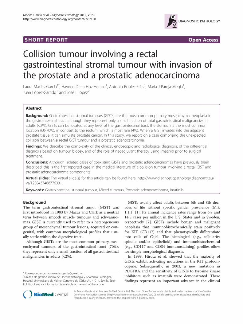
Figure 1 A: Rectal GIST: Excrescent and ulcerated rectal masswith stenosis of the rectal lumen. B: Rectal GIST with prostaticinfiltration: rectal tumour with direct invasion of both adjacentlobules of the prostate. UB: urinary bladder; ADC: prostaticadenocarcinoma; AC: Anal canal; R: rectum; P: prostate.
Materials and methodsCase presentationThe patient examined in this study was a 52-year-oldmale with no relevant personal or family history of dis-ease, who presented with rectal tenesmus, rectal bleed-ing, and obstructive urinary symptoms. A digital rectalexam revealed a fixed submucosal mass in the anteriorrectal wall, attached to the prostate and located 2–3 cmfrom the anal margin. The prostate specific antigen(PSA) value was 3.16 mg/ml, with a pathologic PSA ratioof 0.0937. Rectoscopic and echo-endoscopic evaluationsrevealed a rectal neoplasia with prostatic infiltration.A computed tomography (CT) scan performed at 5months prior to the surgical intervention revealed a re-latively well-defined heterogeneous mass of 60 mm ×36 mm × 45 mm affecting the anterior wall of the rectumin contact with the prostate, with effacement of the fatseparating the two organs.Magnetic resonance imaging (MRI) performed at the
same time revealed a large (7–8 cm) tumour in the distalrectum, affecting the adjacent prostate and causing sten-osis of the rectal lumen. The CT scan and the MRIresults both revealed an absence of metastasis in thelocal and distant lymph nodes.A biopsy of the tumour revealed a profile that was
consistent with a mesenchymal neoplasia of uncertainmalignancy that was positive for CD117 (cytoplasmicand membranous staining), the expression of which ischaracteristic of GISTs.The treatment protocol consisted of neoadjuvant treat-
ment with imatinib abdominoperineal rectal amputationwith a radical cystectomy and prostatectomy. A pre-operative CT scan demonstrated an apparent reductionin the tumour size, measuring 50 mm × 30 mm, where
the separation between the rectum and prostate was dif-ficult to delineate.
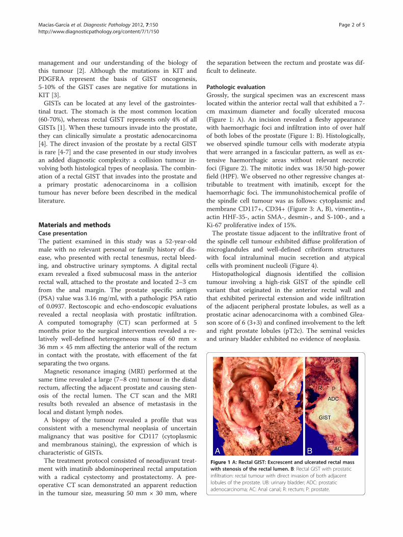
Pathologic evaluationGrossly, the surgical specimen was an excrescent masslocated within the anterior rectal wall that exhibited a 7-cm maximum diameter and focally ulcerated mucosa(Figure 1: A). An incision revealed a fleshy appearancewith haemorrhagic foci and infiltration into of over halfof both lobes of the prostate (Figure 1: B). Histologically,we observed spindle tumour cells with moderate atypiathat were arranged in a fascicular pattern, as well as ex-tensive haemorrhagic areas without relevant necroticfoci (Figure 2). The mitotic index was 18/50 high-powerfield (HPF). We observed no other regressive changes at-tributable to treatment with imatinib, except for thehaemorrhagic foci. The immunohistochemical profile ofthe spindle cell tumour was as follows: cytoplasmic andmembrane CD117+, CD34+ (Figure 3: A, B), vimentin+,actin HHF-35-, actin SMA-, desmin-, and S-100-, and aKi-67 proliferative index of 15%.The prostate tissue adjacent to the infiltrative front of
the spindle cell tumour exhibited diffuse proliferation ofmicroglandules and well-defined cribriform structureswith focal intraluminal mucin secretion and atypicalcells with prominent nucleoli (Figure 4).Histopathological diagnosis identified the collision
tumour involving a high-risk GIST of the spindle cellvariant that originated in the anterior rectal wall andthat exhibited perirectal extension and wide infiltrationof the adjacent peripheral prostate lobules, as well as aprostatic acinar adenocarcinoma with a combined Glea-son score of 6 (3+3) and confined involvement to the leftand right prostate lobules (pT2c). The seminal vesiclesand urinary bladder exhibited no evidence of neoplasia.
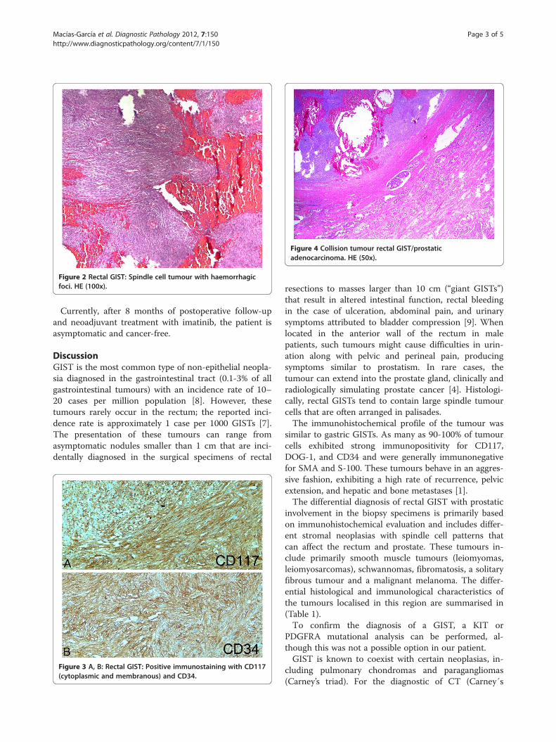
Figure 2 Rectal GIST: Spindle cell tumour with haemorrhagicfoci. HE (100x).
Figure 4 Collision tumour rectal GIST/prostaticadenocarcinoma. HE (50x).
Macías-García et al. Diagnostic Pathology 2012, 7:150 Page 3 of 5http://www.diagnosticpathology.org/content/7/1/150
Currently, after 8 months of postoperative follow-upand neoadjuvant treatment with imatinib, the patient isasymptomatic and cancer-free.
DiscussionGIST is the most common type of non-epithelial neopla-sia diagnosed in the gastrointestinal tract (0.1-3% of allgastrointestinal tumours) with an incidence rate of 10–20 cases per million population [8]. However, thesetumours rarely occur in the rectum; the reported inci-dence rate is approximately 1 case per 1000 GISTs [7].The presentation of these tumours can range fromasymptomatic nodules smaller than 1 cm that are inci-dentally diagnosed in the surgical specimens of rectal
Figure 3 A, B: Rectal GIST: Positive immunostaining with CD117(cytoplasmic and membranous) and CD34.
resections to masses larger than 10 cm (“giant GISTs”)that result in altered intestinal function, rectal bleedingin the case of ulceration, abdominal pain, and urinarysymptoms attributed to bladder compression [9]. Whenlocated in the anterior wall of the rectum in malepatients, such tumours might cause difficulties in urin-ation along with pelvic and perineal pain, producingsymptoms similar to prostatism. In rare cases, thetumour can extend into the prostate gland, clinically andradiologically simulating prostate cancer [4]. Histologi-cally, rectal GISTs tend to contain large spindle tumourcells that are often arranged in palisades.The immunohistochemical profile of the tumour was
similar to gastric GISTs. As many as 90-100% of tumourcells exhibited strong immunopositivity for CD117,DOG-1, and CD34 and were generally immunonegativefor SMA and S-100. These tumours behave in an aggres-sive fashion, exhibiting a high rate of recurrence, pelvicextension, and hepatic and bone metastases [1].The differential diagnosis of rectal GIST with prostatic
involvement in the biopsy specimens is primarily basedon immunohistochemical evaluation and includes differ-ent stromal neoplasias with spindle cell patterns thatcan affect the rectum and prostate. These tumours in-clude primarily smooth muscle tumours (leiomyomas,leiomyosarcomas), schwannomas, fibromatosis, a solitaryfibrous tumour and a malignant melanoma. The differ-ential histological and immunological characteristics ofthe tumours localised in this region are summarised in(Table 1).To confirm the diagnosis of a GIST, a KIT or
PDGFRA mutational analysis can be performed, al-though this was not a possible option in our patient.GIST is known to coexist with certain neoplasias, in-
cluding pulmonary chondromas and paragangliomas(Carney’s triad). For the diagnostic of CT (Carney´s
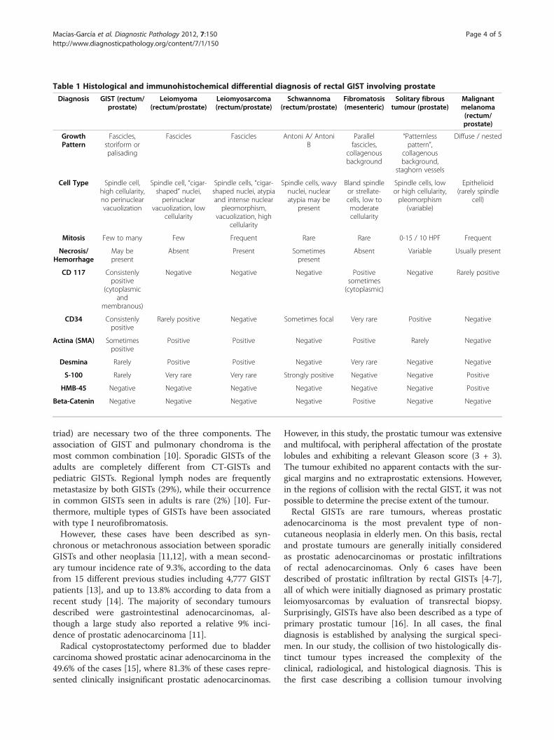
Table 1 Histological and immunohistochemical differential diagnosis of rectal GIST involving prostate
Diagnosis GIST (rectum/prostate)
Leiomyoma(rectum/prostate)
Leiomyosarcoma(rectum/prostate)
Schwannoma(rectum/prostate)
Fibromatosis(mesenteric)
Solitary fibroustumour (prostate)
Malignantmelanoma(rectum/prostate)
GrowthPattern
Fascicles,storiform orpalisading
Fascicles Fascicles Antoni A/ AntoniB
Parallelfascicles,
collagenousbackground
“Patternlesspattern”,
collagenousbackground,
staghorn vessels
Diffuse / nested
Cell Type Spindle cell,high cellularity,no perinuclearvacuolization
Spindle cell, “cigar-shaped” nuclei,perinuclear
vacuolization, lowcellularity
Spindle cells, “cigar-shaped nuclei, atypiaand intense nuclearpleomorphism,
vacuolization, highcellularity
Spindle cells, wavynuclei, nuclearatypia may be
present
Bland spindleor strellate-cells, low tomoderatecellularity
Spindle cells, lowor high cellularity,pleomorphism
(variable)
Epithelioid(rarely spindle
cell)
Mitosis Few to many Few Frequent Rare Rare 0-15 / 10 HPF Frequent
Necrosis/Hemorrhage
May bepresent
Absent Present Sometimespresent
Absent Variable Usually present
CD 117 Consistenlypositive
(cytoplasmicand
membranous)
Negative Negative Negative Positivesometimes(cytoplasmic)
Negative Rarely positive
CD34 Consistenlypositive
Rarely positive Negative Sometimes focal Very rare Positive Negative
Actina (SMA) Sometimespositive
Positive Positive Negative Positive Rarely Negative
Desmina Rarely Positive Positive Negative Very rare Negative Negative
S-100 Rarely Very rare Very rare Strongly positive Negative Negative Positive
HMB-45 Negative Negative Negative Negative Negative Negative Positive
Beta-Catenin Negative Negative Negative Negative Positive Negative Negative
Macías-García et al. Diagnostic Pathology 2012, 7:150 Page 4 of 5http://www.diagnosticpathology.org/content/7/1/150
triad) are necessary two of the three components. Theassociation of GIST and pulmonary chondroma is themost common combination [10]. Sporadic GISTs of theadults are completely different from CT-GISTs andpediatric GISTs. Regional lymph nodes are frequentlymetastasize by both GISTs (29%), while their occurrencein common GISTs seen in adults is rare (2%) [10]. Fur-thermore, multiple types of GISTs have been associatedwith type I neurofibromatosis.However, these cases have been described as syn-
chronous or metachronous association between sporadicGISTs and other neoplasia [11,12], with a mean second-ary tumour incidence rate of 9.3%, according to the datafrom 15 different previous studies including 4,777 GISTpatients [13], and up to 13.8% according to data from arecent study [14]. The majority of secondary tumoursdescribed were gastrointestinal adenocarcinomas, al-though a large study also reported a relative 9% inci-dence of prostatic adenocarcinoma [11].Radical cystoprostatectomy performed due to bladder
carcinoma showed prostatic acinar adenocarcinoma in the49.6% of the cases [15], where 81.3% of these cases repre-sented clinically insignificant prostatic adenocarcinomas.
However, in this study, the prostatic tumour was extensiveand multifocal, with peripheral affectation of the prostatelobules and exhibiting a relevant Gleason score (3 + 3).The tumour exhibited no apparent contacts with the sur-gical margins and no extraprostatic extensions. However,in the regions of collision with the rectal GIST, it was notpossible to determine the precise extent of the tumour.Rectal GISTs are rare tumours, whereas prostatic
adenocarcinoma is the most prevalent type of non-cutaneous neoplasia in elderly men. On this basis, rectaland prostate tumours are generally initially consideredas prostatic adenocarcinomas or prostatic infiltrationsof rectal adenocarcinomas. Only 6 cases have beendescribed of prostatic infiltration by rectal GISTs [4-7],all of which were initially diagnosed as primary prostaticleiomyosarcomas by evaluation of transrectal biopsy.Surprisingly, GISTs have also been described as a type ofprimary prostatic tumour [16]. In all cases, the finaldiagnosis is established by analysing the surgical speci-men. In our study, the collision of two histologically dis-tinct tumour types increased the complexity of theclinical, radiological, and histological diagnosis. This isthe first case describing a collision tumour involving
Macías-García et al. Diagnostic Pathology 2012, 7:150 Page 5 of 5http://www.diagnosticpathology.org/content/7/1/150
rectal GIST with direct prostatic invasion and a prostaticadenocarcinoma. This pathological situation greatly con-founded the clinical diagnosis, particularly given thepresence of a rectal mass, rectal and prostatic symptoms,and pathological levels of PSA.Until recently, the treatment of giant rectal GISTs has
been limited to surgical resection, except for cases of in-operable tumours or metastases that contraindicated thesurgical treatment (indicating that the tumour was in-curable under those specific circumstances). The use ofimatinib as a neoadjuvant treatment offers the possibilityof a partial tumour regression and stabilisation of thedisease [9]. In general, the best therapeutic results havebeen obtained in tumours with KIT mutations in exons11 (85%) and 9 (45%) [3], although a global response rateof only 40% in inoperable tumours indicates that resec-tion is a non-curative procedure in many cases [17].In our study, neoadjuvant therapy with imatinib did
not substantially reduce the tumour size, and histolo-gically, we only observed extensive haemorrhagic fociwithout a decrease in the number of tumour cells orthe presence of myxohyaline stroma, necrosis, or cys-tic degeneration, as previously described in cases thatexhibited an initial response to treatment [3]. How-ever, both tumours were completely resected, and after 8months of postoperative follow-up and adjuvant therapywith imatinib, the patient remains asymptomatic andcancer-free.
ConsentThe authors obtained the rights for publication of thisreport and any accompanying images.
AbbreviationsGIST: Gastrointestinal stromal tumours; PSA: Prostate specific antigen;CT: Computed Tomography; MRI: Including Magnetic Resonance; HPF: High-Power Field.
Competing interestsThe authors declare that they have no competing interests.
Authors’ contributionsAll authors read and approved the final manuscript.
Author details1Unidad de gestión clínica de Oncohematología y Anatomía Patológica,Hospital Universitario de Valme, Carretera de Cádiz s/n, 41014, Sevilla, Spain.2Department of Anatomic Pathology, Hospital Universitario Cruces, InstitutoBioCruces, University of the Basque Country, Barakaldo, Bizkaia, Spain.
Received: 2 August 2012 Accepted: 26 September 2012Published: 30 October 2012
References1. Miettinen M, Fletcher CDM, Kindblom LG, Tsui WMS: Mesenchymal
Tumours of the colon and rectum. In Who Classification of Tumours of theDigestive System. 4th edition. Edited by Bosman FT, Carneiro F, Hruban RH,Theise ND. France: Lyon; 2010:181–182.
2. Ortega Medina L, Artigas Raventos V, Díaz De Liaño Argüelles A, FernándezHernández JA, López Pousa A, de la Poza JL L, Ovejero Delgado C:Anatomía patológica de los tumores del estroma gastrointestinal
primarios no metastásicos en España. Resultados del estuido GRISK. RevEsp Patol 2010, 43(1):3–7.
3. Díaz Delgado M, Hernández Amate A, Sánchez León M, Pereira Gallardo S,González-Cámpora R: Avances en los tumores del estromagastrointestinal. Rev Esp Patol 2010, 43(1):16–23.
4. Madden JF, Burchette JL, Raj GV, Daly JT, Tannenbaum M: Anterior rectalwall gastrointestinal stromal tumor presenting clinically as prostaticmass. Urol Oncol 2005, 23:268–272.
5. Yoelzke BB, Sakamoto K, Hantel A, Paner GP, Kash J, Waters WB, CampbellSC: Gastrointestinal stromal tumor: involvement in urologic patients andrecent therapeutic advances. Urology 2002, 60:218–222.
6. Dickson BC, Srigley JR, Pollett AF, Blackstein ME, Honey JD, Juco JW: Rectalgastrointestinal stromal tumor mimicking a primary prostatic lesion. CanJ Urol 2008, 15:4112–4114.
7. Yaman E, Coskun U, Sozen S, Yamac D, Buyukberber S: Coexistence ofgastrointestinal stromal tumor (GIST) of the rectum and adenocarcinomaof the prostate in a patient with familial GIST. Onkologie 2008,31(12):697–699.
8. Miettinen M, Lasota J: Gastrointestinal stromal tumors-definition, clinical,histological, immuno-histochemical, and molecular genetics features anddifferential diagnosis. Virchows Arch 2001, 438:1–12.
9. Dickhoff C, Leguit RL, Slors JFM, Vervenne WL, Bemelman WA: Giant rectalgastrointestinal stromal tumors: A report of two cases. Case RepGastroenterol 2008, 2:54–59.
10. Otto C, Agaimy A, Braun A, Rädecke J, Hoeppner J, Illerhaus G, Werner M,Kontny U, Haller F: Multifocal gastric gastrointestinal stromal tumors(GISTs) with lymph node metastases in children and young adults: Acomparative clinical and histomorphological study of three casesincluding a new case of Carney triad. Diagn Pathol 2011, 6:52.
11. Agaimy A, Wunsch PH, Sobin LH, Lasota J, Miettinen M: Occurrence ofother malignancies in patients with gastrointestinal stromal tumors.Semin Diagn Pathol 2006, 23:120–129.
12. Liszka L, Zieli Ska-Palak E, Pajak J, Golka D, Huszno J: Coexistence ofgastrointestinal stromal tumors with other neoplasms. J Gastroenterol2007, 42:641–649.
13. Agaimy A, Wuensch PH: Gastrointestinal stromal tumors in patients withother-type cancer: a mere coincidence or an etiological association? Astudy of 97 GIST cases. Z Gastroenterol 2005, 43(9):1025–1030.
14. Gonzalves R, Linhares E, Albagli R, Valadao M, Vilhena B, Romano S, GilFerreira C: Ocurrence of other tumors in patients with GIST. Surg Oncol2010, 19:e140–e143.
15. Mazzucchelli R, Barbisan F, Scarpelli M, López-Beltran A, Van der Kwast TH,Cheng L, Montironi R: Is incidentally detected prostate cancer in patientsundergoing radical cystoprostatectomy clinically significant? Am J ClinPathol 2009, 131:279–283.
16. Lee CH, Lin YH, Lin HY, Lee CM, Chu JS: Gastrointestinal stromal tumors ofthe prostate: a case report and literature review. Hum Pathol 2006,37:1361–1365.
17. Scaife CL, Hunt KK, Patel SR, Benjamin RS, Burgess MA, Chen LL: Is there arole for surgery in patients with “unresectable” c-KIT + gastrointestinalstromal tumors treated with imatinib mesylate? Am J Surg 2003,186:665–669.
doi:10.1186/1746-1596-7-150Cite this article as: Macías-García et al.: Collision tumour involving arectal gastrointestinal stromal tumour with invasion of the prostate anda prostatic adenocarcinoma. Diagnostic Pathology 2012 7:150.