tumour-associated macrophages are a distinct m2 polarised population promoting tumour progression...
DESCRIPTION
Tumour-Associated Macrophages Are a Distinct M2 Polarised Population Promoting Tumour Progression Potential Targets of Anti-cancer TherapyTRANSCRIPT
Seediscussions,stats,andauthorprofilesforthispublicationat:http://www.researchgate.net/publication/7258136
SicaA,SchioppaT,MantovaniA,AllavenaP..Tumour-associatedmacrophagesareadistinctM2polarisedpopulationpromotingtumourprogression:potentialtargetsofanti-cancerth...
ARTICLEinEUROPEANJOURNALOFCANCER·MAY2006
ImpactFactor:4.82·DOI:10.1016/j.ejca.2006.01.003·Source:PubMed
CITATIONS
553
DOWNLOADS
145
VIEWS
130
4AUTHORS,INCLUDING:
AntonioSica
AmedeoAvogadroUniversityofEasternPie…
131PUBLICATIONS19,856CITATIONS
SEEPROFILE
TizianaSchioppa
UniversitàdegliStudidiBrescia
16PUBLICATIONS2,272CITATIONS
SEEPROFILE
Availablefrom:TizianaSchioppa
Retrievedon:05September2015
E U R O P E A N J O U R N A L O F C A N C E R 4 2 ( 2 0 0 6 ) 7 1 7 – 7 2 7
. sc iencedi rec t . com
ava i lab le a t wwwjournal homepage: www.ejconl ine.com
Tumour-associated macrophages are a distinct M2polarised population promoting tumour progression:Potential targets of anti-cancer therapy
Antonio Sicaa,b,*, Tiziana Schioppab, Alberto Mantovania,c, Paola Allavenaa
aIstituto Clinico Humanitas, 20089 Rozzano, Milan, ItalybIstituto di Ricerche Farmacologiche Mario Negri, Via Eritrea 62, 20157 Milan, ItalycCentro IDET, Institute of General Pathology, University of Milan, Via Mangiagalli 31, 20133 Milan, Italy
A R T I C L E I N F O
Article history:
Received 11 January 2006
Accepted 11 January 2006
Available online 7 March 2006
Keywords:
TAM
Cancer
Inflammation
0959-8049/$ - see front matter � 2006 Elsevidoi:10.1016/j.ejca.2006.01.003
* Corresponding author: Tel.: +39 02 8224 511E-mail address: antonio.sica@humanitas.
A B S T R A C T
Tumour-associated macrophages (TAM) represent the major inflammatory component of
the stroma of many tumours, and can affect different aspects of the neoplastic tissue. Many
observations indicate that TAM express several M2-associated pro-tumoural functions,
including promotion of angiogenesis, matrix remodelling and suppression of adaptive
immunity. The pro-tumoural role of TAM in cancer is further supported by clinical studies
that found a correlation between the high macrophage content of tumours and poor patient
prognosis. Evidence is presented here supporting the view that TAM represent a unique and
distinct M2-skewed myeloid population and are a potential target for anti-cancer therapy.
� 2006 Elsevier Ltd. All rights reserved.
1. Introduction
Accumulation of leukocyte subpopulations is the hallmark
of several pathological conditions, including tumours.1 A
prominent component of solid tumours is represented by
non-tumoural cells, including stromal cells (fibroblasts and
endothelial cells) and leukocytes. Among the latter, macro-
phages are the major component.2 Tumour-associated mac-
rophages (TAM) have been studied extensively for their
relationship with tumour cells and their multi-faceted func-
tions in the tumour micro-environment. Immunologists
have long considered the presence of TAM as evidence of
a host response against the growing tumour. Several studies
have demonstrated that macrophages have the potential to
kill tumour cells in vitro when appropriately stimulated, e.g.
following treatment with lipopolysaccharides (LPS) and
interferon (IFN)-c. However, bacterial stimuli and Th1 cyto-
kines inducing M1 type polarisation are usually not present
er Ltd. All rights reserved
1; fax: +39 02 8224 5101.it (A. Sica).
at the tumour site. Here, in contrast, differentiating macro-
phages are likely to encounter factors that most frequently
polarise them toward M2 type macrophages (e.g. interleukin
(IL)-10). Over the years it has become increasingly clear that
TAM are active players in the process of tumour progression
and invasion. In several experimental tumour models, the
activation of an inflammatory response (most frequently
mediated by macrophages) is essential for full neoplastic
transformation and progression.3 Furthermore, in clinical
studies high numbers of intra-tumour macrophages corre-
late with high vessel density and tumour progression.
The strategic location of TAM suggests that these cells are
important regulators of anti-tumour immunity. Characterisa-
tion of the phenotype of TAM is therefore essential to the
understanding of tumour-derived signals guiding polarisation
of innate and adaptive immunity in cancer bearers and to the
identification of molecular mechanisms that might be ame-
nable to therapeutic intervention.
.
718 E U R O P E A N J O U R N A L O F C A N C E R 4 2 ( 2 0 0 6 ) 7 1 7 – 7 2 7
2. Recruitment of myeloid cells at thetumour site
2.1. Monocyte-macrophages
Since the first observation by Rudolf Virchow, who noticed the
infiltration of leukocytes into malignant tissues and sug-
gested that cancers arise at regions of chronic inflammation,
the origin of TAM has been studied in terms of recruitment,
survival and proliferation. TAM derive from circulating mono-
cytes and are recruited at the tumour site by a tumour-derived
chemotactic factor for monocytes, originally described by this
group4 and later identified as the chemokine CCL2/MCP-15,6
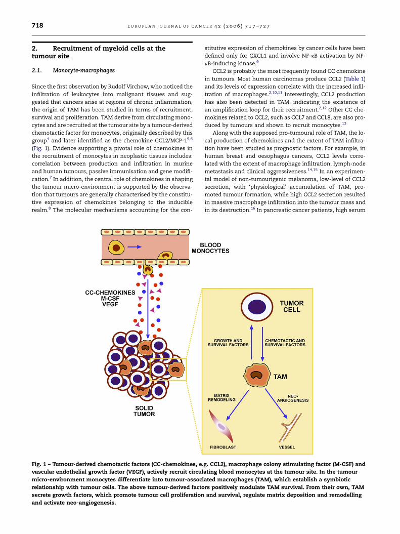
(Fig. 1). Evidence supporting a pivotal role of chemokines in
the recruitment of monocytes in neoplastic tissues includes:
correlation between production and infiltration in murine
and human tumours, passive immunisation and gene modifi-
cation.7 In addition, the central role of chemokines in shaping
the tumour micro-environment is supported by the observa-
tion that tumours are generally characterised by the constitu-
tive expression of chemokines belonging to the inducible
realm.8 The molecular mechanisms accounting for the con-
Fig. 1 – Tumour-derived chemotactic factors (CC-chemokines, e.
vascular endothelial growth factor (VEGF), actively recruit circul
micro-environment monocytes differentiate into tumour-associ
relationship with tumour cells. The above tumour-derived facto
secrete growth factors, which promote tumour cell proliferation
and activate neo-angiogenesis.
stitutive expression of chemokines by cancer cells have been
defined only for CXCL1 and involve NF-jB activation by NF-
jB-inducing kinase.9
CCL2 is probably the most frequently found CC chemokine
in tumours. Most human carcinomas produce CCL2 (Table 1)
and its levels of expression correlate with the increased infil-
tration of macrophages.2,10,11 Interestingly, CCL2 production
has also been detected in TAM, indicating the existence of
an amplification loop for their recruitment.2,12 Other CC che-
mokines related to CCL2, such as CCL7 and CCL8, are also pro-
duced by tumours and shown to recruit monocytes.13
Along with the supposed pro-tumoural role of TAM, the lo-
cal production of chemokines and the extent of TAM infiltra-
tion have been studied as prognostic factors. For example, in
human breast and oesophagus cancers, CCL2 levels corre-
lated with the extent of macrophage infiltration, lymph-node
metastasis and clinical aggressiveness.14,15 In an experimen-
tal model of non-tumourigenic melanoma, low-level of CCL2
secretion, with ‘physiological’ accumulation of TAM, pro-
moted tumour formation, while high CCL2 secretion resulted
in massive macrophage infiltration into the tumour mass and
in its destruction.16 In pancreatic cancer patients, high serum
g. CCL2), macrophage colony stimulating factor (M-CSF) and
ating blood monocytes at the tumour site. In the tumour
ated macrophages (TAM), which establish a symbiotic
rs positively modulate TAM survival. From their own, TAM
and survival, regulate matrix deposition and remodelling
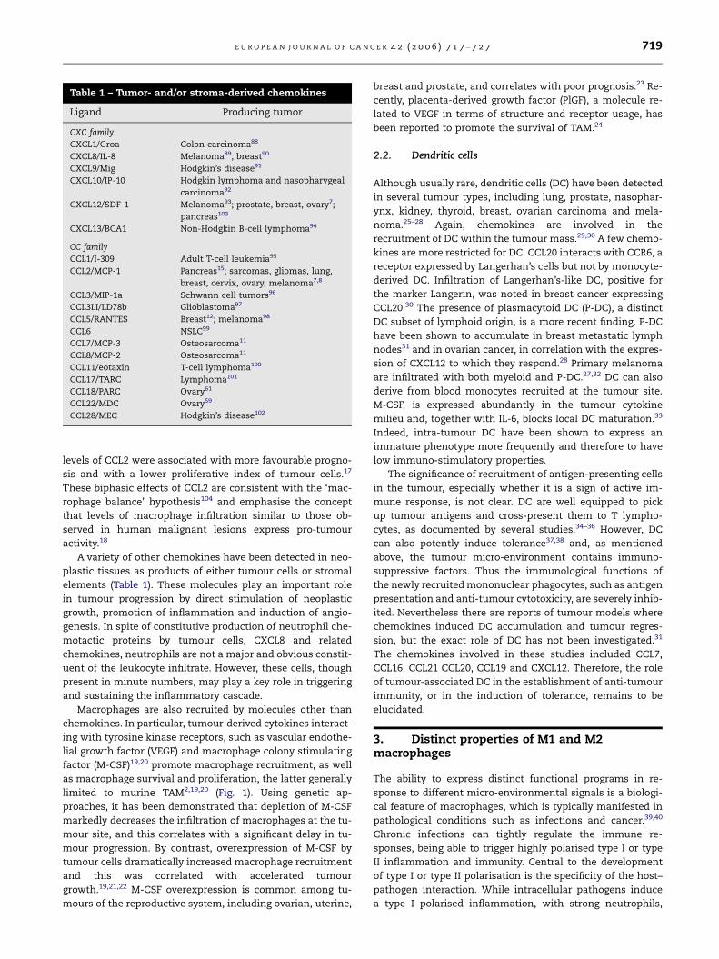
Table 1 – Tumor- and/or stroma-derived chemokines
Ligand Producing tumor
CXC family
CXCL1/Groa Colon carcinoma88
CXCL8/IL-8 Melanoma89, breast90
CXCL9/Mig Hodgkin’s disease91
CXCL10/IP-10 Hodgkin lymphoma and nasopharygeal
carcinoma92
CXCL12/SDF-1 Melanoma93; prostate, breast, ovary7;
pancreas103
CXCL13/BCA1 Non-Hodgkin B-cell lymphoma94
CC family
CCL1/I-309 Adult T-cell leukemia95
CCL2/MCP-1 Pancreas15; sarcomas, gliomas, lung,
breast, cervix, ovary, melanoma7,8
CCL3/MIP-1a Schwann cell tumors96
CCL3LI/LD78b Glioblastoma97
CCL5/RANTES Breast12; melanoma98
CCL6 NSLC99
CCL7/MCP-3 Osteosarcoma11
CCL8/MCP-2 Osteosarcoma11
CCL11/eotaxin T-cell lymphoma100
CCL17/TARC Lymphoma101
CCL18/PARC Ovary61
CCL22/MDC Ovary59
CCL28/MEC Hodgkin’s disease102
E U R O P E A N J O U R N A L O F C A N C E R 4 2 ( 2 0 0 6 ) 7 1 7 – 7 2 7 719
levels of CCL2 were associated with more favourable progno-
sis and with a lower proliferative index of tumour cells.17
These biphasic effects of CCL2 are consistent with the ‘mac-
rophage balance’ hypothesis104 and emphasise the concept
that levels of macrophage infiltration similar to those ob-
served in human malignant lesions express pro-tumour
activity.18
A variety of other chemokines have been detected in neo-
plastic tissues as products of either tumour cells or stromal
elements (Table 1). These molecules play an important role
in tumour progression by direct stimulation of neoplastic
growth, promotion of inflammation and induction of angio-
genesis. In spite of constitutive production of neutrophil che-
motactic proteins by tumour cells, CXCL8 and related
chemokines, neutrophils are not a major and obvious constit-
uent of the leukocyte infiltrate. However, these cells, though
present in minute numbers, may play a key role in triggering
and sustaining the inflammatory cascade.
Macrophages are also recruited by molecules other than
chemokines. In particular, tumour-derived cytokines interact-
ing with tyrosine kinase receptors, such as vascular endothe-
lial growth factor (VEGF) and macrophage colony stimulating
factor (M-CSF)19,20 promote macrophage recruitment, as well
as macrophage survival and proliferation, the latter generally
limited to murine TAM2,19,20 (Fig. 1). Using genetic ap-
proaches, it has been demonstrated that depletion of M-CSF
markedly decreases the infiltration of macrophages at the tu-
mour site, and this correlates with a significant delay in tu-
mour progression. By contrast, overexpression of M-CSF by
tumour cells dramatically increased macrophage recruitment
and this was correlated with accelerated tumour
growth.19,21,22 M-CSF overexpression is common among tu-
mours of the reproductive system, including ovarian, uterine,
breast and prostate, and correlates with poor prognosis.23 Re-
cently, placenta-derived growth factor (PlGF), a molecule re-
lated to VEGF in terms of structure and receptor usage, has
been reported to promote the survival of TAM.24
2.2. Dendritic cells
Although usually rare, dendritic cells (DC) have been detected
in several tumour types, including lung, prostate, nasophar-
ynx, kidney, thyroid, breast, ovarian carcinoma and mela-
noma.25–28 Again, chemokines are involved in the
recruitment of DC within the tumour mass.29,30 A few chemo-
kines are more restricted for DC. CCL20 interacts with CCR6, a
receptor expressed by Langerhan’s cells but not by monocyte-
derived DC. Infiltration of Langerhan’s-like DC, positive for
the marker Langerin, was noted in breast cancer expressing
CCL20.30 The presence of plasmacytoid DC (P-DC), a distinct
DC subset of lymphoid origin, is a more recent finding. P-DC
have been shown to accumulate in breast metastatic lymph
nodes31 and in ovarian cancer, in correlation with the expres-
sion of CXCL12 to which they respond.28 Primary melanoma
are infiltrated with both myeloid and P-DC.27,32 DC can also
derive from blood monocytes recruited at the tumour site.
M-CSF, is expressed abundantly in the tumour cytokine
milieu and, together with IL-6, blocks local DC maturation.33
Indeed, intra-tumour DC have been shown to express an
immature phenotype more frequently and therefore to have
low immuno-stimulatory properties.
The significance of recruitment of antigen-presenting cells
in the tumour, especially whether it is a sign of active im-
mune response, is not clear. DC are well equipped to pick
up tumour antigens and cross-present them to T lympho-
cytes, as documented by several studies.34–36 However, DC
can also potently induce tolerance37,38 and, as mentioned
above, the tumour micro-environment contains immuno-
suppressive factors. Thus the immunological functions of
the newly recruited mononuclear phagocytes, such as antigen
presentation and anti-tumour cytotoxicity, are severely inhib-
ited. Nevertheless there are reports of tumour models where
chemokines induced DC accumulation and tumour regres-
sion, but the exact role of DC has not been investigated.31
The chemokines involved in these studies included CCL7,
CCL16, CCL21 CCL20, CCL19 and CXCL12. Therefore, the role
of tumour-associated DC in the establishment of anti-tumour
immunity, or in the induction of tolerance, remains to be
elucidated.
3. Distinct properties of M1 and M2macrophages
The ability to express distinct functional programs in re-
sponse to different micro-environmental signals is a biologi-
cal feature of macrophages, which is typically manifested in
pathological conditions such as infections and cancer.39,40
Chronic infections can tightly regulate the immune re-
sponses, being able to trigger highly polarised type I or type
II inflammation and immunity. Central to the development
of type I or type II polarisation is the specificity of the host–
pathogen interaction. While intracellular pathogens induce
a type I polarised inflammation, with strong neutrophils,
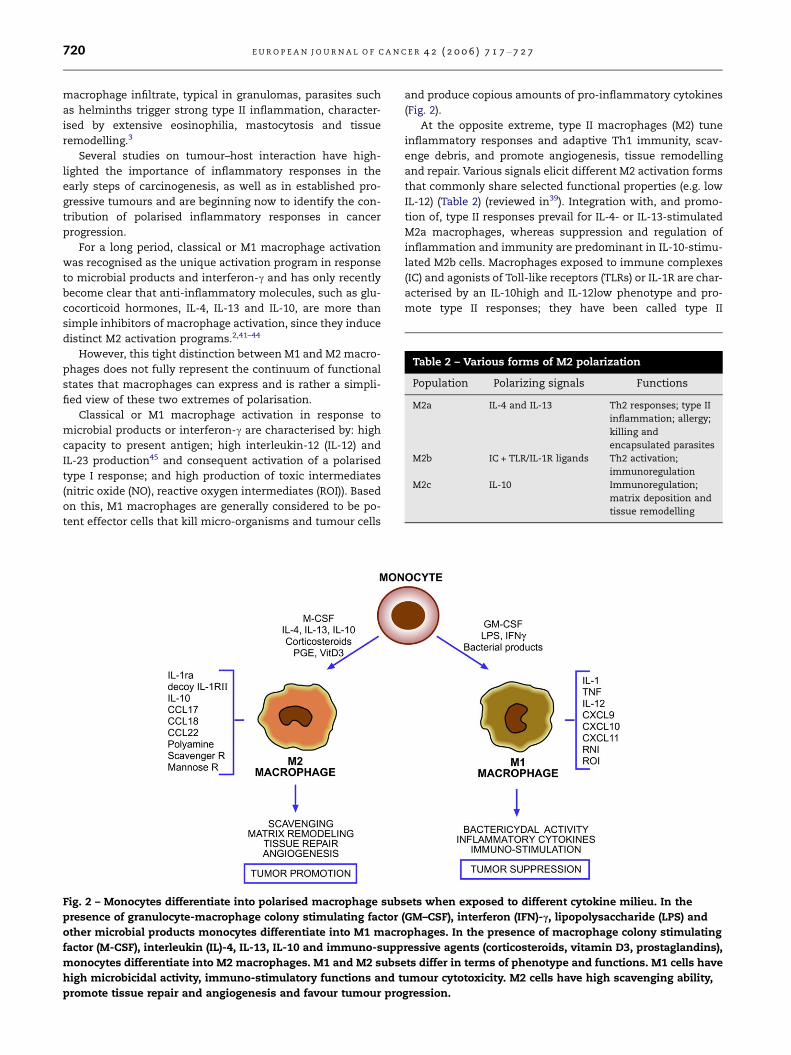
Table 2 – Various forms of M2 polarization
Population Polarizing signals Functions
M2a IL-4 and IL-13 Th2 responses; type II
inflammation; allergy;
killing and
encapsulated parasites
M2b IC + TLR/IL-1R ligands Th2 activation;
immunoregulation
M2c IL-10 Immunoregulation;
matrix deposition and
tissue remodelling
720 E U R O P E A N J O U R N A L O F C A N C E R 4 2 ( 2 0 0 6 ) 7 1 7 – 7 2 7
macrophage infiltrate, typical in granulomas, parasites such
as helminths trigger strong type II inflammation, character-
ised by extensive eosinophilia, mastocytosis and tissue
remodelling.3
Several studies on tumour–host interaction have high-
lighted the importance of inflammatory responses in the
early steps of carcinogenesis, as well as in established pro-
gressive tumours and are beginning now to identify the con-
tribution of polarised inflammatory responses in cancer
progression.
For a long period, classical or M1 macrophage activation
was recognised as the unique activation program in response
to microbial products and interferon-c and has only recently
become clear that anti-inflammatory molecules, such as glu-
cocorticoid hormones, IL-4, IL-13 and IL-10, are more than
simple inhibitors of macrophage activation, since they induce
distinct M2 activation programs.2,41–44
However, this tight distinction between M1 and M2 macro-
phages does not fully represent the continuum of functional
states that macrophages can express and is rather a simpli-
fied view of these two extremes of polarisation.
Classical or M1 macrophage activation in response to
microbial products or interferon-c are characterised by: high
capacity to present antigen; high interleukin-12 (IL-12) and
IL-23 production45 and consequent activation of a polarised
type I response; and high production of toxic intermediates
(nitric oxide (NO), reactive oxygen intermediates (ROI)). Based
on this, M1 macrophages are generally considered to be po-
tent effector cells that kill micro-organisms and tumour cells
Fig. 2 – Monocytes differentiate into polarised macrophage sub
presence of granulocyte-macrophage colony stimulating factor
other microbial products monocytes differentiate into M1 macr
factor (M-CSF), interleukin (IL)-4, IL-13, IL-10 and immuno-supp
monocytes differentiate into M2 macrophages. M1 and M2 subs
high microbicidal activity, immuno-stimulatory functions and t
promote tissue repair and angiogenesis and favour tumour pro
and produce copious amounts of pro-inflammatory cytokines
(Fig. 2).
At the opposite extreme, type II macrophages (M2) tune
inflammatory responses and adaptive Th1 immunity, scav-
enge debris, and promote angiogenesis, tissue remodelling
and repair. Various signals elicit different M2 activation forms
that commonly share selected functional properties (e.g. low
IL-12) (Table 2) (reviewed in39). Integration with, and promo-
tion of, type II responses prevail for IL-4- or IL-13-stimulated
M2a macrophages, whereas suppression and regulation of
inflammation and immunity are predominant in IL-10-stimu-
lated M2b cells. Macrophages exposed to immune complexes
(IC) and agonists of Toll-like receptors (TLRs) or IL-1R are char-
acterised by an IL-10high and IL-12low phenotype and pro-
mote type II responses; they have been called type II
sets when exposed to different cytokine milieu. In the
(GM–CSF), interferon (IFN)-c, lipopolysaccharide (LPS) and
ophages. In the presence of macrophage colony stimulating
ressive agents (corticosteroids, vitamin D3, prostaglandins),
ets differ in terms of phenotype and functions. M1 cells have
umour cytotoxicity. M2 cells have high scavenging ability,
gression.
E U R O P E A N J O U R N A L O F C A N C E R 4 2 ( 2 0 0 6 ) 7 1 7 – 7 2 7 721
activated macrophages.46 Finally, human monocytes differen-
tiated with granulocyte-macrophage colony stimulating fac-
tor (GM–CSF) or M–CSF have M1 and M2 properties,
respectively, and have been referred to as Mø1 and Mø2.45
A comprehensive picture of the polarised functions of
macrophages is represented in Fig. 1. M1 macrophages ex-
posed to the classic activation signals IFN-c and LPS express
opsonic receptors (e.g. FccRIII (CD16)), whereas M2 macro-
phages express preferentially non-opsonic receptor (e.g. man-
nose receptor and scavenger receptors).
Arginine metabolism gives rise to high levels of inducible
nitric oxide synthase (iNOS; NOS2) in the M1 population. In
contrast, M2 macrophages express a predominant activation
of the arginase pathway and the consequent production of
ornithine and polyamines. This metabolic switch occurs
preferentially during the activation of the M2a and M2c
polarisation programs.39 The LPS receptor TLR4 and the
adapter molecule MyD88 are increased by IFN-c, while in
contrast IL-10 inhibits their expression. By analogy, the IL-1
system appears to be differentially regulated by M1 and M2
signals. IFN-c and LPS foster the IL-1-mediated functions
by inhibiting the decoy receptor IL-1RII and upregulating
the signalling IL-1RI, and IL-1R accessory protein.2 In con-
trast, IL-4, IL-13 and glucocorticoid hormones attenuate the
IL-1 system by inducing expression of the decoy receptor
IL-1RII. Moreover, IL-4 and IL-13 induce IL-1 receptor antago-
nist (IL-1ra) production and inhibit IL-1.47 While M1 macro-
phages express high levels of pro-inflammatory cytokines
(IL-1, TNF, IL-6 and IL-23), M2 cells are generally character-
ised by their low production.
The M1 phenotype is typically IL-12high and IL-10low,
whereas M2 macrophages are typically IL-10high and IL-
12low. However, macrophages exposed to IC and LPS (M2b)
(Fig. 2) are an exception, in that they retain high levels of
inflammatory cytokine production with concomitant high
IL-10 and low IL-12.46 In spite of their high production of
inflammatory cytokines and toxic molecules, M2b cells pro-
tect mice against LPS toxicity,46,48 promote Th2 differentiation
and humoural antibody production.
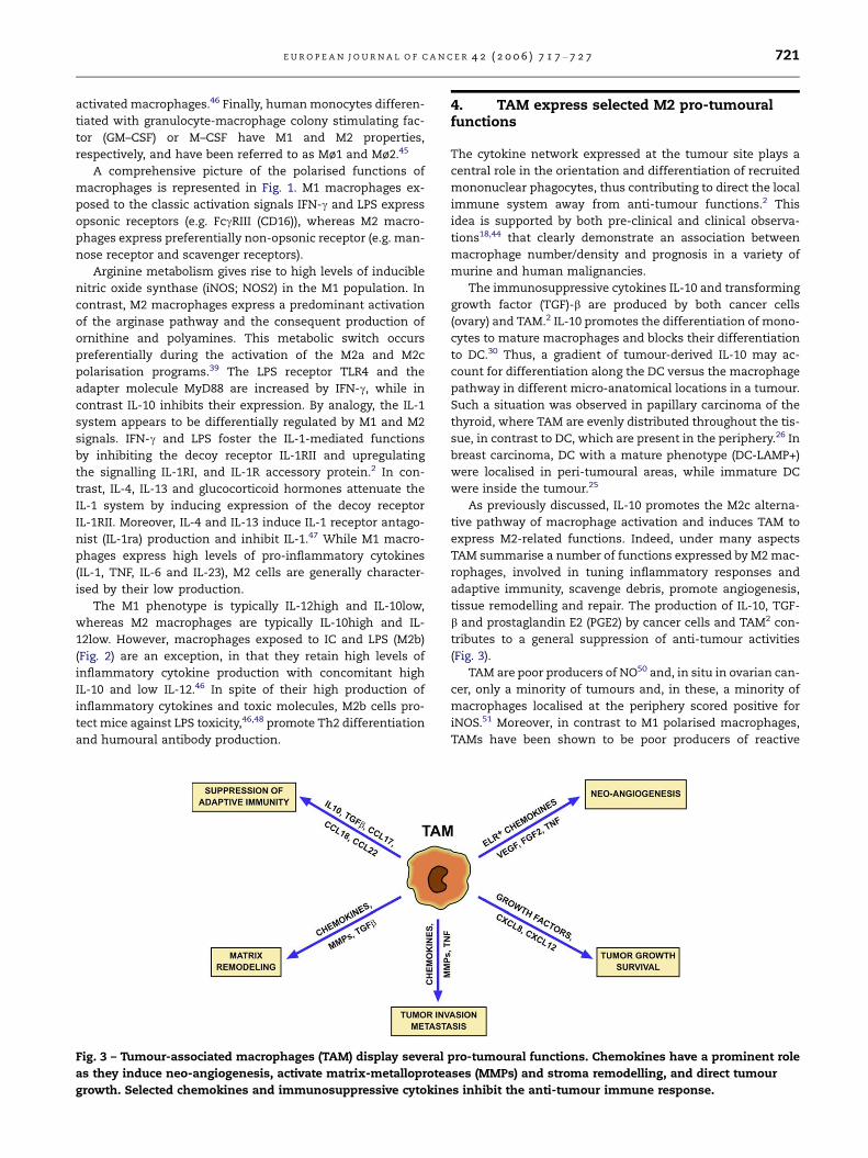
Fig. 3 – Tumour-associated macrophages (TAM) display several
as they induce neo-angiogenesis, activate matrix-metalloprotea
growth. Selected chemokines and immunosuppressive cytokine
4. TAM express selected M2 pro-tumouralfunctions
The cytokine network expressed at the tumour site plays a
central role in the orientation and differentiation of recruited
mononuclear phagocytes, thus contributing to direct the local
immune system away from anti-tumour functions.2 This
idea is supported by both pre-clinical and clinical observa-
tions18,44 that clearly demonstrate an association between
macrophage number/density and prognosis in a variety of
murine and human malignancies.
The immunosuppressive cytokines IL-10 and transforming
growth factor (TGF)-b are produced by both cancer cells
(ovary) and TAM.2 IL-10 promotes the differentiation of mono-
cytes to mature macrophages and blocks their differentiation
to DC.30 Thus, a gradient of tumour-derived IL-10 may ac-
count for differentiation along the DC versus the macrophage
pathway in different micro-anatomical locations in a tumour.
Such a situation was observed in papillary carcinoma of the
thyroid, where TAM are evenly distributed throughout the tis-
sue, in contrast to DC, which are present in the periphery.26 In
breast carcinoma, DC with a mature phenotype (DC-LAMP+)
were localised in peri-tumoural areas, while immature DC
were inside the tumour.25
As previously discussed, IL-10 promotes the M2c alterna-
tive pathway of macrophage activation and induces TAM to
express M2-related functions. Indeed, under many aspects
TAM summarise a number of functions expressed by M2 mac-
rophages, involved in tuning inflammatory responses and
adaptive immunity, scavenge debris, promote angiogenesis,
tissue remodelling and repair. The production of IL-10, TGF-
b and prostaglandin E2 (PGE2) by cancer cells and TAM2 con-
tributes to a general suppression of anti-tumour activities
(Fig. 3).
TAM are poor producers of NO50 and, in situ in ovarian can-
cer, only a minority of tumours and, in these, a minority of
macrophages localised at the periphery scored positive for
iNOS.51 Moreover, in contrast to M1 polarised macrophages,
TAMs have been shown to be poor producers of reactive
pro-tumoural functions. Chemokines have a prominent role
ses (MMPs) and stroma remodelling, and direct tumour
s inhibit the anti-tumour immune response.
722 E U R O P E A N J O U R N A L O F C A N C E R 4 2 ( 2 0 0 6 ) 7 1 7 – 7 2 7
oxygen intermediates (ROIs), consistent with the hypothesis
that these cells represent a skewed M2 population.51
Moreover, TAM were reported to express low levels of
inflammatory cytokines (e.g. IL-12, IL-1b, TNF-a, IL-6).2 Activa-
tion of NF-jB is a necessary event promoting transcription of
several pro-inflammatory genes. Our previous studies49 indi-
cated that TAM display defective NF-jB activation in response
to the M1 polarising signal LPS, and we observed similar re-
sults in response to the pro-inflammatory cytokine IL-1b
(Schioppa and colleagues, Istituto di Ricerche Farmacologiche
Mario Negri, Milan, Italy). Thus, in terms of cytotoxicity and
expression of inflammatory cytokines TAM resemble M2
macrophages.
In agreement with the M2 signature, TAM also express
high levels of both the scavenger receptor-A (SR-A) (Biswas
S, unpublished observation) and the mannose receptor (MR)
(P. Allavena, Istituto di Ricerche Farmacologiche Mario Negri,
Milan, Italy). Furthermore, TAM are poor antigen-presenting
cells.2
Arginase expression in TAM has not been studied. How-
ever, it has been proposed recently that the carbohydrate-
binding protein galectin-1, which is abundantly expressed
by ovarian cancer52 and shows specific anti-inflammatory ef-
fects, tunes the classic pathway of L-arginine, resulting in a
strong inhibition of NO production by LPS-activated
macrophages.
Angiogenesis is an M2-associated function, which repre-
sents a key event in tumour growth and progression. In sev-
eral studies in human cancer, accumulation of TAM has
been associated with angiogenesis and with the production
of angiogenic factors such as VEGF and platelet-derived endo-
thelial cell growth factor.2 More recently, in human cervical
cancer, VEGF-C production by TAMs was proposed to play a
role in peri-tumoural lympho-angiogenesis and subsequent
dissemination of cancer cells with formation of lymphatic
metastasis53 Additionally, TAM participate to the pro-angio-
genic process by producing the angiogenic factor thymidine
phosphorylase (TP), which promotes endothelial cell migra-
tion in vitro and whose levels of expression are associated
with tumour neovascularisation.54 TAM also contribute to tu-
mour progression by producing pro-angiogenic and tumour-
inducing chemokines, such as CCL2.29 Moreover, TAM accu-
mulate in hypoxic regions of tumours and hypoxia triggers
a pro-angiogenic program in these cells (see below). There-
fore, macrophages recruited in situ represent an indirect
pathway of amplification of angiogenesis, in concert with
angiogenic molecules directly produced by tumour cells. On
the anti-angiogenic side, in a murine model, GM–CSF released
from a primary tumour upregulated TAM-derived metallo-
elastase and angiostatin production, thus suppressing tu-
mour growth of metastases.55
Finally, TAM express molecules which affect tumour cell
proliferation, angiogenesis and dissolution of connective tis-
sues. These include epidermal growth factor (EGF), members
of the FGF family, TGF-b, VEGF and chemokines. In lung can-
cer, TAM may favour tumour progression by contributing to
stroma formation and angiogenesis through their release of
platelet derived growth factor (PDGF), in conjunction with
TGF-b1 production by cancer cells.2 Macrophages can produce
enzymes and inhibitors that regulate the digestion of the
extracellular matrix, such as matrix-metalloproteases
(MMPs), plasmin, urokinase-type plasminogen activator
(uPA) and the uPA receptor. Direct evidence has been pre-
sented that MMP-9 derived from haematopoietic cells of host
origin contributes to skin carcinogenesis.56 Chemokines have
been shown to induce gene expression of various MMPs and,
in particular, MMP-9 production, along with the uPA recep-
tor.57 Evidence suggests that MMP-9 has complex effects be-
yond matrix degradation including promotion of the
angiogenesis switch and release of growth factors.56
5. Modulation of adaptive immunity by TAM
It has long been known that TAM have poor antigen-present-
ing capacity and can actually suppress T cell activation and
proliferation.2 The suppressive mediators produced by TAM
include prostaglandins, IL-10 and TGF-b and indoleamine
dioxigenase (IDO) metabolites.2,44 Moreover, TAM are unable
to produce IL-12, even upon stimulation by IFN-c and LPS.49
With this cytokine profile, which is characteristic of M2 mac-
rophages, TAM are unable to trigger Th1 polarised immune
responses, but rather induce T regulatory cells (Treg) (Fig. 4).
Treg cells possess a characteristic anergic phenotype and
strongly suppress the activity of effector T cells and other
inflammatory cells, such as monocytes. Suppression of T cell
mediated anti-tumour activity by Treg cells is associated with
increased tumour growth and hence decreased survival.58 For
instance, in patients with advanced ovarian cancer, an in-
crease in the number of functionally active Treg cells present
in the ascites was predictive of reduced survival.59 Immature
myeloid suppressor cells present in the neoplastic tissue of
some tumours have been shown potently to inhibit T cell re-
sponses.60 The relationship if any, of immature myeloid sup-
pressor cells with TAM remains to be defined.
The complex network of chemokines present at the tu-
mour site can play a role also in the induction of adaptive
immunity. Chemokines also regulate the amplification of
polarised T cell responses (Fig. 4). Some chemokines may en-
hance specific host immunity against tumours, but on the
other hand other chemokines may contribute to escape from
the immune system, by recruiting Th2 effectors and Treg
cells.39 As mentioned above, in addition to being a target for
chemokines, TAM are a source of a selected set of these medi-
ators (CCL2, CCL17, CCL18, CCL22). CCL18 was recently identi-
fied as the most abundant chemokine in human ovarian
ascites fluid. When the source of CCL18 was investigated, it
was tracked to TAM, with no production by ovarian carci-
noma cells.61 CCL18 is a CC chemokine produced constitu-
tively by immature DC and inducible in macrophages by
IL-4, IL-13 and IL-10. Since IL-4 and IL-13 are not expressed
in substantial amounts in ovarian cancer, it is likely that
IL-10, produced by tumour cells and macrophages them-
selves, accounts for CCL18 production by TAM. CCL18 is an
attractant for naive T cells by interacting with an unidentified
receptor.62 Attraction of naive T cells in a peripheral micro-
environment dominated by M2 macrophages and immature
DC is likely to induce T cell anergy.
Work in gene-modified mice has shown that CCL2 can ori-
ent specific immunity in a Th2 direction. Although the exact
mechanism for this action has not been defined, it may
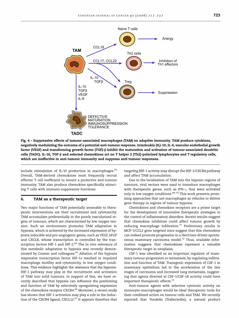
Fig. 4 – Suppressive effects of tumour-associated macrophages (TAM) on adaptive immunity. TAM produce cytokines,
negatively modulating the outcome of a potential anti-tumour response. Interleukin (IL)-10, IL-6, vascular endothelial growth
factor (VEGF) and transforming growth factor (TGF)-b inhibit the maturation and activation of tumour-associated dendritic
cells (TADC). IL-10, TGF-b and selected chemokines act on T helper 2 (Th2)-polarised lymphocytes and T regulatory cells,
which are ineffective in anti-tumour immunity and suppress anti-tumour responses.
E U R O P E A N J O U R N A L O F C A N C E R 4 2 ( 2 0 0 6 ) 7 1 7 – 7 2 7 723
include stimulation of IL-10 production in macrophages.63
Overall, TAM-derived chemokines most frequently recruit
effector T cell inefficient to mount a protective anti-tumour
immunity. TAM also produce chemokine specifically attract-
ing T cells with immuno-suppressive functions.
6. TAM as a therapeutic target
Two major functions of TAM potentially amenable to thera-
peutic interventions are their recruitment and cytotoxicity.
TAM accumulate preferentially in the poorly vascularised re-
gion of tumours, which are characterised by low oxygen ten-
sion. Such an environment promotes TAM adaptation to
hypoxia, which is achieved by the increased expression of hy-
poxia inducible and pro-angiogenic genes, such as VEGF, bFGF
and CXCL8, whose transcription is controlled by the tran-
scription factors HIF-1 and HIF-2.64 The in vivo relevance of
this metabolic adaptation to hypoxia was recently demon-
strated by Cramer and colleagues.65 Ablation of the hypoxia
responsive transcription factor HIF-1a resulted in impaired
macrophage motility and cytotoxicity, in low oxygen condi-
tions. This evidence highlights the relevance that the hypoxia-
HIF-1 pathway may play in the recruitment and activation
of TAM into solid tumours. In support of this, we have re-
cently described that hypoxia can influence the positioning
and function of TAM by selectively upregulating expression
of the chemokine receptor CXCR4.66 Moreover, a recent study
has shown that HIF-1 activation may play a role in the induc-
tion of the CXCR4 ligand, CXCL12.67 It appears therefore that
targeting HIF-1 activity may disrupt the HIF-1/CXCR4 pathway
and affect TAM accumulation.
Due to the localisation of TAM into the hypoxic regions of
tumours, viral vectors were used to transduce macrophages
with therapeutic genes, such as IFN-c, that were activated
only in low oxygen conditions.68–70 This work presents prom-
ising approaches that use macrophages as vehicles to deliver
gene therapy in regions of tumour hypoxia.
Chemokines and chemokine receptors are a prime target
for the development of innovative therapeutic strategies in
the control of inflammatory disorders. Recent results suggest
that chemokine inhibitors could affect tumour growth by
reducing macrophage infiltration.71 Preliminary results in
MCP-1/CCL2 gene targeted mice suggest that this chemokine
can indeed promote progression in a Her2/neu-driven sponta-
neous mammary carcinoma model.72 Thus, available infor-
mation suggests that chemokines represent a valuable
therapeutic target in neoplasia.
CSF-1 was identified as an important regulator of mam-
mary tumour progression to metastasis, by regulating infiltra-
tion and function of TAM. Transgenic expression of CSF-1 in
mammary epithelium led to the acceleration of the late
stages of carcinoma and increased lung metastasis, suggest-
ing that agents directed at CSF-1/CSF-1R activity could have
important therapeutic effects.19
Anti-tumour agents with selective cytotoxic activity on
monocyte-macrophages would be ideal therapeutic tools for
their combined action on tumour cells and TAM. We recently
reported that Yondelis (Trabectedin), a natural product
724 E U R O P E A N J O U R N A L O F C A N C E R 4 2 ( 2 0 0 6 ) 7 1 7 – 7 2 7
derived from the marine organism Ecteinascidia turbinata, with
potent anti-tumour activity73 is specifically cytotoxic to mac-
rophages and TAM, while sparing the lymphocyte sub-set. In
addition, Yondelis inhibits the production of CCL2 and IL-6
both by TAM and tumour cells.74 These anti-inflammatory
properties of Yondelis may be an extended mechanism of
its anti-tumour activity.
Linomide, an anti-angiogenic agent, caused significant
reduction of the tumour volume, in a murine prostate cancer
model, by inhibiting the stimulatory effects of TAM on tu-
mour angiogenesis.75 Based on this, the effects of Linomide,
or other anti-angiogenic drugs, on the expression of pro-
and anti-angiogenic molecules by TAM may be considered
valuable targets for anti-cancer therapy.76
The bisphosphonate zoledronic acid is a prototypical MMP
inhibitor. In cervical cancer this compound suppressed MMP-
9 expression by infiltrating macrophages and inhibited metal-
loprotease activity, reducing angiogenesis and cervical
carcinogenesis.77
Defective NF-jB activation in TAM correlates with im-
paired expression of NF-jB-dependent inflammatory func-
tions (e.g. expression of cytotoxic mediators, NO) and
cytokines (TNFa, IL-1, IL-12).2,49 Restoration of NF-jB activity
in TAM is therefore a potential strategy to restore M1 inflam-
mation and intra-tumoural cytotoxicity. In agreement, recent
evidence indicates that restoration of an M1 phenotype in
TAM may provide therapeutic benefit in tumour-bearing mice.
In particular, combination of CpG plus an anti-IL-10 receptor
antibody switched infiltrating macrophages from M2 to M1
and triggered innate response debulking large tumours with-
in 16 h.78 It is likely that this treatment may restore NF-jB
activation and inflammatory functions by TAM. Moreover,
TAM from STA6 –/– tumour-bearing mice display an M1 phe-
notype, with low level of arginase and high level of NO. As a
result, these mice immunologically rejected spontaneous
mammary carcinoma.79 These data suggest that switching
the TAM phenotype from M2 to M1 during tumour progres-
sion may promote anti-tumour activities. In this regard, Src
homology 2 domain-containing inositol 5-phosphatase 1
(SHIP1) was shown to play a critical role in programming mac-
rophage M1 versus M2 functions. Mice deficient for SHIP1 dis-
play a skewed development away from M1 macrophages
(which have high inducible NOS levels and produce NO), to-
wards M2 macrophages (which have high arginase levels
and produce ornithine).80
The IFN-c-inducible enzyme indoleamine 2,3-dioxygen-
ase (IDO) is a well-known suppressor of T cell activation.
It catalyses the initial rate-limiting step in tryptophan
catabolism, which leads to the biosynthesis of nicotinamide
adenine dinucleotide. By depleting tryptophan from the
local micro-environment, IDO blocks activation of T lym-
phocytes.81 It was reported recently that the BAR adapter-
encoding gene Bin-1 inhibits IDO expression in cancer cells
and macrophages and that inhibitors of IDO, such as
methyl-thiohydantoin-tryptophan (MTH-trp), co-operate
with cytotoxic agents to elicit regression of established
tumours.82
Finally, recent reports have identified a myeloid M2-
biased cell population in lymphoid organs and peripheral
tissues of tumour-bearing hosts, referred to as the myeloid
suppressor cells (MSC), which are suggested to contribute
to the immunosuppressive phenotype.83 These cells are phe-
notypically distinct from TAM and are characterised by the
expression of Gr-1 and CD11b markers. MSC use two en-
zymes involved in arginine metabolism to control T cell re-
sponse: inducible nitric oxide synthase (NOS2) and arginase
(Arg1), which deplete the milieu of arginine, causing perox-
initrite generation, as well as lack of CD3f chain expression
and T cell apoptosis.83,84 In prostate cancer, selective antag-
onists of these two enzymes have been proved beneficial in
restoring T cell-mediated cytotoxicity.85
7. Conclusion
Though the presence of TAM has long been considered as
evidence for a host response against the growing tumour,
it has become increasingly clear that TAM are active players
in the process of tumour progression and invasion. Molecu-
lar and biological studies have been supported by a large
number of clinical studies that have found a significant cor-
relation between the high macrophage content of tumours
and poor patient prognosis. TAM share many similarities
with prototypic polarised M2 mononuclear phagocyte popu-
lation, in terms of gene expression and functions. In line
with known properties of M2 macrophage populations, sev-
eral studies suggest that TAM promote tumour progression
and metastasis by activating circuits that regulate tumour
growth, adaptive immunity, stroma formation and angio-
genesis (for review, see.1,2,56) Analysis of the mechanisms
mediating this phenotype involve defective NF-jB activa-
tion,49 an event thought to be responsible for the inability
of TAM to mount effective M1 inflammatory responses.39,49
Thus, the M2-associated pro-tumoural properties of TAM
are in apparent contrast with emerging evidence supporting
the view that NF-jB activation in the surrounding stroma
acts as a tumour promoter at distinct phases of malignant
progression.86,87 This discrepancy appears even more strik-
ing when one considers a number of works that have re-
ported diversion of the innate and adaptive immunity
toward an M2 and Th2 polarisation state, respectively, in
several murine and human malignancies.2,105 While addi-
tional studies are required fully to clarify mechanisms con-
trolling the immune responses in tumour bearers, it is
possible that this apparent discrepancy may reflect differen-
tial involvement of NF-jB-driven inflammation in early
steps of carcinogenesis, as compared with advanced neopla-
sia, where the established tumour micro-environment
would guide TAM functions toward an M2 suppressive and
tumour-promoting phenotype. This hypothesis is now
accruing new supporting evidence indicating that in vivo
functional switching of infiltrating M2 macrophages towards
an M1 phenotype provides therapeutic benefit in tumour-
bearing mice.78,79 Identification of mechanisms promoting
functional diversion of macrophages towards an M2 direc-
tion may disclose new valuable therapeutic targets against
tumours.
Conflict of interest statement
None declared.
E U R O P E A N J O U R N A L O F C A N C E R 4 2 ( 2 0 0 6 ) 7 1 7 – 7 2 7 725
Acknowledgements
This work was supported by Associazione Italiana Ricerca sul
Cancro (AIRC), Italy; by Ministero Istruzione Universita
Ricerca (MIUR), Italy; Istituto Superiore Sanita’ (ISS); by
Progetto NOBEL by Fondazione Cariplo; European Commis-
sion (FP6, DC-THERA project no 512074).
R E F E R E N C E S
1. Balkwill F, Mantovani A. Inflammation and cancer: back toVirchow? Lancet 2001;357:539–45.
2. Mantovani A, Sozzani S, Locati M, Allavena P, Sica A.Macrophage polarization: tumor-associated macrophages asa paradigm for polarized M2 mononuclear phagocytes.Trends Immunol 2002;23:549–55.
3. Balkwill F, Charles KA, Mantovani A. Smoldering andpolarized inflammation in the initiation and promotion ofmalignant disease. Cancer Cell 2005;7:211–7.
4. Bottazzi B, Polentarutti N, Acero R, et al. Regulation of themacrophage content of neoplasms by chemoattractants.Science 1983;220:210–2.
5. Matsushima K, Larsen CG, DuBois GC, Oppenheim JJ.Purification and characterization of a novel monocytechemotactic and activating factor produced by a humanmyelomonocytic cell line. J Exp Med 1999;169:1485–90.
6. Yoshimura T, Robinson EA, Tanaka S, Appella E, Kuratsu J,Leonard EJ. Purification and aminoacid analysis of twohuman glioma-derived monocyte chemoattractants. J ExpMed 1989;169:1449–59.
7. Rollins B. Chemokines and Cancer. Totowa, NJ: Humana Press;1999.
8. Mantovani A. The chemokine system: redundancy for robustoutputs. Immunol Today 1999;20:254–7.
9. Yang J, Richmond A. Constitutive IkappaB kinase activitycorrelates with nuclear factor-kappaB activation in humanmelanoma cells. Cancer Res 2001;61:4901–9.
10. Balkwill F. Cancer and the chemokine network. Nat RevCancer 2004;4:540–50.
11. Conti I, Rollins BJ. CCL2 (monocyte chemoattractantprotein-1) and cancer. Semin Cancer Biol 2004;14:149–54.
12. Ueno T, Toi M, Saji H, et al. Significance of macrophagechemoattractant protein-1 in macrophage recruitment,angiogenesis, and survival in human breast cancer. ClinCancer Res 2000;6:3282–389.
13. Van Damme J, Proost P, Lenaerts JP, Opdenakker G. Structuraland functional identification of two human, tumor-derivedmonocyte chemotactic proteins (MCP-2 and MCP-3)belonging to the chemokine family. J Exp Med 1992;176:59–65.
14. Azenshtein E, Luboshits G, Shina S, et al. The CC chemokineRANTES in breast carcinoma progression: regulation ofexpression and potential mechanisms of promalignantactivity. Cancer Res 2002;62:1093–102.
15. Saji H, Koike M, Yamori T, et al. Significant correlation ofmonocyte chemoattractant protein-1 expression withneovascularization and progression of breast carcinoma.Cancer 2001;92:1085–91.
16. Nesbit M, Schaider H, Miller TH, Herlyn M. Low-levelmonocyte chemoattractant protein-1 stimulation ofmonocytes leads to tumor formation in nontumorigenicmelanoma cells. J Immunol 2001;166:6483–90.
17. Monti P, Leone BE, Marchesi F, et al. The CC chemokineMCP-1/CCL2 in pancreatic cancer progression: regulation ofexpression and potential mechanisms of antimalignantactivity. Cancer Res 2003;63:7451–61.
18. Bingle L, Brown NJ, Lewis CE. The role of tumour-associatedmacrophages in tumour progression: implications for newanticancer therapies. J Pathol 2000;196:254–65.
19. Lin EY, Nguyen AV, Russell RG, Pollard JW. Colony-stimulatingfactor 1 promotes progression of mammary tumors tomalignancy. J Exp Med 2001;193:727–40.
20. Duyndam MC, Hilhorst MC, Schluper HM, et al. Vascularendothelial growth factor-165 overexpression stimulatesangiogenesis and induces cyst formation and macrophageinfiltration in human ovarian cancer xenografts. Am J Pathol2002;160:537–48.
21. Nowicki A, Szenajch J, Ostrowska G, et al. Impaired tumorgrowth in colony-stimulating factor 1 (CSF-1)-deficient,macrophage-deficient op/op mouse: evidence for a role ofCSF-1-dependent macrophages in formation of tumorstroma. Int J Cancer 1996;65:112–9.
22. Aharinejad S, Abraham D, Paulus P, et al. Colony-stimulating factor-1 antisense treatment suppressesgrowth of human tumor xenografts in mice. Cancer Res2002;62:5317–24.
23. Pollard JW. Tumour-educated macrophages promote tumourprogression and metastasis. Nat Rev Cancer 2004;4:71–8.
24. Adini A, Kornaga T, Firoozbakht F, Benjamin LE. Placentalgrowth factor is a survival factor for tumor endothelial cellsand macrophages. Cancer Res 2002;62:2749–52.
25. Bell D, Chomarat P, Broyles D, et al. In breast carcinomatissue, immature dendritic cells reside within the tumor,whereas mature dendritic cells are located in peritumoralareas. J Exp Med 1999;190:1417–26.
26. Scarpino S, Stoppacciaro A, Ballerini F, et al. Papillarycarcinoma of the thyroid: hepatocyte growth factor(HGF) stimulates tumor cells to release chemokinesactive in recruiting dendritic cells. Am J Pathol2000;156:831–7.
27. Vermi W, Bonecchi R, Facchetti F, et al. Recruitment ofimmature plasmacytoid dendritic cells (plasmacytoidmonocytes) and myeloid dendritic cells in primarycutaneous melanomas. J Pathol 2003;200:255–68.
28. Zou W, Machelon V, Coulomb-L’Hermin A, et al.Stromal-derived factor-1 in human tumors recruits andalters the function of plasmacytoid precursor dendritic cells.Nat Med 2001;7:1339–46.
29. Vicari AP, Caux C. Chemokines in cancer. Cytokine GrowthFactor Rev 2002;13:143–54.
30. Allavena P, Sica A, Vecchi A, Locati M, Sozzani S, MantovaniA. The chemokine receptor switch paradigm and dendriticcell migration: its significance in tumor tissues. Immunol Rev2000;177:141–9.
31. Vicari AP, Treilleux I, Lebecque S. Regulation of thetrafficking of tumour-infiltrating dendritic cells bychemokines. Semin Cancer Biol 2004;14:161–9.
32. Salio M, Cella M, Vermi W, et al. Plasmacytoid dendritic cellsprime IFN-gamma-secreting melanoma-specific CD8lymphocytes and are found in primary melanoma lesions.Eur J Immunol 2003;33:1052–62.
33. Chomarat P, Banchereau J, Davoust J, Palucka AK. IL-6switches the differentiation of monocytes from dendriticcells to macrophages. Nat Immunol 2000;1:510–4.
34. Banchereau J, Palucka AK. Dendritic cells as therapeuticvaccines against cancer. Nat Rev Immunol 2005;5:296–306.
35. Steinman RM, Mellman I. Immunotherapy: bewitched,bothered, and bewildered no more. Science2004;305:197–200.
36. Ardavin C, Amigorena S, Reis e Sousa C. Dendritic cells:immunobiology and cancer immunotherapy. Immunity2004;20:17–23.
37. Steinman RM, Hawiger D, Nussenzweig MC. Tolerogenicdendritic cells. Annu Rev Immunol 2003;21:685–711.
726 E U R O P E A N J O U R N A L O F C A N C E R 4 2 ( 2 0 0 6 ) 7 1 7 – 7 2 7
38. Rutella S, Lemoli RM. Regulatory T cells and tolerogenicdendritic cells: from basic biology to clinical applications.Immunol Lett 2004;94:11–26.
39. Mantovani A, Sica A, Sozzani S, Allavena P, Vecchi A, LocatiM. The chemokine system in diverse forms of macrophageactivation and polarization. Trends Immunol 2004;25:677–86.
40. Sher A, Pearce E, Kaye P. Shaping the immune response toparasites: role of dendritic cells. Curr Opin Immunol2003;15:421–9.
41. Gordon S. Alternative activation of macrophages. Nat RevImmunol 2003;3:23–35.
42. Anderson CF, Mosser DM. novel phenotype for an activatedmacrophage: the type 2 activated macrophage. J Leukoc Biol2002;72:101–6.
43. Goerdt S, Orfanos CE. Other functions, other genes: alter-native activation of antigen-presenting cells. Immunity1999;10:137–42.
44. Mantovani A, Allavena P, Sica A. Tumour-associatedmacrophages as a prototypic type II polarised phagocytepopulation: role in tumour progression. Eur J Cancer2004;40:1660–7.
45. Verreck FA, de Boer T, Langenberg DM, et al. HumanIL-23-producing type 1 macrophages promote butIL-10-producing type 2 macrophages subvert immunity to(myco) bacteria. Proc Natl Acad Sci USA 2004;101:4560–5.
46. Mosser DM. The many faces of macrophage activation. JLeukoc Biol 2003;73:209–12.
47. Dinarello CA. Interleukin-1 and interleukin-1 antagonism.Blood 1991;77:1627–52.
48. Mosser DM, Karp CL. Receptor mediated subversion ofmacrophage cytokine production by intracellular pathogens.Curr Opin Immunol 1999;11:406–11.
49. Sica A, Saccani A, Bottazzi B, et al. Autocrine production ofIL-10 mediates defective IL-12 production and NF-jBactivation in tumor-associated macrophages. J Immunol2000;164:762–7.
50. Dinapoli MR, Calderon CL, Lopez DM. The alteredtumoricidal capacity of macrophages isolated fromtumor-bearing mice is related to reduced expression of theinducible nitric oxide synthase gene. J Exp Med1996;183:1323–9.
51. Klimp AH, Hollema H, Kempinga C, van der Zee AG, deVries EG, Daemen T. Expression of cyclooxygenase-2 andinducible nitric oxide synthase in human ovarian tumorsand tumor-associated macrophages. Cancer Res2001;61:7305–9.
52. Van den Brule F, Califice S, Garnier F, Fernandez PL, BerchuckV, Castronovo V. Galectin-1 accumulation in the ovarycarcinoma peritumoral stroma is induced by ovarycarcinoma cells and affects both cancer cell proliferationand adhesion to laminin-1 and fibronectin. Lab Invest2003;83:377–86.
53. Schoppmann SF, Birner P, Stockl J, et al. Tumor-associatedmacrophages express lymphatic endothelial growth factorsand are related to peritumoral lymphoangiogenesis. Am JPathol 2002;161:947–56.
54. Hotchkiss KA, Ashton AW, Klein RS, Lenzi ML, Zhu GH,Schwartz EL. Mechanisms by which tumor cells andmonocytes expressing the angiogenic factor thymidinephosphorylase mediate human endothelial cell migration.Cancer Res 2003;63:527–33.
55. Dong Z, Yoneda J, Kumar R, Fidler IJ. Angiostatin-mediatedsuppression of cancer metastases by primary neoplasmsengineered to produce granulocyte/macrophagecolony-stimulating factor. J Exp Med 1998;188:755–63.
56. Coussens LM, Tinkle CL, Hanahan D, Werb Z. MMP-9supplied by bone marrow-derived cells contributes to skincarcinogenesis. Cell 2000;103:481–90.
57. Locati M, Deuschle U, Massardi ML, et al. Analysis of thegene expression profile activated by the CC chemokineligand 5/RANTES and by lipopolysaccharide in humanmonocytes. J Immunol 2002;168:3557–62.
58. Sakaguchi S. Naturally arising Foxp3-expressing CD25+CD4+regulatory T cells in immunological tolerance to self andnon-self. Nat Immunol 2005;6:345–52.
59. Curiel TJ, Coukos G, Zou L, et al. Specific recruitment ofregulatory T cells in ovarian carcinoma fosters immuneprivilege and predicts reduced survival. Nat Med2004;10:942–9.
60. Bronte V, Serafini P, Mazzoni A, Segal DM, Zanovello P.L-arginine metabolism in myeloid cells controlsT-lymphocyte functions. Trends Immunol 2003;24:302–6.
61. Schutyser E, Struyf S, Proost P, et al. Identification ofbiologically active chemokine isoforms from ascitic fluid andelevated levels of CCL18/pulmonary and activation-regulatedchemokine in ovarian carcinoma. J Biol Chem2002;277:24584–93.
62. Adema GJ, Hartgers F, Verstraten R, et al. Adendritic-cell-derived C-C chemokine that preferentiallyattracts naive T cells. Nature 1997;387:713–7.
63. Gu L, Tseng S, Horner RM, Tam C, Loda M, Rollins BJ. Controlof TH2 polarization by the chemokine monocytechemoattractant protein-1. Nature 2000;404:407–11.
64. Knowles H, Leek R, Harris AL. Macrophage infiltration andangiogenesis in human malignancy. Novartis Found Symp2004;256:189–200.
65. Cramer T, Yamanishi Y, Clausen BE, et al. HIF-1a is essentialfor myeloid cell-mediated inflammation. Cell2003;112:645–57.
66. Schioppa T, Uranchimeg B, Saccani A, et al. Regulation of thechemokine receptor CXCR4 by hypoxia. J Exp Med2003;198:1391–402.
67. Ceradini DJ, Kulkarni AR, Callaghan MJ, et al.Progenitor cell trafficking is regulated by hypoxicgradients through HIF-1 induction of SDF-1. Nat Med2004;10:858–64.
68. Griffiths L, Binley K, Iqball S, et al. The macrophage – a novelsystem to deliver gene therapy to pathological hypoxia. GeneTher 2000;7:255–62.
69. Carta L, Pastorino S, Melillo G, Bosco MC, Massazza S, VaresioL. Engineering of macrophages to produceIFN-gamma in response to hypoxia. J Immunol2001;166:5374–80.
70. Burke B, Sumner S, Maitland N, Lewis CE. Macrophages ingene therapy: cellular delivery vehicles and in vivo targets. JLeukoc Biol 2002;72:417–28.
71. Robinson SC, Scott KA, Wilson JL, Thompson RG, ProudfootFR, Balkwill FR. A chemokine receptor antagonist inhibitsexperimental breast tumor growth. Cancer Res2003;63:8360–5.
72. Conti I, Rollins BJ. CCL2 (monocyte chemoattractantprotein-1) and cancer. Semin Cancer Biol 2004;14:149–54.
73. Sessa C, De Braud F, Perotti A, et al. Trabectedin for womenwith ovarian carcinoma after treatment with platinum andtaxanes fails. J Clin Oncol 2005;23:1867–74.
74. Allavena P, Signorelli M, Chieppa M, et al.Anti-inflammatory properties of the novel antitumor agentyondelis (trabectedin): inhibition of macrophagedifferentiation and cytokine production. Cancer Res2005;65:2964–71.
75. Joseph IB, Isaacs JT. Macrophage role in the anti-prostatecancer response to one class of antiangiogenic agents. J NatlCancer Inst 1998;90:1648–53.
76. Wahl L, Kleinman HK. Tumor-associated macrophages astargets for cancer therapy. J Natl Cancer Inst1998;90:1583–4.
E U R O P E A N J O U R N A L O F C A N C E R 4 2 ( 2 0 0 6 ) 7 1 7 – 7 2 7 727
77. Giraudo E, Inoue M, Hanahan D. An amino-bisphosphonatetargets MMP-9-expressing macrophages and angiogenesisto impair cervical carcinogenesis. J Clin Invest2004;114:623–33.
78. Guiducci C, Vicari AP, Sangaletti S, Trinchieri G, Colombo MP.Redirecting in vivo elicited tumor infiltrating macrophagesand dendritic cells towards tumor rejection. Cancer Res2005;65:3437–46.
79. Sinha P, Clements VK, Ostrand-Rosenberg S. Reduction ofmyeloid-derived suppressor cells and induction of M1macrophages facilitate the rejection of establishedmetastatic disease. J Immunol 2005;174:636–45.
80. Rauh MJ, Sly LM, Kalesnikoff J, et al. The role of SHIP1 inmacrophage programming and activation. Biochem Soc Trans2004;32:785–8.
81. Grohmann U, Fallarino F, Puccetti P. Tolerance, DCs andtryptophan: much ado about IDO. Trends Immunol2003;24:242–8.
82. Muller AJ, DuHadaway JB, Donover PS, Sutanto-Ward E,Prendergast GC. Inhibition of indoleamine 2,3-dioxygenase,an immunoregulatory target of the cancer suppression geneBin1, potentiates cancer chemotherapy. Nat Med2005;11:312–9.
83. Bronte V, Serafini P, Mazzoni A, Segal DM, Zanovello P.L-arginine metabolism in myeloid cells controlsT-lymphocyte functions. Trends Immunol 2003;24:302–6.
84. Rodriguez PC, Quiceno DG, Zabaleta J, et al. Arginase Iproduction in the tumor microenvironment by maturemyeloid cells inhibits T-cell receptor expression andantigen-specific T-cell responses. Cancer Res2004;64:5839–49.
85. Bronte V, Kasic T, Gri G, et al. Boosting antitumor responsesof T lymphocytes infiltrating human prostate cancers. J ExpMed 2005;201:1257–68.
86. Pikarsky E, Porat RM, Stein I, et al. NF-kappaB functions as atumour promoter in inflammation-associated cancer. Nature2004;431:461–6.
87. Greten FR, Eckmann L, Greten TF, et al. IKKbeta linksinflammation and tumorigenesis in a mouse model ofcolitis-associated cancer. Cell 2004;118:285–96.
88. Li A, Varney ML, Singh RK. Constitutive expression of growthregulated oncogene (gro) in human colon carcinoma cellswith different metastatic potential and its role in regulatingtheir metastatic phenotype. Clin Exp Metastasis2004;21:571–9.
89. Haghnegahdar H, Du J, Wang D, et al. The tumorigenic andangiogenic effects of MGSA/GRO proteins in melanoma. JLeukoc Biol 2000;67:53–62.
90. Azenshtein E, Meshel T, Shina S, Barak N, Keydar I,Ben-Baruch A. The angiogenic factors CXCL8 and VEGF inbreast cancer: regulation by an array of pro-malignancyfactors. Cancer Lett 2005;217:73–86.
91. Teruya-Feldstein J, Tosato G, Jaffe ES. The role ofchemokines in Hodgkin’s disease. Leuk Lymphoma2000;38:363–71.
92. Teichmann M, Meyer B, Beck A, Niedobitek G. Expression ofthe interferon-inducible chemokine IP-10 (CXCL10), achemokine with proposed anti-neoplastic functions, inHodgkin lymphoma and nasopharyngeal carcinoma. J Pathol2005;20:68–75.
93. Scala S, Ottaiano A, Ascierto PA, et al. Expression of CXCR4predicts poor prognosis in patients with malignantmelanoma. Clin Cancer Res 2005;11:1835–41.
94. Smith JR, Braziel RM, Paoletti S, Lipp M, Uguccioni M,Rosenbaum JT. Expression of B-cell-attracting chemokine 1(CXCL13) by malignant lymphocytes and vascularendothelium in primary central nervous system lymphoma.Blood 2003;101:815–21.
95. Ruckes T, Saul D, Van Snick J, Hermine O, Grassmann R.Autocrine antiapoptotic stimulation of cultured adult T-cellleukemia cells by overexpression of the chemokine I-309.Blood 2001;98:1150–9.
96. Mori K, Chano T, Yamamoto K, Matsusue Y, Okabe H.Expression of macrophage inflammatory protein-1alpha inSchwann cell tumors. Neuropathology 2004;24:131–5.
97. Kouno J, Nagai H, Nagahata T, et al. Up-regulation of CCchemokine, CCL3L1, and receptors, CCR3, CCR5 in humanglioblastoma that promotes cell growth. J Neurooncol2004;70:301–7.
98. Payne AS, Cornelius LA. The role of chemokines inmelanoma tumor growth and metastasis. J Invest Dermatol2002;118:915–22.
99. Yi F, Jaffe R, Prochownik EV. The CCL6 chemokine isdifferentially regulated by c-Myc and L-Myc, andpromotes tumorigenesis and metastasis. Cancer Res2003;63:2923–32.
100. Kleinhans M, Tun-Kyi A, Gilliet M, et al. Functionalexpression of the eotaxin receptor CCR3 in CD30+ cutaneousT-cell lymphoma. Blood 2003;101:1487–93.
101. Vermeer MH, Dukers DF, ten Berge RL, et al. Differentialexpression of thymus and activation regulated chemokineand its receptor CCR4 in nodal and cutaneous anaplasticlarge-cell lymphomas and Hodgkin’s disease. Mod Pathol2002;15:838–44.
102. Hanamoto H, Nakayama T, Miyazato H, et al. Expression ofCCL28 by Reed-Sternberg cells defines a major subtype ofclassical Hodgkin’s disease with frequent infiltration ofeosinophils and/or plasma cells. Am J Pathol2004;164:997–1006.
103. Marchesi F, Monti P, Leone BE, et al. Increased survival,proliferation, and migration in metastatic human pancreatictumor cells expressing functional CXCR4. Cancer Res2004;64:8420–7.
104. Mantovani A, Bottazzi B, Colotta F, Sozzani S, Ruco L. Theorigin and function of tumor-associated macrophages.Immunol Today 1992;13:265–70.
105. Clerici M, Shearer GM, Clerici E. Cytokine dysregulation ininvasive cervical carcinoma and other human neoplasias:time to consider the TH1/TH2 paradigm. J Natl Cancer Inst1998;90:261–3.