tumor biology and multidisciplinary strategies of
TRANSCRIPT

Contents lists available at ScienceDirect
Seminars in Cancer Biology
journal homepage: www.elsevier.com/locate/semcancer
Review
Tumor biology and multidisciplinary strategies of oligometastasis ingastrointestinal cancers
Yue Zhaoa,b,⁎, Jiahui Lia, Dai Lia,c, Zhefang Wanga, Jiangang Zhaoa,d, Xiaolin Wua, Qiye Suna,Peter Ping Line, Patrick Pluma,f, Alexander Damanakisa, Florian Gebauera, Menglong Zhoug,Zhen Zhangg, Hans Schlössera,h,i, Karl-Walter Jauchd, Peter J. Nelsonj, Christiane J. Brunsa,i,⁎
a Department of General, Visceral und Tumor Surgery, University Hospital Cologne, Kerpener Straße 62, 50937, Cologne, GermanybDepartment of General, Visceral und Vascular Surgery, Otto von Guericke University, Magdeburg, Germanyc Department of Anethesiology, Changhai Hospital, Naval Medical University, Shanghai, PR ChinadDepartment of General, Visceral und Vascular Surgery, Ludwig-Maximilian-University (LMU), Munich, Germanye Cytelligen, San Diego, CA, 92121, USAf Institute for Pathology, University Hospital Cologne, Cologne, Germanyg Department of Radiation Oncology, Fudan University Shanghai Cancer Center, Shanghai, Chinah Center for Molecular Medicine Cologne, University of Cologne, Cologne, Germanyi Center for Integrated Oncology (CIO) Achen, Bonn, Cologne and Düsseldorf, Cologne, GermanyjDepartment of Internal Medicine IV, University Hospital of Munich, Ludwig-Maximilians-University Munich, Germany
A R T I C L E I N F O
Keywords:OligometastasisMolecular biologyBiomarkersSurgeryRadiation therapy
A B S T R A C T
More than 70% of gastrointestinal (GI) cancers are diagnosed with metastases, leading to poor prognosis. Forsome cancer patients with limited sites of metastatic tumors, the term oligometastatic disease (OMD) has beencoined as opposed to systemic polymetastasis (PMD) disease. Stephan Paget first described an organ-specificpattern of metastasis in 1889, now known as the "seed and soil" theory where distinct cancer types are found tometastasize to different tumor-specific sites. Our understanding of the biology of tumor metastasis and speci-fically the molecular mechanisms driving their formation are still limited, in particular, as it relates to thegenesis of oligometastasis. In the following review, we discuss recent advances in general understanding of thismetastatic behavior including the role of specific signaling pathways, various molecular features and bio-markers, as well as the interaction of carcinoma cells with their tissue microenvironments (both primary andmetastatic niches). The unique features that underlie OMD provide potential targets for localized therapy. As itrelates to clinical practice, OMD is emerging as treatable with surgical resection and/or other local therapyoptions. Strategies currently being applied in the clinical management of OMD will be discussed includingsurgical, radiation-based therapy, ablation procedures, and the results of emerging clinical trials involving im-munotherapy.
1. Introduction
More than 70% of gastrointestinal (GI) cancers are diagnosed withmanifest metastasis, either at the time of diagnosis (synchronous), or atlater stages (metachronous) [1]. Whereas many primary tumors can beeffectively controlled by local therapies (e.g. surgery, radiation orthermoablation), the effective treatment of metastatic disease re-presents an important clinical challenge. Given our aging populationsworldwide, and the increasing incidence of GI cancers, the medicalneed to more effectively treat metastatic disease is urgent. In a sub-group of patients, metastasis can be limited to an individual lesion, or
even a few foci, for a relatively long period of time and thus can beeffectively treated using localized strategies for long term tumor con-trol.
In 1995 Hellman and Weichselbaum proposed a new term for si-tuations where limited tumor metastasis is seen that was termed "oli-gometastatic disease" or OMD. This has eventually led to a paradigmshift in our understanding and treatment of some metastatic diseases[2]. Importantly, current treatments for metastatic cancer are still lar-gely based on the paradigm that metastatic spread beyond local lymphnodes is uniformly a systemic disease. This often results in non-selec-tive, non-individualized systemic treatments which do not take into
https://doi.org/10.1016/j.semcancer.2019.08.026Received 10 July 2019; Accepted 20 August 2019
⁎ Corresponding authors at: Department of General, Visceral und Tumor Surgery, University Hospital Cologne, Kerpener Straße 62, 50937, Cologne, Germany.E-mail addresses: [email protected] (Y. Zhao), [email protected] (C.J. Bruns).
Seminars in Cancer Biology 60 (2020) 334–343
Available online 21 August 20191044-579X/ © 2019 Published by Elsevier Ltd.
T

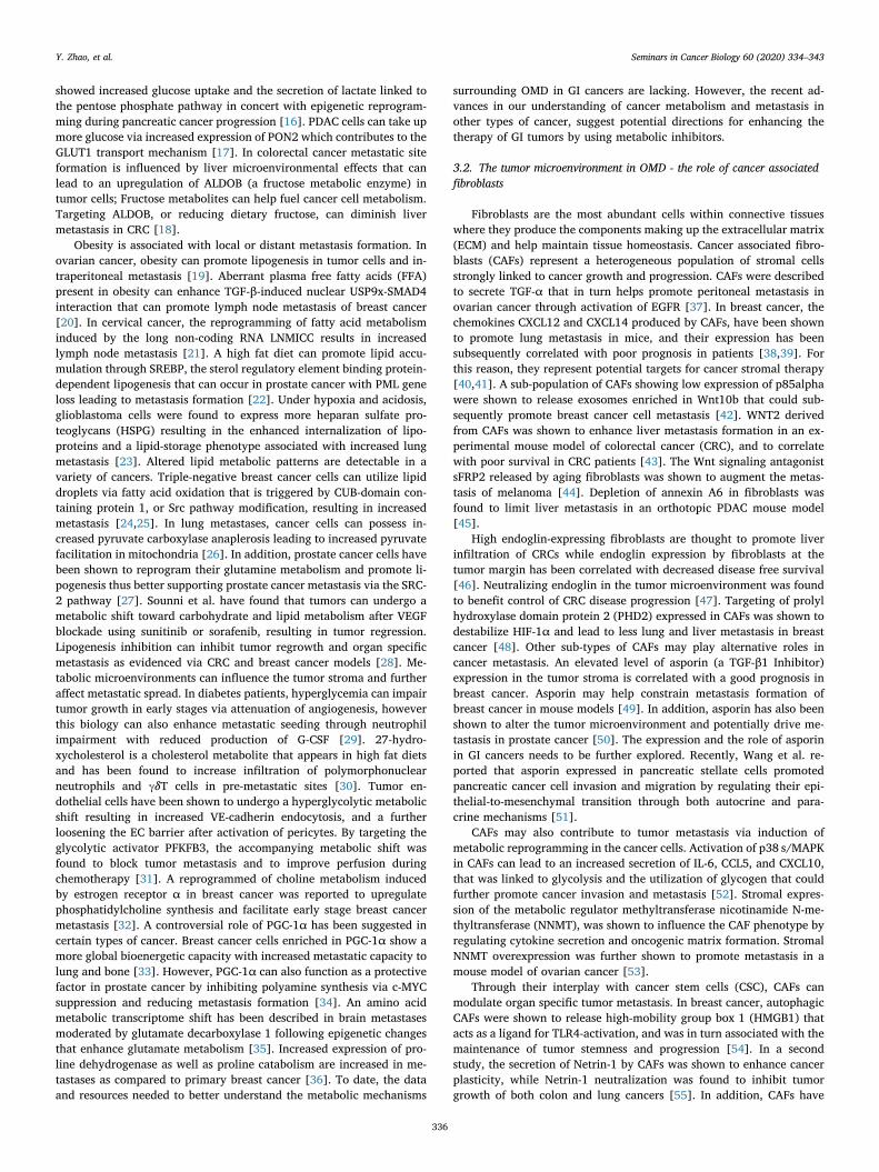
consideration the substantial variability of potential clinical outcomes,and thus may exclude a subgroup to patients that could respond fa-vorably to local or multimodal treatment regimens with a dramaticeffect on disease outcome. In some instances of colorectal cancer (CRC),local as well as systemic treatments are currently being employed as apotential strategy for selected cases where single or few metastases areseen (Fig. 1). This treatment is still based largely on clinical experiencerather than on scientific evidence. Importantly, systemic therapies areoften associated with substantial adverse effects and a significant risk ofinducing therapeutic resistance ultimately leading to uncontrollabletumor progression.
The identification of an oligometastatic state as a separate clinicalentity suggests that a subgroup of patients could be effectively cured bya combination of local (surgery, radiotherapy, local ablative treatment)and systemic therapeutic strategies. The treatment success of multi-modal approaches for OMD is dependent on effective diagnosis, clinicaltolerability and optimized therapeutic algorithms. The integration ofemerging genetic, epigenetic and environmental analyses, and the ap-plication of new therapeutic options such as immunotherapy and mo-lecular targeted therapy may impact oligometastatic disease and thusrepresents an important emerging field in cancer research.
In the following sections we will discuss a series of biologicallydistinct mechanisms that can lead either to oligo- or polymetastases ofgastrointestinal cancer [comprising mainly colorectal cancer (CRC),pancreatic ductal adenocarcinoma (PDAC), and esophageal adeno-carcinoma (EAC)]. A better understanding of the pathophysiology ofthese clinically distinct settings can enhance clinical practice by pro-viding a set of therapy decision points based on biological, im-munological and anatomical characteristics. Recent advances detailingthe mechanisms and emerging biomarkers for the metastatic behaviorof GI cancers will be highlighted, as well as new strategies that arebeing applied for the clinical management of OMD.
2. Definition and clinical significance of oligometastases
Weinberg et al. described the biologic characteristics of active in-vasion and metastasis, with the reseeding and colonization of sometumor cells with "metastatic signatures" in specific tissues, as centralhallmarks of cancer [3]. The metastatic capacity of primary tumors can
be defined based in part on the size and volume of the tumor, as well asthe resident organ. The observation of oliogometastasis, and the relatedphenomenon of oligorecurrence (relapsed oligometastatic disease),suggests that local cancer treatments may be largely curative in asubpopulation of patients with metastases/recurrence [1]. The spec-trum model for oligometastases has been described as a "cancer dia-spora in cancer demography". It includes the passive migration from theprimary lesion with mild hypoxia and unlimited nutrients that does notlead to evolutionary clonal pressure, with the homing of the cancer to anew niche. The origin of this cancer is known and only limited me-tastases are observed. The immune system has not addressed the po-tential threat, and reduced inflammation leads to fewer leukocyteswithin the tumor [4,5]. At one side of the spectrum, some cancers re-main a local disease and do not metastasize. At the other extreme, thecancers are widely metastatic. Then there are cancers that exhibit abehavior intermediate between these two states, with clonal evolutionconferring varying degrees of metastatic potential [6]. The intermediatephase is considered the oligometastatic state, where patients develop alimited number of metastases, and the disease does not quickly progressto a widespread distribution of cancer [2]. Niibe et al. further sub-classified OMD into synchronous oligometastasis (a clinical scenario inwhich oligometastatic disease is detected at the time of diagnosis of theprimary tumour [1]) and metachronous oligometastasis (the develop-ment of oligometastatic disease after treatment of the primary tumor).However the time interval for classification of metachronous versussynchronous has not been standardized [1]. A search in Pub-Med:MEDLINE in early 2011 returned 46 titles containing <oligometast*> [7]. Applying the same search terms today yields 1413articles, with 416 reviews. This increased interest in OMD has emergedfollowing advances in early diagnosis of metastatic disease, expandedtechniques in multidisciplinary tumor management, and new directionsin metastatic research. The oligometastatic state can now be seen as animportant therapeutic opportunity that involves collaboration betweensurgical, radiotherapy and oncology perspectives [8]. To date, little isunderstood about the molecular basis of oligometastatic tumors, theirinteractions with the surrounding microenvironment, or the signalingpathways regulating polymetastatic versus oligometastatic spread. Abetter understanding of this biology will help in the development ofmore effective therapeutic strategies for this disease “phenotype’’.
3. Hallmarks of tumor biology of oligometastatic disease
3.1. Cancer metabolism in OMD
It has been suggested that the metastatic cascade of a primary tumoris associated with "metabolic rewiring" where the environment dictatesmetabolic changes in secondary tumors [9]. A sugar diet has shown topromote breast cancer growth and metastasis with increased expressionof 12-lipoxygenase and its metabolite contributing to lung metastasis[10]. A robust oxidation of glucose rather than glutamine in the citricacid cycle is seen in glioblastomas and brain metastases that can becorrelated with the expression of acetyl-CoA synthetase enzyme 2 [11].Similarly, aerobic glycolysis remodeling in breast cancer induces me-thylglyoxal (MG) formation and sustains YAP nuclear localizationpromoting tumor metastasis [12]. The Warburg pathway associatedenzyme PFKFB4 couples glycoses and stimulates Steroid receptorcoactive-3 (SRC-3) that helps promote metastasis formation [13]. Themetabolic reprogramming of cancer cells can be evidenced by thepresence of varied glycolysis or Oxidative phosphorylation (OXPHOS)balance in metastatic sites. Breast cancer cells with a high liver meta-static potential have been found to show increased expression of pyr-uvate dehydrogenase kinase-1 and an increased metastatic potential[14]. Inhibition of the glycolytic enzyme pyruvate kinase M2 can revertthis metabolic switch from OXPHOS to glycosis and subsequently re-duce lung metastasis in breast cancer [15]. In GI cancer studies, apancreatic cancer model that displayed distant metastatic clones
Fig. 1. Common sites of oligometastatic disease in GI cancers. For gastro-intestinal cancers (including esophageal cancer, gastric cancer, liver cancer,pancreatic cancer, colorectal cancer etc.), liver and lung are relative commonorgans with localized spread of metastasis. It might be great benefit to achievethe control of oligometastatic disease. (Thickness of black arrows reflects thegeneral frequencies of primary tumor metastasizes to the indicated distantorgan site.).
Y. Zhao, et al. Seminars in Cancer Biology 60 (2020) 334–343
335
showed increased glucose uptake and the secretion of lactate linked tothe pentose phosphate pathway in concert with epigenetic reprogram-ming during pancreatic cancer progression [16]. PDAC cells can take upmore glucose via increased expression of PON2 which contributes to theGLUT1 transport mechanism [17]. In colorectal cancer metastatic siteformation is influenced by liver microenvironmental effects that canlead to an upregulation of ALDOB (a fructose metabolic enzyme) intumor cells; Fructose metabolites can help fuel cancer cell metabolism.Targeting ALDOB, or reducing dietary fructose, can diminish livermetastasis in CRC [18].
Obesity is associated with local or distant metastasis formation. Inovarian cancer, obesity can promote lipogenesis in tumor cells and in-traperitoneal metastasis [19]. Aberrant plasma free fatty acids (FFA)present in obesity can enhance TGF-β-induced nuclear USP9x-SMAD4interaction that can promote lymph node metastasis of breast cancer[20]. In cervical cancer, the reprogramming of fatty acid metabolisminduced by the long non-coding RNA LNMICC results in increasedlymph node metastasis [21]. A high fat diet can promote lipid accu-mulation through SREBP, the sterol regulatory element binding protein-dependent lipogenesis that can occur in prostate cancer with PML geneloss leading to metastasis formation [22]. Under hypoxia and acidosis,glioblastoma cells were found to express more heparan sulfate pro-teoglycans (HSPG) resulting in the enhanced internalization of lipo-proteins and a lipid-storage phenotype associated with increased lungmetastasis [23]. Altered lipid metabolic patterns are detectable in avariety of cancers. Triple-negative breast cancer cells can utilize lipiddroplets via fatty acid oxidation that is triggered by CUB-domain con-taining protein 1, or Src pathway modification, resulting in increasedmetastasis [24,25]. In lung metastases, cancer cells can possess in-creased pyruvate carboxylase anaplerosis leading to increased pyruvatefacilitation in mitochondria [26]. In addition, prostate cancer cells havebeen shown to reprogram their glutamine metabolism and promote li-pogenesis thus better supporting prostate cancer metastasis via the SRC-2 pathway [27]. Sounni et al. have found that tumors can undergo ametabolic shift toward carbohydrate and lipid metabolism after VEGFblockade using sunitinib or sorafenib, resulting in tumor regression.Lipogenesis inhibition can inhibit tumor regrowth and organ specificmetastasis as evidenced via CRC and breast cancer models [28]. Me-tabolic microenvironments can influence the tumor stroma and furtheraffect metastatic spread. In diabetes patients, hyperglycemia can impairtumor growth in early stages via attenuation of angiogenesis, howeverthis biology can also enhance metastatic seeding through neutrophilimpairment with reduced production of G-CSF [29]. 27-hydro-xycholesterol is a cholesterol metabolite that appears in high fat dietsand has been found to increase infiltration of polymorphonuclearneutrophils and γδT cells in pre-metastatic sites [30]. Tumor en-dothelial cells have been shown to undergo a hyperglycolytic metabolicshift resulting in increased VE-cadherin endocytosis, and a furtherloosening the EC barrier after activation of pericytes. By targeting theglycolytic activator PFKFB3, the accompanying metabolic shift wasfound to block tumor metastasis and to improve perfusion duringchemotherapy [31]. A reprogrammed of choline metabolism inducedby estrogen receptor α in breast cancer was reported to upregulatephosphatidylcholine synthesis and facilitate early stage breast cancermetastasis [32]. A controversial role of PGC-1α has been suggested incertain types of cancer. Breast cancer cells enriched in PGC-1α show amore global bioenergetic capacity with increased metastatic capacity tolung and bone [33]. However, PGC-1α can also function as a protectivefactor in prostate cancer by inhibiting polyamine synthesis via c-MYCsuppression and reducing metastasis formation [34]. An amino acidmetabolic transcriptome shift has been described in brain metastasesmoderated by glutamate decarboxylase 1 following epigenetic changesthat enhance glutamate metabolism [35]. Increased expression of pro-line dehydrogenase as well as proline catabolism are increased in me-tastases as compared to primary breast cancer [36]. To date, the dataand resources needed to better understand the metabolic mechanisms
surrounding OMD in GI cancers are lacking. However, the recent ad-vances in our understanding of cancer metabolism and metastasis inother types of cancer, suggest potential directions for enhancing thetherapy of GI tumors by using metabolic inhibitors.
3.2. The tumor microenvironment in OMD - the role of cancer associatedfibroblasts
Fibroblasts are the most abundant cells within connective tissueswhere they produce the components making up the extracellular matrix(ECM) and help maintain tissue homeostasis. Cancer associated fibro-blasts (CAFs) represent a heterogeneous population of stromal cellsstrongly linked to cancer growth and progression. CAFs were describedto secrete TGF-α that in turn helps promote peritoneal metastasis inovarian cancer through activation of EGFR [37]. In breast cancer, thechemokines CXCL12 and CXCL14 produced by CAFs, have been shownto promote lung metastasis in mice, and their expression has beensubsequently correlated with poor prognosis in patients [38,39]. Forthis reason, they represent potential targets for cancer stromal therapy[40,41]. A sub-population of CAFs showing low expression of p85alphawere shown to release exosomes enriched in Wnt10b that could sub-sequently promote breast cancer cell metastasis [42]. WNT2 derivedfrom CAFs was shown to enhance liver metastasis formation in an ex-perimental mouse model of colorectal cancer (CRC), and to correlatewith poor survival in CRC patients [43]. The Wnt signaling antagonistsFRP2 released by aging fibroblasts was shown to augment the metas-tasis of melanoma [44]. Depletion of annexin A6 in fibroblasts wasfound to limit liver metastasis in an orthotopic PDAC mouse model[45].
High endoglin-expressing fibroblasts are thought to promote liverinfiltration of CRCs while endoglin expression by fibroblasts at thetumor margin has been correlated with decreased disease free survival[46]. Neutralizing endoglin in the tumor microenvironment was foundto benefit control of CRC disease progression [47]. Targeting of prolylhydroxylase domain protein 2 (PHD2) expressed in CAFs was shown todestabilize HIF-1α and lead to less lung and liver metastasis in breastcancer [48]. Other sub-types of CAFs may play alternative roles incancer metastasis. An elevated level of asporin (a TGF-β1 Inhibitor)expression in the tumor stroma is correlated with a good prognosis inbreast cancer. Asporin may help constrain metastasis formation ofbreast cancer in mouse models [49]. In addition, asporin has also beenshown to alter the tumor microenvironment and potentially drive me-tastasis in prostate cancer [50]. The expression and the role of asporinin GI cancers needs to be further explored. Recently, Wang et al. re-ported that asporin expressed in pancreatic stellate cells promotedpancreatic cancer cell invasion and migration by regulating their epi-thelial-to-mesenchymal transition through both autocrine and para-crine mechanisms [51].
CAFs may also contribute to tumor metastasis via induction ofmetabolic reprogramming in the cancer cells. Activation of p38 s/MAPKin CAFs can lead to an increased secretion of IL-6, CCL5, and CXCL10,that was linked to glycolysis and the utilization of glycogen that couldfurther promote cancer invasion and metastasis [52]. Stromal expres-sion of the metabolic regulator methyltransferase nicotinamide N-me-thyltransferase (NNMT), was shown to influence the CAF phenotype byregulating cytokine secretion and oncogenic matrix formation. StromalNNMT overexpression was further shown to promote metastasis in amouse model of ovarian cancer [53].
Through their interplay with cancer stem cells (CSC), CAFs canmodulate organ specific tumor metastasis. In breast cancer, autophagicCAFs were shown to release high-mobility group box 1 (HMGB1) thatacts as a ligand for TLR4-activation, and was in turn associated with themaintenance of tumor stemness and progression [54]. In a secondstudy, the secretion of Netrin-1 by CAFs was shown to enhance cancerplasticity, while Netrin-1 neutralization was found to inhibit tumorgrowth of both colon and lung cancers [55]. In addition, CAFs have
Y. Zhao, et al. Seminars in Cancer Biology 60 (2020) 334–343
336

been proposed to circulate together with circulating tumor cells (CTCs)to help support cancer metastasis. The presence of CAFs in the per-ipheral blood of breast cancer patients has been linked to metastaticdisease and suggested the potential of using circulating CAFs as apromising biomarker for metastasis [56]. Serum levels of the TGF-β/BMP family member GDF15 (MIC-1) that is secreted by CAFs is thoughtto help stimulate tumor growth at distant sites in prostate cancer [57].CAFs have been reported to recruit integrin α5 high ascitic tumor cellsthat act as "metastatic units" of ovarian cancer that in turn guide peri-toneal invasion and promote transcoelomic metastasis [58].
The interaction between fibroblasts and immune cells can stronglyinfluence cancer progression. CCL19 expression by fibroblasts in thetumor microenvironment of lung carcinoma can enhance the recruit-ment and activation of anti-tumor CD8+T cells and thereby help re-strict cancer progression through an enhanced immune response [59].Similarly, deletion of aSMA+myofibroblasts in pancreatic cancerleads to reduced immune surveillance and increased levels of CD4+Foxp3+ regulatory T cells (Tregs) [60]. Other CAF subtypes wereshown to promote an immunosuppressive microenvironment that canaccelerate cancer progression. Chitinase 3-like 1(Chi3L1) releasing fi-broblasts are highly enriched in the microenvironments surroundingprimary and pulmonary metastases of breast cancer. The deletion ofChi3L1 in fibroblasts was found to attenuate tumor growth and me-tastases, which was associated with an increased filtration of CD8+ andCD4+T cells and decreased macrophage M2 reprogramming [61].These findings highlight the importance of tumor stromal CAFs in themodulation of tumor metastasis. Although direct evidence relating tosimilar phenomenon in GI cancers is currently lacking, the results frombreast cancer, prostate and ovarian cancer studies provide clues as tohow organ-specific metastatic progression may be controlled in gas-trointestinal cancers.
3.3. The tumor microenvironments linked to OMD - the role of exosomes inoligometastasis
Primary tumors can promote pre-metastatic niche formation intarget organs in advance of the metastatic cancer cells [62]. We haveemphasized the importance of preventing the formation of metastaticniche concerning modern strategies targeting metastasis previously[63]. The potential interaction between a primary tumor and distantsites may play an important role in the development of oligometastasisin gastrointestinal cancers. Various cellular and molecular mechanismshave been linked to the formation of the pre-metastatic niche. To thisend, exosomes have been found to act as important mediators of in-tercellular communication [64]. Exosomes are extracellular vesiclesproduced by both tumor cells and CAFs that contain proteins, nucleicacids and lipids that reflect the biology of the parental cell [65]. Costa-Silva et al. found that pancreatic cancer-derived-exosomes induce afibrotic microenvironment and help recruit bone marrow-derivedmacrophages for the development of liver metastasis [66]. Shao et al.reported that exosomes derived from colorectal cancer cells can inducemacrophage polarization and establish an inflammatory pre-metastaticniche in the liver [67]. Interruption of exosome release, or their uptake,may affect the biological behavior of tumors and interfere with for-mation of the pre-metastatic niche. The role of exosomes in pre-meta-static niche formation in oligometastatic gastrointestinal cancers is atpresent unclear. However, several studies have revealed remarkablegenetic and epigenetic heterogeneity between the primary tumors andmetastatic sites linked to exosome biology [68,69]. Kim et al. dis-covered distinct patterns of exosome proteome expression and tyrosinekinase activities in different metastatic sites of pancreatic cancer, in-cluding liver, lung, and peritoneum [70]. Yu et al. compared the pro-teomic profiling of exosomes derived from weakly metastatic murinepancreatic cancer cells (Panc02), and highly metastatic subline-Panc02-H7 cells. They found that highly enriched proteins in thePanc02-H7-derived exosomes could be strongly associated with tumor
growth, invasion, and metastasis [71]. Our group has established ahighly metastatic pancreatic cancer cell line L3.6 pl after several cyclesof in vivo selection. The L3.6 pl cells exhibit a higher incidence of livermetastases than the parental cells [72]. Recently, based on studies usingthis cell line model, we reported an immunosuppressive role of pan-creatic cancer-derived EVs on NK cell dysfunction as it relates to pre-metastatic niche formation of PDAC [73]. A better understanding of thebiology of exosomes will make impact on how the treatment of sys-tematic metastatic disease is addressed in future.
3.4. Molecular features of oligometastatic disease
Patients with oligometastatic disease appear to benefit from loca-lized therapy, however the molecular characteristics that distinguisholigo- from polymetastasis need to be better defined [74,75]. It hasbeen suggested that oligo- or poly-metastasis may either originate fromdifferent clones, or may be part a sequential development with oligo-metastasis representing a transient state in the metastatic process [76].Currently, the selection of oligometastatic patients is largely based onthe number of metastases present, and the length of the disease-freeinterval [8,77]. The transition of the metastatic state from oligo- topolymetastasis has been observed in mouse models [78]. A better un-derstanding of the molecular features, expression signatures or otherhallmarks that help distinguish between oligo- and polymetastasiswould clearly allow a more reliable selection of patients who couldbenefit from the OMD-guided therapy [8,77,79]. While information inthis regard is still quite limited, initial transcriptional expression pro-filing based signatures have been proposed to help distinguish oligo-from polymetastasis. Lussier et al. reported that oligo- and poly-metastasis could be clustered and characterized by prioritized mi-croRNA (miRNA) signatures [77], including analysis of the miRNA-200family, widely reported to be involved in metastasis [80]. In addition,high expression of miR-200c in metastatic tumor was shown to predictprogression towards polymetastasis through regulation of epithelial-mesenchymal transition (EMT)-related pathways. Sun et al. reported asimilar role of miR-200c in the transition from oligo- to polymetastasisthrough the target gene Sec23a, which appears to suppress oligo- topoly-metastatic progression by modifying the tumor microenvironment[81]. Subsequently, Lussier et al. described a larger and more homo-genous group of patients with lung-derived metastases. In this study, aset of miRNAs were identified that were down-regulated in a highprogression (HRP) group, as compared to a low rate of progression(LRP) group [82]. The presence of these miRNAs could be used todistinguish HRP from LRP. Wang et al. identified a panel of 10 miRNAsthat could distinguish the oligo- from polymetastatic lung cancer [83].In patients with oligometastatic liver disease, Fromme et al. reportedthat over-expression of FGFR3 in oligometastatic colorectal cancers wassignificantly associated with shorter overall survival. They suggestedthat FGFR3 overexpression could define a clinical subgroup with pooroutcome and may thus represent a potential therapeutic target [84].
The molecular signatures proposed from the studies described abovesuggest that it should be able to further optimize and better distinguisholigo- from polymetastatic disease in GI cancers. To date, only a fewgenes and miRNAs have been found to overlap between GI tumors andthe signatures detailed above [78,82,83]. It has been proposed thatanalysis of shared pathways, and pathway-based approaches may helpovercome some of the inconsistency among individual gene basedanalysis. A re-analysis of the published miRNA datasets by Uppal et al.identified a series of Kyoto Encyclopedia of Genes and Genomes(KEGG)-derived pathways that are targeted by relevant miRNAs inoligometastasis [85]. Further enrichment of this dataset have identifiedthree distinct functional pathway groups for; adhesion, invasion, andmobility (AIM); intracellular signaling pathways (ICS); and cancer-specific signaling pathways (CSS) group, that could be linked to specificcancer metastatic patterns. Wang et al. also discussed three pathways ofaxon guidance, cancer metastasis, and proteoglycan biology believed to
Y. Zhao, et al. Seminars in Cancer Biology 60 (2020) 334–343
337
contribute to the lung cancer with OMD and PMD phenotypes viamiRNA effects [83].
Pitroda et al. proposed a consensus molecular subtype for limitedCRC liver metastases based on the integrated transcriptional analysis ofboth mRNA and miRNA. Three molecular subtypes were identified: 1.)canonical subtype, 2.) immune subtype and 3.) a stromal subtype [79].Notably, the immune subtype 2-related metastases showed significantlyover-expressed innate and adaptive immune genes as compared to thesubtype 1 and subtype 3-type metastases, showed a robust immuneinfiltration in the original metastatic lesions. The patients with subtype2 metastases showed a lower recurrence rate, and longer survival afterhepatic resection of their metastatic lesions as compared to subtype 1 orsubtype 3 metastases. By contrast, the poor-survival subtype 1 and 3metastases showed an enrichment of expression patterns associatedwith stromal infiltration, presence of EMT, extracellular matrix re-modeling, and angiogenesis. Integration of the resultant molecularsubtypes with clinical risk stratification yielded three prognostic riskgroups: low-risk, intermediate-risk, and high-risk. The integrated low-risk group showed significantly longer distant metastasis-free survivaland overall survival, and was largely represented the oligometastaticphenotype. By targeting the subset of patients with predicted oligo-metastatic phenotype, the molecular signatures alone, or in combina-tion with clinical risk score, may provide a rational strategy for theestablishment of criteria for enhancing curative local therapies.
3.5. Tumor genomics and oligometastasis
To date, there are relatively few studies that have exclusively ex-plored the potential gene mutations found between primary GI cancersand their metastatic disease. Zehir et al. compared data from advancedor metastatic cancers to primary tumors from TCGA [86]. The resultsfrom a metastatic cancer study were consistent with the TCGA findings.They identified major differences between the two cohorts. First, TP53mutations were more frequent in metastatic gastric cancer, second;TP53 mutations were also found to be present in primary gastric cancer,and finally; the PIK3CA mutation was less frequent in metastatic gastriccancer than in primary cancer. In support of this observation, Ikari et al.also found that the TP53 mutation rate was significantly higher ingastric cancer patients with liver metastasis, as compared to thosewithout metastasis [87]. In addition, the status of TP53 mutations inthe liver metastatic group was linked to lymph node stage and venousinvasion. Pectasides et al. found that PIK3CA mutations and amplifi-cation of EGFR, ERBB2, CKD4/6 and MET were frequently found to bedifferent between primary gastric cancers and their metastatic lesions.Inconsistent gene mutations between the primary and matched meta-static gastric cancer tumors was found to occur in 45% patents [88].Zhang et al. identified mutations in GPI, JAK3, PRSS8 and IDH3G thatwere more common in gastric cancer patients without peritoneal me-tastasis, than in peritoneal metastatic patients. Four mutations(PRDM1, c.950 G > A; XPC, c.1315 G > C; CD68, c.554A > C;ACVR1B, c.1345C > A) were only seen in peritoneal metastasis pa-tients [89]. In one gastric adenocarcinoma with peritoneal metastasis,23 somatic mutations were found in the primary tumor, while 12 so-matic variants were associated with metastatic cancer . Four somaticvariations (RP1L1, PRB1, HS6ST3 and DCTN1) were found to si-multaneously occur in both primary and peritoneal metastatic cancer.Genomic profiling has been used for predicting lymph node metastasisin patients with gastric cancer, which may prove useful for the identi-fication of patients for extended lymph node resection [90,91].
Intratumoral heterogeneity (ITH) is an important topic in cancergenomics, suggesting that a single tumor consists of different cell sub-populations [92]. APC, TP53, and KRAS mutations frequently occur inheterogeneous colorectal cancers which could be used to predict a highpotential for liver metastasis [93]. Gene mutation and methylationevents are observed in different disease stages of colorectal cancers.Compared to the corresponding lymph node metastases, KRAS
mutations and p16INK4a methylation were found to be significantlylower in stage III CRCs, while in stage IV CRC KRAS, CDH1, andp16INK4a mutations were decreased, but did not reach significance. Acomparison between stage IV primary tumors and their correspondingliver metastases showed an increase in RASSF1a methylation and adecrease in p16INK4a methylation. In lymph node metastases fromstage III and IV CRCs, KRAS, BRAF and p53 mutations tend to increase.KRAS and p16IN4a mutations, as well as RASSF1a methylation werefound to be higher in liver metastases than in lymph node metastasesoriginating from stage IV CRC [94]. Comprehensive analysis of tumorgenomics should provide unique signatures that could be used to clas-sify OMD with more biological and genetic relevance.
3.6. Other biomarkers associated with the characterization ofoligometastases
3.6.1. Alteration of ctDNAs in oligo- and polymetastasisCirculating tumor DNA (ctDNA), the double-stranded DNA frag-
ments released from tumor cells into the circulation during apoptosis orthe necrotic process, represents an important target for current appli-cations of liquid biopsy. Although the half-life of ctDNA is rather short,ctDNA levels and the degree of tumor burden remain consistent.CtDNAs also carry important genomic information regarding the tumorincluding copy number variation (CNV), genomic integrity, mutationburden, and gene methylation status. The genomic information fromctDNA may be seen to accurately reflect dynamic changes that occur intumors with the context of metastatic tumor progression. The overallanalysis of ctDNA may thus help to assess the genetic alterations oftumor burden and the presence of tumor heterogeneity that could be ofbenefit for precise cancer treatment [95].
Mutant allele frequencies (MAFs) of many cancer hot spot mutationgenes (TP53, RET, FGFR3, and APC) have been shown to increase sig-nificantly in patients with metastasis as compared to patients withsingle malignant lesion [96]. The detection of KRAS mutations inctDNA samples has shown potential application in pancreatic canceroligometastasis. Bernard et al. used ctDNA analysis of pancreatic ductaladenocarcinoma (PDAC) patients to show that patients from the me-tastatic group carry higher levels of KRAS MAF than do patients withlocalized disease. KRAS MAF levels as detected by ctDNA analysisshowed an association with poor progression-free survival (PFS) andoverall survival (OS) based on univariate analysis [97]. In a study ofmetastatic colorectal cancer (mCRC), analysis of ctDNA pattern frompatients with high tumor burden (> 1 metastasis) showed that RAS,BRAF, and ERBB2 alterations were detected in 84.6% cases, which wasconcordant with direct analysis of the tumor tissue [98]. A furtheranalysis of ctDNA for presence of the gene RAS mutation has shown ahigh level of sensitivity (93.3%) and specificity (100%). Additionalchanges in ctDNA levels provided predictive information much earlierthan changes of CEA and CA19-9 levels could be detected [99].
Alternations in methylation patterns in specific genes can be as-sessed by using ctDNA analysis that provide important information formetastasis associated studies. Methylation of the MT1M and MT1Gpromoters was found at a higher incidence in patients with lymph nodemetastasis or extrahepatic metastasis [100]. With identification of alink between DNA epigenetic modifications and cancer progression, thedetection of 5-Hydroxymethylcytosine (5hmC) an important DNAmodification factor, can be measured via ctDNA analysis for clinicalrelevance. Loss of 5hmc in tumor DNA has been observed with stage-dependent variations in lung cancer as they progress from early stage tolate stage disease with metastasis. In this study, the number of 5hmC-enriched regions (5hMR) was found to be decreased in lung cancerpatients with metastasis as compared to non-metastatic patients, orhealthy individuals [101].
The limitations of ctDNA profiling are also important. Strickler,et al. found that in nearly 15% of metastatic colorectal cancer casesgenomic alterations in ctDNA could not be detected, presumably
Y. Zhao, et al. Seminars in Cancer Biology 60 (2020) 334–343
338

because of the low tumor burden leading to reduced levels of ctDNA[102]. Based on the trend of ctDNA application in various tumors types,ctDNA analysis could represent an important tool for distinguishingoligo- from polymetastasis following more detailed study.
3.6.2. Aneuploid CTCs in oligo- and polymetastasisCirculating tumor cells (CTCs) are carcinoma cells shed from pri-
mary or metastatic solid tumors into the peripheral blood, whereascirculating endothelial cells (CECs) are derived from endothelial cells(ECs) which make up the blood vessels. Clinical relevance of CTCs inthe context of cancer metastases/prognosis [103,104], and CECs withrespect to assessing tumor angiogenesis, have been well detailed else-where [105]. CTC is now accepted as a breast cancer biomarker by theAmerican Society of Clinical Oncology (ASCO) [106]. In the pastdecade, the detection of CTCs has been used to evaluate cancer patientprognosis [107], therapeutic efficacy [108,109], and to monitor post-surgical cancer relapse [110,111], as well as drug resistance in carci-noma patients [112,113] as well as in metastatic “patient-derived xe-nograft tumor mouse models” (mPDX) [114]. Aneuploidy (ap) is ahallmark of malignant cells [115,116]. The existence of aneuploid CTCs(apCTCs) can be detected in the peripheral circulation [117]. Recently,a comprehensive breast cancer study demonstrated that EpCAM+
apCTCs and aneuploid disseminated tumor cells (apDTCs) in bonemarrow are able to guide oligometastasis to lung in breast carcinomapatients [118].
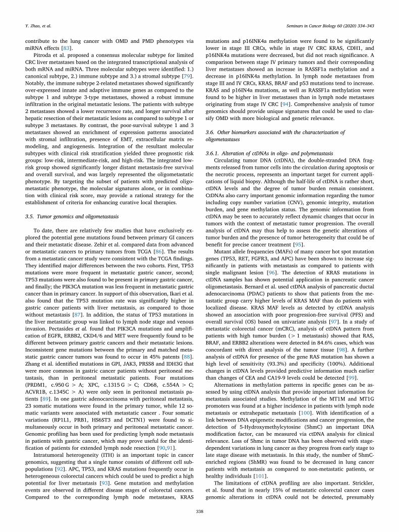
In addition to aneuploid carcinoma cells, some aneuploid tumorendothelial cells (TECs) in neoplastic tissues [119] express inhibitorymolecules such as PD-L1, that can attenuate the anti-cancer immuneresponse mediated by CD8+ T lymphocytes [120]. A recent studycombining anti-angiogeneic agents and anti-PD-1 checkpoint blockadeimmunotherapy demonstrated a durable synergetic clinical response tomalignant cancer cells [121]. The significance of TEC analysis suggeststhat analysis of circulating tumor endothelial cells (CTECs) may alsoshow utility in clinical utilities [122,123]. Recent studies using SE-iFISH analysis [124] of specimens from various carcinoma patientsindicated that in addition to PD-L1, several other tumor biomarkers areexpressed by aneuploid CTECs (Fig. 2). The analysis of apCTECs couldprovide additional information regarding potential invasive cancer
growth, metastasis and disease progression [122,123].Recent results suggest that as a counterpart to ‘nucleotide circu-
lating tumor biomarker’ ctDNA data, non-hematologic aneuploid CTCs,and CTECs may constitute important targets as ‘cellular circulatingtumor biomarkers’ [125]. apCTCs and apCTECs possess distinct clinicalsignificance and will provide insight for the evaluation of cancer me-tastases and therapeutic efficacy in carcinoma patients.
4. Multidisciplinary management of oligometastatic disease
Metastasis represents the main cause of treatment failure in tumormanagement. The concept of oligometastasis revealed an importantpotential disease process and therapeutic state that has changed ourunderstanding of advanced malignant tumors and the choice of treat-ments [2,126]. There is still controversy regarding the definition ofoligometastatic disease. Most clinical trial protocols and clinicians ac-cept a definition of 1–3 or 1–5 metastatic lesions [8,127,128]. At thisrelatively early stage in cancer progression, the bio-invasiveness of thetumor is still relatively low reflected by a limited number of organsinvolved with few metastatic numbers [129,130]. As discussed, thebetter detection of oligometastatic patients can provide an importanttherapeutic window.
While cancer patients with oligometastasis can have a relativelygood prognosis [131,132], the diagnosis of oligometastasis can be dif-ficult in older patients where efficient diagnosis can be complicated bythe presence of other diseases. A multidisciplinary team (MDT) ap-proach can help integrate diverse sets of information regarding thephysical conditions of the patients, pathological features of tumors,imaging, staging, etc. to provide optimal diagnosis and treatment forpatients with OMD [4,133]. As at present, there is no specific set ofuniform standards for MDT diagnosis of GI cancer OMD, a team ap-proach can help with identification and management of the disease. Thepresent data and guidelines support systemic therapy, or a multi-dis-ciplinary, multi-therapeutic approach involving surgery as the maintreatment option [127,134,135]. Surgery, radiotherapy, chemotherapy,targeted therapy and immunotherapy etc., are central to effective MDT[129,136].
Fig. 2. In situ phenotypic and karyotypiccharacterization of aneuploid CTECs ex-pressing tumor biomarkers. Following sub-traction enrichment (SE) of non-hematologiccirculating rare cells from variety of carcinomapatients, specimens are subjected to compre-hensive characterization performed by im-munofluorescence staining-FISH (iFISH)strategy. Several tumor biomarkers, includingthe stemness marker CD44v6, EpCAM, HER2and PD-L1, are respectively expressed on theaneuploid CTECs (CD31+/ CD45−, ≥trisomy8).
Y. Zhao, et al. Seminars in Cancer Biology 60 (2020) 334–343
339

4.1. Surgical treatment of OMD
Surgery is an important treatment option for GI cancers with OMD.Liver is a common site of solid tumor metastasis, especially metastasisfrom colorectal cancer. The resection of liver metastases from colorectalcancer has improved its prognosis. The median survival time of un-treated liver metastases was previously only 6.9 months, and the 5-yearsurvival rate of patients who could not be surgically treated was lessthan 5%. The median survival time of patients with completely resectedliver metastases or no evidence of metastatic disease is now around 35months, and the 5-year survival rate can reach 30–57% [137–139].Complete surgical resection of liver metastases is still probably the bestoption to cure GI cancer liver metastases [127,140–142]. A retro-spective analysis by Sinn et al. has found that patients with advancedpancreatic cancer (locally advanced or locally resectable with hepaticoligometastasis) can benefit from pancreatoduodenectomy followed bygemcitabine-based chemotherapy [143].
4.2. Non-surgical treatment of OMD
For the treatment of colorectal cancer liver metastases, theEuropean 2016 ESMO mCRC consensus has also highlighted the im-portance of surgical resection, but has also recommended other non-surgical treatments, including ablation procedures (RF, microwave,cryoablation, etc.), stereotactic body radiotherapy (SBRT), and selectiveinternal radiation therapy (SIRT). All locally destructive treatments areclassified as the “toolbox of local ablative treatment” (Fig. 3), which canbe combined with surgery to expand therapeutic efficacy towardsachieving no evidence of disease (NED) [127].
Retrospective studies from North America, Europe and Asia showthat the application of SBRT for the treatment of oligometastatic diseasecan reach or exceed 90% of local tumor control [129,144,145]. Phase 3studies are currently underway and show excellent preliminary results.Ongoing trials in the USA (eg, NCT02759783, NCT02089100,NCT02364557, NC01446744, NCT02893332 and NCT02417662), inconjunction with international studies should help inform as to whichOMD patients will benefit the most from local intervention [135,146].
Although many short-term studies have suggested that SBRT is an ap-propriate strategy for OMD patients, the optimal dose and segmentationmethod for SBRT is still not well defined [126,147]. More prospectivestudies are clearly needed to help clarify the overall value and appli-cation of SBRT in the treatment of OMD in GI cancers.
In addition, the emerging breakthrough therapy approach of im-mune modulation has been assessed in the context of SBRT. The po-tential immunomodulatory effects of SBRT when used in conjunctionwith checkpoint inhibitors represents an important step in the clinicalcontrol of OMD [148,149]. Work to date suggests that SBRT can beexploited to benefit the immune response to cancer [4,150]. “ISABR”(Immunotherapy and stereotactic ablative radiotherapy) represents acombination of the two techniques - immunotherapy and SABR forcancer therapies [151]. These new therapeutic strategies combiningradiotherapy with immunotherapy have already yielded significant ef-fects in a variety of tumor models [152]. Early clinical trials haveshown that this combined therapeutic approach can achieve promisingresults for metastatic solid tumors, especially in metastatic kidney andlung cancer patients [153]. Results of a series of ongoing clinical trials(eg, NCT02444741, NCT02298946, NCT02239900, NC01497808,NCT01769222 and NCT01401062) should help establish better clinicaladministration protocols for immunotherapy and radiotherapy ap-proaches.
Radiofrequency ablation (RFA) technology is increasingly beingused in the field of OMD management [77,154]. Radiofrequency ab-lation has its own unique advantages as a local treatment technique.RFA shows less trauma to patients and can be combined with surgery,or applied in postoperative recurrence, even in multiple relapses aftertreatment [152,155]. The combination RFA for liver metastases withimmune checkpoint inhibitor therapies may help enhance antitumorimmunity in a manner that mirrors that seen with ISABR [156]. Localchemotherapy including hepatic arterial infusion (HAI) therapy, isoften used to treat hepatic metastases from CRC in both the resectableand non-resectable settings [157]. A prospective, multicenter, rando-mized, phase III trial of the RENAISSANCE (AIO-FLOT5) initiated bythe AIO/CAO-V/CAOGI group in Germany was registered in October2015 (NCT02578368). The aim of this trial is to investigate the
Fig. 3. Multidiscipinary management of OMD in GI cancers. Multidisciplinary mangement of OMD mainly involves surgical treatment, ablative treatments,chemotherapy, immunotherapy and targeted treatment. Appropriate treatment should be selected according to the patient‘s specific conditions, either alone or incombination, requiring careful MDT assessment. OMD, oligometastatic disease; RT, radiation therapy; SIRT, selective internal radiation therapy; TACE, transarterialchemoembolization.
Y. Zhao, et al. Seminars in Cancer Biology 60 (2020) 334–343
340

therapeutic effect of chemotherapy alone vs. chemotherapy followed bysurgical resection on survival and quality of life in patients with lim-ited-metastatic gastric and gastroesophageal junction cancer [158]. Theresults of this study should be forthcoming.
Surgical options should be actively considered for the treatment ofOMD. Local ablative treatment can be helpful as adjuvant therapies.Their use as a stand-alone approach can reduce their therapeutic sig-nificance. However, non-surgical treatment may still be an appropriateapproach for patients with limited surgical options, or for patientsunable or unwilling to undergo surgery due to complications. Althoughwe are at present limited by a lack of large prospective randomizedcontrolled trials, retrospective analysis has demonstrated that localtreatment can substantially improve the prognosis of patients witholigometastatic disease from different GI cancer entities. To helpidentify the best option forward for complex clinical situations such asthe effective treatment of patients in old age with confounding issues,an interdisciplinary approach using the expertise of surgeons, phy-siotherapists, social workers, psychologists and geriatricians will beexpected to improve outcome effectively [159].
5. Conclusion
In this review, we have summarized recent advances and estab-lished knowledge regarding tumor biology and multidisciplinary ther-apeutic strategies of OMD especially in GI Cancers. Further knowledgewith respect to cancer metabolism, tumor microenvironment, mole-cular signaling pathways and tumor genomics will help to better un-derstand the process of OMD development, and will facilitate to dis-tinguish OMD in GI cancer from polymetastatic disease. Since amajority of all GI cancers progress to a metastatic state, individualizedtherapy according to the underlying metastases should ideally be per-formed before therapy initiation. Therefore, treatment of metastatic GIcancer should be based on the molecular and cellular traits of thecancer cells in context with alterations of their local environment in thedirection of precision medicine differentiating between oligo- andpolymetastatic disease in GI cancer. Except for surgery, new targetedand individualized OMD treatment methods have emerged, includingvarious radiotherapy, immunotherapy combined with radiotherapy, etc.Some therapeutic strategies have achieved initial success, but need tobe expanded and validated in diverse settings.
Finally, it is important to note that current OMD diagnosis andtreatment still face various challenges in the clinic including a lack ofspecific and unified standards for clinical practice. The most criticalissue remains how to diagnose patients with OMD in GI cancer.However, with increasing knowledge of the underlying biology, it isexpected that clinicians and researchers will pay more attention to thediversity of metastatic cancer diseases and will be able to developcomprehensive, tailored treatment strategies to maximize the ther-apeutic benefits and improve the quality of life for patients with me-tastatic GI cancer.
Funding source
This study was supported by European Fund for RegionalDevelopment (EFRE) for the project “Autonomy in old age - potentialbiomarker for individualization of cancer therapy of elderly patients”and Köln Fortune Program/Faculty of Medicine, University of Cologne.
Disclosures
The authors declare that they have no competing financial interests.
Acknowledgements
This study was supported by European Fund for RegionalDevelopment (EFRE) for the project “Autonomy in old age - potential
biomarker for individualization of cancer therapy of elderly patients”and Köln Fortune Program/Faculty of Medicine, University of Cologne.
References
[1] Y. Niibe, K. Hayakawa, Oligometastases and oligo-recurrence: the new era ofcancer therapy, Jpn. J. Clin. Oncol. 40 (2) (2010) 107–111.
[2] S. Hellman, R.R. Weichselbaum, Oligometastases, J. Clin. Oncol. 13 (1) (1995)8–10.
[3] D. Hanahan, R.A. Weinberg, Hallmarks of cancer: the next generation, Cell 144 (5)(2011) 646–674.
[4] D.K. Reyes, K.J. Pienta, The biology and treatment of oligometastatic cancer,Oncotarget 6 (11) (2015) 8491–8524.
[5] K.J. Pienta, et al., The cancer diaspora: metastasis beyond the seed and soil hy-pothesis, Clin. Cancer Res. 19 (21) (2013) 5849–5855.
[6] S. Hellman, Karnofsky Memorial Lecture. Natural history of small breast cancers,J. Clin. Oncol. 12 (10) (1994) 2229–2234.
[7] T. Treasure, Oligometastatic cancer: an entity, a useful concept, or a therapeuticopportunity? J. R. Soc. Med. 105 (6) (2012) 242–246.
[8] D.A. Palma, et al., The oligometastatic state - separating truth from wishfulthinking, Nat. Rev. Clin. Oncol. 11 (9) (2014) 549–557.
[9] I. Elia, G. Doglioni, S.M. Fendt, Metabolic hallmarks of metastasis formation,Trends Cell Biol. 28 (8) (2018) 673–684.
[10] Y. Jiang, et al., A sucrose-enriched diet promotes tumorigenesis in mammarygland in part through the 12-Lipoxygenase pathway, Cancer Res. 76 (1) (2016)24–29.
[11] T. Mashimo, et al., Acetate is a bioenergetic substrate for human glioblastoma andbrain metastases, Cell 159 (7) (2014) 1603–1614.
[12] M.J. Nokin, et al., Methylglyoxal, a glycolysis side-product, induces Hsp90 gly-cation and YAP-mediated tumor growth and metastasis, Elife 5 (2016).
[13] S. Dasgupta, et al., Metabolic enzyme PFKFB4 activates transcriptional coactivatorSRC-3 to drive breast cancer, Nature 556 (7700) (2018) 249–254.
[14] F. Dupuy, et al., PDK1-dependent metabolic reprogramming dictates metastaticpotential in breast Cancer, Cell Metab. 22 (4) (2015) 577–589.
[15] F. Liu, et al., PKM2 methylation by CARM1 activates aerobic glycolysis to promotetumorigenesis, Nat. Cell Biol. 19 (11) (2017) 1358–1370.
[16] O.G. McDonald, et al., Epigenomic reprogramming during pancreatic cancerprogression links anabolic glucose metabolism to distant metastasis, Nat. Genet.49 (3) (2017) 367–376.
[17] A. Nagarajan, et al., Paraoxonase 2 facilitates pancreatic Cancer growth and me-tastasis by stimulating GLUT1-Mediated glucose transport, Mol. Cell 67 (4) (2017)685–701 e6.
[18] P. Bu, et al., Aldolase B-Mediated fructose metabolism drives metabolic repro-gramming of Colon Cancer liver metastasis, Cell Metab. 27 (6) (2018)1249–1262 e4.
[19] Y. Liu, et al., Obesity contributes to ovarian Cancer Metastatic success throughincreased lipogenesis, enhanced vascularity, and decreased infiltration of M1macrophages, Cancer Res. 75 (23) (2015) 5046–5057.
[20] Y. Wu, et al., Aberrant phosphorylation of SMAD4 Thr277-Mediated USP9x-SMAD4 interaction by free fatty acids promotes breast Cancer metastasis, CancerRes. 77 (6) (2017) 1383–1394.
[21] C. Shang, et al., LNMICC promotes nodal metastasis of cervical Cancer by repro-gramming fatty acid metabolism, Cancer Res. 78 (4) (2018) 877–890.
[22] M. Chen, et al., An aberrant SREBP-dependent lipogenic program promotes me-tastatic prostate cancer, Nat. Genet. 50 (2) (2018) 206–218.
[23] J.A. Menard, et al., Metastasis stimulation by hypoxia and acidosis-induced ex-tracellular lipid uptake is mediated by proteoglycan-dependent endocytosis,Cancer Res. 76 (16) (2016) 4828–4840.
[24] H.J. Wright, et al., CDCP1 drives triple-negative breast cancer metastasis throughreduction of lipid-droplet abundance and stimulation of fatty acid oxidation, Proc.Natl. Acad. Sci. U. S. A. 114 (32) (2017) E6556–E6565.
[25] J.H. Park, et al., Fatty acid oxidation-driven src links mitochondrial energy re-programming and oncogenic properties in triple-negative breast Cancer, Cell Rep.14 (9) (2016) 2154–2165.
[26] S. Christen, et al., Breast cancer-derived lung metastases show increased pyruvatecarboxylase-dependent anaplerosis, Cell Rep. 17 (3) (2016) 837–848.
[27] S. Dasgupta, et al., Coactivator SRC-2-dependent metabolic reprogrammingmediates prostate cancer survival and metastasis, J. Clin. Invest. 125 (3) (2015)1174–1188.
[28] N.E. Sounni, et al., Blocking lipid synthesis overcomes tumor regrowth and me-tastasis after antiangiogenic therapy withdrawal, Cell Metab. 20 (2) (2014)280–294.
[29] T. Fainsod-Levi, et al., Hyperglycemia impairs neutrophil mobilization leading toenhanced metastatic seeding, Cell Rep. 21 (9) (2017) 2384–2392.
[30] A.E. Baek, et al., The cholesterol metabolite 27 hydroxycholesterol facilitatesbreast cancer metastasis through its actions on immune cells, Nat. Commun. 8 (1)(2017) 864.
[31] A.R. Cantelmo, et al., Inhibition of the glycolytic activator PFKFB3 in endotheliuminduces tumor vessel normalization, impairs metastasis, and improves che-motherapy, Cancer Cell 30 (6) (2016) 968–985.
[32] M. Jia, et al., Estrogen receptor alpha promotes breast Cancer by reprogrammingcholine metabolism, Cancer Res. 76 (19) (2016) 5634–5646.
[33] S. Andrzejewski, et al., PGC-1alpha promotes breast Cancer metastasis and confersbioenergetic flexibility against metabolic drugs, Cell Metab. 26 (5) (2017)
Y. Zhao, et al. Seminars in Cancer Biology 60 (2020) 334–343
341

778–787 e5.[34] L. Kaminski, et al., PGC-1alpha inhibits polyamine synthesis to suppress prostate
cancer aggressiveness, Cancer Res. (2019).[35] P.M. Schnepp, et al., GAD1 upregulation programs aggressive features of Cancer
cell metabolism in the brain metastatic microenvironment, Cancer Res. 77 (11)(2017) 2844–2856.
[36] I. Elia, et al., Proline metabolism supports metastasis formation and could be in-hibited to selectively target metastasizing cancer cells, Nat. Commun. 8 (2017)15267.
[37] T.S. Lau, et al., A loop of cancer-stroma-cancer interaction promotes peritonealmetastasis of ovarian cancer via TNFalpha-TGFalpha-EGFR, Oncogene 36 (25)(2017) 3576–3587.
[38] E. Sjoberg, et al., A novel ACKR2-Dependent role of fibroblast-derived CXCL14 inEpithelial-to-Mesenchymal transition and metastasis of breast Cancer, Clin. CancerRes. 25 (12) (2019) 3702–3717.
[39] D.K. Ahirwar, et al., Fibroblast-derived CXCL12 promotes breast cancer metastasisby facilitating tumor cell intravasation, Oncogene 37 (32) (2018) 4428–4442.
[40] Q. Zhang, et al., A novel autocrine CXCL14/ACKR2 Axis: the achilles' heel ofCancer metastasis? Clin. Cancer Res. 25 (12) (2019) 3476–3478.
[41] M. Augsten, et al., Cancer-associated fibroblasts expressing CXCL14 rely uponNOS1-derived nitric oxide signaling for their tumor-supporting properties, CancerRes. 74 (11) (2014) 2999–3010.
[42] Y. Chen, et al., Aberrant low expression of p85alpha in stromal fibroblasts pro-motes breast cancer cell metastasis through exosome-mediated paracrine Wnt10b,Oncogene 36 (33) (2017) 4692–4705.
[43] N. Kramer, et al., Autocrine WNT2 signaling in fibroblasts promotes colorectalcancer progression, Oncogene 36 (39) (2017) 5460–5472.
[44] A. Kaur, et al., sFRP2 in the aged microenvironment drives melanoma metastasisand therapy resistance, Nature 532 (7598) (2016) 250–254.
[45] J. Leca, et al., Cancer-associated fibroblast-derived annexin A6+ extracellularvesicles support pancreatic cancer aggressiveness, J. Clin. Invest. 126 (11) (2016)4140–4156.
[46] M. Paauwe, et al., Endoglin expression on cancer-associated fibroblasts regulatesinvasion and stimulates colorectal Cancer metastasis, Clin. Cancer Res. 24 (24)(2018) 6331–6344.
[47] L.M. Becker, V.S. LeBleu, Endoglin Targeting in Colorectal TumorMicroenvironment, Clin. Cancer Res. 24 (24) (2018) 6110–6111.
[48] C.D. Madsen, et al., Hypoxia and loss of PHD2 inactivate stromal fibroblasts todecrease tumour stiffness and metastasis, EMBO Rep. 16 (10) (2015) 1394–1408.
[49] P. Maris, et al., Asporin is a fibroblast-derived TGF-beta1 inhibitor and a tumorsuppressor associated with good prognosis in breast Cancer, PLoS Med. 12 (9)(2015) e1001871.
[50] R.M. Hughes, et al., Asporin restricts mesenchymal stromal cell differentiation,alters the tumor microenvironment, and drives metastatic progression, Cancer Res.(2019).
[51] L. Wang, et al., Asporin promotes pancreatic cancer cell invasion and migration byregulating the epithelial-to-mesenchymal transition (EMT) through both autocrineand paracrine mechanisms, Cancer Lett. 398 (2017) 24–36.
[52] M. Curtis, et al., Fibroblasts mobilize tumor cell glycogen to promote proliferationand metastasis, Cell Metab. 29 (1) (2019) 141–155 e9.
[53] M.A. Eckert, et al., Proteomics reveals NNMT as a master metabolic regulator ofcancer-associated fibroblasts, Nature 569 (7758) (2019) 723–728.
[54] X.L. Zhao, et al., High-mobility group box 1 released by autophagic cancer-asso-ciated fibroblasts maintains the stemness of luminal breast cancer cells, J. Pathol.243 (3) (2017) 376–389.
[55] P.J. Sung, et al., Cancer-associated fibroblasts produce Netrin-1 to control cancercell plasticity, Cancer Res. (2019).
[56] Z. Ao, et al., Identification of cancer-associated fibroblasts in circulating bloodfrom patients with metastatic breast Cancer, Cancer Res. 75 (22) (2015)4681–4687.
[57] F. Bruzzese, et al., Local and systemic protumorigenic effects of cancer-associatedfibroblast-derived GDF15, Cancer Res. 74 (13) (2014) 3408–3417.
[58] Q. Gao, et al., Heterotypic CAF-tumor spheroids promote early peritoneal metas-tatis of ovarian cancer, J. Exp. Med. 216 (3) (2019) 688–703.
[59] H.W. Cheng, et al., CCL19-producing fibroblastic stromal cells restrain lung car-cinoma growth by promoting local antitumor T-cell responses, J. Allergy Clin.Immunol. 142 (4) (2018) 1257–1271 e4.
[60] B.C. Ozdemir, et al., Depletion of carcinoma-associated fibroblasts and fibrosisinduces immunosuppression and accelerates pancreas cancer with reduced sur-vival, Cancer Cell 25 (6) (2014) 719–734.
[61] N. Cohen, et al., Fibroblasts drive an immunosuppressive and growth-promotingmicroenvironment in breast cancer via secretion of Chitinase 3-like 1, Oncogene36 (31) (2017) 4457–4468.
[62] H. Peinado, et al., Pre-metastatic niches: organ-specific homes for metastases, Nat.Rev. Cancer 17 (5) (2017) 302–317.
[63] Y. Zhao, et al., Targeting cancer stem cells and their niche: perspectives for futuretherapeutic targets and strategies, Semin. Cancer Biol. 53 (2018) 139–155.
[64] I. Wortzel, et al., Exosome-mediated metastasis: communication from a distance,Dev. Cell 49 (3) (2019) 347–360.
[65] C. Thery, L. Zitvogel, S. Amigorena, Exosomes: composition, biogenesis andfunction, Nat. Rev. Immunol. 2 (8) (2002) 569–579.
[66] B. Costa-Silva, et al., Pancreatic cancer exosomes initiate pre-metastatic nicheformation in the liver, Nat. Cell Biol. 17 (6) (2015) 816–826.
[67] Y. Shao, et al., Colorectal cancer-derived small extracellular vesicles establish aninflammatory premetastatic niche in liver metastasis, Carcinogenesis 39 (11)(2018) 1368–1379.
[68] I. Dagogo-Jack, A.T. Shaw, Tumour heterogeneity and resistance to cancertherapies, Nat. Rev. Clin. Oncol. 15 (2) (2018) 81–94.
[69] D.A. Lawson, K. Kessenbrock, Tumour heterogeneity and metastasis at single-cellresolution, Nat. Cell Biol. 20 (12) (2018) 1349–1360.
[70] M.S. Kim, et al., Heterogeneity of pancreatic cancer metastases in a single patientrevealed by quantitative proteomics, Mol. Cell Proteomics 13 (11) (2014)2803–2811.
[71] Z. Yu, et al., Pancreatic cancer-derived exosomes promote tumor metastasis andliver pre-metastatic niche formation, Oncotarget 8 (38) (2017) 63461–63483.
[72] C.J. Bruns, et al., In vivo selection and characterization of metastatic variants fromhuman pancreatic adenocarcinoma by using orthotopic implantation in nudemice, Neoplasia 1 (1) (1999) 50–62.
[73] J. Zhao, et al., Tumor-derived extracellular vesicles inhibit natural killer cellfunction in pancreatic Cancer, Cancers (Basel) 11 (6) (2019).
[74] B.W. Renz, et al., Oligometastatic disease in pancreatic Cancer - how to proceed?Visc. Med. 33 (1) (2017) 36–41.
[75] A. Conti, et al., Oligometastases in genitourinary tumors: recent insights and fu-ture molecular diagnostic approach, Eur Urol suppl (16) (2017) 12.
[76] A. Uppal, et al., Towards a molecular basis of oligometastatic disease: potentialrole of micro-RNAs, Clin. Exp. Metastasis 31 (6) (2014) 735–748.
[77] S.S. Lo, et al., The role of local therapy in the management of lung and liveroligometastases, Nat. Rev. Clin. Oncol. 8 (7) (2011) 405–416.
[78] Y.A. Lussier, et al., MicroRNA expression characterizes oligometastasis(es), PLoSOne 6 (12) (2011) e28650.
[79] S.P. Pitroda, et al., Integrated molecular subtyping defines a curable oligometa-static state in colorectal liver metastasis, Nat. Commun. 9 (1) (2018) 1793.
[80] M. Korpal, Y. Kang, The emerging role of miR-200 family of microRNAs in epi-thelial-mesenchymal transition and cancer metastasis, RNA Biol. 5 (3) (2008)115–119.
[81] Z. Sun, et al., Sec23a mediates miR-200c augmented oligometastatic to poly-metastatic progression, EBioMedicine 37 (2018) 47–55.
[82] Y.A. Lussier, et al., Oligo- and polymetastatic progression in lung metastasis(es)patients is associated with specific microRNAs, PLoS One 7 (12) (2012) e50141.
[83] Z. Wang, et al., Exploring the microRNA profiles as potential diagnostic probes foroligo- and polymetastatic prognosis of lung metastasis(es) patients, Medicine(Baltimore) 97 (23) (2018) e10958.
[84] J.E. Fromme, et al., FGFR3 mRNA overexpression defines a subset of oligometa-static colorectal cancers with worse prognosis, Oncotarget 9 (63) (2018)32204–32218.
[85] A. Uppal, et al., 14q32-encoded microRNAs mediate an oligometastatic pheno-type, Oncotarget 6 (6) (2015) 3540–3552.
[86] A. Zehir, et al., Mutational landscape of metastatic cancer revealed from pro-spective clinical sequencing of 10,000 patients, Nat. Med. 23 (6) (2017) 703–713.
[87] N. Ikari, et al., Near-comprehensive resequencing of cancer-associated genes insurgically resected metastatic liver tumors of gastric Cancer, Am. J. Pathol. 189 (4)(2019) 784–796.
[88] E. Pectasides, et al., Genomic heterogeneity as a barrier to precision medicine inGastroesophageal Adenocarcinoma, Cancer Discov. 8 (1) (2018) 37–48.
[89] J. Zhang, et al., Whole genome and transcriptome sequencing of matched primaryand peritoneal metastatic gastric carcinoma, Sci. Rep. 5 (2015) 13750.
[90] M.M. Weiss, et al., Genomic profiling of gastric cancer predicts lymph node statusand survival, Oncogene 22 (12) (2003) 1872–1879.
[91] D. Izumi, et al., A genomewide transcriptomic approach identifies a novel geneexpression signature for the detection of lymph node metastasis in patients withearly stage gastric cancer, EBioMedicine 41 (2019) 268–275.
[92] F. Schmidt, T. Efferth, Tumor Heterogeneity, Single-Cell Sequencing, and DrugResistance, Pharmaceuticals Basel (Basel) 9 (2) (2016).
[93] J.G. Joung, et al., Tumor heterogeneity predicts metastatic potential in colorectalCancer, Clin. Cancer Res. 23 (23) (2017) 7209–7216.
[94] E. Miranda, et al., Genetic and epigenetic alterations in primary colorectal cancersand related lymph node and liver metastases, Cancer 119 (2) (2013) 266–276.
[95] R.B. Corcoran, B.A. Chabner, Application of cell-free DNA analysis to cancertreatment, N. Engl. J. Med. 379 (18) (2018) 1754–1765.
[96] G. He, et al., Application of plasma circulating cell-free DNA detection to themolecular diagnosis of hepatocellular carcinoma, Am. J. Transl. Res. 11 (1943-8141) (2019) 1428–1445.
[97] V. Bernard, et al., Circulating nucleic acids are associated with outcomes of pa-tients with pancreatic cancer, Gastroenterology 156 (1) (2019) 108–118 e4.
[98] G. Germano, et al., Parallel evaluation of circulating tumor DNA and circulatingtumor cells in metastatic colorectal cancer, Clin. Colorectal Cancer 17 (1) (2018)80–83.
[99] N. Jia, et al., Serial monitoring of circulating tumor DNA in patients with meta-static colorectal cancer to predict the therapeutic response, Front. Genet. 10(2019) 470.
[100] X.F. Ji, et al., MT1M and MT1G promoter methylation as biomarkers for hepato-cellular carcinoma, World J Gastroenterology 20 (16) (2014) 4723–4729.
[101] C.X. Song, et al., 5-hydroxymethylcytosine signatures in cell-free DNA provideinformation about tumor types and stages, Cell Res. 27 (10) (2017) 1231–1242.
[102] J.H. Strickler, et al., Genomic landscape of cell-free DNA in patients with color-ectal cancer, Cancer Discov. 8 (2) (2018) 164–173.
[103] S.A. Joosse, T.M. Gorges, K. Pantel, Biology, detection, and clinical implications ofcirculating tumor cells, EMBO Mol. Med. 7 (1) (2015) 1–11.
[104] H. Wang, et al., Circulating and disseminated tumor cells: diagnostic tools andtherapeutic targets in motion, Oncotarget 8 (1) (2017) 1884–1912.
[105] F. Bertolini, et al., The multifaceted circulating endothelial cell in cancer: towardsmarker and target identification, Nat. Rev. Cancer 6 (11) (2006) 835–845.
Y. Zhao, et al. Seminars in Cancer Biology 60 (2020) 334–343
342

[106] L. Harris, et al., American Society of Clinical Oncology 2007 update of re-commendations for the use of tumor markers in breast cancer, J. Clin. Oncol. 25(33) (2007) 5287–5312.
[107] G. Heller, et al., Circulating tumor cell number as a response measure of prolongedsurvival for metastatic castration-resistant prostate Cancer: a comparison withprostate-specific antigen across five randomized phase III clinical trials, J. Clin.Oncol. 36 (6) (2018) 572–580.
[108] C. Alix-Panabieres, K. Pantel, Clinical applications of circulating tumor cells andcirculating tumor DNA as liquid biopsy, Cancer Discov. 6 (5) (2016) 479–491.
[109] Y.L. Li, et al., Clinical significance of phenotyping and karyotyping of circulatingtumor cells in patients with advanced gastric cancer, Oncotarget 5 (16) (2014)6594–6602.
[110] C. Bayarri-Lara, et al., Circulating tumor cells identify early recurrence in patientswith non-small cell lung Cancer Undergoing radical resection, PLoS One 11 (2)(2016) e0148659.
[111] L. Wang, et al., Quantified postsurgical small cell size CTCs and EpCAM(+) cir-culating tumor stem cells with cytogenetic abnormalities in hepatocellular carci-noma patients determine cancer relapse, Cancer Lett. 412 (2018) 99–107.
[112] A. Gradilone, et al., Circulating tumor cells (CTCs) in metastatic breast cancer(MBC): prognosis, drug resistance and phenotypic characterization, Ann. Oncol.22 (1) (2011) 86–92.
[113] Y. Li, et al., Evolutionary expression of HER2 conferred by chromosome aneu-ploidy on circulating gastric Cancer cells contributes to developing targeted andchemotherapeutic resistance, Clin. Cancer Res. 24 (21) (2018) 5261–5271.
[114] J. Jiang, et al., Comprehensive characterization of chemotherapeutic efficacy onmetastases in the established gastric neuroendocrine cancer patient derived xe-nograft model, Oncotarget 6 (17) (2015) 15639–15651.
[115] D.J. Gordon, B. Resio, D. Pellman, Causes and consequences of aneuploidy incancer, Nat. Rev. Genet. 13 (3) (2012) 189–203.
[116] G.J. Kops, B.A. Weaver, D.W. Cleveland, On the road to cancer: aneuploidy andthe mitotic checkpoint, Nat. Rev. Cancer 5 (10) (2005) 773–785.
[117] P.P. Lin, et al., Comprehensive in situ co-detection of aneuploid circulating en-dothelial and tumor cells, Sci. Rep. 7 (1) (2017) 9789.
[118] X. Liu, et al., Epithelial-type systemic breast carcinoma cells with a restrictedmesenchymal transition are a major source of metastasis, Sci. Adv. 5 (6) (2019)eaav4275.
[119] K. Hida, M. Klagsbrun, A new perspective on tumor endothelial cells: unexpectedchromosome and centrosome abnormalities, Cancer Res. 65 (7) (2005)2507–2510.
[120] N. Rodig, et al., Endothelial expression of PD-L1 and PD-L2 down-regulates CD8+T cell activation and cytolysis, Eur. J. Immunol. 33 (11) (2003) 3117–3126.
[121] E. Allen, et al., Combined antiangiogenic and anti-PD-L1 therapy stimulates tumorimmunity through HEV formation, Sci. Transl. Med. 9 (385) (2017).
[122] A.C. Dudley, Tumor endothelial cells, Cold Spring Harb. Perspect. Med. 2 (3)(2012) a006536.
[123] K. Hida, et al., Contribution of tumor endothelial cells in Cancer progression, Int.J. Mol. Sci. 19 (5) (2018) 1272.
[124] P.P. Lin, Integrated EpCAM-independent subtraction enrichment and iFISH stra-tegies to detect and classify disseminated and circulating tumors cells, Clin. Transl.Med. 4 (1) (2015) 38.
[125] P.P. Lin, Aneuploid C.T.C. and CEC, Diagnostics (Basel) 8 (2) (2018) 26.[126] F. Huang, G. Wu, K. Yang, Oligometastasis and oligo-recurrence: more than a
mirage, Radiat. Oncol. 9 (2014) 230.[127] E. Van Cutsem, et al., ESMO consensus guidelines for the management of patients
with metastatic colorectal cancer, Ann. Oncol. 27 (8) (2016) 1386–1422.[128] F. Reeves, et al., Targeted local therapy in oligometastatic prostate cancer: a
promising potential opportunity after failed primary treatment, BJU Int. 116 (2)(2015) 170–172.
[129] S. Otake, T. Goto, Stereotactic radiotherapy for oligometastasis, Cancers (Basel) 11(2) (2019).
[130] R.R. Weichselbaum, S. Hellman, Oligometastases revisited, Nat. Rev. Clin. Oncol.8 (6) (2011) 378–382.
[131] A.C. Wong, et al., Clinical and molecular markers of long-term survival after oli-gometastasis-directed stereotactic body radiotherapy (SBRT), Cancer 122 (14)(2016) 2242–2250.
[132] P. Ost, et al., Progression-free survival following stereotactic body radiotherapyfor oligometastatic prostate Cancer Treatment-naive recurrence: a multi-institu-tional analysis, Eur. Urol. 69 (1) (2016) 9–12.
[133] T. Steuber, et al., Standard of care versus metastases-directed therapy for PET-
detected nodal oligorecurrent prostate Cancer Following multimodality treatment:a multi-institutional case-control study, Eur. Urol. Focus (2018).
[134] National Health Commission of the People’s Republic of, C., Chinese guidelines fordiagnosis and treatment of pancreatic cancer 2018 (English version), Chin. J.Cancer Res. 31 (2) (2019) 278–294.
[135] E.M. Jaffee, et al., Future cancer research priorities in the USA: a Lancet OncologyCommission, Lancet Oncol. 18 (11) (2017) e653–e706.
[136] N.E. Kemeny, B. Rau, Oligometastases of gastrointestinal Cancer origin, Visc. Med.33 (1) (2017) 8–9.
[137] M. Rees, et al., Evaluation of long-term survival after hepatic resection for meta-static colorectal cancer: a multifactorial model of 929 patients, Ann. Surg. 247 (1)(2008) 125–135.
[138] J.S. Tomlinson, et al., Actual 10-year survival after resection of colorectal livermetastases defines cure, J. Clin. Oncol. 25 (29) (2007) 4575–4580.
[139] A. Noren, H.G. Eriksson, L.I. Olsson, Selection for surgery and survival of syn-chronous colorectal liver metastases; a nationwide study, Eur. J. Cancer 53 (2016)105–114.
[140] K.S. Corbin, S. Hellman, R.R. Weichselbaum, Extracranial oligometastases: asubset of metastases curable with stereotactic radiotherapy, J. Clin. Oncol. 31 (11)(2013) 1384–1390.
[141] S.H. Chon, et al., Gastric cancer treatment in the world: germany, Transl.Gastroenterol. Hepatol. 2 (2017) 53.
[142] A.K. Siriwardena, et al., Management of colorectal cancer presenting with syn-chronous liver metastases, Nat. Rev. Clin. Oncol. 11 (8) (2014) 446–459.
[143] M. Sinn, et al., Perioperative treatment options in resectable pancreatic cancer -how to improve long-term survival, World J. Gastrointest. Oncol. 8 (3) (2016)248–257.
[144] A. Haridass, Developments in stereotactic body radiotherapy, Cancers (Basel) 10(12) (2018).
[145] K. Jingu, et al., Stereotactic radiotherapy for pulmonary oligometastases fromcolorectal Cancer: a systematic review and meta-analysis, Technol. Cancer Res.Treat. 17 (2018) 1533033818794936.
[146] A.C. Tree, et al., Stereotactic body radiotherapy for oligometastases, Lancet Oncol.14 (1) (2013) e28–37.
[147] K.J. Redmond, et al., A multinational report of technical factors on stereotacticbody radiotherapy for oligometastases, Future Oncol. 13 (12) (2017) 1081–1089.
[148] T.P. Robin, D. Raben, T.E. Schefter, A contemporary update on the role of ste-reotactic body radiation therapy (SBRT) for liver metastases in the evolvinglandscape of oligometastatic disease management, Semin. Radiat. Oncol. 28 (4)(2018) 288–294.
[149] M.A. Postow, et al., Immunologic correlates of the abscopal effect in a patient withmelanoma, N. Engl. J. Med. 366 (10) (2012) 925–931.
[150] X. Ge, et al., Stereotactic body radiotherapy in the era of radiotherapy with im-munotherapy, J. Thorac. Dis. 8 (11) (2016) 2968–2970.
[151] M.B. Bernstein, et al., Immunotherapy and stereotactic ablative radiotherapy(ISABR): a curative approach? Nat. Rev. Clin. Oncol. 13 (8) (2016) 516–524.
[152] E. Muraro, et al., Local high-dose radiotherapy induces systemic im-munomodulating effects of potential therapeutic relevance in oligometastaticbreast Cancer, Front. Immunol. 8 (2017) 1476.
[153] I. Desideri, et al., Benefit of ablative versus palliative-only radiotherapy in com-bination with nivolumab in patients affected by metastatic kidney and lungcancer, Clin. Transl. Oncol. (2018).
[154] Y.Q. Hua, et al., Radiofrequency ablation for hepatic oligometastatic pancreaticcancer: an analysis of safety and efficacy, Pancreatology 17 (6) (2017) 967–973.
[155] D. Ligresti, et al., EUS-guided radiofrequency ablation of small pancreatic ade-nocarcinoma: a new therapeutic option for patients unfit for surgery, VideoGIE 4(1) (2019) 29–31.
[156] Y. Minami, N. Nishida, M. Kudo, Radiofrequency ablation of liver metastasis:potential impact on immune checkpoint inhibitor therapy, Eur. Radiol. (2019).
[157] A. Zervoudakis, T. Boucher, N.E. Kemeny, Treatment Options in Colorectal LiverMetastases: Hepatic Arterial Infusion, Visc. Med. 33 (1) (2017) 47–53.
[158] S.E. Al-Batran, et al., The RENAISSANCE (AIO-FLOT5) trial: effect of che-motherapy alone vs. Chemotherapy followed by surgical resection on survival andquality of life in patients with limited-metastatic adenocarcinoma of the stomachor esophagogastric junction - a phase III trial of the German AIO/CAO-V/CAOGI,BMC Cancer 17 (1) (2017) 893.
[159] E. Soto-Perez-de-Celis, et al., Functional versus chronological age: geriatric as-sessments to guide decision making in older patients with cancer, Lancet Oncol. 19(6) (2018) e305–e316.
Y. Zhao, et al. Seminars in Cancer Biology 60 (2020) 334–343
343