tumor and stem cell biology cancer research membrane type 1
TRANSCRIPT

Published OnlineFirst June 29, 2010; DOI: 10.1158/0008-5472.CAN-10-0346
Tumor and Stem Cell Biology
CancerResearch
Membrane Type 1-Matrix Metalloproteinase Cleaves Off theNH2-Terminal Portion of Heparin-Binding Epidermal GrowthFactor and Converts It into a Heparin-IndependentGrowth Factor
Naohiko Koshikawa1, Hiroto Mizushima2, Tomoko Minegishi1, Ryo Iwamoto2,Eisuke Mekada2, and Motoharu Seiki1
Abstract
Authors' AMedical Scof Cell BioUniversity,
Note: SupResearch O
N. Koshika
CorresponUniversityJapan. Phoims.u-tokyo
doi: 10.115
©2010 Am
www.aacr
Downl
Epidermal growth factor (EGF) receptors (ErbB) and EGF family members represent promising targets forcancer therapy. Heparin-binding EGF (HB-EGF) is a member of the EGF family and is an important target fortherapy in some types of human cancers. Processing of HB-EGF by proprotein convertases, and successively,by ADAM family proteases, generates a soluble growth factor that requires heparin as a cofactor. Althoughheparin potentiates HB-EGF activity in vitro, it is not clear how the heparin-binding activity of HB-EGF isregulated. Here, we show that membrane type 1-matrix metalloproteinase (MT1-MMP; MMP14), a potent in-vasion-promoting protease, markedly enhances HB-EGF–dependent tumor formation in mice. MT1-MMP ad-ditionally cleaves HB-EGF and removes the NH2-terminal 20 amino acids that are important for bindingheparin. Consequently, the processing of HB-EGF by MT1-MMP converts HB-EGF into a heparin-independentgrowth factor with enhanced mitogenic activity, and thereby, expression of both proteins costimulates tumorcell growth in vitro and in vivo. The ErbB family of receptors expressed in human gastric carcinoma cells play arole in mediating enhanced HB-EGF activity by MT1-MMP during invasive cell growth in collagen. Thus, weshed light on a new mechanism whereby HB-EGF activity is regulated that should be considered whendesigning HB-EGF–targeted cancer therapy. Cancer Res; 70(14); 6093–103. ©2010 AACR.
Introduction
Heparin-binding epidermal growth factor (HB-EGF), amember of the EGF family, transduces extracellular signalsvia ErbB receptors and plays a pivotal role in many physio-logic and pathologic processes (1–3). Most HB-EGF–deficientmice do not survive beyond the neonatal stage and exhibitsevere defects in multiple tissues and organs (4). HB-EGF isalso expressed in a variety of human carcinomas such aspancreatic, esophageal, colon, gastric, ovarian, and bladdercancers (5), and targeting it therapeutically is under clinicalevaluation (6). HB-EGF is synthesized as a proform ofmembrane-bound HB-EGF (pro-mHB-EGF), which containsa propeptide sequence that is eliminated by proprotein con-
ffiliations: 1Division of Cancer Cell Research, Institute ofience, University of Tokyo, Tokyo, Japan and 2Departmentlogy, Research Institute for Microbial Diseases, OsakaOsaka, Japan
plementary data for this article are available at Cancernline (http://cancerres.aacrjournals.org/).
wa and H. Mizushima contributed equally to this work.
ding Author: Motoharu Seiki, Institute of Medical Science,of Tokyo, 4-6-1 Shirokanedai, Minato-ku, Tokyo 108-8639,ne: 81-35449-5255; Fax: 81-35449-5414; E-mail: [email protected].
8/0008-5472.CAN-10-0346
erican Association for Cancer Research.
journals.org
on April 3, 201cancerres.aacrjournals.org oaded from
vertases such as furin (7), as indicated in Fig. 1A. The ectodo-main of mHB-EGF comprises an NH2-terminal heparin-binding domain containing a core stretch of basic aminoacids, an EGF-like domain, and a juxtamembrane domain(1). The ectodomain of mHB-EGF is shed by the action of pro-teases that belong to the ADAM (a disintegrin andmetallopro-tease) family (3), thereby releasing sHB-EGF from the cells.The importance of shedding of the sHB-EGF fragment wasshown in mutant mice in which HB-EGF was replaced witha mutated HB-EGF that was resistant to cleavage by ADAMs.These mice exhibited phenotypes similar to HB-EGF–nullmice indicating that most of the biological activity of HB-EGF is attributable to sHB-EGF (8).HB-EGF has heparin-binding activity, and the binding of
heparin enhances the mitogenic activity of HB-EGF (9).Therefore, heparin or heparan-sulfate proteoglycans(HSPG) are believed to activate HB-EGF activity in vivo.The heparin-binding domain of HB-EGF masks the abilityof the EGF-like domain to bind its cognate ErbB receptors,and binding of heparin to the sequence releases this inhi-bition. The basic amino acid motif within the NH2-terminal domain comprises the core sequence for heparinbinding, and binding of heparin to this site might induceconformational changes in the EGF-like domain to an ap-propriate form that could bind the receptors (10); however,the precise molecular mechanisms by which this occursare still unclear.
6093
9. © 2010 American Association for Cancer Research.

Koshikawa et al.
6094
Published OnlineFirst June 29, 2010; DOI: 10.1158/0008-5472.CAN-10-0346
The major heparin-like molecules in tissue, HSPGs, existabundantly on the surface of most cells and in tissues (11).HSPGs are required for HB-EGF activity in cell cultureconditions (12); however, they may act as a reservoir forHB-EGF in tissue and restrict the availability of HB-EGFto the receptors. Thus, the regulatory roles of HSPGs onHB-EGF activity in vivo and in vitro are not necessarilyclear.Membrane type 1-matrix metalloproteinase (MT1-MMP) is
an integral membrane protease for which many substrateslocated in the vicinity of cells have been identified (13, 14).These are mostly extracellular matrix (ECM) proteins andcell surface molecules. To identify new substrates for MT1-MMP, we recently undertook an approach to survey proteinsassociating with MT1-MMP using a systemic whole cell
Cancer Res; 70(14) July 15, 2010
on April 3, 201cancerres.aacrjournals.org Downloaded from
analysis (15, 16). Although HB-EGF was not identified as anMT1-MMP–associating protein directly, we noticed thatsome membrane proteins that have been reported to interactwith HB-EGF, such as integrins α3 and β1, CD63, and CD9(17, 18), were identified in the assay. Because MT1-MMPmight encounter HB-EGF through interaction with theseproteins, MT1-MMP might cleave HB-EGF like other MT1-MMP–associating proteins (15, 16). Indeed, we observed thatexpression of HB-EGF and MT1-MMP in MT1-MMP–deficient cells induces the processing of HB-EGF. Further-more, the expression of both proteins in cells synergisticallyenhances tumor growth in mice. MT1-MMP cleaves off theNH2-terminal portion of HB-EGF, and the cleavage negatesthe heparin-binding activity of HB-EGF and converts it froma heparin-dependent to a heparin-independent growth factor
9. © 2010 American Associati
Figure 1. MT1-MMP cooperateswith HB-EGF to stimulate theproliferation of MEFs. A, domainstructure and processing sites ofHB-EGF. Arrows, sites ofpro-mHB-EGF cleavage by furinand ADAMs. B, mHB-EGFs andMT1-MMP were detected inMT1-MMP knockout (MT1-KO) orwild-type (WT) MEF cells byWestern blotting with anti-FLAG(M2) antibody [Lysate (HB)], andanti–MT1-MMP antibody [Lysate(MT1)], respectively. sHB-EGFswas detected by Western blottingusing anti–HB-EGF polyclonalantibody in serum-free CMs ofboth MEFs (34). β-Actin wasdetected as a loading control.C, synergistic effect of HB-EGFand MT1-MMP on MEF cell growthin collagen gels. MT1-KO cellsexpressing either exogenousMT1-MMP or HB-EGF, or bothproteins simultaneously, werecultured in collagen for a week.Columns, mean of triplicateexperiments; bars, SD.D, MT1-MMP and HB-EGFsynergistically promotetumorigenesis. MT1-KO cellsexpressing either exogenousMT1-MMP or HB-EGF alone, orboth proteins simultaneously, wereinjected into nude mice. Tumorweight was measured 6 wk afterinjections. Student's t test wasused for statistical analyses.
Cancer Research
on for Cancer Research.

Processing of HB-EGF by MT1-MMP Promotes Tumor Cell Growth
Published OnlineFirst June 29, 2010; DOI: 10.1158/0008-5472.CAN-10-0346
resulting in its potentiation as a growth factor. Thus, the reg-ulation of heparin-binding activity of HB-EGF by MT1-MMPis an important step for HB-EGF to promote tumor growth.
Materials and Methods
Cells and cell culture32D cells expressing EFG receptor (DER cells) are EGF
receptor (EGFR)–expressing derivatives of interleukin-3–dependent hematopoietic 32D cells. These cells respondto HB-EGF in suspension culture (10). Human gastric can-cer TMK-1 cells were a gift from Dr. Tahara (HiroshimaUniversity, Hiroshima, Japan), and STKM-1 and STKM-2cells were from Dr. Yanoma (Kanagawa Cancer Center,Yokohama, Japan). Madin-Darby canine kidney (MDCK),buffalo rat liver (BRL), myelomonocyte WEHI-3, and gas-tric cancer MKN7 and MKN28 cells were provided byHealth Science Research Resources Bank (Osaka, Japan).Mouse embryonic fibroblasts (MEF) obtained from MT1-MMP knockout or wild mouse whole embryos were estab-lished in our laboratory (19). MDCK, BRL, DER, and MEFcells were cultured in DMEM. Other cells were cultured inRPMI 1640. Both media (Sigma) were supplemented with10 mmol/L of HEPES; 1.2 mg/mL of NaHCO3, 2 mmol/L ofglutamate, and 10% FCS (HyClone) were used as basal me-dia. These cells were cultured in a humidified atmosphere of5% CO2/95% air.
Transfection of expression plasmidsThese expression plasmids were transfected into cells us-
ing FuGENE6 (Roche-Diagnostics). Transfected cells were se-lected in the presence of 400 μg/mL of zeocine (Invitrogen).
Knockdown of MT1-MMP and HB-EGF mRNAs inSTKM-2 cellsSmall interfering RNAs targeting MT1-MMP (no. 1, 5′-
ggauggacacggagaauuu-3′; no. 2, 5′-gcgaugaagucuucacuua-3′)and HB-EGF (no. 1, 5′-ggacccaugucuucggaaa-3′; no. 2, 5′-gagguuaugauguggaaaa-3′) were designed and prepared byB-Br idge , and trans fect ion was carr ied out us ingLipofectamine RNAi-MAX (Invitrogen). Cells were culturedfor 36 hours, and seeded into collagen gels where cell growthwas monitored. The knockdown efficiency of those mRNAswas analyzed by reverse transcription-PCR (RT-PCR). Primersto detect mRNAs for MT1-MMP, HB-EGF, and ErbB familygenes are indicated in Supplementary Table S1.
Heparin-binding activity and growth factor activity ofHB-EGFsmHB-EGF fragments were applied to a HiTrap-Heparin
column (GE Healthcare) on an AKTA-Purifier (GE Health-care) and eluted with a linear gradient of NaCl. Each fractionwas analyzed by Western blotting using an M2 antibody.To measure growth factor activity of N1 and N3 fragments,
DER cells (5 × 10⁵ cells/mL)were plated in eachwell of a 96-wellplate in DMEM containing 10% FCS. Purified mHB-EGF frag-ments (0.25 nmol/L each) or 5% (w/v) WEHI-3 CM as a sourceof interleukin-3 were added to the wells and the cells were
www.aacrjournals.org
on April 3, 201cancerres.aacrjournals.org Downloaded from
incubated in the presence or absence of heparin (50 μg/mL)for 30 hours at 37°C. The number of cells was counted usinga hemocytometer under phase-contrast microscope.
ReagentsAnti–MT1-MMP monoclonal antibody was a gift from
Daiichi Fine Chemical (Takaoka, Japan), and anti-FLAG(M2) monoclonal antibody was purchased from Sigma-Aldrich; HB-EGF polyclonal antibodies were from R&D Sys-tems; EGFR and erbB4 neutralizing antibodies and β-actinmonoclonal antibodies were from Millipore and Cell Signal-ing Technology; erbB2 neutralizing antibody was from Chu-gai Pharmaceutical. EGFR, erbB2, and erbB4 polyclonalantibodies were from Santa Cruz Biotechnology.
Results
MT1-MMP cooperates with HB-EGF to stimulate theproliferation of mouse fibroblastsTo test the possible cooperation between MT1-MMP and
HB-EGF in cell growth, we used MEFs derived from wild-typeand MT1-MMP–deficient (MT1-KO) mice (19) immortalizedwith SV40 T-antigen. These MEFs did not express endoge-nous HB-EGF that was detectable by Western blotting(Fig. 1A). Expression of HB-EGF in wild-type MEFs generatedtwo major mHB-EGF fragments of 18 and 7 kDa (Fig. 1B, HB),whereas MT1-KO MEFs generated 25-, 21-, and 7-kDa frag-ments. Thus, MT1-MMP seems to affect the processing ofHB-EGF. Additional expression of MT1-MMP in MT1-KOcells converted the processing pattern of HB-EGF to that ofthe wild-type cells (Fig. 1B, MT1); that is, the amount of the25- and 21-kDa fragments was reduced, and the 18-kDa frag-ment was increased. We designated these membrane-an-chored forms of NH2-terminally processed fragments asmN1 to mN4 according to their molecular sizes. We inferthat the 25-kDa (mN1) fragment corresponds to the maturemembrane-bound HB-EGF produced following the proces-sing of pro-mHB-EGF by proteases belonging to PCs and thatthe 7-kDa fragment (mN4) corresponds to the residual por-tion after ectodomain shedding by the ADAM family of pro-teases (Fig. 1B). The 21 kDa (mN2) and 18 kDa (mN3) likelycorrespond to mHB-EGF fragments produced by cleavagesomewhere between the cleavage sites by PCs and ADAMs(Fig. 1B). In particular, MT1-MMP seems to cleave mN1and mN2 fragments and generates the mN3 fragment. Solu-ble HB-EGF fragments (sN1, sN2, and sN3) detected in theculture medium (Fig. 1B, CM) are most likely the productsof shedding of mN1, mN2, and mN3 by ADAMs, respectively.Indeed, the sN3 fragment was detected in medium obtainedfrom cells that generated the mN3 fragment.To evaluate the effect of MT1-MMP–dependent processing
of HB-EGF on cell growth, MT1-KO MEFs expressing eitherHB-EGF or MT1-MMP, or expressing both proteins, were cul-tured in collagen gels and cell proliferation was measured(Fig. 1C). Expression of HB-EGF alone promoted cell growthby 1.8-fold whereas MT1-MMP alone promoted cell growthby 1.6-fold. However, coexpression of both proteins promot-ed cell growth by 4.5-fold (Fig. 1C). The cooperation of both
Cancer Res; 70(14) July 15, 2010 6095
9. © 2010 American Association for Cancer Research.

Koshikawa et al.
6096
Published OnlineFirst June 29, 2010; DOI: 10.1158/0008-5472.CAN-10-0346
proteins was more apparent on tumor growth in vivo. MT1-KO MEFs formed a tumor-like aggregate when cells were im-planted s.c. in syngeneic mice, and the effect of expression ofeither HB-EGF or MT1-MMP alone promoted tumor growthslightly (Fig. 1D). It is notable that the effect of HB-EGF alonewas not that dramatic even in the HSPG-rich in vivo environ-ment. In contrast, vigorous tumor growth (∼12-fold) was ob-served when both proteins were coexpressed (Fig. 1D). Thus,HB-EGF and MT1-MMP cooperate to regulate cell growth inculture and tumor formation in mice, which correlates withthe effect of MT1-MMP on the processing of HB-EGF.
Cancer Res; 70(14) July 15, 2010
on April 3, 201cancerres.aacrjournals.org Downloaded from
MT1-MMP cleaves HB-EGF and converts it to aheparin-independent growth factorWe used MDCK cells that lack the expression of MT1-
MMP to prepare processed mHB-EGF fragments for bio-chemical analysis. Processing of HB-EGF by MT1-MMP inthe cells was similar to that observed in MT1-KO MEFs(Supplementary Fig. S1). HB-EGF with a FLAG-tag at theCOOH terminus was expressed in the cells, and mHB-EGFfragments containing mN1, mN2, and mN4 were purifiedfrom the cells using anti-FLAG antibody (Fig. 2A). The puri-fied proteins were incubated with a catalytic fragment of
9. © 2010 Americ
Figure 2. MT1-MMP cleaves HB-EGF and convertsit into a heparin-independent growth factor.A, digestion of HB-EGF by MT1-MMP in vitro andanalysis of the products by immunoblotting usingan M2 antibody. B, mHB-EGF prepared from MDCKcells expressing HB-EGF and MT1-MMP wasapplied to a heparin HPLC and eluted with a lineargradient of NaCl. Original sample (Input),flow-through (FT), and fraction numbers areindicated (top). Arrowheads indicate mN1, mN2,mN3, and mN4 fragments. C, mitogenic activityof mHB-EGF fragments was analyzed using DERcells in the presence/absence of heparin. Columns,average values from experiments performed intriplicate. Student's t test was used forstatistical analyses.
Cancer Research
an Association for Cancer Research.
Processing of HB-EGF by MT1-MMP Promotes Tumor Cell Growth
Published OnlineFirst June 29, 2010; DOI: 10.1158/0008-5472.CAN-10-0346
MT1-MMP to confirm whether MT1-MMP directly cleavesmN1 and mN2. MT1-MMP reduced the amount of themN1 and mN2 fragments and converted them to mN3,whereas mN4 was not affected (Fig. 2A). To purify mHB-EGF fragments further, fragments obtained from the cellsexpressing both HB-EGF and MT1-MMP were applied to aheparin-sepharose column (Fig. 2B). The mN1 and mN2fragments were retained in the heparin column and theywere eluted in response to a linear concentration gradientof NaCl (Fig. 2B). We observed that mN3 and mN4 weremostly in the flow-through fraction (Fig. 2B). Thus, mN1and mN2 fragments have heparin-binding activity, whereasthe mN3 fragment does not. mN4 lacks all the extracellulardomains, judging from its molecular size.We next examined the mitogenic activity of two major
forms of mHB-EGF fragments (mN1 and mN3) using a DERcell that could grow in a suspension culture. These cells havelow levels of HSPGs on the cell surface (20). Interleukin-3 is aheparin-independent growth factor for the cells (21), and in-deed, it stimulated the cells to grow without requiring hepa-rin (Fig. 2C). In contrast, the mN1 fragment showed onlyweak mitogenic activity at the concentrations used in thisassay, and this activity was enhanced by the addition of
www.aacrjournals.org
on April 3, 201cancerres.aacrjournals.org Downloaded from
heparin (Fig. 2C), as reported previously (10). In contrast,the mN3 fragment exhibited a potent mitogenic activitywithout requiring heparin and the activity was comparableto interleukin-3. The activity of mN3 was not further en-hanced by the addition of heparin (Fig. 2C). Thus, processingof mHB-EGF by MT1-MMP converted HB-EGF to a heparin-independent growth factor and enhanced the mitogenicactivity at the same time.
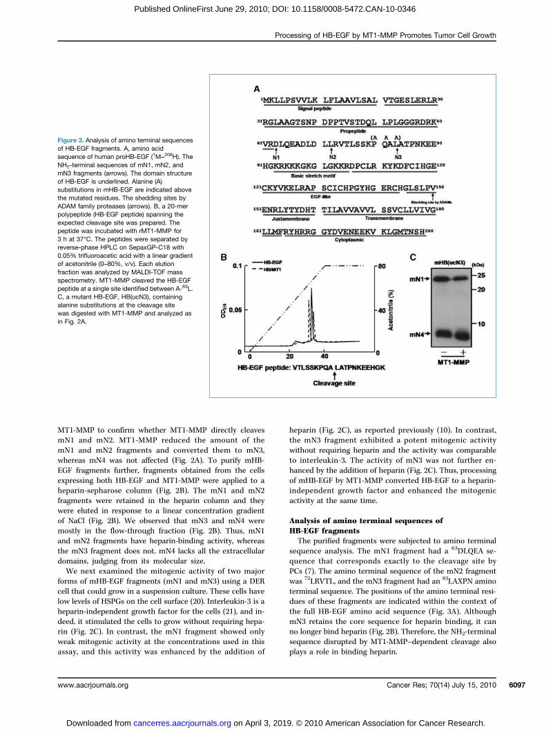
Analysis of amino terminal sequences ofHB-EGF fragmentsThe purified fragments were subjected to amino terminal
sequence analysis. The mN1 fragment had a 63DLQEA se-quence that corresponds exactly to the cleavage site byPCs (7). The amino terminal sequence of the mN2 fragmentwas 72LRVTL, and the mN3 fragment had an 83LAXPN aminoterminal sequence. The positions of the amino terminal resi-dues of these fragments are indicated within the context ofthe full HB-EGF amino acid sequence (Fig. 3A). AlthoughmN3 retains the core sequence for heparin binding, it canno longer bind heparin (Fig. 2B). Therefore, the NH2-terminalsequence disrupted by MT1-MMP–dependent cleavage alsoplays a role in binding heparin.
Figure 3. Analysis of amino terminal sequencesof HB-EGF fragments. A, amino acidsequence of human proHB-EGF (1M–208H). TheNH2-terminal sequences of mN1, mN2, andmN3 fragments (arrows). The domain structureof HB-EGF is underlined. Alanine (A)substitutions in mHB-EGF are indicated abovethe mutated residues. The shedding sites byADAM family proteases (arrows). B, a 20-merpolypeptide (HB-EGF peptide) spanning theexpected cleavage site was prepared. Thepeptide was incubated with rMT1-MMP for3 h at 37°C. The peptides were separated byreverse-phase HPLC on SepaxGP-C18 with0.05% trifluoroacetic acid with a linear gradientof acetonitrile (0–80%, v/v). Each elutionfraction was analyzed by MALDI-TOF massspectrometry. MT1-MMP cleaved the HB-EGFpeptide at a single site identified between A-83L.C, a mutant HB-EGF, HB(ucN3), containingalanine substitutions at the cleavage sitewas digested with MT1-MMP and analyzed asin Fig. 2A.
Cancer Res; 70(14) July 15, 2010 6097
9. © 2010 American Association for Cancer Research.

Koshikawa et al.
6098
Published OnlineFirst June 29, 2010; DOI: 10.1158/0008-5472.CAN-10-0346
A synthetic 20-mer polypeptide corresponding to aminoacids 74 to 93 of HB-EGF was incubated with MT1-MMPin vitro and was cleaved at a single site between A-83L, whichis consistent with the NH2-terminal sequence of mN3(Fig. 3B). Amino acid sequences flanking the cleavage sitefit the reported consensus ones (PXA-83L) for cleavage sitesby MT1-MMP. Substitution of three amino acids in the con-sensus sequence with alanine (PQA-83L to AAA-83A) made thepeptide resistant to MT1-MMP (Supplementary Fig. S2). Mu-tant HB-EGF [HB(ucN3)] with the same substitutions at thecleavage site was no longer cleavable by MT1-MMP (Fig. 3C).
HB-EGF and MT1-MMP expressed in human gastriccarcinoma cells support their invasive growthWe focused our attention on human gastric carcinoma cells
to investigate the significance of MT1-MMP–dependent HB-EGF processing in tumor cell growth because gastric carcino-ma is one of the most common malignancies in Japan andcarcinoma cells are reported to overexpress HB-EGF (22) aswell as MT1-MMP (23) compared with normal tissues. To ex-amine whether MT1-MMP and HB-EGF contribute to their
Cancer Res; 70(14) July 15, 2010
on April 3, 201cancerres.aacrjournals.org Downloaded from
cell growth, we first analyzed the expression of mRNAs encod-ing HB-EGF andMT1-MMP in five gastric carcinoma cell lines(Fig. 4A). MKN7, MKN28, and STKM-2 cells express both MT1-MMP and HB-EGF at significant levels, reflecting clinical si-tuations and showing that these cells could grow in collagengels (Fig. 4B). The growth of these cells was inhibited by anHB-EGF inhibitor (CRM197) derived fromDiphtheria toxin (24), ora synthetic MMP inhibitor (MMI270; ref. 25; Fig. 4B), suggest-ing the contribution of HB-EGF and MT1-MMP to the growthof these cells in the collagen matrix. To confirm this further,we knocked down the expression of HB-EGF or MT1-MMP inSTKM-2 cells using small interfering RNA. Knockdown effi-ciency was confirmed by RT-PCR (Supplementary Fig. S3).The growth of the cells was suppressed by treatment with ei-ther small interfering RNA targeting MT1-MMP or HB-EGF(Fig. 4C). Knockdown of either MT1-MMP or HB-EGF was alsofound to inhibit the invasion of the cells into the collagen(Fig. 4D). We also confirmed that MT1-MMP expressed inSTKM-2 cells produced mN3 fragments in these cells (Supple-mentary Fig. S4). Both EGFR and ErbB2 are expressed inSTKM-2 cells, as shown in Fig. 5A.
Figure 4. HB-EGF and MT1-MMP expressed in human gastric carcinoma cells are supporting their invasive growth. A, expression of HB-EGF andMT1-MMP mRNAs in human gastric cancer cells was analyzed by RT-PCR. GAPDH mRNA was used as an internal control and cDNAs for MT1-MMP andHB-EGF were used as positive controls. B, inhibition of tumor cell growth by CRM197 (5 μg/mL) and MMI270 (5 μg/mL). These cells were cultured for4 d in the presence/absence of the indicated inhibitors. The results represent the average of experiments performed in triplicate. The Student's t test wasused for statistical analyses. C and D, comparison of cell growth activity (C) and morphology (D) of STKM-2 in collagen gels after incubation for 4 d.
Cancer Research
9. © 2010 American Association for Cancer Research.

Processing of HB-EGF by MT1-MMP Promotes Tumor Cell Growth
Published OnlineFirst June 29, 2010; DOI: 10.1158/0008-5472.CAN-10-0346
Processing of HB-EGF by MT1-MMP is essential forthe promotion of invasive growth of gastriccarcinoma cellsAmong the gastric carcinoma cells tested in Fig. 4A, TMK-
1 is unique in that the cells lack the expression of both genes.Therefore, we used this cell line to reconstitute the proces-sing of HB-EGF by MT1-MMP by expressing both proteinsand observed the effect on cell growth in collagen gels. Ex-pression of HB-EGF alone in TMK-1 cells gave rise to thegeneration of several processed fragments but the mN3 frag-ment was not detected, whereas coexpression of HB-EGF andMT1-MMP in these cells led to the specific generation ofmN3 (Fig. 6A). However, mN3 was not produced followingthe coexpression of MT1-MMP and the processing mutantof HB(ucN3) (Fig. 6A).We next examined the effect of coexpression of HB-EGF
and MT1-MMP on the proliferation of TMK-1 cells grown incollagen matrix. Control mock-transfected cells multiplied1.4-fold under these conditions during this period (Fig. 6B).Expression of MT1-MMP alone did not stimulate cell growth,whereas expression of HB-EGF alone enhanced cell growth by
www.aacrjournals.org
on April 3, 201cancerres.aacrjournals.org Downloaded from
2-fold. Coexpression of MT1-MMP and HB-EGF enhanced cellgrowth by 4-fold (Fig. 6B). This effect was not observed whenMT1-MMP was coexpressed with the mutant HB-EGF, HB(ucN3) (Fig. 6B). We confirmed that HB(ucN3) retainedheparin-dependent mitogenic activity similar to mN1 andmN2 as shown in Supplementary Fig. S5. Thus, processing ofHB-EGFbyMT1-MMPis important topotentiate themitogenicactivity of HB-EGF on cell growth in the collagen environment.We also noted an alteration in cell morphology during the
growth of TMK-1 cells in collagen gels following the coex-pression of HB-EGF and MT1-MMP. Mock-transfected cellsformed compact spheroids whose morphology was not sig-nificantly altered by the expression of MT1-MMP (Fig. 6C).Expression of HB-EGF led to the generation of slightly largerspheroids (Fig. 6C). However, we observed a dramatic changein cell morphology following coexpression of MT1-MMP andHB-EGF (Fig. 6C). The cells did not form spheroids, but rath-er showed spindle-like mesenchymal morphology and spreadout invasively into the collagen gel as observed with STKM-2cells. In contrast, coexpression of HB(ucN3) and MT1-MMPdid not lead to an invasive cell morphology (Fig. 6C). The
Figure 5. ErbB receptor-mediatedsignals regulate invasive growthof human gastric cancer cells.A, expression of ErbB receptorsin TMK-1 and STKM-2 cells wasanalyzed by RT-PCR. B, effect ofneutralizing antibodies againstErbB receptors on TMK-1 cellgrowth expressing HB-EGF andMT1-MMP. HB/MT1-TMK-1 cellswere incubated with the indicatedantibodies, anti-EGFR (5 μg/mL),anti-ErbB2 (10 μg/mL), anti-ErbB3monoclonal antibodies (5 μg/mL),and the combined anti-EGFR +anti-ErbB2 monoclonal antibodiestreatment. Student's t test wasused for statistical analyses. Theresults represent the averageof experiments performed intriplicate. C, effects of neutralizingantibodies to the ErbB receptorson invasive morphology of theTMK-1 cells induced by HB-EGFand MT1-MMP coexpression incollagen gels (bar, 100 μm).
Cancer Res; 70(14) July 15, 2010 6099
9. © 2010 American Association for Cancer Research.

Koshikawa et al.
6100
Published OnlineFirst June 29, 2010; DOI: 10.1158/0008-5472.CAN-10-0346
Figure 6. Processing of HB-EGF by MT1-MMP promotes invasive growth of TMK-1 cells in collagen gels. A, detection of HB-EGFs (HB) and MT1-MMP(MT1) in TMK-1 cells by Western blotting. HB-EGF was detected by Western blotting using a M2 antibody and MT1-MMP was detected withanti–MT1-MMP antibody. CMs were obtained from mock and transfectants. sHB-EGFs in CMs were analyzed by Western blotting using anti–HB-EGFantibody. sHB-EGF concentration was estimated to be ∼1.5 ng/mL according to an ELISA assay using the antibody (data not shown). β-Actin wasused as a loading control. *, cytoplasmic HB-EGF. B, effect of HB-EGF processing by MT1-MMP on TMK-1 cell growth in collagen gels. Mock andtransfectants were cultured for 8 d. C, morphology of cells grown in collagen gels under light microscopy (bar, 100 μm). D, invasion of the cells intocollagen was analyzed using Transwell chambers using 10% FCS as a chemoattractant. The data represent average values from experiments performed intriplicate. Student's t test was used for statistical analyses.
Cancer Res; 70(14) July 15, 2010 Cancer Research
on April 3, 2019. © 2010 American Association for Cancer Research. cancerres.aacrjournals.org Downloaded from

Processing of HB-EGF by MT1-MMP Promotes Tumor Cell Growth
Published OnlineFirst June 29, 2010; DOI: 10.1158/0008-5472.CAN-10-0346
invasive growth of TMK-1 cells expressing HB-EGF and MT1-MMP was nearly completely inhibited by exposure to eitherMMI270 or CRM197 (Fig. 6C). The invasive property of thecells into collagen was further confirmed by an assay usingtranswell chambers, as shown in Fig. 6D. Expression of MT1-MMP alone enhanced cell invasion significantly, but the ex-pression of HB-EGF or HB(ucN3) alone had little effect oninvasion. We observed a major enhancement of invasion fol-lowing the coexpression of MT1-MMP and HB-EGF. This ef-fect was not observed when MT1-MMP was coexpressed withHB(ucN3) (Fig. 6D). The results again indicate that the clea-vage of HB-EGF between A-83L by MT1-MMP markedlychanged growth factor activity.The extracellular portion of HB-EGF shed by ADAM ac-
tivity is the entity that conveys the mitogenic activity ofHB-EGF. However, recent studies have shown that the re-sidual portion of HB-EGF following ectodomain sheddingcould have additional stimulatory effects on the cells (26,27). An mN4 fragment translocates to the perinuclearregion where it interacts with regulators of transcriptionfactors and modulates cellular functions in cooperationwith ErbB receptor-mediated signals elicited by the extra-cellular portion of HB-EGF (28). Thus, the NH2- andCOOH-terminal fragments of HB-EGF may cooperate toregulate cell function after being processed by ADAMs.Therefore, we examined whether the shed fragments weresufficient to elicit invasive cell growth or if the mN4COOH-terminal fragment plays a role in the mitogenicresponse of cells expressing HB-EGF and MT1-MMP. Totest this, we prepared conditioned medium (CM) fromTMK-1 expressing HB-EGF either alone or together withMT1-MMP, and examined the effect of these media on pa-rental TMK-1 cells that did not express both proteins. Weconfirmed the presence of sN3 fragments in CM from cellsexpressing MT1-MMP and HB-EGF by Western blotting(Fig. 6A, CM), whereas the sN3 fragment was not detectedin CM from mock-transfected cells or cells expressingMT1-MMP, HB-EGF, or HB(ucN3)/MT1-MMP. Only CMprepared from cells expressing both MT1-MMP and HB-EGF stimulated the growth of TMK-1 cells in collagen gels(Supplementary Fig. S6). Thus, the sN3 fragment containedin CM is important to stimulate the invasive growth ofTMK-1 cells. The COOH-terminal mN4 fragment might reg-ulate other cell functions rather than the invasive growthin collagen.
Stimulation of ErbB receptors mediates the invasivegrowth of gastric tumor cellsTo identify the receptors responsible for transducing the
signals mediated by HB-EGF and MT1-MMP, we examinedthe expression of ErbB family members in TMK-1 andSTKM-2 cells by RT-PCR (Fig. 5A). We detected the expres-sion of EGFR (ErbB1) and ErbB2 but not ErbB3 and ErbB4mRNAs in these cells. We next used neutralizing antibodiesto EGFR, ErbB2, and ErbB4 to test whether these receptorswere used for the growth of TMK-1 cells expressing MT1-MMP and HB-EGF. Anti-EGFR and anti-ErbB2 suppressedcell growth by 40% and 50%, respectively (Fig. 5B), and these
www.aacrjournals.org
on April 3, 201cancerres.aacrjournals.org Downloaded from
antibodies also inhibited the invasive property of the cells(Fig. 5C). EGFR and ErbB2 could also form a heterodimerand treatment of the cells with a combination of anti-EGFRand anti-ErbB2 antibodies resulted in a more efficient inhibi-tion of growth (Fig. 5B). Finally, it is important to confirmthat sN3 has the potential to stimulate the receptor. TheCM containing the sN3 fragment (Fig. 5A) indeed inducedphosphorylation of EGFR efficiently in TMK-1 cells (Supple-mentary Fig. S7).
Discussion
HB-EGF and MT1-MMP have been independently impli-cated in malignant tumor growth and invasion, and bothproteins have been recognized as potential targets for cancertherapy (29–32). The present study shows that HB-EGF is anew substrate of MT1-MMP and the growth factor activity ofHB-EGF is greatly enhanced by its processing. Interestingly,removal of the NH2-terminal fragment of HB-EGF by MT1-MMP converted HB-EGF into a potent mitogen that doesnot require heparin as a cofactor (Fig. 2C).NH2-terminal processing of HB-EGF by metalloproteinases
has been reported to occur at multiple sites (7, 33). Amongthem, MMP7 is reported to have processing activity againstHB-EGF. Therefore, we examined whether MMP7 couldcleave HB-EGF and generate mN3–HB-EGF. MMP7 generat-ed mN2-like fragments and increased the amount of mN4-like fragments, although it did not generate any mN3-likefragments (Supplementary Fig. S8).The NH2-terminal heparin-binding sequence suppresses
growth factor activity of HB-EGF, and binding of heparinto the sequence releases the suppression at least in vitro asreported previously (10). In vivo, HSPGs are abundantlypresent in tissues and are expected to activate HB-EGF likeheparin (11). Indeed, HB-EGF binds to HSPGs and elimina-tion of HSPGs from the cell surface by treating cells with he-paritinase diminishes the cell's response to HB-EGF (12). Toour surprise, however, we observed that exogenous expres-sion of HB-EGF alone in MT1-KO MEFs was not sufficientto promote tumor growth in mice (Fig. 1D), whereas coex-pression of MT1-MMP together with HB-EGF enhanced tu-mor growth synergistically. These results indicate thatHSPGs in tissues are not sufficient to activate HB-EGF, andthat MT1-MMP is required to induce the full mitogenic ac-tivity of HB-EGF. However, the abundance of HSPGs in tissuemight suggest the alternative possibility that HSPGs play anegative regulatory role on HB-EGF in vivo, which may bereleased by the MT1-MMP cleavage of the HSPGs binding do-main. For example, HSPGs may trap sN1– and sN2–HB-EGFsin vivo and prevent them from accessing their target cells,whereas sN3 could move through HSPG-rich tissues. Thisrole may not be readily detectable in culture.We observed that both proteins were coexpressed in most
gastric carcinoma cells (Fig. 4A) and that the growth of cells inculture exhibited dependence on both proteins as evaluatedby knockdown experiments or by using inhibitors (CRM197and MMI270). Cooperative action between HB-EGF andMT1-MMP is mediated by the processing of HB-EGF between
Cancer Res; 70(14) July 15, 2010 6101
9. © 2010 American Association for Cancer Research.

Koshikawa et al.
6102
Published OnlineFirst June 29, 2010; DOI: 10.1158/0008-5472.CAN-10-0346
the A-83L by MT1-MMP. This was proven by demonstratingthat the uncleavable mutant HB(ucN3) against MT1-MMPcould not cooperate with MT1-MMP to stimulate cellgrowth and invasion (Fig. 6). Soluble HB-EGF fragmentsshed by ADAM proteases act as ligands for ErbB receptors(1), and the gastric carcinoma cells expressed EGFR andErbB2 (Fig. 5A). The growth-promoting effect of HB-EGFand MT1-MMP on TMK-1 cells is mediated by the EGFRsas the effect was suppressed by neutralizing antibody toeach receptor (Fig. 5B and C).In conclusion, our present study uncovered the crucial role
of MT1-MMP as a regulator of HB-EGF activity. Thus, al-though MT1-MMP is important for tumor invasion, as hasbeen reported, it also promotes tumor growth by directlymodulating HB-EGF activity. The specific HB-EGF inhibitorCRM197 is currently being evaluated in a phase I clinical trialin patients with ovarian carcinoma (6), and the present studysuggests that combination therapy targeting the HB-EGF/ErbB pathway and the proteolytic activity of MT1-MMPwould provide a better therapeutic outcome for the treat-ment of malignant gastric carcinomas, and that detection
Cancer Res; 70(14) July 15, 2010
on April 3, 201cancerres.aacrjournals.org Downloaded from
of the sN3 fragment would be a good biomarker for selectingappropriate patients.
Disclosure of Potential Conflicts of Interest
No potential conflicts of interest were disclosed.
Acknowledgments
We acknowledge Dr. Roy Zent (Vanderbilt University, Nashville, TN) forcritical reading of this manuscript.
Grant Support
Scientific Research on Priority Areas “Integrative Research Toward the Con-quest of Cancer” (N. Koshikawa, M. Seiki, and E. Mekada) and the Global COEProgram “Center of Education and Research for the Advanced Genome-BasedMedicine—for personalized medicine and the control of worldwide infectiousdiseases,” MEXT, Japan (M. Seiki).
The costs of publication of this article were defrayed in part by the paymentof page charges. This article must therefore be hereby marked advertisement inaccordance with 18 U.S.C. Section 1734 solely to indicate this fact.
Received 01/29/2010; revised 04/06/2010; accepted 05/20/2010; publishedOnlineFirst 06/29/2010.
References
1. Raab G, Klagsbrun M. Heparin-binding EGF-like growth factor. Bio-chim Biophys Acta 1997;1333:F179–99.2. Harris RC, Chung E, Coffey RJ. EGF receptor ligands. Exp Cell Res
2003;284:2–13.3. Higashiyama S, Nanba D. ADAM-mediated ectodomain shedding of
HB-EGF in receptor cross-talk. Biochim Biophys Acta 2005;1751:110–7.
4. Iwamoto R, Yamazaki S, Asakura M, et al. Heparin-binding EGF-likegrowth factor and ErbB signaling is essential for heart function. ProcNatl Acad Sci U S A 2003;100:3221–6.
5. Miyamoto S, Yagi H, Yotsumoto F, Kawarabayashi T, Mekada E.Heparin-binding epidermal growth factor-like growth factor as a noveltargeting molecule for cancer therapy. Cancer Sci 2006;97:341–7.
6. Yagi H, Yotsumoto F, Sonoda K, Kuroki M, Mekada E, Miyamoto S.Synergistic anti-tumor effect of paclitaxel with CRM197, an inhibitorof HB-EGF, in ovarian cancer. Int J Cancer 2009;124:1429–39.
7. Nakagawa T, Higashiyama S, Mitamura T, Mekada E, Taniguchi N.Amino-terminal processing of cell surface heparin-binding epidermalgrowth factor-like growth factor up-regulates its juxtacrine but not itsparacrine growth factor activity. J Biol Chem 1996;271:30858–63.
8. Yamazaki S, Iwamoto R, Saeki K, et al. Mice with defects in HB-EGFectodomain shedding show severe developmental abnormalities.J Cell Biol 2003;163:469–75.
9. Aviezer D, Yayon A. Heparin-dependent binding and autophosphor-ylation of epidermal growth factor (EGF) receptor by heparin-bindingEGF-like growth factor but not by EGF. Proc Natl Acad Sci U S A1994;91:12173–7.
10. Takazaki R, Shishido Y, Iwamoto R, Mekada E. Suppression of thebiological activities of the epidermal growth factor (EGF)-like domainby the heparin-binding domain of heparin-binding EGF-like growthfactor. J Biol Chem 2004;279:47335–43.
11. Bernfield M, Gotte M, Park PW, et al. Functions of cell surface hepar-an sulfate proteoglycans. Annu Rev Biochem 1999;68:729–77.
12. Higashiyama S, Abraham JA, Klagsbrun M. Heparin-binding EGF-like growth factor stimulation of smooth muscle cell migration: de-pendence on interactions with cell surface heparan sulfate. J CellBiol 1993;122:933–40.
13. Butler GS, Dean RA, Tam EM, Overall CM. Pharmacoproteomics of ametalloproteinase hydroxamate inhibitor in breast cancer cells: dy-namics of membrane type 1 matrix metalloproteinase-mediatedmembrane protein shedding. Mol Cell Biol 2008;28:4896–914.
14. Itoh Y, Seiki M. MT1-MMP: a potent modifier of pericellular microen-vironment. J Cell Physiol 2006;206:1–8.
15. Niiya D, Egawa N, Sakamoto T, et al. Identification and characteriza-tion of Lutheran blood group glycoprotein as a new substrate ofMT1-MMP: a systemic whole-cell analysis of MT1-MMP-associatingproteins in A431 cells. J Biol Chem 2009;284:27560–69.
16. Tomari T, Koshikawa N, Uematsu T, et al. High throughput analysisof proteins associating with a proinvasive MT1-MMP in human ma-lignant melanoma A375 cells. Cancer Sci 2009;100:1284–90.
17. Nakamura K, Iwamoto R, Mekada E. Membrane-anchored heparin-binding EGF-like growth factor (HB-EGF) and diphtheria toxinreceptor-associated protein (DRAP27)/CD9 form a complex with in-tegrin α3β1 at cell-cell contact sites. J Cell Biol 1995;129:1691–705.
18. Nakamura K, Mitamura T, Takahashi T, Kobayashi T, Mekada E. Im-portance of the major extracellular domain of CD9 and the epidermalgrowth factor (EGF)-like domain of heparin-binding EGF-like growthfactor for up-regulation of binding and activity. J Biol Chem 2000;275:18284–90.
19. Taniwaki K, Fukamachi H, Komori K, et al. Stroma-derived matrixmetalloproteinase (MMP)-2 promotes membrane type 1-MMP-dependent tumor growth in mice. Cancer Res 2007;67:4311–9.
20. Baba I, Shirasawa S, Iwamoto R, et al. Involvement of deregulatedepiregulin expression in tumorigenesis in vivo through activated Ki-Ras signaling pathway in human colon cancer cells. Cancer Res2000;60:6886–9.
21. Otani H, Siegel JP, Erdos M. Interleukin (IL)-2 and IL-3 induce distinctbut overlapping responses in murine IL-3-dependent 32D cells trans-duced with human IL-2 receptor β chain: involvement of tyrosine ki-nase(s) other than p56lck. Proc Natl Acad Sci U S A 1992;89:2789–93.
22. Naef M, Yokoyama M, Friess H, Buchler MW, Korc M. Co-expressionof heparin-binding EGF-like growth factor and related peptides in hu-man gastric carcinoma. Int J Cancer 1996;66:315–21.
23. Shim KN, Jung SA, Joo YH, Yoo K. Clinical significance of tissuelevels of matrix metalloproteinases and tissue inhibitors of metallo-proteinases in gastric cancer. J Gastroenterol 2007;42:120–8.
24. Mitamura T, Higashiyama S, Taniguchi N, Klagsbrun M, Mekada E.Diphtheria toxin binds to the epidermal growth factor (EGF)-like do-main of human heparin-binding EGF-like growth factor/diphtheriatoxin receptor and inhibits specifically its mitogenic activity. J BiolChem 1995;270:1015–9.
Cancer Research
9. © 2010 American Association for Cancer Research.

Processing of HB-EGF by MT1-MMP Promotes Tumor Cell Growth
Published OnlineFirst June 29, 2010; DOI: 10.1158/0008-5472.CAN-10-0346
25. Levitt NC, Eskens FA, O'Byrne KJ, et al. Phase I and pharmacologicalstudy of the oral matrix metalloproteinase inhibitor, MMI270(CGS27023A), in patients with advanced solid cancer. Clin CancerRes 2001;7:1912–22.
26. Nanba D, Mammoto A, Hashimoto K, Higashiyama S. Proteolytic re-lease of the carboxy-terminal fragment of proHB-EGF causes nucle-ar export of PLZF. J Cell Biol 2003;163:489–502.
27. Hieda M, Isokane M, Koizumi M, et al. Membrane-anchored growthfactor, HB-EGF, on the cell surface targeted to the inner nuclearmembrane. J Cell Biol 2008;180:763–9.
28. Higashiyama S, Iwabuki H, Morimoto C, HiedaM, Inoue H,MatsushitaN. Membrane-anchored growth factors, the epidermal growth factorfamily: beyond receptor ligands. Cancer Sci 2008;99:214–20.
29. Miyamoto S, Yagi H, Yotsumoto F, et al. New approach to cancer ther-apy: heparin binding-epidermal growth factor-like growth factor asa novel targeting molecule. Anticancer Res 2007;27:3713–21.
30. Yagi H, Yotsumoto F, Sonoda K, Kuroki M, Mekada E, Miyamoto S.
www.aacrjournals.org
on April 3, 201cancerres.aacrjournals.org Downloaded from
Synergistic anti-tumor effect of paclitaxel with CRM197, an inhibitorof HB-EGF, in ovarian cancer. Int J Cancer 2008;124:1429–39.
31. Devy L, Huang L, Naa L, et al. Selective inhibition of matrix metallo-proteinase–14 blocks tumor growth, invasion, and angiogenesis.Cancer Res 2009;69:1517–26.
32. Nonaka T, Nishibashi K, Itoh Y, Yana I, Seiki M. Competitive disrup-tion of the tumor-promoting function of membrane type 1 matrix me-talloproteinase/matrix metalloproteinase-14 in vivo. Mol Cancer Ther2005;4:1157–66.
33. Higashiyama S, Lau K, Besner GE, Abraham JA, Klagsbrun M. Struc-ture of heparin-binding EGF-like growth factor. Multiple forms, pri-mary structure, and glycosylation of the mature protein. J BiolChem 1992;267:6205–12.
34. Koshikawa N, Minegishi T, Sharabi A, Quaranta V, Seiki M.Membrane-type matrix metalloproteinase-1 (MT1-MMP) is a pro-cessing enzyme for human laminin γ2 chain. J Biol Chem 2005;280:88–93.
Cancer Res; 70(14) July 15, 2010 6103
9. © 2010 American Association for Cancer Research.
2010;70:6093-6103. Published OnlineFirst June 29, 2010.Cancer Res Naohiko Koshikawa, Hiroto Mizushima, Tomoko Minegishi, et al. FactorFactor and Converts It into a Heparin-Independent Growth -Terminal Portion of Heparin-Binding Epidermal Growth2
Membrane Type 1-Matrix Metalloproteinase Cleaves Off the NH
Updated version
10.1158/0008-5472.CAN-10-0346doi:
Access the most recent version of this article at:
Material
Supplementary
http://cancerres.aacrjournals.org/content/suppl/2010/06/28/0008-5472.CAN-10-0346.DC1
Access the most recent supplemental material at:
Cited articles
http://cancerres.aacrjournals.org/content/70/14/6093.full#ref-list-1
This article cites 34 articles, 21 of which you can access for free at:
Citing articles
http://cancerres.aacrjournals.org/content/70/14/6093.full#related-urls
This article has been cited by 12 HighWire-hosted articles. Access the articles at:
E-mail alerts related to this article or journal.Sign up to receive free email-alerts
Subscriptions
Reprints and
To order reprints of this article or to subscribe to the journal, contact the AACR Publications
Permissions
Rightslink site. Click on "Request Permissions" which will take you to the Copyright Clearance Center's (CCC)
.http://cancerres.aacrjournals.org/content/70/14/6093To request permission to re-use all or part of this article, use this link
on April 3, 2019. © 2010 American Association for Cancer Research. cancerres.aacrjournals.org Downloaded from
Published OnlineFirst June 29, 2010; DOI: 10.1158/0008-5472.CAN-10-0346