tuberculosis paroxysmal tachycardia - heart · elsewhere. in the pathogenesis of this disease,...
TRANSCRIPT
MYOCARDIAL TUBERCULOSIS WITH PAROXYSMALVENTRICULAR TACHYCARDIA
BY
ROBERT SCHNITZER
From the Essex County Hospital, WansteadReceived April 4, 1947
Tuberculous myocarditis is extremely rare, and even in tuberculous pericarditis it is un-common. The reason for this is difficult to explain. Since the lungs are a common site oftuberculous infection, one might expect myocardial infection to occur much more frequently.It has been suggested that the constant movement of the musculature is not conducive to thelodgement of tubercle bacilli or to the development of tubercles, and that the lactic acidproduced by cardiac muscular activity offers some protection against Koch's bacilli (Raviart,1906). The following unusual case is reported.
CASE RECORDSA Petty Officer in the French Navy, aged 32, was admitted to hospital in August 1945.
He looked extremely ill and slightly cyanosed and complained of severe breathlessness andpalpitation.
History. He had always enjoyed good health. In 1940, during the evacuation fromDunkirk, he was wounded in the left forearm and subsequently the whole limb had to beamputated and he was fitted with an artificial arm. Soon afterwards he began to complain oftiredness and shortness of breath on the slightest exertion and this was followed by attacks ofpalpitation. These attacks started and stopped abruptly, and were accompanied by severedyspncea, an aching sensation in the amputation stump, and occasionally by slight hwmoptysis.He had lost 12 lb. in weight within five months and was sent to a sanatorium for furtherinvestigation. X-rays of his chest suggested some infiltration in both lungs but were atfirst not quite typical of tuberculosis, and repeated sputum examination, including cultures,failed to reveal acid-fast bacilli. A bronchogram showed no abnormality. After his dischargefrom the sanatorium he experienced a severe attack of paroxysmal tachycardia in which he wasadmitted to hospital twelve hours after the onset.
Examination. A youthful looking man, ashen-grey and cyanosed, dyspnceic, and sweatingprofusely. Temperature 99.40 F., pulse very rapid and regular; respiration 36. Neck veinsdistended; apex beat in sixth inteispace 3-5 cm. outside mid-clavicular line; no murmurs.Blood pressure 130/80 mm. No clubbing of fingers. Trachea central. A few bronchiticsounds scattered over both lungs, especially over the right mid-zone. Liver not enlarged;spleen not palpable; no-cedema.
Progress. Quinidine, intramuscularly and orally, and carotid sinus pressure failed tostop the paroxysm and acute pulmonary cedema developed. Morphine had only a limitedeffect. A cardiogram during this attack showed paroxysmal ventricular tachycardia (P. V. T.)with a ventricular rate of 280-300 a minute, similar but not identical with the paroxysm shownlater. X-ray of the chest suggested infiltration in the right lung, and to a lesser degree in theleft, and some enlargement of the paratracheal gland. Intravenous injection of 0 5 mg.R 213
on July 10, 2020 by guest. Protected by copyright.
http://heart.bmj.com
/B
r Heart J: first published as 10.1136/hrt.9.3.213 on 1 July 1947. D
ownloaded from
214 SCHNITZER
t,Lt i-^-r- ~~-~- _
r r i- !. . ~~~~ - --:-r ;1
..FIG. 1 ^ - - ;+..FIG 2
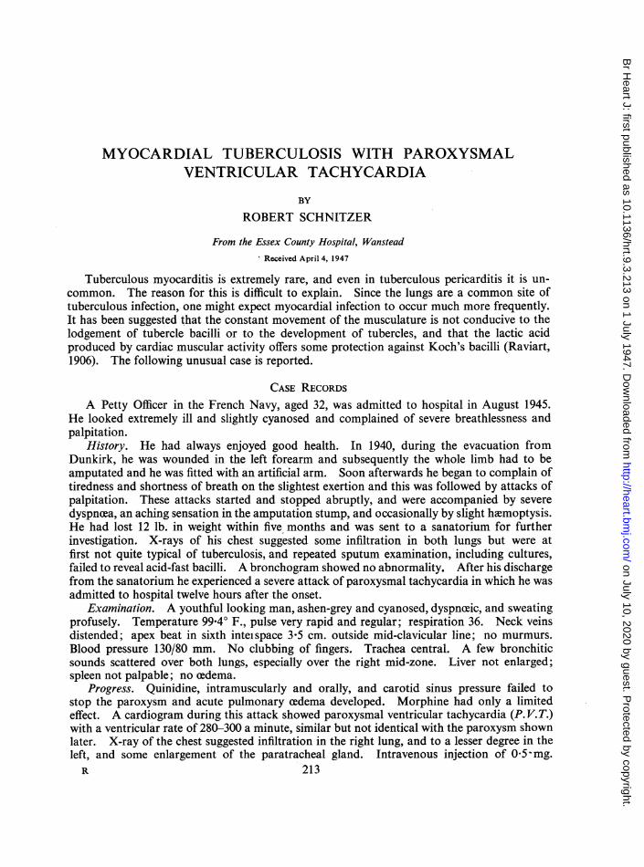
05 mg. digoxin intaeosy Chne sugs mycrda inactoof- T--t-II~§typ or posil effectofrecnt aroys,orsoofdigitalis.j,O
=. _. _ ~~~~~~~~~~~~~~~~~~~~~~~~~~~~~~~~~~~~..* ttt-I:it
XR--- -r. - -X. ...., .... -- _, X -- 0:- .. ..~~.
A-~~~~~~~~~~~~~~~~~~~~~~~~~~~~~~ ~ ~ ~ ~ ~ ~ ~ ~ ~ ~
N ----- - T s.r...
FIG. 3. FIG. 4.
FIG.~ ~ ~~IG3.-etocrigamatraparoxysmovetricularstac36hycardia Thathylastdi5hours.datrijcino
FIG.4 .-Ten Way before deathr. Changes segensnthpycrevious trfarctiong hav alos completel
u.be..d...
FIG _ -T1 -. F IG 2. -e
t~ ~FG 2 Tw wek laer Chage sugs myocardia infarctio of4T I type. 04
. i
,~~~~~~~~~~~~~~~~~~~~~~~~~~~~~~~~~~~~~....._Vk.tv. .s .....
FIG. 3. FIG.-T
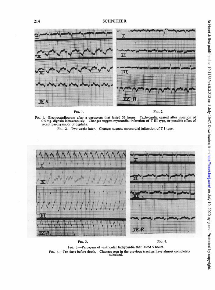
FIG. 3.-Paroxysm of ventricular tachycardia that lasted S hours.FIG. 4.-Ten days before death. Changes seen in the previous tracings have almost completely
subsided.
on July 10, 2020 by guest. Protected by copyright.
http://heart.bmj.com
/B
r Heart J: first published as 10.1136/hrt.9.3.213 on 1 July 1947. D
ownloaded from

MYOCARDIAL TUBERCULOSIS
digoxin reduced the apex rate to 120, and within one hour a further drop to 108 was observed.Digitalis was continued by mouth in small doses and only a few short attacks of P. V. T.occurred, but after four days it had to be stopped because of frequent extrasystoles. Cardio-grams between the attacks showed transient T wave changes which might have been attributedto pulmonary infarction, to posterior cardiac infarction, to the effect of the paroxysmaltachycardia, or to digitalis (Fig. 1). Later, however, inversion of the T I suggested anteriorcardiac infarction (Fig. 2). One night he complained of weakness and a sinking feeling, andtachycardia recurred and persisted until death early the next morning. A record of one of hisparoxysms of ventricular tachycardia is shown in Fig. 3. A cardiogram taken only ten daysbefore his death showed none of the changes seen in previous tracings (Fig. 4).
Investigations. On admission, white blood cells, 27,700, with 89 per cent polymorphs; 10days later, 5600, with 71 per cent polymorphs. Blood sedimentation rate on admission, 25 mm.in one hour (Westergren); 3 weeks later 10 mm. in one hour. Blood urea 35 mg. per 100 ml.Wassermann and Kahn negative. Urine, slight traces of albumin with many pus cells;culture sterile. Sputum, tubercle bacilli not found; culture for tubercle bacilli negative.
FIG. 5.-Section showing giant celled nodules in myocardium. Magnification: x 52.
215
on July 10, 2020 by guest. Protected by copyright.
http://heart.bmj.com
/B
r Heart J: first published as 10.1136/hrt.9.3.213 on 1 July 1947. D
ownloaded from
SCHNITZER
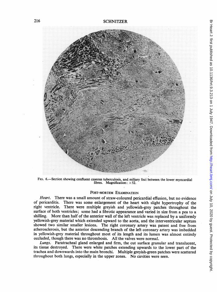
FIG. 6.-Section showing confluent caseous tuberculosis, and miliary foci between the lower myocardialfibres. Magnification: x 52.
POST-MORTEM EXAMINATIONHeart. There was a small amount of straw-coloured pericardial effusion, but no evidence
of pericarditis. There was some enlargement of the heart with slight hypertrophy of theright ventricle. There were multiple greyish and yellowish-grey patches throughout thesurface of both ventricles; some had a fibrotic appearance and varied in size from a pea to ashilling. More than half of the anterior wall of the left ventricle was replaced by a uniformlyyellowish-grey material which extended upward to the aorta, and the interventricular septumshowed two similar smaller lesions. The right coronary artery was patent and free fromatherosclerosis, but the anterior descending branch of the left coronary artery was imbeddedin yellowish-grey material throughout most of its length and its lumen was almost entirelyoccluded, though there was no thrombosis. All the valves were normal.
Lungs. Paratracheal gland enlarged and firm, the cut surface granular and translucent,its tissue destroyed. There were white patches extending upwards to the lower part of the-trachea and downwards into the main bronchi. Multiple greyish-green patches were scatteredthroughout both lungs, especially in the upper zones. No cavities were seen.
216
on July 10, 2020 by guest. Protected by copyright.
http://heart.bmj.com
/B
r Heart J: first published as 10.1136/hrt.9.3.213 on 1 July 1947. D
ownloaded from

MYOCARDIAL TUBERCULOSIS
The spleen was enlarged and soft, with multiple irregular greyish patches on the surfaceand in the parenchyma. The liver was not enlarged, but showed similar lesions and a fewdarkhlemorrhagic areas. Kidneys and adrenals were normal.
Histological report. Sections of the lung show numerous miliary and confluent tubercleswith central caseation in some. Fibrosis is present within and around some isolated tuberclesand is marked in the confluent areas, some of which are reduced almost to hyaline scars.Tuberculous arteritis is present in one small branch of the pulmonary artery. The remainderof the lung parenchyma shows cedema and congestion, and alveoli are occupied by an eosino-phil coagulum containing free macrophages. A bronchial gland consists of an acellular massof collagen, enclosing patches of amorphous debris and cholesterol crystals and surrounded bya capsule of fibrotic granulation tissue containing miliary tubercles.
Blocks from the left ventricle and interventricular septum of the heart show similarmiliaryand confluent miliary tubercles in the myocardium with fibrosis within and around thetubercles. In the left ventricle, fibrous granulation tissue containing tubercles replaces theouter part of the myocardium for a depth of at least 1 cm. except for a few included groupsof muscle fibres (Fig. 5 and 6). Inflammatory infiltration is slight in this confluent area, andconsists of macrophages, lymphocytes, plasma cells, and a few neutrophil leucocytes.
The dark areas in the liver proved to be cavernous hemangiomata and in one sectionmeasured up to1l5 cm. in diameter. The remainder-of the liver in sections shows albuminousdegeneration of the parenchymal cells and scattered miliary tubercles, one having a caseouscentre. The spleen shows miliary and confluent miliary tubercles involving Malpighianbodies. The kidneys show albuminous degeneration of the tubular epithelium.
The tubercles in the lung, liver, spleen, and myocardium are composed of collagen, epi-thelioid and multinuclear giant histiocytes, fibroblasts, and a few lymphocytes. In manytubercles, most or all of the cells are necrosed, and in some the necrosed cells form a homo-geneous caseous mass. Caseation affects only a part, usually central, of a tubercle. The giantcells are variable in appearance; some are typical Langhans's cells, but others are more of theforeign body type in that the nuclei, of which there are as many as 40, are scattered andchiefly central. In all sites some giant cells contain eosinophil, crystalloid, star-like, radialcytoplasmic inclusions similar to, but probably not so well developed as, those in the Stengel-Wolbach disease. Acid-fast bacilli could not be found in spite of prolonged search. Thepresence of caseation in many of the tubercles indicates almost certainly that the condition istuberculous. The absence of tubercle bacilli might be expected in a tuberculous inflammationof such low grade intensity as was evident in this case.
DISCUSSIONIn the case described, the diagnosis of myocardial tuberculosis is established beyond doubt
by the histological findings, despite the absence of tubercle bacilli from the sputum and fromtissue sections. The route of infection is difficult to explain in the absence of pericarditis,though the lesions in the paratracheal gland, the bronchi and lungs, suggest a primarylymphatic spread. The tubercle bacilli may have gained access to the venous blood streamvia the lymphatic duct, then infected the lungs and eventually entered the general circulationvia the pulmonary capillaries, whence the myocardium, liver, and spleen became involved.
A case of myocardial tuberculosis was reported by Townsend (1832) who described "atumour-like auricular growth." In Anders's (1902) extensive review of the subject, the totalof reported cases was brought up to 72. From the time of Bollinger's report in 1890 andRaviart's study in 1906, 101 cases had been observed over a period of 16 years, while only 79cases had been recognized previously. Norris (1904), in a study of 1764 hearts of tuberculoussubjects, observed only 6 cases.
Tubterculosis of the myocardium is almost invariably, secondary to a tuberculous focuss
217
on July 10, 2020 by guest. Protected by copyright.
http://heart.bmj.com
/B
r Heart J: first published as 10.1136/hrt.9.3.213 on 1 July 1947. D
ownloaded from
elsewhere. In the pathogenesis of this disease, three possible routes of infection havebeen considered, (a) by the blood stream, (b) by retrograde lymph extension, and (c) by con-tiguous tissues. Myocardial involvement is usually secondary to tuberculous pericarditis.Caseous tuberculous mediastinal lymph nodes, particularly the bronchial or paratrachealgroup, may be the source of infection of the pericardium and heart. Infection may occurby means of direct contact or through the lymphatics; this latter would necessitate a reversalof the lymph flow, since the drainage of the pericardium has been shown to be upwards intothe bronchial lymph nodes (Recklinghausen, 1885). Another possible mode of spread is bydirect extension from the pleura.
Three types of myocardial tuberculosis are usually recognized, the nodular, the miliary,and the diffuse infiltrating form. Raviart described a fourth type which he called " chronicinterstitial tuberculous myocarditis "; this included the so-called " sclerosis of the myo-cardium." The existence of this last form has been disputed and it is generally held thatfibrous myocardial lesions which may be found in the hearts of those dying from tuberculosisare not necessarily of tuberculous origin. Some such cases, in which tubercles or tuberclebacilli have been found in sections, probably belong to the diffuse infiltrative form of myo-cardial tuberculosis. The nodular variety is the commonest, and nodules varying from thesize of a pea to that of an egg have been reported. Next in frequency is the miliary varietyand rarest is the diffuse infiltrative form. According to Moenckeberg (1924), the last men-tioned is a " specific diffuse productive tuberculous myocarditis " most frequently a sequel topericarditis. Here the myocardium appears to be taken up by a uniformly grey or yellow-grey firm material, which may almost entirely replace the muscle in the areas involved. Mostauthors report that Ziehl-Neelsen staining has failed to reveal tubercle bacilli in spite oftypical histological changes. The infiltrating type most often involves the auricles and maycause auricular extrasystoles and rarely auricular fibrillation (Sweeney, 1940). The ventriclesare sometimes involved, and occasionally extensive destruction of the myocardium andconducting tissues has been observed. Caseous infiltration may destroy half or more of thethickness of the ventricular wall. Coronary occlusion is seldom observed in cases of tuber-culosis, and tuberculous arteritis has been observed very rarely in the coronary arteries whereit is usually confined to the smaller branches. In one reported case, however, a comparativelylarge coronary branch was involved, and, in the present case, almost the whole length of theleft descending coronary artery was surrounded and partly occluded by tuberculous infiltra-tion. The initial leucocytosis of 27,700, which disappeared within 10 days, may have beendue to the ventricular tachycardia rather than to coronary occlusion; for a leucocytosis of20,000 to 30,000 which subsides rapidly after termination of the paroxysm may be a feature ofparoxysmal tachycardia (Levine and Golden, 1922).
SUMMARYA case of myocardial tuberculosis complicated by paroxysmal ventricular tachycardia is
reported. X rays showed pulmonary infiltration, but sputum tests for tubercle bacilli werenegative.
Necropsy showed miliary tuberculosis with extensive involvement of the myocardium,including the interventricular septum, without pericarditis. The tuberculous nature ofthe lesions in the heart and other organs was established by the histological findings, which aredescribed.
The publications relating to tuberculous myocarditis are briefly reviewed and the pathologyis discussed.
I wish to thank Dr. O'Brien and Dr. J. Gilmour for their detailed histological report, and Dr. M. Brown andDr. D. Gutman for their assistance at the necropsy. I am also indebted to Dr. John Parkinson and Dr. J. W.Linnell for their interest and help, and to Dr. P. J. W. Mills, Medical Superintendent, Lister Emergency liospital.Hitchin, for permission to publish this case.
218 SCHNITZER
on July 10, 2020 by guest. Protected by copyright.
http://heart.bmj.com
/B
r Heart J: first published as 10.1136/hrt.9.3.213 on 1 July 1947. D
ownloaded from
MYOCARDIAL TUBERCULOSIS 219
REFERENCESAnders, J. M. (1902). J. Amer. med. Ass., 39, 1081.Bollinger, 0. (1890). Munch. med. Wschr., 37, 567.Levine, S. A., and Golden, R. (1922). Arch. intern. Med., 29, 836.Moenckeberg, J. G. (1924). Handbuch der Speciellen. Path. Anat. und. Histol. Henke-Lubarsch, Berlin,
Julius Springer.Norris, G. W. (1904). Amer. J. med. Sci., 128, 649.Raviart, G. (1906). Arch. Mid. exper., 18, 141.Recklinghausen, R. (1885). Virchows Arch., 100, 503.Sweeney, J. A. (1940). Amer. Heart. J., 20, 345.Townsend, R. (1832).- Dublin J. med. Sci., 1, 176.
on July 10, 2020 by guest. Protected by copyright.
http://heart.bmj.com
/B
r Heart J: first published as 10.1136/hrt.9.3.213 on 1 July 1947. D
ownloaded from