tta-p2 is a potent and selective blocker of t-type...
TRANSCRIPT

TTA-P2 Is a Potent and Selective Blocker of T-Type CalciumChannels in Rat Sensory Neurons and a Novel AntinociceptiveAgent
WonJoo Choe, Richard B. Messinger, Emily Leach, Veit-Simon Eckle,Aleksandar Obradovic, Reza Salajegheh, Vesna Jevtovic-Todorovic,and Slobodan M. TodorovicDepartment of Anesthesiology, InJe University Ilsan Paik Hospital and College of Medicine, Seoul, Korea (WJ.C.); Departmentsof Anesthesiology (R.B.M., E.L., A.O., R.S., V.J.-T., S.M.T.) and Neuroscience (V.J.-T., S.M.T.) and Neuroscience GraduateProgram (S.M.T., V.J.-T.), University of Virginia Health System, Charlottesville, Virginia; and Department of Anesthesiology andIntensive Care Medicine, Tuebingen University Hospital, Eberhard-Karls University, Tuebingen, Germany (V.-S.E.)
Received April 21, 2011; accepted August 5, 2011
ABSTRACTSeveral agents that are preferential T-type calcium (T-channel)blockers have shown promise as being effective in alleviatingacute and chronic pain, suggesting an urgent need to identifyeven more selective and potent T-channel antagonists. We usedsmall, acutely dissociated dorsal root ganglion (DRG) cells of adultrats to study the in vitro effects of 3,5-dichloro-N-[1-(2,2-dimethyl-tetrahydro-pyran-4-ylmethyl)-4-fluoro-piperidin-4-ylmethyl]-benzamide (TTA-P2), a derivative of 4-aminomethyl-4-fluoropip-erdine, on T currents, as well as other currents known to modulatepain transmission. We found that TTA-P2 potently and reversiblyblocked DRG T currents with an IC50 of 100 nM and stabilizedchannel in the inactive state, whereas high-voltage-activated cal-cium and sodium currents were 100- to 1000-fold less sensitive tochannel blocking effects. In in vivo studies, we found that intra-
peritoneal injections of 5 or 7.5 mg/kg TTA-P2 reduced painresponses in mice in phases 1 and 2 of the formalin test. Further-more, TTA-P2, at 10 mg/kg i.p., selectively and completely re-versed thermal hyperalgesia in diabetic rats treated with strepto-zocin but had no effect on the nociceptive response of healthyanimals. The antihyperalgesic effects of TTA-P2 in diabetic ratswere completely abolished by administration of oligonucleotideantisense for CaV3.2 isoform of T channels. Thus, TTA-P2 is notonly the most potent and selective blocker of T channels in sen-sory neurons yet described, but it also demonstrates the potentialfor the pharmacological effectiveness of this approach in address-ing altered nociceptive responses in animal models of both inflam-matory and neuropathic pain.
IntroductionOn the basis of the membrane potential at which they gate
ion currents, voltage-gated calcium channels are classified ashigh-voltage-activated (HVA) or sustained currents and low-voltage-activated or transient (T-type) currents (Perez-Reyes, 2003). Different subtypes of HVA currents in the
central nervous system (CNS) are important in regulatingfast synaptic transmission. In contrast, neuronal T channelshave key functions in neuronal membrane oscillations and inlowering the threshold for action potential firing in both theperipheral and central nervous systems. On the basis ofcloned sequences of the pore-forming �1 subunit, at leastthree isoforms of T channels exist: CaV3.1 (�1G) (Perez-Reyes et al., 1998), CaV3.2 (�1H) (Cribbs et al., 1998), andCaV3.3 (�1I) (Lee et al., 1999a). Recent electrophysiological,genetic, molecular, and behavioral studies have suggestedthat T channels are crucial in controlling the excitability ofsensory neurons, act as signal amplifiers, and make a previ-ously unrecognized contribution to both peripheral and cen-tral pain processing (Todorovic et al., 2001; Jevtovic-Todoro-
This work was supported by The National Institutes of Health NationalInstitute on Drug Abuse [Grant R21-DA029342]; the Harold Carron Endow-ment Fund; the American Diabetes Association [Basic Research Grant 7-09-BS-190]; the Department of Anesthesiology at the University of Virginia, InJeUniversity; and a gift from Joseph C. Palumbo and Sandra C. Palumbo.
Article, publication date, and citation information can be found athttp://molpharm.aspetjournals.org.
doi:10.1124/mol.111.073205.
ABBREVIATIONS: HVA, high-voltage-activated; DRG, dorsal root ganglion; HEK, human embryonic kidney; PWL, paw withdrawal latency; STZ,streptozocin; TTX, tetrodotoxin; TTA-P2, 3,5-dichloro-N-[1-(2,2-dimethyl-tetrahydro-pyran-4-ylmethyl)-4-fluoro-piperidin-4-ylmethyl]-benzamide);ECN, [(3�, 5�, 17�)-17-hydroxyestrane-3-carbonitrile]; CNS, central nervous system; P1, phase 1; P2, phase 2; AS, antisense-CaV3.2; MIS,Mismatch-CaV3.2; I-V, current-voltage.
0026-895X/11/8005-900–910$25.00MOLECULAR PHARMACOLOGY Vol. 80, No. 5Copyright © 2011 The American Society for Pharmacology and Experimental Therapeutics 73205/3723390Mol Pharmacol 80:900–910, 2011 Printed in U.S.A.
900
at ASPE
T Journals on Septem
ber 14, 2018m
olpharm.aspetjournals.org
Dow
nloaded from
vic and Todorovic, 2006; Snutch and David, 2005; Choi et al.,2007). Furthermore, in vivo experiments showed that use ofsubtype-specific antisense for knockdown of CaV3.2 channelsin DRG cells completely reversed hyperalgesia in animalmodels of mechanical injury to the sciatic nerve (Bourinet etal., 2005) and painful diabetic neuropathy (Messinger et al.,2009). This creates interest in further developing pharmaco-logical tools for studies of the function of T channels in painpathways and for clinical development of specific pain ther-apies targeting ion channels in sensory neurons.
Although there are many natural toxins or venom compo-nents that could be used to study the multiple HVA currents,only recently have substances been identified that are moreuseful blockers of T currents. Consistent with this, systemicinjections of mibefradil, a peripherally acting pan-T-channelblocker (Clozel et al., 1997), suppressed cutaneous, thermal,and mechanical nociception in healthy rats (Todorovic et al.,2002), as well as visceral nociception in healthy mice (Kim etal., 2003). Furthermore, the reversal of symptoms of neuro-pathic pain associated with chronic constrictive injury of thesciatic nerve has been demonstrated by systemic and localintraplantar injections of mibefradil (Dogrul et al., 2003).Some clinically used antiepileptics such as phenytoin andethosuximide are in vitro blockers of T channels in sensoryneurons (Todorovic and Lingle, 1998) and potent analgesicswhen injected into peripheral receptive fields of whole ani-mals (Todorovic et al., 2003). It is noteworthy that some otherclinically used analgesic drugs target voltage-gated calciumchannels. For example, voltage-gated calcium channels areconsidered a major cellular target for the anticonvulsantsgabapentin and pregabalin, which can relieve diabetes-in-duced neuropathic pain in some populations of patients (Ro-gawski and Loscher, 2004). However, the use of gabapentinand related drugs is associated with side effects such asexcessive sedation in many patients, which necessitates thesearch for other novel therapies (Edwards et al., 2008).
The usefulness of mibefradil, a prototypical T-channelblocker, has been questioned because recent studies havedemonstrated that this molecule, in addition to affecting Tchannels, can affect several other ion channels, includingvoltage-gated sodium channels in sensory neurons, with asimilar potency (Coste et al., 2007). Likewise, the antiepilep-tic ethosuximide, another representative T-channel blockerand analgesic in animal pain models (Todorovic et al., 2003),affects T channels and sodium channels in neurons of theCNS with similar potency (Leresche et al., 1998). This raisesthe question of whether the analgesic effects of these drugs inbehavioral pain paradigms can be attributed only to theantagonism of T channels. Thus, despite the progress madein recent years, pharmacological tools for the study of Tcurrents are still very limited [see reviews by McGivern(2006) and Lory and Chemin (2007)], precluding any clinicalstudies aimed at establishing the potential value of T-chan-nel blockers in treating various pain disorders.
The recent discovery of novel selective T-channel antagonistssuch as 4-aminomethyl-4-fluoropiperdine is promising becausethese agents completely block recombinant T-current isoformswith high potency (IC50 � 20–100 nM) (Shipe et al., 2008).However, the selectivity, potency, and mechanisms of action ofthese novel blockers in native cells in pain pathways have not,as yet, been systematically examined. Thus, we hypothesizedthat one such compound, 3,5-dichloro-N-[1-(2,2-dimethyl-
tetrahydro-pyran-4-ylmethyl)-4-fluoro-piperidin-4-ylmethyl]-benzamide (TTA-P2; see Fig. 1), may be a potent blocker ofnative T currents in rat sensory neurons and, accordingly, auseful tool for studies of the role of T channels in pain signaling.To test this hypothesis, we have done biophysical studies usingpatch-clamp experiments with acutely dissociated sensory neu-rons of rat dorsal root ganglia and whole-animal behavioralpain experiments with adult rats and mice.
Materials and MethodsElectrophysiological In Vitro Studies
Before the harvest of tissues, rats were deeply anesthetized withisoflurane and rapidly decapitated. For one experiment, we dissectedsix to eight lumbar dorsal root ganglia from both sides of rats. Weprepared acutely dissociated DRG cells and used them within 6 to 8 hfor whole-cell recordings as described previously (Todorovic et al.,1998; Nelson et al., 2005, 2007). We focused on small cells (i.e., thosewith an average soma diameter of 20–30 �m), because functionalstudies have indicated that most of them are likely polymodal noci-ceptors belonging to unmyelinated C-type sensory fibers that arecapable of responding to noxious mechanical, chemical, and thermalstimuli in vivo (McCleskey and Gold, 1999; Campbell and Meyer,2006). Our previous studies have confirmed that the majority ofacutely dissociated small DRG cells express T currents that areimportant for control of membrane excitability (Todorovic et al.,1998; Nelson et al., 2005, 2007).
Recordings were made using standard whole-cell techniques. Se-ries resistance (Rs) and capacitance (Cm) values were taken directlyfrom readings of the amplifier after electronic subtraction of thecapacitive transients. Series resistance was compensated to the max-imum extent possible (usually �60–80%). In most experiments, weused a P/5 protocol for online leak subtractions. The percentagereductions in peak current at various concentrations of TTA-P2were used to generate a concentration-response curve. Mean val-ues were fit to the following Hill function:
PB([TTA-P2]) � PBmax/(1 � (IC50/[TTA-P2])n) (1)
where PBmax is the maximal percentage block of peak current, IC50
is the concentration that produces 50% inhibition, and n is theapparent Hill coefficient for blockade. The fitted value is reportedwith 95% linear confidence limits. The voltage dependencies of acti-vation and steady-state inactivation were described with singleBoltzmann distributions of the following forms.
Activation:G(V) � Gmax/(1 � exp[ � (V � V50)/k]) (2)
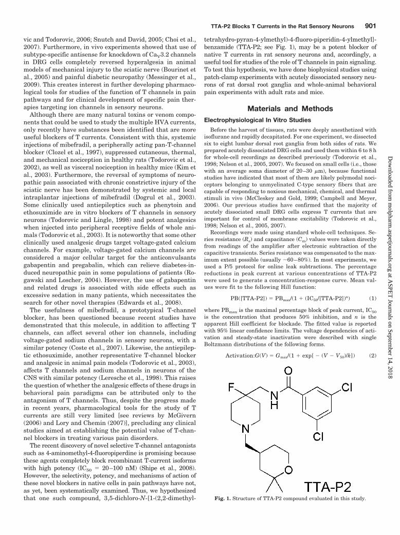
Fig. 1. Structure of TTA-P2 compound evaluated in this study.
TTA-P2 Blocks T Currents in the Rat Sensory Neurons 901
at ASPE
T Journals on Septem
ber 14, 2018m
olpharm.aspetjournals.org
Dow
nloaded from
Inactivation:I(V) � Imax/(1 � exp[(V � V50)/k]) (3)
In these forms, Imax is the maximal amplitude of current, Gmax is themaximal conductance, V50 is the voltage at which half of the currentis activated or inactivated, and k represents the voltage dependence(slope) of the distribution.
The time course of macroscopic T-current inactivation and deac-tivating tail currents were fitted using a single-exponential equation:
y � A1 � e� � x/�1� � y0 (4)
where A1 is the amplitude, �1 is the decay constant, and y0 is theoffset.
For fitting the time course of recovery from inactivation, a double-exponential function was used, yielding two time constants (�1 and�2) and their corresponding amplitudes (A1 and A2):
y � y0 � A1e� � x/�1� � A2e� � x/�2� (5)
TTA-P2 was prepared as 100- to 300-mM stock solutions in dimethylsulfoxide. The final concentrations of dimethyl sulfoxide had nosignificant effect on T-current amplitude in DRG and human embry-onic kidney (HEK) 293 cells (data not shown).
The external solution used to isolate T currents contained 2 mMCaCl2, 152 mM TEA-Cl, and 10 mM HEPES adjusted to pH 7.4 withTEA-OH. To minimize contamination of T currents with even mini-mal HVA components, we used only fluoride (F � )-based internalsolution to facilitate HVA calcium current rundown; this solutioncontained 135 mM tetramethylammonium hydroxide, 10 mM EGTA,40 mM HEPES, and 2 mM MgCl2, adjusted to pH 7.2 with hydro-fluoric acid. This allowed studies of well isolated and well clamped Tcurrents in small DRG cells. All chemicals were obtained fromSigma-Aldrich (St. Louis, MO) unless noted otherwise. Statisticalcomparisons were made, where appropriate, using an unpaired Stu-dent’s t test, Mann-Whitney sum test, and 2 test. All quantitativedata are expressed as means of multiple experiments � S.E.M.unless stated otherwise. The amplitude of T current was measuredfrom the peak, which was subtracted from the current at the end of thedepolarizing test potential to avoid contamination with residual HVAcurrents that were present at more positive membrane potentials(typically � 20 mV and higher). To record HVA calcium currents inDRG and HEK cells, we used the same external solution except thatequimolar BaCl2 was substituted for CaCl2 and the internal solutioncontained 110 mM Cs-methane sulfonate, 14 mM phosphocreatine, 10mM HEPES, 9 mM EGTA, 5 mM Mg-ATP, and 0.3 mM Tris-GTP,adjusted to pH 7.3 with CsOH. For recordings of voltage-gated sodiumcurrents in DRG cells, we used the same fluoride-based internal solu-tion as for recordings of T currents. The external solution contained 140mM NaCl, 4 mM KCl, 2 mM MgCl2, 2 mM CaCl2, 0.5 mM CdCl2, 10 mMglucose, and 10 mM HEPES, adjusted to pH 7.4. In some experiments,this solution was supplemented with 1 �M tetrodotoxin (TTX).
In Vivo Studies
Chemicals and Animals. In all experiments, Sprague-Dawleyadult female rats (retired breeders) and adult C57BL/6 mice of bothsexes were used. Streptozocin (STZ) was purchased from Sigma-Aldrich. Antisense oligonucleotides and mismatched oligonucleo-tides [using the sequence previously published by Bourinet et al.(2005); Messinger et al., 2009] were purchased from Invitrogen(Carlsbad, CA). Antisense-CaV3.2 (AS) (CCACCTTCTTACGCC-AGCGG), which was used to knock down the T-type-channel pore-forming subunit of the gene encoding the �1H (Cav3.2) or Mismatch-CaV3.2 (Mis-CaV3.2; MIS) (TACTGTACTTGCGAGGCCAC) weredissolved in sterile neutral pH buffer solution. Vehicle experimentswere performed using sterile saline neutral pH buffer solution. As inprevious studies (Messinger et al., 2009), vehicle and MIS werefound to have no effect on thermal nociception.
TTA-P2 was kindly provided by Drs. John Renger and Victor N.Uebele (Merck Research Laboratories, West Point, PA). For all of our
in vivo studies TTA-P2 was dissolved in 15% cyclodextrin and in-jected intraperitoneally at the doses of 5, 7.5, or 10 mg/kg. Cyclodex-trin [(2-hydroxypropyl)-�-cyclodextrin] solution (Sigma-Aldrich) wasbalanced at pH 7.4 just before injection.
Induction of Peripheral Diabetic Neuropathy with Strep-tozocin. All experimental protocols were approved by the Universityof Virginia Animal Care and Use Committee and were in accordancewith the Guide for the Care and Use of Laboratory Animals (Instituteof Laboratory Animal Resources, 1996). All possible efforts weremade to minimize animals’ suffering and to minimize the number ofanimals used.
To induce peripheral diabetic neuropathy, we intravenously in-jected freshly dissolved STZ solution at pH 5 to 6, using a dose of 50mg/kg. This dose causes severe hyperglycemia and pain-like behav-ior within the first few days after injection but does not cause severegeneralized sickness (e.g., ketoacidosis, malaise, wasting) (Aley andLevine, 2001; Jagodic et al., 2007; Messinger et al., 2009). Controlrats received the same volume per kilogram of intravenous sterilesaline. The animals were studied for 10 days after the day of intra-venous injection.
Three days after injecting STZ (or saline), at which point STZ-injected rats had developed peripheral diabetic neuropathy, we in-trathecally injected 12.5 �g/25 �l of either AS or MIS (or 25 �l ofsaline) into the L5–6 region of the spinal cord every 12 h for 4 days (atotal of eight injections) to test the effects of oligonucleotides. Allsolutions were pH balanced to 7.4 to avoid spinal cord irritation. Ratswere maintained in a surgical plane of anesthesia with isoflurane(2–3% in oxygen delivered via nose cone) throughout the injectionprocedure. We previously showed that intrathecal injections as de-scribed lead to preferential uptake of AS by DRG sensory neuronsand minimal uptake by spinal cord tissues (Messinger et al., 2009).This method of AS application reliably reversed neuropathic hyper-algesia in diabetic rats and concomitantly reversed up-regulation ofT currents in rat sensory neurons, whereas injections of saline orMIS intrathecally in the same manner did not affect pain responsesand T-current density in DRG cells (Messinger et al., 2009).
Behavioral Studies
Assessment of Thermal Sensitivity. The nociceptive responseto thermal stimulation was measured using a paw thermal stimula-tion system consisting of a clear plastic chamber (10 � 20 � 24 cm)that sits on a clear elevated glass floor and is temperature regulatedat 30°C. As described previously (Messinger et al., 2009), each rat isplaced in the plastic chamber for 15 min to acclimate. A radiant heatsource mounted on a movable holder beneath the glass floor ispositioned to deliver a thermal stimulus to the plantar side of thehind paw. When the rat withdraws the paw, a photocell detectsinterruption of a light-beam reflection, and the automatic timershuts off. This method has a precision of �0.05 s for the measure-ment of paw withdrawal latency (PWL). To prevent thermal injury,the light beam is automatically discontinued at 20 s if the rat fails towithdraw its paw. Pain testing was done before STZ or vehicleinjection (day 0) and daily thereafter for up to 10 days. The stabilityof daily pain recordings was confirmed by saline-injected controls.
Statistical Analysis. PWLs were subjected to analysis of vari-ance containing one within-subject variable, test session (before theadministration of STZ or vehicle versus each post-treatment day upto 10 days), and one between-subject variable, AS versus TTA-P2.Relevant pairwise comparisons were done, and � levels were ad-justed using the Bonferroni procedure when appropriate.
Assessment of Inflammatory Pain in Mice. Behavioral testsfor inflammatory pain after formalin injection were done with micein a clear Plexiglas chamber prefilled with air at a flow of 6 l/min, aswe reported recently (Orestes et al., 2011). Each mouse was firstplaced in the prefilled chamber to accommodate for 30 min and thenremoved from the chamber, injected in the plantar side of the rightpaw with 20 �l of 5% formalin or vehicle (15% cyclodextrin), andreturned to the test chamber. The time in seconds that the mouse
902 Choe et al.
at ASPE
T Journals on Septem
ber 14, 2018m
olpharm.aspetjournals.org
Dow
nloaded from
spent licking and biting the paw was measured for 1 h and recordedper every 5-min interval. Afterward, the mouse’s temperature wasmeasured to ensure normothermia. To test the effects of TTA-P2, amouse was injected intraperitoneally with fresh TTA-P2 solution (5or 7.5 mg/kg) or an equal volume of vehicle. Thirty minutes later, themouse was placed inside the chamber to equilibrate and becomefamiliar with the environment. One hour after the injection ofTTA-P2 or vehicle, the mouse was given an injection of formalin. Therest of the experiment was performed as described in the precedingsection.
Assessment of Sensorimotor Abilities. The sensorimotor bat-tery in mice consisted of three tests: ledge, platform, and inclinedscreen. These tests are designed to assess agility and fine motorabilities, as we described in a recent publication (Latham et al.,2009).
ResultsThe in vitro results presented here were obtained from a
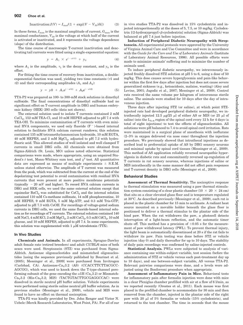
total of 144 small DRG cells having an average soma diam-eter of 25 � 3 �m (mean � S.D.) and an average membranecapacitance of 24 � 4 pF (mean � S.D.). We began our studyby testing the effects of TTA-P2 on well isolated T currents inrat sensory neurons. Traces (Fig. 2A) and time course(Fig. 2B) from the same representative DRG cell indicatethat at 1 �M, TTA-P2 inhibited most of the T current thatwas recorded at holding potentials (Vh) of � 90 mV. Figure2B shows that the inhibitory effect of TTA-P2 had a fast onset
but was slowly and only partially reversible. Interestingly,similarly fast onset and slow reversibility of the inhibition ofT currents with TTA-P2 in thalamic relay neurons has alsobeen reported (Dreyfus et al., 2010). Furthermore, the tracesshown in Fig. 2C demonstrate that TTA-P2 at 10 �M hadlittle effect (less than 10%) on the amplitude of HVA Ca2�
currents in acutely dissociated DRG cells. For recordings ofHVA currents, cells were held at � 50 mV to separate thatsignal from T currents that are almost completely inactivatedat positive membrane potentials.
Some of the calcium channel blockers thought to be selec-tive for T currents (e.g., mibefradil, nickel, ethosuximide)may also affect the R-type (CaV2.3) subtype of HVA calciumcurrents in the same concentration range (Randall andTsien, 1997; Nakashima et al., 1998). The above studies havealso shown that separation of T currents from R-type cur-rents in native cells is further complicated by the fact thatthey appear to inactivate at somewhat comparable rates.Thus, we tested the ability of TTA-P2 to inhibit humanrecombinant CaV2.3 channels stably coexpressed with �3calcium channel subunits in HEK 293 cells (Nakashima etal., 1998). Traces from a representative HEK 293 cell shownin Fig. 2D demonstrate that TTA-P2 at 10 �M inhibited onlyapproximately 10% of the recombinant CaV2.3 current. Wealso tested the ability of TTA-P2 to inhibit voltage-gatedsodium currents (INa�), because these channels are critical
Fig. 2. TTA-P2 selectively inhibits T currents in acutelydissociated adult rat sensory neurons. A, traces of T cur-rent in a representative DRG cell before and after (blacktraces) and during (gray trace) bath application of 1 �MTTA-P2, which reversibly inhibited most of the peak in-ward current. Currents were evoked from a holding poten-tial (Vh) of � 90 mV and stepping to test potential (Vt)of � 30 mV. Bars indicate calibration. B, temporal recordfrom the same cell presented in A. The gray bar indicatesduration of TTA-P2 application. C, traces of HVA currentfrom another DRG cell before (black trace) and during(gray trace) the bath application of 10 �M TTA-P2. TTA-P2inhibited less than 10% of peak current. Currents wereevoked from Vh of � 50 mV and stepping to Vt of � 10 mV.Bars indicate calibration. D, traces of recombinant CaV2.3current from a representative HEK 293 cell before (blacktrace) and during (gray trace) the bath application of 10 �MTTA-P2, which inhibited approximately 20% of the peakcurrent. Currents were evoked from Vh of � 80 mV andstepping to Vt of � 20 mV. Bars indicate calibration.E, traces of total sodium current (INa�) from a representa-tive DRG cell are shown on the left. Traces of TTX-resistantsodium current (INa� TTXR) from another DRG cell are pre-sented on the right. Note that there is little differencebetween baseline current (black traces) and during thebath application (gray traces) of 1 �M TTA-P2. Currentsare evoked from Vh of � 90 mV and stepping to Vt of � 20mV. Bars indicate calibration.
TTA-P2 Blocks T Currents in the Rat Sensory Neurons 903
at ASPE
T Journals on Septem
ber 14, 2018m
olpharm.aspetjournals.org
Dow
nloaded from
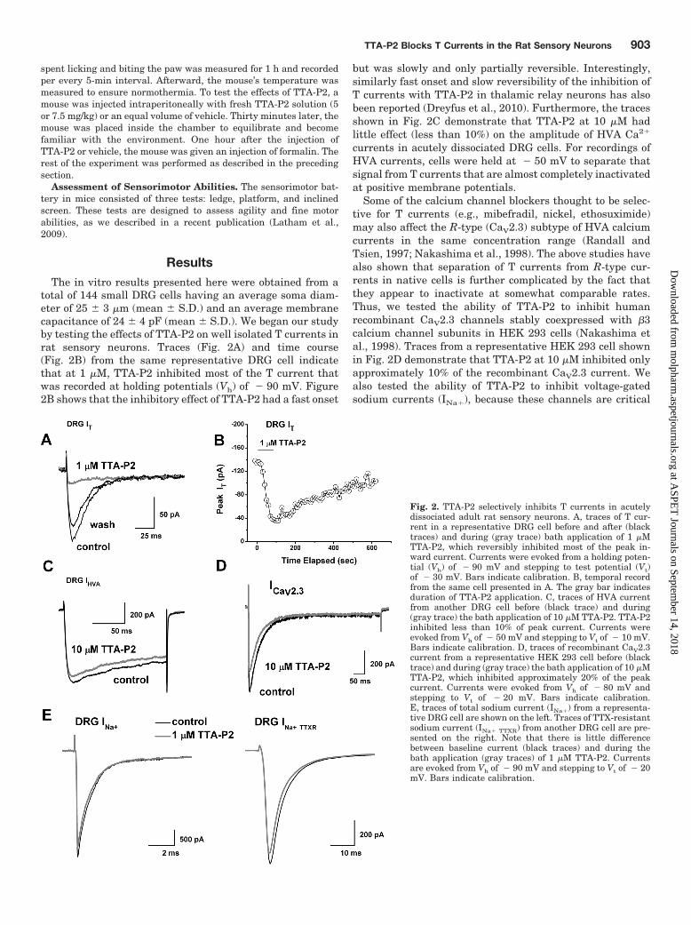
regulators of the excitability of nociceptive sensory neuronsand are implicated in neuropathic pain (Dib-Hajj et al., 1999;Lai et al., 2002; Hong et al., 2004). Traces from representa-tive DRG cells shown in Fig. 2E demonstrate that 1 �MTTA-P2 had little effect on the amplitude of total voltage-gated sodium currents (Fig. 2E, left, INa�) or the tetrodoxin-resistant component of voltage-gated sodium currents (Fig.2E, right, INa�TTXR). On average, the effects of 1 �M TTA-P2on the amplitude of DRG sodium currents were as follows:total INa�, 1 � 4% change (p 0.05, n � 8); INa�TTXR, 1 � 7%change (p 0.05, n � 5). To compare the potency of TTA-P2in inhibiting T currents and HVA currents in DRG cells, aswell as recombinant CaV2.3 currents, we obtained multiplepoints on concentration-response relationships and gener-ated best fits using eq. 1 (Fig. 3). These experiments indi-cated, impressively, that TTA-P2 was 2 to 3 orders of mag-nitude more potent in inhibiting DRG T currents (Fig. 3,filled circles; IC50 � �100 nM) than it was in inhibiting totalDRG HVA currents (Fig. 3, filled squares; IC50 � �165 �M)and recombinant CaV2.3 currents (Fig. 3, open triangles;IC50 � �35 �M).
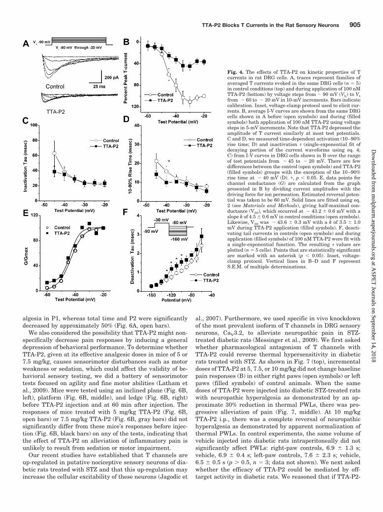
We then set out to discern the biophysical mechanisms ofT-current inhibition by TTA-P2, which could contribute tothe inhibition of current and, consequently, diminish thecellular excitability of DRG cells. To determine the effects ofTTA-P2 on the kinetic properties of DRG T currents, wemeasured current-voltage (I-V) relationships in the presenceand absence of 0.1 �M TTA-P2 ((Fig. 4A), finding that, com-pared with controls (open symbols), TTA-P2 (filled symbols)reduced T-current amplitudes at all test potentials between� 50 and � 20 mV (Fig. 4B) but had little effect on thekinetics of macroscopic current inactivation (Fig. 4C) or ac-tivation (Fig. 4D). The only significant effect was that
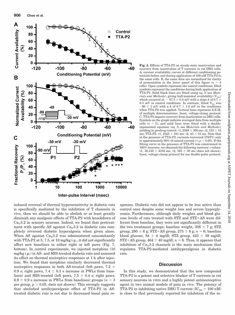
TTA-P2 slowed 10 to 90% rise time at test potential of � 40mV for approximately 50% (Fig. 4D). Likewise, TTA-P2 didnot induce significant alterations of channel gating, as shownby similar half-maximal activation (V50) of the T channelsbefore and during drug application (Fig. 4E). It is worthnoting that TTA-P2 decreased the rate of channel closure afterrepolarization, as demonstrated by significantly slower deacti-vation time constants (� values) at � 130, � 120, � 110,and � 100 mV (Fig. 4F). Binding to inactivated states and offrate of the compound are important properties of drugs thatmodulate ion channels because it allows them to have tissueselectivity based on the different membrane potential cyclingconditions. Transitions from closed to inactivated states can bemeasured using long prepulses at different potentials, produc-ing what are commonly referred to as steady-state inactivationcurves. We assessed steady-state inactivation curves using astandard double-pulse protocol with 3.6-s-long prepulses tovariable voltages (from � 110 to � 45 mV) and test potentialsto � 30 mV. As shown in Fig. 5, A and B, TTA-P2 (filledsymbols), compared with control conditions (open symbols), hada great effect on the voltage-dependent kinetics of channel in-activation, as determined by a hyperpolarizing shift in steady-state inactivation curves of approximately 20 mV. These datasuggest that TTA-P2 binds to and stabilizes inactive states ofthe channel and thus is a more potent blocker at depolarizedmembrane potentials. For example, it is evident in Fig. 5A that100 nM TTA-P2 inhibits approximately 40% of maximal T cur-rent at � 110 mV, whereas the same concentration inhibits Tcurrent almost 100% at � 65 and � 60 mV. T channels canrecover from inactivation during sufficiently long hyperpolar-izations of the neuronal membrane caused by the effects ofneuromodulators such as serotonin in DRG neurons (Nelson etal., 2005). This can drastically influence firing properties of cellsthat express T channels. Thus, we studied the effects of 100 nMTTA-P2 on recovery from inactivation using our standard dou-ble-pulse protocol with variable interpulse duration at � 90mV (Fig. 5C) after a 500-ms-long inactivating pulse (Vh � � 90mV; Vt � � 30 mV). Figure 5C depicts these data indicatingthat in the presence of TTA-P2 (filled squares), T currentsrecover partially to only approximately 60% of the T-currentamplitudes of predrug control values (Fig. 5C, open circles). Thehyperpolarizing shift in steady-state inactivation and incom-plete recovery from inactivation could be highly useful proper-ties for a channel inhibitor because, when applied in vivo, it willaffect actively firing neurons more potently than it will restingcells. Thus, we tested the efficacy of TTA-P2 in two frequentlyused animal models of pain.
Previous studies with CaV3.2 knockout mice have estab-lished the role of T channels in inflammatory pain (Choi etal., 2007). Thus, to explore the interaction of TTA-P2 and Tchannels in vivo, we used injections of formalin into the hindpaws of mice to measure inflammatory pain. The amount oftime mice spend licking and biting the injected paw in thefirst 5 min after injection is a response to direct activation ofperipheral nociceptors [phase 1 (P1)]. In contrast, responses10 to 60 min after injection reflect central sensitization ofpain [phase 2 (P2)]. As shown in Fig. 6A, TTA-P2 at 7.5 mg/kgi.p. (gray bars), compared with vehicle controls (black bars),significantly reduced licking and biting of the affected pawsin P1, P2, and total time from 30 to 50%. When TTA-P2 wasinjected at a dose of 5 mg/kg i.p., mice demonstrated some-what reduced, but not significant (approximately 20%), an-
Fig. 3. TTA-P2 inhibits T currents in DRG cells more potently than itdoes HVA currents in DRG cells and recombinant CaV2.3 currents inHEK 293 cells. The graphs illustrate concentration-response relation-ships for TTA-P2 inhibition of T currents in rat DRG cells (filled circles),HVA currents in DRG cells (filled squares), and recombinant CaV2.3currents in HEK 293 cells (open triangles) (n � 4–15 cells per data point).The solid line is the best fit (eq. 1; see Materials and Methods) forT-current inhibition (IC50 � 0.11 � 0.01 �M; slope coefficient, 0.8 � 0.1;maximal inhibition, 90 � 3% of the peak current), HVA current inhibition(IC50 � 165 � 35 �M; slope coefficient, 1.3 � 0.5; maximal inhibitionconstrained to 100%), and CaV2.3 current inhibition (IC50 � 35 � 9 �M;slope coefficient, 0.9 � 0.3; maximal inhibition constrained to 100%).
904 Choe et al.
at ASPE
T Journals on Septem
ber 14, 2018m
olpharm.aspetjournals.org
Dow
nloaded from
algesia in P1, whereas total time and P2 were significantlydecreased by approximately 50% (Fig. 6A, open bars).
We also considered the possibility that TTA-P2 might non-specifically decrease pain responses by inducing a generaldepression of behavioral performance. To determine whetherTTA-P2, given at its effective analgesic doses in mice of 5 or7.5 mg/kg, causes sensorimotor disturbances such as motorweakness or sedation, which could affect the validity of be-havioral sensory testing, we did a battery of sensorimotortests focused on agility and fine motor abilities (Latham etal., 2009). Mice were tested using an inclined plane (Fig. 6B,left), platform (Fig. 6B, middle), and ledge (Fig. 6B, right)before TTA-P2 injection and at 60 min after injection. Theresponses of mice treated with 5 mg/kg TTA-P2 (Fig. 6B,open bars) or 7.5 mg/kg TTA-P2 (Fig. 6B, gray bars) did notsignificantly differ from these mice’s responses before injec-tion (Fig. 6B, black bars) on any of the tests, indicating thatthe effect of TTA-P2 on alleviation of inflammatory pain isunlikely to result from sedation or motor impairment.
Our recent studies have established that T channels areup-regulated in putative nociceptive sensory neurons of dia-betic rats treated with STZ and that this up-regulation mayincrease the cellular excitability of these neurons (Jagodic et
al., 2007). Furthermore, we used specific in vivo knockdownof the most prevalent isoform of T channels in DRG sensoryneurons, CaV3.2, to alleviate neuropathic pain in STZ-treated diabetic rats (Messinger et al., 2009). We first askedwhether pharmacological antagonism of T channels withTTA-P2 could reverse thermal hypersensitivity in diabeticrats treated with STZ. As shown in Fig. 7 (top), incrementaldoses of TTA-P2 at 5, 7.5, or 10 mg/kg did not change baselinepain responses (B) in either right paws (open symbols) or leftpaws (filled symbols) of control animals. When the samedoses of TTA-P2 were injected into diabetic STZ-treated ratswith neuropathic hyperalgesia as demonstrated by an ap-proximate 30% reduction in thermal PWLs, there was pro-gressive alleviation of pain (Fig. 7, middle). At 10 mg/kgTTA-P2 i.p., there was a complete reversal of neuropathichyperalgesia as demonstrated by apparent normalization ofthermal PWLs. In control experiments, the same volume ofvehicle injected into diabetic rats intraperitoneally did notsignificantly affect PWLs: right-paw controls, 6.9 � 1.3 s;vehicle, 6.9 � 0.4 s; left-paw controls, 7.6 � 2.3 s; vehicle,6.5 � 0.5 s (p 0.5, n � 3; data not shown). We next askedwhether the efficacy of TTA-P2 could be mediated by off-target activity in diabetic rats. We reasoned that if TTA-P2-
Fig. 4. The effects of TTA-P2 on kinetic properties of Tcurrents in rat DRG cells. A, traces represent families ofaveraged T currents evoked in the same DRG cells (n � 5)in control conditions (top) and during application of 100 nMTTA-P2 (bottom) by voltage steps from � 90 mV (Vh) to Vtfrom � 60 to � 20 mV in 10-mV increments. Bars indicatecalibration. Inset, voltage-clamp protocol used to elicit cur-rents. B, average I-V curves are shown from the same DRGcells shown in A before (open symbols) and during (filledsymbols) bath application of 100 nM TTA-P2 using voltagesteps in 5-mV increments. Note that TTA-P2 depressed theamplitude of T current similarly at most test potentials.C and D, we measured time-dependent activation (10–90%rise time; D) and inactivation � (single-exponential fit ofdecaying portion of the current waveforms using eq. 4;C) from I-V curves in DRG cells shown in B over the rangeof test potentials from � 45 to � 20 mV. There are fewdifferences between the control (open symbols) and TTA-P2(filled symbols) groups with the exception of the 10–90%rise time at � 40 mV (D). �, p 0.05. E, data points forchannel conductance (G) are calculated from the graphpresented in B by dividing current amplitudes with thedriving force for ion permeation. Estimated reversal poten-tial was taken to be 60 mV. Solid lines are fitted using eq.2 (see Materials and Methods), giving half-maximal con-ductance (V50), which occurred at � 43.2 � 0.6 mV with aslope k of 4.5 � 0.6 mV in control conditions (open symbols).Likewise, V50 was � 43.6 � 0.3 mV with a k of 3.5 � 1.0mV during TTA-P2 application (filled symbols). F, deacti-vating tail currents in controls (open symbols) and duringapplication (filled symbols) of 100 nM TTA-P2 were fit witha single-exponential function. The resulting � values areplotted (n � 5 cells). Points that are statistically significantare marked with an asterisk (p 0.05). Inset, voltage-clamp protocol. Vertical lines in B–D and F representS.E.M. of multiple determinations.
TTA-P2 Blocks T Currents in the Rat Sensory Neurons 905
at ASPE
T Journals on Septem
ber 14, 2018m
olpharm.aspetjournals.org
Dow
nloaded from
induced reversal of thermal hypersensitivity in diabetic ratsis specifically mediated by the inhibition of T channels invivo, then we should be able to abolish or at least greatlydiminish any analgesic effects of TTA-P2 with knockdown ofCaV3.2 in sensory neurons. Indeed, we found that pretreat-ment with specific AS against CaV3.2 in diabetic rats com-pletely reversed diabetic hyperalgesia when given alone.When AS against CaV3.2 was administered concomitantlywith TTA-P2 at 5, 7.5, or 10 mg/kg i.p., it did not significantlyaffect new baselines in either right or left paws (Fig. 7,bottom). In control experiments, we injected morphine (10mg/kg i.p.) in AS- and MIS-treated diabetic rats and assessedits effect on thermal nociceptive responses at 1 h after injec-tion. We found that morphine similarly decreased thermalnociceptive responses in both AS-treated (left paws, 7.2 �0.9 s; right paws, 7.4 � 0.3 s increase in PWLs from base-lines) and MIS-treated (left paws, 7.5 � 0.4 s; right paws,6.9 � 0.9 s increase in PWLs from baselines) groups (n � 3per group, p 0.05, data not shown). This strongly suggeststhat abolished antihyperalgesic effect of TTA-P2 in AS-treated diabetic rats is not due to decreased basal pain re-
sponses. Diabetic rats did not appear to be less active thancontrol ones despite some weight loss and severe hypergly-cemia. Furthermore, although daily weights and blood glu-cose levels of rats treated with STZ and STZ�AS were dif-ferent from baseline, they were not significantly different inthe two treatment groups: baseline weight, 308 � 7 g; STZgroup, 280 � 6 g; STZ�AS group, 275 � 8 g; n � 6; baselineblood glucose, 84 � 4 mg/dl; STZ group, 423 � 39 mg/dl;STZ�AS group, 464 � 40 mg/dl; n � 6. Thus, it appears thatinhibition of CaV3.2 channels is the main mechanism thatregulates TTA-P2-mediated antihyperalgesia in diabeticrats.
DiscussionIn this study, we demonstrated that the new compound
TTA-P2 is a potent and selective blocker of T currents in ratsensory neurons in vitro and a highly potent antinociceptiveagent in two animal models of pain in vivo. The potency ofTTA-P2 in inhibiting native DRG T current (IC50 � 100 nM)is close to that previously reported for inhibition of the re-
Fig. 5. Effects of TTA-P2 on steady-state inactivation andrecovery from inactivation of T currents in rat DRG cells.A, current availability curves at different conditioning po-tentials before and during application of 100 nM TTA-P2 tothe same cells. B, the same data are normalized for clarityof presentation in the lower panel of this figure (n � 4cells). Open symbols represent the control conditions; filledsymbols represent the conditions during bath application ofTTA-P2. Solid black lines are fitted using eq. 3 (see Mate-rials and Methods), giving half-maximal availability (V50),which occurred at � 67.5 � 0.3 mV with a slope k of 6.7 �0.3 mV in control conditions. In contrast, fitted V50 was�90 � 1 mV with a k of 8.7 � 1.0 mV in the conditionswhen TTA-P2 was applied. Vertical lines represent S.E.M.of multiple determinations. Inset, voltage-clamp protocol.C, TTA-P2 impairs recovery from inactivation in DRG cells.Symbols on the graph indicate averaged data from multiplecells (n � 5), and solid lines were fitted with a double-exponential equation (eq. 5; see Materials and Methods);yielding in predrug control: �1, 2388 � 300 ms; �2, 155 � 15ms; TTA-P2: �1, 1045 � 181 ms; �2, 43 � 15 ms. Note thatin the presence of TTA-P2, currents recovered (P2/P1) onlyto approximately 60% of control current (�, p 0.05). If thefitting curve in the presence of TTA-P2 was constrained to100% recovery, we obtained the following recovery � values:�1, 16,130 � 4316 ms; �2, 165 � 50 ms (data not shown).Inset, voltage-clamp protocol for our double-pulse protocol.
906 Choe et al.
at ASPE
T Journals on Septem
ber 14, 2018m
olpharm.aspetjournals.org
Dow
nloaded from

combinant CaV3 currents CaV3.1 (93 nM), CaV3.2 (196 nM),and CaV3.3 (84 nM) (Shipe et al., 2008). Indeed, we foundthat TTA-P2 in DRG cells even at 100-fold higher concentra-tions has little effect on other voltage-gated currents in sen-sory neurons. We also found that TTA-P2 inhibited DRG Tcurrents in a voltage-dependent manner. Figure 4 summa-rizes our biophysical experiments, which indicate thatTTA-P2 induced mild slowing of channel closure after briefdepolarizations (deactivation), whereas it had a minimal ef-fect on channel gating and macroscopic inactivation and ac-tivation rates.
It is noteworthy that the results shown in Fig. 5 indicatethat TTA-P2 caused a marked voltage-dependent blockade ofT channels, as demonstrated by a large hyperpolarizing shiftof the steady-state inactivation curve. This voltage depen-dence indicates the drug’s preference for inactive states of thechannel, resulting in higher fractional block of the channel atdepolarized membrane potentials. This is in contrast tomechanisms of inhibition in thalamic relay neurons in brain
slices, where TTA-P2 acts as a state-independent antagonistof T channels (Dreyfus et al., 2010). However, it is possiblethat the difference between the mechanisms of channel inhi-bition in native thalamic and sensory neurons is due to theexpression of different T-channel isoforms in thalamic relayneurons (CaV3.1) and DRG cells (CaV3.2). Our finding issomewhat unexpected, given that a previous study foundthat recombinant CaV3.2 channels were also inhibited byTTA-P2 in a voltage-independent manner (Shipe et al., 2008).In contrast, the structurally unrelated compound TTA-A2shows state-dependent features of recombinant T-channelinhibition (Kraus et al., 2010). It is possible that differentsplice variants of CaV3.2 are expressed in adult DRG cells;alternatively, another unknown ancillary subunit that canmodify the interaction of TTA-P2 with T channels may becoexpressed in DRG cells. It is noteworthy that we also havedescribed substantial differences in the mechanism of T-channel blockade of native DRG cells and recombinantCaV3.2 channels for the anticonvulsants phenytoin and suc-cinimide (Todorovic et al., 2000).
Additional molecular studies will be needed to resolve thisissue. However, regardless of the precise basis for differencesin the observations, voltage-dependent inhibition of the chan-nel may be useful for clinical applications of TTA-P2, becauseit seems preferably to inhibit the neuronal excitability ofactively firing DRG cells. This effect may explain the selec-tive reversal of neuropathic hyperalgesia in diabetic ratswith a dose that was completely ineffective in naive rats (Fig.7). Thus, lowering the dose of drug used in vivo may greatlydecrease the risk of adverse side effects. It is also importantto note that some in vitro studies reported prominent expres-sion of CaV3.2 T currents in non-nociceptive subpopulationsof putative mechanosensitive DRG cells (Dubreuil et al.,2004; Coste et al., 2007). However, to our knowledge, the roleof T channels in mechanosensation has not been validatedin vivo.
The analgesic effect of TTA-P2 in the formalin inflamma-tory pain model in mice and antihyperalgesic effect in dia-betic rats are achieved at concentrations of 5 to 10 mg/kg(Figs. 6 and 7). Previous pharmacokinetic studies in rodentsand other species have shown that at these doses of plasmaconcentrations of TTA-P2 reach 0.2 to 1.0 �M and thatTTA-P2 penetrates well into the CNS (Shipe et al., 2008).These findings strongly suggest that the analgesic and anti-hyperalgesic effects of TTA-P2 observed in our study could berelated to the pharmacological antagonism of T currents inperipheral sensory neurons, the CNS, or both. It is notewor-thy that we directly implicated the CaV3.2 isoform of T chan-nels in the effects of TTA-P2 in diabetic neuropathic pain bydemonstrating that injections of antisense oligonucleotidesspecific for CaV3.2 completely abolished effects of TTA-P2 onthermal PWLs (Fig. 7).
Our study strongly suggests that TTA-P2 is more suit-able for functional studies than are many currently avail-able compounds thought to be selective T-channel block-ers. By virtue of their unique activation, deactivation, andinactivation properties, T currents are relatively easy tostudy in vitro in isolation from other calcium current com-ponents. However, pharmacological tools for identifyingand investigating T currents have been very limited. Forexample, it was reported that a scorpion toxin, kurtoxin,potently blocks recombinant T currents at nanomolar
Fig. 6. TTA-P2 has antinocicpetive properties in the formalin pain modeland has no effect on sensorimotor tests in mice. A, Adult mice experiencesignificant analgesia after intraperitoneal injections of TTA-P2 at 5mg/kg (open bars; n � 8) or 7.5 mg/kg (gray bars; n � 14) compared withexperiments with vehicle (control, black bars; n � 15). Vertical barsindicate S.E.M.; the asterisks indicate a significance of p 0.01 byStudent’s t test. B, histograms of average time in seconds in differentsensorimotor tests using an inclined plane (left), platform (middle), andledge (right) for mice given injections of 5 mg/kg TTA-P2 (white bars) or7.5 mg/kg TTA-P2 (gray bars). Baseline measurements were taken 2 daysbefore (controls, black bars). Note that black bars indicate controls withtwo different groups of animals receiving injections subsequently of ei-ther 5 or 7.5 mg/kg TTA-P2. Vertical bars indicate S.E.M. of multipledeterminations. At either dose, TTA-P2 had no significant effect on eithertest, because p 0.05 in comparisons of time points between controls andafter injections of TTA-P2 (n � 6 mice in each group).
TTA-P2 Blocks T Currents in the Rat Sensory Neurons 907
at ASPE
T Journals on Septem
ber 14, 2018m
olpharm.aspetjournals.org
Dow
nloaded from

range with an IC50 for CaV3.1 of 15 nM (Chuang et al.,1998). However, kurtoxin has limited usefulness because italso blocks voltage-gated Na� currents and native HVAcurrents within the same concentration range (Chuang etal., 1998; Sidach and Mintz 2002). Of the other describedcompounds with known effects on T currents, includingtraditional agents such as ethosuximide (Coulter et al.,1989) and amiloride (Tang et al., 1988), most are reportedto block with an IC50 in excess of 100 �M. At these con-centrations, effects on other ion channels also occur, rais-ing questions about the usefulness of these agents as spe-cific probes of T-current function. Among the otheravailable pharmacological agents, nickel at low micromo-lar concentrations (Lee et al., 1999b) and gaseous anes-thetic/analgesic nitrous oxide up to 80% (Todorovic et al.,2001) can be used for selective definition of CaV3.2 Tcurrents as opposed to other CaV3 currents. However, atsimilar concentrations, nickel blocks CaV2.3 R-type HVAcurrents (Zamponi et al., 1996), whereas nitrous oxideinhibits N-methyl-D-aspartate receptors in the CNS (Jev-tovic-Todorovic et al., 1998).
Some new experimental agents are more promising in
selectively targeting T channels. We have previously re-ported that a neuroactive steroid with a 5� configurationat the steroid A,B ring fusion [(�)-ECN] is a potent, volt-age dependent, but only a partial blocker of T channels inrat sensory neurons (IC50 � 300 nM; maximal block, 40%)(Todorovic et al., 1998). In addition, we recently describedanother novel steroid, a voltage-dependent blocker of Tchannels in rat sensory neurons; it has a 5� configurationat the steroid A,B ring fusion ([(3�,5�,17�)-3-hydroxyan-drostane-17-carbonitrile]), which more completely blocks Tcurrents, but with decreased potency with an IC50 of 2.8�M (Todorovic et al., 2004). Moreover, our previous studieshave documented that both ECN and [(3�,5�,17�)-3-hydroxyandrostane-17-carbonitrile] are approximately 10-fold more potent in inhibiting T-type currents than ininhibiting HVA currents in rat sensory neurons (Todorovicet al., 1998, 2004). Compared with neuroactive steroids, itseems that TTA-P2 is even more potent and more selectivein inhibiting T currents in DRG cells and thus is a bettertool for the study of these channels. Although no preclini-cal data are currently available on the long-term use ofECN and related neuroactive steroids, evaluation of the
Fig. 7. TTA-P2 effectively reverses hyperalgesia in diabeticSTZ-treated rats but does not change baseline nociceptivethresholds in healthy rats. Top, baseline (B) PWLs in rightpaws (open bars) and left paws (filled bars) remain stable1 h after intraperitoneal injections of TTA-P2 at 5, 7.5, or10 mg/kg. Middle, dose-dependent antihyperalgesic effectof TTA-P2, which, at 10 mg/kg i.p., completely reversedthermal hyperalgesia in STZ-treated diabetic rats. Bottom,note that AS treatment also completely reversed diabetichyperalgesia, as indicated by the apparent normalizationof PWLs, and that subsequent applications of TTA-P2 atdifferent doses did not change PWLs. Vertical bars indicateS.E.M. of multiple experiments (n � 4–8 per group). �, p 0.05 baseline (B) versus treatment at different doses ofTTA-P2; �, p 0.05 for treatment with TTA-P2 versus newbaseline in diabetic rats (STZ).
908 Choe et al.
at ASPE
T Journals on Septem
ber 14, 2018m
olpharm.aspetjournals.org
Dow
nloaded from
safety of TTA compounds in dogs has disclosed no cardio-vascular or renal side effects (Shipe et al., 2008).
This study confirms our previously published conclusionthat T-channel blockers are important and novel agents forthe treatment of pain disorders. No presently available treat-ments can completely reverse the significantly adverse ef-fects of intractable pain on quality-of-life measures. We hopethat our studies may inspire future clinical trials that willlead to better treatments for intractable inflammatory painand, in particular, the painful symptoms of patients withperipheral diabetic neuropathy.
Acknowledgments
We thank Damir Bojadzic for technical assistance and Dr. ToniSchneider for providing stable cell lines expressing CaV2.3�3 sub-units. We thank Drs. Victor N. Uebele and John J. Renger forproviding the TTA-P2 compound, supporting the experimental de-sign, and reviewing this manuscript.
Authorship Contributions
Participated in research design: Choe, Messinger, Salajegheh, Jev-tovic-Todorovic, and Todorovic.
Conducted experiments: Choe, Messinger, Leach, Eckle, Obra-dovic, and Salajegheh.
Performed data analysis: Messinger, Leach, Obradovic, and Todorovic.Wrote or contributed to the writing of the manuscript: Jevtovic-
Todorovic and Todorovic.
ReferencesAley KO and Levine JD (2001) Rapid onset pain induced by intravenous streptozo-
tocin in the rat. J Pain 2:146–150.Bourinet E, Alloui A, Monteil A, Barrere C, Couette B, Poirot O, Pages A, McRory J,
Snutch TP, Eschalier A, et al.(2005) Silencing of the Cav3.2 T-type calcium channelgene in sensory neurons demonstrates its major role in nociception. EMBO J24:315–324.
Campbell JN and Meyer RA (2006) Mechanisms of neuropathic pain. Neuron 52:77–92.Choi S, Na HS, Kim J, Lee J, Lee S, Kim D, Park J, Chen CC, Campbell KP, and Shin
HS(2007) Attenuated pain responses in mice lacking Ca(V)3.2 T-type channels.Genes Brain Behav 6:425–431.
Chuang RS, Jaffe H, Cribbs L, Perez-Reyes E, and Swartz KJ (1998) Inhibition ofT-type voltage-gated calcium channels by a new scorpion toxin. Nat Neurosci1:668–674.
Clozel JP, Ertel EA, and Ertel SI (1997) Discovery and main pharmacologicalproperties of mibefradil (Ro 40-5967), the first selective T-type calcium channelblocker. J Hypertens Suppl 15:S17–S25.
Coste B, Crest M, and Delmas P (2007) Pharmacological dissection and distributionof NaN/Nav1.9, T-type Ca2� currents, and mechanically activated cation currentsin different populations of DRG neurons. J Gen Physiol 129:57–77.
Coulter DA, Huguenard JR, and Prince DA (1989) Characterization of ethosuximidereduction of low-threshold calcium current in thalamic neurons. Ann Neurol 25:582–593.
Cribbs LL, Lee JH, Yang J, Satin J, Zhang Y, Daud A, Barclay J, Williamson MP, FoxM, Rees M, et al. (1998) Cloning and characterization of �1H from human heart,a member of the T-type Ca2� channel gene family. Circ Res 83:103–109.
Dib-Hajj SD, Fjell J, Cummins TR, Zheng Z, Fried K, LaMotte R, Black JA, andWaxman SG (1999) Plasticity of sodium channel expression in DRG neurons in thechronic constriction injury model of neuropathic pain. Pain 83:591–600.
Dogrul A, Gardell LR, Ossipov MH, Tulunay FC, Lai J, and Porreca F (2003)Reversal of experimental neuropathic pain by T-type calcium channel blockers.Pain 105:159–168.
Dreyfus FM, Tscherter A, Errington AC, Renger JJ, Shin HS, Uebele VN, Crunelli V,Lambert RC, and Leresche N (2010) Selective T-type calcium channel block inthalamic neurons reveals channel redundancy and physiological impact of I(T)win-dow. J Neurosci 30:99–109.
Dubreuil AS, Boukhaddaoui H, Desmadryl G, Martinez-Salgado C, Moshourab R,Lewin GR, Carroll P, Valmier J, and Scamps F (2004) Role of T-type calciumcurrent in identified D-hair mechanoreceptor neurons studied in vitro. J Neurosci24:8480–8484.
Edwards JL, Vincent AM, Cheng HT, and Feldman EL (2008) Diabetic neuropathy:mechanisms to management. Pharmacol Ther 120:1–34.
Hong S, Morrow TJ, Paulson PE, Isom LL, and Wiley JW (2004) Early painfuldiabetic neuropathy is associated with differential changes in tetrodotoxin-sensitive and -resistant sodium channels in dorsal root ganglion neurons in therat. J Biol Chem 279:29341–29350.
Institute of Laboratory Animal Resources (1996) Guide for the Care and Use ofLaboratory Animals 7th ed. Institute of Laboratory Animal Resources, Commis-sion on Life Sciences, National Research Council, Washington DC.
Jagodic MM, Pathirathna S, Nelson MT, Mancuso S, Joksovic PM, Rosenberg ER,
Bayliss DA, Jevtovic-Todorovic V, and Todorovic SM (2007) Cell-specific altera-tions of T-type calcium current in painful diabetic neuropathy enhance excitabilityof sensory neurons. J Neurosci 27:3305–3316.
Jevtovic-Todorovic V, Todorovic SM, Mennerick S, Powell S, Dikranian K, BenshoffN, Zorumski CF, and Olney JW (1998) Nitrous oxide (laughing gas) is an NMDAantagonist, neuroprotectant and neurotoxin. Nat Med 4:460–463.
Jevtovic-Todorovic V and Todorovic SM (2006) The role of peripheral T-type calciumchannels in pain transmission. Cell Calcium 40:197–203.
Kim D, Park D, Choi S, Lee S, Sun M, Kim C, and Shin HS (2003) Thalamic controlof visceral nociception mediated by T-type Ca2� channels. Science 302:117–119.
Kraus RL, Li Y, Gregan Y, Gotter AL, Uebele VN, Fox SV, Doran SM, Barrow JC,Yang ZQ, Reger TS, et al. (2010) In vitro characterization of T-type calciumchannel antagonist TTA-A2 and in vivo effects on arousal in mice. J PharmacolExp Ther 335:409–417.
Lai J, Gold MS, Kim CS, Bian D, Ossipov MH, Hunter JC, and Porreca F (2002)Inhibition of neuropathic pain by decreased expression of the tetrodotoxin-resistant sodium channel, NaV1.8. Pain 95:143–152.
Latham JR, Pathirathna S, Jagodic MM, Choe WJ, Levin ME, Nelson MT, Lee WY,Krishnan K, Covey DF, Todorovic SM, et al.(2009) Selective T-type calcium chan-nel blockade alleviates hyperalgesia in ob/ob mice. Diabetes 58:2656–2665.
Lee JH, Daud AN, Cribbs LL, Lacerda AE, Pereverzev A, Klockner U, Schneider T,and Perez-Reyes E (1999a) Cloning and expression of a novel member of the lowvoltage-activated T-type calcium channel family. J Neurosci 19:1912–1921.
Lee JH, Gomora JC, Cribbs LL, and Perez-Reyes E (1999b) Nickel block of threecloned T-type calcium channels: low concentrations selectively block alpha1H.Biophys J 77:3034–3042.
Leresche N, Parri HR, Erdemli G, Guyon A, Turner JP, Williams SR, Asprodini E,and Crunelli V (1998) On the action of the anti-absence drug ethosuximide in therat and cat thalamus. J Neurosci 18:4842–4853.
Lory P and Chemin J (2007) Towards the discovery of novel T-type calcium channelblockers. Expert Opin Ther Targets 11:717–722.
McCleskey EW and Gold MS (1999) Ion channels of nociception. Annu Rev Physiol61:835–856.
McGivern JG (2006) Pharmacology and drug discovery for T-type calcium channels.CNS Neurol Disord Drug Targets 5:587–603.
Messinger RB, Naik AK, Jagodic MM, Nelson MT, Lee WY, Choe WJ, Orestes P,Latham JR, Todorovic SM, and Jevtovic-Todorovic V (2009) In vivo silencing of theCa(V)3.2 T-type calcium channels in sensory neurons alleviates hyperalgesia inrats with streptozocin-induced diabetic neuropathy. Pain 145:184–195.
Nakashima YM, Todorovic SM, Pereverzev A, Hescheler J, Schneider T, and LingleCJ (1998) Properties of Ba2� currents arising from human �1E and �1E�3 con-structs expressed in HEK293 cells: physiology, pharmacology, and comparison tonative T-type Ba2� currents. Neuropharmacology 37:957–972.
Nelson MT, Joksovic PM, Perez-Reyes E, and Todorovic SM (2005) The endogenousredox agent L-cysteine induces T-type Ca2� channel-dependent sensitization of anovel subpopulation of rat peripheral nociceptors. J Neurosci 25:8766–8775.
Nelson MT, Woo J, Kang HW, Vitko I, Barrett PQ, Perez-Reyes E, Lee JH, Shin HS,and Todorovic SM (2007) Reducing agents sensitize C-type nociceptors by relievinghigh-affinity zinc inhibition of T-type calcium channels. J Neurosci 27:8250–8260.
Orestes P, Bojadzic D, Lee J, Leach E, Salajegheh R, Digruccio MR, Nelson MT, andTodorovic SM (2011) Free radical signalling underlies inhibition of CaV3.2 T-typecalcium channels by nitrous oxide in the pain pathway. J Physiol 589:135–148.
Perez-Reyes E (2003) Molecular physiology of low-voltage-activated T-type calciumchannels. Physiol Rev 83:117–161.
Perez-Reyes E, Cribbs LL, Daud A, Lacerda AE, Barclay J, Williamson MP, Fox M,Rees M, and Lee JH (1998) Molecular characterization of a neuronal low-voltage-activated T-type calcium channel. Nature 391:896–900.
Randall AD and Tsien RW (1997) Contrasting biophysical and pharmacologicalproperties of T-type and R-type calcium channels. Neuropharmacology 36:879–893.
Rogawski MA and Loscher W (2004) The neurobiology of antiepileptic drugs for thetreatment of nonepileptic conditions. Nat Med 10:685–692.
Shipe WD, Barrow JC, Yang ZQ, Lindsley CW, Yang FV, Schlegel KA, Shu Y, RittleKE, Bock MG, Hartman GD, et al. (2008) Design, synthesis, and evaluation of anovel 4-aminomethyl-4-fluoropiperidine as a T-type Ca2� channel antagonist.J Med Chem 51:3692–3695.
Sidach SS and Mintz IM (2002) Kurtoxin, a gating modifier of neuronal high- andlow-threshold ca channels. J Neurosci 22:2023–2034.
Snutch TP and David LS (2005) T-type calcium channels: an emerging therapeutictarget for the treatment of pain. Drug Dev Res 67:404–415.
Tang CM, Presser F, and Morad M (1988) Amiloride selectively blocks the lowthreshold (T)-type calcium channel. Science Wash DC 240:213–215.
Todorovic SM, Jevtovic-Todorovic V, Meyenburg A, Mennerick S, Perez-Reyes E,Romano C, Olney JW, and Zorumski CF (2001) Redox modulation of T-typecalcium channels in rat peripheral nociceptors. Neuron 31:75–85.
Todorovic SM and Lingle CJ (1998) Pharmacological properties of T-type Ca2�
current in adult rat sensory neurons: effects of anticonvulsant and anestheticagents. J Neurophysiol 79:240–252.
Todorovic SM, Meyenburg A, and Jevtovic-Todorovic V (2002) Mechanical and ther-mal antinociception in rats following systemic administration of mibefradil, aT-type calcium channel blocker. Brain Res 951:336–340.
Todorovic SM, Meyenburg A, and Jevtovic-Todorovic V (2004a) Redox modulation ofperipheral T-type Ca2� channels in vivo: alteration of nerve injury-induced ther-mal hyperalgesia. Pain 109:328–339.
Todorovic SM, Pathirathna S, Brimelow BC, Jagodic MM, Ko SH, Jiang X, NilssonKR, Zorumski CF, Covey DF, and Jevtovic-Todorovic V (2004b) 5�-Reduced neu-roactive steroids are novel voltage-dependent blockers of T-type Ca2� channels inrat sensory neurons in vitro and potent peripheral analgesics in vivo. Mol Phar-macol 66:1223–1235.
Todorovic SM, Perez-Reyes E, and Lingle CJ (2000) Anticonvulsants but not general
TTA-P2 Blocks T Currents in the Rat Sensory Neurons 909
at ASPE
T Journals on Septem
ber 14, 2018m
olpharm.aspetjournals.org
Dow
nloaded from
anesthetics have differential blocking effects on different T-type current variants.Mol Pharmacol 58:98–108.
Todorovic SM, Prakriya M, Nakashima YM, Nilsson KR, Han M, Zorumski CF,Covey DF, and Lingle CJ (1998) Enantioselective blockade of T-type Ca2� currentin adult rat sensory neurons by a steroid that lacks gamma-aminobutyric acid-modulatory activity. Mol Pharmacol 54:918–927.
Todorovic SM, Rastogi AJ, and Jevtovic-Todorovic V (2003) Potent analgesic effectsof anticonvulsants on peripheral thermal nociception in rats. Br J Pharmacol140:255–260.
Zamponi GW, Bourinet E, and Snutch TP (1996) Nickel block of a family of neuronalcalcium channels: subtype- and subunit-dependent action at multiple sites. JMembr Biol 151:77–90.
Address correspondence to: Slobodan M. Todorovic, Department of Anes-thesiology, University of Virginia Health System, Mail Box 800710, Charlot-tesville, VA 22908-0710. E-mail: [email protected]
910 Choe et al.
at ASPE
T Journals on Septem
ber 14, 2018m
olpharm.aspetjournals.org
Dow
nloaded from