treatment techniques for edema reduction during the …
TRANSCRIPT

Graduate Theses, Dissertations, and Problem Reports
2011
Treatment Techniques for Edema Reduction During the Acute Treatment Techniques for Edema Reduction During the Acute
Stage of the Inflammatory Cycle: A Systematic Review Stage of the Inflammatory Cycle: A Systematic Review
Adam D. Graham West Virginia University
Follow this and additional works at: https://researchrepository.wvu.edu/etd
Recommended Citation Recommended Citation Graham, Adam D., "Treatment Techniques for Edema Reduction During the Acute Stage of the Inflammatory Cycle: A Systematic Review" (2011). Graduate Theses, Dissertations, and Problem Reports. 4723. https://researchrepository.wvu.edu/etd/4723
This Thesis is protected by copyright and/or related rights. It has been brought to you by the The Research Repository @ WVU with permission from the rights-holder(s). You are free to use this Thesis in any way that is permitted by the copyright and related rights legislation that applies to your use. For other uses you must obtain permission from the rights-holder(s) directly, unless additional rights are indicated by a Creative Commons license in the record and/ or on the work itself. This Thesis has been accepted for inclusion in WVU Graduate Theses, Dissertations, and Problem Reports collection by an authorized administrator of The Research Repository @ WVU. For more information, please contact [email protected].

Treatment Techniques for Edema Reduction During the Acute Stage of the Inflammatory Cycle: A Systematic Review
Adam D. Graham, BS, ATC
Thesis submitted to the College of Physical Activity and Sport Sciences at West Virginia University in partial fulfillment of the requirements for the degree of
Master of Science in
Athletic Training
Michelle A. Sandrey, Ph.D., ATC, Chair Benjamin Moorehead, MD Elizabeth Bunn, MS., ATC
Damien Clement, Ph.D., ATC
Department of Sport Sciences
Morgantown, WV
2011
Keywords: lymphatic, acute, treatment, edema

ABSTRACT
Treatment Techniques for Edema Reduction During the Acute Stage of the Inflammatory Cycle: A Systematic Review
Adam D. Graham
Objective: To evaluate the methodological quality of edema reduction during acute injury studies found in the current literature. Data Sources: Pubmed (1951-2010), MEDLINE, ScienceDirect (1995-2010), CINAHL with Full Text (1982-2010), SPORTDiscus Full Text (1800-2010), MDConsult (1980-2010), Science Direct (1940-2010), Google Scholar were searched using the terms lymphatic system,edema reduction, inflammation process individually. Second, the term lymphatic system was combined with each of the following words: injury, acute, treatment,
edema, cryotherapy, electrical stimulation, compression, deep oscillation and Kinesio Tape.
Third, citations were cross referenced from studies to include literature not found in the original search. Study Selection: Studies were included based on the following inclusion criteria: 1) written in the English language; 2) edema reduction or a form of the word lymphatic must be in the title; 3) the abstract must include the terms lymphatic system, edema, modalities and treatment; 4) acute injury must be the chief complaint in the study; 5) and the study must be a randomized controlled trial. The exclusion criteria included any chronic injuries or conditions. Data Extraction: All the studies that meet the inclusion criteria were collected and evaluated via the PEDro Scale and a comparison of their effect size. Each study was read first without the use of the PEDro Scale, then upon completion it was read again with the PEDro Scale by both evaluators. Depending on the checklists completed by the evaluators each study was given a score from 0-10. Upon completion the final scores of the investigators were compared, and differences were discussed until a final score was agreed upon. Data Synthesis: There were 13 studies extracted in this review. They varied in population, location, type of treatment, inclusion/exclusion criteria, methodological quality and effect size. The methodological quality ranged from a 2 to a 8 with an average of 4.5. The effect size comparison ranged from -3.27 to 0.93 which shows the confidence interval crossed 0 demonstrating that most of the treatments were ineffective with only one massage study proven to have a significant treatment effect. Conclusions: Treatment methods for edema reduction through the lymphatic system is not well researched. There are several different treatments that have yet to be studied. Athletic trainers currently perform treatments without empirical evidence to support it.

iii
ACKNOWLEDGEMENTS
To my mother and my hero, Debi, for everything that she has given up and sacrificed to help me get to where I am today. I am so very thankful for her unyielding support, guidance and patience throughout the years. To the rest of my family, and Creighton, their continual support through the years helped me to realize who I wanted to be and for continuing to push me to strive to be the best that I can. I would like to thank Dr. Benjamin Moorhead, Liz Bunn and Dr. Damien Clement for being on my committee and for all of the help. I am very grateful for the help, and Dr. Clement for joining so late, in guiding me through this lengthy process. I would also like to thank Liz Bunn for helping me score the studies using the PEDro scale. Thank you for taking the time to assist me with this, I appreciate the time and effort that went in to scoring these studies. To my friends throughout the years, and especially the new ones I’ve made here at WVU, for their support, understanding and great memories that have kept us going through these two years. I would like to thank Dr. Sandrey for her numerous hours of support throughout this process. I
am very grateful for your assistance and guidance throughout this difficult process.

iv
TABLE OF CONTENTS
ACKNOWLEDGEMENTS...............................................................................................iii
LIST OF TABLES..............................................................................................................v
LIST OF FIGURES...........................................................................................................vi
INTRODUCTION..............................................................................................................1
METHODS..........................................................................................................................3
DATA SYNTHESIS............................................................................................................6
DISCUSSION......................................................................................................................18
CONCLUSION....................................................................................................................25
REFERENCES.....................................................................................................................26
APPENDICES......................................................................................................................29
APPENDIX A. THE PROBLEM.............................................................................30
APPENDIX B. LITERATURE REVIEW...............................................................34
APPENDIX C. ADDITIONAL METHODS............................................................63
APPENDIX D. ADDITIONAL RESULTS..............................................................67
APPENDIX E. RECOMMENDATIONS FOR FURTHER RESEARCH...............95
ADDITIONAL REFERENCES............................................................................................96
v
LIST OF TABLES Tables Page
B1. Comparison of Cryotherapy Studies…………………………………………………….51
B2. Comparison of Massage Treatment Studies……………………………………………..55
B3. Comparison of Electrical Stimulation Studies…………………………………………..59
B4. Comparison of Kinesio Tape Studies……………………………………………………61
C1. PEDro Scale....................................................................................................................63
C2. PEDro Scale Checklist with Explanations......................................................................64
C3. Protocol For Determining Studies..................................................................................66
D1. Cryotherapy Treatment Studies…...…………………………………………………….67
D2. Compression Treatment Studies……………...…………………………………………69
D3. Massage Treatment Studies……………………………………………………………..70
D4. Electrical Stimulation Treatment Studies…..……………………………………………71
D5. Comparison of Overall PEDro Scores.............................................................................72
D6. PEDro Checklist for Buzzard et al……….......................................................................73
D7. PEDro Checklist for Cheing et al.....................................................................................73
D8. PEDro Checklist for Haren et al ……...………………………………………………….74
D9. PEDro Checklist for Kessler et al ………………………………………………………..74
D10. PEDro Checklist for Knygsand et al …………….……………………………………...75
D11. PEDro Checklist for Man et al ………..………………………………...………………75
D12. PEDro Checklist for Meeusen et al....................... .........................................................76
D13. PEDro Checklist for Rucinski et al……….....................................................................76
D14. PEDro Checklist for Scheffler et al.................................................................................77
D15. PEDro Checklist for Stockle et al …………….………………………………………...77
vi
D16. PEDro Checklist for Thordarson et al …………..……………………...…………….…78
D17. PEDro Checklist for Tsang et al......................................................................................78
D18. PEDro Checklist for Weston et al....................................................................................79

vii
LIST OF FIGURES Figures Page
B1. Blood Flow Into the Interstitial Space ............................................................................44
B2. Funnel Arrangement of Lymphatic Valves ....................................................................44
D1. Figure of Studies Used: CINAHL...….………………………………………………….80
D2. Figure of Studies Used: SPORTdiscus.……...…………………………………………..81
D3. Figure of Studies Used: MEDLINE..….………………………………………………...82
D4. Figure of Studies Used: MDConsult………..……………………………………………83
D5. Figure of Studies Used: PubMed…….............................................................................84
D6. Figure of Studies Used: Cochran.. ………......................................................................85
D7. Figure of Studies Used: Google Scholar..........................................................................86
D8. Figure of Studies Used: Science Direct.………………………………………………….87
D9. Figure of Studies Used: PEDro Database….……………………………………………..88
D10. Effect Size Comparison………………………………………………………………….89

1
INTRODUCTION Acute musculoskeletal injuries can often have associated inflammation producing
swelling and edema. The resulting inflammation of the traumatic event is not entirely
destructive in nature as the inflammatory process is important due to a “consistent and lasting
response”1 which allows for tissue healing. The more damage an area sustains the more necrotic
tissue arises resulting in a longer time for the removal, thus increasing return to play timelines.2
The tissue repair process is marked by three distinct stages, the inflammatory stage, the
proliferation stage and the maturation stage.1,3,4,5 Each stage has a specific role in the healing of
tissues, however, there is still very little agreement on the exact timeline of these phases and it is
generally believed to overlap.1,3,4,5 If the initial stage is not managed properly then there is
potential risk of secondary injury due to ischemia and hypoxia.2 There has been little research
conducted to further examine secondary injury since Kinght’s secondary injury model presented
over 20 years ago.2
Removal of edema is important and the role of the lymphatic system is paramount.
Edema along with range of motion and equal strength all have an effect on making a return to
play decision. There are several modalities and lymphatic drainage techniques that have been
evaluated in the literature that assist edema reduction by including the lymphatic system. These
include but are not limited to cryotherapy, compression units, manual lymphatic drainage
techniques, bandaging and exercising.6 Cryotherapy is one of the oldest and most widely used
modalities. Cryotherapeutic treatments cause a decrease in tissue temperature which in turn
causes a decrease in cell metabolism, vasoconstriction, decrease in spasm and a decrease in
inflammation.7 These effects can be increased with the application of compression with
cryotherapy, moreover, compression can also be used alone in treatment.8 Compression alone

2
causes a change in the pressure forcing the edema out of the affected tissues and into non-
compressed areas.8 Manual lymphatic drainage techniques are interventions employed by
medical personnel and allied health clinicians during a rehabilitation protocol.9 These techniques
are used to open up new pathways in the superficial lymphatic system in the skin which will
assist in moving the fluid into the reservoir to be filtered and drained.6 Other recent techniques
are Hivamat and Kinesio Tape. Despite their popularity, there is very little research on the use
of Hivamat as compared to other modalities such as electrical stimulation or ultrasound.
Anecdotal evidence suggests the Hivamat “creates a fascial change by applying an intermittent
electrostatic charge to the collagen matrix.”11 Specifically the “HIVAMAT® 200 operates at the
level of the connective tissue using a pulsing electrostatic field, producing an intense resonant
vibration within the tissues involved. The repetition of this phenomenon in rapid succession
generates rhythmic deformations of the tissue. This action permits fiber and tissue layers to
reacquire motility and malleability.”11 Depending on the goals of the treatment Kineaio Tape
can be used to improve active range of motion, relieve pain, adjust misalignment, or improve
lymphatic circulation.12 Increased lymphatic circulation is achieved via the waves in the tape as
it raises and folds the layers of the epidermis lifting the skin.12
Several studies9,12,13,14,15 have been performed examining the effects of therapeutic
modalities and manual therapy. Evaluation of the methodological quality of these studies has
been conducted in treating acute injuries. Despite the present investigations there has been little
research conducted to examine the effects of these treatments on edema reduction through the
lymphatic system. Because of this, it can be difficult for athletic trainers to determine whether
treatments are truly effective and whether or not they can be applied clinically. Determining a
treatment effect does not appear to be examined in the literature. This comparison can be useful

3
when making the transition from basic science to practice. A methodologically sound study does
not necessarily have clinical implications. Therefore, comparing the effect sizes of the group or
treatment will demonstrate if that treatment favors the experimental group over the control. If
the effect size favors the treatment group versus the control then it shows that the treatment was
effective. From there it can be determined the treatment can be applied clinically since it was
shown to be effective.
This study will evaluate the literature as it pertains to treatment methods of edema
reduction through the lymphatic system during the acute phase of the inflammation process. The
treatments investigated will include cryotherapy, compression, electrical stimulation, lymphatic
drainage techniques and alternative methods. Evaluation will be performed using two separate
techniques: 1) to evaluate the methodological quality of included studies using the PEDro scale
and 2) to evaluate the effect size of these studies. Though several previous systematic reviews
have examined the methodological quality, this review will empirically evaluate group and
treatment results. This will assist the clinician to determine the effectiveness of a treatment and
whether to use in clinical practice.
METHODS
Design
The design of this study is a systematic review. Studies were obtained through searches
on specific databases using keywords, combinations of keywords, or cross referencing which
were then evaluated using the PEDro Scale and a comparison of the effect sizes. Randomized
control trials (RCTs) often give the medical field information about a certain treatment, however,
a systematic review of those RCTs can offer methodological evaluation of those studies.16 These
reviews have been believed by many authors to offer important information about the

4
effectiveness of medical interventions. Evaluation can reveal lesser quality studies that may
provide biased results of treatment effectiveness.
Instrumentation
The Physiotherapy Evidence Database (PEDro) scale is a tool used to evaluate the
methodological quality of RCTs.16 The PEDro scale is an 11 item scale which was developed
based on the 9 item Delphi scale 17,18 and the 3 item Jadad scale.16 Each item receives one point
which is added to the overall total score that ranges from 0-10 with the exception of item one,
which examines external validity and does not receive a point.16,18 In a study performed by
Maher et al.16 using the PEDro scale in two separate studies, intracalss correlation coefficient
(ICC) for interrater reliability was .55 at a 95% confidence interval (CI) in the first study, and .56
at a 95% CI in the second study. The ICC for consensus rating was .68 at a 95% CI. Therefore,
due to these findings it can be concluded that the PEDro score can be assessed with “fair” to
“good” reliability.16 Further information can be found regarding the PEDro score in Table C1
and C2.
Data Sources First, Pubmed (1951-2010), MEDLINE, CINAHL with Full Text (1982-2010),
SPORTDiscus Full Text (1800-2010), MDConsult (1980-2010), Science Direct (1940-2010),
Google Scholar and PEDro were searched using the terms lymphatic system, inflammation
process individually. Second, the term lymphatic system was combined with each of the
following words: injury, acute, treatment, edema, cryotherapy, electrical stimulation,
compression, deep oscillation and Kinesio Tape. Third, citations were cross referenced from
studies to include literature not found in the original search. This was performed for each
individual database. All results were limited to peer reviewed studies available in the English
5
language using human subjects. Once studies were selected they were compared to the
inclusion/exclusion criteria to determine final inclusion into the study. The included studies
were then evaluated with the PEDro Scale and a comparison of effect sizes was performed if data
was provided.
Study Selection
Studies were included based on the following inclusion criteria: 1) available in the
English language; 2) edema reduction or a form of the word lymphatic must be in the title; 3) the
abstract must include the terms lymphatic system, edema, modalities and treatment; 4) acute
injury must be the chief complaint in the study; and 5) the study must be a randomized controlled
trial. The exclusion criteria included any chronic injuries or conditions.
Data Extraction
There were two forms of data extraction in this review. The first was the use of the
PEDro scale to evaluate the methodological quality. The second was an examination of the
effect size estimate based on the determination of Cohen’s d.
Both investigators obtained training using the PEDro scale and met the requirements set
forth by the developers. This was achieved through the use of the PEDro website. Once the
training was completed, the studies that had been selected based on the inclusion criteria were
evaluated. The studies were read first without the use of the PEDro scale, followed by a second
reading which were then evaluated using the PEDro scale. Once this had been completed the
studies were given a score from 0 to 10 based on the checklist. Both investigators compared
their scores and if discrepancies occurred, discussion continued until a final score was agreed
upon. The PEDro scale was chosen because it has been shown to have sufficient reliability when
used in systematic reviews.16

6
The effect size estimate was utilized to examine the results of the study to determine if
the final results can be applied to the population and whether the results offered enough of a
difference to be considered a valid treatment. The Conhen’s d effect size comparison has a
standard scale in which less than a 0.4 is considered weak, from 0.41 to 0.7 as moderate and
greater than 0.7 as strong.19 To determine which treatments or combination thereof was most
effective at reducing edema, along with the hypotheses, this study had two questions. First, is
cryotherapy combined with compression is more effective at reducing edema than the other
treatments? Secondly, which treatment method alone is more effective in reducing edema? The
effect size comparisons used the difference between the means at pre test and post test
measurements for the experimental and the control groups. Due to the fact that there have been
several previous systematic reviews, the effect size estimate will provide a quantitative approach
in addition to the systematic review.
DATA SYNTHESIS
Study Quality
The 13 studies20, 21, 22, 23, 24, 25, 26, 27, 28, 29, 30, 31, 32 that met the inclusion criteria and were
evaluated varied in population, location, injury, inclusion/exclusion criteria and methodological
quality. The subject populations consisted of volunteers and hospitalized patients that were
recruited. Of the 13 studies, 5 of the studies had specific criteria for inclusion and exclusion
from the study.21, 23, 24, 28, 30 The remaining 8 studies had a detailed inclusion or exclusion criteria
but not both or had both but one was not thoroughly detailed.20, 22, 25, 26,27, 29, 31, 32 The inclusion
criteria included acute injuries,20, 27, 30, 31, 32 surgical intervention,22, 23, 28, 30, and one study used
healthy subjects26. The exclusion criteria included any chronic lymphatic conditions,20 21, 23, 24, 25,

7
28, 29 excessive musculoskeletal injury,20, 21, 23, 25, 29, 30 and several other general medical
exclusions that were inconsistent between studies.20, 21, 23, 24, 25, 29
The methodological quality ranged from a 228 to 825 with a mean average score of 4.5.
The PEDro scores can be found in Table D5-D18. There were some discrepancies between the
two raters, especially when evaluating for criteria 9. Steps were taken to insure understanding of
the criteria and applied to the studies. Of the 13 studies most met some aspect of the inclusion
criteria of this study. One of the inclusion criteria that was not met by all studies pertained to the
chief complaint must be an acute injury. Six of the studies22, 23, 24, 28, 29, 30,29 used subjects
following a surgical repair,23, 28, 29, 30 while others were used after an immobilization period.22,24
One other study used healthy subjects.26 This study was included because it was a rare study that
examined the effects of lymphatic flow when ice and compression was applied. All but 4
studies20,26,28,32 used random allocation for group assignment. Blinding was rarely performed in
any of these studies. None of the studies blinded the therapist providing the therapy, while one25
blinded the evaluators and only three21,24,25 blinded the subjects. Haren et al22 was the only study
that did not perform a baseline measure. Information of the studies with PEDro scores can be
found in Table D1-D4.
Treatment Techniques
Cryotherapy: This area of treatment was by far the most investigated with six of the 13
studies evaluating the effects on the lymphatic system and edema reduction. The greater number
of studies performed on cryotherapy in comparison to other techniques is based on use in the
clinical setting. Buzzard et al.20 examined the effects of pulsed shortwave diathermy versus ice
therapy in the reduction of edema following calcaneal fractures. The author did not describe any
of the allocation used to determine the experimental or control groups. Once demographic

8
information and delay from injury to starting treatment were recorded baseline circumference
measurements were taken of the affected foot and ankle. The measurements were performed
“around the malleoli, around the calcaneum diagonally, and the forefoot at the level of the head
of the fifth metatarsal.”20 A goniometer was used to measure dorsiflexion, plantarflexion and
subtalar inversion and eversion. These measurements were performed daily at 8:00 AM and
4:00 PM during the hospitalization in order to measure swelling, range of motion and any
fracture blisters. The pulsed shortwave diathermy group received treatment twice a day using a
pulse duration of 200 ms, a pulse frequency of 26 Hz and an intensity of 35 W for 15 minutes.
The cryotherapy group received Cryocuff treatment 6 times a day for 20 minutes. The author
suggested that there is little evidence to support an optimum protocol for use of the cryocuff.
Significant increases were reported for ankle range of motion (P = 0.007)20 however, there was
very little reduction in edema around the calcaneum, malleoli or forefoot in either group (P =
0.22)20. In the Cryocuff group, circumferential measurements were taken from day 1 to day 5
around the calcaneus (33±2, 33±2), malleolar (27±2, 27±2) and forefoot (24±2, 24±2). In the
shortwave diathermy treatment there was only one difference in the calcaneal measurement
(34±2, 33±2), with no other difference noted. Scheffler et al.28 observed the effect of the
Cryocuff on the control of postoperative pain and edema. Allocation utilized to group the
patients was not mentioned. The patients were instructed to use the Cryocuff for 30 min out of
every hour and only perform this when awake. The Cryocuff was evaluated using three different
methods consisting of the circumference of the experimental foot versus the control foot, the
patients perceived pain at the first, second, third, and fourth postoperative visits, and the overall
comfort of the Cryocuff. The only data presented by the study was that a standard deviation of
0.032 below circumference mean was noted in the group using the device, while a standard

9
deviation for the control group was +0.167 above circumference mean. Individuals using this
device on the test foot reported 80% less pain than the control foot.28 The perceived comfort was
graded on a 0 to 5 scale with 0 being worst and 5 being best. The perceived comfort score using
the Cryocuff was between a 4.64 and a 4.8 on the given criteria. This can be seen in Table D1.
Stockle et al.29 randomized 60 patients by assigning a continuous number according to
the date of admission and then alternated the subjects into one of the three treatment groups.
Each of the three groups consisted of 20 patients; the cool pack cryotherapy, continuous
cryotherapy and an intermittent impulse compression group. Circumference measurements were
taken at the ankle, midfoot and forefoot prior to surgery, the second day after surgery and then
every 24 hours until the patient was discharged which was typically within six days. The cool
pack therapy was changed four times a day and fixed around the swollen area with an elastic
dressing. Continuous cryotherapy used the Polar Care 500 device which is similar to the
Cryocuff, except instead of gravity this device uses a low-voltage pump to move the water.29.
Temperature of the water was maintained at 12°C for 8 hours with the ice water changed twice a
day. During the day the unit was used nearly continuously. For the intermittent compression
group the A-V Impulse system was used. This unit is designed to simulate weight bearing by
compressing and stretching the venae comitantes of the lateral plantar artery. The pad was
inflated with air to 130mmHg for 1 second every 20 seconds. The unit was used nearly
continuously throughout the day with the patient allowed to turn the unit off at night. Prior to
surgery, continuous cryotherapy and the A-V intermittent compression were both effective in
decreasing edema, however, the compression unit reduced posttraumatic swelling even faster
than continuous cryotherapy. This study did not present any of the statistics performed and a
brief summation of their results can be found in Table D1.
10
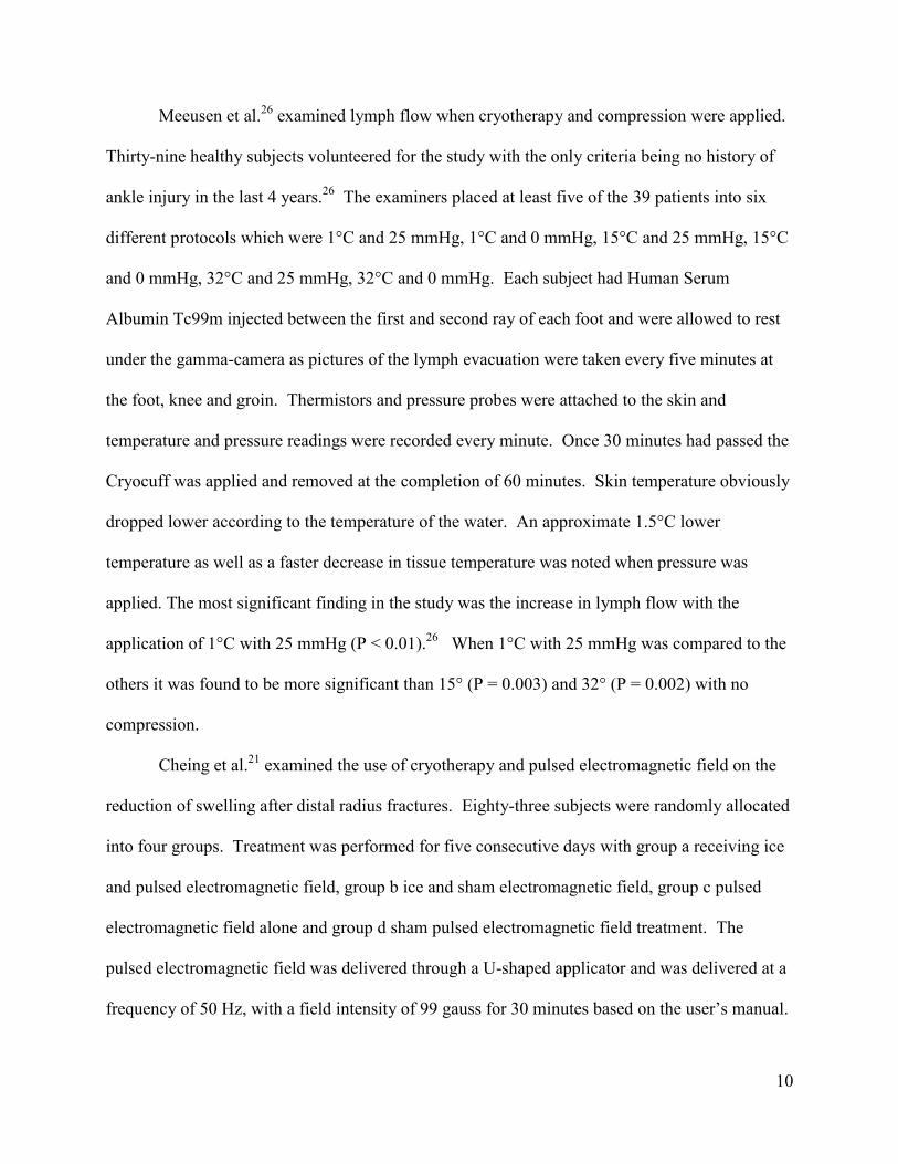
Meeusen et al.26 examined lymph flow when cryotherapy and compression were applied.
Thirty-nine healthy subjects volunteered for the study with the only criteria being no history of
ankle injury in the last 4 years.26 The examiners placed at least five of the 39 patients into six
different protocols which were 1°C and 25 mmHg, 1°C and 0 mmHg, 15°C and 25 mmHg, 15°C
and 0 mmHg, 32°C and 25 mmHg, 32°C and 0 mmHg. Each subject had Human Serum
Albumin Tc99m injected between the first and second ray of each foot and were allowed to rest
under the gamma-camera as pictures of the lymph evacuation were taken every five minutes at
the foot, knee and groin. Thermistors and pressure probes were attached to the skin and
temperature and pressure readings were recorded every minute. Once 30 minutes had passed the
Cryocuff was applied and removed at the completion of 60 minutes. Skin temperature obviously
dropped lower according to the temperature of the water. An approximate 1.5°C lower
temperature as well as a faster decrease in tissue temperature was noted when pressure was
applied. The most significant finding in the study was the increase in lymph flow with the
application of 1°C with 25 mmHg (P < 0.01).26 When 1°C with 25 mmHg was compared to the
others it was found to be more significant than 15° (P = 0.003) and 32° (P = 0.002) with no
compression.
Cheing et al.21 examined the use of cryotherapy and pulsed electromagnetic field on the
reduction of swelling after distal radius fractures. Eighty-three subjects were randomly allocated
into four groups. Treatment was performed for five consecutive days with group a receiving ice
and pulsed electromagnetic field, group b ice and sham electromagnetic field, group c pulsed
electromagnetic field alone and group d sham pulsed electromagnetic field treatment. The
pulsed electromagnetic field was delivered through a U-shaped applicator and was delivered at a
frequency of 50 Hz, with a field intensity of 99 gauss for 30 minutes based on the user’s manual.

11
The sham group was able to see the readout on the display but did not receive any treatment
because the circuit was not complete in the back.21 A baseline visual analogue scale (VAS),
volumetric measurement and range of motion assessment were performed and assessed again at
days 1, 3 and 5. At the completion of day three there was no significant difference in VAS
between groups, although group d experienced the least amount of reduction in pain. By day 5
the reduction in VAS score was greater in group a (1.8±0.8) when compared to the other groups
(b = 1.2±0.8, c = 1.0±0.8, d =0.7±0.6). For the volumetric measurements, from one to day three
there was a significant difference (P = 0.005) in the reduction between three groups(a =
10.4±10.3, b = 5.2±8.1, c = 6.6±7.0) when compared to the control (d = -0.94±12.5). The study
claimed significant differences from day one to day five (a = 25.0±16.2, b = 20.2±11.5, c =
13.9±7.2, d = 6.9±18.4) but did not present a significance. Once the study was completed post
hoc testing was performed showing group a was considerably better than groups c and d, while
group b was better than group d. However, there was no significant difference between groups b
and c. Range of motion results varied based on the direction of motion. The difference in flexion
improved significantly (P = 0.034) from day one to day three in the two pulsed electromagnetic
field groups (a = 6.3±5.7, c = 7.1±6.1) versus the two sham groups (b = 3.4±4.5, d = 3.1±3.6)
and again a similar increase was noted on day five, however it was not significant (P = 0.084).
Wrist pronation did not improve from day one to three (a = 6.3±5.9, b = 7.5±5.7, c = 5.0±4.6, d =
4.4±6.0), but was more significant (P = 0.021) by day five (a =18.0±10.8, b = 15.9±9.3, c =
13.4±7.0, d = 9.1±8.0) . There was a difference between groups as groups a and b decreased
more edema than group d. The difference between groups b and c were minimal.21
Weston et al.32 used 15 subjects who were suffering from mild to moderate level
inversion ankle sprains. Part of the inclusion criteria was the use of a written questionnaire to
12
rule out certain conditions. Data was collected using a bilateral tetrapolar impedance
plethysmograph to measure changes in fluid volume. The subject was first allowed to sit for 10
minutes before lying prone for 10 minutes with their knees placed in 15-20° of flexion
throughout the remainder of the session. When 10 minutes had passed the resting data was
collected. A blood pressure cuff was placed around the patients’ thigh and inflated to 20 mmHG
below the resting diastolic blood pressure to allow arterial blood flow but obstruct venous return.
Over the next 20 minutes 10 measurements were taken before a cold gel pack was placed on the
ankle for an additional 20 minutes. The study claimed that there was approximately a 50%
reduction in volume increase when the cold gel pack was applied. There was no significant
difference found in the patients systolic(P = 0.31), diastolic(P = 0.20) and heart rate (P = 0.42).
An examination of the effect size of the two cryotherapy studies20,21 compared the
treatment to the control at post test. Within the control cryocuff treatment, there was no change
in the measurements (d = 0.00) while within the treatment group there was a moderate effect of -
0.50 which demonstrated that the treatment favored the post test and edema decreased. This
effect, however, was not significant as the confidence intervals crossed over zero. Cheing et al.21
used four treatment groups as effect sizes ranged from -0.44 to -0.10 with a small to moderate
effect for group b and group d, respectively. Group a had an effect size of -0.37 and group c had
an effect size of -0.17. Treatments favored the post test resulting in a reduction of edema. The
confidence intervals crossed zero which demonstrated that the treatment effect was not
significant.(Figure D10)
Studies in the cryotherapy treatment group scored 2,28 320, 29, 26, 32 and one study scored as
high as 6.21 These studies did not blind the subjects, therapist or assessor or presented their data
appropriately. Individual PEDro scores can be found in Table D5-D18.

13
Compression: Compression is used frequently by athletic trainers. As shown in the
Meeusen et al.26 study, cryotherapy treatment improved when combined with compression.
Rucinski et al.27 used 30 subjects with sprained ankles and randomly assigned the subjects to
three different treatment groups. The treatments were an elastic treatment group, intermittent
compression group and an elevated control group. For the first group a plain ace bandage was
used from the metatarsal heads to approximately 12.7 centimeters above the malleoli and was
applied in the typical tighter to looser fashion with 45° elevation for 30 minutes. The second
group used a single cell intermittent compression device while the leg was elevated to 45°. The
study utilized a protocol following Prentice with 60 seconds on and 15 seconds off with the
pressure set to a range of 40 – 50 mmHG for 30 minutes. Control group subjects elevated the leg
45° for 30 minutes with no compression treatment. Baseline and post measurements were
performed with a volumetric tank. Surprisingly the control group showed the most significant
(P < 0.01) decrease in edema (pre = 1350.4±201.6, post = 1335.5±203.2) while the other two
(compression pre = 1222.0±166.7, post = 1229.4±166.5; wrap pre = 1340.2±128, post =
1343.9±127.4) treatments had an increase in edema with intermittent compression providing the
most.
Tsang et al.31 examined the effect of intermittent compression versus elevation as a
control condition. This study used 12 subjects presenting with a post-acute inversion ankle
sprain. The subjects started in a dependent position for 10 minutes before a baseline volumetric
measurement and blood pressure were taken. The subjects were then placed in a supine position
with the injured ankle elevated either with or without intermittent compression for 30 minutes.
Once treatment was completed the subject went back to a gravity dependent position with both
feet flat on the floor. Measurements were taken every five minutes for the first 30 minutes then

14
once at 45 and 60 minutes. The intermittent compression treatment was 45 seconds of inflation
with 15 seconds of deflation at a pressure based on the patients’ diastolic blood pressure. The
results did not directly compare the two treatment methods, but both showed a significant (P <
0.05) reduction in edema from baseline to immediately after treatment (17.25±4.05). However,
once the patient resumed the gravity dependent position the effects were lost within five minutes
as there was only a difference of 8.91 versus 17.25.
Thordarson et al.30 used 30 patients with a closed Weber B or C ankle fracture. The
patients were randomized into a pneumatic pedal compression (PPC) treatment group and a
control group. The control treatment consisted of a posterior splint, ice and elevation while
awaiting surgery. The PPC treatment consisted of the placement of the device on the foot with a
posterior splint applied as the leg was elevated. The PPC device “works by intermittently
compressing the veins in the foot, thus facilitating venous drainage…it inflates in less than one
second, compressing the foot temporarily, and then deflates more gradually.”30 Volumetric
baseline measurements were taken daily up until the day of surgery. The results for this study
are only presented as a change in volume from day one to day two and day one to day three.
There was a decrease in volume in the PPC group by 88 mL from day one to two while control
increased 33 mL (P = 0.027). From day one to day three the experimental group decreased an
addition 31 mL while the control increased 32 mL (P = 0.049).
Rucinski et al.27 used three treatment groups and compared compression and elevation to
an elastic wrap and elevation and elevation alone. The examination of the effect size confirms
the findings in the study. The compression and elevation group had a weak effect size of 0.04
and 95% CI’s ranging from -0.83 to 0.92. That the treatment favored the pre-test which means
the treatment increased edema. In the elastic wrap group there was a weak effect size of 0.03
15
with 95% CI’s ranging from -0.85 to 0.91. Again the treatment favored the pre-test resulting in
an increase of edema. The elevation group showed the only edema reduction with a weak effect
size of -0.07 and 95% CI’s ranging from -0.95 to 0.80, showing the treatment favored the post
test. Though these findings were unexpected none of them were significant as the confidence
intervals crossed zero ranging from -0.95 to 0.92 collectively.(Figure D10)
Studies in this section scored a 3,30 531 and a 627 . All the studies did not receive a yes
score for blinding the subjects, therapists or assessors. Thordarson et al.30 did not present any
data, which made it difficult to determine the treatments effectiveness.
Massage: Massage appears to be used on occasion by athletic trainers but certainly not as
often as the previous two treatments. Haren et al.22 examined the effects of manual lymph
drainage (MLD) to reduce edema of the hand after fracture of the distal radius. Twenty-six
patients were divided randomly into an experimental group and control group. All patients
regardless of group received the same conventional treatment of elevation, active and passive
exercises, and compression with elastic bandage while the external fixation was in place. Once
the fixation was removed wrist exercises were added using a home program. The difference was
in the 10 minutes of MLD as described by Vodder. In short, this method utilized a light surface
massage starting proximally near the drainage into the right and left subclavian veins, and slowly
continued distally into the injured area. Measurements were performed with a volumeter at 3,
17, 33 and 68 days after removal of the external fixation. Results showed a decrease in edema
for the control (day 3 = 64±41, day 17 = 50±35, day 33 = 35±26, day 68 = 24±20) and
experimental (day 3 = 39±12, day 17 = 27±9, day 33 = 19±9, day 68 = 12±11), with the effect
size favoring the experimental group.

16
Knygsand et al.24 used 30 patients randomly assigned into a control group receiving
traditional edema treatment and a experimental group receiving modified manual edema
mobilization (MEM). Treatments were three times a week for four weeks, then twice a week for
two weeks.24 Treatment continued until the therapist felt functional requirements were met and
the patient perceived it was at a level to suit their needs. The traditional treatment included
elevation, compression and functional training along with Coban applied around the digits and
proximal to the wrist. The modified MEM included “deep diaphragmatic breathing, exercises
that started proximally and ended distally, terminus (supraclavicular area) stimulation, axillary
stimulation in the uninvolved side, and MEM to the trunk region followed by MPP stimulation to
the involved upper extremity.”24 Measurements were performed using a volumeter and the
subjects edema, pain, AROM and ADL at inclusion and at one, three, six, nine and 26 weeks.
No significant difference between the two groups reduction of edema at inclusion (experimental
= 86.8 mL, control = 96.3), one week (68.2, 77.3), three week (41.1, 54.0), six weeks (28.6,
43.3) or nine weeks (12.1, 28.3) was evident. (P = 0.33, 0.40, 0.31, 0.13, 0.06) 24
Kessler et al.23 randomized 23 subjects with hindfoot surgery into two groups to assess
the effectiveness of manual lymph drainage versus a control. Both groups received the same
standard physiotherapy protocol consisting of thrombosis prophylaxis instructions, active and
passive ankle movements and daily gait training on crutches. The ankle movements consisted of
dorsiflexion and plantarflexion performed 50 times without resistance as well as 25 with
resistance the same time of the day prior to the MLD. For the intervention group “gentle manual
pressure was applied to each of the dermal lymphotomes to direct lymph flow to the non-
obstructed lymph nodal areas…a firm pressure was applied to watershed areas…”23 for 30
minutes a day. This treatment was unique to each patient’s needs and performed by the same

17
licensed physiotherapist. A volumeter was utilized to measure the potential edema reduction
measured two days after surgery and again at time of discharge. The study claimed that based on
the results there was a significant (P = 0.032) decrease in lower extremity swelling when
compared to the control group. Data was only presented using a graph.
Haren et al.22 was the only study in this treatment group to present complete data to
assess effect size. Measurements were at baseline then at three more points within the control
group from baseline to the first measurement and then again from baseline to the end of the study
the effect sizes were weak (d = -0.34) and large (d = -0.98) effect, respectively. The 95%
confidence intervals for the shorter time period ranged from -1.09 to 0.40 and the longer time
period ranged from -1.76 to -0.19. Thus demonstrating the treatment favored the post test in
both measures but was only significant and therefore effective over the longer period of time.
The treatment group showed there was a large effect (d = -1.00, d = -2.25) for the treatment
group favoring the post test with both demonstrating a significant and effective treatment with
95% CI’s ranging from -1.85 to -0.15 and -3.27 to -1.23, respectively.(Figure D10)
This treatment group as a whole had higher scores than the previous two treatment
groups. Two studies22,23 scored a 5 while one24 scored a 7. These studies were not awarded
points for the blinding of subjects, therapists and evaluators.
Electrical stimulation: Man et al.25 used 34 subjects with an acute ankle sprain and
randomly assigned into either the neuromuscular electrical stimulation group (NMES), the
submotor electrical stimulation or a group with electrodes but no electrical stimulation applied.
Ankle girth measurements as well volumetric measurements were performed at baseline. The
electrodes were placed over the tibialis anterior muscle of the injured limb. The electrical
stimulation treatments lasted 30 minutes. Intensity was increased with the NMES group to the
18
subjects maximum tolerance. The submotor group used an EMS unit turned up until a flicker of
muscle contraction was seen, then the intensity was lowered. The subjects in the sham treatment
received the same electrode placement but no electrical stimulation. No statistically significant
differences were found in any of the three groups (NMES group = pre 1522±262, post
1515±230; submotor group = pre 1597±339, post 1573±314, sham = pre 1324±215, post
1352±213) when compared pre-test and post test (P > 0.017).25
Examining the measures for the treatments in this study, the sham treatment had a weak
effect size of 0.13 with 95% CI’s ranging from -0.67 to 0.93, indicating that the treatment
favored the pre-test, but the treatment was not significant and ineffective as the CI’s crossed
zero. The submotor group had a weak effect size of -0.07 with CI’s ranging from -0.91 to 0.77,
which demonstrated that the treatment favored the post-test measurement but was not a
significant effect as the CI’s crossed zero. Examination of the NMES group showed a weak
effect with an effect size of -0.03 and 95% CI’s ranging from -0.86 to 0.81. The treatment
favored the post treatment measurement but the CI’s crossed zero.(Figure D10)
Though there was only one study performed on humans pertaining to lymph treatment the
study scored well and received an 8. One point was not awarded as the therapist providing the
treatment was not blinded and the intention to treat analysis was not provided.
Included in this search was alternative treatments which included the Hivamat and the
Kinesio Tape method. No treatments pertained to the reduction of edema in an acute injury and
were not used in this study. The Hivamat studies were on lymphedema which did not pertain to
this study. The Kinesio Tape studies primarily dealt with pain reduction and chronic
lymphedema treatment also.

19
DISCUSSION
The purpose of this study was to examine the methodological quality of treatment studies
for edema reduction and lymphatic evacuation found in the current literature. Along with the
hypotheses, this study also asked two questions: First, is cryotherapy combined with
compression is more effective at reducing edema than the other treatments? Secondly, which
treatment method alone is more effective in reducing edema? Based on the effect sizes it can be
stated that cryotherapy and compression, although effective in their respective studies, were
ultimately ineffective as the confidence intervals of each group crossed zero. As per the second
question, there was only one treatment to be truly effective. Haren et al.22 reported that the
treatment group that received massage had a large significant effect demonstrating the treatment
favored the post-test measurement. Therefore, the only treatment shown to be effective was
manual lymphatic drainage. All of the other treatments were found to be ineffective. One
problem, however, with the studies that received the effect size comparison is the sample size
used. Therefore, there might be a larger effect size if the subject population size was larger.
This can be seen further in Figure D10.
This review had three experimental hypotheses. The first one stated that there will be a
difference in outcomes between the different treatment techniques. The second one stated that
the severity of the traumatic event can have an effect on the type and amount of lymphatic
management. The third one stated that there will be enough information in each study to be
scored via the PEDro scale. Given the information provided, the first and third hypotheses were
confirmed. There was not enough information presented in the studies reviewed to confirm or
reject the second hypothesis.

20
The methodological scores of the studies ranged from a 2 to an 8 with an average score of
4.5 which demonstrated that overall the studies were of poor quality. Most of the studies in this
review used volumetric or girth measurement to evaluate the effectiveness of the treatments.
Some studies examined range of motion and pain, however, due to the focus of this systematic
review did not pertain. The outcomes of the studies had some positive findings and some
unexpected ones as well. Several of the cryotherapy studies21,26,28, supported the use of
cryotherapy while other studies20,29,32 did not discredit but did not state that cryotherapy assisted
in the reduction of edema. Compression treatments were shown to be effective,31 however,
elevation is more effective and compression had an adverse effect.26 Tsang et al.31 noted the
effects last less than five minutes before edema begins to re-enter the area. All three of the
massage treatment studies22,23,24 found success with treatment. The one stimulation study25 noted
some changes but stated that it could have been attributed to the difference at baseline.
Evaluation of Positive Outcomes
Cryotherapy and Compression: Some of the studies evaluated did find some positive
outcomes for their respective treatments. The cryotherapy group rarely utilized a cryotherapy
treatment alone and when it did it was found to be the least effective.29,32 The remainder of the
studies utilized some form of compression mainly with a Cryocuff unit or similar. Buzzard et
al.20 was the only study that did not notice a difference with the use of the Cryocuff. In their
study shortwave diathermy was used in comparison instead of a control treatment or another
cryotherapy treatment. Meanwhile, Meeusen et al.26 reported that a cryotherapy treatment was
effective but the application of compression increased the effectiveness. Stockle et al.29 reported
that the compression unit was the most effective but that the combination was more effective
than a cryotherapy treatment alone. These effects could be due to vasoconstriction that occurs
21
during the application of cryotherapy treatment as well as the decreased metabolic rate of the
cells.29,26 Vasoconstriction can in fact reverse if the treatment is too cold but this was not the
case in the studies reviewed. Meeusen et al.26 adds that the compression makes the treatment
even more effective because the outside pressure results in an increase in hydrostatic pressure
causing the fluid to flow out of the area.
The compression studies provided the most unexpected results found in this review. In
the Rucinski et al.27 study the control group had the largest decrease in edema while the other
two increased the amount of edema present. After the elevation or compression treatment ended
and as soon as the athlete regained a gravity dependent position the effects gained in the
treatment started to decrease within five minutes.31 The last study30 in this group used a non-
traditional type of compression with a pneumatic pedal compression unit. This unit is applied to
the foot and the attempt is to mimic the venous flow during ambulation. This is performed by
allowing the vessels to fill, as they would during the swing phase, when the pump is off, then
emptying, as they would during the stance phase, as the pump inflates.30 Positive results showed
change by 121 mL by day one and an additional 63 mL on day two. Some explanation of the
results was presented by Rucinski et al.27as to why there was an increase in findings. The
uniform pressure caused by compression can allow for a back flow of lymph, known as the
rebound phenomenon, which can occur when the athlete changes position from elevation to a
gravity dependent position.31 There was an increase in blood volume due to the response of the
intermittent compression and a potential increase in temperature due to the application. As with
the cryotherapy compression studies it is believed that the compression caused that change in the
pressure gradient with an increase in lymphatic flow.30

22
Massage: The massage treatment studies scored well with PEDro scores of 522,23 and 7.24
The limitation to this section is the subjects used and not using acute injuries. Two of the
studies22,24 used subjects who were recovering from immobilization after a distal radius fracture.
They were included because the patients were treated during an acute stage of rehabilitation
where edema is present. The results varied on the effectiveness of massage as a helpful tool for
the reduction of edema. Haren et al.22 found significantly less edema in the treatment group
receiving the manual lymph drainage than in the control. Another type of massage was
investigated by Knygsand et al.24 and manual edema mobilization was found to be statistically
insignificant in edema reduction. The remaining study23 examined patients’ immediately after
surgery and discovered there was a significant difference in the group receiving the manual
lymph drainage. While each of the studies presented different methods for performing the
massage techniques, all used light circles that started up the chain working in an area ahead of
the swelling.22,23,24
Electrical Stimulation: Electrical stimulation treatment has been very well supported
using animal models, however, when applied to humans it appeared to be ineffective.25 A
significant difference however, was attributed to a difference in baseline measurements. In the
discussion by Man et al.,25 possibilities are provided as to why the protocols were not effective.
The greatest factor is the protocols could potentially be more effective in a situation where the
swelling had occurred due to prolonged motionless standing causing venous return problems
versus a capillary permeability.25 A motor reaction would cause stimulation of the musculo-
venous pump. Man et al.25 continues to theorize that such capillary permeability present in
injuries for example ligament sprains, might respond better to submotor ES such as high volt.

23
This was evaluated by Mendel et al.63 with positive results and can be read in further detail in
Appendix B.
Limitations of the Study
Even though there were positive outcomes in the reported studies used, there are
limitations that should be discussed. The first area pertains to the blinding of the subjects by the
studies reviewed. Following the outline of the criteria for the PEDro scale only three
studies21,24,25 out of 13 blinded the subjects and even less blinded the assessor.25 It is difficult to
speculate as to the reasons there was such little blinding performed in the studies. Blinding of
subjects during treatments may be difficult in that the patient can feel ice or massage. Because
blinding was not performed many of the studies were not awarded points. Six
studies20,26,28,29,30,31 out of the 13 received a score less than a 5/10. Mosley et al.17 points out that
score of 5 or higher out of 10 is considered to have moderate to high methodological quality.
Therefore, it can make it difficult to apply the results of those studies to clinical practice due to
poor study design and potential biases.
The largest limitation of this study by far has to be the lack of literature on edema
reduction for acute injuries and more specifically as it pertains to the lymphatic system. Athletic
trainers are often charged with returning athlete’s to play in a more accelerated mode and
utilization of the lymphatic system may assist with this, but little evidence based support is
available. Along with a lack of literature was a lack of data presented by the studies included,
which meant evaluation of the effect size could not be performed on all studies. This could
potentially be due to the time at which the studies were published where a graph of the data was
more acceptable than presenting a table of means and standard deviations. Use of different
treatment protocols for compression and cryotherapy are used but little is known about how

24
much pressure to apply or the temperature of the ice bag. Meeusen et al.26 suggested that
temperature of the water should be 1°C 25 mmHg of pressure.
One of the inclusion criteria was that the study must be available in the English language,
or an English translation. This could have caused a number of foreign studies not available in
English to be excluded from the study that might have pertained to the other inclusion criteria.
There were no studies on acute treatment for two widely used clinical products in Kinesio Tape
and the Hivamat machine. The studies that were found in the search focused primarily on pain
and chronic lymphedema, particular post mastectomy. Both are used on a daily basis in many
athletic training rooms without knowledge of the exact mechanism of treatment effectiveness.
Though the PEDro scale is a widely accepted tool to grade the methodological quality
there are still many others that are in use today. Though there is training to assist the raters in
understanding the criteria there is still the human element and it is possible that the raters missed
some of the information and scored a study either higher or lower than it was worthy. It is also
unclear based on the criteria if some criteria are worth more than others despite only getting one
point. For instance, many of the studies did not blind the subjects, therapists or assessors but that
does not mean they were not good quality studies.
Clinical Relevance
The information in this review does provide some useful information to athletic trainers
in that lymphatic treatment is not very well understood at this moment in time. Until more
research can be conducted, the best option is clinical practice with some assistance from
evidence based to determine treatment of acute injuries. Even the current studies lack high
methodological quality resulting in potentially poor application to treatment of athletes. This is
supported by an examination of the effect size of five studies.27,20,21,22,25 Comparing pre and

25
post test within the group, one study demonstrated a strong effective size in both the treatment
and control. Haren’s study22 used a standard treatment for both groups which significantly
reduced edema. The treatment group did slightly better and received the same treatment plus 10
minutes of manual lymphatic drainage technique. Therefore it can be concluded that the
treatment was effective and the manual lymphatic drainage technique did assist in edema
reduction.
Of the treatments reviewed in this study despite some low methodological quality studies,
cryotherapy combined with compression is better than cryotherapy alone. Massage can be an
effective tool to treat the lymphatic system after casting and immobilization. It may be a worthy
treatment in an acute injury as pain allows. With only one EMS study it is difficult to
recommend electrical stimulation in the treatment of edema. It might be good for pain
modulation in an acute injury, but not to reduce edema.
CONCLUSIONS
Return to play is based on several different markers including, but not limited to, range of
motion, strength, functional ability and edema. This study only examined one of those aspects.
The results of this study show that the treatments employed by athletic trainers for edema
reduction may not perform to the level that is expected despite their clinical following. Based on
the studies found in this review and the only treatment that could potentially be effective is the
manual lymphatic drainage massage technique. Research on edema reduction via the lymphatic
system is an area that needs to be explored more in depth. It can be an effective resource if
athletic trainers activate the lymphatic system to assist in edema reduction. Currently, the
methods athletic trainers use are best clinical practice and not evidence based.

26
REFERENCES
1. Smith C, Kruger M, Smith R, Myburgh K. The inflammatory response to skeletal muscle injury: illuminating complexities. Sports Med. 2008;38(11):947-969. 2. Merrick M. Secondary injury after musculoskeletal trauma: a review and update. J Athl
Train. 2002;37(2):209-217. 3. Houglum P. Soft tissue healing and its impact on rehabilitation. J Sport
Rehabil. 1992;1:19-39. 4. Sharma P, Maffulli N. Tendon injury and tendinopathy: healing and repair. J. Bone Joint
Surg. Am. 2005;87:187-202. 5. Kellet J. Acute soft tissue injuries – a review of the literature. Med Sci Sports Exerc. 1986;18(5):489-500. 6. Ellis S. Structure and function of the lymphatic system: an overview. The Lymphoedema
Supplement. 2006;S4-S6. 7. Knight K. Cryotherapy in sports injury management. Int Perspect Physiother. 1989;4:163-185 8. Block J. Cold and compression in the management of musculoskeletal injuries and orthopedic operative procedures: a narrative review. J Sports
Med. 2010;1:105-113 9. Vairo G, Miller S, McBrier N, Buckley W. Systematic review of efficacy for manual lymphatic drainage techniques in sports medicine and rehabilitation: an evidence-based practice approach. J Manual Manipulative Ther. 17(3):E80-E89. 10. Comeaux Z. Dynamic fascial release and the role of mechanical/vibrational assist devices in manual therapies. J Bodywork Movement Ther. 2010;1-7. 11. Gasbarro V. Ruolo dell’oscilazione profunda (HIVAMAT 200) nel trattamento fisico del linfedema degil arti(Abstract only). La Medicina Estetica. 2006;30:473-479. 12. Tsai H, Hung H, Yang J, Huang C, Tsauo J. Could kinesio tape replace the bandage in decongestive lymphatic therapy for breast-cancer-related lymphedema? a pilot study. Support Care Cancer. 2009;17:1353-1360. 13. Bleakley C, McDonough S, MacAuley D. The use of ice in the treatment of acute soft- tissue injury: a systematic review of randomized controlled trials. Am J Sports Med. 2004;32(1):251-261. 14. Hubbard T, Aronson S, Denegar C. Does cryotherapy hasten return to participation? a systematic review. J Athl Train. 2004;39(1):88-94.

27
15. Hubbard T, Denegar C. Does cryotherapy improve outcomes with soft tissue injury? (Abstract) J Athl Train. 2004;39(3):278-279. 16. Maher C, Sherrington C, Herbert R, Moseley A, Elkins M. Reliability of the PEDro scale for rating quality of randomized controlled trials. Phys Ther. 2003;83(8):713-721. 17. Moseley A, Herbert R, Sherrington C, Maher C. Evidence for physiotherapy practice: a survey of the physiotherapy evidence database (PEDro). Australian J Physiotherapy. 2002;48:43-49. 18. Sherrington C, Herbert R, Maher C, Moseley A. PEDro. a database of randomized trials and systematic reviews in physiotherapy. Manual Ther. 2000;5(4):223-226. 19. Mckeon P, Hertel J. Systematic review of postural control and lateral ankle instability, part 1: can deficits be detected with instrumented testing. J Ath Train. 2008;43(3):293-304. 20. Buzzard BM, Pratt RK, Briggs PJ, Siddique MS, Tasker A, Robinson S. Is pulsed shortwave diathermy better than ice therapy for the reduction of oedema following calcaneal fractures? Preliminary trial. Physiotherapy. 2003;89(12):734-742. 21. Cheing G, Wan J, Lo S. Ice and pulsed electromagnetic field to reduce pain and swelling after distal radius fractures. J Rehabil Med. 2005;37:372-377. 22. Haren K, Backman C, Wiberg M. Effect of manual lymph drainage as described by vodder on oedema of the hand after fracture of the distal radius: a prospective clinical study. Scand J
Plast Reconstr Hand Surg. 2000;34:367-372. 23. Kessler T, Bruin E, Brunner F, Vienne P, Kissling R. Effect of manual lymph drainage after hindfoot operations. Physio Research International. 2003;8(2)101-110. 24. Knygsand-Roenhoej K, Maribo T. A randomized clinical controlled study comparing the effect of modified manual edema mobilization treatment with traditional edema technique in patients with a fracture of the distal radius. J Hand Ther. 2011. 25. Man I, Morrissey M, Cywinski J. Effect of neuromuscular electrical stimulation on ankle swelling in the early period after ankle sprain. Phys Ther. 2007;87:53-65. 26. Meeusen R, van der Veen P, Joos E, Roeykens J, Bossuyt A, De Meirlier K. The influence of cold and compression on lymph flow at the ankle. Clin J Sport Med. 1998;8:266-271. 27. Rucinski T, Hooker D, Prentice W, Shields E, Cote-Murray D. The effects of intermittent compression on edema in postacute ankle sprains. J Orthop Sport Phys Ther.
1991;14(2):65-69.

28
28. Scheffler N, Sheitel P, Lipton M. Use of Cryo/Cuff for the control of postoperative pain and edema. J Foot Surg. 1992:31(1):141-148. 29. Stockle U, Hoffmann R, Schutz M, von Fournier C, Sudkamp N, Haas N. Fastest reduction of posttraumatic edema: continuous cryotherapy or intermittent impulse compression. Foot
Ankle Int. 1997;18(7):432-438. 30. Thordarson D, Ghalambor N, Perlman M. Intermittent pneumatic pedal compression and edema resolution afte acute ankle fracture: a prospective, randomized study. Foot Ankle Int.
1997;18(6):347-350. 31. Tsang K, Hertel J, Denegar C. Volume decreases after elevation and intermittent compression of postacute ankle sprains are negated by gravity-dependent positioning. J Athl
Train. 2003;38(4):320-324. 32. Weston M, Taber C, Casagranda L, Cornwall M. Changes in local blood volume during cold gel pack application to traumatized ankles. J Orthop Sport Phys Ther. 1994;19(4):197-199

29
APPENDICIES
30
APPENDIX A
THE PROBLEM Research Question Clinicians have experience in an array of different acute injuries and often these injuries
have associated swelling and edema. The resulting inflammation of the traumatic event is not
entirely destructive in nature as the inflammatory process is important due to the “consistent and
lasting response.”1 Injury requires time to heal and the more damage an area sustains the more
necrotic tissue arises resulting in a longer time for the removal, thus increasing return to play
timelines.2 The tissue repair process is marked by three distinct stages, the inflammatory stage,
the proliferation stage and the maturation stage.1,3,4,5 Each stage has a specific role in the healing
of tissues, however, there is still very little agreement on the exact time line of these phases and
is generally believed that they overlap.1,3,4,5 If the initial stage is not managed properly then
there is risk of secondary injury due to ischemia and hypoxia. In Merrick’s review, Knight’s
secondary injury model is examined by evaluating the different theories pertaining to the
phenomenon of secondary injury, however, there has been little research conducted since then to
assess the true mechanism of secondary injury.2
Primary injury can involve several musculoskeletal structures including muscles,
ligaments and other connective tissue as well as neurovascular structures.2 As the arteries carry
fluids to the area of injury, these fluids must be reabsorbed. Once the blood is pumped into the
area, the interstitial fluid between the tissues increases so that the cells can absorb nutrients. At
the same time waste is being excreted,6 thus creating a balance in the interstitial space. Should
an increase in pressure from the waste and increased blood flow occur, an occlusion of the
vascular and lymphatic structures may take place. This results in decreased arterial flow as well

31
as decreased venous return. To balance the pressure in the area the fluid enters the prelymphatic
channels.6 From there, the lymphatic system drains to the closest “watershed” area which are
located at each axilla and groin region.6 The lymphatic system may malfunction and if this
occurs the edema remains in the area prolonging secondary injury. The removal of excessive
fluid is important to maintain a functioning lymphatic system.
There are several modalities and manual therapy techniques that the clinician can employ
to assist the lymphatic system. These include but are not limited to compression units, manual
lymphatic drainage techniques, bandaging and exercising.6 Manual lymphatic drainage and
edema reduction techniques are interventions employed by medical personnel and allied health
clinicians during a rehabilitation protocol.7 These techniques are used to open up new pathways
in the superficial lymphatic system in the skin which will assist in moving the fluid into the
reservoir where it can be filtered and drained.6 Other new techniques are Kinesio Tape and the
use of the Hivamat. There is very little research on the use of Hivamat as compared to other
modalities such as electrical stimulation or ultrasound. This unit claims to “create a fascial
change by applying an intermittent electrostatic charge to the collagen matrix.”10 Specifically
the “HIVAMAT® 200 operates at the level of the connective tissue using a pulsing electrostatic
field, producing an intense resonant vibration within the tissues involved. The repetition of this
phenomenon in rapid succession generates rhythmic deformations of the tissue. This action
permits fiber and tissue layers to reacquire motricity and malleability.”11
With a lack of understanding the lymphatic system and the many techniques available for
clinicians to use, little has been reported in the literature to suggest the most effective way for
lymphatic drainage. At this time, clinicians do not have a basis of comparison for evidence
based and best clinical practice. Thus, how are clinicians to know which method is the most

32
effective and the method that provides the most benefit? Therefore the following research
question is purposed: using a methodological quality assessment tool are there any studies to
suggest that one lymphatic management or edema reduction tool will be better alone or in
combination with other treatments?
Experimental Hypothesis 1. There will be a difference in outcomes between different management techniques. 2. The severity of the traumatic event can have an effect on the type and amount of lymphatic management. 3. There will be enough information in each study to be scored via the PEDro scale. Assumptions 1. All studies will meet the inclusion criteria. The inclusion criteria will include but not limited to: 1. The studies were written in or translated to English.
2. Edema reduction must appear in the title.
3. The abstract must include the terms lymphatic system, edema, modalities and
treatment.
4. Acute injury must be the chief complaint in the study.
5. The study must be a randomized controlled trial.
2. No studies will meet exclusion criteria. 3. Both primary and secondary reviewers using the PEDro scale will be reliable in scoring. 4. Coding for each study using the PEDro scale will be reliable. Delimitations 1. All studies that were used were in the English language or English translation, thus eliminating other foreign studies of importance. Limitations 1. There may be differences between the first and second reviewer.

33
2. The inexperience of the reveiwers to properly score the study.
3. If there are disagreements about a studies score, the higher score will be used to show the study in the best light possible
4. Only studies published in the English language or in an English translation will be included.
Significance of the Study
As long as there is an athletic population there will be injuries sustained, resulting in time
loss. Currently clinicians may not thoroughly understand the lymphatic system and therefore
may not be aware of further effective treatments to decrease this time loss. Treatment at this
time is predominantly best clinical practice represented by the RICE principle instead of a
treatment protocol that is evidence based. Although the RICE principle has been the standard for
several years it is important to stay current on new information that may dictate a change in
treatment. To explore these new techniques clinicians may or may not be consistently
investigating additional treatments that may enhance the healing process to decrease time loss
from competition. To decrease time loss, the goal is to increase the flow of nutrients to the area
while removing the necrotic tissue to prevent secondary injury, as a result lymphatic system and
edema reduction management is an area of interest. This study will examine the literature to see
if one method, or a combination there of, will be more effective to enhance lymphatic drainage.
Creating an understanding in the role of the lymphatic system for edema reduction via
presentations and publications for athletic trainers and sports medicine professionals is a goal of
this study. Furthermore, multiple experimental studies will be evaluated and critiqued in regard
to methodological quality as well as comparisons of effect size to help differentiate an effective
acute injury treatment.
34
APPENDIX B
LITERATURE REVIEW
Introduction
After an injury is sustained fluids and cells rush to the site to start debridement and the
tissue healing process. The resulting inflammation of the traumatic event is not entirely
destructive in nature as the inflammatory process is important due to the “consistent and lasting
response.”1 Injury requires time to heal and the more damage an area sustains the greater the
increase in necrotic tissue resulting in a longer time for damaged tissue removal thus increasing
return to play timelines.2 The tissue repair process is marked by three distinct stages, the
inflammatory stage, the proliferation stage and the maturation stage.1,3,4,5 Each stage has a
specific role in the healing of tissues, however, there is still very little agreement on the exact
time line of these phases and is generally believed that all three phases overlap,1,3,4,5 with the
exception of one author who states that only the last two phases overlap.33 If the initial stage is
not managed properly there is risk of secondary injury due to ischemia and hypoxia. Knight’s
secondary injury model was reviewed while examining the different theories pertaining to the
phenomenon of secondary injury, however, there has been little research since then conducted to
assess the true mechanism of secondary injury.2
The lymphatic system plays a critical role in assisting in the removal of cellular waste.
As the arteries carry fluids to the area of injury, these fluids must be reabsorbed. Once the blood
is pumped into the area, the interstitial fluid between the tissues increases so that the cells can
absorb nutrients. At the same time a balance is created as waste is excreted.6 Should an increase
in pressure from the waste and increased blood flow occur, an occlusion of the vascular and
lymphatic structures may take place. This results in decreased arterial flow as well as decreased
35
venous return. To balance the pressure in the area the fluid enters the prelymphatic channels.6
From there, the lymphatic system drains into the closest “watershed” area which are located at
each axilla and groin region.6 The lymphatic system may malfunction and if this occurs the
edema remains in the area prolonging secondary injury. The removal of excessive fluid is
important to maintain a functioning lymphatic system.
There are several different techniques that clinicians can employ to assist the lymphatic
system. These methods can be but are not limited to compression units, manual lymphatic
drainage techniques, bandaging and exercising.6 Manual lymphatic drainage techniques are
interventions employed by medical personnel and allied health clinicians during a rehabilitation
protocol.7 These techniques are used to open up new pathways in the superficial lymphatic
system in the skin which will assist in moving the fluid into the reservoir where it can be filtered
and drained.6 Other new techniques are Kinesio Tape and use of the Hivamat. There is very
little research on the use of Hivamat as compared to other modalities such as electrical
stimulation or ultrasound, but clinically the use has been advocated. This literature review will
focus on the inflammatory process, including all three stages, the lymphatic system and the
importance of fluid removal, the treatment modalities and the theory behind assistance with
lymphatic removal.
Inflammatory Process
Once an injury has occurred there is an immediate inflammatory response by the body to
address the traumatic damage. The inflammatory process is divided into three phases, the
inflammatory phase, proliferation phase, and remodeling phase. At the beginning of the process
the following signs and symptoms of heat, redness, swelling, and pain are evident.3,5,34-35 Of

36
these, swelling can cause the most damage by inducing secondary injury. The second phase
begins the repair of tissues while the last phase starts to prepare the tissues to return to a near
normal functional level. Tendons and ligaments have similar structures36 of “bundles of parallel
fibers made of dense connective tissue that play an important role in mediating normal
movement and stability of joints, respectively.”37 The difference in ligaments and tendons lies in
the composition, ligaments contain approximately two-thirds water with the remaining one-third
collagen, elastin, glycosaminoglycans(GAGs), fibroblasts and other substances.36 The amount of
collagen in ligaments is less than that of protein, with greater proportions of type III collagen and
GAGs than tendons.36 When muscle, tendon or ligaments are injured they are repaired via scar
formation that connects the two ends of the torn tissue, and unlike bone, muscle, ligament and
tendon cells do not regenerate.5,33 These soft-tissue structures all follow a similar healing
process, impacted only by the severity and size of the damaged area.3
Inflammatory phase: The inflammatory phase should not be thought of as a negative
episode but as an essential component of the healing process. During this time the foundation
for the later stages of healing is developed.3 Immediately after the injury occurs there is a
disruption of vascular structures and the capillaries become more permeable allowing for tissue
fluids to enter the area.1,3,4,5,35 This increase in permeability and fluids results in the formation of
a hematoma4,33,38 and causes a change in the hydrostatic and osmotic pressure.39 These two
processes are responsible for maintaining the equilibrium for fluid exchange.39 The platelets
entering the area attach to the exposed collagen fibers and release phospholipids to stimulate the
clotting mechanism.3 Fibrin and fibronectin then enter the area and form cross-links with tissue
collagen to construct a temporary and fragile plug to reduce the hemorrhaging and acts as the
tissues only source of tensile strength.3

37
Neutrophils are responsible for the destruction of tissue via the production of destructive
oxidizing agents and the removal of the resulting necrotic tissue from the area through a process
called phagocytosis. There is however discrepancy in the time frame as to just how long the
neutrophils are present. Oakes et al.40 stated that initially the inflammatory cells and
polymorphonuclear cells are present first and within 24 hours monocytes and macrophages enter.
Smith et al.1 stated that the neutrophils peak at 24-48 hours following injury and remain for up to
5 days, while Butterfield34 stated that neutrophils are dominant for the first four to 24 hours.
Sharma et al.4 agreed with Oakes et al.40 stating that it is the monocytes and macrophages which
are present and responsible for the phagocytosis of the necrotic tissue during the first 24 hours.
The disagreement in the literature reflects a discrepancy in an exact timeline for the
inflammatory process. Logic would argue that the reduced time that neutrophils are present
would result in a decrease of injury, however, the interaction between these destructive enzymes
and the repairing macrpophages is an important one. Without the presence of neutrophils the
macrophage repair would occur at a slower rate.4 As a result, the neutrophils cause tissue
damage to assist in tissue healing.
Along with these early cellular mediators are significant vasodiolators in the form of
bradykinin and prostaglandins (PGs). The bradykinin is triggered by enzymes released after the
injury and acts as a potent vasodiolator as well as increasing vascular permeability.3 Bradykinin
also plays a role in the amount of calcium present in a cell which can be problematic during
injury as an increase in bradykinin can lead to an increase in calcium causing damage to the
mitochondria and the cell.41 Prostaglandins are released by the kinin system3 and PGE2 is one of
the major prostanoids produced at the beginning of the inflammation process.42 PGE2 is a
significant vasodilator but has to be paired with a separate enzyme to increase permeability.42
38
The PGs also have an anti-inflammatory action as well due to an immunosuppressive effect
which suppresses lymphocyte proliferation, migration, cell mediated cytotoxicity and secretion
of cytokines.42
As the process continues the macrophages arrive at the site of injury providing three main
functions; antigen presentation, phagocytosis, and immunomodulation through production of
various cytokines and growth factors.35 These cells originate in the bone marrow then circulate
in the blood before maturation and are activated in different tissues. Prior to entering an injured
tissue a monocyte “undergoes transformation into a larger phagocytic cell, the macrophage.”35
The macrophages have three specific actions. Antigen presentation is a cellular function that
produces antigens that lymphocytes receive to promote lymphocyte initiation. Macrophages
assist in phagocytosis,4 which is the elimination of waste and debris as well as the termination of
invading pathogens.35 The macrophages action, unlike the neutrophils, is specific to necrotic
tissue and does not destroy healthy cells. These cells also express receptors that activate cells to
produce cytokines and microbicidal substances and receptors that stimulate the
migration/chemotaxis of the cells to sites of infection.35 The final role of the macrophages
involves the release of cytokines that affect the regulation of immune and inflammatory
responses. These cytokines act on the Th cells and either cause a cell-mediated response or a
humoral response. Cell-mediated responses are used to defend against intracellular pathogens
such as mononuclear and granuloma cells, while humoral responses are used in the defense in
extracellular pathogens such as neutrophils. The later type of response assists with the mediation
of tissue repair. These cells release more cytokines that are responsible for the recruiting and
activation of fibroblasts which will ultimately secrete collagen to start the repair process.34 The
cytokines triggered by the macrophages can also become pro or anti-inflammatory. This is

39
determined based on the macrophages and the challenges faced in the injured area as well as
enzyme exposure.43
Once these two primary inflammatory cells have completed preliminary tasks the
resolution of inflammation can begin. Although the complete process nor the body’s ability to
switch from a pro-inflammatory state to an anti-inflammatory process is not entirely understood
some components are a cellular response called apoptosis, formation of soluble mediators, and
production of direct effectors.35 Apoptosis is a program in eukaryotic cells that causes cells to
die and marks them for removal by the phagocytes. Although cellular death typically causes
inflammation due to this process there is limited elevation of inflammation. This process is the
normal route by which inflammatory cells are removed from the injured area. Should this
process become defective, or there is a persistence of apoptotic cells that escape removal, chronic
inflammation may result. The second aspect of inflammation resolution is soluble mediators.
These are cells that are released by the body that decrease the inflammation process such as anti-
inflammatory cytokines and suppressor of cytokine signaling proteins. The final component
utilizes antioxidants and protease inhibitors. The antioxidants are used to protect the host cells
and prevent tissue damage. During the inflammatory process protease tissue destruction is
normal. To prevent excessive tissue damage the body releases protease inhibitors.34,35
Should any of the aforementioned enzymes and cells not receive the appropriate signals
or if there are large amounts of inflammation in the area, then the area can be subject to
secondary injury. Secondary injury occurs when the edema from the primary injury causes
damage to healthy uninjured cells. Merrick et al.2 suggested that there are two types of
secondary injury. The first one is based on enzymes released from the lysosomes of the dead and
dying cells. Although it is not well understood, some possibilities of enzymes release are acid

40
hydrolases, phospholipases, various proteases, and possibly neutrophills. The more believed
theory is secondary injury is due to hypoxic injury. This ischemia is achieved due to a
combination of events: hemorrhage from damaged blood vessels, hemostasis from the clotting
cascade, reduced blood flow from the inflammation induced increase in blood viscosity, and the
increased extravscular pressure from an expanding hematoma and muscle spasm.2 This
combination of events creates a hypoxic period which causes a metabolic imbalance and can
result in oncosis, acidosis, and lysosomal digestion. This is important because vascular
structures are unable to deliver oxygen to the uninjured cells and therefore are unable to use the
oxygen for oxidative phosphorylation leading to dependence on the glycolytic pathway for
adenosine triphosphate. This method is acceptable but only for a short period of time before the
glycolytic pathway can no longer provide adequate ATP thus resulting in failure of systems
within the cell consequentially causing cellular death and necrosis.2
The timing of this phase, and the entire inflammatory process, is not well understood,
which causes discrepancies in the length. Houglum et al.3 presented some of these discrepancies.
In a review of the literature, one study40 agreed with Kellet et al.5 that this phase lasts less than 4
days, while others disagree stating it lasts anywhere from 3 to 4 to 533,44 days, 4 to 6 days and
even 5 days or maybe longer1. Nonetheless, the overlapping of time between the phases during
the inflammation process does exist, despite no definitive timeline. This is an area of research
that needs to be emphasized as there are only theories as to the mechanisms during this phase but
there is very little research to substantiate these theories. This discrepancy in the timeline could
be resolved by conducting more in depth studies.
Proliferative phase: The second phase of the inflammatory process is the proliferative
phase. This stage is marked by the removal of necrotic debris and an increase in friboblasts,
41
myofibroblasts in musculoskeletal structures, and endothelial cells.3 The fibroblasts begin to
synthesize the extracellular matrix (ECM) while the capillary buds develop5 to begin
communication with existing capillaries. This combination of tissues is referred to as granulation
tissue. The ECM plays an important role in the structure of the cell and soft-tissues by providing
a scaffolding structure but also functions as a medium for the transportation of nutrients.38,45
Houglum et al.3 hypothesized that this structure plays a major role in tensile strength by
dispersing stress throughout the structure, which prevents large amounts of force to be applied to
weak points. The ground substance formed by proteoglycans and
glycosaminoglycans3,4,38,45which interact with the fibrous components of the ECM, continueing
to help increase the stability.45 These roles can be hindered by the increase in fluid flux45 or
damaged sustained from the injury resulting in cellular death. The rebuilding of the ECM is
important during this phase to return stability to the injured soft-tissue by acting as an anchor for
the fibroblasts.33,38 The fibroblasts are able to preserve the ECM by undergoing proliferation and
secreting proteins involved in the production and remodeling.42 The proper reconstruction of the
ECM is necessary for soft-tissue repair.43
As the fibroblasts increase, and the ECM regains stability, the original clot decreases and
is replaced with a more densely packed permanent one. Although this structure is more stable
than the previous one constructed during the inflammatory phase, the tensile strength of the
structure is at the lowest level and is very prone to damage.3,5 The fibroblasts and fibronectin
become more active and begin to lay down type III collagen4 which has thin, weak fibers.33,38
The type III collagen formed at this stage is “disorganized and gelatinous” until cross-linking38
occurs with the soluble tropocollagen molecules to create insoluble tropocollagen molecules that
will form collagen fibrils.4 This cross-linking leads to maturation of the collagen giving it
42
mechanical stability.33 Collagen fibers are organized in a hierarchichal levels that increase in
complexity.4 This hierarchy begins with tropocollagen, fibers, fascicles, tertiary bundles and the
soft-tissue structure itself. Collagen fibers are the smallest unit within the soft-tissue that can be
tested mechanically.4 Although the scar continues to develop pressure sensitive and tension
sensitive nerve endings, the vascular system remains immature and weak and can tear and bleed
easily. As the scar continues to mature, around 10 days post-injury it has reached the stage at
which it is not the weakest link.33 According to Houglum et al.3 by day seven there is a
significant increase in the amount of collagen in the area which increases the tensile strength of
the wound and by day 12 the type III collagen is replaced by type I which is more mature and
much stronger. Ng et al.36 stated that collagen synthesis reaches its peak at about 3 weeks post
injury. The final stages of this phase result in a reduction of the size of the hematoma, the
capillaries decrease, the collagen fibers are more mature and are more densely packed.3 Some
authors believe this stage extends to approximately 21 days after injury while others believe it
can extend up to 6 weeks40 with the range somewhere between 3 to 14 weeks.3,4,5
Maturation phase: The final phase of the inflammatory process is the maturation phase.
The literature agrees that this phase may take up to and potentially a year or longer post
injury.3,4,5,36,38,40 During this phase the amount of type I collagen fibers continue to increase and
begins to form bundles as the scar thickens. During the earlier portion of this phase the repair
tissue changes from cellular to fibrous.4 The scar becomes more defined gaining strength from
the increased tensile load3 as collagen fibers become aligned in the direction of stress.4 The
originally random placement of type III collagen rearranges and reorients itself to a normal linear
orientation.38 During the rehabilitation process it is important to stress the tissues in a controlled
manner that will simulate how the structures will be stressed during activity.

43
Lymphatic System
The lymphatic system has a significant role in the flow of body fluids, which can be even
more important in the presence of an injury. The lymphatic system consists of lymph, lymphatic
capillaries, pre-collector vessels, collectors, lymphatic trunks and lymphatic ducts, the structural
layers of a lymphatic vessel, lymph nodes and lymphatic ducts.39,46 Each structure is another
step closer to the elimination of excess fluid and cellular waste from the body. To assist in the
movement of lymph through the system the body relies on built in pumps as well as muscular
contractions.
Lymphatic anatomy: The smallest portion of the lymphatic system is not technically part
of the lymphatic system. The ECM plays a critical role in the structure39,45 and beginning flow of
the lymph from the interstitial space.39 The scaffolding support is important because of
assistance in maintaining the structure and port of entry for the lymphatic fluid through the
attachments of the anchoring filaments which sit perpendicular to the capillaries. The first stage
of the lymphatic system is the formation of lymph which is clear or a pale-light yellow and
consists of water, macromolecules, dying cells and other waste.46 Blood travels through the
arterial capillaries plasma fluid and proteins are forced into the interstitial space.39 A majority of
this solution is reabsorbed into the post-capillary venules but due to osmotic forces resulting
from protein exravasation, there is some fluid left behind. This vacant fluid is absorbed through
the interstitium and into the non-contractile initial lymphatics.47 This can be seen in Figure B1.
Once this fluid is formed, it is collected in the first lymphatic vessels located in the superficial
layer of the dermis which are permeable to macromolecules and serve a primary role in
maintaining osmotic and hydrostatic pressures within the interstitial space.39 These vessels are
referred to as capillaries and are described as “blind-ended” tubes consisting of endothelial layers

44
and are made of attenuated epithelium.39,46,47 At this level the vessels begin to produce one way
valves and smooth muscle proceeding into the lower levels of the dermis. The space between the
valves are known as lymphangions and each one serves as a contractile element which force the
lymph to the next section.39,46 These elements can be seen in Figure B2.
From there the lymph enter the collector vessels which resemble venous structures and
less like the initial lymphatic structures, containing more valves and stronger contractile
elements and no longer require attachments to the ECM.39,46 As the collector vessels course
through the body in fewer numbers than the initial capillaries, they pass through the hundreds of
lymph nodes scattered throughout. The lymph nodes vary in size and are covered in a fibrous
tissue that enters into the node forming compartment dividers known as trabeculae which divide
the node into three sections. These sections are the cortex, paracortex and medulla. In the cortex
lymphocytes are packed densely into lymphatic nodules. The outer circle of the nodules contain
Figure B1. Seen here is blood traveling through the capillaries releasing plasma proteins and fluid into the interstitial space. What is not reabsorbed by the post-capillary venules the remainder is absorbed into the lymphatic capillaries.39
Figure B2. Depiction of funnel arrangement of lymphatic valves. The funnel is a thin, soft connective tissue. The buttresses on either side prevents inversion of the funnel. Cross sections can be seen at a,b and c.39

45
T-lymphocytes, or T-cells, and macrophages, while the inner circle contains B-lymphocytes, or
B-cells. The paracortex contains T-cells and the medulla contains both T- and B-
lymphocytes.39,46
The final phase of the lymphatic system is for the collector vessels to enter the lymphatic
trunks.39,46 The exception to this are the lymph ascending from the lower limbs, pelvic and
abdominal cavities, left side of the chest, head and neck and the left arm. These regions all drain
into the lymphatic duct known as the cisterna chyli. The remaining vessels of the right side of
the chest, head, neck and right arm enter the right lymphatic duct located at the base of the neck.
Both the cistern chyli and the right lymphatic duct then drain into the left and right subclavian
vein, respectively.39,46
Lymphatic flow: The edema from secondary injury can cause further damage and one
cause can be due to a disruption of lymphatic drainage.39 When edema is present it distorts the
mechanical equilibrium. This failure to maintain equilibrium can be due to a blockage of the
lymph node, buildup of osmotic forces within the interstitium,47 damage to the interstitial
lymphatic connections or changes in the mechanical properties or composition of the ECM.39
Once the tissues begin to swell, cells may release enzymes that recruit inflammatory cells which
aggravate the situation causing ECM degradation. Once this happens the forces normally placed
on the anchoring filaments by the ECM would decrease due to the loss in structure resulting in a
collapse of the beginning lymphatics.39
Unlike the circulatory system the lymphatic system lacks any sort of mechanical pump
to force fluids through the system.39,46 The lymph enters the lymphatic capillaries due to the
increase of lymph placing radial stress on the anchoring filaments from the ECM. The diameter
of the openings are increased and the overlapping of the sides act as a one-way valve to prevent

46
any back flow. Once this takes place the continuation of the pressure within these tissues forces
the lymph along through the pre-collectors and into the collectors.39,46,47 The one way valves and
the lymphangions assist in maintaining the flow. As the lymph fluid enters one lymphangion the
smooth muscle of the vessel walls contracts47 forcing lymph through the one way valve and into
the next lymphangion. There are several external forces also acting on the lymphatic system.39, 46
Due to the often close proximity to vascular structures of the circulatory system, the pulsations of
the neighboring structures assists in the flow.39,46 Another method is the contraction of the
surrounding muscles which help to force the lymph through the vessels when compressed by the
muscle.39,46,47 The final external force on the lymphatic system is the low pressure force created
in the thoracic cavity during respiration. Due to the location of the ducts in the thoracic cavity
the low pressure helps pull the lymph through the vessels towards the ducts.39,46
Lymphatic course: The course of the lymphatic system is very extensive ranging from the
head, down each arm and into the legs. As the arterial vessel enter the tissues there is
communication with the venular vessels to create a meshwork of arterioles called arcade
arterioles.47 These arterioles continue and connect to the capillaries via the transverse arterioles.
The lymphatic vessels lie adjacent to the arterioles and follow their course. They follow the
transverse minimally but do not follow the capillaries.47 The first level of the lymphatic system
are the capillaries and therefore the structures discussed will be primarily the collecting vessels.
The collecting vessels in the foot have two layers a superficial and a deep layer that track with
the superficial and major vascular bundles, respectively.48 The medial superficial lymphatic
vessels tend to be larger and more numerous than those on the lateral side and are responsible for
draining the medial side of the dorsum and sole of the foot. These vessels congregate on the
great saphenous vein and run with it to the vertical group of the superficial inguinal lymph nodes
47
and then to the deep inguinal lymph nodes along the proximal femoral vein as well as possibly
enter the external iliac lymph nodes located along the external iliac vein. On the opposite side
the lateral superficial lymphatic vessels drain the lateral side of the dorsum and sole of the foot.
A majority of these vessels pass posterior to the lateral malleolus and accompany the small
saphenous vein to the popliteal fossa entering the popliteal lymph nodes along with the deep
lymphatic vessels of the foot. In the popliteal fossa there are two lymph nodes, the superficial
popliteal group and the deep popliteal group. The superficial nodes are small and are located in
the subcutaneous tissue. A node is located at the termination of the small saphenous vein and
receives lymph from the lymphatic vessel that corresponds with the vein. The deep popliteal
nodes envelop the vessels and receive lymph from the joint capsule of the knee and the vessels
that accompany the deep veins of the leg. From there the lymph pass through the femoral vessels
to the inguinal nodes and then on to the iliac nodes. Drainage of the gluteal region follows the
superior and inferior gluteal lymph nodes to the internal, external and common iliac lymph nodes
and end in the lateral aortic lumbar nodes. Lymph from the superficial gluteal tissues enters the
superficial inguinal lymph nodes.48
Drainage of the upper limbs starts with superficial lymphatic vessels arising from the
lymphatic capillaries in the fingers, palm and dorsum of the hand and course with the cephalic
and basilic veins.48 The vessels running with the basilic vein enter the cubital nodes which are
located proximal to the medial epicondyle and medial to the basilic vein. From this point the
vessels ascend the arm into the humeral axillary lymph nodes. The majority of the superficial
lymphatic vessels that run with the cephalic vein cross the proximal portion of the arm and the
anterior aspect of the glenohumeral joint to enter the apical axillary lymph nodes although some
vessels may enter the deltopectoral lymph nodes.

48
The deeper vessels are less numerous and run with the major deep veins in the upper limb
and terminate in the humeral axillary lymph nodes. They are responsible for draining the joint
capsules, periosteum, tendons, nerves, muscles and ascend with the veins and encounter fewer
lymph nodes than superficial. The axillary lymph nodes are divided into five groups, pectoral,
subscapular, humeral, central and apical. The pectoral nodes are located along the medial wall of
the axilla, around the lateral thoracic vein and the inferior border of the pectoralis minor and
consists of three to five nodes. These nodes receive lymph from the thoracic wall including the
breast quadrant and subareolar plexus. The subscapular nodes are located along the posterior
axillary fold, subscapular blood vessel, consist of six or seven nodes and are fed by the posterior
aspect of the thoracic wall and scapular region. The humeral nodes are located along the lateral
wall of the axilla, medial and posterior to the axillary vein and consist of four to six nodes.
These nodes are responsible for almost all the lymph from the upper limb except for what is
carried by the lymphatic vessels running with the cephalic vein which drains to the apical and
infraclavicular nodes. After passing through one of these three lymph nodes the lymph moves to
the central nodes, which are comprised of three to four large nodes located deep to the pectoralis
minor near the base of the axilla along with the second part of the axillary artery. After moving
through the central nodes lymph passes through the apical nodes which are located at the apex of
the axilla along the medial side of the axillary vein and the first part of the axillary artery. These
nodes receive lymph from the other axillary lymph nodes along with the lymphatics running with
the cephalic vein. The efferent vessels from the apical nodes ultimately form the subclavian
lymphatic trunk however, some vessels might drain through the clavicular nodes. Once the
subclavian trunk is formed it may be joined by the jugular and bronchomediastinal trunks on the
49
right side to form the right lymphatic duct. On the left side the subclavian trunk most commonly
joins the thoracic duct.48
Lymphatic Treatment and Edema Reduction Assistance
The secondary injury is a process that should be avoided and thus it is believed, and
many athletic trainers practice, that decreasing the present edema and preventing further edema
in the area is important. Treatment of the lymphatic system and edema reduction in the
occurrence of acute injuries has not been studied as much as other areas in the treatment of
athletes. The typical treatment of acute injuries consists of cryotherapy49,50 and compression8.
This section will aim to discuss the physiological effects and theory behind cryotherapy,
compression, massage, electrical stimulation, other less studied modalities and manual therapy.
Cryotherapy: Cryotherapy is one of the most common modalities for the treatment of
acute musculoskeletal injuries. Athletic trainers typically use this when treating athletes without
prior knowledge of the cellular effects. This may be because there is be little research into the
exact physiological response to cryotherapy.14 It is generally believed that when the tissue
temperature is decreased, pain, cellular metabolism and muscle spasm decreases. 50 In some
studies, cryotherapy decreased tissue temperature which reduced oxygen demand and cellular
metabolic activity and decreased vasodilation decreasing the circulating blood volume.51 The
result of this is an attenuation of the hydrostatic pressure which causes local edema in the
presence of damaged cells.51 It is also believed that by using cryotherapy the inflammatory
response will decrease and therefore decrease recovery time. Pain and muscle spasm is thought
to decrease by the slowing of the nerve conduction velocity when the skin reaches 15°C. By
decreasing the cellular metabolism secondary injury is believed to be decreased.
Vasoconstriction occurs first, therefore limiting the future delivery of more tissue fluids into the

50
area of insult, and secondly, reduces the hypoxic injury due to the decrease in metabolic
requirements.
Currently there is little research to support the length of treatment parameters. Bleakley
et al.49 used treatment parameters ranging from 10-20 minutes two to four times a day, up to 20-
30 minutes and even 30-45 minutes every 2 hours. Hubbard et al.14 reported that there have been
no studies with good methodological quality of clinical trials of cryotherapy as well as no studies
evaluating the effects of cryotherapy on muscle contusions or strains. Schaser et al.51 studied the
effects of prolonged cryotherapy after inflicted trauma on rats. They found that continuous
cryotherapy for six hours after insult within the first 24 hours improved nutritive tissue
perfusion, preserved cellular viability and attenuated leukocyte mediated tissue destruction. In
addition, most of the studies reviewed by the previously mentioned authors, included an adjunct
of compression and/or elevation, therefore making it difficult to evaluate the true effects of the
cryotherapy treatment. The brief review of the literature shows that though there have been
many experimental studies performed on human subjects, very few have been of good
methodological quality. Bleakley et al.49 performed one of the more extensive searches
comparing several treatments methods involving cryotherapy. Despite the number of studies
examining cryotherapy not one study was graded higher than a 5/10 on the PEDro scale. The
PEDro scale was developed to rate randomized control trials on the Physiotherapy Evidence
Database.16 This scale is an 11 item scale to assess the methodological quality. For each criteria
that is satisfied, with the exception of item one, one point is awarded to the total score.16 Though
perfect scores on the PEDro scale are not obtainable due to the inability to blind subjects to the
exposure of cold, higher scores should be achieved.15 Each of the studies examined in systematic

51
Table B1. Comparison of Cryotherapy Studies ____
Study Subjects Treatment Results
Schaser51 21 Male Sprague-Dawley Rats
3 groups, 1) room temp saline, 2) cold saline 8° C, 3) No treatment
Cryotherapy was confirmed to decrease secondary tissue damage by a decrease in posttraumatic capillary dysfuntion and leukocyte-edothelial cell interaction
Bleakley and McDonough49
70 patients who had a grade 1 or grade 2 lateral ankle sprain
2 groups, 1) ice bag application 20 minutes every two hours, 2) intermittent group applied ice for 10 minutes, removed for 10 mintues then reapplied for 10 minutes
Intermitent cryotherapy significantly reduced the level of subjective pain on activity, one week after injury when compared. There was no other signifcant differences between groups.
Block8 Systematic review, 21 studies
Examined cryotherapy on ankle sprains, knee ligament repair, total knee arthroplasty, four miscellaneous.
The studies were not uniform in their choices of experimental and control groups, study duration, sample size or surgical procedure
Bleakley, McDonough and MacAuley 50
Systematic review, 22 studies
Examined randomized controlled trials describing human subjects recovering from acute soft tissue injuries, and employed cryotherapy in isolation or in combination with other treatments
Mean PEDro scale score of 3.4/10. Little evidence to supports the use of cryotherapy in a wide realm of settings, no optimal mode or duration was established either.
Hubbard and Aronson14
Systematic review, 4 studies
Randomized controlled trials of cryotherapy on return to participation measures
Scored from 2 to 4/10 on the PEDro scale. Despite the wide use of cryotherapy few authors have examined the effect of cryotherapy alone on return to participation
Hubbard and Denegar15
Systematic review, 22 studies
Examined cryotherapy on a number of levels, including with other therapies, comparisons with no treatment, placebo, different protocols, outcome measures of function, pain, swelling, ROM
PEDro scores ranged from 1 to 5 out of 10, avg score 3.4. Based on current evidence cryotherapy seems to be effective in decreasing pain. The exact effect should be studied.

52
reviews were able to present positive findings, however, caution must be taken when applying
them due to the low PEDro scores. Studies need to be performed to preserve methodological
quality. In addition, it is evident that cryotherapy needs to be further researched to truly
understand the effects on many criteria, particularly, on the inflammation process, lymphatic
absorption and flow.
Compression therapy: Cryotherapy typically is used with some form of compression to
force the edema away from the compression where the lymphatic system is less affected and can
be reduced more effectively. The two combined modalities collectively increased the rate,
magnitude and depth of penetration as well as increased lymph evacuation.8 The compression
can be of varying types to assist in static, modified compression and intermittent compression.52
The effects of compression include action on the lymphatic system, reducing edema, skeletal
muscle contractions and create local pressure gradients.52 For edema reduction, the external
pressure gradient is the critical component. After the acute injury and the inflammatory process,
compression can cause the tissues to return to a normal hydrostatic pressure overcoming the
osmotic pressure and can cause the effusion to slow or even stop. The other positive component
to compression is the decreased space between tissues for more swelling to accumulate.52
Guidelines should be followed to achieve maximum benefit and prevent further damage
or regression. Application of compression, unlike cryotherapy, can and should be applied
continuously for the first 72 hours according to Kreamer et al.52 Next, it is beneficial to have a
pressure gradient that forces the tissue fluids and edema from distal to proximal. Lastly, when
applying compression it is important to insure that the pressure does not exceed the diastolic
pressure. Pressure between 35 and 55 mmHg has shown an increase in venous flow.52
53
Compression has recently been accepted as an adjunct modality and research has found
compression helpful in diminishing the swelling presented with acute soft tissue injury,
improving alignment and mobility of scar tissue and progress proprioception.52 Similar to
cryotherapy, compression therapy studies have difficulty satisfying methodological
qualifications. Two of the studies examined in Table B1 for cryotherapy also used a
compression component.8,49 Block’s review of the literature was a narrative review and
presented the studies versus a critical evaluation. Bleakley et al.49 critically evaluated several
cryotherapy studies, including those in which were paired with compression. In one such study
comparing ice with different modes of compression, insufficient data was reported and few
conclusions could be drawn. Some of the other studies that Bleakley et al.49 examined compared
ice and compression versus no ice, ice and compression versus ice, and ice and compression
versus compression. One treatment of ice and compression was just as beneficial as no treatment
when it came to reducing pain, edema and increasing ROM, however, insufficient data was
provided. Though there was no control across subjects in the lone study, it was found that the
combination of treatment was more effective than icing alone. This was determined by the
observation of a reduction in intramuscular and oral analgesia after ACL reconstruction. There
were several studies that attempted to determine the effects of compression without ice. Of these
studies four did not use the same mode of compression between the intervention and the control
group thus making it difficult to confirm the efficacy of the treatment. In an additional eight
studies, type of compression applied was strictly controlled across groups. Of these studies only
two found significant support for the use of ice and compression versus compression alone,
however, the PEDro scores were low.49 Research performed to evaluate the treatments that
54
many athletic trainers are utilizing on a daily basis cannot be validated as a methodological study
and therefore it is difficult to apply the findings clinically.
Lymphatic massage: Lymphatic massage is the hands on approach to edema reduction. It
has been utilized for centuries throughout civilization.53,54,55 There are several different
techniques that can be instituted during a regular tissue massage, however, there are special
manual techniques that can be used when targeting the lymphatic system. As cited in
Williams,56 manual lymphatic drainage was originally illustrated by Vodder and is often used in
the treatment of lymphedema patients. It is believed that this method is effective because of the
sensitive nature of the lymph vessels to mechanical stimulation.22 The increase of the lymph
fluid flow53,57 into the lymphatic capillaries as well as increasing the collector contraction rate is
promoted.56 The basis for the use of manual lymphatic drainage lies in the theories that
stimulating the lymphatic system increases lymph circulation.7 Expediting the removal of
biochemical wastes from body tissues, enhancing body fluid dynamics resulting in edema
reduction, decreasing muscle tone to create a non stressed body frame work state are some of the
positive results.7
Techniques for massage treatment will vary between clinicians even though some basic
procedures have remained unchanged.54 The basic types of massage are effleurage, petrissage,
friction, tapotement and vibration.54,58 The resulting action of these causes a manipulation,
methodical pressure, friction, rubbing and kneading type effect depending on the stroke
utilized.55 Some other techniques that can be utilized is friction massage, particularly augmented
soft tissue mobilization (ASTM)57,59 to assist in the healing of tendons.53,57,59 The pressure of the
massage induces an increase in fibroblast presence thus the resulting fibroblast activation and
proliferation causes an increased rate in tendon healing.57,59

55
This soft tissue mobilization technique is often utilized in the management of different
conditions including pre- and post-exercise inflammatory response, muscle soreness and
increased lymphatic flow.57 However, despite these claims, there is very little evidence to
support massage usage,7,54,57,58 therefore, this may be poorly understood.55 In reviewing the
literature there is very little evidence to support the use of massage. One study59 was able to find
and report evidence of a form of massage having a positive effect yet at the same time, another
study60 negated the claims as to why massage works. There are several literature reviews
available, however, these reviews do not evaluate the literature for methodological quality. One
study did, but due to the inclusion criteria only three studies fulfilled the inclusion criteria to be
evaluated. While these studies scored well, there is still not enough research available on the
physiologic effects of massage or to confirm wide spread usage as is evident throughout the
literature.7,53,54,55,57,58.
Table B2. Comparison of Massage Treatment Studies
Study Subjects Treatment Results Gehlsen59 30 male White
Sprague-Dawley rats 5 groups, 1)tendinitis, 2)
tendinitis plus light ASTM, 3) tendinitis plus medium ASTM, 4) tendinitis plus extreme ASTM, 5) control with surgery only
ASTM stimulates fibroblasts proliferation, the response is dependent upon the magnitude of the pressure.
Shoemaker
60 10 healthy subjects One group, massaged forearm
and thigh in all subjects, used effleurage, petrissage and tapotement
None of the massage techniques proved to increase arterial blood flow.
Vairo7 3 human-subject studies were selected for PEDro grading, 6 others were examined
Examined studies if they were systematic reviews, randomized control trials, or cohort studies evaluating the effects of manual lymphatic drainage.
The 3 studies scored 6 or a 7 out of 10. These techniques are based upon hypothesis, theory and preliminary evidence. Little research is present to determine the physiologic response.
56
Electrical stimulation: Another clinical modality used by athletic trainers is high voltage
electric stimulation.61 High voltage has shown to be effective at curbing edema in laboratory
animals.61 It is believed this is due to the decrease in permeability of microvessels, however, the
exact mechanism remains unknown. According to Cook et al.62 there are two schools of thought.
The first is that edema is prevented and the second is that edema is reduced. It is hypothesized
that the high voltage stimulation restricts leakage from the microvasculature by decreasing
permeability to proteins. A reduction in the already present edema is hypothesized to occur by
the “high voltage stimulation enhancing the movement of charged proteins into the lymphatic
channels.”62 More specifically once the electric field is induced the charged proteins are forced
to move and advancement of the lymph into the lymphatic channels is accelerated. Therefore,
the osmotic pressure of the lymphatic channels is increased resulting in faster fluid absorption
from the interstitial space.62 Mendel et al.63 examined the effect of different treatment
parameters on acute edema in frog legs. Upon completion, they found that “cathodal high
voltage pulsed current (CHVPC) at 120 pps at 90% of visible motor threshold delivered via
immersion technique” curbed edema. A single 30 minute treatment could slow edema for more
than four hours but that a series of four 30 minute treatments with significant rest time would
curb edema for up to 24 hours. Therefore, they recommend that it be applied for 30 minutes at a
time every four hours.63 In a similar study performed by Dolan et al.61 findings were confirmed
that an extended treatment was more effective. They applied the CHVPC for three continuous
hours and noted that swelling was limited to 50% as compared to control limbs.
Man et al.25 evaluated the effects of motor level electrical stimulation on edema
reduction. With this type of stimulation the desired effect is to elicit some form of muscle
contraction which can cause a compression of the venous and lymphatic vessels. There appear to

57
be many benefits of this activation of the muscle pump including the decrease in edema,
reduction in venous pooling, increase lymph flow, and increase in lymph propulsion. Despite the
potentially positive effects, the results of the study by Man et al.25 did not support the theory that
motor level stimulation would decrease edema. However, Cook et al.62 examined the
effectiveness of high voltage stimulation on the effect of lymph flow via the transportation of a
blue dye and found the experimental group showed a change in position in the blue dye but not
in decreased limb volume. These two studies present a good case as to why the treatments
should be effective, yet are unable to present data in support.
Recent studies of electrical stimulation appear to show that CHVPC is an effective
modality to use and though the tests have been on laboratory animals it can be transferred to
human subjects given the similar tissue responses. Though these studies support the use of this
treatment they must be critically evaluated for methodological quality. In the examination of
Table B3 it can be seen that a majority of the studies were performed on animals. Frogs and rats
have been used in multiple studies to examine the inflammatory response as we share a large
portion of genomes with these fellow vertebrates.63,64 It is difficult to apply results of these
studies to human subjects directly, however, it is possible that these results are predictors of
human response and merit clinical trials.63 There still remains one area of electric stimulation
that has yet to be studied and that is the physiological effects. Mendel et al.63 openly stated that
studies provided no solid hypothesis as to how HVPC works due to the fact that the short duty
cycle of the HVPC or the placement of the anodal HVPC did not increase the edema, and
therefore it makes it difficult to hypothesize that it is due to the electrical field effect. Studies
examining the exact effect of CHVPC on the tissues may reveal a modality that can truly reduce
edema and may then provide insight into other treatments that may assist.

58
Miscellaneous Treatments
Research is available on Kinesio tape and Deep Oscillation (Hivamat 200) but
information is limited and effects have yet to be fully evaluated.
Kinesio tape was originally developed in 1963 as elastic tape to mimic human skin. It is
approximately the thickness of the epidermis and can be stretched 30% to 40% of its resting
length. The tape is the key to this method and when applied correctly it can facilitate weakened
muscles and relaxed overused muscles. Depending on the goals the tape can be used to improve
active range of motion, relieve pain, adjust misalignment, or improve lymphatic circulation.67,68,8
Lymphatic circulation is achieved via the waves in the tape as it raises and folds the layers of the
epidermis67 lifting the skin.8 Supposedly, this causes an increase in blood flow capacity in the
area and leads to improvement of the lymph transportation from the lymph capillaries of the
papillae to the subpapillary network of blood vessels.67,11 Therefore, as “normal fasciae tension
occurs, blood and lymph flow is activated, pain decreased and function is rectified.”67
Most of the information available on kinesio tape has been reported from studies
examining the effects on structural and muscular conditions and breast cancer lymphedema. The
effects of kinesio tape on acute edema has not yet been studied. Therefore, more studies need to
be conducted in the realm of acute edema before it can be confirmed as a useful modality to be
used by athletic trainers. Deep oscillation has very little evidence based research even though it
is advocated on the website and is used by most professional teams. This treatment is an
“electromechanical therapeutic procedure using resonant vibration of the tissue with deep
oscillation equipment (Hivamat).

59
Table B3. Comparison of Electrical Stimulation Studies
Study Subjects Treatment Results
Cook62 28 Male rats 2 groups injected with albumin labeled with evans blue dye (AL-EBD), 1) 1 hour of subcontraction high-voltage stimulation (SC-HVS); 2) sham treatmetn
Higher amounts of AL-EBD were taken up by the lymph of the experimental group animals compared to control group
Man25 34 patients who were recovering from a lateral ankle sprain
3 groups, 1) NMES applied to lower leg; 2) sub motor ES applied to lower leg; 3) electrodes applied with no current
No statistically significant differences between ankle-foot volume and self-assessed ankle function. Differences in ankle girth could have been due to difference in values at baseline. Only NMES showed significant differences from session 1 to 3
Mohr65 40 male rats 2 groups, 1) control group; 2) group treated with high voltage stimulation
No significant difference in traumatic paw edema
Dolan61 21 rats
3 groups, 1) IBU, 2) cathodal high-voltage pulsed current, 3) Combination of both
All treatments decreased edema when compared to control limbs, but no one treatment was better than the other
Taylor66 24 bullfrogs Both legs received trauma, one leg was treated with cathodal HVPC, the other no treatment
HVPC treatment significantly curbed edema formation for at least 4 hours as compared to untreated limbs.
Thornton
64 19 Zucker-Lean rats, 14 brown Norway rats, 11 Sprague-Dawley rats
Both legs received trauma, one leg was treated with cathodal HVPC, the other no treatment
Sprague-Dawley rats showed no difference between limbs. Zucker-Lean rats showed a decrease in treated limbs after second treatment. Brown Norway rats showed a decrease in treated limbs after first treatment.

60
Aliyev et al.69 claimed this is achieved via an electrostatic attraction and friction, to produce
mechanical vibrations in treated tissues of the body that penetrates to deeper tissues. In another
review, Comeaux et al.10 claims the unit is used to “create a fascial change by applying an
intermittent electrostatic charge to the collagen matrix.” Specifically the “HIVAMAT® 200
operates at the level of the connective tissue using a pulsing electrostatic field, producing an
intense resonant vibration within the tissues involved. The repetition of this phenomenon in rapid
succession generates rhythmic deformations of the tissue. This action permits fiber and tissue
layers to reacquire motricity and malleability.”11 Despite widespread use, there is very little
research available to the American consumer on the product. Many of the studies have been
performed in its country of origin but have yet to be performed in the United States. There are
many verbal accounts of positive results with this modality but until more studies are performed
it will remain less utilized in the treatment of acute edema.
SUMMARY
Returning athletes to participation after an injury is one of the many roles athletic trainers
have. Therefore, an understanding of the inflammatory process, the lymphatic system and
different treatment methods can assist in this. The inflammatory process is divided into three
phases, the inflammatory phase, proliferation phase and the maturation phase. Timelines of
these phases are of great controversy and are continuing to be studied. The inflammatory phase
is marked by an influx of cellular tissues including leukocytes, neutrophils and macrophages. If
this phase is not properly managed then secondary injury can ensue and can increase the time it
takes to return the athlete. The proliferation phase begins the repair of the injury by the
formation of ECM and type III collagen.
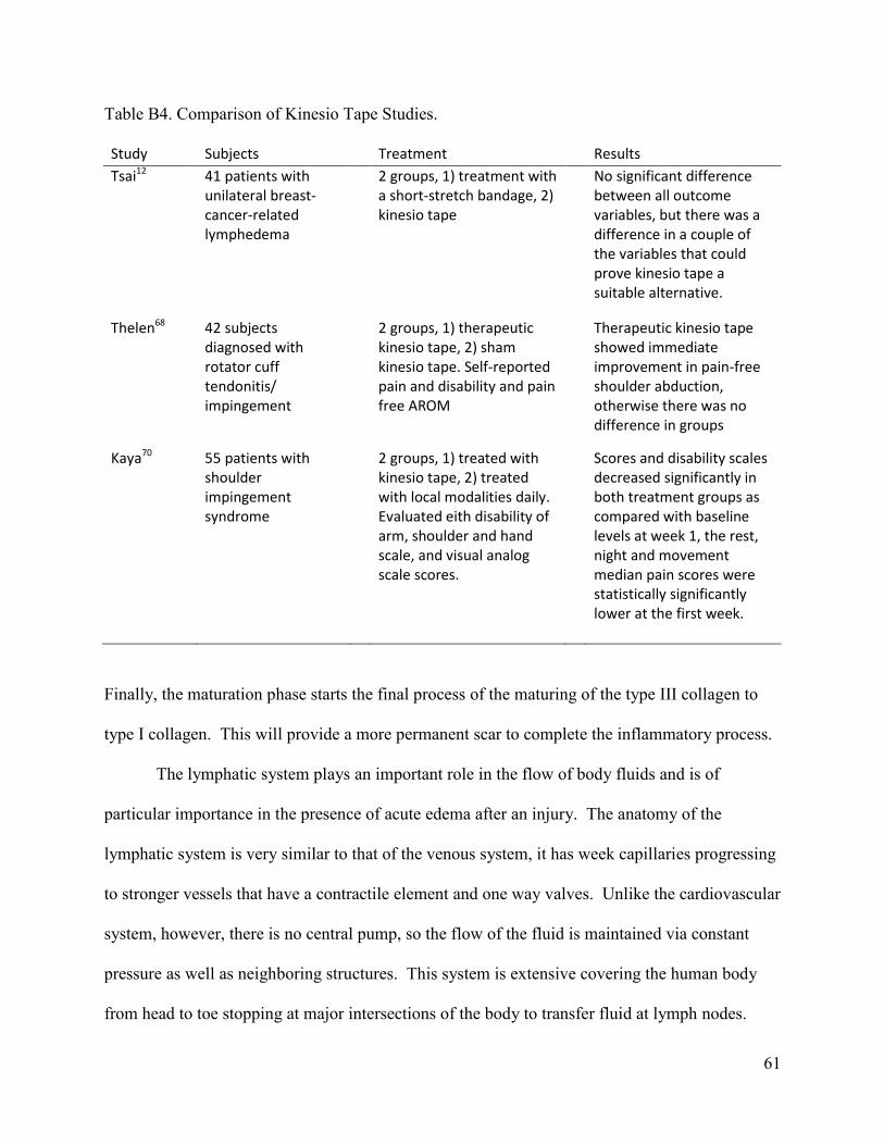
61
Table B4. Comparison of Kinesio Tape Studies.
Study Subjects Treatment Results
Tsai12 41 patients with unilateral breast-cancer-related lymphedema
2 groups, 1) treatment with a short-stretch bandage, 2) kinesio tape
No significant difference between all outcome variables, but there was a difference in a couple of the variables that could prove kinesio tape a suitable alternative.
Thelen68 42 subjects diagnosed with rotator cuff tendonitis/ impingement
2 groups, 1) therapeutic kinesio tape, 2) sham kinesio tape. Self-reported pain and disability and pain free AROM
Therapeutic kinesio tape showed immediate improvement in pain-free shoulder abduction, otherwise there was no difference in groups
Kaya70 55 patients with shoulder impingement syndrome
2 groups, 1) treated with kinesio tape, 2) treated with local modalities daily. Evaluated eith disability of arm, shoulder and hand scale, and visual analog scale scores.
Scores and disability scales decreased significantly in both treatment groups as compared with baseline levels at week 1, the rest, night and movement median pain scores were statistically significantly lower at the first week.
Finally, the maturation phase starts the final process of the maturing of the type III collagen to
type I collagen. This will provide a more permanent scar to complete the inflammatory process.
The lymphatic system plays an important role in the flow of body fluids and is of
particular importance in the presence of acute edema after an injury. The anatomy of the
lymphatic system is very similar to that of the venous system, it has week capillaries progressing
to stronger vessels that have a contractile element and one way valves. Unlike the cardiovascular
system, however, there is no central pump, so the flow of the fluid is maintained via constant
pressure as well as neighboring structures. This system is extensive covering the human body
from head to toe stopping at major intersections of the body to transfer fluid at lymph nodes.

62
Any breakdown in this system can cause chronic edema and treatment methods to prevent that is
very important.
Currently treatment includes but is not limited to cryotherapy, compression, massage,
electrical stimulation, kinesio tape and Hivamat. Cryotherapy is a staple in the treatment of acute
edema by the athletic trainer. Despite this being the most accepted treatment, there has been
very little research to study the physiology behind the use of cryotherapy. The same can be said
for compression, however, there have been even fewer studies to support the use. Massage is
one of the oldest treatments used and there have been more literature reviews to present the
experimental studies instead of critically evaluating them. One type of massage has been
confirmed to assist in the healing of tendonous injury. The most researched and supported
modality in the treatment of edema is cathodal high voltage pulsed current. Despite studies not
examining the physiological effects, a decrease and even prevention of edema from entering the
injured area is evident. Kinesio tape and Deep Oscillation are less studied than the previously
mentioned modalities. Kinesio tape is designed to mimic the skins properties and claims to,
along with other actions, increase lymphatic flow. Deep Oscillation therapy was developed in
Germany and to this point the studies conducted on this modality have been performed there
with very little research conducted in the United states. Though treatment of acutely injured
athlete’s is what sets athletic trainers apart from other health care professionalsdolan there is still
much to be learned about the modalities used.

63
APPENDIX C
ADDITIONAL METHODS
Table C1. PEDro Scale71 1. Eligibility criteria were specified no _ yes _ 2. Subjects were randomly allocated to groups (in a crossover study, subjects were randomly allocated an order in which treatments were received) no _ yes _ 3. Allocation was concealed no _ yes _ 4. The groups were similar at baseline regarding the most important prognostic indicators no _ yes _ 5. There was blinding of all subjects no _ yes _ 6. There was blinding of all therapists who administered the therapy no _ yes _ 7. There was blinding of all assessors who measured at least one key outcome no _ yes _ 8. Measures of at least one key outcome were obtained from more than 85% of the subjects initially allocated to groups no _ yes _ 9. All subjects for whom outcome measures were available received the treatment or control condition as allocated or, where this was not the case, data for at least one key outcome was analysed by “intention to treat” no _ yes _ 10. The results of between-group statistical comparisons are reported for at least one key outcome no _ yes _ 11. The study provides both point measures and measures of variability for at least one key outcome no _ yes _

64
Table C2. Pedro Scale with Explanations.16,71 Criteria 1. Eligibility criteria were specified.
Explanation: This criterion is satisfied if the report describes the source of the subjects and a list of criteria used to determine who was eligible to participate in the study.
Criteria 2. Subjects were randomly allocated to groups (in a crossover study, subjects were
randomly allocated an order in which treatments were received) Explanation: A study is considered to have used random allocation if the report states that allocation was random. The precise method of randomization need not be specified. Procedures such as coin tossing and dice rolling should be considered random. Quasi-randomization allocation procedures such as allocation procedures such as allocation by hospital record number or birth date, or alternation, do not satisfy this criterion.
Criteria 3. Allocation was concealed.
Explanation: Concealed allocation means that the person who determined if a subject was eligible for inclusion in the trial was unaware, when this decision was made, of which group the subject would be allocated to. A point is awarded for this criterion, even if it is not stated that allocation was concealed, when the report states that allocation was by sealed opaque envelopes or that allocation involved contacting the holder of the allocation schedule who was “off-site.”
Criteria 4. The groups were similar at baseline regarding the most important prognostic
indicators. Explanation: At a minimum, in studies of therapeutic interventions, the report must describe at least one measure of the severity of the condition being treated and at least one (different) key outcome measure at baseline. The rater must be satisfied that the groups’ outcomes would not be expected to differ, on the basis of baseline differences in prognostic variables alone, by a clinically significant amount. This criterion is satisfied even if only baseline data of subjects completing the study are presented.
Criteria 4, 7-11.
Explanation: Key outcomes are those outcomes that provide the primary measure of the effectiveness (or lack of effectiveness)of the therapy. In most studies, more than one variable is used as an outcome measure.
Criteria 5-7.
Explanation: Blinding means the person in question (subject, therapist, or assessor) did not know which group the subject had been allocated to. In addition, subjects and therapists are only considered to be “blind” if it could be expected that they would have been unable to distinguish reported (eg, visual analog scale, pain diary), the assessor is considered to be blind if the subject was blind.

65
Criteria 8. Measures of at least one key outcome were obtained from more than 85%
of the subjects initially allocated to groups.
Explanation: This criterion is satisfied only if the report explicitly states both the number of subjects initially allocated to groups and the number of subjects from whom key outcome measurements were obtained. In trials in which outcomes are measured at several points in time, a key outcome must have been measured in more than 85% of subjects at one of those points in time.
Criteria 9. All subjects for whom outcome measures were available received the treatment
or control condition as allocated or, where this was not the case, data for at least one key
outcome was analysed by “intention to treat.” Explanation: An intention-to-treat analysis means that, where subjects did not receive treatment (or the control condition) as allocated and where the measures of outcomes were available, the analysis was performed as if subjects received the treatment (or control condition) they were allocated to. This criterion is satisfied, even if there is no mention of analysis by intention to treat, if the report explicitly states that all subjects received treatment or control conditions allocated.
Criteria 10. The results of between-group statistical comparisons are reported for at least
one key outcome.
Explanation: A between-group statistical comparison involves statistical comparison of one group with another. Depending on the design of the study, this may involve comparison of 2 or more treatments of comparison of treatment with a control condition. The analysis may be a simple comparison of outcomes measured after the treatment was administered or a comparison of the change in one group with the change in another (when a factorial analysis of variance has been used to analyze the data, the latter is often reported as a group X time interaction.) The comparison may be in the form of hypothesis testing (which provides a P value, describing the probability that the groups differed only by chance) or in the form of an estimate (eg. The mean or median difference, or a difference in proportions, number needed to treat a relative risk or hazard ratio) and its confidence interval.
Criterion 11. The study provides both point measures and measures of variability for at
least one key outcome.
Explanation: A point measure is a measure of the size of the treatment effect. The treatment effect may be described as a difference in group outcomes or as the outcome in (each of ) all groups. Measures of variability include standard deviations, standard errors, confidence intervals, interquartile ranges (or other quartile ranges), and ranges. Point measures and/or measures of variability may be provided graphically (eg, standard deviations may be given as error bars in a figure) as long as it is clear what is being graphed (eg, as long as it is clear whether error bars represent standard deviations or standard errors). Where outcomes are categorical, this criterion is considered to have been met if the number of subjects in each category is given for each group.

66
Table C3. Protocol for Determining Studies 1. The studies were written in or translated to English.
2. Edema reduction or a form of the word lymphatic must appear in the title.
3. The abstract must include the terms lymphatic system, edema, modalities and treatment.
4. Acute injury must be the chief complaint in the study.
5. The study must be a randomized controlled trial.
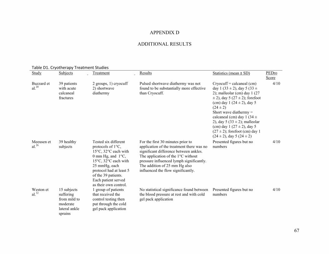
67
APPENDIX D
ADDITIONAL RESULTS
Table D1. Cryotherapy Treatment Studies Study Subjects Treatment Results Statistics (mean ± SD) PEDro
Score Buzzard et al.20
39 patients with acute calcaneal fractures
2 groups, 1) cryocuff 2) shortwave diathermy
Pulsed shortwave diathermy was not found to be substantially more effective than Cryocuff.
Cryocuff = calcaneal (cm) day 1 (33 ± 2), day 5 (33 ± 2); malleolar (cm) day 1 (27 ± 2), day 5 (27 ± 2); forefoot (cm) day 1 (24 ± 2), day 5 (24 ± 2) Short wave diathermy = calcaneal (cm) day 1 (34 ± 2), day 5 (33 ± 2); malleolar (cm) day 1 (27 ± 2), day 5 (27 ± 2); forefoot (cm) day 1 (24 ± 2), day 5 (24 ± 2)
4/10
Meeusen et al.26
39 healthy subjects
Tested six different protocols of 1°C, 15°C, 32°C each with 0 mm Hg, and 1°C, 15°C, 32°C each with 25 mmHg, each protocol had at least 5 of the 39 patients. Each patient served as their own control.
For the first 30 minutes prior to application of the treatment there was no significant difference between ankles. The application of the 1°C without pressure influenced lymph significantly. The addition of 25 mm Hg also influenced the flow significantly.
Presented figures but no numbers
4/10
Weston et al.32
15 subjects suffering from mild to moderate lateral ankle sprains
1 group of patients that received the control testing then put through the cold gel pack application
No statistical significance found between the blood pressure at rest and with cold gel pack application
Presented figures but no numbers
4/10

68
Stockle et al.29
60 patients with lower leg trauma
3 groups 1) cool pack cryotherapy, 2) continuous cryotherapy, 3) intermittent impulse compression
After first 24 hours of treatment there was 47% reduction in swelling with the AV impulse system, 33% with continuous cryotherapy and 17% with cold packs. After 4 days the AV impulse showed a 74% reduciton while continuous cryotherapy and cold packs had 70% and 45% respectively.
cool pack = ankle (mm) pre (272), day (265); midfoot (mm) pre (272) day 6 (254); forefoot (mm) pre (238) day 6 (232). Continuous = ankle (mm) pre (279), day (262); midfoot (mm) pre (268) day 6 (258); forefoot (mm) pre (244) day 6 (234). Compression = ankle (mm) pre (280), day (265); midfoot (mm) pre (277) day 6 (258); forefoot (mm) pre (253) day 6 (243).
4/10
Cheing et al.21
83 subjects with stable distal radius fractures treated with closed reduction
4 groups 1) ice plus PEMF, 2) ice plus sham PEMF, 3) PEMF alone, 4) sham PEMF
Evaluated based on VAS, volumetric measurements and ROM. By day 5 the a greater cumulative reduction in the VAS score was noted in group 1 over the other 3. For volumeteric measurements group 1 performed better than group 4 but not group 2.
Group A = day 1 (462 ± 73), day 3 (452 ± 72), day 5 (437 ± 67); Group B = day 1 (430 ± 45), day 3 (425 ± 44), day 5 (410 ± 47); Group C = day 1 (442 ± 83), day 3 (436 ± 81), day 5 (428 ± 82) Group D = day 1 (445 ± 68), day 3 (446 ± 72), day 5 (438 ± 73)
7/10
Scheffler et al.28
25 patients with post surgical trauma
1 group of patients that had a treatment foot that received cryocuff and control that did not
Evaluated based on circumference, pain assessment and by evaluation of comfort of the cryocuff which was rated on a 5 point scale. There was a standard deviation of .032 lower in circumference mean in the cryocuff treatment. 80% of patients noted less pain in the treatment foot. Comfort: 4.64, ease of use: 4.64, amt of cold: 4.8, leaking: 4.56, safety: 4.88, convenience: 4.6, compression: 4.8, stays in place: 4.8.
No calculations presented 3/10

69
Table D2. Compression Treatment Studies
Study Subjects Treatment Results Statistics (mean ± SD) PEDro Score
Rucinski et al.27
30 patients with postactute ankle sprains(>24 hrs)
3 treatment groups, 1) elastic wrap, 2) intermittent compresion, 3) elevated control group.
Found that the elevated group had the least amount of edema and the two compression protocols increased edema.
elastic wrap = Pre (1340.2 ± 128) post (1343.9 ± 127.4) Compression tx = pre (1220 ± 166.7) post (1229.4 ± 166.5) Control = pre (1350.4 ± 201.6) post (1335.5 ± 302.2)
6/10 Thordarson et al.29
30 patients with an ankle fracture requiring open reduction
2 groups, 1) received pneumatic pedal compression, 2) control group
Found a difference between groups of 121 mL in support of the PPC on day one and 63 mL on day two.
No data presented
3/10 Tsang et al.31
12 college students with inversion ankle sprains
2 groups, an elevation group and an elevation with compression group
Both groups provided a decrease in edema immediately after treatment however after 5 minutes the effects became less seen.
Elevation = 616.67 ± 121.41 At 0' ΔmL = -22.5, es = -0.19, at 5' ΔmL = -14.5, es = -0.12, at 60' ΔmL = -8.34, -0.07 Elevation & compression = 875.5 ± 169.21 At 0' ΔmL = -12, es = -0.07, at 5' ΔmL = -3.33, es = -0.02, at 60' ΔmL = -4.33, -0.03 6/10

70
Table D3. Massage Treatment Studies
Study Subjects Treatment Results Statistics (mean ± SD) PEDro Score
Haren et al.22
26 patients with distal radius fractures that were repaired with external fixation
2 groups, both received a standard protocol, 1) experimental group received 10 treatments of manual lymphatic drainage(MLD), 2) the control that did not receive MLD
Via volumeteric measurements it was found that the experimental group had significantly less edema in the injured hand than the control.
Experimental = (mL) day 3 (39 ± 12), day 17 (27 ± 9), day 33 (19 ± 9), day 68 (12 ± 11). Control = (mL) day 3 (64 ± 41), day 17 (50 ± 35), day 33 (35 ± 26), day 68 (24 ± 20)
6/10
Kessler et al.23
23 subjects who underwent hindfoot surgery
2 Groups, 1) standard physiotherapy plus manual lymph drainage, 2) standard phyiotherapy but no manual lymph drainage
When compared to the control group there was a significant reduction in post-operative swelling in the experimentral group.
Presented figures but no numbers.
6/10
Knygsand et al.24
30 subjects with distal radius fractures
2 groups, 1) received manual edema mobilization, 2) control
There was no statistically significant reduction in edema between the groups at six and nine weeks via volumetric measurements. There was a significant difference noted at three weeks in activities of daily living in the treatment group as well as there were fewer treatments needed in group 1.
Experimental = (mL) baseline (86.8 ± 26.1), 1 wk (68.2), 3 wk (41.1), 6 wk (28.6), 9 wk (12.1), 26 wk (2.5). Control = (mL) baseline (96.3 ± 28.3), 1 wk (77.3), 3 wk (54), 6 wk (43.3), 9 wk (28.3), 26 wk (15.7).
8/10
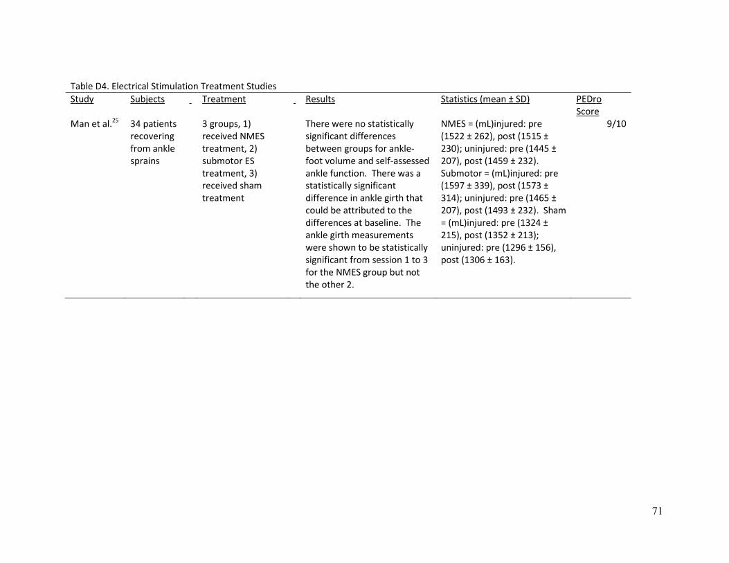
71
Table D4. Electrical Stimulation Treatment Studies
Study Subjects Treatment Results Statistics (mean ± SD) PEDro Score
Man et al.25 34 patients recovering from ankle sprains
3 groups, 1) received NMES treatment, 2) submotor ES treatment, 3) received sham treatment
There were no statistically significant differences between groups for ankle-foot volume and self-assessed ankle function. There was a statistically significant difference in ankle girth that could be attributed to the differences at baseline. The ankle girth measurements were shown to be statistically significant from session 1 to 3 for the NMES group but not the other 2.
NMES = (mL)injured: pre (1522 ± 262), post (1515 ± 230); uninjured: pre (1445 ± 207), post (1459 ± 232). Submotor = (mL)injured: pre (1597 ± 339), post (1573 ± 314); uninjured: pre (1465 ± 207), post (1493 ± 232). Sham = (mL)injured: pre (1324 ± 215), post (1352 ± 213); uninjured: pre (1296 ± 156), post (1306 ± 163).
9/10
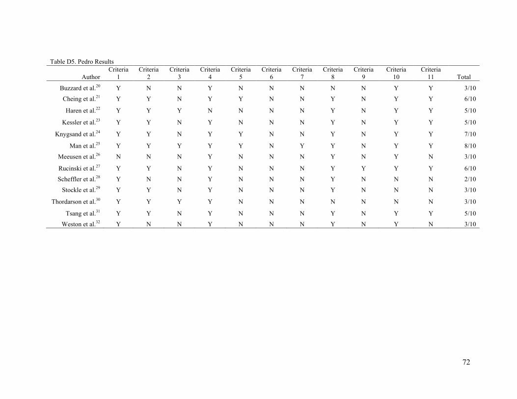
72
Table D5. Pedro Results
Author
Criteria 1
Criteria 2
Criteria 3
Criteria 4
Criteria 5
Criteria 6
Criteria 7
Criteria 8
Criteria 9
Criteria 10
Criteria 11 Total
Buzzard et al.20 Y N N Y N N N N N Y Y 3/10
Cheing et al.21 Y Y N Y Y N N Y N Y Y 6/10
Haren et al.22 Y Y Y N N N N Y N Y Y 5/10
Kessler et al.23 Y Y N Y N N N Y N Y Y 5/10
Knygsand et al.24 Y Y N Y Y N N Y N Y Y 7/10
Man et al.25 Y Y Y Y Y N Y Y N Y Y 8/10
Meeusen et al.26 N N N Y N N N Y N Y N 3/10
Rucinski et al.27 Y Y N Y N N N Y Y Y Y 6/10
Scheffler et al.28 Y N N Y N N N Y N N N 2/10
Stockle et al.29 Y Y N Y N N N Y N N N 3/10
Thordarson et al.30 Y Y Y Y N N N N N N N 3/10
Tsang et al.31 Y Y N Y N N N Y N Y Y 5/10
Weston et al.32 Y N N Y N N N Y N Y N 3/10
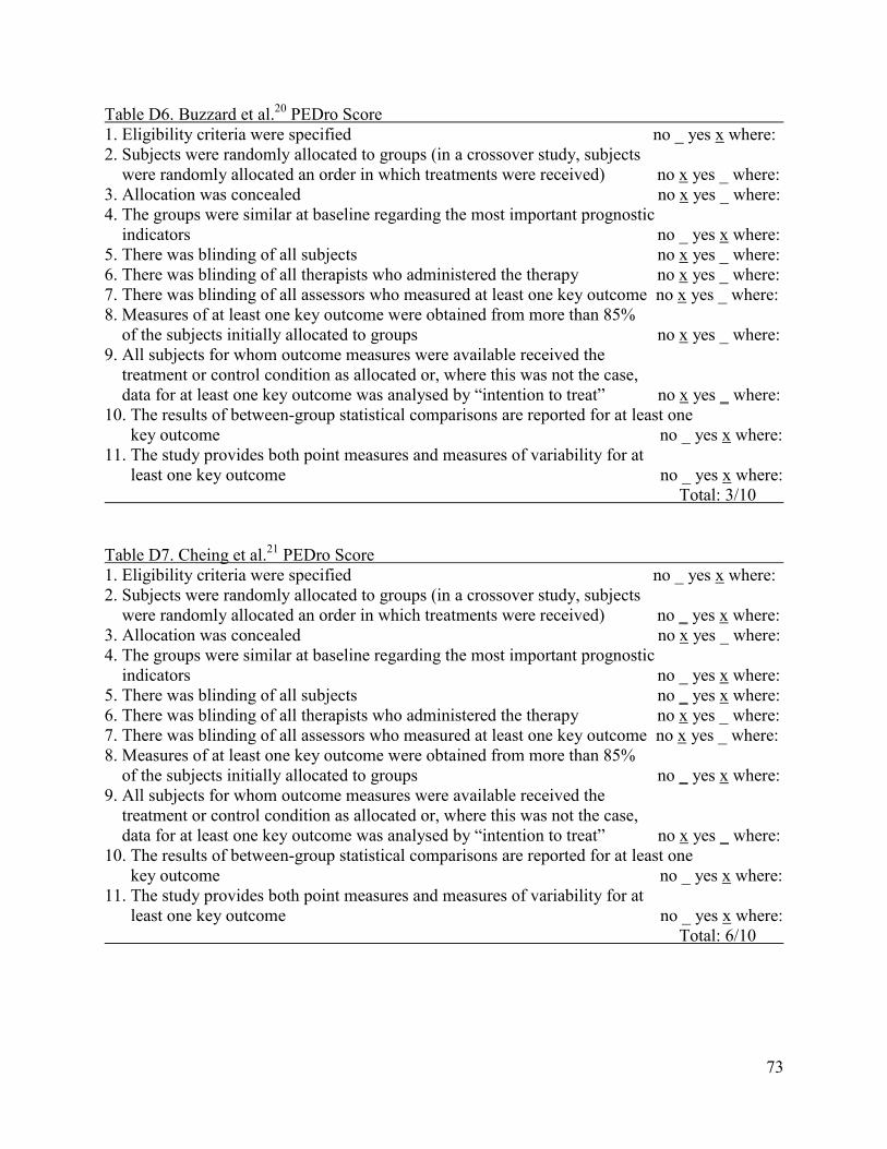
73
Table D6. Buzzard et al.20 PEDro Score 1. Eligibility criteria were specified no _ yes x where: 2. Subjects were randomly allocated to groups (in a crossover study, subjects were randomly allocated an order in which treatments were received) no x yes _ where: 3. Allocation was concealed no x yes _ where: 4. The groups were similar at baseline regarding the most important prognostic indicators no _ yes x where: 5. There was blinding of all subjects no x yes _ where: 6. There was blinding of all therapists who administered the therapy no x yes _ where: 7. There was blinding of all assessors who measured at least one key outcome no x yes _ where: 8. Measures of at least one key outcome were obtained from more than 85% of the subjects initially allocated to groups no x yes _ where: 9. All subjects for whom outcome measures were available received the treatment or control condition as allocated or, where this was not the case, data for at least one key outcome was analysed by “intention to treat” no x yes _ where: 10. The results of between-group statistical comparisons are reported for at least one key outcome no _ yes x where: 11. The study provides both point measures and measures of variability for at least one key outcome no _ yes x where: Total: 3/10 Table D7. Cheing et al.21 PEDro Score 1. Eligibility criteria were specified no _ yes x where: 2. Subjects were randomly allocated to groups (in a crossover study, subjects were randomly allocated an order in which treatments were received) no _ yes x where: 3. Allocation was concealed no x yes _ where: 4. The groups were similar at baseline regarding the most important prognostic indicators no _ yes x where: 5. There was blinding of all subjects no _ yes x where: 6. There was blinding of all therapists who administered the therapy no x yes _ where: 7. There was blinding of all assessors who measured at least one key outcome no x yes _ where: 8. Measures of at least one key outcome were obtained from more than 85% of the subjects initially allocated to groups no _ yes x where: 9. All subjects for whom outcome measures were available received the treatment or control condition as allocated or, where this was not the case, data for at least one key outcome was analysed by “intention to treat” no x yes _ where: 10. The results of between-group statistical comparisons are reported for at least one key outcome no _ yes x where: 11. The study provides both point measures and measures of variability for at least one key outcome no _ yes x where: Total: 6/10

74
Table D8. Haren et al.22 PEDro Score 1. Eligibility criteria were specified no _ yes x where: 2. Subjects were randomly allocated to groups (in a crossover study, subjects were randomly allocated an order in which treatments were received) no _ yes x where: 3. Allocation was concealed no _ yes x where: 4. The groups were similar at baseline regarding the most important prognostic indicators no x yes _ where: 5. There was blinding of all subjects no x yes _ where: 6. There was blinding of all therapists who administered the therapy no x yes _ where: 7. There was blinding of all assessors who measured at least one key outcome no x yes _ where: 8. Measures of at least one key outcome were obtained from more than 85% of the subjects initially allocated to groups no _ yes x where: 9. All subjects for whom outcome measures were available received the treatment or control condition as allocated or, where this was not the case, data for at least one key outcome was analysed by “intention to treat” no x yes _ where: 10. The results of between-group statistical comparisons are reported for at least one key outcome no _ yes x where: 11. The study provides both point measures and measures of variability for at least one key outcome no _ yes x where: Total: 5/10 Table D9. Kessler et al.23 PEDro Score 1. Eligibility criteria were specified no _ yes x where: 2. Subjects were randomly allocated to groups (in a crossover study, subjects were randomly allocated an order in which treatments were received) no _ yes x where: 3. Allocation was concealed no x yes _ where: 4. The groups were similar at baseline regarding the most important prognostic indicators no _ yes x where: 5. There was blinding of all subjects no x yes _ where: 6. There was blinding of all therapists who administered the therapy no x yes _ where: 7. There was blinding of all assessors who measured at least one key outcome no x yes _ where: 8. Measures of at least one key outcome were obtained from more than 85% of the subjects initially allocated to groups no _ yes x where: 9. All subjects for whom outcome measures were available received the treatment or control condition as allocated or, where this was not the case, data for at least one key outcome was analysed by “intention to treat” no x yes _ where: 10. The results of between-group statistical comparisons are reported for at least one key outcome no _ yes x where: 11. The study provides both point measures and measures of variability for at least one key outcome no _ yes x where: Total: 5/10

75
Table D10. Knygsand et al.24 PEDro Score 1. Eligibility criteria were specified no _ yes x where: 2. Subjects were randomly allocated to groups (in a crossover study, subjects were randomly allocated an order in which treatments were received) no _ yes x where: 3. Allocation was concealed no x yes _ where: 4. The groups were similar at baseline regarding the most important prognostic indicators no _ yes x where: 5. There was blinding of all subjects no _ yes x where: 6. There was blinding of all therapists who administered the therapy no x yes _ where: 7. There was blinding of all assessors who measured at least one key outcome no _ yes x where: 8. Measures of at least one key outcome were obtained from more than 85% of the subjects initially allocated to groups no _ yes x where: 9. All subjects for whom outcome measures were available received the treatment or control condition as allocated or, where this was not the case, data for at least one key outcome was analysed by “intention to treat” no x yes _ where: 10. The results of between-group statistical comparisons are reported for at least one key outcome no _ yes x where: 11. The study provides both point measures and measures of variability for at least one key outcome no _ yes x where: Total: 7/10 Table D11. Man et al.25 PEDro Score 1. Eligibility criteria were specified no _ yes x where: 2. Subjects were randomly allocated to groups (in a crossover study, subjects were randomly allocated an order in which treatments were received) no _ yes x where: 3. Allocation was concealed no _ yes x where: 4. The groups were similar at baseline regarding the most important prognostic indicators no _ yes x where: 5. There was blinding of all subjects no _ yes x where: 6. There was blinding of all therapists who administered the therapy no x yes _ where: 7. There was blinding of all assessors who measured at least one key outcome no _ yes x where: 8. Measures of at least one key outcome were obtained from more than 85% of the subjects initially allocated to groups no _ yes x where: 9. All subjects for whom outcome measures were available received the treatment or control condition as allocated or, where this was not the case, data for at least one key outcome was analysed by “intention to treat” no x yes _ where: 10. The results of between-group statistical comparisons are reported for at least one key outcome no _ yes x where: 11. The study provides both point measures and measures of variability for at least one key outcome no _ yes x where: Total: 8/10

76
Table D12. Meeusen et al.26 PEDro Score 1. Eligibility criteria were specified no x yes _ where: 2. Subjects were randomly allocated to groups (in a crossover study, subjects were randomly allocated an order in which treatments were received) no x yes _ where: 3. Allocation was concealed no x yes _ where: 4. The groups were similar at baseline regarding the most important prognostic indicators no _ yes x where: 5. There was blinding of all subjects no x yes _ where: 6. There was blinding of all therapists who administered the therapy no x yes _ where: 7. There was blinding of all assessors who measured at least one key outcome no x yes _ where: 8. Measures of at least one key outcome were obtained from more than 85% of the subjects initially allocated to groups no _ yes x where: 9. All subjects for whom outcome measures were available received the treatment or control condition as allocated or, where this was not the case, data for at least one key outcome was analysed by “intention to treat” no x yes _ where: 10. The results of between-group statistical comparisons are reported for at least one key outcome no _ yes x where: 11. The study provides both point measures and measures of variability for at least one key outcome no x yes _ where: Total: 3/10 Table D13. Rucinski et al.27 PEDro Score 1. Eligibility criteria were specified no _ yes x where: 2. Subjects were randomly allocated to groups (in a crossover study, subjects were randomly allocated an order in which treatments were received) no _ yes x where: 3. Allocation was concealed no x yes _ where: 4. The groups were similar at baseline regarding the most important prognostic indicators no _ yes x where: 5. There was blinding of all subjects no x yes _ where: 6. There was blinding of all therapists who administered the therapy no x yes _ where: 7. There was blinding of all assessors who measured at least one key outcome no x yes _ where: 8. Measures of at least one key outcome were obtained from more than 85% of the subjects initially allocated to groups no _ yes x where: 9. All subjects for whom outcome measures were available received the treatment or control condition as allocated or, where this was not the case, data for at least one key outcome was analysed by “intention to treat” no _ yes x where: 10. The results of between-group statistical comparisons are reported for at least one key outcome no _ yes x where: 11. The study provides both point measures and measures of variability for at least one key outcome no _ yes x where: Total: 6/10

77
Table D14. Scheffler et al.28 PEDro Score 1. Eligibility criteria were specified no _ yes x where: 2. Subjects were randomly allocated to groups (in a crossover study, subjects were randomly allocated an order in which treatments were received) no x yes _ where: 3. Allocation was concealed no x yes _ where: 4. The groups were similar at baseline regarding the most important prognostic indicators no _ yes x where: 5. There was blinding of all subjects no x yes _ where: 6. There was blinding of all therapists who administered the therapy no x yes _ where: 7. There was blinding of all assessors who measured at least one key outcome no x yes _ where: 8. Measures of at least one key outcome were obtained from more than 85% of the subjects initially allocated to groups no _ yes x where: 9. All subjects for whom outcome measures were available received the treatment or control condition as allocated or, where this was not the case, data for at least one key outcome was analysed by “intention to treat” no x yes _ where: 10. The results of between-group statistical comparisons are reported for at least one key outcome no x yes _ where: 11. The study provides both point measures and measures of variability for at least one key outcome no x yes _ where: Total: 2/10 Table D15. Stockle et al.29 PEDro Score 1. Eligibility criteria were specified no _ yes x where: 2. Subjects were randomly allocated to groups (in a crossover study, subjects were randomly allocated an order in which treatments were received) no _ yes x where: 3. Allocation was concealed no x yes _ where: 4. The groups were similar at baseline regarding the most important prognostic indicators no _ yes x where: 5. There was blinding of all subjects no x yes _ where: 6. There was blinding of all therapists who administered the therapy no x yes _ where: 7. There was blinding of all assessors who measured at least one key outcome no x yes _ where: 8. Measures of at least one key outcome were obtained from more than 85% of the subjects initially allocated to groups no _ yes x where: 9. All subjects for whom outcome measures were available received the treatment or control condition as allocated or, where this was not the case, data for at least one key outcome was analysed by “intention to treat” no x yes _ where: 10. The results of between-group statistical comparisons are reported for at least one key outcome no x yes _ where: 11. The study provides both point measures and measures of variability for at least one key outcome no x yes _ where: Total: 3/10

78
Table D16. Thordarson et al.30 PEDro Score 1. Eligibility criteria were specified no _ yes x where: 2. Subjects were randomly allocated to groups (in a crossover study, subjects were randomly allocated an order in which treatments were received) no _ yes x where: 3. Allocation was concealed no _ yes x where: 4. The groups were similar at baseline regarding the most important prognostic indicators no _ yes x where: 5. There was blinding of all subjects no x yes _ where: 6. There was blinding of all therapists who administered the therapy no x yes _ where: 7. There was blinding of all assessors who measured at least one key outcome no x yes _ where: 8. Measures of at least one key outcome were obtained from more than 85% of the subjects initially allocated to groups no x yes _ where: 9. All subjects for whom outcome measures were available received the treatment or control condition as allocated or, where this was not the case, data for at least one key outcome was analysed by “intention to treat” no x yes _ where: 10. The results of between-group statistical comparisons are reported for at least one key outcome no x yes _ where: 11. The study provides both point measures and measures of variability for at least one key outcome no x yes _ where: Total: 3/10 Table D17. Tsang et al.31 PEDro Score 1. Eligibility criteria were specified no _ yes x where: 2. Subjects were randomly allocated to groups (in a crossover study, subjects were randomly allocated an order in which treatments were received) no _ yes x where: 3. Allocation was concealed no x yes _ where: 4. The groups were similar at baseline regarding the most important prognostic indicators no _ yes x where: 5. There was blinding of all subjects no x yes _ where: 6. There was blinding of all therapists who administered the therapy no x yes _ where: 7. There was blinding of all assessors who measured at least one key outcome no x yes _ where: 8. Measures of at least one key outcome were obtained from more than 85% of the subjects initially allocated to groups no _ yes x where: 9. All subjects for whom outcome measures were available received the treatment or control condition as allocated or, where this was not the case, data for at least one key outcome was analysed by “intention to treat” no x yes _ where: 10. The results of between-group statistical comparisons are reported for at least one key outcome no _ yes x where: 11. The study provides both point measures and measures of variability for at least one key outcome no _ yes x where: Total: 5/10
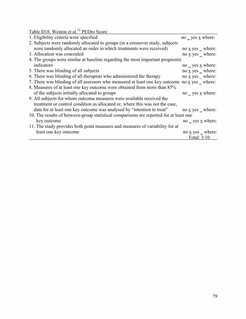
79
Table D18. Weston et al.32 PEDro Score 1. Eligibility criteria were specified no _ yes x where: 2. Subjects were randomly allocated to groups (in a crossover study, subjects were randomly allocated an order in which treatments were received) no x yes _ where: 3. Allocation was concealed no x yes _ where: 4. The groups were similar at baseline regarding the most important prognostic indicators no _ yes x where: 5. There was blinding of all subjects no x yes _ where: 6. There was blinding of all therapists who administered the therapy no x yes _ where: 7. There was blinding of all assessors who measured at least one key outcome no x yes _ where: 8. Measures of at least one key outcome were obtained from more than 85% of the subjects initially allocated to groups no _ yes x where: 9. All subjects for whom outcome measures were available received the treatment or control condition as allocated or, where this was not the case, data for at least one key outcome was analysed by “intention to treat” no x yes _ where: 10. The results of between-group statistical comparisons are reported for at least one key outcome no _ yes x where: 11. The study provides both point measures and measures of variability for at least one key outcome no x yes _ where: Total: 3/10
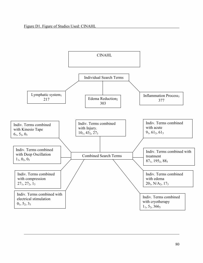
80
Figure D1. Figure of Studies Used: CINAHL
CINAHL
Individual Search Terms
Lymphatic system1 217 Edema Reduction2
303
Inflammation Process3 377
Combined Search Terms
Indiv. Terms combined with Injury. 101, 432, 273
Indiv. Terms combined with acute 91, 612, 613
Indiv. Terms combined with Kinesio Tape 61, 52, 03
Indiv. Terms combined with Deep Oscillation 11, 02, 03
Indiv. Terms combined with treatment 871, 1952, 883
Indiv. Terms combined with compression 271, 272, 13
Indiv. Terms combined with edema 201, N/A2, 173
Indiv. Terms combined with cryotherapy 11, 52, 3663
Indiv. Terms combined with electrical stimulation 01, 52, 33
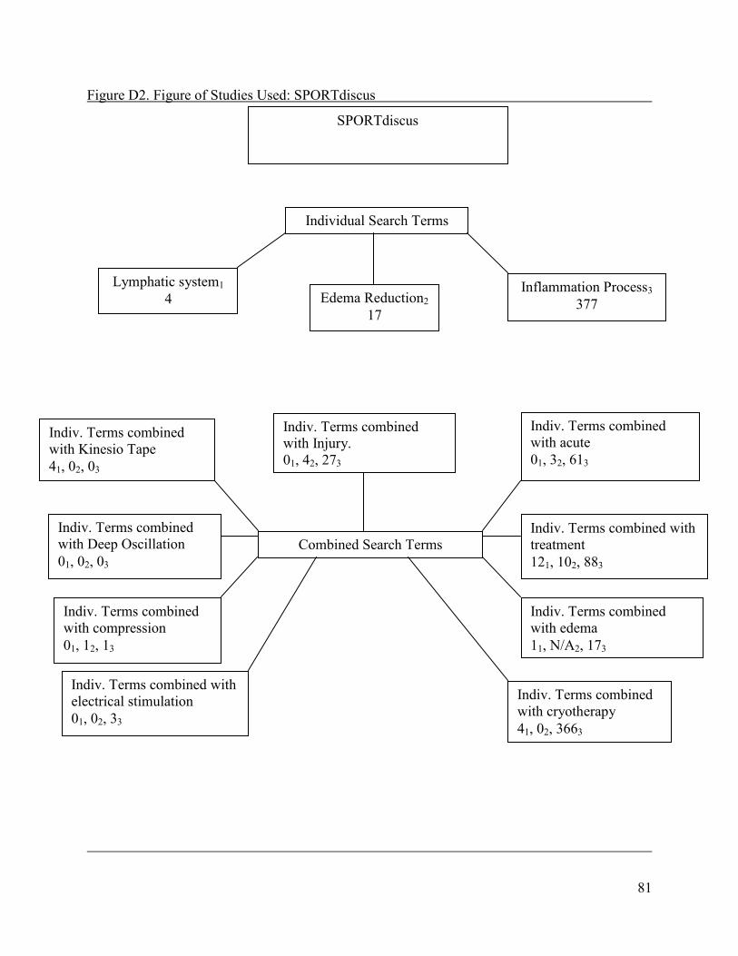
81
Figure D2. Figure of Studies Used: SPORTdiscus
SPORTdiscus
Individual Search Terms
Lymphatic system1 4 Edema Reduction2
17
Inflammation Process3 377
Combined Search Terms
Indiv. Terms combined with Injury. 01, 42, 273
Indiv. Terms combined with acute 01, 32, 613
Indiv. Terms combined with Kinesio Tape 41, 02, 03
Indiv. Terms combined with Deep Oscillation 01, 02, 03
Indiv. Terms combined with treatment 121, 102, 883
Indiv. Terms combined with compression 01, 12, 13
Indiv. Terms combined with edema 11, N/A2, 173
Indiv. Terms combined with cryotherapy 41, 02, 3663
Indiv. Terms combined with electrical stimulation 01, 02, 33

82
Figure D3. Figure of Studies Used: MEDLINE
MEDLINE
Individual Search Terms
Lymphatic system1 4,758 Edema Reduction2
60
Inflammation Process3 377
Combined Search Terms
Indiv. Terms combined with Injury. 1641, 82, 273
Indiv. Terms combined with acute 2171, 112, 613
Indiv. Terms combined with Kinesio Tape 01, 02, 03
Indiv. Terms combined with Deep Oscillation 11, 02, 03
Indiv. Terms combined with treatment 6971, 382, 883
Indiv. Terms combined with compression 541, 162, 13
Indiv. Terms combined with edema 3791, N/A2, 173
Indiv. Terms combined with cryotherapy 21, 02, 3663
Indiv. Terms combined with electrical stimulation 91, 12, 33

83
Figure D4. Figure of Studies Used: MDConsult
MDConsult Database
Individual Search Terms
Lymphatic system1 539 Edema Reduction2
31
Inflammation Process3 131
Combined Search Terms
Indiv. Terms combined with Injury. 591, 92, 193
Indiv. Terms combined with acute 721, 102, 283
Indiv. Terms combined with Kinesio Tape 01, 02, 03
Indiv. Terms combined with Deep Oscillation 01, 02, 03
Indiv. Terms combined with treatment 1911, 242, 543
Indiv. Terms combined with compression 301, 92, 03
Indiv. Terms combined with edema 701, N/A2, 83
Indiv. Terms combined with cryotherapy 41, 22, 03
Indiv. Terms combined with electrical stimulation 11, 12, 03

84
Figure D5. Figure of Studies Used: PubMed
PubMed
Individual Search Terms
Lymphatic system1 12,113 Edema Reduction2
73
Inflammation Process3 328
Combined Search Terms
Indiv. Terms combined with Injury. 4761, 152, 333
Indiv. Terms combined with acute 4061, 122, 613
Indiv. Terms combined with Kinesio Tape 01, 02, 03
Indiv. Terms combined with Deep Oscillation 01, 02, 03
Indiv. Terms combined with treatment 3,7271, 712, 1613
Indiv. Terms combined with compression 721, 182, 13
Indiv. Terms combined with edema 7891, N/A2, 203
Indiv. Terms combined with cryotherapy 111, 12, 03
Indiv. Terms combined with electrical stimulation 581, 42, 33

85
Figure D6. Figure of Studies Used: Cochran Database
Cochran Database
Individual Search Terms
Lymphatic system1 86 Edema Reduction2
10
Inflammation Process3 1
Combined Search Terms
Indiv. Terms combined with Injury. 31, 12, 13
Indiv. Terms combined with acute 31, 12, 43
Indiv. Terms combined with Kinesio Tape 01, 02, 13
Indiv. Terms combined with Deep Oscillation 01, 02, 03
Indiv. Terms combined with treatment 31, 62, 13
Indiv. Terms combined with compression 11, 72, 03
Indiv. Terms combined with edema 41, N/A2, 03
Indiv. Terms combined with cryotherapy 01, 02, 03
Indiv. Terms combined with electrical stimulation 01, 02, 03
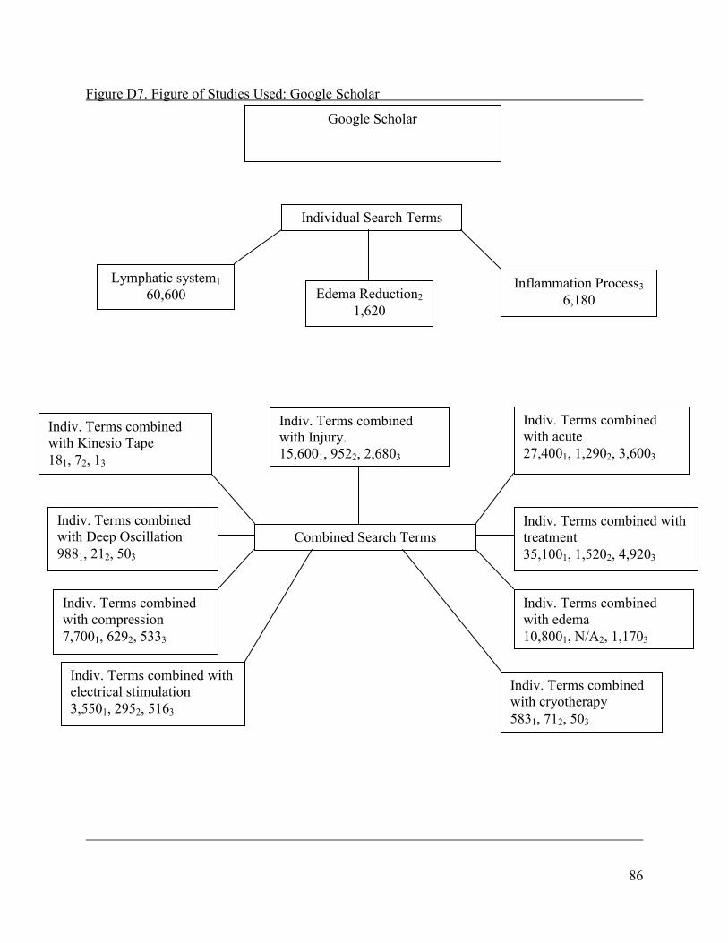
86
Figure D7. Figure of Studies Used: Google Scholar
Google Scholar
Individual Search Terms
Lymphatic system1 60,600 Edema Reduction2
1,620
Inflammation Process3 6,180
Combined Search Terms
Indiv. Terms combined with Injury. 15,6001, 9522, 2,6803
Indiv. Terms combined with acute 27,4001, 1,2902, 3,6003
Indiv. Terms combined with Kinesio Tape 181, 72, 13
Indiv. Terms combined with Deep Oscillation 9881, 212, 503
Indiv. Terms combined with treatment 35,1001, 1,5202, 4,9203
Indiv. Terms combined with compression 7,7001, 6292, 5333
Indiv. Terms combined with edema 10,8001, N/A2, 1,1703
Indiv. Terms combined with cryotherapy 5831, 712, 503
Indiv. Terms combined with electrical stimulation 3,5501, 2952, 5163

87
Figure D8. Figure of Studies Used: Science Direct
Science Direct
Individual Search Terms
Lymphatic system1 10,517 Edema Reduction2
482
Inflammation Process3 1,897
Combined Search Terms
Indiv. Terms combined with Injury. 3,0361, 2992, 7993
Indiv. Terms combined with acute 4,4711, 3452, 1,0593
Indiv. Terms combined with Kinesio Tape 31, 32, 03
Indiv. Terms combined with Deep Oscillation 1291, 72, 93
Indiv. Terms combined with treatment 7,8441, 4442, 1,5053
Indiv. Terms combined with compression 1,4131, 1602, 743
Indiv. Terms combined with edema 2,7241, N/A2, 3533
Indiv. Terms combined with cryotherapy 1201, 292, 143
Indiv. Terms combined with electrical stimulation 4801, 802, 383

88
Figure D9. Figures of Studies Used: PEDro Database
PEDro Database
Individual Search Terms
Lymphatic system1 1 Edema Reduction2
3
Inflammation Process3 1
Combined Search Terms
Indiv. Terms combined with Injury. 01, 02, 03
Indiv. Terms combined with acute 11, 02, 13
Indiv. Terms combined with Kinesio Tape 01, 02, 03
Indiv. Terms combined with Deep Oscillation 01, 02, 03
Indiv. Terms combined with treatment 11, 22, 13
Indiv. Terms combined with compression 01, 02, 03
Indiv. Terms combined with edema 01, N/A2, 03
Indiv. Terms combined with cryotherapy 01, 02, 03
Indiv. Terms combined with electrical stimulation 01, 02, 03
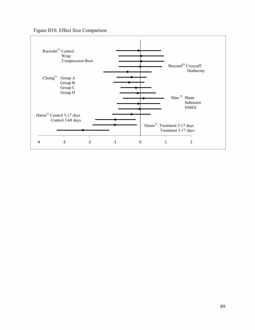
89
-4 -3 -2 -1 0 1 2
Rucinski27 Control Wrap Compression Boot
Buzzard20 Cryocuff Diathermy
Cheing21 Group A Group B Group C Group D
Man 25 Sham Submotor NMES
Haren22 Control 3-17 days Control 3-68 days
Haren22 Treatment 3-17 days Treatment 3-17 days
Figure D10. Effect Size Comparison

90
APPENDIX E
RECOMMENDATIONS FOR FUTURE RESEARCH
1. Future studies should include foreign language database and studies. This may expand the number of studies found.
2. Expanding the injuries from acute to chronic edema in traumatic injuries may provide more information.
3. Perform a study examining the further effects of each treatment, particularly massage,
with larger populations.
4. Altering the inclusion criteria to expand the study to include chronic edema but excluding chronic diseases.
91
ADDITIONAL REFERENCES 33. Jarvinen TA, Jarvinen TL, Kaariainen M, Kalimo H, Jarvinen M. Muscle injuries: biology and breatment. Am J Sports Med. 2005;33(5):745-764. 33. Butterfield T, Best T, Merrick M. The dual roles of nuetrophils and macrophages in inflammation: a critical balance between tissue damage and repair. J Athl
Train. 2008;41(4)457-465. 35. Fujiwara N, Kobayashi K. Macrophage in inflammation. Current Drug Targets-
Inflammation Allergy. 2005;4(3):281-286. 36. Ng, G. Ligament injury and repair: current concepts. Hong Kong Physiotherapy J. 2002;20:22-29 37. Aslan H, Kimelman-Belch N, Pelled G, Gazit D. Molecular targets for tendon neoformation. . Clin Invest. 2008;118:439-444. 38. Sandrey M. Acute and chronic tendon injuries: factors affecting the healing response and treatment. J Sport Rehabil. 2003;12:70-91. 39. Swartz M. The physiology of the lymphatic system. Advanced Drug Delivery Reviews. 2001;50:3-20. 40. Oakes B. Tissue healing and repair: tendons and ligaments. In: Frontera W. Editor. Rehabilitation of Sports Injuries: Scientific Basis. Boston: Blackwell Science;2003;56-98. 41. Chiang W, Chen Y, Lin S, Wu K, Tsai T. Bradykinin enhances reactive oxygen species generation, mitochondrial injury, and cell death induced by ATP depletion – a role of the phospholipase C-Ca2+ Pathway. Free Radical Biology Medicine. 2007;43:702-710. 42. Gilroy D, Lawrence T, Colville-Nash P, Willoughby D. New insights into inflammatory resolution. Inflammopharmacology. 2001;9(1,2):125-130. 43. Glaros T, Larsen M, Li L. Macrophages and fibroblasts during inflammation, tissue damage and organ Injury. Frontiers in Bioscience. 2009;14:3988-3993. 44. Enwemeka C. Inflammation, cellularity, and fibrillogenesis in regeneration tendon: implications for tendon rehabilitation. Phys Ther. 1989;69(10):816-825. 45. Reed R, Rubin K. Transcapillary exchange: role and importance of the interstitial fluid pressure and the extracellular matrix. Cardiovascular Research. 2010;87:211-217. 46. Board J, Harlow W. Lymphoedema 1: components and function of the lymphatic system. Bri J of Nursing. 2002;11(5):304-309.

92
47. Schmid-Schonbein G. Microlymphatics and lymph flow. Physiological Reviews. 1990;70(4):987-1028 48. Moore K, Dalley A. Clinical Oriented Anatomy. Baltimore, MD. Lippincott Williams & Wilkins. 2006. 584, 625, 672, 748-750. 49. Bleakly C, McDonough S, MacAuley. Cryotherapy for acute ankle sprains: a randomised controlled study of two different icing protocols. Br J Sports Med. 2006;40:700-705 50. Bleakly C, O’Connor S, Tully M, Rocke L, MacAuley D, McDonough S. The Price (Protection Rest Ice Compression Elevation): design of a randomised controlled trial comparing standard versus cryokinetic ice applications in the management of acute ankle sprain. BMC Musculoskeletal Disorders. 2007;8:125-133. 51. Schaser K, Disch A, Stover J, Lauffer A, Bail H, Mittlmeier T. Prolonged superficial local cryotherapy attenuates microcirculatory impairment, regional inflammation, and muscle necrosis after closed soft tissue injury in rats. Am J Sports Med. 2007;35:93-102. 52. Kreamer W, French D, Spiering B. Compression in the treatment of acute muscle injuries in sport. Int SportMed J. 2004;5(3):200-208. 53. Ramey D, Tiidus P. Massage therapy in horses: assessing its effectiveness from empirical data in humans and animals. Compendium. 2002;24(5)418-423. 54. Callaghan M. The role of massage in the management of the athlete: a Review. Br J Sport
Med. 1993;27(1):28-33. 55. Hemmings B. Physiological, psychological and performance effects of massage therapy in sport: a review of the literature. Phys Ther Sport. 2001;2:165-170. 56. Williams A. Manual lymphatic drainage: exploring the history and evidence dase. Br
J Community Nursing. 2010:S18-S24. 57. Tiidus P. A review of human massage therapy: assessing effectiveness primarily from empirical data in the human species. AAEP Proceedings. 2000;46:302-305. 58. Tiidus P. Massage and ultrasound as therapuetic modalities in exercise-induced muscle damage. Can J App. Physiol. 1999;24(3):267-278. 59. Gehlsen G, Gannon L, Helfst R. Fibroblasts responses to variation in soft tissue mobilization pressure. Med Sci Sports Exerc. 1999;31(4):531-535. 60. Shoemaker J, Tiidus P, Mader R. Failure of manual massage to alter limb blood flow: measures by doppler ultrasound. Med Sci Sports Exerc. 1997;29(5):610-614.

93
61. Dolan M, Graves P, Nakazawa C, Delano T, Hutson A, Mendel F. Effects of ibuprofen and high-voltage electric stimulation on acute edema formation after blunt trauma to limbs of rats. J Athl Train. 2005;40(2):111-115. 62. Cook H, Morales M, La Rosa E, Dean J, Donnelly M, McHugh P, Otradovec A, Wright K, Kula T, Tepper S. Effects of electrical stimulation on lymphatic flow and limb volume in the rat. Phys Ther. 1994;74(11):1040-1046. 63. Mendel F, Fish D. New perspective in edema control via electrical stimulation. J
Athl Train. 1993;28(1):63-74. 64. Thornton R, Mendel F, Fish D. Effects of electrical stimulation on edema formation in different strains of rats. Phys Ther. 1998;78(4):386-394. 65. Mohr T, Akers T, Landry R. Effect of high voltage stimulation on edema reduction in the rat hind limb. Phys Ther. 1987;67(11):1703-1707. 66. Taylor K, Fish D, Mendel F, Burton H. Effect of a single 30 minute treatment of high voltage pulsed current on edema formation in frog hind limbs. Phys Ther. 1992;72(1):73-78. 67. Zajt-Kwiatkowska J, Rajkowska-Labon E, Skrobot W, Bakula S, Szamotulska J. Application of kinesio taping for treatment of sports injuries. Research Yearbook. 2007;13(1)130-134. 68. Thelen M, Dauber J, Stoneman P. The clinical efficacy of kinesio tape for shoulder pain: a randomized, double-blinded, clinical trial. J Orthop Sport Phys Ther. 2008;38(7):389- 395. 69. Aliyev R. Clinical effects of the therapy mthod deep oscillation in treatment of sports injuries (Abstract only). XIX International Congress on Sports Rehabilitation and Traumatology. 70. Kaya E, Zinnuroglu M, Tugeu I. Kinesio taping compared to physical therapy modalities for the treatment of shoulder impingement syndrome. Clin Rheumatol. 2010. 71. Physiotherapy Evidence Database: Scale. Available at: http:// http://www.pedro.org.au/english/downloads/pedro-scale/. Accessed December 6, 2010