treatment of staphylococcus aureus-induced chronic osteomyelitis
TRANSCRIPT

© 2015 Yan et al. This work is published by Dove Medical Press Limited, and licensed under Creative Commons Attribution – Non Commercial (unported, v3.0) License. The full terms of the License are available at http://creativecommons.org/licenses/by-nc/3.0/. Non-commercial uses of the work are permitted without any further
permission from Dove Medical Press Limited, provided the work is properly attributed. Permissions beyond the scope of the License are administered by Dove Medical Press Limited. Information on how to request permission may be found at: http://www.dovepress.com/permissions.php
Drug Design, Development and Therapy 2015:9 3665–3676
Drug Design, Development and Therapy Dovepress
submit your manuscript | www.dovepress.com
Dovepress 3665
O r i g i n a l r e s e a r c h
open access to scientific and medical research
Open access Full Text article
http://dx.doi.org/10.2147/DDDT.S84486
Treatment of Staphylococcus aureus-induced chronic osteomyelitis with bone-like hydroxyapatite/poly amino acid loaded with rifapentine microspheres
ling Yan1
Dian-Ming Jiang2
Zhi-Dong cao2
Jun Wu2
Xin Wang3
Zheng-long Wang4
Ya-Jun li5
Yong-Fen Yi1
1Department of Pathology, college of Basic Medicine, 2Department of Orthopaedic surgery, The First affiliated hospital, 3Pediatric hospital, 4Department of cardiology, The First affiliated hospital, 5Department of radiology, college of Basic Medicine, chongqing Medical University, chongqing, People’s republic of china
Purpose: The purpose of this study was to investigate the curative effect of bone-like
hydroxyapatite/poly amino acid (BHA/PAA) as a carrier for poly(lactic-co-glycolic acid)-coated
rifapentine microsphere (RPM) in the treatment of rabbit chronic osteomyelitis induced by
Staphylococcus aureus.
Methods: RPM was prepared through an oil-in-water emulsion solvent evaporation method,
and RPM was combined with BHA/PAA to obtain drug-loaded, slow-releasing materials.
Twenty-six New Zealand white rabbits were induced to establish the animal model of chronic
osteomyelitis. After debridement, the animals were randomly divided into three groups (n=8):
the experimental group (with RPM-loaded BHA/PAA), the control group (with BHA/PAA), and
the blank group. The RPM-loaded BHA/PAA was evaluated for antibacterial activity, dynamics
of drug release, and osteogenic ability through in vitro and in vivo experiments.
Results: In vitro, RPM-loaded BHA/PAA released the antibiotics slowly, inhibiting the bacterial
growth of S. aureus for up to 5 weeks. In vivo, at week 4, the bacterial colony count was sig-
nificantly lower in the experimental group than in the control and blank groups (P,0.01). At
week 12, the chronic osteomyelitis was cured and the bone defect was repaired in the experimen-
tal group, whereas the infection and bone defect persisted in the control and blank groups.
Conclusion: In vitro and in vivo experiments demonstrated that RPM-loaded BHA/PAA
effectively cured S. aureus-induced chronic osteomyelitis. Therefore, BHA/PAA has potential
value as a slow-releasing material in clinical setting. Further investigation is needed to determine
the optimal dosage for loading rifapentine.
Keywords: osteomyelitis, antibiotics-loaded bone materials, bone defect, antimicrobial,
osteoconduction
IntroductionChronic osteomyelitis is mainly caused by Staphylococcus aureus,1 in particular,
methicillin-resistant S. aureus (MRSA). Despite the progress and development of
surgical techniques and antibiotic drugs,2 it is difficult to treat chronic osteomyelitis
in clinical setting. With traditional treatment methods, it is difficult to achieve effec-
tive bactericidal drug concentrations at the infection site through intravenous or oral
antibiotics. In addition, it is difficult for the patients to accept the treatment because it
is prolonged and accompanied by high toxicity and adverse side effects.
In the 1970s, Buchholz and Engelbrecht3 and Wahlig et al4 reported that
treatment of osteomyelitis through the application of gentamicin combined with
poly-methylmethacrylate (PMMA) bone cement at the bone infection site resulted in
correspondence: Yong-Fen YiDepartment of Pathology, college of Basic Medicine, chongqing Medical University, no. 1 Xueyuan road, Yuzhong District, chongqing 400016, People’s republic of chinaTel +86 13 7083 10802Fax +86 23 6848 5540email [email protected]
Journal name: Drug Design, Development and TherapyArticle Designation: Original ResearchYear: 2015Volume: 9Running head verso: Yan et alRunning head recto: Treatment of Staphylococcus aureus-induced chronic osteomyelitisDOI: http://dx.doi.org/10.2147/DDDT.S84486

Drug Design, Development and Therapy 2015:9submit your manuscript | www.dovepress.com
Dovepress
Dovepress
3666
Yan et al
significant improvements. However, PMMA bone cement
does not degrade and thus has many disadvantages: 1) it
releases a large amount of heat in the polymerization pro-
cess, which leads to some antibiotic failures;5 2) it lacks the
nano-microporous structure of the natural bone, so antibiotics
are not completely released, which partially neutralizes the
antibiotic effect;6 3) it cannot be degraded after implantation
in the body, which requires a secondary operation to take
out PMMA and reconstruct the bone defects; and 4) it can
induce secondary infection.7
The development of tissue bioengineering and forging
technologies has resulted in many new releasing materials that
have good biocompatibility, are biodegradable, and are capa-
ble of osteogenesis, which can be used to replace the PMMA
bone cement, such as hydroxyapatite (HA), chitosan, and
synthetic polymers. One of the most studied materials is HA.
Because HA constitutes the main component of bone, it
has good biocompatibility and biological activity and is
widely used for bone tissue repairing and as a replacement
material.8,9
The local placement of biodegradable, slow-releasing
antibiotic materials is considered the most effective treatment
method for osteomyelitis.10,11 The carrier provides a sustained
release of antibiotics at the infection site to eradicate the bac-
terial infection and avoid the side effects of systemic drugs.
Simultaneously, as the drug is released and the material is
gradually degraded and absorbed, the material induces and
promotes new bone formation to reconstruct the bone defect,
which avoids the second reconstruction of bone defect.12
Bone-like hydroxyapatite (BHA) is carbonated nano-
hydroxyapatite (n-HA) and is similar to a normal bone
structure. It exhibits good biodegradation performance,
biocompatibility, and osteogenic activity, and its biological
activity is stronger than that of n-HA.13 Good biocompatibility
and bone conduction of BHA combines with good mechani-
cal performance and processability of poly amino acid (PAA)
to create a potential BHA/PAA bone repair material.
Poly(lactic-co-glycolic acid) (PLGA) has good bio-
compatibility, safety, and biodegradability, and can be easily
made into clusters or membranes. In many studies, PLGA was
used as a carrier material for the controlled drug release.14
Rifapentine (RPT) is a derivative of rifampicin that has an
antibacterial spectrum similar to that of rifampin, a stronger
antibacterial activity, and a drug elimination half-life that
is longer than that of rifampin. Its ability to penetrate is
stronger than that of vancomycin,15 and it can penetrate into
infected bone, dead bone, biofilm, and inflammatory cells to
effectively eradicate bacterial infections.
This study used BHA/PAA as the carrier of rifapentine
microsphere (RPM). In vitro and in vivo, various assays to
determine the drug releasing ability, antibacterial activity,
and osteogenesis induction ability of RPT-loaded BHA/PAA
were conducted, which provide a new approach for the clini-
cal treatment of chronic osteomyelitis.
Materials and methodsPreparation of materialsRPM was prepared through an oil-in-water emulsion sol-
vent evaporation method. Briefly, 200 mg of PLGA was
dissolved in 10 mL of methylene chloride, and 50 mg of
RPT was dissolved in the polymer solution. RPMs were
prepared through an oil-in-water emulsion solvent evapo-
ration method, with a coating rate of 85.78%±2.00%, a
loading drug dose of 17.16%±0.40%, and a mean diameter
of 25.26±5.45 μm.
The BHA/PAA materials, A0 (no drug), were prepared
using the standard atmospheric pressure solution method.16
RPMs of different RPT doses were composited with BHA/
PAA to make RPM-loaded materials A1 and A2. The RPT
doses of materials A1 and A2 were 4 wt% and 2 wt%,
respectively. The three wafer materials were 8 mm diameter,
1 mm thick, and weighed 100.48±1.45 mg.
Using the same method, we prepared cylindrical BHA/
PAA and RPM-loaded BHA/PAA, with size 15×5×5 mm3,
weight 750.50±8.54 mg, and aperture size 100–500 μm. The
RPT dose of RPM-loaded BHA/PAA was 30 mg (4 wt%).
The materials and testing data were from the Sichuan
University Material Analysis Inspection Center and the
Sichuan University National Nano-Materials Incubation
Base. The materials mentioned earlier were disinfected by
low-temperature plasma.
in vitro experimentDetermination of releasing profile and antibacterial activityThe releasing profile and antibacterial activity of
RPM-loaded BHA/PAA was determined in vitro. We
used the three kinds of wafer materials, ie, A0, A1,
and A2 for the bacterial inhibition assay. Each of the
three kinds of wafer materials and RPT drug-sensitive
piece was inoculated onto 90 mm agar medium coated
with S. aureus and Escherichia coli (concentration
of approximately 3×10⁸ CFU (colony forming units)/
mL) and incubated at 37°C in a constant-temperature
incubator for 1 day. Then, the bacterial inhibition
zones were measured, and the materials were taken out

Drug Design, Development and Therapy 2015:9 submit your manuscript | www.dovepress.com
Dovepress
Dovepress
3667
Treatment of Staphylococcus aureus-induced chronic osteomyelitis
and replaced on the new medium with S. aureus and
E. coli being continuously cultivated (the RPT drug-sensi-
tive piece was used on the first day as a positive reference
only). Every 3 days, the diameter of the inhibition zone
was measured and recorded, and the culture media were
replaced. Finally, the change in the antibacterial activity
over time was determined through the disappearance of the
bacterial inhibition zone. This experiment was repeated five
times for S. aureus and E. coli.
For the antibacterial experiment, each of the three kinds
of wafer materials, ie, A0, A1, and A2 was placed on 24-hole
culture plates containing S. aureus and E. coli (concentra-
tion of approximately 3×10⁸ CFU/mL) in under the standard
incubation conditions (37°C, 5% CO2, 5% air). After 5 hours,
the wafer materials were taken out and processed as follows:
fixation, dehydration, substitution, critical point drying, and
gold spraying. Finally, under the conditions of high vacuum
and 20 kV, the anti bacterial effects were observed through
scanning electron microscopy (SEM) by examining the adhe-
sion and surface growth of the bacteria on the material. This
experiment was repeated three times.
Determination of osteogenic ability in vitroThe BHA/PAA wafer materials were cleaned three times with
PBS and then placed in the 24-hole culture plate. MG63 cells
(3×10⁴ cells/hole) were incubated under the standard envi-
ronmental conditions (37°C, 5% CO2, 5% air). Cell adhesion
and growth was observed under inverted microscope. After
inoculation for 7 days, one wafer was taken out and processed
as described earlier for SEM analysis. MG63 cells on the
material surface were observed through SEM for adhesion,
growth, and calcium nodule formation.
in vivo experimentAnimal models with chronic osteomyelitis were established
and treated. All the surgical procedures were approved and
performed by the Animal Care Committee of Chongqing
Medical University in the People’s Republic of China. We
used 26 New Zealand white rabbits that were 4 months old
and weighed 2.5–3.0 kg (an average of 2.73 kg). After breed-
ing and observation for 5 days, the New Zealand rabbits were
given pentobarbital (30 mg/kg) anesthesia intravenously in
the ear margin and then were placed on the operating table.
The right side of the medial upper tibia was shaved and disin-
fected with povidone-iodine solution. An approximately 2 cm
long, proximal tibial medial, longitudinal incision was made
to expose the tibial metaphysis. A 4.5 mm Kirschner wire was
inserted into the medullary cavity, letting out a small amount
of bone marrow. Approximately, 0.2 mL of 5% sodium
morrhuate (as scleromate;17 Shanghai Donghai Pharmaceutical
Factory, Shanghai, People’s Republic of China) was injected
through this hole. After 5 minutes, 0.2 mL of 3×10⁸ CFU/mL
ATCC25923 S. aureus suspension was injected into this aper-
ture. Then, a syringe with a small amount of saline was used
to flush the residual bacteria into the injection hole, to ensure
that all bacteria entered into the medullar cavity. The hole
was filled with an approximately 4.5 mm diameter foreign
cotton ball. Finally, the hole was immediately closed with
bone wax to prevent bacterial leakage into the surrounding
soft tissues. The wound was washed and then closed with
layered sutures.
Four weeks after the induction of osteomyelitis, in
the rabbit tibia, osteomyelitis was confirmed by X-ray
according to the descriptive criteria of Norden et al18 and
Pathological HE staining to use the descriptive criteria of
Smeltzer et al19 and the G-staining and coagulase tube test
of culture bacteria of lesion bone tissue. Two rabbits were
conducted for pathological hematoxylin and eosin (HE)
staining to evaluate the presence of osteomyelitis inflam-
mation. The remaining 24 rabbits were treated according to
the previously stated surgical operations for the removal of
necrotic, hardening, and infected soft and bone tissue. After
the infected bone and soft tissue were cleaned, the wounds
were washed with 2% hydrogen peroxide solution and
saline. The 24 animals were then randomly divided into three
groups (n=8): the experimental group received 15×5×5 mm3
RPM-loaded BHA/PAA material, the control group received
15×5×5 mm3 BHA/PAA (unloaded) material, and the blank
group received only debridement without filling with any
material. The wounds were washed with saline and closed
with layered sutures. The animals were kept postoperatively
in individual cages, fed a routine diet, and allowed unre-
stricted activity.
Postoperatively, the animals were observed for the
following conditions: diet, hair, activities, gait, and wound.
The relevant indicators that were detected pre- and postop-
eratively were weight, white blood cell (WBC) count, and
C-reactive protein changes.
The radiological evaluation of chronic osteomyelitis
was performed 4 weeks after induction, and the chronic
osteomyelitis established in the rabbit tibia was determined
using radiographs. To verify the presence of osteomyelitis,
two radiologists independently evaluated the radiographs
in a double-blind manner using the criteria described by
Norden et al.18 At a different postoperative time point after the
debridement and treatment of chronic osteomyelitis, X-ray

Drug Design, Development and Therapy 2015:9submit your manuscript | www.dovepress.com
Dovepress
Dovepress
3668
Yan et al
was used to evaluate the presence of bone lesion changes,
degradation and absorption of the implant material, and new
bone formation.
Pathological and immunohistochemical examination
was performed 4 weeks after the induction of osteomyelitis.
The bone tissue lesions of two sacrificed animals and from
all animals after debridement surgery were collected and
processed as follows: fixation with 10% formaldehyde
solution, decalcification, paraffin embedding, sectioning
into 5-μm-thick slices, and staining with HE. To verify the
histologic diagnosis of chronic osteomyelitis, two patholo-
gists independently evaluated the samples in a double-blind
manner. Twelve weeks after the debridement and treatment,
the animals were sacrificed, and bone tissue specimens from
each group were collected for pathological HE staining and
immunohistochemical staining of collagen fibers in hard
tissue slices to evaluate the inflammation and osteogenesis
changes of chronic osteomyelitis.
For micriobial examination, samples were collected from
the bone tissue lesion, immediately stored in sterile tubes,
and sent to the laboratory. Then samples were inoculated on
blood agar plates for bacterial culture. The cultured bacteria
were subjected to G-staining and coagulase tube test to detect
the presence of S. aureus. In addition, 4 weeks after the
debridement and treatment, four animals were sacrificed in
each group by giving an overdose of sodium pentobarbital.
Bone tissue lesions were collected and pulverized, and
then, 1 g of bone sample was precisely weighed and mixed
with 1 mL of saline to prepare a suspension. After tenfold
serial dilution with saline, 0.1 mL of the diluted sample was
inoculated onto blood agar plates and incubated at 37°C for
48 hours. Finally, colony counts per gram of bone tissue
were calculated. All tests were carried out in triplicate and
under aseptic conditions.
statistical analysisThe experimental data were analyzed using the SPSS18.0 sta-
tistical software package, and the data were expressed as the
mean ± standard deviation (x̄ ± s). The differences between
groups were analyzed using Student’s t-test. P-values ,0.05
were considered to be statistically significant.
Resultsin vitro experimentThe bacterial inhibition assays showed that the maximum inhi-
bition zones in the medium containing S. aureus and E. coli
were 23.25±0.24 and 18.42±0.38 mm, respectively. The inhi-
bition effect lasted for 5 weeks and 4 weeks in the medium
containing S. aureus and E. coli, respectively (Figure 1).
For antibacterial experiment assay, materials A0, A1, and
A2 were cultivated with S. aureus and E. coli for 5 hours.
Via SEM observation, there were a large number of bacteria
adhesion and growth adhered on the materials with no drug-
loaded. The bacterial adhesion and growth clearly decreased
with the drugs-loaded materials. The decrease was dose-
dependent and depended on the doses of the loaded drug
(Figures 2 and 3).
MG63 cells that were cultivated together with BHA/
PAA for 1 week were observed under inverted microscope
and SEM. Cell adhesion on the material was good, and there
was extensive growth with the formation of many calcium
nodules (Figure 4).
general observations after osteomyelitis induction, debridement, and treatmentWithin 1 week of osteomyelitis induction and the debride-
ment surgery, it was observed that the hair of all animals
had become coarse and the limbs on the side of the surgery
dangled without touching the ground. Within 3 days after
the surgery, the animals became lethargic, and all animals
consumed less than normal. However, this change in behavior
gradually recovered after 1 week. Three days after osteomy-
elitis induction, the wound was red and had swelling. After
10–14 days, the wound formed fistulas and had discharge,
which leaked until the debridement surgery was performed.
One week after the debridement surgery, the early redness
and swelling of the lesions in the experimental group showed
improvement. After 2 weeks, the normal gait was restored
and the hair of the animals was smooth and comparable to
the preoperative condition. In the control and blank groups,
the redness and swelling continued for 3 weeks and then
gradually improved with persistent mild swelling at the
local site.
evaluation of weight changes and relevant laboratory indicatorsAfter osteomyelitis induction, the weights of the animals
initially decreased and then later increased, but were still lower
than normal values. 1 week after the debridement surgery,
the weights of the animals gradually increased in the drug-
loaded group, and after 2 weeks, the weights had largely
returned to normal. In the control and blank groups, the
weights slowly increased, but were still lower than normal
at week 4. WBC count and C-reactive protein analysis were
performed. The WBC count of normal rabbits was approxi-
mately 8.0×10⁹ cells/L. The WBC count was notably high
(14.25±1.32×10⁹ cells/L) on the day before the debridement
surgery because of the presence of osteomyelitis. Within the

Drug Design, Development and Therapy 2015:9 submit your manuscript | www.dovepress.com
Dovepress
Dovepress
3669
Treatment of Staphylococcus aureus-induced chronic osteomyelitis
first 3 days after the debridement surgery, the WBC counts of
the three groups were elevated above the preoperative levels
because of the surgical trauma. There was no statistically
significant difference in the three groups (P.0.5). The WBC
count gradually decreased in the experimental group, and
after 3 weeks, it was close to normal. In contrast, the WBC
count decreased only slowly in the control group and blank
group, but it was still higher than normal at postoperative
week 4. The difference between the experimental group and
other two groups was significant (P,0.05). On the day before
the debridement surgery, the level of C-reactive protein was
clearly high in the rabbit venous blood test because of the
existence of osteomyelitis. On the third day after the debride-
ment surgery, the amount of C-reactive protein in the three
groups was not significantly different (P.0.05) compared
with the preoperative levels, due to the surgical trauma. The
C-reactive protein gradually decreased in the experimental
group, and at postoperative week 3, it was close to normal
levels. In the other two groups, it slowly decreased and was
still higher than normal levels at postoperative week 4. The
difference between the experimental group and the other two
groups was significant (P,0.01, Figure 5).
± ±
Figure 1 The bacterial inhibition experiments.Notes: (A) and (B) are the maximal bacterial inhibition zones (diameter) on day 1 with strains aTcc25923 and aTcc25922, respectively. a0, no loading drug; a1, 4% rPT, a2, 2% rPT, r, the sensitive drug piece of rPT. (C) and (D) are mean diameters of bacterial inhibition changes of aTcc25923 and aTcc25922 strains, respectively, with time.Abbreviation: rPT, rifapentine.

Drug Design, Development and Therapy 2015:9submit your manuscript | www.dovepress.com
Dovepress
Dovepress
3670
Yan et al
Figure 2 aTcc25923 was cultivated with three types of materials for 5 hours.Notes: SEM: the first row ×500, the second row ×1,500. (A, D) no loaded medicine; (B, E) loaded with 2% rPT; (C, F) loaded with 4% rPT. The bacterial adhesion and growth were clearly different on the three kinds of materials.Abbreviations: seM, scanning electron microscopy; rPT, rifapentine.
× × ×
×××
Figure 3 aTcc25922 was cultivated with three kinds of materials for 5 hours.Notes: SEM: the first row ×500, the second row ×1,500). (A, D) no loaded medicine; (B, E) loaded with 2% rP; C, F – loaded with 4% rP. The bacterial adhesion and growth were clearly different on the three types of materials.Abbreviations: seM, scanning electron microscopy; rPT, rifapentine.
× × ×
×××

Drug Design, Development and Therapy 2015:9 submit your manuscript | www.dovepress.com
Dovepress
Dovepress
3671
Treatment of Staphylococcus aureus-induced chronic osteomyelitis
Figure 4 Mg63 cells were cultivated with Bha for 1 week.Notes: (A) inverted microscopy observation (×100) shows a large amount of cell growth around the material. red arrow is the material and white arrow is the Mg63 cell. (B) seM (×1,000) shows many calcium nodules on the material. M, materials; ca, calcium nodules; c, Mg63 cell.Abbreviations: Bha, bone-like hydroxyapatite; seM, scanning electron microscopy.
×
Figure 5 changes in weight (A), WBc count (B), and c-reactive protein (C) after the debridement surgery and treatment.Notes: The changes of weight, WBC count, and C-reactive protein were significantly different in the experimental group compared with the other two groups (weight, P,0.05; WBc count and c-reactive protein, P,0.01). Weight, WBc count, and c-reactive protein were not different in the control group compared with the blank group (P.0.05).Abbreviation: WBc, white blood cells.
± ±
±

Drug Design, Development and Therapy 2015:9submit your manuscript | www.dovepress.com
Dovepress
Dovepress
3672
Yan et al
Figure 6 X-ray photograph inspections before and after treatment.Notes: (A) Four weeks after the induction of osteomyelitis, the proximal tibia has significant broadening, bone destruction, trabecular bone thinning, and periosteal hyperplasia. (B) and (C) show implants with the drug-loaded material and with no drug-loaded material, respectively, at day 1. (D) after treatment with drug-loaded material for 12 weeks, the proximal tibia bone shape largely returned to normal, with faintly visible material. (E) after treatment with no drug-loaded material for 12 weeks, the proximal tibia damage was deteriorating, and the material was clearly visible, without new bone formation.
using the methods described by Smeltzer et al19 includes
four categories: acute inflammation, chronic inflammation,
periosteal inflammation, and bone necrosis. At 12 weeks after
the debridement surgery and treatment in the experimental
group, inflammatory cells were not observed, most of the
material was degraded and absorbed, and new bone and
cartilage had formed. In contrast, inflammatory cells were
clearly visible in the control and blank groups. In addition,
the material was clearly visible due to inflammation and
no new bone had formed in the control group (Figure 7).
Twelve weeks after the debridement surgery and treatment,
specimens were collected from the bone tissue lesion and
stained using immunohistochemical techniques of collagen
fiber. The formation of collagen fibers was clearly visible
in the the experimental group around the carrier materials,
whereas the formation was not observed in the control group
(Figure 8).
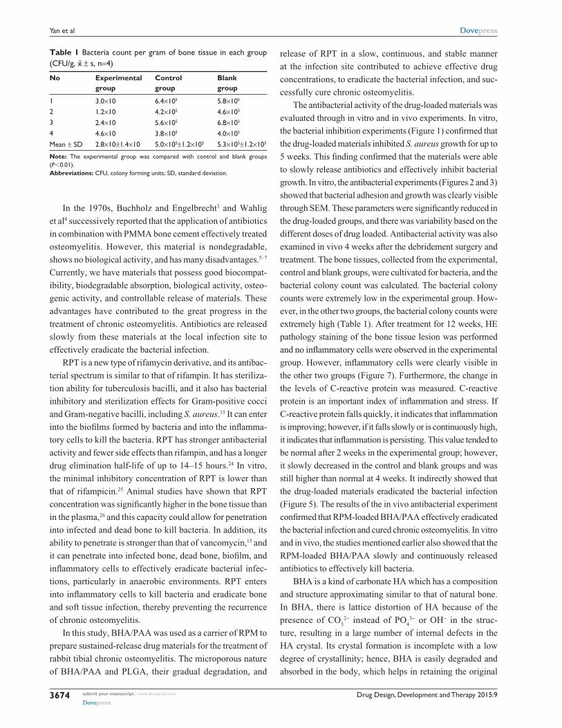
Microbial examinationFour weeks after induction of osteomyelitis, the bacte-
rial culture results of lesion tissue showed an extensive
bacterial growth on the culture plate, and G-staining and
coagulase test confirmed the bacteria as S. aureus. Four
weeks after the debridement surgery and treatment, bone
tissue lesions were collected from the three groups and
cultivated for bacteria, and then the bacterial CFU/g bone
was calculated. A lower CFU/g was observed in the experi-
mental group (2.8×10±1.4×10 CFU/g), and the concentra-
tion of bacteria was more in the control and blank groups
(5.0×10⁵±1.2×10⁵ CFU/g and 5.3×10⁵±1.2×10⁵ CFU/g,
respectively). The difference between the experimental
group and the other two groups was significant (P,0.01,
Table 1).
DiscussionThe clinical treatment of chronic osteomyelitis induced
by S. aureus is extremely difficult, particularly involving
MRSA. S. aureus in the infective site causes ischemia
and necrosis of the bone and the surrounding tissue, and
it causes the formation of a biofilm that blocks blood flow
and prevents antibiotic penetration.22 S. aureus simultane-
ously produces coagulase on its surface, which results in a
state of immune tolerance that allows the bacteria to escape
removal by inflammatory cells and immune cells. In addition,
S. aureus is resistant to multiple antibiotics, which can lead
to persistent infection. In summary, it is difficult to achieve
effective antibiotic drug concentrations at the local site by
intravenous injection or oral routes, which makes the treat-
ment effect not ideal.23
radiological evaluationThere was no animal death during modeling or during the
treatment. Four weeks after the osteomyelitis induction, the
right proximal tibia of all rabbits had clear chronic osteomy-
elitis on radiographic inspection with the following findings:
reduced bone density, the trabecular bone being thin and
sparse, dead bone, subperiosteal abscesses, and new periosteal
bone formation. We confirmed that a chronic osteomyelitis
animal model was successfully established, as described by
Norden et al.18 The osteomyelitis model severity classification
was level 3, 4 (including 9 in level 3 and 15 in level 4) using
gross observation and radiological detection standards.20,21
After the debridement surgery, the radiographic results of ani-
mals in the three groups at week 12 showed that the proximal
tibia shape had largely returned to the normal structure, that
new trabecular bone had formed, and that the materials were
mainly degraded and absorbed in the experimental group;
whereas the shape of the proximal tibia was not recovered
in the control or blank group, and showed the following
deterioration trend: bone destruction, subperiosteal abscesses,
and periosteal new bone formation. The carrier material was
clearly visible in the control group (Figure 6).
Pathological and immunohistochemical examinationFour weeks after induction of osteomyelitis, the HE stain-
ing of bone tissue lesion showed a large number of clearly
visible inflammatory cells, interstitial hemorrhage, fibrous
hyperplasia, and bone necrosis extending from periosteum
to bone marrow cavity. Grading the severity of osteomyelitis

Drug Design, Development and Therapy 2015:9 submit your manuscript | www.dovepress.com
Dovepress
Dovepress
3673
Treatment of Staphylococcus aureus-induced chronic osteomyelitis
Figure 7 he staining of bone tissue lesion (×100).Notes: (A) Four weeks after the induction of chronic osteomyelitis, there was clearly visible inflammatory cell infiltration, fibrous hyperplasia, dead bone from periosteum to bone marrow cavity. (B–D) Treatment of osteomyelitis for 12 weeks. (B) in the drug-loaded group, the new bone and cartilage formed and materials were largely absorbed. (C) and (D), no drug-loaded group and blank group, respectively, inflammatory cells, fibrous hyperplasia, and dead bone were clearly visible, and new bone did not form. Long white arrow, inflammatory cells; short white arrow, bleeding; long black arrow, trabecular bone; short black arrow, dead bone; long red arrow, cartilage and chondrocytes; short red arrow, materials long green arrow, fibrous hyperplasia and necrosis; short green arrow, the tissue of medullary cavity.Abbreviation: he, hematoxylin and eosin.
Figure 8 Collagen fiber immunohistochemical examination of the bone tissue lesion (×200) 12 weeks after the second surgical implantation of materials.Notes: (A) Drug-loaded group, a large number of collagen fibers formed around the materials. (B) No drug-loaded groups, collagen fiber formation was not notable, only a small amount of formation was observed. F, collagen fiber; M, material.
F
FF
M M
M
M
BA
Drug Design, Development and Therapy 2015:9submit your manuscript | www.dovepress.com
Dovepress
Dovepress
3674
Yan et al
Table 1 Bacteria count per gram of bone tissue in each group (CFU/g, x̄ ± s, n=4)
No Experimental group
Control group
Blank group
1 3.0×10 6.4×10⁵ 5.8×10⁵
2 1.2×10 4.2×10⁵ 4.6×10⁵
3 2.4×10 5.6×10⁵ 6.8×10⁵
4 4.6×10 3.8×10⁵ 4.0×10⁵
Mean ± sD 2.8×10±1.4×10 5.0×10⁵±1.2×10⁵ 5.3×10⁵±1.2×10⁵
Note: The experimental group was compared with control and blank groups (P,0.01).Abbreviations: cFU, colony forming units; sD, standard deviation.
In the 1970s, Buchholz and Engelbrecht3 and Wahlig
et al4 successively reported that the application of antibiotics
in combination with PMMA bone cement effectively treated
osteomyelitis. However, this material is nondegradable,
shows no biological activity, and has many disadvantages.5–7
Currently, we have materials that possess good biocompat-
ibility, biodegradable absorption, biological activity, osteo-
genic activity, and controllable release of materials. These
advantages have contributed to the great progress in the
treatment of chronic osteomyelitis. Antibiotics are released
slowly from these materials at the local infection site to
effectively eradicate the bacterial infection.
RPT is a new type of rifamycin derivative, and its antibac-
terial spectrum is similar to that of rifampin. It has steriliza-
tion ability for tuberculosis bacilli, and it also has bacterial
inhibitory and sterilization effects for Gram-positive cocci
and Gram-negative bacilli, including S. aureus.15 It can enter
into the biofilms formed by bacteria and into the inflamma-
tory cells to kill the bacteria. RPT has stronger antibacterial
activity and fewer side effects than rifampin, and has a longer
drug elimination half-life of up to 14–15 hours.24 In vitro,
the minimal inhibitory concentration of RPT is lower than
that of rifampicin.25 Animal studies have shown that RPT
concentration was significantly higher in the bone tissue than
in the plasma,26 and this capacity could allow for penetration
into infected and dead bone to kill bacteria. In addition, its
ability to penetrate is stronger than that of vancomycin,15 and
it can penetrate into infected bone, dead bone, biofilm, and
inflammatory cells to effectively eradicate bacterial infec-
tions, particularly in anaerobic environments. RPT enters
into inflammatory cells to kill bacteria and eradicate bone
and soft tissue infection, thereby preventing the recurrence
of chronic osteomyelitis.
In this study, BHA/PAA was used as a carrier of RPM to
prepare sustained-release drug materials for the treatment of
rabbit tibial chronic osteomyelitis. The microporous nature
of BHA/PAA and PLGA, their gradual degradation, and
release of RPT in a slow, continuous, and stable manner
at the infection site contributed to achieve effective drug
concentrations, to eradicate the bacterial infection, and suc-
cessfully cure chronic osteomyelitis.
The antibacterial activity of the drug-loaded materials was
evaluated through in vitro and in vivo experiments. In vitro,
the bacterial inhibition experiments (Figure 1) confirmed that
the drug-loaded materials inhibited S. aureus growth for up to
5 weeks. This finding confirmed that the materials were able
to slowly release antibiotics and effectively inhibit bacterial
growth. In vitro, the antibacterial experiments (Figures 2 and 3)
showed that bacterial adhesion and growth was clearly visible
through SEM. These parameters were significantly reduced in
the drug-loaded groups, and there was variability based on the
different doses of drug loaded. Antibacterial activity was also
examined in vivo 4 weeks after the debridement surgery and
treatment. The bone tissues, collected from the experimental,
control and blank groups, were cultivated for bacteria, and the
bacterial colony count was calculated. The bacterial colony
counts were extremely low in the experimental group. How-
ever, in the other two groups, the bacterial colony counts were
extremely high (Table 1). After treatment for 12 weeks, HE
pathology staining of the bone tissue lesion was performed
and no inflammatory cells were observed in the experimental
group. However, inflammatory cells were clearly visible in
the other two groups (Figure 7). Furthermore, the change in
the levels of C-reactive protein was measured. C-reactive
protein is an important index of inflammation and stress. If
C-reactive protein falls quickly, it indicates that inflammation
is improving; however, if it falls slowly or is continuously high,
it indicates that inflammation is persisting. This value tended to
be normal after 2 weeks in the experimental group; however,
it slowly decreased in the control and blank groups and was
still higher than normal at 4 weeks. It indirectly showed that
the drug-loaded materials eradicated the bacterial infection
(Figure 5). The results of the in vivo antibacterial experiment
confirmed that RPM-loaded BHA/PAA effectively eradicated
the bacterial infection and cured chronic osteomyelitis. In vitro
and in vivo, the studies mentioned earlier also showed that the
RPM-loaded BHA/PAA slowly and continuously released
antibiotics to effectively kill bacteria.
BHA is a kind of carbonate HA which has a composition
and structure approximating similar to that of natural bone.
In BHA, there is lattice distortion of HA because of the
presence of CO3
2- instead of PO4
3- or OH- in the struc-
ture, resulting in a large number of internal defects in the
HA crystal. Its crystal formation is incomplete with a low
degree of crystallinity; hence, BHA is easily degraded and
absorbed in the body, which helps in retaining the original

Drug Design, Development and Therapy 2015:9 submit your manuscript | www.dovepress.com
Dovepress
Dovepress
3675
Treatment of Staphylococcus aureus-induced chronic osteomyelitis
good biocompatibility, biological activity, and osteogenic
effect of HA.27 BHA exchanges Ca2+, OH-, HPO42-, PO
43-,
and CO3
2- through autolysis in the body fluid when it is
implanted in the body, and it is bound with the host bone as
a whole through hydrogen bonding. It provides an excellent
physiological scaffold for new bone formation and regenera-
tion. In addition, BHA gradually degrades during the process
of bone healing until the defected bone is entirely replaced.
Because the material is naturally porous and has a large
surface area, it is conducive to new blood vessel growth and
formation,28 which increases the ability of local tissue blood
supply to tissues and improves defence against inflammation.
BHA also is also conducive to osteoblast and bone marrow
stem cell adhesion, proliferation, growth, as well as to the
formation of new bone tissue on the material.29,30 Addition-
ally, nanocrystals of BHA are smaller than those of HA, so
their surface area is increased, which increases the surface
area and hence the drug-loading capacity. This allows for
the achievement of high local antibiotics concentrations of
antibiotics in a short period of time when implanted in the
body, which results in the effective killing of pathogens in the
infective lesions. However, pure BHA is highly brittle, and
hence is not conducive to processing and plasticity. When it
combines with PAA, its toughness and mechanical property is
greatly enhanced and provides good support for bone defects.
PAA, with a similar molecular structure to collagen, has good
biocompatibility and is biodegradable,9 and its degradation
products includes glycine, proline, and hydroxyproline, which
promote the formation of collagen fibers. Collagen fibers are
the scaffold of bone formation and attract calcium deposits,
which promotes new bone formation. Preliminary experi-
ments31 had shown that the BHA/PAA material had good
biological safety, compatibility, and bone conductibility.
For the in vitro osteogenesis experiments, MG63 cells
were cultivated together with BHA/PAA for 7 days. Inverted
microscopy observation showed that a large number of cells
adhered and grew on the material. Under SEM, extensive cell
adhesion, growth, and calcium nodule formation was visible
on the material. These results showed that the material had
good biocompatibility, biological activity, and osteogenic
activity (Figure 4).
The degradation and osteogenic ability of the RPM-
loaded BHA/PAA material was evaluated in vivo. After
12 weeks of treatment, I collagen fibers were detected in
the bone tissue lesions via through immunohistochemical
staining. A large number of I collagen fibers formed around
the materials in the experimental group. However, there was
no or only a small amount of I collagen fiber formation in
the control group (Figure 8), due to persistent inflammation.
Collagen fibers are a scaffold for calcium salt deposition and
the formation of new bone. Studies have shown that collagen
fibers are important indicators of reflexion bone formation,
and they play an important role in calcium nodule formation
and osteoblast adhesion, differentiation, and maturation.32
HE staining showed that most of the material was degraded
and new trabecular bone and cartilage formed in the experi-
mental group of containing drug-loaded material. In contrast,
degradation of material and the formation of new bone were
not observed in the group where no drug was loaded into
the material (Figure 7), due to persistent inflammation. The
results of immunohistochemical staining for collagen fibers
and HE staining confirmed that the RPM-loaded BHA/PAA
had good biological activity and osteogenesis induction abil-
ity, promoting new bone formation and rebuilding the bone
defect with gradual degradation and absorption.
For radiographic detection, 24 rabbits with chronic
osteomyelitis were conducted by debridement surgery. The
results of the treatment confirmed that the bone shape gradu-
ally improved in the RPM-loaded BHA/PAA experimental
group, and it largely returned to normal at 12 weeks. The
material was faintly visible, thus showing that the material
was largely degraded and absorbed. However, in the BHA/
PAA control group, the bone shape was not improved, and
there was a deterioration trend with clearly visible material,
which indicated no degradation and absorption (Figure 6).
The imaging examination results showed that the drug-loaded
material eradicated the bacterial infection, promoted new
bone formation, and rebuilt the bone defect.
ConclusionThrough in vitro and in vivo antibacterial experiments, this
study confirmed that RPM-loaded BHA/PAA effectively
restrained and killed bacteria, and controlled rabbit tibial
infection. The material slowly and continuously released
the antibiotic, RPT, which had an effective sterilization
effect against S. aureus. In vitro, the osteogenesis experi-
ments showed that BHA/PAA promoted MG63 adhesion,
aggregation, differentiation, and calcium nodule formation.
In vivo, the osteogenesis experiments showed that RPM-
loaded BHA/PAA induced osteogenesis, promoted new
bone formation, and reconstructed bone defects while the
material was gradually degraded and absorbed. In conclu-
sion, the current study shows that RPM-loaded BHA/PAA
is effective for the treatment of chronic osteomyelitis and for
the reconstruction of bone defects. This method could be a
promising method for the treatment of orthopedic infections
induced by MRSA and should be investigated further in
future research and clinical work.

Drug Design, Development and Therapy
Publish your work in this journal
Submit your manuscript here: http://www.dovepress.com/drug-design-development-and-therapy-journal
Drug Design, Development and Therapy is an international, peer-reviewed open-access journal that spans the spectrum of drug design and development through to clinical applications. Clinical outcomes, patient safety, and programs for the development and effective, safe, and sustained use of medicines are a feature of the journal, which
has also been accepted for indexing on PubMed Central. The manu-script management system is completely online and includes a very quick and fair peer-review system, which is all easy to use. Visit http://www.dovepress.com/testimonials.php to read real quotes from published authors.
Drug Design, Development and Therapy 2015:9submit your manuscript | www.dovepress.com
Dovepress
Dovepress
Dovepress
3676
Yan et al
AcknowledgmentThis study was financially supported by the National Natural
Science Foundation Program of China (81171685).
DisclosureThe authors report no conflicts of interest in this work.
References 1. Lew DP, Waldvogel FA. Osteomyelitis. Lancet. 2004;364:369–379. 2. Sia IG, Berbari EF. Osteomyelitis. Best Pract Res Clin Rheumatol.
2006;20:1065–1081. 3. Buchholz HW, Engelbrecht H. Depot effects of various antibiotics
mixed with Palacos resins. Chirurg. 1970;41:511–515. 4. Wahlig H, Dingeldein E, Bergmann R, Reuss K. The release of gen-
tamicin from polymethylm ethacrylate beads. An experimental and pharmacokinetic study. J Bone Joint Surg Br. 1978;60-B:270–275.
5. Wong MW, Hui M. Development of gentamicin resistance after gentamicin-PMMA beads for treatment of foot osteomyelitis: report of two cases. Foot Ankle Int. 2005;26:1093–1105.
6. Gürsel I, Korkusuz F, Türesin F, Alaeddinoglu NG, Hasirci V. In vivo application of biodegradable controlled antibiotic release systems for the treatment of implant-related osteomyelitis. Biomaterials. 2001;22:73–80.
7. Wei G, Kotoura Y, Oka M, et al. A bioabsorbable delivery system for antibiotic treatment of osteomyelitis: the use of lactic acid oligomer as a carrier. J Bone Joint Surg Br. 1991;73:246–252.
8. Genile P, Chiono V, Boccafoschi F. Composite films of gelatin and hydroxyapatite/bioactive glass for tissue-engineering applications. J Biomater Sci Polym Ed. 2010;21(8/9):1207–1226.
9. Roohani-Esfahani SI, Nour-iKhorasani S, Lu Z, Appleyard R, Zreiqat H. The influence hydroxyapatite nanoparticle shape and size on the properties of biphasic calcium phosphate scaffolds coated with hydroxyapatite-PCL composites. Biomaterials. 2010;31:5498–5509.
10. Stallmann HP, Faber C, Bronckers AL, Nieuw Amerongen AV, Wuisman PI. In vitro gentamicin release from commercially available calcium-phosphate bone substitutes influence of carrier type on duration of the release profile. BMC Musculoskelet Disord. 2006;7:18.
11. Koort JK, Suokas E, Veiranto M, et al. In vitro and in vivo testing of bioabsorbable antibiotic containing bone filler for osteomyelitis treat-ment. J Biomed Mater Res A. 2006;78:532–540.
12. Orhan Z, Cevher E, Mülazimoglu L, et al. The preparation of cipro-floxacin hydrochloride-loaded chitosan and pectin microspheres: their evaluation in an animal osteomyelitis model. J Bone Joint Surg Br. 2006;88:270–275.
13. Wu Y, Bose S. Nanocrystalline hydroxyapatite: micelle templated synthesis and characterization. Langmuir. 2005;21:3232–3234.
14. Freitas S, Merkle HP, Gander B. Microencapsulation by solvent extraction/evaporation: reviewing the state of the art of microspheres preparation process technology. J Control Release. 2005;102:313–332.
15. Petri WA. Chemotherapy of tuberculosis, Mycobacterium avium complex disease, and leprosy. In: Goodman LS, Gilman A, Brunton LL, Lazo JS, Parker KL, editors. Goodman and Gillman’s The Pharmacological Basis Of Therapeutics, 11th ed. New York: Churchill Livingstone. 2006;1207–1210.
16. Peng X, Li Y, Wang X, et al. Medical nano hydroxyapatite/polyamide 66 composite material in vitro immersion behavior research. J Funct Mater. 2004;2:253–255.
17. O’Reilly T, Mader JT. Rat model of bacterial osteomyelitis of the tibia. In: Zak O, Sande MA, editors. Handbook of Animal Models of Infection: Experimental Model in Antimicrobial Chemotherapy. San Diego, CA: Academic Press. 1999;560–575.
18. Norden CW, Myerowitz RL, Keleti E. Experimental osteomyelitis due to Staphylococcus aureus or Pseudomonas aeruginosa. A radiographic-pathological correlative analysis. Br J Exp Path. 1980;61:451–460.
19. Smeltzer MS, Thomas JR, Hickmon SG, et al. Characterization of a rabbit model of Staphylococcal osteomyelitis. J Orthop Res. 1997;15:414–421.
20. Shirtliff ME, Calhoun JH, Mader JT. Experimental osteomyelitis treatment with antbiotic impregnated hydroxyapatite. Clin Orthop Relat Res. 2002;40:239–247.
21. Mader JT, Stevens CM, Stevens JH, Ruble R, Lathrop JT, Calhoun JH. Treatment of experimental osteomyelitis with a fibrin sealant antibiotic implant. CIin Orthop Relat Res. 2002;403:58–72.
22. Costerton JW. Biofilm theory can guide the treatment of device-related orthopaedic infections. Clin Orthop Relat Res. 2005;437:7–11.
23. Stewart PS, Costerton JW. Antibiotic resistance of bacteria in biofilms. Lancet. 2001;358:135–138.
24. Keung AC, Eller MG, McKenzie KA, Weir SJ. Single and multiple dose pharmacokinetics of rifapentine in man: part II. Int J Tuberc Lung Dis. 1999;3:437–444.
25. Venkataraman P, Paramasivan CN, Prabhakar R. In vitro activity of rifampicin, rifapentine and rifabutin against south Indian isolates of Mycobacterium tuberculosis. Indian J Tuberc. 1993;40:17–20.
26. Assandri A, Ratti B, Cristina T. Pharmacokinetics of rifapentine, a new long lasting rifamycin, in the rat, the mouse and the rabbit. J Antibiot (Tokyo). 1984;37:1066–1075.
27. Gentile P, Chiono V, Boccafoschi F, et al. Composite films of gelatin and hydroxyapatite/bioactive glass for tissue-engineering applications. J Biomater Sci Polym Ed. 2010;21:1207–1226.
28. Chanda A, Singha Oy R, Xue W, Bose S, Bandyopadhyay A. Bone cell-materials interaction on alumina ceramics with different grain sizes. Mater Sci Eng C. 2009;29:1201–1206.
29. Mastrogiacomo M, Scaglione S, Martinetti R, et al. Role of scaffold internal structure on in vivo bone formation in macroporous calcium phosphate bioceramics. Biomaterials. 2006;27:3230–3237.
30. Turco G, Marsich E, Bellomo F, et al. Alginate/hydroxyapatite biocom-posite for bone ingrowth: a trabecular structure with high and isotropic connectivity. Biomacromolecules. 2009;10:1575–1583.
31. Dai Y, Li J, Zhao ZH, et al. Nano amino apatite/poly (amino acid com-posite safety evaluation. J Third Military Medical University. 2010;32: 294–299.
32. Yang ZM, Yu XJ, Huang FG, et al. Exogenous type I collagen of embry-onic periosteum ossification cell biological characteristics influence. Journal of West China Major Medical Journal. 2001;32:1.