treatment of atherosclerosis by macrophage-biomimetic
TRANSCRIPT
ARTICLE
Treatment of atherosclerosis by macrophage-biomimetic nanoparticles via targetedpharmacotherapy and sequestration ofproinflammatory cytokinesCheng Gao 1,2, Qiaoxian Huang1,2, Conghui Liu 1,2, Cheryl H. T. Kwong1, Ludan Yue1, Jian-Bo Wan1,
Simon M. Y. Lee1✉ & Ruibing Wang 1✉
Vascular disease remains the leading cause of death and disability, the etiology of which
often involves atherosclerosis. The current treatment of atherosclerosis by pharmacotherapy
has limited therapeutic efficacy. Here we report a biomimetic drug delivery system derived
from macrophage membrane coated ROS-responsive nanoparticles (NPs). The macrophage
membrane not only avoids the clearance of NPs from the reticuloendothelial system, but also
leads NPs to the inflammatory tissues, where the ROS-responsiveness of NPs enables spe-
cific payload release. Moreover, the macrophage membrane sequesters proinflammatory
cytokines to suppress local inflammation. The synergistic effects of pharmacotherapy and
inflammatory cytokines sequestration from such a biomimetic drug delivery system lead to
improved therapeutic efficacy in atherosclerosis. Comparison to macrophage internalized
with ROS-responsive NPs, as a live-cell based drug delivery system for treatment of ather-
osclerosis, suggests that cell membrane coated drug delivery approach is likely more suitable
for dealing with an inflammatory disease than the live-cell approach.
https://doi.org/10.1038/s41467-020-16439-7 OPEN
1 State Key Laboratory of Quality Research in Chinese Medicine, Institute of Chinese Medical Sciences, University of Macau, Taipa, Macao, China. 2Theseauthors contributed equally; Cheng Gao, Qiaoxian Huang, Conghui Liu. ✉email: [email protected]; [email protected]
NATURE COMMUNICATIONS | (2020) 11:2622 | https://doi.org/10.1038/s41467-020-16439-7 | www.nature.com/naturecommunications 1
1234
5678
90():,;
Cardiovascular disease remains the leading cause of deathglobally1. The disease generally involves atherosclerosis.Although the atherosclerosis process is not well under-
stood, it is often initiated by the endothelial layers’ dysfunction,which accumulates oxidized form of low-density lipoprotein(LDL) in the intimal layer and leads to the local inflammationwhere reactive oxygen species (ROS) are overproduced2,3. Theinflammatory areas recruit monocytes and differentiate intomacrophages. Upon ingestion of LDL, macrophages would die oreven lead to cellular rupture, providing positive feedback torecruit additional immune cells to these areas4,5. Subsequently,the inflammation leads to formation of atheromatous plaques inthe arterial tunica intima. Treatment of established plaques mayinclude medications to lower cholesterol, such as statins, ormedications that decrease clotting, such as aspirin6. However,only limited efficacy has been observed in clinics when the drugsare administered systemically, likely attributed to the rapid drugclearance and unsatisfactory accumulation at the arterial injurysite7. Considering these issues, significant efforts have beendevoted to the design of various nanomaterials, which couldcontrol payload release at the plaque sites in response to thehighly produced ROS or the acidic environment, or may enhancetargeted drug delivery via a targeting ligand, or extend systemiccirculation8,9.
However, once administered into the body, nanomaterialsencounter extremely complexed physiological environments anddefense (immune) system that is actively recognizing and clearingmatters that are foreign to our human body10,11. Thus, very fewNPs-based formulations coated with targeting ligands have pas-sed phase III clinical trials. One of the major hurdles for theclinical applications of NPs is that most NPs are taken andremoved by the reticuloendothelial system (RES) before reachingthe target tissues12,13. Very recently, Chan and co-workers pre-pared trastuzumab and folic acid coated gold and silica NPs, andquantified their cancer cell targeting efficiencies14. Their resultsdemonstrated that only 0.7% i.v. administered NPs reached thetumor site and only 0.0014% reached inside targeted cancer cells,suggesting that NPs, even tagged with targeting ligands, might notachieve the expected delivery efficiency.
As one of the most fundamental units of life, cells get alongwell with its surrounding environments and may accumulate insome specific microenvironments depending on their nature15.Biomimetic drug delivery systems, especially cell membranecoated NPs, have attracted rapidly increasing attentions16,17. Forinstance, Tasciotti et al. reported the first macrophage membrane(leukocyte membranes) coated nanoparticles that enhanced cir-culation time and improved accumulation in the tumor18. Zhanget al. recently reported dual‐cell membrane‐coated NPs from thefused membranes originated from both red blood cells (RBC) andplatelets, and neutrophil membrane coated nanoparticles toalleviate inflammatory arthritis19,20. For the treatment of cardi-ovascular disease, platelet membrane coated nanoparticles werefirstly reported to show enhanced therapeutic effects againstcoronary restenosis21. Similarly, Wang and coworkers developedRBC membrane coated nanocomplexes to minimize macrophage‐mediated phagocytosis in the blood and enhance accumulation ofnanoparticles in the established atherosclerotic plaques forimproved atherosclerosis management22. In spite of the sig-nificantly reduced RES clearance and improved payload delivery,none of these previous carriers may specifically release payload inthe disease sites. Considering the accumulation of macrophageand overproduction of ROS during the development of athero-sclerosis23, herein we developed a delivery system derived frommacrophage membrane coated ROS-responsive NPs (MM-NPs)for the treatment of atherosclerosis. The macrophage membranenot only may improve targeted delivery of NPs and payload to the
lesion site, but also may act as a scavenger for proinflammatoryfactors. Meanwhile, live cells have also recently emerged aspotential drug carriers with various advantages24. For instance,neutrophils were applied as carriers for liposomes loaded withpaclitaxel to fight against postoperative glioma recurrence, andhaematopoietic stem cells were used for the delivery of antibodiesto augment antileukemia efficacy25,26. As the inflammatoryresponse of macrophage is not only related to the proteins in cellmembrane, but also involves some signal pathways insidecells27,28, we thought that ROS-responsive NPs internalized insidemacrophage (NPs/MAs) might as well have a high targetingefficiency and excellent therapeutic efficacy on atherosclerosis.Thus, a thorough comparative study between MM-NPs and NPs/MAs was conducted for the treatment of atherosclerosis in vivo.
In our experimental design, atorvastatin (AT) is selected as themodel drug and positive control. The attenuation effects of ATloaded ROS-NPs (AT-NPs) and macrophage membrane coatedAT-NPs (MM-AT-NPs) on lipopolysaccharide (LPS) and oxLDLinduced macrophage inflammation and foam cell formation are,respectively, demonstrated. The escape behavior of MM-NPsfrom macrophage, as well as the intracellular drug release in ROSoverproduced cells are exhibited in vitro. Biocompatibility ofMM-NPs and macrophage is respectively confirmed in vivo. In anatherosclerotic mouse model, the targeting efficiency and ther-apeutic efficacy of MM-AT-NPs and AT-NPs internalized insidemacrophages (AT-NPs/MAs) are systemically evaluated andcompared against those of AT alone. Finally, the underlyingtherapeutic mechanisms are thoroughly investigated at a mole-cular level, and very interestingly, MM-AT-NPs exhibit hints ofmoderately better therapeutic efficacy than AT-NPs/MAs to treatthe inflammatory disease.
ResultsPreparation of ROS responsive NPs and MM-NPs. ROSresponsive NPs were prepared via self-assembly of amphiphilicoxidation-sensitive chitosan oligosaccharide (Oxi-COS). Thesynthetic procedure of Oxi-COS was shown in SupplementaryFig. 1, and the successful chemical conjugation was confirmed by1H NMR (Supplementary Fig. 2). In an aqueous solution, thephenylboronic acid pinacol ester serves as the hydrophobic sidechain, which would aggregate to form a hydrophobic core, andthe hydrophilic backbone of COS would spread outside aroundthe hydrophobic core to form NPs with a micelle structure.Subsequently, NPs loaded with hydrophobic AT (AT-NPs), witha drug encapsulation efficiency (DEE) of 48.3% and a drugloading content (DLC) of 5.1%, were obtained by a simple self-assembly process (Fig. 1a). Transmission electron microscope(TEM) analysis showed that these NPs possessed roughly sphe-rical morphology with a mean size of ca. 149 nm (Fig. 1b). It iswell known that H2O2 oxidizes arylboronic esters29,30, thus Oxi-COS would degrade into pinacol borate, COS, and p-hydroxy-methylphenol in the presence of H2O2 (overproduced atinflammatory tissues). In order to confirm the ROS responsive-ness, NPs were placed in phosphate-buffered saline (PBS) withdifferent concentrations of H2O2 and their transmittance wasmeasured at 500 nm at different time points. The concentrationsof H2O2 used in the present study were based on previous lit-erature papers that simulated the level of H2O2 in vitro foratherosclerotic lesion9,31. As shown in Fig. 1c, the particle solu-tion in the presence of 1.00 mM H2O2 became completelytransparent within 2 h. In contrast, the particles in PBS exhibitedonly moderate decrease over 4 h in absorbance due to partialprecipitation of NPs. Accordingly, AT-NPs exhibited a highaccumulative drug release rate up to ∼80% in the presence of1.00 mM H2O2, whereas only ∼20% of drug was released without
ARTICLE NATURE COMMUNICATIONS | https://doi.org/10.1038/s41467-020-16439-7
2 NATURE COMMUNICATIONS | (2020) 11:2622 | https://doi.org/10.1038/s41467-020-16439-7 | www.nature.com/naturecommunications

peroxide treatment (Fig. 1d), indicating the excellent ROSresponsiveness of these NPs.
To synthesize MM-NPs, macrophage membrane derived frommurine macrophage cell line (RAW264.7 cells) was coated on thesurface of ROS responsive NPs via an extrusion method (Fig. 1e).As shown in Fig. 1f, MM-NPs exhibited a spherical core–shellstructure under TEM, and each NP was wrapped with a single
layer of cell membrane, as the thickness of the wrapped layer was∼9 nm, in line with the thickness of cell membrane. Dynamiclaser scattering (DLS) indicated that the zeta potential of MM-NPs was more negative than that of free NPs (Fig. 1g), butconsistent with the zeta potential of macrophage surface.Furthermore, DLS analysis revealed that, after cell membranecoating, the diameter of nanoparticles was increased from ∼204
Self-assembling
AT
Time (min)
Hyd
roly
sis
perc
enta
ge (
%)
0 100 200 300
150
100
50
0
PBS0.01 mM H2O2
0.25 mM H2O2
0.5 mM H2O2
1 mM H2O2
Time (h)
Cum
ulat
ive
drug
rel
ease
(%
)
0 10 20 30
Macrophage Membrane derived vesicles ROS responsive NPs MM-NPs
Hypotonic treatment
Extrusion
Extrusion
=
Dia
met
er (
nm)
300
200
100
0
*****
ζ (m
v)
NPs
MM
-NPs
Mac
roph
age
vesic
les
0
–10
–20
–30
***
Inte
grat
ed d
ensi
ty 10,000
5000
0
TNFR2
***
***CCR2
***
***
CD36
CCR2
TNFR270
100
70
100
55
40
a
b
e
f g
c d
h
i
j
kDa
AT-NPsOxi-COS
150
100
50
0
1 mM H2O2
0 mM H2O2
NPs
MM
-NPs
Mac
roph
age
vesic
les
NPs
MM
-NPs
Mac
roph
age
vesic
les
Inte
grat
ed d
ensi
ty 10,000
5000
0
NPs
MM
-NPs
Mac
roph
age
vesic
les
NPs
MM
-NPs
Mac
roph
age
vesic
les
O
O
O
HOO
OHO
HO
OH
nOHO
NHO
OO
O
BOO OO
NATURE COMMUNICATIONS | https://doi.org/10.1038/s41467-020-16439-7 ARTICLE
NATURE COMMUNICATIONS | (2020) 11:2622 | https://doi.org/10.1038/s41467-020-16439-7 | www.nature.com/naturecommunications 3
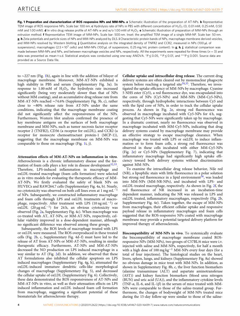
to ∼227 nm (Fig. 1h), again in line with the addition of bilayer ofmacrophage membrane. Moreover, MM-AT-NPs exhibited ahigh stability in PBS and serum (Supplementary Fig. 3a). Inresponse to 1.00 mM of H2O2, the hydrolysis rate increasedsignificantly (being very moderately slower than that of NPswithout MM coating), and an accumulative drug release rate fromMM-AT-NPs reached ∼74.6% (Supplementary Fig. 3b, c), ratherclose to ∼80% release rate from AT-NPs under the sameconditions, indicating that the macrophage membrane coatingdid not significantly affect the responsiveness of the NPs.Furthermore, Western blot analysis confirmed the presence ofkey membrane antigens on the surfaces of both macrophagemembrane and MM-NPs, such as tumor necrosis factor receptorreceptor 2 (TNFR2), CD36 (a receptor for oxLDL), and CCR2 (areceptor for monocyte chemoattractant protein-1 (MCP-1)),suggesting that the macrophage membrane on MM-NPs wascomparable to those on macrophage (Fig. 1i, j).
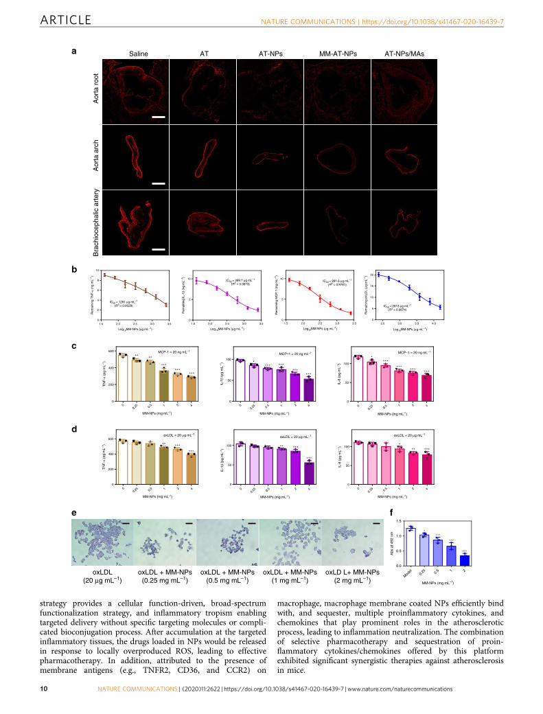
Attenuation effects of MM-AT-NPs on inflammation in vitro.Atherosclerosis is a chronic inflammatory disease and the for-mation of foam cells plays a key role in disease development32,33.Therefore, LPS-induced inflammation in macrophage andoxLDL-treated macrophage (foam cells formation) were selectedas in vitro models for evaluating the therapeutic efficacy of MM-AT-NPs. We firstly evaluated the safety of blank NPs onHUVECs and RAW264.7 cells (Supplementary Fig. 4a, b). Nearly,no cytotoxicity was observed on both cell lines even at 1 mgmL−1
of NPs. Subsequently, we constructed inflammatory macrophageand foam cells through LPS and oxLDL treatments of macro-phage, respectively. After treatment with LPS (10 ng mL−1) oroxLDL (20 µg mL−1) for 24 h, an obvious cytotoxicity wasobserved (Fig. 2a, Supplementary Fig. 4c). When macrophage wasco-treated with AT, AT-NPs, or MM-AT-NPs, respectively, cel-lular viability improved in a dose-dependent manner, althoughno significant difference was observed among these groups.
Subsequently, the ROS levels of macrophage treated with LPSor oxLDL were measured. The ROS overproduced in these treatedcells (Fig. 2b, c, Supplementary Fig. 4d–f) must have led to therelease of AT from AT-NPs or MM-AT-NPs, resulting in similartherapeutic efficacy. Furthermore, AT-NPs and MM-AT-NPsdecreased the NO production on LPS induced macrophage in away similar to AT (Fig. 2d). In addition, we observed that theseAT formulations also inhibited the cellular apoptosis on LPSinduced macrophages (Supplementary Fig. 4g, h), attenuated theoxLDL-induced immature dendritic cells-like morphologicalchanges of macrophage (Supplementary Fig. 5), and decreasedthe cellular uptake of oxLDL (Supplementary Fig. 6). Collectively,these data demonstrated the ROS responsiveness of AT-NPs andMM-AT-NPs in vitro, as well as their attenuation effects on LPSinduced inflammation and oxLDL induced foam cell formationfrom macrophage, suggesting a significant potential of thesebiomaterials for atherosclerosis therapy.
Cellular uptake and intracellular drug release. The current drugdelivery systems are often cleared out by mononuclear phagocytesystem before reaching a targeted site34,35. Therefore, we inves-tigated the uptake efficiency of MM-NPs by macrophage. Cyanine5 NHS ester (Cy5), a red fluorescence dye, was encapsulated intothe cores of NPs (Cy5-NPs) and MM-NPs (MM-Cy5-NPs),respectively, through hydrophobic interactions between Cy5 andwith the lipid core of NPs, in order to track the cellular uptakeprocess. As shown in Fig. 2e, strong red fluorescence wasobserved in macrophage incubated with Cy5-NPs for 4 h, sug-gesting that Cy5-NPs were significantly taken up by macrophage.In a dramatic contrast, nearly no fluorescence was observed inmacrophage incubated with MM-Cy5-NPs, indicating that drugdelivery systems coated by macrophage membrane may providean effective strategy to escape macrophage clearance. Whenmacrophage was treated with LPS or oxLDL to induce inflam-mation or to form foam cells, a strong red fluorescence wasobserved in these cells incubated with either MM-Cy5-NPs(Fig. 2e) or Cy5-NPs (Supplementary Fig. 7), indicating thatinflammatory macrophage had significantly high uptake effi-ciency toward both delivery systems without discriminationagainst MM-NPs.
To examine the intracellular drug release behavior, Nile red(NR), a lipophilic stain with little fluorescence in a polar solutionbut strong red fluorescence in a lipid environment36, was loadedinto MM-NPs (MM-NR-NPs), and incubated with LPS- andoxLDL-treated macrophage, respectively. As shown in Fig. 2f, thered fluorescence of NR increased in an incubation-timedependent manner, indicating the NR was released in LPS andoxLDL treated, inflammatory macrophages, respectively (Fig. 2b,Supplementary Fig. 4e). Taken together, the escape of MM-NPsfrom macrophages, their efficient cellular uptake and responsivepayload release in inflammatory macrophages and foam cellssuggested that the ROS-responsive NPs coated with macrophagemembrane may provide a potential targeted delivery platform forimproved therapy of atherosclerosis.
Biocompatibility of MM-NPs in vivo. To systemically evaluatethe biocompatibility of macrophage membrane coated ROS-responsive NPs (MM-NPs), two groups of C57BL/6 mice were i.v.injected with saline and MM-NPs, respectively, for half a monthwith a high dose of 100 mg kg−1 MM-NPs every four days (for atotal of four injections). The histological studies on the heart,livers, spleen, lungs, and kidneys (Supplementary Fig. 8a) showedno obvious damage in mice treat with MM-NPs. In addition, asshown in Supplementary Fig. 8b, c, the liver function biomarkers(alanine transaminase (ALT) and aspartate aminotransferase(AST)) and kidney function biomarkers (blood urea nitrogen(BUN) and uric acid (UA)), and the inflammatory cytokine levels(TNF-α, IL-6, and IL-1β) in the serum of mice treated with MM-NPs were comparable to those of the saline-treated group. Fur-thermore, the changes of bodyweight of MM-NPs treated miceduring the 15-day follow-up were similar to those of the saline-
Fig. 1 Preparation and characterization of ROS responsive NPs and MM-NPs. a Schematic illustration of the preparation of AT-NPs. b RepresentativeTEM image of ROS responsive NPs. Scale bar: 100 nm. c Hydrolysis rate of NPs in PBS with different concentration of H2O2 (0, 0.01 mM, 0.25mM, 0.50mM and 1.00mM). d In vitro drug release profile of AT-NPs w and w/o 1.00mM of H2O2. e Schematic illustration of preparation of MM-NPs through anextrusion method. f Representative TEM image of MM-NPs. Scale bar: 500 nm. Inset: the amplified TEM image of a single MM-NP. Scale bar: 50 nm.g, h Zeta potentials and particle sizes of NPs and MM-NPs analyzed by DLS. i Characteristic protein bands of NPs, macrophage membrane derived vesicles,and MM-NPs resolved by Western blotting. j Quantitative analysis on the integrated density of TNFR2 and CCR2, measured in NPs (100 μL ofsuspensions), macrophages (2.5 × 10⁷ cells) and MM-NPs (100 μL of suspensions, 0.25 mg/mL protein content). In g, h, j, statistical comparison wasmade between MM-NPs and NPs, and between macrophage vesicles and NPs, respectively. All the experiments were repeated for three times (n= 3) anddata was presented as mean ± s.d. Statistical analysis was conducted using one-way ANOVA. *P≤ 0.05, **P≤ 0.01, and ***P≤ 0.001. Source data areprovided as a Source Data file.
ARTICLE NATURE COMMUNICATIONS | https://doi.org/10.1038/s41467-020-16439-7
4 NATURE COMMUNICATIONS | (2020) 11:2622 | https://doi.org/10.1038/s41467-020-16439-7 | www.nature.com/naturecommunications

treated control group (Supplementary Fig. 8d). Collectively, thisset of data support the good biocompatibility of MM-NPs in vivo.
Targeted delivery in atherosclerotic mouse. In the develop-mental process of atherosclerosis, atheromatous plaquessecrete inflammatory cytokines and chemokines, leading to the
recruitment of macrophages37,38. Taking this into account, livemacrophage might be an ideal drug delivery system for AT-NPsdue to their immune response and proactive migration toinflammatory sites39. In this study, RAW264.7 cells were selectedas live cell based carriers for AT-NPs. Thus, we preparedAT-NPs/MAs (Fig. 3a) to investigate their therapeutic efficacyagainst atherosclerosis in mice, and to compare with that of
Cel
l via
bilit
y (%
of c
ontr
ol)
100
50
150
0
AT + LPSAT-NPs + LPSMM-AT-NPs + LPS
AT (μM) – – 1.25 5 20
LPS (10 ng mL–1) – + + + +
* ****
********
* ****** ******
Cou
nt
0 104 105 106
Control
LPS
AT + LPS
AT-NPs + LPS
MM-AT-NPs + LPS Mea
n D
CF
H-D
Aflu
ores
cenc
e (f
old)
Contro
lLP
S
AT + L
PS
AT-NPs +
LPS
MM
-AT-N
Ps + L
PS
0
1
2
3
4
5 *****
******
NO
pro
duct
ion
(%) 120
90
60
30
0
***
**** **
Cy5
-NP
sM
M-C
y5-N
Ps
RA
W26
4.7
cells
1 h 2 h 3 h 4 h
1 h 2 h 3 h 4 h
MM
-Cy5
-NP
s
LPS
trea
ted
mac
roph
ages
1 h 2 h 3 h 4 h
MM
-Cy5
-NP
s
Foa
m c
ells
1 h 2 h 3 h 4 h
LPS
trea
ted
mac
roph
ages
Foa
mce
llsMM
-NR
- NP
s
1 h 2 h 4 h 8 h
1 h 2 h 4 h 8 h
Time (h)
Mea
n flu
ores
cenc
e in
tens
ity(×
103 )
4 h3
h2 h
1 h
0
1
2
3
4
5
a cb d
e
f
Contro
lLP
S
AT + L
PS
AT-NPs +
LPS
MM
-AT-N
Ps + L
PS
Time (h)4
h3 h2
h1
h
Time (h)
4 h
3 h2
h1
h
Time (h)
4 h
3 h2
h1
h
Time (h)
4 h
3 h2
h1
h
Time (h)
4 h
3 h2
h1
h
Mea
n flu
ores
cenc
e in
tens
ity(×
103 )
0
1
2
3
4
5
Mea
n flu
ores
cenc
e in
tens
ity(×
103 )
0
1
2
3
4
5
Mea
n flu
ores
cenc
e in
tens
ity(×
103 )
0
2
4
6
8
10
Mea
n flu
ores
cenc
e in
tens
ity(×
103 )
0
1
2
3
4
5
Mea
n flu
ores
cenc
e in
tens
ity(×
103 )
0
1
2
3
4
5
107
NATURE COMMUNICATIONS | https://doi.org/10.1038/s41467-020-16439-7 ARTICLE
NATURE COMMUNICATIONS | (2020) 11:2622 | https://doi.org/10.1038/s41467-020-16439-7 | www.nature.com/naturecommunications 5

MM-AT-NPs. Firstly, we investigated the systemic biocompat-ibility of RAW264.7 cells in 6-week-old female ApoE−/− mouse.After i.v. administration with different number of macrophagesinto mice once every four days continuously for half a month, itwas found that the body weight (Supplementary Fig. 9a), andcounts of immune-associated cells including monocyte, lym-phocyte and neutrophil in the blood (Supplementary Fig. 9b) ofthe treated mice were similar to those of the mice in the controlgroup. In addition, the histological analysis of the main organs(including the liver, lungs, kidneys, and spleen) of the micetreated by macrophage indicated no toxicity (SupplementaryFig. 9c). These results demonstrated that no obvious immuno-toxicity was caused in mice administered with external macro-phage, even at the highest dosage (109 macrophages kg−1). Uponinternalization of a dye-loaded NPs, Cy5-NPs, by macrophage,red fluorescence was mainly distributed in cytoplasm (Supple-mentary Fig. 10a). The DEE of AT in the AT-NPs/MAs (5 × 10⁷
cells) was determined by HPLC to be ∼14.7%, and moreimportantly, AT-NPs showed negligible cytotoxicity to macro-phage even at a highest concentration of 20 μM used in our study(Supplementary Fig. 10b).
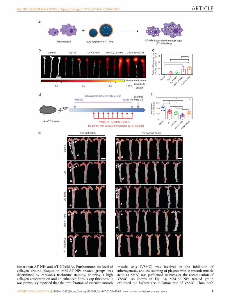
To study the targeting capability of MM-NPs and NPs/MAs toatheromatous plaques, cyanine 7.5 NHS ester (Cy7.5), a syntheticdye with near infrared emission (excitation/emission 788/808nm), were employed for preparing Cy7.5-loaded NPs (Cy7.5-NPs), macrophage membrane coated Cy7.5-NPs (MM-Cy7.5-NPs), and Cy7.5-NPs internalized inside macrophage (Cy7.5-NPs/MAs). As was previously reported, atherosclerotic plaquesbegin to develop in ApoE−/− mouse after being fed with high-fatdiet for 1 month, during which process macrophages migrate toplaques in large numbers40. Thus, 6-week-old female ApoE−/−
mice having received high-fat diet for 1 month were employed forour investigation of the targeted delivery. To determine a suitableimaging time point for plaque targeting studies, in vitropharmacokinetic studies of Cy7.5-NPs, MM-Cy7.5-NPs, andCy7.5-NPs/MAs were determined by IVIS (in vivo imagingspectrum) system by testing on blood samples taken from mice atpredetermined time points after i.v. administration of each ofthese formulations. As shown in Supplementary Fig. 11, thefluorescence intensities became significantly weaker after admin-istration of these formulations for longer than 6 h, and got almostcompletely cleared after 12 h. The circulation half-life (t1/2) ofMM-Cy7.5-NPs, 9.82 h, was much longer than Cy7.5-NPs (t1/2=5.43 h), and Cy7.5-NPs/MAs (t1/2= 13.32 h) exhibited the longestcirculation time among all groups. Therefore, at 6 h after i.v.injection of different formulations (free Cy7.5, Cy7.5-NPs, MM-Cy7.5-NPs, and Cy7.5-NPs/MAs, respectively) with the samedosage of Cy7.5 (2 mg kg−1) into mice, the aorta harvested fromall treated groups of mice exhibited different levels of fluorescence(Fig. 3b), whereas the blank control groups exhibited nofluorescence (Fig. 3c). Among which, Cy7.5-NPs treated groupshowed slightly stronger fluorescence than that of free Cy7.5treated group in the aorta tissues, likely due to the specificrelease of Cy7.5-NPs in response to overproduced ROS in the
inflammatory aorta. In particular, strong fluorescence wasobserved in the aorta tissues isolated from both MM-Cy7.5-NPsand Cy7.5-NPs/MAs treated groups, likely due to their inherentimmune propensity for inflammation. Of note, Cy7.5-NPs/MAseven exhibited a moderately higher accumulation rate in aortatissue than MM-Cy7.5-NPs. The enhanced targeting efficiency ofCy7.5-NPs/MAs was likely attributed to active recruitment ofmacrophages in the development process of atherosclerosis.
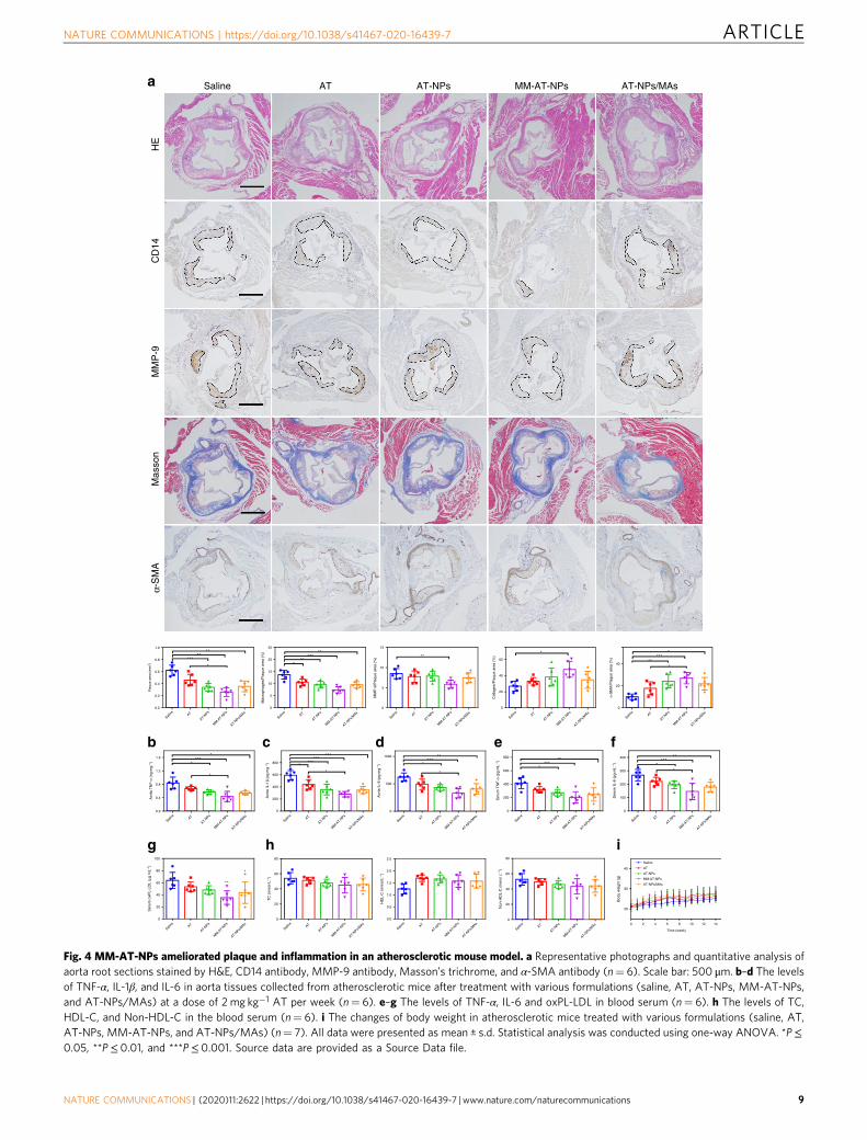
Therapeutic efficacy in atherosclerotic mouse. We next exam-ined the therapeutic efficacy of different formulations (saline, AT,AT-NPs, MM-AT-NPs, and AT-NPs/MAs) against athero-sclerotic development. After receiving high-fat diet for 1 month,6-week-old female ApoE−/− mice were randomly andinvestigator-blindly divided into 5 groups (n= 10 in each groupfrom two batches of studies), and intravenously administeredwith different formulations, respectively, once a week, in com-bination with high-fat food for another 2 months (Fig. 3d). Atendpoint of the experiment, the aorta was collected and stainedby ORO and the resultant red region indicated plaque area(Fig. 3e). As shown in Fig. 3f, the saline-treated group showed ahighest plaque area of ∼20% of the total aorta tissue area,determined by using en face analysis of lesions on the intimalsurface of the aorta41. The free drug AT moderately reduced theplaque area down to ∼15%. Benefiting from the ROS respon-siveness, AT-NPs exhibited slightly improved therapeutic efficacyin comparison to that of free AT. To our surprise, the aorta ofmice treated with AT-NPs/MAs, which we expected to haveexcellent therapeutic efficacy against inflammatory plaques due totheir best payload accumulation in the targeted site, exhibitedsimilar plaque area (∼14%) with that of the aorta of mice treatedwith AT-NPs (Fig. 3e, f). Very interestingly, MM-AT-NPs sig-nificantly decreased plaque area down to ∼8% of the total aortatissue area, showing moderate, yet obvious improvement incomparison with AT-NPs/MAs. Subsequently, we conductedORO staining with sequential 10 cryosections at 100 μm intervalsfrom the aorta tissues (Supplementary Fig. 12). In MM-AT-NPstreated group, an obvious decrease of plaque area was observed inthe aorta root, in comparison to other groups.
Furthermore, histological and immunohistochemical analysiswas conducted on the atherosclerotic plaques from the aorta root.As shown in Fig. 4a, hematoxylin and eosin (H&E) staining onthe aorta root showed that the plaques from the saline-treatedgroup (the control) and AT-treated group were largely necroticcores. In contrast, the areas of plaques and necrotic cores weresignificantly reduced in the MM-AT-NPs treated group. Separatestaining with anti-CD14 antibody and anti-matrix metallopro-teinase-9 (MMP-9) antibody, respectively, exhibited that MM-AT-NPs effectively reduced the number of monocytes and theexpression of MMP-9 in plaques of the aorta arch. As the totalarea of necrotic cores and monocyte filtration were positivelyrelated to plaque development and disease severity, MM-AT-NPseffectively prevented the atherosclerotic process, moderately
Fig. 2 Attenuation effects on LPS-induced inflammation and oxLDL-induced foam cells formation by MM-AT-NPs. a Viability of RAW264.7 co-treatedwith LPS (10 ngmL−1) and AT, AT-NPs, and MM-AT-NPs, respectively, at 0, 1.25, 5, and 20 μM AT. b, c Intracellular ROS levels (by flow cytometryanalysis) in RAW264.7 cells treated with LPS (400 ngmL−1), w or w/o AT, AT-NPs, or MM-AT-NPs, respectively at 0.4 mM AT for 24 h. d NO productionof RAW264.7 cells treated with LPS (100 ngmL−1), w or w/o AT, AT-NPs or MM-AT-NPs, respectively at 0.1 mM AT for 24 h. e Cellular uptake (includingquantitative analysis) of Cy5-NPs and MM-Cy5-NPs by RAW264.7 cells treated with LPS and oxLDL, respectively. Scale bar: 50 μm. f Intracellular payloadrelease (including quantitative analysis) of MM-NR-NPs in LPS- and oxLDL-treated macrophage. Scale bar: 50 μm. The experiments were repeated forthree times (n= 3) and data were presented as mean ± s.d. Statistical analysis for cell viability was performed using two-way ANOVA. Analysis for meanDCFH-DA fluorescence, NO production and apoptosis rate were conducted using one-way ANOVA. *P≤ 0.05, **P≤ 0.01, and ***P≤ 0.001. Source dataare provided as a Source Data file.
ARTICLE NATURE COMMUNICATIONS | https://doi.org/10.1038/s41467-020-16439-7
6 NATURE COMMUNICATIONS | (2020) 11:2622 | https://doi.org/10.1038/s41467-020-16439-7 | www.nature.com/naturecommunications
better than AT-NPs and AT-NPs/MAs. Furthermore, the level ofcollagen around plaques in MM-AT-NPs treated groups wasdetermined by Masson’s trichrome staining, showing a highcollagen concentration and an enhanced fibrous cap thickness. Itwas previously reported that the proliferation of vascular smooth
muscle cells (VSMC) was involved in the inhibition ofatherogenesis, and the staining of plagues with α-smooth muscleactin (α-SMA) was performed to measure the accumulation ofVSMC. As shown in Fig. 4a, MM-AT-NPs treated groupexhibited the highest accumulation rate of VSMC. Thus, both
Macrophage ROS responsive AT-NPs AT-NPs internalized macrophages(AT-NPs/MAs)
p/s/cm2/sr
μW/cm2
Radiant efficiency
108
MM-Cy7.5-NPs Cy7.5-NPs/MAsControl
l
Cy7.5 Cy7.5-NPs
Saline
Cy7.5
Cy7.5
-NPs
MM
-Cy7
.5-N
Ps
Cy7.5
-NPs/M
As
45
30
15
0Rad
iant
effi
cien
cy p
er m
g(1
06 , P
/s μ
M/c
m2–
1 m
g–1)
*
* *
* *
*
Cholesterol-rich and high-fat diet
Week 6 Week 19
Week 11~19 (once a week)
Treatment with various formulations by i.v. injection
Sacrificeweek 20
:
ApoE–/– mouse
Aor
tic le
sion
s(%
of t
otal
are
a)
Saline AT
AT-NPs
MM
-AT-N
Ps
AT-NPs/M
As
40
30
20
10
0
*****
**
***
*
**
Sal
ine
AT
AT
-NP
sM
M-A
T-N
Ps
AT
-NP
s/M
As
The first batch The second batch
a
b c
d
e
f
×1.0 2.0 3.0
NATURE COMMUNICATIONS | https://doi.org/10.1038/s41467-020-16439-7 ARTICLE
NATURE COMMUNICATIONS | (2020) 11:2622 | https://doi.org/10.1038/s41467-020-16439-7 | www.nature.com/naturecommunications 7

the enhanced collagen concentration around plaques andincreased accumulation of VSMC suggested that MM-AT-NPsstabilized the plaques from further development, and inhibitedatherogenesis. Similarly, CD31 immunohistochemical studyshowed that MM-AT-NPs reduced the number of perivascularCD31(+) neovessels, and KI67 staining analysis results exhibitedthat MM-AT-NPs effectively inhibited the endothelial prolifera-tion (Supplementary Fig. 13).
In addition, as shown in Fig. 4b–f, the lowest expression ofmajor pro-inflammatory cytokines in both the aorta tissues(TNF-α, IL-1β, and IL-6) and the blood serums (TNF-α and IL-6)were observed in the MM-AT-NPs treated group of ApoE−/−
mice with atherosclerosis, when compared with those from allother formulations-treated groups. In line with these observa-tions, MM-AT-NPs treated group also displayed the lowest levelof oxLDL (measured in the phospholipid form, oxPL-LDL, by anassay kit) in the aorta tissue (Fig. 4g). Therefore, these datasupported that MM-AT-NPs effectively decreased the systemicinflammation as well as oxPL-LDL levels and local inflammationin the aorta. Furthermore, the total cholesterol (TC) level did notchange obviously in serum of the mouse treated with MM-AT-NPs, while high density lipoprotein cholesterol (HDL-C) levelwas increased moderately in the serum of all treated groups.Meanwhile, non-HDL-C levels in the treated groups exhibitedlittle changes when compared with the control group (Fig. 4h). Inaddition, as shown in Fig. 4i, these formulations had littleinfluence on the changes of body weight of the treated mice.Collectively, a series of evidences suggested that MM-AT-NPsexhibited excellent therapeutic effects against atherosclerosisin mice and showed hints of better treatment effects than AT-NPs/MAs.
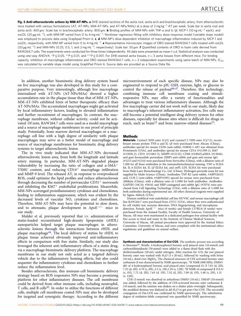
Anti-atherosclerotic mechanism of MM-NPs. To furtherinvestigate the mechanism responsible for in vivo atherosclerotictreatment of these formulations, dihydroethidium (DHE) stainingwas conducted on sections of the aorta root, aorta arch, andbrachiocephalic artery collected from atherosclerotic mice toevaluate their ROS levels. As shown in Fig. 5a, bright red fluor-escence was observed in the saline-treated group (the controlgroup), indicating that a high level of ROS was produced in theseaorta tissues. Moreover, the saline-treated group also showed thehighest level of H2O2 (Supplementary Fig. 14a), revealing thatoxidative stress was significantly increased in atheroscleroticmice. As was discussed in the previous section, NPs had a goodROS responsiveness in the presence of a high level of H2O2
(Fig. 1c, d), or overproduced ROS in LPS induced macrophage(Fig. 2b, c) and foam cell (Supplementary Fig. 4e). After i.v.injection with different formulations, these NPs may respond toover-produced ROS in the inflammatory plaques, and release AT,exhibiting their anti-atherosclerotic effects. In comparison to freeAT, ROS-responsive release in the plaque site gave these NPs aclear advantage in atherosclerotic therapy. Thus, a weak fluores-cence intensity and a low level of H2O2 were observed in the AT-NPs, MM-AT-NPs, and AT-NPs/MAs treated group.
Attributed to the presence of membrane antigens (e.g., TNFR2,CD36, and CCR2) on macrophage, we hypothesized thatmacrophage membranes from MM-NPs might sequester proin-flammatory cytokines or chemokines (Fig. 1i, j), which have beenshown to play prominent roles in the atherosclerotic process4. Infact, we discovered the lower concentrations of inflammatorycytokines in the plaques of mice treated with MM-AT-NPs, incomparison to those of AT-NPs/MAs-treated groups (Fig. 4b–f).Herein, we studied the interactions between MM-NPs andinflammatory cytokines or chemokines. The representativecytokines (TNF-α and IL-1β) were incubated with different dosesof MM-NPs, and MM-NPs dose dependent sequestration profileswere observed for both cytokines (Fig. 5b). The IC50 (halfmaximal inhibition concentration) values of MM-NPs were 1219and 399.7 µg mL−1, respectively for TNF-α and IL-1β clearance.Meanwhile, it was reported that both MCP-1 and oxLDLcontribute to the plaque formation42,43. As shown in Fig. 5b,MM-NPs exhibited a good binding affinity toward both MCP-1and oxLDL in a dose dependent manner. IC50 values were 281.6and 2813 µg mL−1, respectively for MCP-1 and oxLDL inhibition.In addition, the blood serums collected from atherosclerotic micewere incubated with different doses of MM-NPs and similarbinding kinetics were obtained (Supplementary Fig. 14b–e). Thus,these results revealed that MM-NPs may sequester proinflamma-tory cytokines and chemokines. Subsequently, the interaction ofRAW264.7 cells with MCP-1 and oxLDL was also investigated forcomparative purpose. After treatment of macrophage with MCP-1 (20 ng mL−1) or oxLDL (20 µg mL−1) for 24 h, significantactivation and inflammation of macrophage was detected, asevidenced by a high expression of TNF-α, IL-1β, and IL-6 in thecellular medium (Fig. 5c, d). Interestingly, the levels of thesecytokines and chemokines were decreased by the co-treatment ofMM-NPs with a dose-dependent inhibitory effect. Furthermore,ORO staining was conducted on RAW264.7 cells after treatmentwith oxLDL in the absence and presence of various doses of MM-NPs (Fig. 5e). Large red area in macrophage was detected in thegroup treated with oxLDL only, suggesting a high uptakeefficiency of oxLDL by macrophage. With co-treatment of MM-NPs, the red area was significantly reduced, with a MM-NPs-dosedependent manner (Fig. 5f), indicating that efficient bindingbetween MM-NPs and oxLDL may have prevented the uptake ofoxLDL. Taken together, our studies suggested that live macro-phage may get easily activated by inflammatory cytokines orchemokines to release more cytokines, whereas macrophagemembrane may effectively sequester key proinflammatorycytokines or chemokines to decrease their levels in theenvironment thereby decreasing local inflammation.
DiscussionIn summary, our study described a macrophage-biomimetic drugdelivery system, in which ROS responsive NPs were coated withmacrophage membrane. Unlike the existing PEGylation andantibody-based targeted delivery strategies, the macrophagemembrane coating strategy alone may lead to the escape of NPsfrom the RES and actively target inflammatory tissues. This
Fig. 3 Targeting efficiency and therapeutic efficacy of MM-AT-NPs and AT-NPs/MAs. a Schematic illustration of preparation of AT-NPs/MAs. b, c Exvivo fluorescence bio-imaging and quantitative analysis of Cy7.5 fluorescent signal in aorta tissues. ApoE−/− mice fed with high fat food for 1 month werei.v. administered with Cy7.5, Cy7.5-NPs, MM-Cy7.5-NPs, and Cy7.5-NPs/MAs, respectively. n= 3 aorta tissues from different mice. Scale bar: 15 mm.d Schematic illustration of atherosclerotic mouse model development and treatment with various formulations (AT, AT-NPs, MM-AT-NPs, and AT-NPs/MAs). e ORO stained aorta tissues collected from atherosclerotic mice after treatment with various formulations (AT, AT-NPs, MM-AT-NPs, and AT-NPs/MAs) at equivalent dosage of 2 mg kg−1 AT per week. n= 10 aorta tissues from different mice. Scale bar: 5 mm. f Quantitative analysis of lesion area inaorta tissues (n= 10). All data were presented as mean ± s.d. Statistical analysis was conducted using one-way ANOVA. *P≤ 0.05, **P≤ 0.01, and ***P≤0.001. Source data are provided as a Source Data file.
ARTICLE NATURE COMMUNICATIONS | https://doi.org/10.1038/s41467-020-16439-7
8 NATURE COMMUNICATIONS | (2020) 11:2622 | https://doi.org/10.1038/s41467-020-16439-7 | www.nature.com/naturecommunications
AT-NPs/MAs
HE
Mas
son
CD
14M
MP
-9α-
SM
A
Saline AT AT-NPs MM-AT-NPs
Ser
um o
xPL-
LDL
(μg
mL–1
)
100
80
60
40
20
0
**
*
Ser
um T
NF
-α (p
g m
L–1)
800
600
400
200
0
*****
*
Ser
um IL
-6 (
pg m
L–1)
400
300
200
100
0
*****
**
Aor
ta T
NF
-α (n
g m
g–1)
1.6
1.2
0.8
0.4
0.0
****
*
*
Aor
ta IL
-1β
(pg
mg–1
) 800
600
400
200
0
******
****
*
Aor
ta IL
-6 (
pg m
g–1)
1000
500
0
*****
*
*
b c d e f
g h
TC
(m
mol
L–1
)
80
60
40
20
0
HD
L-C
(m
mol
L–1
)
2.5
2.0
1.5
1.0
0.5
0.0
i
Time (week)
Bod
y w
eigh
t (g)
0 2 4 6 8 10 12 14
40
30
20
Saline
AT
AT-NPs
MM-AT-NPs
AT-NPs/MAs
Pla
que
area
(m
m2 )
Saline AT
AT-NPs
MM
-AT-N
Ps
AT-NPs/M
As
1.0
0.8
0.6
0.4
0.2
0.0
**
*****
*
Mac
roph
ages
/Pla
que
area
(%
)
25
20
15
10
5
0
**
*****
*
MM
P-9
/Pla
que
area
(%
)
15
10
5
0
**
Col
lage
n/P
laqu
e ar
ea (
%)
60
40
20
0
*
α-S
MA
/Pla
que
area
(%
)
40
20
0
***
****
Non
-HD
L-C
(m
mol
L–1
)
a
Saline AT
AT-NPs
MM
-AT-N
Ps
AT-NPs/M
As
Saline AT
AT-NPs
MM
-AT-N
Ps
AT-NPs/M
As
Saline AT
AT-NPs
MM
-AT-N
Ps
AT-NPs/M
AsSali
ne AT
AT-NPs
MM
-AT-N
Ps
AT-NPs/M
As
Saline AT
AT-NPs
MM
-AT-N
Ps
AT-NPs/M
As
Saline AT
AT-NPs
MM
-AT-N
Ps
AT-NPs/M
As
Saline AT
AT-NPs
MM
-AT-N
Ps
AT-NPs/M
AsSali
ne AT
AT-NPs
MM
-AT-N
Ps
AT-NPs/M
As
Saline AT
AT-NPs
MM
-AT-N
Ps
AT-NPs/M
As
Saline AT
AT-NPs
MM
-AT-N
Ps
AT-NPs/M
As
Saline AT
AT-NPs
MM
-AT-N
Ps
AT-NPs/M
AsSali
ne AT
AT-NPs
MM
-AT-N
Ps
AT-NPs/M
As
Saline AT
AT-NPs
MM
-AT-N
Ps
AT-NPs/M
As
80
60
40
20
0
Fig. 4 MM-AT-NPs ameliorated plaque and inflammation in an atherosclerotic mouse model. a Representative photographs and quantitative analysis ofaorta root sections stained by H&E, CD14 antibody, MMP-9 antibody, Masson’s trichrome, and α-SMA antibody (n= 6). Scale bar: 500 μm. b–d The levelsof TNF-α, IL-1β, and IL-6 in aorta tissues collected from atherosclerotic mice after treatment with various formulations (saline, AT, AT-NPs, MM-AT-NPs,and AT-NPs/MAs) at a dose of 2 mg kg−1 AT per week (n= 6). e–g The levels of TNF-α, IL-6 and oxPL-LDL in blood serum (n= 6). h The levels of TC,HDL-C, and Non-HDL-C in the blood serum (n= 6). i The changes of body weight in atherosclerotic mice treated with various formulations (saline, AT,AT-NPs, MM-AT-NPs, and AT-NPs/MAs) (n= 7). All data were presented as mean ± s.d. Statistical analysis was conducted using one-way ANOVA. *P≤0.05, **P≤ 0.01, and ***P≤ 0.001. Source data are provided as a Source Data file.
NATURE COMMUNICATIONS | https://doi.org/10.1038/s41467-020-16439-7 ARTICLE
NATURE COMMUNICATIONS | (2020) 11:2622 | https://doi.org/10.1038/s41467-020-16439-7 | www.nature.com/naturecommunications 9
strategy provides a cellular function-driven, broad-spectrumfunctionalization strategy, and inflammatory tropism enablingtargeted delivery without specific targeting molecules or compli-cated bioconjugation process. After accumulation at the targetedinflammatory tissues, the drugs loaded in NPs would be releasedin response to locally overproduced ROS, leading to effectivepharmacotherapy. In addition, attributed to the presence ofmembrane antigens (e.g., TNFR2, CD36, and CCR2) on
macrophage, macrophage membrane coated NPs efficiently bindwith, and sequester, multiple proinflammatory cytokines, andchemokines that play prominent roles in the atheroscleroticprocess, leading to inflammation neutralization. The combinationof selective pharmacotherapy and sequestration of proin-flammatory cytokines/chemokines offered by this platformexhibited significant synergistic therapies against atherosclerosisin mice.
Saline AT AT-NPs MM-AT-NPs AT-NPs/MAs
Bra
chio
ceph
alic
art
ery
Aor
ta r
oot
Aor
ta a
rch
a
Rem
aini
ng IL
-1β
(ng
mL–1
)
0
5
10
Log10MM-NPs (μg mL–1)
Rem
aini
ng T
NF
-α (n
g m
L–1)
1.5 2.0 2.5 3.0 3.50
2
4
6
8
10
IC50 = 1291 μg mL–1
(R2 = 0.9528)
Rem
aini
ng o
xLD
L (μ
g m
L–1)
2.5 3.0 3.5 4.0
20
15
10
5
0
Rem
aini
ng M
CP
-1 (
ng m
L–1)
0
5
10
IL-1
β (p
g m
L–1)
100
50
0
**** ***
******
MM-NPs (mg mL–1)
TN
F-α
(pg
mL–1
)
00.
25 0.5 1 2 4
600
400
200
0
MCP-1 = 20 ng mL–1
** **
******
***
IL-6
(pg
mL–1
) 100
50
0
****
****** ***
oxLDL = 20 μg mL–1
*** ***
** ***
***
** ***
***
b
c
d
oxLDL(20 μg mL–1)
oxLD L+ MM-NPs(2 mg mL–1)
oxLDL + MM-NPs(1 mg mL–1)
oxLDL + MM-NPs(0.5 mg mL–1)
oxLDL + MM-NPs(0.25 mg mL–1) MM-NPs (mg mL–1)
Abs
at 4
92 n
m
Mod
el0.
25 0.5 1 2
1.5
1.0
0.5
0.0
****
***
***
e f
Log10MM-NPs (μg mL–1)
1.5 2.0 2.5 3.0 3.5
Log10MM-NPs (μg mL–1)
1.5 2.0 2.5 3.0 3.5
Log10MM-NPs (μg mL–1)
IC50 = 399.7 μg mL–1
(R2 = 0.9673)
IC50 = 281.6 μg mL–1
(R2 = 0.9781)
IC50 = 2813 μg mL–1
(R2 = 0.9674)
MM-NPs (mg mL–1)
00.
25 0.5 1 2 4
MM-NPs (mg mL–1)
00.
25 0.5 1 2 4
IL-6
(pg
mL–1
) 100
50
0
IL-1
β (p
g m
L–1)
100
50
0
TN
F-α
(pg
mL–1
)
600
400
200
0
MM-NPs (mg mL–1)
00.
25 0.5 1 2 4
MM-NPs (mg mL–1)
00.
25 0.5 1 2 4
MM-NPs (mg mL–1)
00.
25 0.5 1 2 4
MCP–1 = 20 ng mL–1 MCP–1 = 20 ng mL–1
oxLDL = 20 μg mL–1oxLDL = 20 μg mL–1
ARTICLE NATURE COMMUNICATIONS | https://doi.org/10.1038/s41467-020-16439-7
10 NATURE COMMUNICATIONS | (2020) 11:2622 | https://doi.org/10.1038/s41467-020-16439-7 | www.nature.com/naturecommunications
In addition, another biomimetic drug delivery system basedon live macrophage was also developed in this study for a com-parative purpose. Very interestingly, although live macrophageinternalized with AT-NPs (AT-NPs/MAs) showed a higheraccumulation rate in the plaque tissue than that of MM-AT-NPs,MM-AT-NPs exhibited hints of better therapeutic efficacy thanAT-NPs/MAs. The accumulated macrophages might get activatedby local inflammatory factors, leading to elevated inflammationand further recruitment of macrophages. In contrast, the mac-rophage membrane, without cellular activity, could not be acti-vated. Of note, RAW264.7 cells were used as a model macrophageand a source of macrophage membranes in our proof of principlestudy. Potentially, bone marrow derived macrophages or a mac-rophage cell line with a high degree of similarity with plaquemacrophages may serve as a better model of macrophage andsource of macrophage membranes for biomimetic drug deliverysystems to target atherosclerotic lesion.
The in vivo study indicated that MM-AT-NPs decreasedatherosclerotic lesion area, from both the longitude and latitudeartery staining. In particular, MM-AT-NPs degraded plaquevulnerability by increasing the ratio of collagen and α-SMA+
VSMC content, and reducing CD14+ macrophage infiltrationand MMP-9 level. The released AT, in response to overproducedROS, could optimize the lipid profiles and reduce atherosclerosisthrough decreasing the number of perivascular CD31+ neovesselsand inhibiting the KI67+ endothelial proliferations. Meanwhile,MM-NPs scavenged proinflammatory cytokines and chemokines,leading to inflammation suppression, which was evident by thedecreased levels of vascular NO, cytokines and chemokines.Therefore, MM-AT-NPs may have the potential to slow down,and to attenuate, plaque development in ApoE−/− mice inour study.
Mulder et al. previously reported that i.v. administration ofstatin-loaded reconstituted high-density lipoprotein (rHDL)nanoparticles helped target plaque macrophages in athero-sclerotic lesions through the interactions between rHDL andplaque macrophage44. The local delivery of statins by rHDL toplaque tissue achieved obviously improved anti-inflammatoryeffects in comparison with free statin. Similarly, our study alsoleveraged the inherent anti-inflammatory effects of a statin drug,via a macrophage-biomimetic delivery platform. The macrophagemembrane in our study not only acted as a targeted deliveryvehicle due to the inflammatory homing effects, but also couldsequester the inflammatory cytokines and chemokines to furtherreduce the inflammation level.
Besides atherosclerosis, this immune-cell biomimetic deliverystrategy based on ROS responsive NPs may become a promisingplatform for other inflammatory diseases. The cell membranecould be derived from other immune cells, including neutrophil,T cells, and B cells45. In order to utilize the functions of differentcells, multiple cell membrane coated NPs may also be developedfor targeted and synergistic therapy. According to the different
microenvironment of each specific disease, NPs may also beengineered to respond to pH, GSH, enzyme, light, or glucose tocontrol the release of payload46,47. Therefore, this technology,combining immune cell membrane coating and stimuli-responsive NPs, may offer a variety of functionalities andadvantages to treat various inflammatory diseases. Although thelive macrophage carrier did not work well in our study, likely dueto macrophage’s inherent inflammatory activation, live cells maystill become a potential intelligent drug delivery system for otherdiseases, especially for disease sites where is difficult for drugs toreach, such as brain22 and bone marrow-related diseases23.
MethodsMaterials. Cynine5 NHS ester (Cy5) and cyanine7.5 NHS ester (Cy7.5), recom-binant mouse protein TNF-α and IL-1β were purchased from Abcam (China),antibodies special for mouse CD36 (anti-rabbit, #18836-1-AP) was obtained fromProteintech (USA), and antibodies special for mouse CD14 (#11390-1), Ki67(#13030-2), CD31 (#11063-3), MMP9 (#12132), CD68 (#14043),αSMA (#13044),and goat horseradish peroxidase (HRP)-anti-rabbit and goat anti-mouse IgG(#1213 and #1214) were purchased from Servicebio (China), with a dilution ratio of1:200 for all these antibodies in the immunohistochemical staining. TNF-α ELISAkit, IL-6 ELISA kit, IL-1β ELISA kit, and oxPL-LDL ELISA kit were purchasedfrom Hefei Laier Biotechnology Co., Ltd. (China). Hydrogen peroxide assay kit wassupplied by Multi Science (China). Antibodies TNF-R2 (anti-rabbit, #ABP52623)and, CCR-2 (anti-rabbit, #ABP53395) special for mouse were purchased fromAbbkine (China), with a dilution ratio of 1:1000 for both antibodies. AntibodiesGAPDH (14C10, #5014) and HRP-conjugated anti-rabbit IgG (#7074) were pur-chased from Cell Signaling Technology (USA), with a dilution ratio of 1:1000 forboth antibodies during experimental use. All other chemical reagents were suppliedby Sigma Aldrich (USA).
Human umbilical vein endothelial cell line HUVEC and mouse macrophage cellline RAW264.7 were purchased from ATCC (USA), where they were authenticatedby cell vitality test, isozyme detection, DNA fingerprinting, and mycoplasmadetection. Female ApoE−/− mice (6 weeks) and female C57BL/6 (6 weeks) werepurchased from the Animal Facility, Faculty of Health Sciences, University ofMacau. All mice were maintained in a dedicated pathogen-free animal facility withfree access to food and water in the Institute of Chinese Medical Sciences,University of Macau. All animal procedures were approved by the Animal EthicsCommittee, University of Macau, and were compliant with the institutional ethicsregulations and guidelines on animal welfare.
Synthesis and characterization of Oxi-COS. The synthetic process was accordingto literature41. Briefly, 4-hydroxyphenyl boronic acid pinacol ester (10 mmol) andcarbonyldiimidazole (50 mmol) were added to a flame-dried flask with drydichloromethane (20 mL) under nitrogen. After reaction for 12 h, the raw pinacolboronic ester was washed with H2O (3 × 20mL), followed by washing with brine(10 mL), dried over MgSO4. The chemical structure of CDI-activated boronic estercarbamate 2 was characterized by NMR spectroscopy. 1H NMR (600 MHz, DMSO-d6) of 4-hydroxyphenyl boronic acid pinacol ester (compound 1): δ 7.65 (d, 2H),7.35 (d, 2H), 4.79 (s, 2H), 2.5 (s, 1H),1.29 (s, 12H). 1H NMR of compound 2: δ 8.31(s, 1H), 7.72 (d, 2H), 7.63 (d. 1H), 7.52 (d, 2H), 7.09 (d, 1H), 5.48 (s, 2H), 1.31(s, 12H).
COS (5 mmol) was dissolved in anhydrous DMSO (10 mL). DMAP (10 mmol)was added, followed by the addition of CDI-activated boronic ester carbamate 2(10 mmol), and the mixture was shaken on a shaker plate overnight. Subsequently,the modified dextran was dialyzed in dd-H2O to get pure Oxi-COS. The successfulconjugation of Oxi-COS was confirmed by NMR spectroscopy. The substitutiondegree of oxidation-labile compound was quantified by NMR spectroscopy.
Fig. 5 Anti-atherosclerotic actions by MM-AT-NPs. a DHE-stained sections of the aorta root, aorta arch and brachiocephalic artery, from atheroscleroticmice treated with various formulations (AT, AT-NPs, MM-AT-NPs, and AT-NPs/MAs) at a dose of 2 mg kg−1 AT per week. Scale bar in aorta root andaorta arch: 400 μm. Scale bar in brachiocephalic artery: 800 μm. b Binding profiles of MM-NPs with TNF-α and IL-1β, MCP-1 (10 ngmL−1 each), andoxLDL (20 μg mL−1), with MM-NP varied from 0 to 4mgmL−1. Nonlinear regression fitting with inhibitory dose–response model (variable slope model)was employed to process data using Graphpad Prism 6. c, d MM-NP’s dose-dependent inhibition of macrophage inflammation induced by MCP-1 andoxLDL, respectively, with MM-NP varied from 0 to 4mgmL−1. e Representative microscopic images of ORO stained RAW264.7 cells treated with oxLDL(20 μg mL−1) and MM-NPs (0.25, 0.5, 1, and 2mgmL−1, respectively). Scale bar: 50 μm. f Quantified contents of ORO in foam cells derived fromRAW264.7 cells. The experiments were conducted for three times independently. All data were presented as mean ± s.d. Statistical analysis was conductedusing one-way ANOVA. *P≤ 0.05, **P≤ 0.01, and ***P≤ 0.001. For DHE-stained aorta tissues, n= 3 aorta tissues from different mice. For bindingcapacity, inhibition of macrophage inflammation and ORO stained RAW264.7 cells, n= 3 independent experiments using same batch of MM-NPs. IC50
was calculated by variable slope model using GraphPad Prism 6. Source data are provided as a Source Data file.
NATURE COMMUNICATIONS | https://doi.org/10.1038/s41467-020-16439-7 ARTICLE
NATURE COMMUNICATIONS | (2020) 11:2622 | https://doi.org/10.1038/s41467-020-16439-7 | www.nature.com/naturecommunications 11
Preparation of ROS responsive NPs and AT-NPs. Oxi-COS (30 mg) was dis-solved in water (deionized, 300 mL) and the mixture was stirred for 1 h. Subse-quently, the resulting solution was passed through a syringe filter (with a pore sizeof 0.45 micron) to have solids removed, and the aqueous solution of NPs wasachieved. Upon lyophilization, the resultant powder was put away for use in otherexperiments. The NPs’ morphology was studied by a TEM (H-7650, Hitachi Ltd.)and analyzed by Gatan Digital Micrograph 3.9. The size and size-distribution of theNPs was further characterized via DLS. Subsequently, the NPs were incubated for48 h in PBS in the presence of a various concentrations of H2O2 (0, 0.1, 0.2, 0.5, and1 mM). At predetermined time intervals, the diameters were determined by DLSwith a Zetasizer (Nano-ZS, Malvern) system and analyzed by Zetasizer Software(version 7.11).
The preparative process of AT-NPs was similar to that of the micelles asdescribed above. OXi-COS and AT were co-dissolved into deionized water, and thefeeding ratio of OXi-COS: AT was 90: 10 (w/w). Upon being stirred for 1 h, thesolution was dialyzed with a dialysis bag (WMCO= 12,000) to remove theunencapsulated AT. Subsequently, the solution was subject for filtration andlyophilization for use in the following experiments. The quantity of AT in the AT-NPs was measured by high performance liquid chromatography (HPLC) withreversed-phase column (Agilent TC-C18, 4.6 × 250 mm, 5 μm). The mobile phaseconsisted of ammonium acetate (adjusted pH to 4 with glacial acetic acid) andacetonitrile (6:4, V%), and the detection wavelength was 244 nm. The DEE andDLC were subsequently calculated by using Eqs. (1) and (2), respectively.
DEE %ð Þ ¼ Mass of drug in NPsMass of AT in feed
´ 100% ð1Þ
DLC %ð Þ ¼ Mass of drug in NPsMass of AT� NPs
´ 100% ð2Þ
Release profile of AT-NPs. The release profiles of AT from AT-NPs were studiedunder various concentrations of H2O2 (0, 0.1, and 1 mM, respectively). AT-NPs(10 mg) dispersed in deionized water (4 mL) were put in a dialysis bag (MWCO=12,000) that was subsequently placed in 35 mL of buffer solution (sitting in a 37 °Cshaker with 30 × g shaking rate). At various time intervals, 2 mL of incubationmedia was taken out for HPLC analysis, and 2 mL of medium (fresh) was refilledinto the buffer solution. The cumulative release rate of AT was calculated using thefollowing equation, Eq. (3).
Cumulative drug release %ð Þ ¼ Mt
M0´ 100% ð3Þ
where Mt was the amount of drug released at time t and M0 was the initial amountof drug in the NPs. The release experiments were conducted in triplicate.
Isolation of macrophage membrane. RAW264.7 cells were suspended at a densityof 2.0 × 10⁷ cells mL−1 in ice-cold TM buffer solution (pH 7.4; 10 mM Tris+ 1 mMMgCl2) and subsequently were extruded through a mini-extruder for at least 30times in order to disrupt the cells. 1 M sucrose was subsequently mixed with thecell homogenate to eventually reach 0.25 M sucrose concentration, and the mixturewas centrifuged at 2000 × g and 4 °C for approximately 10 min. The resultingsupernatant was subject for collection upon further centrifugation at 3000 × g foradditional 30 min. The cell membranes were collected and washed with ice-coldTM buffer in the presence of 0.25 M of sucrose twice for purification. Thebicinchoninic acid assay (BCA) protein assay was employed to analyze the totalprotein content in the purified macrophage membrane. Approximately, 500 millionRAW264.7 cells were able to yield 0.5 mg membrane material (total proteinweight). Membrane material was stored at −80 °C for future study.
Preparation of MM-NPs and MM-AT-NPs. After isolating the macrophagemembrane, the membrane coating was subsequently completed by fusing macro-phage membrane vesicles with NPs and AT-NPs (respectively) via a mini-extruderfor at least 20 times, and substantial sonication using a bath sonicator at a fre-quency of 40 kHz and a power of 100W for 2 min. The resulting solution wascentrifuged at 3000 × g for 30 min to remove the uncoated membrane, and wassubsequently centrifuged at 12,000 × g for 5 min to remove soluble membraneproteins to obtain the precipitate, crude membrane coated NPs. After washing withPBS for several times until no protein was detected in the supernatant by BCAProtein Assay (Thermo Scientific, Rockford, IL), pure MM-NPs and MM-AT-NPswere therefore obtained. The specific surface markers on macrophage, macrophagemembrane and MM-NPs were determined by Western blotting. Membrane proteinsamples extracted from NPs, macrophage vesicles and MM-NPs were preparedusing membrane protein extraction kit (Beyotime, Shanghai, China) according tothe manufacturer’s protocols. The protein concentrations were determined by theBCA Protein Assay (Thermo Scientific, Rockford, IL). Approximately, 30 µg ofprotein samples was separated in 10% sodium dodecyl sulfate polyacrylamide gelelectrophoresis and electrophoretically transferred onto polyvinylidene fluoridemembranes. After blocking with 5% nonfat dry milk in tris-buffered saline,membranes were incubated overnight at 4 °C with primary antibodies, includingCD36 (Proteintech), TNFR2 (Abbkine, Inc, China), and CCR2 (Abbkine, Inc.,China). Followed by incubation of a secondary HRP–conjugated anti-rabbit IgG
antibody (Cell Signaling Technology) at room temperature for 1 h, specific bandswere visualized using an enhanced chemiluminescence detection kit (Bio-Rad).
In vitro safety evaluation. In vitro cytotoxicity of free NPs and MM-NPs wasevaluated with RAW264.7 and HUVECs cells by MTT assays, respectively. Briefly,the cells were seeded in 96-well plate at a density of 105 per mL. After incubationfor 24 h, the media were replaced with fresh media containing NPs or MM-NPs atdifferent concentrations (0.1, 1, 10, and 20 mM), respectively. After incubation foranother 24 h, the media were replaced with DMEM containing MTT and thesurvival number of cells was determined by MTT enzyme-linked immunometricmeter (data analyzed by SoftMax Pro 5.4.1). Results were analyzed using GraphPadPrism 6.
ROS-responsiveness of MM-AT-NPs in vitro. RAW264.7 cells were incubatedwith media containing 10 ng mL−1 LPS or 20 μg mL−1 oxLDL, and concurrentlycontaining MM-AT-NPs at various concentrations of AT (1.25, 5, and 20 μM) for24 h. The treatment cells with LPS or oxLDL, in the absence and presence of AT orAT-NPs, respectively, was also conducted for comparative purposes. After incu-bation, the media were replaced with DMEM containing MTT and the survivalnumber of cells was determined by MTT enzyme-linked immunometric meter.Results were analyzed using GraphPad Prism 6.
Furthermore, because LPS is a typical ROS inducer, the ROS levels wereanalyzed to confirm the ROS production. Briefly, after co-treatment of cells by LPS(400 ng mL−1) and the drug (free AT, AT-NPs, and MM-NT-NPs) at 0.4 mM for12 h, the media were replaced with fresh media containing 2′,7′-dichlorofluorescindiacetate (DCFH-DA). After incubation for 30 min, the cells were washed with PBSfor three times and the fluorescence intensity of cells was assessed by flowcytometry (interfaced with BD Accuri C6 Software (version 1. 0. 264. 21)) at anexcitation wavelength of 488 nm. Results were analyzed using FlowJo software(version 7.6.1).
In addition to cell viability and ROS production, the apoptosis rate was alsomeasured. After respective co-treatment for 6 h, the cells were suspended in 100 μLof binding buffer, and subsequently mixed with 10 μL of annexin V-fluoresceinisothiocyanate (V-FITC) and 10 μL of propidium iodide for 15 min. Another400 μL of binding buffer was added and the cells were analyzed by a flow cytometerto determine the apoptosis rates. Results were analyzed using FlowJo software(version 7.6.1).
Cellular uptake and intracellular drug release. The cellular uptake and intra-cellular drug release tests were conducted in macrophage, LPS-treated macrophageand foam cells. After seeding the cells and incubation at 37 °C for 24 h, the mediawere replaced with fresh media containing Cy5-NPs and MM-Cy5-NPs, and thecells were incubated for additional 1–4 h, respectively. Subsequently, the cells werewashed for three times with PBS buffer and fixed by paraformaldehyde for 15 min.The cells were washed with PBS for three more times and counterstained withHoechst for 15 min. After washing with PBS for three times again, the cells wereobserved via confocal laser scanning microscopy interfaced with LAS X (version3.5.2) software.
For intracellular drug release behavior, NR, another fluorescence probe, wasloaded into the NPs (NR-NPs) and MM-NPs (MM-NR-NPs). NR has nofluorescence itself, but it would show strong red fluorescence once contacting withthe lipid droplet inside cells. LPS treated RAW264.7 cells and foam cells wereincubated in the media containing NR-NPs and MM-NR-NPs, respectively. Thecells were subsequently imaged via microscopy with an excitation wavelength at480 nm after continuous incubation for 1, 2, 4, and 8 h, respectively.
Safety evaluation of MM-NPs i.v. injected in mouse. To evaluate the bio-compatibility of our proposed formulations (macrophage membrane coated ROS-responsive NPs) in vivo, 2 groups of 6-week-old female C57BL/6 mice (n= 6 ineach group) were i.v. injected with saline and MM-NPs, respectively, for half amonth with a high dose of 100 mg/kg MM-NPs every 4 days. The changes ofbodyweight of mice in both groups were recorded during the 15-day follow-upstudy. At the end of experiment, all mice were sacrificed, and the heart, livers,spleen, lungs, and kidneys were collected for histological study. In addition, theblood serum was collected for blood chemistry analysis, including the liver functionbiomarkers (ALT and AST) and kidney function biomarkers (BUN and UA)analysis, as well as inflammatory cytokines (TNF-α, IL-6, and IL-1β) analysis.
Safety evaluation of macrophage i.v. injected in mouse. To evaluate theimmune response and organ toxicity of foreign RAW264.7 cells in mice, femaleApoE−/− mice were randomly and investigator-blindly divided into 4 group (n=6), and intravenously administered with 100 μL of macrophages (2.5 × 10⁸, 5 × 10⁸,and 1 × 10⁹ macrophages kg−1, respectively). The mice were injected once everyfour days for 2 weeks. Their body weight and survival rate were recorded. At theendpoint of experiment, all mice were sacrificed and the blood was collected toquantitate the immune-associated cells. In addition, their livers, spleen, lungs, andkidneys were processed for histological examination.
ARTICLE NATURE COMMUNICATIONS | https://doi.org/10.1038/s41467-020-16439-7
12 NATURE COMMUNICATIONS | (2020) 11:2622 | https://doi.org/10.1038/s41467-020-16439-7 | www.nature.com/naturecommunications

Construction of atherosclerosis in ApoE−/− mice. Six-week-old female ApoE−/−
mice were given high fat diet containing 21.2% lard, 49.1% carbohydrate, 19.8%protein, and 0.2% cholesterol for 3 months, in order to induce atherosclerosis. At theend of treatment, six mice were euthanized and the degree of pathological changeswere evaluated by measuring the lesion area of the aorta from the heart to the iliacbifurcation. To determine the extent of atherosclerosis at the aortic root, aortic arch,and brachiocephalic artery, Oil Red O (ORO) staining was performed to confirm theformation of atherosclerotic plaque in mice.
In vivo targeting of the atherosclerotic plaque. Six-week-old female ApoE−/−
mice fed with high fat food (with the same composition as described above) for1 month were i.v. administered with Cy7.5, Cy7.5-NPs, MM-Cy7.5-NPs, andCy7.5-NPs/MAs with the same dosage of Cy7.5 (2 mg kg−1), respectively, via thetail vein. After allowing these dyes to distribute for 6 h, the mice were euthanizedand subsequently perfused with PBS to remove unbound dyes. The aortas wereisolated for imaging and quantitative analysis using an IVIS (Lumina XR III), withan excitation wavelength of 780 ± 20 nm and an emission wavelength of 840 ± 20nm for the measurements.
In vivo therapeutic efficacy study. Six-week-old female ApoE−/− mice wererandomly and investigator-blindly divided into 5 groups (n= 13, the first batch of6 and second batch of 7), including a control group (saline), a free-AT treatedgroup, and groups separately treated with AT-NPs, MM-AT-NPs, and AT-NPs/MAs. The mice were treated with high-fat food (with the same composition asdescribed above) for 3 months in a row. After treatment with high-fat diet for thefirst month, five groups were i.v. administered with saline, AT, AT-NPs, MM-AT-NPs, and AT-NPs/MAs, respectively at a dosage of 2 mg kg−1 AT (except for thesaline-treated group) per week for additional 2 months. After administration for2 months, all mice were euthanized and atherosclerotic plaques from ten mice ineach group were collected for ORO staining. Imaging of atherosclerotic plaqueswere conducted for evaluating the therapeutic efficacy of these different formula-tions. Furthermore, quantitative analysis of atherosclerotic plaque was also deter-mined by Image-Pro Plus 6.0.
Histological study on aorta tissues. For histological analysis, aortic root, aorticarch and brachiocephalic artery from mice with various treatments were collectedfor hematoxylin–eosin (H&E) staining and Masson’s trichrome staining. Forimmunohistochemistry analysis, sections of aortic root, aortic arch and brachio-cephalic artery were incubated with antibodies, including CD68, CD14, MMP-9,and α-SMA, CD31 and KI67, respectively.
Quantitation of cytokines, chemokines, and cholesterol. In addition, the tissueswere homogenized for analysis of inflammatory cytokines and chemokinesincluding IL-1β, IL6, and oxPL-LDL by Elisa kits from Hefei Laier BiotechnologyCo., Ltd. (China), including IL-1β Elisa kit (catalog number: LE-M0444), IL6 Elisakit (catalog number: LE-M0458), and oxPL-LDL Elisa kit (catalog number: LE-M1000). Briefly described here, 50 μL of standard was added to a standard well, and10 μL of testing sample and 40 μL of kit diluent was added to a testing well.Subsequently, 100 μL of HRP-conjugate reagent was added to each well, followedby coverage with an adhesive strip. After incubation for 60 min at 37 °C, each wellwas washed and any remained solution in the well should be remove completely.Subsequently, 50 μL of chromogen solution A and 50 μL of chromogen solution Bwere added to each well. After incubation for 15 min at 37 °C in dark, 50 μL of StopSolution was added to each well. Finally, optical density (O.D.) at 450 nm wasmeasured by using a microtiter plate reader immediately. The H2O2 levels weredetermined by using fluorimetric hydrogen peroxide assay kit. The serum fromthese groups were also collected and the cytokines including TNF-α, IL6, andoxPL-LDL were quantified by using Elisa kits according to the previously describedprocedure. TC and HDL-C contents in serums were quantified using assay kitsfrom Nanjing Jiancheng Bioengineering Institute (China), including TC assay kit(catalog number: A111-1-1) and HDL cholesterol assay kit (catalog number: A112-1-1). Non-HDL-C was subsequently calculated from “TC minus HDL-C”.
DHE staining analysis of aorta tissues. The samples of aorta root, aorta arch, andbrachiocephalic artery were embedded in Tissue-Tek O.T.C. compound, and then8 μm sections were incubated with 2% Triton X-100 for 10 min at 21 °C, followedby blocking with 5% BSA in PBS. Subsequently, the sections were stained by DHE(Servicebio, Wuhan, China) and incubated for 30 min under dark environment.After washing with PBS for three times, the slides were imaged under fluorescencemicroscopy. The fluorescent intensity was quantified by Image-Pro Plus 6.0.
Binding analysis of cytokine and chemokine by MM-NPs. TNF-α (10 ng mL−1),IL-1β (10 ng mL−1), oxLDL (20 µg mL−1), and MCP-1 (10 ng mL−1) were mixedwith MM-NPs at the concentration from 0 to 10 mgmL−1. The mixture wasincubated at 37 °C for 2 h and then centrifuged at 13,000 × g for 10 min to removethe NPs. The supernatant was analyzed by Elisa kit for the measurements of TNF-α, IL-1β, MCP-1, and oxPL-LDL. The results were further analyzed by GraphPadPrism 6.
Oil Red O staining in macrophage. Macrophages were co-treated with oxLDL anddifferent doses of MM-NPs (0, 1.25, 2.5, 5, and 10 mgmL−1) for 24 h, and the cellswere washed by PBS for three times. Subsequently, macrophages were stained byfreshly prepared ORO working solution for 15 min, and rinsed with 60% iso-propanol. After washing with PBS for three times again, the nuclei of macrophageswere slightly stained with alum haematoxylin. Finally, macrophages were washedwith PBS and measured under microscope.
Statistical analysis. One-way ANOVA and two-way was utilized for statisticalanalysis. Value of *P ≤ 0.05, **P ≤ 0.01 and ***P ≤ 0.001 were applied to annotatestatistical significance. All data were presented as mean value ± the standarddeviation of independent experiments.
Reporting summary. Further information on research design is available inthe Nature Research Reporting Summary linked to this article.
Data availabilityThe source data underlying Figs. 1c, d, g–i, 2a, c–f, 3c, f, 4a, b–i, 5b, c–d, f, andSupplementary Figs. 3a–c, 4a–c, f, h, 6, 8b–d, 9a, b, 10–13, and 14a–e are provided asa Source Data file. The data supporting all the plots within this paper are available fromthe corresponding authors upon request.
Received: 1 May 2019; Accepted: 30 April 2020;
References1. Roth, G. A. et al. Global, regional, and national burden of cardiovascular
diseases for 10 causes, 1990 to 2015. J. Am. Coll. Cardiol. 70, 1–25 (2017).2. Allahverdian, S., Chaabane, C., Boukais, K., Francis, G. A. & Bochaton-Piallat,
M. L. Smooth muscle cell fate and plasticity in atherosclerosis. Cardiovasc. Res.114, 540–550 (2018).
3. Allahverdian, S., Chehroudi, A. C., McManus, B. M., Abraham, T. & Francis,G. A. Contribution of intimal smooth muscle cells to cholesterol accumulationand macrophage-like cells in human atherosclerosis. Circulation 129,1551–1559 (2014).
4. Moore, K. J., Sheedy, F. J. & Fisher, E. A. Macrophages in atherosclerosis: adynamic balance. Nat. Rev. Immunol. 13, 709–721 (2013).
5. Tabas, I. & Bornfeldt, K. E. Macrophage phenotype and function in differentstages of atherosclerosis. Circ. Res. 118, 653–667 (2016).
6. Hansson, G. K., Libby, P. & Tabas, I. Inflammation and plaque vulnerability.J. Intern. Med. 278, 483–493 (2015).
7. Martinet, W., Schrijvers, D. M. & De Meyer, G. R. Pharmacologicalmodulation of cell death in atherosclerosis: a promising approach towardsplaque stabilization? Br. J. Pharmacol. 164, 1–13 (2011).
8. Zhang, Q. et al. Structure–property correlations of reactive oxygenspecies-responsive and hydrogen peroxide-eliminating materials withanti-oxidant and anti-inflammatory activities. Chem. Mater. 29, 8221–8238(2017).
9. Cheng, J. et al. A targeting nanotherapy for abdominal aortic aneurysms. J.Am. Coll. Cardiol. 72, 2591–2605 (2018).
10. Bourquin, J. et al. Biodistribution, clearance, and long-term fate of clinicallyrelevant nanomaterials. Adv. Mater. 30, 1704307 (2018).
11. Moyano, D. F., Liu, Y., Peer, D. & Rotello, V. M. Modulation of immuneresponse using engineered nanoparticle surfaces. Small 12, 76–82 (2016).
12. Mo, J., Xie, Q., Wei, W. & Zhao, J. Revealing the immune perturbation ofblack phosphorus nanomaterials to macrophages by understanding theprotein corona. Nat. Commun. 9, 2480 (2018).
13. Hu, Z. et al. An intelligent re-shieldable targeting system for enhanced tumoraccumulation. J. Control Release 268, 1–9 (2017).
14. Dai, Q. et al. Quantifying the ligand-coated nanoparticle delivery to cancercells in solid tumors. ACS Nano 12, 8423–8435 (2018).
15. Lim, W. A. & June, C. H. The principles of engineering immune cells to treat.Cancer Cell 168, 724–740 (2017).
16. Lang, T., Yin, Q. & Li, Y. Progress of cell-derived biomimetic drug deliverysystems for cancer therapy. Adv. Ther. 1, 1800053 (2018).
17. Yoo, J. W., Irvine, D. J., Discher, D. E. & Mitragotri, S. Bio-inspired,bioengineered and biomimetic drug delivery carriers. Nat. Rev. Drug Discov.10, 521–535 (2011).
18. Parodi, A. et al. Synthetic nanoparticles functionalized with biomimeticleukocyte membranes possess cell-like functions. Nat. Nanotechnol. 8, 61(2012).
19. Dehaini, D. et al. Erythrocyte-platelet hybrid membrane coating for enhancednanoparticle functionalization. Adv. Mater. 29, 1606209 (2017).
NATURE COMMUNICATIONS | https://doi.org/10.1038/s41467-020-16439-7 ARTICLE
NATURE COMMUNICATIONS | (2020) 11:2622 | https://doi.org/10.1038/s41467-020-16439-7 | www.nature.com/naturecommunications 13

20. Zhang, Q. et al. Neutrophil membrane-coated nanoparticles inhibit synovialinflammation and alleviate joint damage in inflammatory arthritis. Nat.Nanotechnol. 13, 1182–1190 (2018).
21. Hu, C.-M. J. et al. Nanoparticle biointerfacing by platelet membrane cloaking.Nature 526, 118 (2015).
22. Wang, Y. et al. Biomimetic nanotherapies: red blood cell based core–shellstructured nanocomplexes for atherosclerosis management. Adv. Sci. 6,1900172 (2019).
23. Luo, Y. et al. Macrophagic CD146 promotes foam cell formation and retentionduring atherosclerosis. Cell Res. 27, 352–372 (2017).
24. Shi, J. et al. Engineered red blood cells as carriers for systemic delivery of a widearray of functional probes. Proc. Natl Acad. Sci. USA 111, 10131–10136 (2014).
25. Xue, J. et al. Neutrophil-mediated anticancer drug delivery for suppression ofpostoperative malignant glioma recurrence. Nat. Nanotechnol. 12, 692–700(2017).
26. Hu, Q. et al. Conjugation of haematopoietic stem cells and platelets decoratedwith anti-PD−1 antibodies augments anti-leukaemia efficacy. Nat. Biomed.Eng. 2, 831–840 (2018).
27. Beste, T. M. et al. Molecular network analysis of endometriosis reveals a role forc-Jun–regulated macrophage activation. Sci. Transl. Med. 6, 222–234 (2014).
28. Tabas, I. & Lichtman, A. H. Monocyte-macrophages and T cells inatherosclerosis. Immunity 47, 621–634 (2017).
29. Saravanakumar, G., Kim, J. & Kim, W. J. Reactive-oxygen-species-responsivedrug delivery systems: promises and challenges. Adv. Sci. 4, 1600124 (2017).
30. Miller, E. W., Albers, A. E., Pralle, A., Isacoff, E. Y. & Chang, C. J. Boronate-based fluorescent probes for imaging cellular hydrogen peroxide. J. Am. Chem.Soc. 127, 16652–16659 (2005).
31. Wang, Y. et al. Targeted therapy of atherosclerosis by a broad-spectrumreactive oxygen species scavenging nanoparticle with intrinsic anti-inflammatory activity. ACS Nano 12, 8943–8960 (2018).
32. Rader, D. J. & Pure, E. Lipoproteins, macrophage function, andatherosclerosis: beyond the foam cell? Cell Metab. 1, 223–230 (2005).
33. Fan, J. et al. CKIP-1 limits foam cell formation and inhibits atherosclerosis bypromoting degradation of Oct-1 by REGgamma. Nat. Commun. 10, 425 (2019).
34. Barua, S. & Mitragotri, S. Challenges associated with penetration ofnanoparticles across cell and tissue barriers: a review of current status andfuture prospects. Nano Today 9, 223–243 (2014).
35. Blanco, E., Shen, H. & Ferrari, M. Principles of nanoparticle design forovercoming biological barriers to drug delivery. Nat. Biotechnol. 33, 941–951(2015).
36. Rumin, J. et al. The use of fluorescent Nile red and BODIPY for lipidmeasurement in microalgae. Biotechnol. Biofuels 8, 42 (2015).
37. van der Vorst, E. P. C. & Weber, C. Novel features of monocytes andmacrophages in cardiovascular biology and disease. Arterioscler. Thromb.Vasc. Biol. 39, e30–e37 (2019).
38. Hirata, Y. et al. Coronary atherosclerosis is associated with macrophagepolarization in epicardial adipose tissue. J. Am. Coll. Cardiol. 58, 248–255 (2011).
39. Bordon, Y. Cell migration: pericytes—route planners. Nat. Rev. Immunol. 13,5 (2013).
40. Chen, Y. et al. Atherosclerotic dyslipidemia revealed by plasma lipidomics onApoE(−/−) mice fed a high-fat diet. Atherosclerosis 262, 78–86 (2017).
41. Daugherty, A. et al. Recommendation on design, execution, and reporting ofanimal atherosclerosis studies: a scientific statement from the American HeartAssociation. Arterioscler. Thromb. Vasc. Biol. 37, e131–e157 (2017).
42. Kanda, H. et al. MCP-1 contributes to macrophage infiltration into adiposetissue, insulin resistance, and hepatic steatosis in obesity. J. Clin. Investig. 116,1494–1505 (2006).
43. Lara-Guzman, O. J. et al. Oxidized LDL triggers changes in oxidative stressand inflammatory biomarkers in human macrophages. Redox Biol. 15, 1–11(2018).
44. Duivenvoorden, R. et al. A statin-loaded reconstituted high-densitylipoprotein nanoparticle inhibits atherosclerotic plaque inflammation. Nat.Commun. 5, 3065 (2014).
45. Libby, P. & Hansson, G. K. Inflammation and immunity in diseases of thearterial tree: players and layers. Circ. Res. 116, 307–311 (2015).
46. Tang, L. et al. Enhancing T cell therapy through TCR-signaling-responsivenanoparticle drug delivery. Nat. Biotechnol. 36, 707–716 (2018).
47. Lee, Y. et al. Multistimuli-responsive bilirubin nanoparticles for anticancertherapy. Angew. Chem. Int. Ed. 55, 10676–10680 (2016).
AcknowledgementsScience and Technology Development Fund (FDCT), Macao SAR (Grant no. 0121/2018/A3), National Science Foundation of China (Grant no. 21871301), and Research Com-mittee at University of Macau (Grant nos. MYRG2017-00010-ICMS and MYRG2016-00165-ICMS-QRCM) are gratefully acknowledged for providing financial support tothis work.
Author contributionsThe project was conceptually designed by C.G., S.L., and R.W. The majority of theexperiments were conducted by C.G., Q.H., and C.L. (with equal contributions), assistedby C.K. and L.Y. Data analysis and interpretation were done by C.G., J.W., S.L., and R.W.The paper was prepared by C.G. and R.W. All authors discussed the results andimplications, and commented on the paper.
Competing interestsThe authors declare no competing interests.
Additional informationSupplementary information is available for this paper at https://doi.org/10.1038/s41467-020-16439-7.
Correspondence and requests for materials should be addressed to S.M.Y.L. or R.W.
Peer review information Nature Communications thanks David Cormode and the other,anonymous, reviewer(s) for their contribution to the peer review of this work. Peerreviewer reports are available.
Reprints and permission information is available at http://www.nature.com/reprints
Publisher’s note Springer Nature remains neutral with regard to jurisdictional claims inpublished maps and institutional affiliations.
Open Access This article is licensed under a Creative CommonsAttribution 4.0 International License, which permits use, sharing,
adaptation, distribution and reproduction in any medium or format, as long as you giveappropriate credit to the original author(s) and the source, provide a link to the CreativeCommons license, and indicate if changes were made. The images or other third partymaterial in this article are included in the article’s Creative Commons license, unlessindicated otherwise in a credit line to the material. If material is not included in thearticle’s Creative Commons license and your intended use is not permitted by statutoryregulation or exceeds the permitted use, you will need to obtain permission directly fromthe copyright holder. To view a copy of this license, visit http://creativecommons.org/licenses/by/4.0/.
© The Author(s) 2020
ARTICLE NATURE COMMUNICATIONS | https://doi.org/10.1038/s41467-020-16439-7
14 NATURE COMMUNICATIONS | (2020) 11:2622 | https://doi.org/10.1038/s41467-020-16439-7 | www.nature.com/naturecommunications