travis9, and michael l. freeman - media.nature.com · accumulation of isolevuglandin-modified...
TRANSCRIPT

Accumulation of isolevuglandin-modified protein in normal and fibrotic lung
Stacey Mont1,2, Sean S. Davies3, L. Jackson Roberts 2nd3, Raymond L. Mernaugh4, W. Hayes
McDonald4,5, Brahm H Segal6, William Zackert3, Jonathan A Kropski7, Timothy S. Blackwell7,
Konjeti R. Sekhar2, James J. Galligan4, Pierre P. Massion7, Lawrence J. Marnett4,8, Elizabeth L.
Travis9, and Michael L. Freeman2*
1Department of Cancer Biology, 2Department of Radiation Oncology, 3Division of Clinical
Pharmacology, Department of Pharmacology, 4Department of Biochemistry, 5Proteomics
Laboratory and Mass Spectrometry Research Center, 7Division of Pulmonary & Critical Care,
Department of Medicine, 8A.B. Hancock Jr. Memorial Laboratory for Cancer Research,
Vanderbilt Institute of Chemical Biology, Vanderbilt-Ingram Cancer Center, Vanderbilt University
Medical Center, Nashville, TN 37240, USA; 6Department of Medicine, Department of
Immunology, Roswell Park Cancer Institute, and University at Buffalo Jacobs School of
Medicine and Biomedical Sciences, Buffalo, NY, 14263; 9Department of Experimental Radiation
Oncology, Division of Radiation Oncology, The University of Texas MD Anderson Cancer
Center, Houston, TX 77230
*Correspondence should be addressed to MLF (email: [email protected])

Supplementary Information: Materials and Methods Quantitative real time PCR. Total RNA was extracted from needle-picked human lung tissues
using Arcturus PicoPure RNA isolation kit (Applied Biosystems). RNA was quantified using
nano-drop methodology with Biotek Synergy HT. RT-PCR was conducted in triplicate using an
iScript One-Step kit with SYBR Green (BioRad). Primers used for RT-PCR were NFE2L2-F:
5’-AGTGGATCTGCCAACTACTC-3’; NFE2L2-R: 5’-CATCTACAAACGGGAATGTCTG-3’;
Actin-F: 5’-TCACCCACACTGTGCCCATCTACGA-3’; Actin-R: 5’-
CAGCGGAACCGCTCATTGCCAATGG-3’. A PCR standard curve was generated using
pcDNA3/NFE2L2 expression construct using iTaq Universal SYBR Green Supermix kit
(BioRad).
Measurement of apoptosis. Percent of apoptosis was measured using the Annexin V-
fluorescein isothiocyanate apoptosis detection kit I (Pharmingen) with flow cytometry according
to the manufacturer’s directions.
MMP-1 degradation assay with Collagen1Alpha1. Purified human recombinant Col1α1 was
incubated with purified IsoLG at 37°C/1hr, after which unreacted IsoLG was quenched. MMP1
was the added to the reaction and incubated at 37°C/0.5hr, pH = 7.0. Col1α1 degradation was
analyzed by 1D SDS PAGE, Coomassie Blue staining.

Legend for Supplementary Fig 1: Human lung tissue sections from organ donors 5 and 6 underwent IHC staining with the D11 antibody, counterstained with methyl green and imaged using wide field microscopy or were immunostained with D11 (Red), counterstained with DAPI (blue) and imaged by confocal microscopy.

Legend for Supplementary Fig 2: De-identified fine-needle bronchial biopsies were obtained from 40 human individuals. (A) Waterfall plot of NFE2L2 mRNA, quantified in triplicate relative to actin mRNA in non-cancerous bronchial tissue by qRT-PCR; (B) Expression of NFE2L2 mRNA as a function of age.

Legend for Supplementary Fig 3: The relationship between expression of NFE2L2 mRNA and expression of NQO1 mRNA in human non-cancerous pulmonary tissue, measured by qRT-PCR.

Supplementary Fig 4A

Legend for Supplementary Fig 4: A) Ionizing radiation and hydrogen peroxide induce formation of IsoLG-modified proteins. Human microvascular endothelial cells were stained for IsoLG-protein-adducts (Red) before and 24 hrs after administration of 5 Gy of γ-rays or 150uM hydrogen peroxide. B) Relative D11 staining intensity normalized to no treatment controls. Staining was measured at 60x magnification by wide-field microscopy and quantified by NIS Elements AR (Nikon).
Supplementary Fig 4B

Legend for Supplementary Fig 5: IsoLGs are cytotoxic. A) Apoptosis of 3B11 and HMVECs exposed to 1µM 15-E2-IsoLG for 1 hr. Sixteen hrs later apoptosis was measured by Annexin V+ PI- stained cells (mean ±SD, N = 3). B) Loss of viability in MLE12 cells exposed to various concentrations of 15-E2-IsoLG for 1 hr. Sixteen hrs later an MTT assay was used to quantify viability (mean ±SD, N = 4). Standard deviations are shown if larger than symbols.
A B

Legend for Supplementary Fig 6: Human lung tissue sections obtained from IPF patients (panels A-C & G) or non-IPF organ donors (panels D-F). FFPE sections were subjected to IHC staining with D11 and counter stained with methyl green. Sections were imaged using wide field microscopy. Black bar represents 30 µM. Red arrowhead denotes positively stained cells in panels A- F.

Legend for Supplementary Fig 7: IsoLG-modified proteins are present in human idiopathic pulmonary fibrotic tissue and colocalize with collagen. Human lung tissue sections from an organ donor (A) or from a subject with IPF (B) were stained with D11 (Red) and collagen type 1 alpha 1 (Alexa 647, green false color) and imaged by confocal microscopy. 20x magnification, N= 150 fields. The white bar represents 30 µm.

Legend for Supplementary Fig 8: MMP1 mediated degradation of collagen 1α1 (Col1α1). A) Purified human recombinant Col1α1 was incubated with the indicated molar ratios of purified IsoLG at 37°C/1hr, after which unreacted IsoLG was quenched. MMP1 was the added to the reaction and incubated at 37°C/0.5hr, pH = 7.0. Col1α1 degradation was analyzed by 1D SDS PAGE, Coomassie Blue staining. B) Intensity of Col1α1 degradation product was quantified and is shown as relative inhibition.

Supplementary Table 1 Patient Characteristics
Subject Number
Age Gender Diagnosis Race/Ethicity Tobacco Use
Pack Years
FVC%
1 67 Female IPF Caucasian Yes 90 68 2 67 Male IPF Caucasian Yes 42 82 3 61 Male IPF Caucasian Yes 15 51 4 59 Female Control Caucasian Yes Unknown Unknown 5 52 Female Control Caucasian Unknown Unknown Unknown 6 32 Female Control Caucasian No NA Unknown
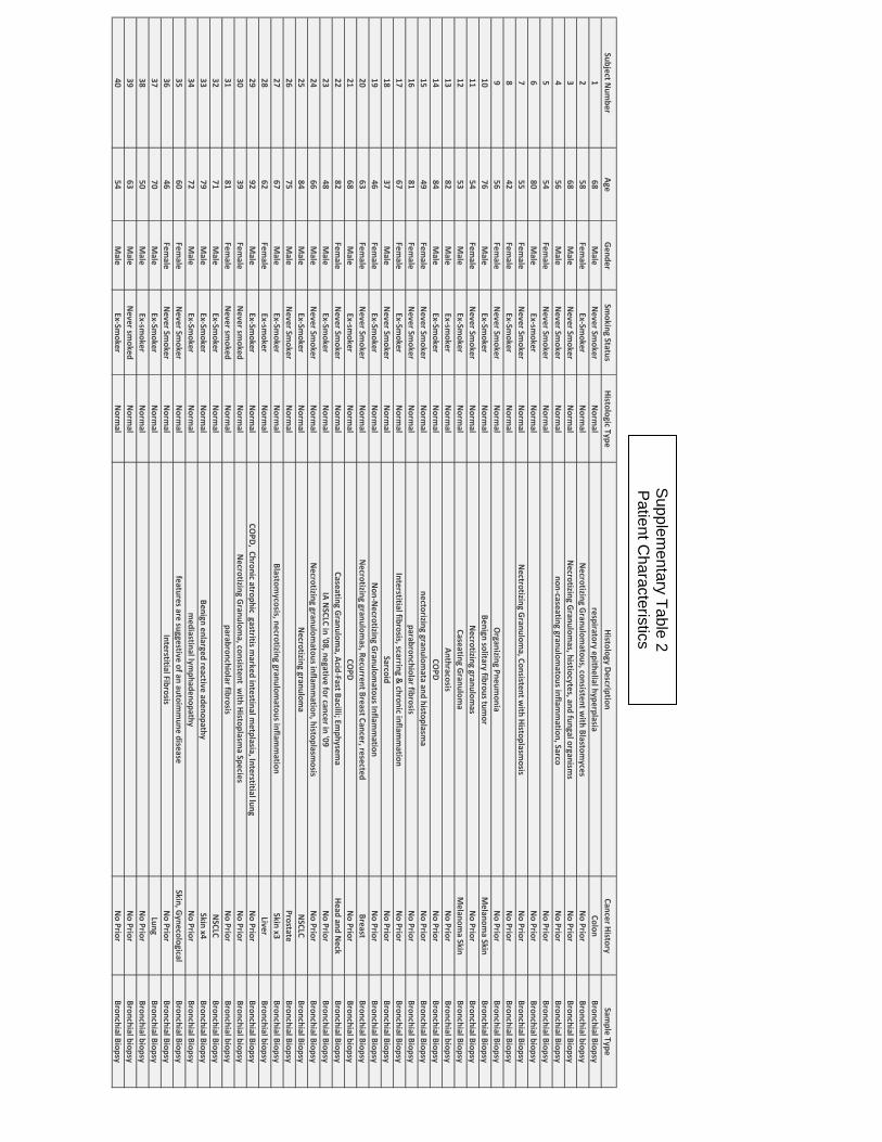
Subject Num
berAge
GenderSm
oking StatusHistologic Type
Histology DescriptionCancer History
Sample Type
168
Male
Never Sm
okerN
ormal
respiratory epithelial hyperplasia Colon
Bronchial Biopsy2
58Fem
aleEx-Sm
okerN
ormal
Necrotizing Granulom
atous, consistent with Blastom
ycesN
o PriorBronchial biopsy
368
Male
Never Sm
okerN
ormal
Necrotizing Granulom
as, histiocytes, and fungal organisms
No Prior
Bronchial Biopsy4
56M
aleN
ever Smoker
Norm
al non-caseating granulom
atous inflamm
ation, SarcoN
o PriorBronchial Biopsy
554
Female
Never Sm
okerN
ormal
No Prior
Bronchial Biopsy6
80M
aleEx-sm
okerN
ormal
No Prior
Bronchial biopsy7
55Fem
aleN
ever Smoker
Norm
alN
ectrotizing Granuloma, Consistent w
ith Histoplasmosis
No Prior
Bronchial Biopsy8
42Fem
aleEx-Sm
okerN
ormal
No Prior
Bronchial Biopsy9
56Fem
aleN
ever Smoker
Norm
alO
rganizing Pneumonia
No Prior
Bronchial Biopsy10
76M
aleEx-Sm
okerN
ormal
Benign solitary fibrous tumor
Melanom
a SkinBronchial Biopsy
1154
Female
Never Sm
okerN
ormal
Necrotizing granulom
asN
o PriorBronchial Biopsy
1253
Male
Ex-Smoker
Norm
alCaseating Granulom
aM
elanoma Skin
Bronchial Biopsy13
82M
aleEx-sm
okerN
ormal
AnthracosisN
o PriorBronchial biopsy
1484
Male
Ex-Smoker
Norm
alCO
PDN
o PriorBronchial Biopsy
1549
Female
Never Sm
okerN
ormal
nectorizing granulomata and histoplasm
a N
o PriorBronchial Biopsy
1681
Female
Never Sm
okerN
ormal
parabronchiolar fibrosisN
o PriorBronchial Biopsy
1767
Female
Ex-Smoker
Norm
alInterstitial fibrosis, scarring &
chronic inflamm
ationN
o PriorBronchial Biopsy
1837
Male
Never Sm
okerN
ormal
SarcoidN
o PriorBronchial Biopsy
1946
Female
Ex-Smoker
Norm
alN
on-Necrotizing Granulom
atous Inflamm
ationN
o PriorBronchial Biopsy
2063
Female
Never Sm
okerN
ormal
Necrotizing granulom
as, Recurrent Breast Cancer, resectedBreast
Bronchial Biopsy21
68M
aleEx-sm
okerN
ormal
COPD
No Prior
Bronchial biopsy22
82Fem
aleN
ever Smoker
Norm
alCaseating Granulom
a, Acid-Fast Bacilli; Emphysem
a Head and N
eckBronchial Biopsy
2348
Male
Ex-Smoker
Norm
alIA N
SCLC in '08, negative for cancer in '09N
o PriorBronchial Biopsy
2466
Male
Never Sm
okerN
ormal
Necrotizing granulom
atous inflamm
ation, histoplasmosis
No Prior
Bronchial Biopsy25
84M
aleEx-Sm
okerN
ormal
Necrotizing granulom
aN
SCLCBronchial Biopsy
2675
Male
Never Sm
okerN
ormal
ProstateBronchial Biopsy
2767
Male
Ex-Smoker
Norm
alBlastom
ycosis, necrotizing granulomatous inflam
mation
Skin x3Bronchial Biopsy
2862
Female
Ex-smoker
Norm
alLiver
Bronchial biopsy29
92M
aleEx-Sm
okerN
ormal
COPD, Chronic atrophic gastritis m
arked intestinal metplasia, Interstitial lung
No Prior
Bronchial Biopsy30
39Fem
aleN
ever smoked
Norm
alN
ecrotizing Granuloma, consistent w
ith Histoplasma Species
No Prior
Bronchial biopsy31
81Fem
aleN
ever smoked
Norm
alparabronchiolar fibrosis
No Prior
Bronchial biopsy32
71M
aleEx-Sm
okerN
ormal
NSCLC
Bronchial Biopsy33
79M
aleEx-Sm
okerN
ormal
Benign enlarged reactive adenopathySkin x4
Bronchial Biopsy34
72M
aleEx-Sm
okerN
ormal
mediastinal lym
phadenopathyN
o PriorBronchial Biopsy
3560
Female
Never Sm
okerN
ormal
features are suggestive of an autoimm
une diseaseSkin, Gynecological
Bronchial Biopsy36
46Fem
aleN
ever Smoker
Norm
alInterstitial Fibrosis
No Prior
Bronchial Biopsy37
70M
aleEx-Sm
okerN
ormal
LungBronchial Biopsy
3850
Male
Ex-smoker
Norm
alN
o PriorBronchial biopsy
3963
Male
Never sm
okedN
ormal
No Prior
Bronchial biopsy40
54M
aleEx-Sm
okerN
ormal
No Prior
Bronchial Biopsy
Supplementary Table 2
Patient Characteristics