transmembrane conductance regulator
TRANSCRIPT
Interactions of liposomes and proteoliposomes with cultured cells : application to the cystic fibrosistransmembrane conductance regulatorby Carol Ann Higginbotham
A thesis submitted in partial fulfillment of the requirements for the degree of Doctor of Philosophy inBiochemistryMontana State University© Copyright by Carol Ann Higginbotham (1996)
Abstract:Cystic Fibrosis (CF) is the most common fatal genetic disease affecting Caucasians. Potentialapproaches to therapy seek to decrease or avoid disease symptoms by correction of the genetic defect.Included in these approaches are attempts to deliver a normal copy of the defective gene, and attemptsto deliver normal protein, to the affected cells of CF patients.
Liposomes composed of artificial, cationic compounds have been found to be effective DNA deliveryvehicles, producing expression of the gene products introduced in a significant percentage of treatedcells. This study investigated the feasibility of using liposomes for delivery of integral membraneproteins, and particularly the protein defective in CF, to cultured eukaryotic cells. Because of theirdemonstrated usefulness in DNA transfection, cationic liposome delivery vehicles were examined indetail.
Fluorescence microscopy was used, in conjunction with fluorescence resonance energy transfer, tocharacterize the interaction occurring between liposomes or proteoliposomes and cultured cells. Studiesof cationic liposomes composed of dioleoylphosphatidylethanolamine (DOPE) andl,2-bis(oleoxy)-3-(trimethylammonio) propane (DOTAP), and DOPE and 3-β[N,N-dimethylaminoethane)-carbamoyl] cholesterol (DC Chol) revealed that these liposomes adheredstrongly to cell surfaces, but that dilution of the probes did not occur, to a measurable degree. Theseresults suggest that these cationic liposomes would not be suitable for integral membrane proteindelivery to the cell lines studied. Other liposome formulations tested did not exhibit interaction withcultured cells.
Systems employing viral envelopes (virosomes) and receptor mediated uptake of liposomes were alsostudied, to see if these systems might be significantly more efficient at delivery of foreign material. Inthe experiments performed, no evidence of lipid transfer to target cells was obtained.
The protein defective in CF is reported to undergo rapid endocytosis from the plasma membrane. Toinvestigate the trafficking of this protein, we identified the CFTR protein by immunoblot from vesicleenriched cell fractions. Cell fractions containing clathrin, a marker protein for coated vesicles, werefound also to contain a protein of approximately 170 kD which was reactive to anti-CFTR antibodies.

INTERACTIONS OF LIPOSOMES AND PROTEOLIPOSOMES WITH
CULTURED CELLS: APPLICATION TO THE CYSTIC FIBROSIS
TRANSMEMBRANE CONDUCTANCE REGULATOR
by
Ccu ui Aim Higginbotham
A thesis submitted in partial fulfillment of the requirements for the degree
Of
Doctor of Philosophy
in
Biochemistry
MONTANA STATE UNIVERSITY-BOZEMAN Bozeman, Montana
December 1996
11
APPROVAL
of a thesis submitted by
Carol Ann Higginbotham
This thesis has been read by each member of the thesis committee and has been found to be satisfactory regarding content, English usage, format, citations, bibliographic style, and consistency, and is ready for submission to the College of Graduate Studies.
Martin Teintze(Signature) (Date)
Approved for the Department of Chemistry and Biochemistry
David M. Dooley
Approved for the College of Graduate Studies
Robert L. Brown(Signature) (Date)
m
STATEMENT OF PERMISSION TO USE
In presenting this thesis in partial fulfilment of the requirements for
a doctoral degree at Montana State University-Bozeman, I agree that the
Library shall make it available to borrowers under rules of the Library. I
further agree that copying of this thesis is allowable only for scholarly
purposes, consistent with “fair use” as prescribed in the U.S. Copyright .
Law. Requests for extensive copying or reproduction of this thesis should
be referred to University Microfilms International, 300 North Zeeb Road,
Ann Arbor, Michigan 48fo6, to whom I have granted “the exclusive right to
reproduce and distribute my dissertation in and from microform along with
the non-exclusive right to reproduce and distribute my abstract in any
format in whole or in part.”
Signature
Date

ACKNOWLEDGEMENTS
I would like to thank my academic and personal mentors who have supported me in immeasurable ways throughout the course of this study. Dr. Martin Teintze is a thorough teacher, and deserves much of the credit for the development of my abilities and this project. Other members of the academic community at Montana State University have also been particularly helpful: Sam Rogers, Ed Dratz, Mark Quinn, and Dave Dooley who have served on my graduate advisory committee, and also CliffBond and Bruce Granger, who have been helpful with technical issues.
Steve Swain has been a particularly important professional and personal cohort. His breadth of knowledge and true understanding of the scientific pursuit challenged and inspired me. His keen perception (in scientific and personal matters) and humor did much to keep my work interesting. His knack for knowing the right time for ice cream breaks was critical to maintaining my perspective, especially at difficult times. I owe him much.
Other coworkers have done their share, as well, to make this work possible, and enjoyable. Thanks to Daphne Moffett, Tami Peters, and Dave Parks from the Teintze group, and to Doreen Brown, Jan Rasmussen, Marcella Alvarez, Christy Ruggiero, and John Bollinger in the Dooley group. Michele McGuirl, also of the Dooley group, has been a special friend and someone who understands the added challenges of parenting while in graduate school.
Undergraduates who have fueled my conviction to teach also deserve thanks. Those who were assigned to my courses, I want to thank for inspiration and for making me aware of how little I know. Those who I have worked with on this project, I want to thank for adding enthusiasm and energy to a long and at times trying endeavor.
As an undergraduate myself, I was affected profoundly by some fabulous teachers: Dave Erkenbrack, Cathy Haustein, Dan Brass, Art Bosch, and John Bowles, none of whom ever doubted my ability to reach my goals, and gave me the courage to try.
Most influential, of course, is my family. My extended family has never questioned my ambition, they have expressed their pride, and I am grateful. My husband David has endured hardships because of my desire to complete an advanced degree, without complaints. He is a wonderfully supportive partner. And finally, there is Patrick, who thinks by now that school is a lifelong endeavor. I hope what I have done will benefit him greatly.
V
TABLE OF CONTENTS
C hapter Page1. INTRODUCTION.................................................................. I
Cystic Fibrosis . . . . . . IThe C F T R .................................................................. 6Emerging Approaches to Therapy . . . 8Endocytosis, Recycling, and CFTR . . . 10Delivery Systems . . . . . . 12Liposomes . . . . . . . 18Mechanism of Liposomal Delivery . . . 23The Contents of This Study . . . . . . 24References Cited . . . . . . 26
2. CFTR PROTEIN EXPRESSION AND PURIFICATION . 35Introduction and Motivation . . . . 35Materials and Methods . . . . . 36
Protein Expression in theBaculoviral System. . . . 36
CFTR Purification . . . . . 39Results . . . . . . . 45Discussion . . . . . . . 46References Cited . . . . . . 49
3. INTERACTIONS OF LIPOSOMES WITHCULTURED CELLS ..................................................................50
Introduction and Motivation . . . . 50Materials and Methods . . . . . 51
Liposome Manufacture . . . . 51Cell Culture . . . . . . 56
He La . . . . . 56E B T r..................................................................57RAW 264.7 . . . . . 58

V l
TABLE OF CONTENTS-ContinuedChapter Page
. CFG-SV40 . . . . 58HT29 . . . 59
Liposome Treatments 60Fluorescence Techniques . . . 62
Microscopy and Confocal Microscopy . 64Resonance Energy Transfer 67
Results . . . . . . . 73Liposome Manufacture . . . 73Microscopy . . . . . . 75Confocal Microscopy . . . . 83Resonance Energy Transfer 83
Discussion . . 90References Cited . . . . 92
4. INTERACTIONS OF PROTEOLIPOSOMES WITHCULTURED CELLS . . . . . . 96
Introduction and Motivation . . . . 96Materials and Methods . . . 1 97
Production of Proteoliposomes 97Band 3 as a Surrogate Protein 100
Results . . . . . . . 103Discussion . . . . 109References Cited . . . . . . 112
5. FUSION PROTEINS AND RECEPTORMEDIATED PROCESSES . . . 115
Introduction and Motivation . . . . 115\

TABLE OF CONTENTS—ContinnpH Chapter Page
Materials and Methods . . . . . i l 8Influenza Virosomes . . . . H gVIP Receptor Studies . . . . 119
Results . . . . . . . 121Influenza Virosomes . . . . 121VIP Receptor Studies . . • . 121
Discussion . . . . . _ 2.22References Cited . . . . . . 124
6. ISOLATION OF CLATHRIN COATED VESICLES:THE ROLE OF VESICLE TRAFFICKING . . . 1 2 7
Introduction and Motivation . . . . 127Materials and Methods . . . . . 128Results . ■ . . . . 131Discussion . . . . . . . 133References Cited . . . . . . 136
7. GENERAL CONCLUSIONS . . . . . 137Expression and Purification of CFTR . . . 138Interactions of Liposomes with Cultured Cells . 138Interactions of Proteohposomes with Cultured Cells . 140Viral and Receptor Mediated Processes . . 141CFTR and Vesicle Trafficking . . . 142References Cited . . . . 144
APPENDICESAppendix A: A Brief Overview of All Liposome and Proteohposome Experiments Performed on HeLa CeUs 146
'
C hapter
V U l
. TABLE OF CONTENTS-Continued
Page
Appendix B: A Summary of All Liposome Experiments Performed on Cell Lines Other than HeLa . . 161
LIST OF FIGURES
Figure Page
1. What goes wrong in cftr mutations causingcystic fibrosis . . . . . . . 5
2. A schematic model of the CFTR protein . . . 7
3. Possible modes of interaction between liposomesand cells . . . . . . . . 17
4. Delivery vehicle types, arranged on a continuumaccording to complexity . ; . . . . 1 7
5. Compounds used to form cationic liposomes forthis study . . . . . . . . 19
6. PCR amplification of DNA from Sf9 cells transformedwith cftr DNA . . . . . . . 38
7. CFTR elution from gel filtration column . . . 42
8. BioCad Sprint chromatography system program for separation of proteins over hydroxyapetite, used forCFTR purification . . . . . . 43
9. BioCad Sprint chromatography system program for separation of proteins by gel permeation, used forCFTR purification . . . . . . . 44
10. Western blot on purified recombinant CFTR . . . 47
11. Extrusion device used to form liposomes . . . 54
12. Fluorescent labels used in this study . . . . 65
13. Theory of resonance energy transfer (RET) . . . 69
ix
X
LIST OF FIGURES—continued
F igure Page
14. Instability of OA/PE liposomes . . . . . 74
15. Fluorescent image of HeLa cells treated withDOTAPZDOPE liposomes . . . . . . 78
16. Fluorescent image of HeLa cells treated with DOTAPZDOPE liposomes, and subsequently fused tocells with polyethylene glycol. . . . . . 7 9
17. Brightfield illumination, same field as figure 15. . . . 80
18. Confocal fluorescent image of liposome-treated cells . " 84
19. RET fluorescence emission scan of HeLa cells treated with labelled DOTAPZDOPE liposomes, fluorescein-PEas RET donor . . . . . . 86
20. RET fluorescence emission scan of HeLa cells treated with labelled DOTAPZDOPE liposomes, NBD-PE asRETdonor . . . . . . . . . 87
21. RET fluorescence emission scan of HeLa cells treatedwith labelled IiposomeZDNA complexes. . . . 89
22. Eosin-5-maleimide, label for covalent linkage to Band 3 . 101
23. Electrophoresis with silver staining of fractions fromBand 3 purification . . . . . . . 104
24. Fluorescent image of HeLa cells treated with Band 3proteoliposomes . . . . . . . 107
25. Fluorescent image of HeLa cells treated with Band 3 proteoliposomes, subsequently treated with polyethylene glycol 109
LIST OF FIGURES-Continued
Figure Page
26. RET fluorescence emission scan of HeLa cells treated withlabelled Band 3 proteoliposomes . . . . . 1 1 0
27. Electrophoresis of fractions from vesicle isolation thoughtto contain clathrin . . . .' . . 132
28. Radioassay on samples from CFTR immunoprecipitation experiment . . . . . . . 134
xd
ABSTRACT
Cystic Fibrosis (CF) is the most common fatal genetic disease affecting Caucasians. Potential approaches to therapy seek to decrease or avoid disease symptoms by correction of the genetic defect. Included in these approaches are attempts to deliver a normal copy of the defective gene, and attempts to deliver normal protein, to the affected cells of CF patients.
Liposomes composed of artificial, cationic compounds have been found to be effective DNA delivery vehicles, producing expression of the gene products introduced in a significant percentage of treated cells. This study investigated the feasibility of using liposomes for delivery of integral membrane proteins, and particularly the protein defective in CF, to cultured eukaryotic cells. Because of their demonstrated usefulness in DNA transfection, cationic liposome delivery vehicles were examined in detail.
Fluorescence microscopy was used, in conjunction with fluorescence resonance energy transfer, to characterize the interaction occurring between liposomes or proteoliposomes and cultured cells. Studies of cationic liposomes composed of dioleoylphosphatidylethanolamine (DOPE) and l,2-bis(oleoxy)-3-(trimethylammonio) propane (DOTAP), and DOPEand 3-(HN,N-dimethylaminoethane)-carbamoyl] cholesterol (DC Choi) revealed tha t these liposomes adhered strongly to cell surfaces, but that dilution of the probes did not occur, to a measurable degree. These results suggest that these cationic liposomes would not be suitable for integral membrane protein delivery to the cell lines studied. Other liposome formulations tested did not exhibit interaction with cultured cells.
Systems employing viral envelopes (virosomes) and receptor mediated uptake of liposomes were also studied, to see if these systems might be significantly more efficient at delivery of foreign material. In the experiments performed, no evidence of lipid transfer to target cells was obtained.
The protein defective in CF is reported to undergo rapid endocytosis from the plasma membrane. To investigate the trafficking of this protein, we identified the CFTE protein by immunoblot from vesicle enriched cell fractions. Cell fractions containing clathrin, a marker protein for coated vesicles, were found also to contain a protein of approximately 170 kD which was reactive to anti-CFTR antibodies.
CHAPTER I
INTRODUCTION
I
Cystic Fibrosis
Cystic Fibrosis (CF) is an autosomal recessive genetic disease
affecting one in 2500 Caucasians [I]. There is no cure, and progressive loss
of function in affected organs generally leads to death before middle age.
The current median age of death from CF is 29, however recent
improvements in treatment are causing the median age of death to rise.
Early and aggressive treatment, general quality of health, and the
particular genetic defect an individual carries all affect the age of mortality.
Cystic Fibrosis was first described in the medical literature in the
late 1930s. The syndrome includes pancreatic exocrine insufficiency, male
infertility, and recurring infection of the lung leading to development of
inelastic scar tissue, or "fibrosis," in the lungs. An elevated sweat chloride
level in affected individuals is diagnostic of the disease [I].
Pancreatic insufficiency can be effectively treated by enzyme
replacement, and most morbidity involves symptoms affecting the lungs.
CF patients have difficulty clearing mucoid secretions from the lungs. It is
suspected that the associated difficulty in removing bacteria increases
susceptibility to colonization and chronic infection [I]. Changes in bacterial
2
adhesion and impaired ability of the body to kill the bacteria have also been
implicated in complications in the lungs [2-5]. Damage caused by colonizing
bacteria and subsequent inflammation results in the development of
inelastic scar tissue and progressively declining lung function.
The metabolic disorder causing the symptoms of Cystic Fibrosis was
not well understood until the genetic defect responsible was identified in
1989. DNA polymorphisms were used to map the locus of the genetic
defect to a region on chromosome 7. Genetic analysis of diseased individuals
and DNA analysis of a sequence thought to contain the CF locus revealed
the location of the gene. The cDNA identified aligns with an open reading
frame, with transcription patterns that correlate with the tissues that are
affected in the disease [6-8].
The gene encodes an intregal membrane protein of 1480 residues
termed the Cystic Fibrosis Transmembrane Conductance Regulator
(CFTRX with a predicted molecular weight of 168 kilodaltons [6]. The
predicted structure contains a pair of similar motifs* with each domain
containing six transmembrane helices and a nucleotide binding fold. There
is a single regulatory domain on the cytoplasmic side of the protein,
containing sites amenable to regulation by phosphorylation by cAMP-
dependent protein kinase [9]. The protein is glycosylated on the
extracellular surface, with the sugars contributing about 30 kilodaltons to
the apparent molecular weight as indicated on polyacrylamide gels [10].
Studies of purified recombinant CFTR indicated that the protein is a
regulated ion channel, with properties consistent with the specific activities
of cAMP regulated chloride channels in cell types that normally express
3
CFTR [8]. However, discussion continues over its potential activity as a
regulator of other cellular functions [11-15]. Glycosylation apparently does
not affect activity [16].
In 1991, it was discovered that introduction of a normal copy of the
gene restores chloride channel activity in c f- /- cells isolated from an
affected individual. Drumm et al. used retroviral delivery of a normal copy
of the gene to a cell line derived from a CF-patient, to demonstrate
correction of the defect [17]. cAMP stimulated ion efflux and patch clamp
experiments revealed the correction of the defect by introduction oicftr
DNA. Rich et al., in a concurrently published paper, utilized a fluorescent
method to trace ion efflux, and also patch clamping, to attain the same
result in cultured CF airway epithelial cells. Additionally, they
demonstrated that expression of a gene carrying a common mutation did
not correct the defect [18]. The connection between CFTR and ion
transport dysfunction was made clear.
Sequence analysis has revealed that approximately 70% of all
diagnosed individuals contain a cftr gene missing 3 base pairs, resulting in
deletion of a phenylalanine at residue 508 in the mature protein (AF508).
The remaining 30% of CF cases involve any one of over 200 different faults
in the gene that have been identified [19].
A wide disparity in disease severity exists among the other 200+
identified mutations existing. Relationships between the effect of a
particular mutation on the protein and severity are unclear [20]. Some
individuals with particularly mild forms of CF can even go undiagnosed until
early adulthood [21]. Environmental factors, the existence of multiple
4
mutations on one copy of the gene or heterozygous mutants, and the
possibility that other genetic factors play a role in disease presentation, all
obscure the correlation [20].
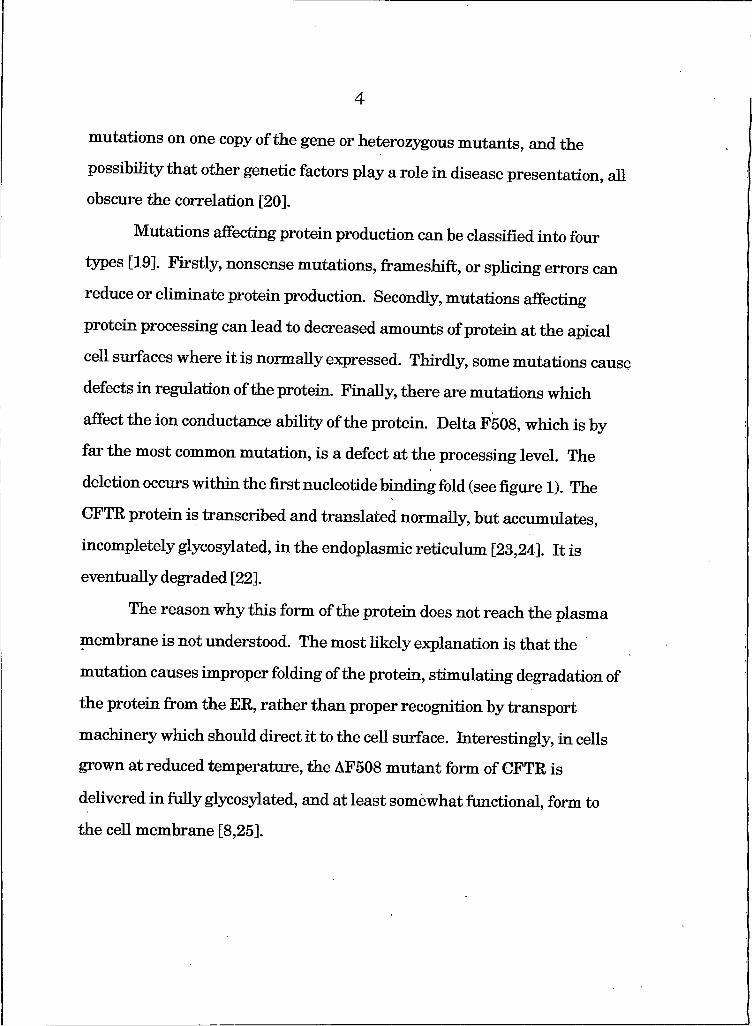
Mutations affecting protein production can be classified into four
types [19]. Firstly, nonsense mutations, frameshift, or splicing errors can
reduce or eliminate protein production. Secondly, mutations affecting
protein processing can lead to decreased amounts of protein at the apical
cell surfaces where it is normally expressed. Thirdly, some mutations cause
defects in regulation of the protein. Finally, there are mutations which
affect the ion conductance ability of the protein. Delta F508, which is by
far the most common mutation, is a defect at the processing level. The
deletion occurs within the first nucleotide binding fold (see figure I). The
CFTR protein is transcribed and translated normally, but accumulates,
incompletely glycosylated, in the endoplasmic reticulum [23,24]. It is
eventually degraded [22].
The reason why this form of the protein does not reach the plasma
membrane is not understood. The most likely explanation is that the
mutation causes improper folding of the protein, stimulating degradation of
the protein from the ER, rather than proper recognition by transport
machinery which should direct it to the cell surface. Interestingly, in cells
grown at reduced temperature, the AF508 m utant form of CFTR is
delivered in fully glycosylated, and at least somewhat functional, form to
the cell membrane [8,25].
5
Plasma Membrane
IV
Golgi
III. amass
E.R. Nucleus
II. I.
Figure I: What goes wrong in cftr mutations causing cystic fibrosis. Four types of mutation altered membrane potential: I. Defective protein production, II. Defective protein processing, III. Defective regulation, and IV. Defective ion
conduction
6
The resultant lack of protein at the apical surface of bronchiolar
epithelium is proposed to cause an imbalance in ion transport at that
surface. Compensatory changes in sodium channel activity compound the
problem by altering water balance across the cell membrane [26]. The
viscosity of the mucoid secretions in the channels of the glands where
CFTR is most actively expressed is thought to increase due to the osmotic effect.
The CFTR
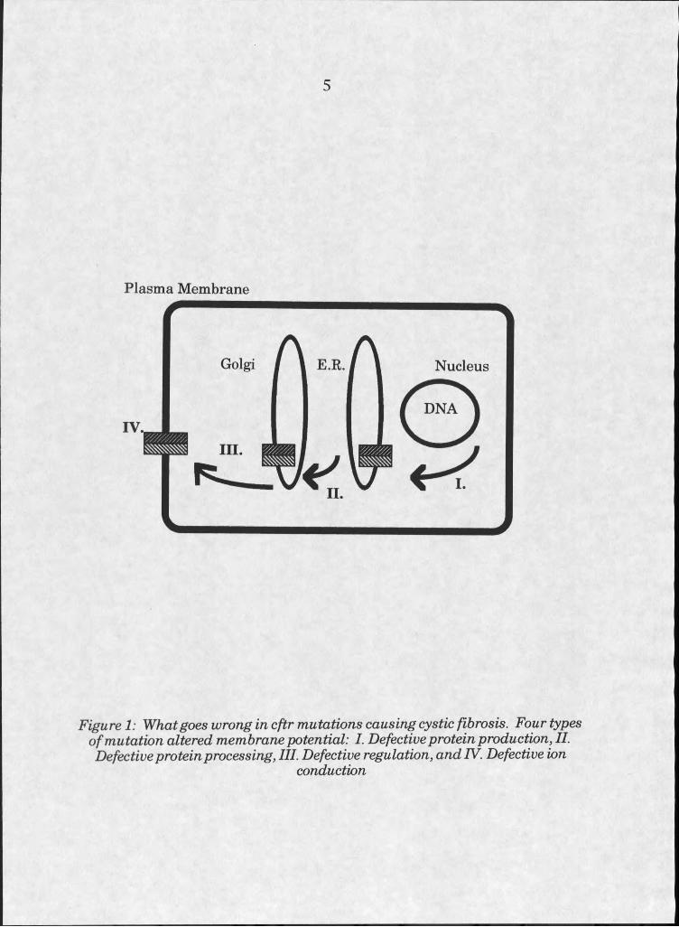
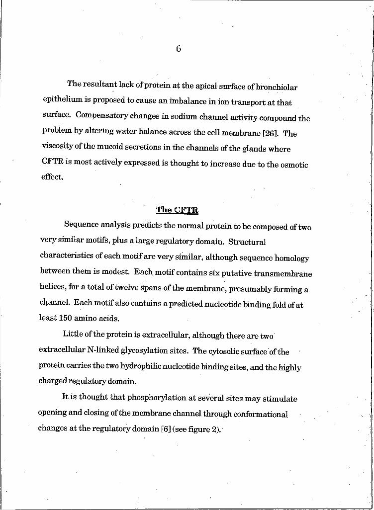
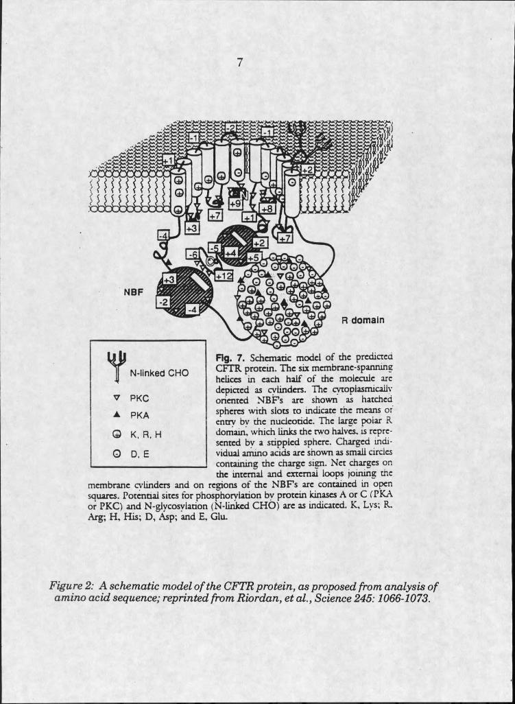
Sequence analysis predicts the normal protein to be composed of two
very similar motifs, plus a large regulatory domain. Structural
characteristics of each motif are very similar, although sequence homology
between them is modest. Each motif contains six putative transmembrane
helices, for a total of twelve spans of the membrane, presumably forming a
channel. Each motif also contains a predicted nucleotide binding fold of at
least 150 amino acids.
Little of the protein is extracellular, although there are two
extracellular N-Iinked glycosylation sites. The cytosolic surface of the
protein carries the two hydrophilic nucleotide binding sites, and the highly
charged regulatory domain.
It is thought that phosphorylation at several sites may stimulate
opening and closing of the membrane channel through conformational
changes at the regulatory domain [6] (see figure 2).
7
R domain
Fig. 7. Schematic model o f the predicted CFTR protein. The six membrane-spanning helices in each half o f the molecule are depicted as cylinders. The cytoplasmicailv oriented N B F s arc shown as hatched spheres with slots to indicate the means or entry bv the nucleotide. The large polar R domain, which links the two halves, is represented bv a stippled sphere. Charged individual ammo acids arc shown as small circles containing the charge sign. Net charges on the internal and external loops joining the
membrane cylinders and on regions o f the N B F s arc contained in open squares. Potential sites for phosphorylation by protein kinases A or C CPKA or PKC) and N -glycosylation (N-Iinkcd CH O ) arc as indicated. K, Lys; R. Arg; H , His; D , Asp; and E, Glu.
7N -Iin k e d C H O
V P K C
▲ P K A
O K, R , H
O D, E
Figure 2: A schematic model o f the CFTR protein, as proposed from analysis o f amino acid sequence; reprinted from Riordan, et a l, Science 245:1066-1073.
8
CFTR belongs to a family of proteins called the ATP binding cassette
proteins, or the ABC family. Included in this family are several other
transmembrane transporters, notably the multidrug resistance protein, or
P-glycoprotein, proposed to be responsible for drug efflux from cells
resistant to chemotherapeutics [27,28].
Debate about CFTR’s function has continued since its original
characterization. As the name implies, CFTR has been considered to be a
regulator, as well as an ion channel itself. The low copy number of the
expressed protein, and the fact that other chloride channels exist in cells
containing CFTR, suggest that CFTR may have functions outside of its role
as chloride channel. In particular, a relationship between CFTR and the
outwardly rectifying chloride channel, or ORCC, may exist [27,30]. It has
also been proposed that CFTR acts as an ATP pump, driving nucleotides to
the extracellular milieu [12].
Emerging Approaches to Therapy
Current therapeutic approaches to treating the pulmonary
symptoms of Cystic Fibrosis are aimed at aggressively treating bacterial
colonization and improving the ability to clear lung secretions. Physical
elimination of the secretions is aided by chest and back percussion therapy.
Enzymatic therapies for decreasing sputum viscosity have been recently
introduced. Recombinant DNase has been used with some success to
destroy viscous DNA deposits in the lung resulting from inflammation [31].
Improvement in the ion balance, and therefore water balance, of cellular
9
secretions has been attempted by blocking sodium channels in the lungs
with the compound amiloride [32]. All of these methods have contributed to
a noticeable increase in life expectancy for patients, but further
developments in therapy will be necessary before those stricken with CF
can enjoy a full life.
A goal of the CF research community is to develop therapies which
more directly affect the molecular deficiency caused by the disease.
Correction of the defect at the molecular level would be the most efficient
kind of therapy, making it possibile to avoid the escalation of pulmonary
damage caused by repeated infections.
Of particular interest are investigations into the use of gene transfer
for correction of the genetic defect. Proponents argue that by introducing a
normal copy of the cftr gene into the cells of an affected person, they can
stimulate production of normal protein and alleviate symptoms. Dehvery of
the cftr gene has been shown to be possible in vitro and in the lungs of
mouse models and cotton rats [33-35], but has yet to be shown adequate to
relieve CF-affected individuals. CFTR gene transfer studies have been
ongoing for several years, without reports of success. The'effects of ongoing
treatment with such therapies are unknown at this time.
Specifically, issues surrounding the immunogenicity and safety of
gene delivery therapies are unresolved [36]. Clinical trials using liposomal
vectors and adenoviral vectors are currently underway [37-40]. Both
methods produce some expression, but expression levels may be inadequate
and tend to be patchy across a tissue. Expression is in all cases transient,
and therapies for gene delivery will need to be administered repeatedly [41].
10
This fact raises questions regarding the involvement of the immune
response, and potential decreases in effectiveness over time. The most
likely scenario is that even after development of gene therapies, a
combination of clinical approaches will be used to combat the disease.
Alternative approaches to restore normal ion conductance include
attempts at manipulating regulation and trafficking of CFTR, and
upregulating the activity of the protein at the plasma membrane. In
addition, CFTR's potential function as a regulator, rather than a significant
ion conductor itself, may open doors to new approaches aimed at correcting
the metabolic defect further down the line; i.e. by finding ways of changing
regulation of proteins normally regulated by CFTR.
Endocytosis. Recycling, and CFTR
Certain plasma membrane proteins are known to be transported
through endosomal pathways and back to the plasma membrane. This
process, termed recycling, occurs through clathiin coated vesicle
endocytosis, but the return of the protein to the cell membrane involves
unidentified vesicles. The process is selective, involving only certain
components of the plasma membrane. CFTR has been reported to cycle
through such a pathway, although why CFTR would participate in such a
pathway is not known [42,43].
Studies of vesicle trafficking are difficult because they rely on the
identification of marker proteins specific to a particular type of vesicle in
order to characterize that subpopulation of vesicles. In some instances,
there are no vesicle-type specific markers [44,45]. Physical identifiers such
11
as size and density are somewhat characteristic of a vesicle subpopulation,
although they generally are not adequately distinctive to separate vesicle populations unequivocally [46].
Studies of yeast secretory mutants have led to a fairly thorough
understanding of the mechanisms involved in vesicle traffic from the
endoplasmic reticulum to the plasma membrane. In all instances,
membrane trafficking along this route is carefully controlled by ligand-
receptor interactions which stimulate fusion of vesicle membranes [47-49],
Identification of the ligands and receptors involved, mutation studies,and
immunocytochemistry allow for detailed studies of these systems.
Similar model systems do not exist for studying recycling. The
existence of proteins regulating the endocytosis selection system, and
subsequent sorting of vesicle components and downstream routing, is
undetermined. Studies rely, rather, on compounds thought to inhibit
endocytosis, such as brefeldin A [50], or the vesicle acidification processes,
such as chloroquine [51]. Because of the general lack of understanding
about the endocytosis/recycling pathway, these studies are inadequate to
draw conclusions about how sorting and trafficking in the recycling
pathway occurs.
What is known is that certain identifiable proteins are selectively
drawn into the recycling pathway, and that among those proteins is the
CFTR. CFTR is functional in endosomes, but does not appear to affect the
acidification or regulation of the endocytic system [53,54]. It is plausible
that CFTR, like most membrane proteins in endosomes, gets recycled to
the plasma membrane [54-56].
12
Vesicles containing compounds bound for endocytosis get internalized
through either coated or noncoated pits. Coated pits, the usual route for
internalization of receptor-ligand complexes, are so called because they are
surrounded by a protein coat at the cytosolic face of the membrane. The
fibrous coat protein clathrin assembles at the site of membrane
invagination as the vesicle buds and dissociates from the vesicle after
internalization. Sometime after dissociation of the clathrin from the
vesicle, sorting occurs and certain components of the vesicle membrane are
routed back to the plasma membrane [45].
The internal pH of endosomes varies from near physiological pH to
as low as 5.0. Separation of ligands from their receptors during endocytosis
and recycling is controlled by the endosomal pH, so that as the pH is
reduced, the ligand dissociates and is free to be trafficked separately from
the receptor [57]. This process provides a way for the cell to sort ligands
from the extracellular milieu to the lysosome for degradation, while
recycling the receptor.
Delivery Systems
Cell membranes act as selective barriers to most substances.
Overcoming this barrier is necessary for efficient delivery of any foreign
molecule, be it DNA, protein, or other compounds, to the cell. The
development of delivery vehicles, or vectors, with adequate efficiency to
cause physiological changes in tissues has proven difficult. In the arena of
DNA transfection, dramatic improvements have come in the last ten
13
years, but the in vivo therapeutic potential of existing methods remains questionable.
Requirements for development of delivery vehicles depend somewhat
on what kind of material needs to be delivered. In the case of DNA delivery,
few copies of the molecule need to be delivered, although they need to get
from outside the cell all the way to the nucleus. In the case of drugs active
in the cytosol, in contrast, a larger number of molecules must be delivered,
but only as far as the cytosol [58].
Engineered dehvery vehicles are therefore designed to interact in
specific ways with cells, to maximize their effectiveness. The rationale
behind a particular approach is influenced by physical theories of
membrane-membrane or membrane-protein interactions, and by the design
of natural biological delivery systems, i.e. viruses. Physical studies of
membrane biophysics have influenced heavily the development of all types
of vehicles. Genetically deficient viruses and virosomes have been
developed to closely mimic naturally occurring delivery processes, based on
empirical evidence that these systems work [60,61]. Their complexity, and
the complexity of the cells they interact with, however, have made their
mechanisms elusive.
Among the better understood viruses, many appear to take
advantage of the cellular internalization system to infect cells [61-63].
Some viral envelopes are composed of phospholipids and proteins which
contribute to their ability to infect the host. Influenza virus envelopes
contain the integral membrane protein hemagglutinin, which binds sialic
acid residues on the surfaces of host cells. Once internalized via
14
endocytosis, and exposed to the reduced pH of an endosome, the protein
triggers fusion ofthe viral coat with the endosomal membrane. The
triggering mechanism involves structural changes in the protein, which
cause destabilization of the membranes in close proximity. In this way
influenza efficiently delivers its genetic material to the host cell [62,63]].
For potential in vivo uses, the requirements for delivery vehicles
become more stringent [64]. In addition to concerns about the efficiency of
a method to deliver exogenous material to cells, in vivo applications also
must be safe. Cellular toxicity and lack of immunogenicity are critical if a
method is to have therapeutic use. Simplicity in design and avoidance of
immunogenic materials is desirable in the development of such vectors.
Generally, the simplest delivery vehicles can be composed of safe,
nonimmunogenic materials, but are less efficient than more complex, virus
like vectors. Among the simpler vehicles are liposomes, composed of
spherical bilayers of lipids or lipid analogues, and large globular complexes of
polycationic, nonlipid compounds [65].
The crux of the delivery problem seems then to be this: the
development of safe, non-immunogenic vectors with the adequate efficiency
to deliver the compound of interest to the target cell. Development of such
a vector requires analysis of possible routes of entry into a cell, and a design
which seeks to maximize the desired vehicle/cell interaction. Additionally,
simplicity and safety must remain considerations.
Delivery of material from a vehicle to a target cell can theoretically
occur in several different ways. Depending on what type of material must
be delivered, vehicle design can be tailored to produce an effective delivery
15
system. In all cases, the material must be brought into close proximity
with the cell. In vitro, this is not difficult; although in vivo there are
mechanistic challenges associated with this stage of delivery [58,66].
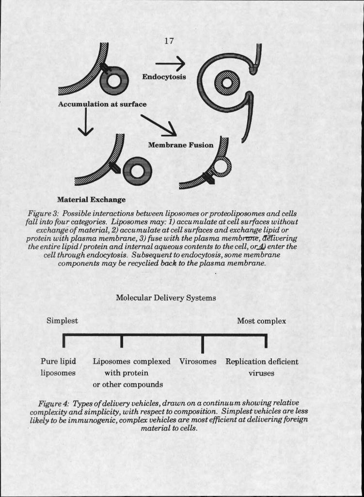
Once a delivery vehicle is present at the surface of a cell, four things
may happen. Firstly, it is possible that there will be no interaction at all; no
exchange of material or entry of foreign material into the cell. Secondly,
there could be exchange of material to or from the cell surface. The third
possibility, in the case of bilayer-containing vehicles, is the fusion of the
vehicle with the cell plasma membrane; incorporation of all the foreign
material into the cell at the surface. Finally, the entire delivery vehicle
could be endocytosed by the cell, and the fate of the vehicle and contents
determined by its routing once in the endosome (see figure 3).
Some interactions would result in greater introduction of foreign
material to the plasma membrane, whereas some would result in greater
concentrations in the cytosol, or lysosomes. Therefore, the route is critical
in the success of a vehicle for delivery of a particular type of compound. For
example, cytosolic delivery would likely be more useful when attempting to
deliver DNA than exchange of material at the cell surface.
Studies of molecular delivery to living cells often do not explore the
route of delivery. Additionally, systems designed for efficient delivery of one
type of molecule are rarely tested for delivery of other molecules to living
systems.
Delivery systems developed to date can be distributed along a
continuum, according to complexity. At the simplest end of the spectrum
are structures containing no protein. In this area are anionic liposomes,
16
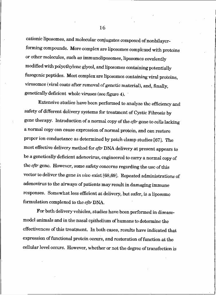
cationic liposomes, and molecular conjugates composed of nonbilayer
forming compounds. More complex are liposomes complexed with proteins
or other molecules, such as immunoliposomes, liposomes covalently
modified with polyethylene glycol, and liposomes containing potentially
fusogenic peptides. Most complex are liposomes containing viral proteins,
virosomes (viral coats after removal of genetic material), and, finally,
genetically deficient whole viruses (see figure 4).
Extensive studies have been performed to analyze the efficiency and
safety of different delivery systems for treatment of Cystic Fibrosis by
gene therapy. Introduction of a normal copy of the cftr gene to cells lacking
a normal copy can cause expression of normal protein, and can restore
proper ion conductance as determined by patch clamp studies [67]. The
most effective delivery method for cftr DNA delivery at present appears to
be a genetically deficient adenovirus, engineered to carry a normal copy of
the cftr gene. However, some safety concerns regarding the use of this
vector to deliver the gene in vivo exist [68,69]. Repeated administrations of
adenovirus to the airways of patients may result in damaging immune
responses. Somewhat less efficient at delivery, but safer, is a liposome
formulation complexed to the cftr DNA.
For both delivery vehicles, studies have been performed in disease-
model animals and in the nasal epithelium of humans to determine the
effectiveness of this treatment. In both cases, results have indicated that
expression of functional protein occurs, and restoration of function at the
cellular level occurs. However, whether or not the degree of transfection is
17
E ndocytosis
A ccum ulation at surface
A
%
M aterial E xchange
Figure 3: Possible interactions between liposomes or proteoliposomes and cells fall into four categories. Liposomes may: I) accumulate at cell surfaces without
exchange o f material, 2) accumulate at cell surfaces and exchange lipid or protein with plasma membrane, 3) fuse with the plasma membrane, delivering the entire lipid I protein and internal aqueous contents to the cell, orjjj enter the
cell through endocytosis. Subsequent to endocytosis, some membrane components may be recyclied back to the plasma membrane.
Molecular Delivery Systems
Simplest Most complex
I I I IPure lipid Liposomes complexed Virosomes Replication deficient liposomes with protein viruses
or other compounds
Figure 4: Types o f delivery vehicles, drawn on a continuum showing relative complexity and simplicity, with respect to composition. Simplest vehicles are less likely to be immunogenic, complex vehicles are most efficient at delivering foreign
material to cells.
18
adequate to alleviate the symptoms of Cystic Fibrosis in the lungs of affected individuals is unknown.
Concerns over the safety and immunogenicity of viral proteins in
adenoviral delivery systems would suggest that a simpler, less viral-like
vehicle should be used to deliver the gene. The poor efficiency of available
liposomes, though, has been a hindrance to their application. The necessity
of understanding HposomeZcell interactions, in the hopes of rationally
designing a more efficient Hposome, is becoming very apparent.
Liposomes
Liposomes are spherical vesicles composed of Hpids arranged in a
bilayer. They mimic biological membranes to the extent that membranes
are also composed of lipid bilayers, primarily of phospholipids. Liposomes
have often been composed of naturaUy occurring phospholipids, but more
recently their content has been varied in order to increase their potential as
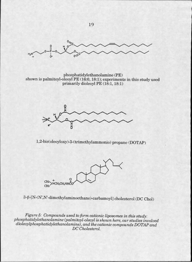
vehicles for delivering a variety of compounds. Particularly interesting has
been the development of liposomes composed of cationic compounds and
the membrane destabilizing phosphohpid phosphatidylethanolamine (PE),
which are easily prepared and relatively efficient vehicles for delivery of
DNA to ceHs in transfection experiments (see figure 5). They are used quite
routinely in the laboratory, and, as discussed before, are being investigated
as carriers of DNA into cells in vivo.
Liposome formulations contain any of a wide variety of compounds,
although some restrictions on content exist. The chemicals used must be
able to incorporate into a bilayer without completely disrupting the
19
o
■h O -
phosphatidylethanolamine (PE)shown is palmitoyl-oleoyl PE (16:0,18:1); experiments in this study used
primarily dioleoyl PE (18:1,18:1)
l,2-bis(oleoyloxy)-3-(trimethylammonio) propane (DOTAP)
3-p-[N-(N’,N’-dimethylaminoethane)-carbamoyl] cholesterol (DC Choi)
Figure 5: Compounds used to form cationic liposomes in this study: phosphatidylethanolamine (palmitoyl-oleoyl is shown here, our studies involved dioleoylphosphatidylethanolamine), and the cationic compounds DOTAPand
DC Cholesterol.
20
structure. Phospholipids, the most common material in liposomes, are
composed of two hydrophobic acyl chains attached to a glycerol moiety, and
a third, hydrophilic group at the “head.” Most phospholipids naturaUy form
bilayers when in aqueous solutions. The bilayer is stabilized by the
hydrophobic effect, along with the limitations imposed by the structures of
the component molecules. The head groups may carry net positive,
negative, or neutral charges, although highly charged elements in close
proximity to one another may destabilize the bilayer. Cholesterol, proteins
and peptides, as well as other amphipathic compounds, can incorporate into
the bilayer, so long as they are not in such high concentrations as to totally disrupt the structure.
Most naturally occurring phospholipids are stable in bilayers, and
carry either a net zero charge (as zwitterions) or a net -I charge. The size
of the head group on naturally occurring phospholipids generally is large
enough to help stabilize the bilayer structure. One exception is PE, whose
head group is small enough that it forms an inverted micellar “hexagonal
phase,” rather than a bilayer, under certain conditions [70].
In naturally occurring bilayers, PE is present in low enough
concentrations that hexagonal phases do not form. In artificial bilayers, PE
concentrations can be manipulated to make the liposome somewhat
unstable.
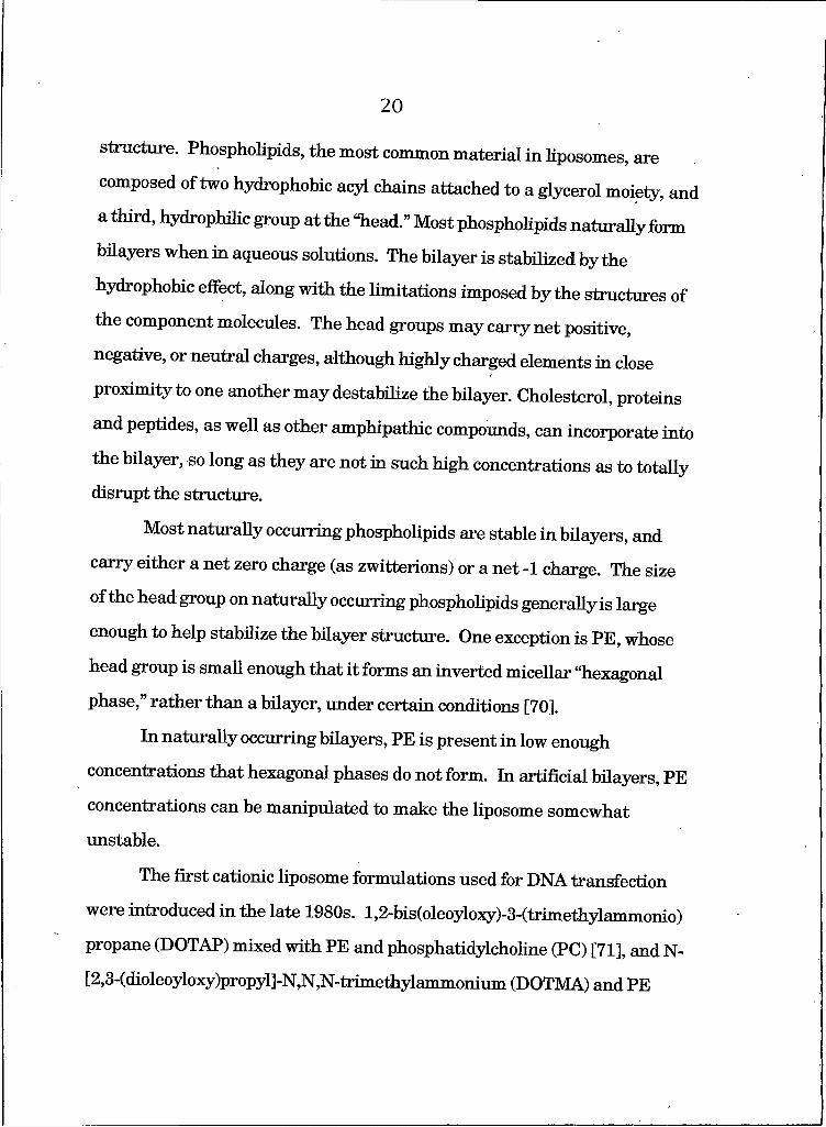
The first cationic liposome formulations used for DNA transfection
were introduced in the late 1980s. l,2-bis(oleoyloxy)-3-(trimethylammonio)
propane (DOTAP) mixed with PE and phosphatidylcholine (PC) [71], and N-
[2,3-(dioleoyloxy)propyl]-N,N,N-trimethylammonium (DOTMA) and PE
21
[72] were the original mixtures used for this purpose. Jn 1991,3p[N-(N’,N’-
dimethylaminoethane)-carbamoyl] cholesterol (DC-Chol) was introduced
[73] , and became the third popularized compound to be used in combination
with PE to form cationic liposomes. DOTMA and DOTAP are phospholipid
analogues, containing two hydrophobic acyl chains which anchor them into
the bilayer. DOTAP has been reported to be less toxic, perhaps due to its
hydrolyzable ester bonds at the glycerol moiety [74]. DC-Chol is a cationic
cholesterol analog, also has been considered less toxic than DOTMA, and
was the first of these three compounds to be approved for clinical trials in human subjects [73].
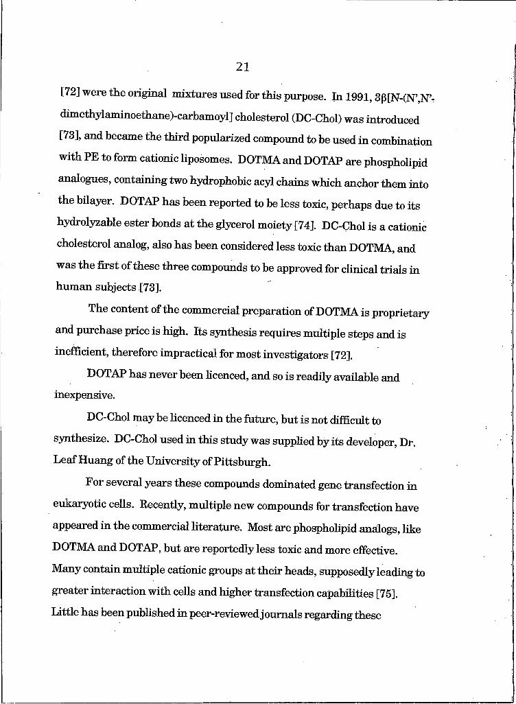
The content of the commercial preparation of DOTMA is proprietary
and purchase price is high. Its synthesis requires multiple steps and is
inefficient, therefore impractical for most investigators [72].
DOTAP has never been licenced, and so is readily available and inexpensive.
DC-Chol may be licenced in the future, but is not difficult to
synthesize. DC-Chol used in this study was supplied by its developer, Dr.
LeafHuang of the University of Pittsburgh.
For several years these compounds dominated gene transfection in
eukaryotic cells. Recently, multiple new compounds for transfection have
appeared in the commercial literature. Most are phospholipid analogs, like
DOTMA and DOTAP, but are reportedly less toxic and more effective.
Many contain multiple cationic groups at their heads, supposedly leading to
greater interaction with cells and higher transfection capabilities [75].
Little has been published in peer-reviewed j oumals regarding these
22
compounds, however, and transfection efficiency and toxicity appear to
vaiy widely among cell types. Various reports of toxicity and efficiency
reach inconsistent conclusions, even in identical cell lines [76-79]. Specific
formulations for transfection are proprietary, and are costly.
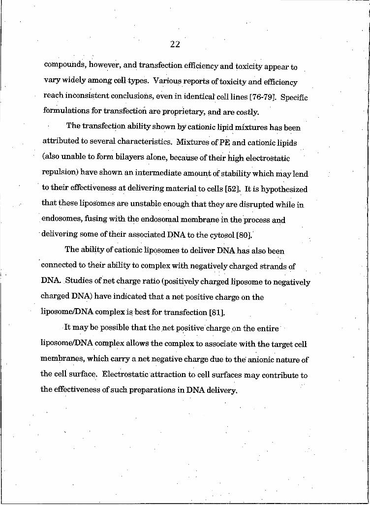
The transfection ability shown by cationic lipid mixtures has been
attributed to several characteristics. Mixtures of PE and cationic lipids
(also unable to form bilayers alone, because of their high electrostatic
repulsion) have shown an intermediate amount of stability which may lend
to their effectiveness at delivering material to cells [52]. It is hypothesized
that these liposomes are unstable enough that they are disrupted while in
endosomes, fusing with the endosomal membrane in the process and
delivering some of their associated DNA to the cytosol [80].'
The ability of cationic liposomes to deliver DNA has also been
connected to their ability to complex with negatively charged strands of
DNA Studies of net charge ratio (positively charged liposome to negatively
charged DNA) have indicated that a net positive charge on the
IiposomeZDNA complex is best for transfection [81].
It may be possible that the net positive charge on the entire
liposome/DNA complex allows the complex to associate with the target cell
membranes, which carry a net negative charge due to the anionic nature of
the cell surface. Electrostatic attraction to cell surfaces may contribute to
the effectiveness of such preparations in DNA delivery.
23
25££Zliil2i§ffiL2jL IB2222£i&IS£Zy2Bi*X
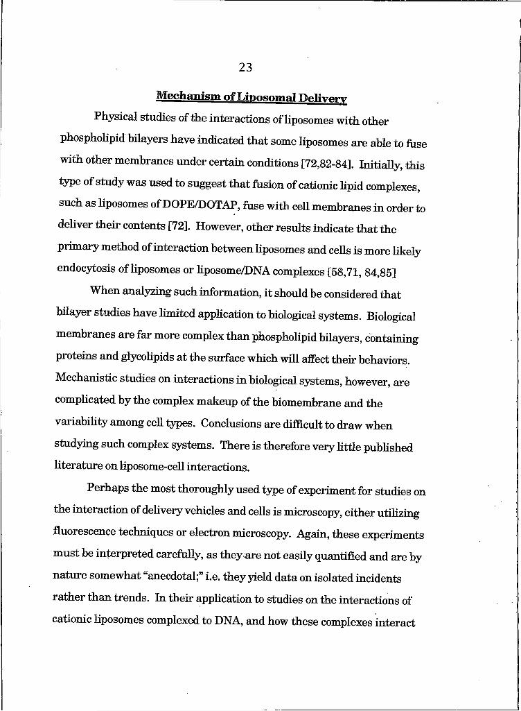
Physical studies of the interactions of liposomes with other
phospholipid bilayers have indicated that some liposomes are able to fuse
with other membranes under certain conditions [72,82-84]. Initially, this
type of study was used to suggest that fusion of cationic lipid complexes,
such as liposomes of DOPE/DOTAP, fuse with cell membranes in order to
deliver their contents [72]. However, other results indicate that the
primary method of interaction between liposomes and cells is more likely
endocytosis of liposomes or liposome/DNA complexes [58,71, 84,85]
When analyzing such information, it should be considered that
bilayer studies have limited application to biological systems. Biological
membranes are far more complex than phospholipid bilayers, containing
proteins and glycolipids at the surface which will affect their behaviors.
Mechanistic studies on interactions in biological systems, however, are
complicated by the complex makeup of the biomembrane and the
variability among cell types. Conclusions are difficult to draw when
studying such complex systems. There is therefore very little published
literature on liposome-cell interactions.
Perhaps the most thoroughly used type of experiment for studies on
the interaction of delivery vehicles and cells is microscopy, either utilizing
fluorescence techniques or electron microscopy. Again, these experiments
must be interpreted carefully, as they ,are not easily quantified and are by
nature somewhat “anecdotal;” i.e. they yield data on isolated incidents
rather than trends. In their application to studies on the interactions of
cationic liposomes complexed to DNA, and how these complexes interact
2 4
with cells, they indicate consistently that the primary mechanism of
interaction with cells is via endocytosis of the IiposomeZDNA complex [80,
86]. How the DNA then gets delivered to the nucleus for transcription is
not understood, but exit from the endosome into the cytoplasm may be the
limiting factor in transfection efficiency, rather than entry into the cell.
The data collected for this study supports the hypothesis that
endocytosis is the primary route of delivery in DNA/liposome complex
mediated transfection, and further suggests that cationic liposomes alone,
without DNA present, interact with cells in a similar way.
The Contents of This Stmdy
It has been the objective of this study to systematically analyze the
mechanism of interaction between a select group of liposomes arid
proteoliposomes with cultured cells, with the applied objective of
determining the feasibility of using such systems for delivering the CFTR
protein to cell membranes.
The types of delivery vehicles studied include primarily the cationic
formulations of DOTAP/DOPE, and DC Cholesterol/DOPE. Also studied, as
alternative approaches, were pH-dependent liposomal vectors composed of
oleic acid and DOPE, and liposomes complexed to a peptide ligand whose
receptor lies on the surface of a target cell;
Recombinant expression of CFTR in a baculoviral system and
purification from that source was performed in an attempt to develop
proteoliposomes carrying that protein, with the goal of transferring the
protein to target cell membranes.
25
Liposomes of various compositions were studied for toxicity and
ability to deliver their lipid and soluble contents to host cells. Fluorescent
analogs of phospholipids and soluble fluorescent markers were traced by
fluorescent microscopy and fluorescence studies utilizing a resonance
energy transfer technique.
Studies have been performed on the use of cationic liposomes as
carriers of membrane proteins to target cells. Specifically, investigations
have been done with the protein Band 3, covalently modified with a
fluorescent tag.
Finally, in an attempt to better understand the potential usefulness
of protein delivery through endocytic vesicles, some experiments have been
performed attempting to identify CFTR within isolated coated endosomes.
This work should have relevance to all who use liposomes for delivery
of DNA to cells, as well as to others who are interested in exploring the
capabilities of liposomes to deliver other compounds to cells.

References Cited
Boat, T.F., Welsh, M.J., and Beaudet, AL., in The M etabolic Basis of Inherited Disease. Scriver, C.L., Beaudet, AL., Sly, W.S., and Valle, D., Eds. (McGraw-Hill, NY, ed. 6) 2649-2680,1989.
Schwab, U.E., Wold, AE., Carson, J.L., Leigh, M.W., Cheng, P.W., Gilligan, P.H., Boat, T.F., “Increased adherence of Staphylococcus aureus from cystic fibrosis lungs to airway epithelial cells,” Am. Rev. Respir. Dis. 148: 365-369, 1993.
Zar, H., Salman, L., Quitted, L, and Prince, A , “Binding of Pseudomonas aeruginosa to respiratory epithelial cells from patients with various mutations in the cystic fibrosis transmembrane regulator,” J . P ed ia tr. 126: 230-233, 1995.
Govan, J.R.W., and Harris, G.S., “Pseudomonas aeruginosa and cystic fibrosis: unusual bacterial adaptation and pathogenesis,” M icrobiological Sci. 3: 302-308, 1986.
Pier, G.B., “Pulmonary disease associated with Pseudomonas aeruginosa in cystic fibrosis: current status of the host-bacterium interaction,” “J . Infect. Dis. 151: 575-580, 1985.
Riordon, J.R., Rommens, J.M., Kerem, B.S., Alon, N., Rozmahel, R., Grzelczak, Z., Zielenski, J., Lok, S., Plavxic, N., Chou, J.L., Drumm, M.L., lannuzzi, M.C., Collins, F.S., and Tsui, L.C., “Identification of the cystic fibrosis gene: cloning and characterization of complementary DNA,” Science 245: 1066-1073, 1989. .
Anderson, M.P., Sheppard, D.N., Berger, HA., and Welsh, M.J., “Chloride channels in the apical membrane of normal and cystic fibrosis airway and intestinal epithelia,” Am. J . PhysioL Lung Cell MoLPhysioL 263: L1-L14, 1992.
Denning, G.M., Ostedgaard, L.S., Cheng, S.H., Smith, AE., and Welsh, M.J., “Localization of cystic fibrosis transmembrane conductance regulator in chloride secretory epithelia,”J .O in Jn v est. 89:339-349, 1992.
Berger, H.A., Travis, S.M., and Welsh, M.J., “Regulation of the cystic fibrosis transmembrane conductance regulator chloride channel by specific protein kinase and protein phosphatases,” J . Biol. C hem . 268: 2037-2047, 1993.
Kartner5 N., Hanrahan5 J.W., Jensen5 T.J., Naismith5 AL., Sun S Ackerley5 C A 5 Keyes5 E-R5 Tsui5 L.C., Kommens5 JJVL5 Bear5 C.E., and Riordan5 J .R., “Expression of the cystic fibrosis gene in non- epithelial invertebrate cells produces a regulated anion conductance * CeU 64: 681-691, 1991.
Gabriel, S.E., Clarke, L.L., Boucher, R.C., and Stutts5 M.J., “CFTR and outward rectifying chloride channels are distinct proteins with a regulatory relationship,” N ature 363: 263-266, 1993.
Reisen51.L., Prat5 AG., Abraham5 E.H., Amara5 J.F., Gregory5 R.J., Ausiello5 D A 5 and Cantiello5 H F., “The cystic fibrosis transmembrane conductance regulator is a dual ATP and chloride channel,” J . Biol. Chem. 269: 20584-20591, 1994.
Reddy5 M., Quinton5 P ., Haws5 C., Wine5 J., Grygorczyk5 R., Tabcharam5 J., “Failure of the cystic fibrosis transmembrane conductance regulator to conduct ATP,” Science 271: 1876-1879 1996.
Ismailov, LL5 Awayda5 M.S., Jovov5 B., Berdiev5 B.K, Fuller, C.M., Dedman5 J.R., Kaetzel5 M A 5 Benos5 D.J., “Regulation of epithelial sodium channels by the cystic fibrosis transmembrane conductance regulator,” J . Biol. Chem. 271: 4725-4732, 1996.
Stutts5 M., Canessa5 C., Olsen, J., Hamrick, M., Cohn5 J., Rossier5 B., and Boucher, R., “CFTR as a cAMP-dependent regulator of sodium channels,” Science 269: 847-850, 1995.
Morris5 AP., Cunningham5 S A 5 Benos5 D.J., and Frizzell, R A 5 “Glycosylation status of endogenous CFTR does not affect cAMP- stimulated chloride secretion in epithelial cells,” Am. J . PhysioL CeU PhysioL 265: C688-C694, 1993.
Drumm5 M.L., Pope5 H A 5 CUff5 W.H., Rommens5 J.M., Marvin5 S A 5 Tsui5 L.C., Collins, F.S., Frizzell, R A 5 and Wilson, J.M., “Correction of the cystic fibrosis defect in vitro by retrovirus-mediated gene transfer,” CeU 62: 1227-1233, 1990.
Rich, D.P., Anderson5 M.P., Gregory5 R.J., Cheng5 S.H., Paul, S., Jefferson5 D.M., McCann, J.D., KUnger5 KW., Smith5 AE., and Welsh, M.J., “Expression of cystic fibrosis transmembrane conductance regulator corrects defective chloride channel regulaiton in cystic fibrosis airway epithelial cells,” N ature 347: 358-363, 1990.
19. Welsh, M.J., and Smith, A.E., “Molecular mechanisms of CFTRchloride channel dysfunction in cystic fibrosis,” Cell 73: 1251-1254,
28
20. Lester, LA., Kraut, J., Lloyd-Still, J., Karrison, T., Mott, C., Billstrand, C., Lemke, A., Ober, C., “Delta F508 genotype does not predict disease severity in an ethnically diverse cystic fibrosispopulation,” P ediatrics 93(1): 114-118, 1994.
21. Halley, D.J., Veeze, H.J., Sandkuyl, LA., Wesby-van. Swaay, E., van Damme, N.H., Deelen, W.H., Witte, J.E., and Niermeijer, M.F., “The mutation delta F508 on Dutch cystic fibrosis chromosomes and frequency and relation to patients’ age at diagnosis,” Hum. Genet. 85(4): 407-408, 1990,
22. Cheng, S.H., Gregoiy, R.J., Marshall, J., Paul, S., souza, D.W., White, GA., OrRiordan, C.R., and Smith, A.E., “Defective intracellular transport and processing of CFTR is the molecular basis of most cystic fibrosis,” Cell 63: 827-834, 1990.
23. Lukacs, G.L., Mohamed, A., Kartner, N., Chang, X.B., Riordan, J.R., and Grinstein, S., “Conformational maturation of CFTR but not its m utant counterpart (AF508) occurs in the endoplasmic reticulum and requires ATP,” EMBO J . 13(24): 6076-6086, 1994.
24. Pasyk, E A., and Foskett, J.K., “Mutant (AF508) cystic fibrosis transmembrane conductance regulator chloride channel is functional when retained in endoplasmic reticulum of mammalian cells,” J . Biol. Chem. 270: 12347-12350, 1995.
25. Li, C., Ramjeesingh, M., Reyes, E., Jensen, T., Chang, X., Rommens, J-M., and Bear, C.E., “The cystic fibrosis mutation (AF508) does not influence the chloride channel activity of CFTR,” N atu re Genet. 3:311-316, 1993.
26. Johnson, L.G., Boyles, S.E., Wilson, J., and Boucher, R.C., “Normalization of raised sodium absorption and raised calcium- mediated chloride secretion by adenovirus-mediated expression of cystic fibrosis transmembrane conductance regulator in primary human cystic fibrosis airway epithelial cells,” J . Clin. Invest. 95: 1377-1382, 1995.
27. Manavalan, P., Smith, A.E., and McPherson, J.M., “Sequence and structural homology among membrane-associated domains of CFTR and certain transporter proteins,” J . P ro te in Chem. 12: 279-290, 1993.
29
28. Wei, L.Y., Stutts, M.J., Hoffinann, M.M., and Roepe, P.D.,Overexpression of the cystic fibrosis transmembrane conductance
regulator in NIH 3T3 cells lowers membrane potential and intracellular pH and confers a multidrug resistance phenotype ” Biophys. J . 69: 883-895, 1995. ’
29. Gabriel, S.E., Clarke, L.L., Boucher, RC., and Stutts, M.J., “CFTR and outward rectifying chloride channels are distinct proteins with a regulatory relationship,” N ature 363: 263-266, 1993.
30. Schweibert, E.M., Egan, M.E., Hwang, TH., fulmer, S.B., Allen, S.S., Cutting, G.R., Guggino, W.B., “CFTR regulates outwardly rectifying chloride channels through an autocrine mechanism involving ATP ” Cell 81: 1063-1073, 1995.
31. Shak, S., “Aerosolized recombinant human DNase I for the treatment of cystic fibrosis,” Chest 107: 65S-70S, 1995.
32. Knowles, M.R., Olivier, K N ., Hohneker, K W., Robinson, J., Bennett, W.D., boucher, R.C., “Pharmacologic treatment of abnormal ion transport in the airway epithelium in cystic fibrosis,” Chest 107" 71S-76S, 1995.
33. Caplen, N.J., Alton, E.W.F.W, Middleton, P.G., Dorin, J.R., Stevenson, B.J., Gao, X., Durham, S.R., Jeffery, P.K, Hodson, M.E., Coutelle, C., Huang, L., Porteous, D.J., Williamson, R., and Geddes, D.M., “Liposome-mediated CFTR gene transfer to the nasal epithelium of patients with cystic fibrosis,” N ature Med. 1:39-46, 1995.
34. Zabner, J., Courure, L.A., Smith, A R , Welsh, M.J., “Correction of cAMP-stimulated fluid secretion in cystic fibrosis airway epithelia: efficiency of adenovirus-mediated gene transfer in vitro,” Hum.Gene Ther. 5: 585-593, 1994.
35. Engelhard!, J.F., Simon, RH., Yang, Y., Zepeda, M., Weber- Pendleton, S., Grossman, M., and Wilson, J.M., “Adenovirus-mediated transfer of the CFTR gene to lung of nonhuman primates: biological efficacy study,” H um an Gene Ther. 4: 759-769, 1993.
36. Yang, Y., Nunes, F A , Berencsi, K , Goenczoel, E., Engelhard!, J.F., Wilson, J.M., “Reactivation of E2a in recombinant adenovirus improves the prospect for gene therapy in cystic fibrosis,” N ature Genet. 7: 362-369, 1994.
Sorsche^Ej;, Logan, J J . , Frizzell, R A , Lyrene, R.K, Bebok, Z.,S r 11S M D ’ Feigner, P.L., Matalon, S., Walker, L.,Wiatrak, B J., Gene therapy for cystic fibrosis using cationic llPosome mediated gene transfer: a phase I trial of safety and 1994 07 m the naSal airway’” Hm n- Gene Ther. 5: 1259-1277,
WilmotLR.W., Whitsett, J A , Trapnell,B., Wert, S .,Baughman,R., ^ apP. ’ Tolstohev, P., “Gene therapy for cystic fibrosisutilizing a replication deficient recombinant adenovirus vector to deliver the human cystic fibrosis transmembrahe conductance regulator cDNA to the airways. A phase I study,” Hum. Gene Ther. 5(8): 1019-1057, 1994.
Boucher R C Knowies, M.R., Johnson, L.G., Olsen, J.C., Pickles, R., Wilson, J.M., Engelhard!, J., Yang, Y., Grossman, M., “Gene therapy tor cystic fibrosis using El-deleted adenovirus: a phase I trial in the nasal cavity. The University of North Carolina at Chapel Hill ” Hum. Gene Ther. 5: 615-639, 1994.
Wilson, J.M., Engelhard!, J.F., Grossman, M., Simon, r.H., and Yang, Y., Gene therapy for cystic fibrosis lung disease using E l deleted adenoviruses: a phase I trial,” Hum. Gene Ther. 5: 501-519, 1994.
Zabner ,J., Couture, L A , Gregory, R.J., Graham, S.M., smith, AE., and Welsh, M.J., “Adenovirs-mediated gene transfer transiently corrects the chloride transport defect in nasal epithelia of patients with cystic fibrosis,” Cell 75: 207-216, 1993.
Schwiebert, E.M., Gesek, F., Ercolani, L., Wjasow, C., Gruenert, D.C., Karlson, K., and Stanton, BA., aHeterotnmeric G proteins, vesicle trafficking, and CFTR chloride channels,” Am. J . PhysioL Cell PhysioL 267: C272-C281, 1994.
Bradbury, N A , Cohn, J.A., Venglarik, C.J., and Bridges, R.J., Biochemical and biophysical identification of cystic fibrosis
transmembrane conductance regulator chloride channels as components of endocytic clathrin-coated vesicles,” J . Biol. Chem. 269” 8296-8302, 1994.
Luzio, J.P., and Banting, G., Eukaryotic membrane traffic: retrieval and retention mechanisms to achieve organelle residence,” Trends in Biochem. Sci. 18: 994-997, 1993.
Sztul, E., Kaplin, A., Saucan, L., Palade, g., “Protein traffic between distinct plasma membrane domains: isolation and characterization
of vesicular carriers involved in transcytosis,” Cell 64: 81-89,1991.
C urren t Protocols in P ro te in Science, “Overview of Cell Fractionation,” Unit 4.1, John Wiley and Sons, 1995.
31
Clary, D.O., Griff, I.C., and Rothman, J.E., “SNAPs, a family of NSF attachment proteins in volved in intracellular membrane fusion in animals and yeast,” Cell 61: 709-721, 1990.
Rothman, J.E., “Mechanisms of intracellular protein transport ” N ature 372: 55-63, 1994. ’
Rothman, J E., “The protein machinery of vesicle buddig and fusion ” Protein Sci. 5: 185-194, 1996.
Gordon, C.M., and Uoyd, K.O., “Endocytosis and recycling of gangliosides in a human malanoma cell line: inhibitory effect of brefeldin A and monensin,” Arch. Biochem. Biophys, 315(2): 339-
Guy, J., Drabek, D., and Antoniou, M., “Delivery of DNA into mammalian cells by receptor-mediated endocytosis and gene therapy,” Mol-BiotechnoL 3(3): 237-248, 1995
Farhood, H., Serbina, N., and Huang, L., “The role of dioleoylphosphatidylethanolamme in cationic liposome mediated gene transfer,” Biochim. Biophys. Acta Bio-memhr. 1235: 289-295,
Dunn, KW., Park, J., Semrad, GE., Gelman, D.L., Shevell, T., and McGraw, T.E., Regulation of endocytic trafficking and acidification are independent of the cystic fibrosis transmembrane conductance regulator,” J . Biol. Chem. 269: 5336-5345, 1994.
Biwersi, J., and Verkman, AS., “Functional CFTR in endosomal compartment of CFTR-expressing fibroblasts and T84 cells ”Am. J . PhysioL 266:C149-C156, 1994.
Prince, L.S., Workman, RB., Jr., and Marchase, RB., “Rapid endocytosis of the cystic fibrosis transmembrane conductance regulator chloride channel,” Proc. NatL Acad. Sci. USA 91: 5192- 5196, 1994.
Root, KV., Engelhardt, J.F., Post, M., Wilson, J.M., and VanDyke,R W., CFTR does not Alter Acidification of L cell Endosomes ” Biochem. Biophys. Res. Comm. 205: 396-401, 1994.
Darnell, J., Lodish, H., and Baltimore, D., Molecular Cell Biology (W.H. Freeman and Company, 2nd ed.), New York, 1990.
??kner,J., Fasbender, A J ., Momnger, T., Poellinger, K A , and Welsh, ,aiJ ind moIecular barriers to gene transfer by a cationic
bpid, J . BioL Chem. 270(32): 18997-19007, 1995.
Hug^ P., and Sleight, R.G., “Fusogenic virosomes prepared by partitioning of vesicular stomatitis virus G protein into preformed vesicles,” J . Biol. Chem. 269: 4050-4056, 1994.
Bron, R , Ortiz, A , Dijkstra, J., Stegmann, T., and Wilschut, J., Preparation, properties, and apphcations of reconstituted influenza
virus envelopes (virosomes),” M ethods EnzymoL 222: 313-331,
De Fiebre, C.M., Wu, P., Notabartolo, D., Millard, W.J., and Meyer, E.M., Differential adenoassociated virus vector-driven expression of a neuropeptide Y gene in primary rat brain astroglial cultures after transfection with Sendai virosomes versus Lipofectin,” N eurochem ical Res. 19(6): 643-648, 1994.
Schoch, C., and Blumenthal, R , “Role of the fusion peptide sequence in initial stages of influenza hemagglutinin-induced cell fusion,” J . BioL Chem. 268: 9267-9274, 1993.
Alford, D., Ellens, H., and Bentz, J ., “Fusion of influenza virus with sialic acid-bearing target membranes,” B iochem istry 33: 1977- 1987, 1994.
Stewart, M.J., Plautz, G.E., del Buono, L., Yang, Z.Y., Xu, L., Gao, X., Huang, L., Nabel, E.G., and Nabel, G.J., “Gene transfer in vivo with DNA-Iiposome complexes: Safety and acute toxicity in mice,” Hum.Gene Ther. 3: 267-275, 1992.
Legendre, J.Y., and Szoka, F.C., “Cyclic amphipathic peptide-DNA complexes mediate high-efficience transfection of adherent mammalian cells,” Proc. NatL Acad. Sci. USA 90(3): 893-897,
Stnblmg, R , Brunette, E., Liggitt, D., Gaensler, K , and Debs, R , “Aerosol gene delivery in vivo” Proc. NatL Acad. Sci. USA 89: 11277-11281, 1992.
Simon, R.H;, Engelhardt, J.F., Yang, Y., Zepeda, M., Weber- Pendleton S Grossman, M., and Wilson, J.M., “Adenovirus-mediated transfer of the CFTR gene to lung of nonhuman primates: toxicity study, Hum. Gene Ther. 4: 771-780, 1993.
Nabel F.G Gordon, D., Yang, Z.Y., Xu, L., San, H., Plautz, G.e., Wu,tvG'a v 0’ Huang’ k., and Nabel, G.J., “Gene transfer in vivo with UNA-Iiposome complexes: lack of autoimmunity and gonadal localization,” Hum. Gene Ther. 3: 649-656, 1992.
Wdkmson, D A , and Nagle, J.F., “Metastability in the phase behavior of dimyristoylphosphatidylethanolamine bilayers ” Biochem istry 23: 1538-1541, 1984. ’
r eTfj^is ’ an^ Silvius, J.R., “Interactions of mammalian cells withiipicl dispersions containing novel metabolizable cationic amphiphiles,” BiochimJBiophysActa 1023: 124-132, 1990.
Duzgtines, N., Golstein, J., Friend, D., and Feigner, P., ’’Fusion of iyofomes containing a novel cationic lipid, N-[2,3-(dioleoxy)-propyl]- N,N,N-trimethylammonium: induction by mustivalent anions and 9279^184 ^989 ^ th PhosPholiPid vesicles,” Biochem istry 28:
Goyal, K , and Huang, L., “Gene therapy using DC-Chol liposomes,”J . Liposome Res. 5: 49-61,1995.
Natasha Caplen, personal communication.
Sells, M A , Li, J., and Chemoff, J., “Delivery of protein into cells using polycationic liposomes,” BioTechniques 19: 72-78, 1995.
Guide to E ukaryotic Transfections with Catirvnin Linid Reagents. Life Technologies, Gaithersburg, MD
SchenbomvE., Oler, J., Goiffon, V., Balasubramaniam, R , Bennett, M.J., Aberle, AM., Nantz, M.H., and Malone, R.W., “Transfection: Tfx-50 Reagent: a new transfection reagent for eukaryotic cells ” Prom ega Notes Magazine, number 52: 2-7,1995'
Hawley-Nelson, P., Ciccarone5 V., Gebeyehu5 G., and Jessee J., -Lipoiectamine Reagent: a new, higher efficiency polycationic
liposome transfection reagent,” Focus 15(3): 73-79, 1993.
^Grnholz5 E., Bantle, E., Weigert, M., Borowski, E., v.d. Eltz5 H., and Hinzpeter5 M., “DOSPER liposomal transfection reagent: a reagent with unique transfection properties ” Biochem ica No. 2, 7-10,1996.
34
Wrobel51., and ColHns5 D ., “Fusion of cationic liposom es with mammalian cells occurs after endocytosis,” Biochhn. Biophys. A cta Bio-membr. 1235: 296-304, 1995.
Egilmez5 N.K., Iwanuma5 Y., and Bankert5 R.B., “Evaluation and optimization of different cationic liposome formulations for in vivo gene transfer,” Biochem. Biophys. Res. Comm. 221: 169-173,
Zellmer5 S., Cevc5 G., and Risse5 P., “Temperature- and pH-controlled fusion between complex lipid membranes. Examples with the diacylphosphatidylchoHne/fatty acid mixed Hposomes,” Biochhn. Biophys. Acta Bio-membr. 1196(2): 101-113, 1994.
Kagiwada, S., Murata5 M., Hishida5 R., Tagaya5 M., Yamashina5 S., and Ohmshi5 S., uIn vitro fusion of rabbit Hver golgi membranes with liposomes,” J . Biol. Chem. 268: 1430-1435, 1988.
Stamatatos5 L., Leyentis5 R., Zuckerman5 M.J., and Silvius5 J.R
3917-3925, 1988.
Gershon5 H., Ghirlando5 R., Guttman5 S.B., and Minsky5 A., “Mode of formation and structural features of DNA-cationic Hposome complexes used for transfection,” Biochem istry 32: 7143-7151 1993.
Fnend5D1S., Papahadjopoulos, D., and Debs5 R.J., “Endocytosis and intracellular processing accompanying transfection mediated by cationic Hposomes,” Biochhn. Biophys. Acta Bio-membr. 1278- 41-50, 1996.
35.
CHAPTER 2
CFTR PROTEIN EXPRESSION AND PURIFICATION
Introduction and Motivation
Expression of CFTR was originally performed from recombinant
Chinese Hamster Ovary (CHO) cells, but with low yield [I]. The ability to
produce more protein was facilitated by production of the protein in
recombinant Spodoptera frugiperda (Sf9) cultures transformed with
baculovirus [2]. Production of quantities of protein adequate for the
manufacture of proteoliposomes containing CFTR required use of this
system. Additionally, the single available CFTR purification scheme,
published by Bear, et al. in 1992 utilized this source of protein [3].
The goals of this study required a source of protein which was pure
and at high enough concentration to make reconstitution into liposomes
possible. The method developed by Bear resulted in pure preparations of
protein at a yield of 0.5 mg protein per liter of transformed cells;
significantly higher than yields from any other available source. It was
hoped that purified CFTR could be incorporated into liposomes and used for
studies of the feasibility of delivering the protein to cultured cells with
liposomes.
36
Materials and Methods
Protein Expression in the Baculoviral System
The baculoviral system of protein expression was developed in the
late 1980s, and provides a relatively simple and efficient way to produce
soluble and membrane proteins from a eukaryotic source [4].
The host cell line,Sf9, is a clonal isolate from Spodoptera frugiperda.
These insect cells fold and transport proteins similarly to human cell lines,
and glycosylate them, albeit incompletely. Foreign genes are introduced
into these cells through the Autographica califomica nuclear polyhedrosis
virus (AcMNPV). Production of recombinant virus is accomplished simply
by cointroduction of wild type AcMNPV and purified plasmid DNA into Sf9
cells during log phase growth. Insertion of the plasmid sequence into the
viral genome disrupts the viral polyhedrin protein production. Detection of
recombinants is possible because these viral plaques do not contain the
large quantities of refractile polyhedrin protein that wild type plaques
contain. Purified recombinant virus harvested from the plaques is used to
infect large cultures of Sf9 cells to produce the protein of interest [5],
A 4.7 kB Pst I fragment containing the cftr cDNA was cut from a
plasmid (supplied by Dr. F. Collins at the National Institues of Health), and
was ligated into the baculoviral transfer vector pVL1393 (available from
Invitrogen). This plasmid was then amplified in E. Coli, and plasmid DNA
purified. Purified plasmid DNA and wild type AcMNPV were co-transfected
into log-phase Sf9 cells using cationic liposomes. Recombinant viral
plaques were identified by analyzing them visually for the absence of
polyhedrin occlusion bodies. Identification was aided by using a dissecting
microscope (40x) and a point source of light to highlight refractive
polyhedrin occlusion-positive wild type plaques. Identification of occlusion
body negative plaques was difficult at first, because the absence of
occlusion bodies is difficult to distinguish visually. Suspected recombinant
virus was used to infect fresh cultures of Sf9 cells. Several putative
recombinants which continued to produce occlusion body negative plaques
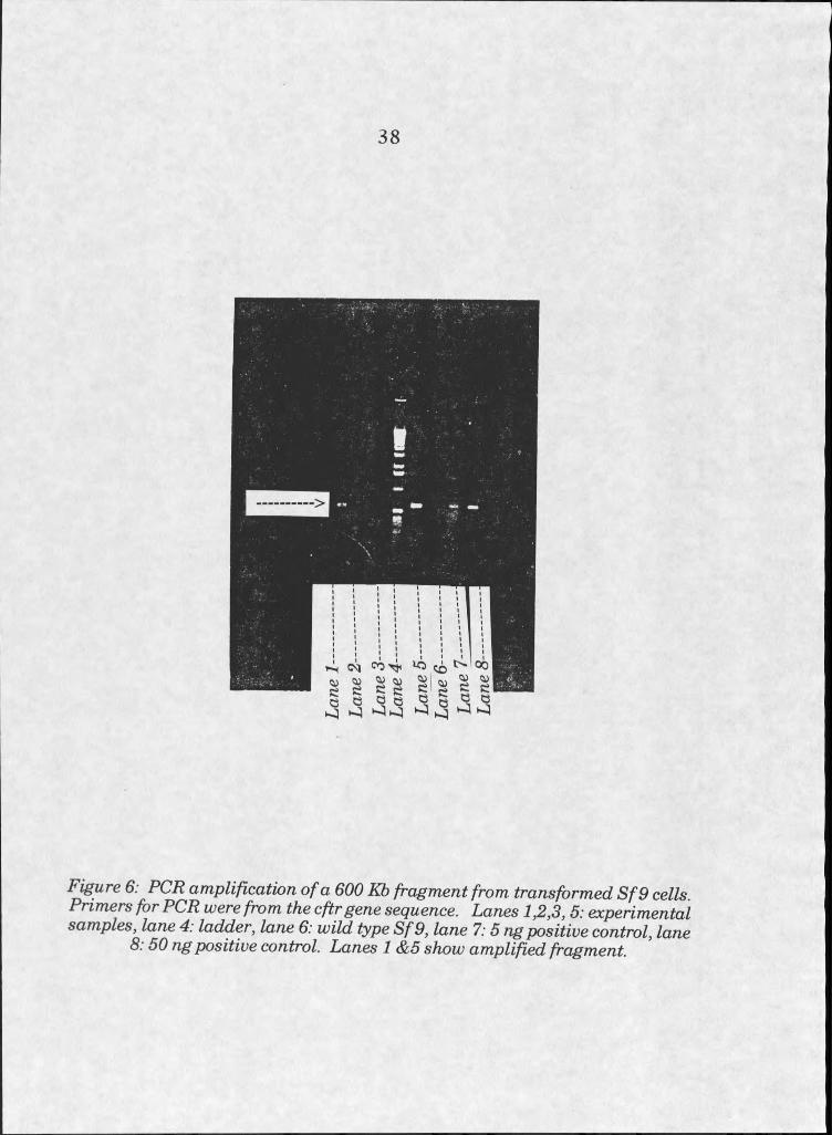
were analyzed for presence of the cftr DNA insert by PCR amplification of
part of the cftr sequence (see figure 6).
This viral stock was used to infect more cells to prepare high volume
viral stock solutions. Virus titer was determined by plaque assay to be 4.5
XlO^ plaque forming units (pfu) /mL. Both the ratio of virus to cells
' (multiplicity of infection, or MOI) and incubation time were analyzed to
optimize production of recombinant protein. Optimum MOI for expression
of CFTR was determined to be 5.0. Optimum incubation time, between
viral inoculation and harvest, was determined to be 48-72 hours.
Suspension cultures of Sf9 cells were then infected with recombinant
virus, and were assayed for production of CFTR by SDS-PAGE and
Western blotting. Coomassie Brilliant Blue staining did not reveal large
quantities of overexpressed protein at the expected molecular weight, but
Western blots detected an immunoreactive protein (to mouse anti-human
CFTR, Genzyme product number 1660-01) of apparent molecular weight
130 kD. This size is compatible with reports of the molecular weight of
CFTR isolated from Sf9 cells [3], The apparent molecular weight of
37
38
Figure 6: PCR amplification o f a 600 Kb fragment from transformed S f 9 cells Primers for PCR were from the cftrgene sequence. Lanes 1,2,3, 5: experimental samples, lane 4: ladder, lane 6: wild type S f 9, lane 7:5 ng positive control, lane
8:50 ng positive control. Lanes I &5 show amplified fragment.
CFTR appears unusually low when compared with its sequence predicted
weight, however other members of the ABC superfamily of proteins run at
apparent molecular weights well under their sequence predicted molecular
weights, as well.
CFTR Purification
Purification of recombinant CFTR, accomplished primarily by the
efforts of Steve Swain, involved modification of the work of Bear et aL,
published in 1992. This paper reported the expression, purification, and
reconstitution of functional protein into liposomes [3]. The applied goal of
the proteoliposome project was related insofar as it required production and
purification of the protein, and reconstitution into a bilayer. It was
therefore natural to draw heavily from the published method. Some
modifications were made to optimize production of pure, functional protein
in our hands.
The purification scheme consists of two general phases: first,
isolation of integral membrane proteins from cell lysates, and second,
isolation of CFTR from other integral membrane proteins.
The first phase of the purification involves cell lysis, membrane
isolation by centrifugation, and an alkali wash to remove peripheral
membrane proteins. Membranes are then disrupted by detergent; in this
case sodium dodecyl sulfate (SDS) is used to solubilize CFTR.
Monolayer cultures of SfS cells were used for protein because yields
appeared somewhat better than from susupension cultures [6].
Approximately 5 x IO^ infected cells were harvested from monolayer
39
cultures 48-56 hours after infection, and spun 10 minutes at 800 x g, 4-
10°C. Supernatant was discarded, cells were resuspended in cold PBS, and
spun as before. After removing the supernatant, cells were lysed in 5 ml
hypotonic buffer per approximate ml of cells: IOmM PIPES with 10 mM
NaCl and 5 mM KC1, pH 6.4, supplemented immediately before use with
protease inhibitors leupeptin (10 ng per ml buffer from a 5 mg/ml stock),
aprotinin (10 ^g per ml buffer from a 5 mg/ml stock), and PMSF (IOpl per
ml buffer of a 10 mg/ml stock in ethanol). Lysed cells were then treated for
I hour at room temperature with 200 |xl of a 2.5 mg/ml stock solution of
DNase, mixing occasionally. After spinning 40 minutes a t 100,000 x g, the
membrane pellet was extracted with cold alkali (10 mM NaOH with 0.1 mM
EDTA) to remove peripheral membrane proteins. Centrifugation at
100,000 x g pelleted the remaining membrane proteins.
Integral membrane proteins were then solubilized in SDS with
dithiothreitol, at room temperature. The extraction buffer contained 10
mM Na2HP04, 2% SDS, and 5 mM DTT, pH 6.4. Pellets were resuspended
in the buffer and incubated 40-60 minutes, with periodic resuspension.
Samples were spun at 100,000 x g for 40 minutes at IO0C. Supernatants
were collected for chromatographic separation.
The second phase of the purification involves separation of CFTR
from other membrane proteins. This was accomplished by two sequential
column chromatography steps. The first involved the use of a matrix
coated with hydroxyapatite, and the second was size exclusion
chromatography.
40
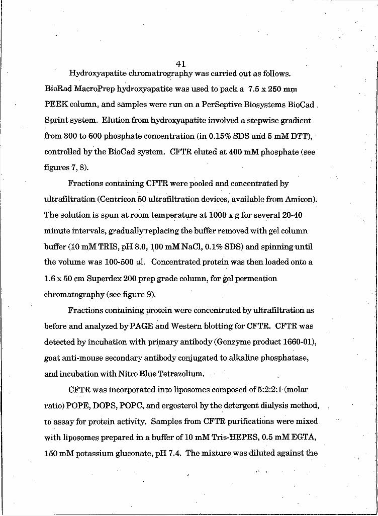
Hydroxyapatite chromatrography was carried out as follows.
BioRad MacroPrep hydroxyapatite was used to pack a 7.5 x 250 mm
PEEK column, and samples were run on a PerSeptive Biosystems BioCad
Sprint system. Elution from hydroxyapatite involved a stepwise gradient
from 300 to 600 phosphate concentration (in 0.15% SDS arid 5 mM DTT),
controlled by the BioCad system. CFTR eluted at 400 mM phosphate (see
figures 7,8).
Fractions containing CFTR were pooled and concentrated by
ultrafiltration (Centricon 50 ultrafiltration devices, available from Amicon).
The solution is spun at room temperature at 1000 x g for several 20-40
minute intervals, gradually replacing the buffer removed with gel column
buffer (10 mM TRIS, pH 8.0,100 mM NaCl, 0.1% SDS) and spinning until
the volume was 100-500 [xl. Concentrated protein was then loaded onto a
1.6 x 50 cm Superdex 200 prep grade column, for gel permeation
chromatography (see figure 9).
Fractions containing protein were concentrated by ultrafiltration as
before and analyzed by PAGE and Western blotting for CFTR. CFTR was
detected by incubation with primary antibody (Genzyme product 1660-01),
goat anti-mouse secondary antibody conjugated to alkaline phosphatase,
and incubation with Nitro Blue Tetrazolium.
CFTR was incorporated into liposomes composed of 5:2:2:1 (molar
ratio) POPE, DOPS, POPC, and ergosterol by the detergent dialysis method,
to assay for protein activity. Samples from CFTR purifications were mixed
with liposomes prepared in a buffer of 10 mM Tris-HEPES, 0.5 mM EGTA,
150 mM potassium gluconate, pH 7.4. The mixture was diluted against the
41
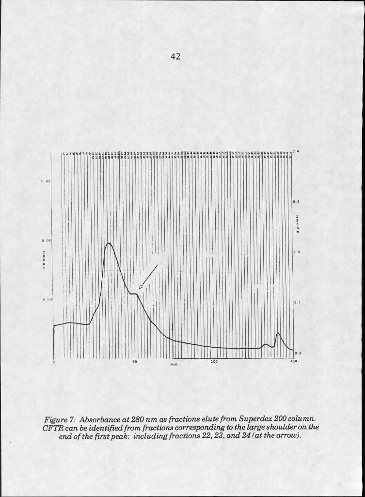
42
o Ol
Figure 7: Absorbance at 280 nm as fractions elute from Superdex 200 column. CFTR can be identified from fractions corresponding to the large shoulder on the
end o f the first peak: including fractions 22,23, and 24 (at the arrow).
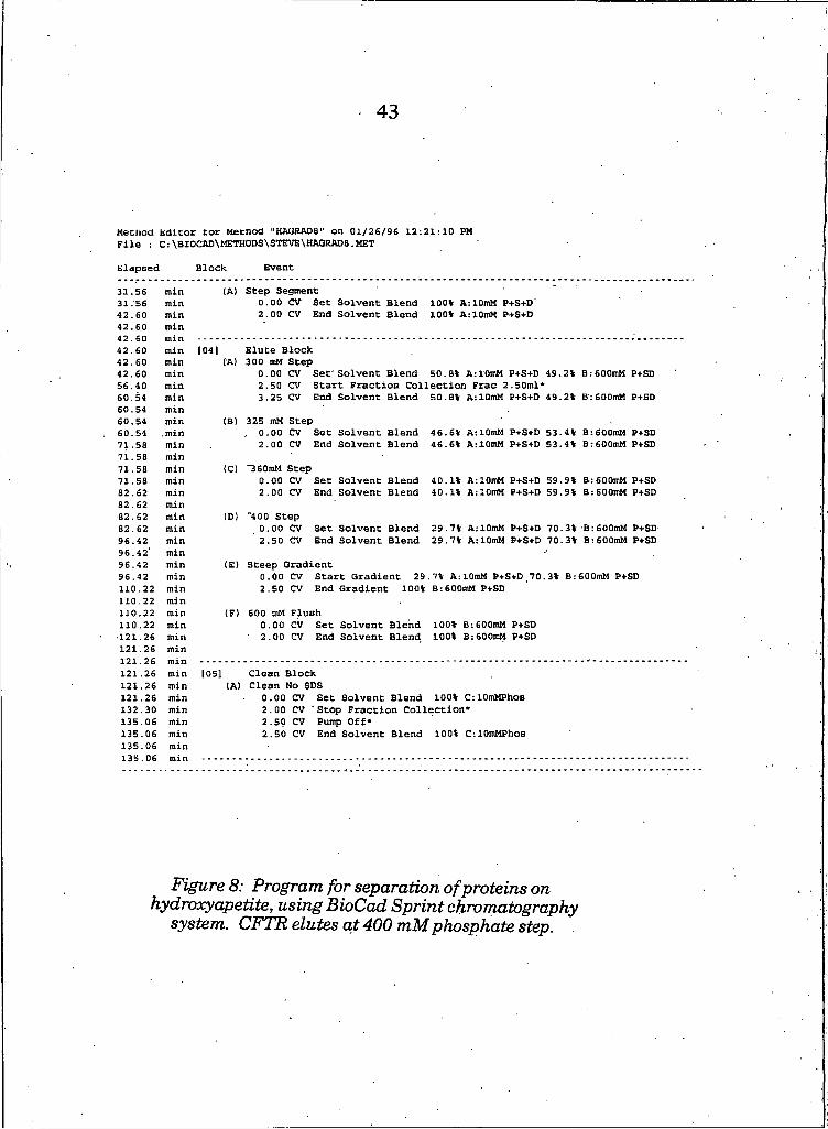
43
Meciiod Editor tor Mecnod "HAtiRADS" on 01/26/96 12:21:10 PM File : C:\BI0CAD\METH0DS\STEVE\HAGRAD8.MET
Elapsed Block Event
31.56 min (A) Step Segment31.56 min 0.00 CV Set Solvent Blend 100% A: IOmM P+S+D"42.60 min 2.00 CV End Solvent Blend 100% AilOmM P+S+D42.60 min
42.60 min 104] Elute Block42.60 min CA) 300 mM Step42.60 min 0.00 CV Set" Solvent Blend 50.8% AilOmM P+S+D 49.2% B:600mM P+SD56.40 min 2.50 CV Start Fraction Collection Frac 2.50ml*60.54 min 3.25 CV End Solvent Blend 50.8% AilOmM P+S+D 49.2% B:600mM P+SD60.54 min60.54 min (B) 325 mM Step60.54 .min , 0.00 CV Set Solvent Blend 46.6% AilOmM P+S+D 53.4% B : 6OOmM P+SD71.58 min 2.00 CV End Solvent Blend 46.6% AilOmM P+S+D 53.4% B:600mM P+SD71.58 min71.58 min (Cl "360mM Step71.58 min 0.00 CV Set Solvent Blend 40.1% AilOmM P+S+D 59.9% B:6OOmM P+SD82.62 min 2.00 CV End Solvent Blend 40:1% AilOmM P+S+D 59.9% B :6OOmM P+SD82.62 min82.62 min (D) "400 Step82.62 min 0.00 CV Set Solvent Blend 29.7% AilOmM P+S+D 70.3% B :6OOmM P+SD96.42 min 2.50 CV End Solvent Blend 29.7% AilOmM P+S+D 70.3% B:600mM P+SD96.42' min96.42 min (E) Steep Gradient96,42 min 0.00 CV Start Gradient 29 .7% AilOmM P+S+D ,70 .3% B : 6d0mM P+SD110.22 min 2.50 CV End Gradient 100% B : 600mM P+SD110.22 min110.22 min (F) 600 mM Flush110.22 min 0.00 CV Set Solvent Blend 100% B :6OOmM P+SD121.26 min 2.00 CV End Solvent Blend 100% B:600mM P+SD121.26 min
121.26 min 105] Clean Block121.26 min (A) Clean No SDS121.26 min 0.00 CV Set Solvent Blend 100% C : IOmMPhoa132.30 min 2.00 CV Stop Fraction Collection*135.06 min 2.50 CV Pump Off*135.06 min 2.50 CV End Solvent Blend 100% C:10mMPhoB135.06135.06
minmin
Figure 8: Program for separation o f proteins on hydroxyapetite, using BioCad Sprint chromatography
system. CFTR elutes at 400 mM phosphate step.
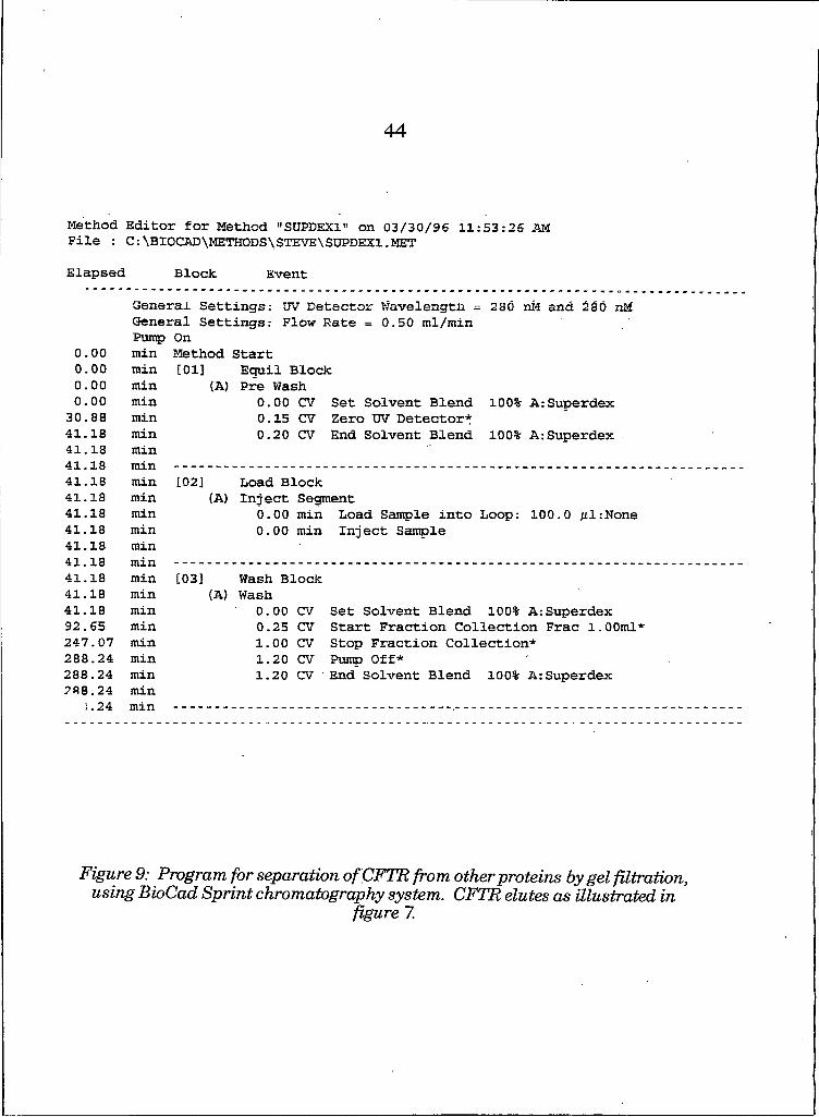
4 4
M e t h o d E d i t o r f o r M e t h o d " S U P D E X l " o n 0 3 / 3 0 / 9 6 1 1 : 5 3 : 2 6 AM F i l e : C : \ B X 0 C A D \ M E T H 0 D S \ S T E V E \ S U P D E X 1 . M E T
E l a p s e d B l o c k E v e n t
G e n e r a l S e t t i n g s : UV D e t e c t o r W a v e l e n g t h = 2 8 0 n M a n d OSO n M G e n e r a l S e t t i n g s : F l o w R a t e = 0 . 5 0 m l / m i n P u m p O n
0 . 0 00 . 0 00 . 0 00 . 0 0
3 0 . 8 84 1 . 1 84 1 . 1 8 4 1 I F
m i nm i nm i nm i nm i nm i nm i n
M e t h o d[ 0 1 ]
(A )
S t a r tE q u i l B l o c k P r e W a s h
0 . 0 0 C V S e t S o l v e n t B l e n d 0 . 1 5 C V Z e r o U V D e t e c t o r * 0 . 2 0 C V E n d S o l v e n t B l e n d
1 0 0 % A : S u p e r d e x
1 0 0 % A : S u p e r d e x .
4 1 . 1 8 m i n [ 0 2 ] L o a d B l o c k4 1 . 1 8 m i n (A ) I n j e c t S e g m e n t4 1 . 1 8 m i n 0 . 0 0 m i n L o a d S a m p l e i n t o L o o p : 1 0 0 . 0 f i l : N o n e4 1 . 1 8 m i n 0 . 0 0 m i n I n j e c t S a m p l e4 1 . 1 8 m i n4 1 1 84 1 . 1 8 m i n [ 0 3 ] W a s h B l o c k4 1 . 1 8 m i n (A ) W a s h4 1 . 1 8 m i n 0 . 0 0 CV S e t S o l v e n t B l e n d 1 0 0 % A : S u p e r d e x9 2 . 6 5 m i n 0 . 2 5 C V S t a r t F r a c t i o n C o l l e c t i o n F r a c 1 . 0 0 m l *2 4 7 . 0 7 m i n 1 . 0 0 C V S t o p F r a c t i o n C o l l e c t i o n *2 8 8 . 2 4 m i n 1 . 2 0 C V P u m p O f f *2 8 8 . 2 4 m i n 1 . 2 0 CV E n d S o l v e n t B l e n d 1 0 0 % A : S u p e r d e x2 8 8 . 2 4 m i n
1 . 2 4 m i n
Figure 9: Program for separation of CFTR from other proteins by gel filtration, using BioCad Sprint chromatography system. CFTRelutesasillustratedin
figure I.
same buffer, plus 1.5% sodium chelate, for I day. Dialysis was carried out
an additional 3 days against Tris-HEPES buffer without additional
detergent.
The fluorophore, 6-methoxy-N-(3-sulfopropyl)quinolinium (SPQ),was
then added to liposomes by adding a concentrated aliquot of SPQ to the
liposome sample, freezing the sample in liquid nitrogen, thawing, and
sonicating for 2 seconds. ExtraHposomal fluorophore was then removed by
passing the liposome solution over a Sepharose CL-6B gel permeation
column, watching for elution of the yellow-colored SPQ loaded Hposomes.
SPQ is a fluorescent molecule which is quenched in the presence of
halide ions. Because CFTR is an ion channel, the degree of SPQ quenching
can be used to monitor changes in the hahde concentration brought about
by CFTR activity [19-25]. Fluorescence measurements were taken on
samples containing CFTR proteohpOsomes, loaded with SPQ and
surrounded by 50 mM KC1, before and after stimulating the channel to
open. Channel opening was stimulated by incubating with MgATP and 250
U/ml Protein Kinase A, to phosphorylate CFTR. SPQ fluorescence was
monitored as MgATP and Protein Kinase were added, and upon lysis of
liposomes with detergent to expose all the SPQ to halide ions.
Results
Recombinant CFTR yields ranged from 20-100 per purification
run (estimated value from silver stained polyacrylamide gels), starting with
5 x 10⁸ cells, grown by monolayer culture in glass bottles. Protein ran as a
single band at a molecular weight of 130 kD on polyacrylamide gels,
45
detected by silver staining. Western blots revealed a single band
immunoreactive for anti-CFTR antibody at the same apparent molecular
weight (see figure 10).
Production of cationic liposomes containing CFTR for analysis of
their interactions with cells could not be carried out, because CFTR
solutions were too dilute for cell treatments (from 2 x 10⁻¹⁰ to I x 10"0
mol/ml), and too little CFTR was available. To create a 1:1000 mole ratio of
protein to lipid, standard volumes for treatment would have to be increased
nearly ten times for normal dosages. Two to five entire purification runs
(necessitating freezing and thawing purified protein) would be required to
produce enough protein to manufacture proteoliposomes adequate for one
complete cell treatment experiment, conducted in an 8 well chamber slide.
Dialysis, necessary for removal of detergent from CFTR solutions, would
likelyresult in even further dilution.
Discussion
Production of recombinant CFTR in the cultured SfB cell line
produced similar amounts of protein to that reported by Bear et al.. Using
this system we were able to achieve reproducible separations and produce
pure CFTR. However, yields remained too low to produce enough pure
protein to perform experiments on cells with CFTR proteoliposomes.
CFTR incorporation into non-fiisogenic liposomes provided evidence
that purified CFTR would incorporate into a phospholipid bilayer. Western
blots revealed the presence of immunoreactive protein in dialyzed
46
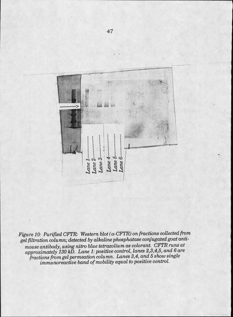
47
imA
yb
III HI
' ' / C S
Figure 10: Purified CFTR: Western blot (a-CFTR) on fractions collected from gel filtration column; detected by alkaline phosphatase conjugated goat anti
mouse antibody, using nitro blue tetrazolium as colorant. CFvTR runs at approximately 130 kD. Lane I: positive control, lanes 2,3,4,5, and 6 are
fractions from gel permeation column. Lanes 3,4, and 5 show single immunoreactive band o f mobility equal to positive control.
liposomes. Assay for protein activity, by the fluorescent SPQ method
[22,23], gave no indication that the purified CFTR was able to channel ions.
48
49References Cited
1. Gregory, R.J., Cheng, S.H., Rich, D.P., Marshall, J., Paul, S., Hehir, K , Ostedgaard, L., Klinger, KW., Welsh, M.J., and Smith, AE., “Expression and characterization of the cystic fibrosis transmembrane conductance regulator,” N ature 347: 382-386,1990.
2. Kartner, N., Hanrahan, J.W., Jensen, T.J., Naismith, AL., Sun, S., Ackerley, C A , Keyes, E.F., Tsui, L.C., Rommens, J.M., Bear, C.E., and Riordan, J.R., “Expression of the cystic fibrosis gene in non- epithelial invertebrate cells produces a regulated anion conductance,” Cell 64: 681-691, 1991.
3. Bear, C.E., Li, C., Kartner, N., Bridges, R.J., Jensen, T.J., Ramjeesingh, M., and Riordan, J.R., “Purification and functional reconstitution of the cystic fibrosis transmembrane conductance regulator (CFTR),” Cell 68: 809-818, 1992.
4. Summers, M.D., and Smith, G.E., A m anual of m ethods for baculoviral vectors an d insect cell cu ltu re procedures. Texas Agricultural Research Station Bulletin number 1555,1987.
5. Ma^Kac Baculoviral Expression System version I .fit A m anual of m ethods for baculoviral vectors an d insect cell cu lture procedures. Invitrogen Corporation, San Diego, CA, 1992.
6 . Steve Swain, personal communication.
I
50
CHAPTERS
INTERACTIONS OF LIPOSOMES WITH CULTURED CELLS
Introduction and Motivatioii
Studies of liposome transfection systems tend to focus on the
application of such systems to produce changes in cell physiology. They
often do not include systematic study of the mechanism of interaction
between the liposome formulation, the DNA used, and the cells affected.
Primarily, studies on efficiency of delivery to cells rely on the expression of
reporter genes in target cells, reporting on the number of transfected cells
but not on the mechanism of DNA entry. Studies of the transfection
mechanism may remain unpublished because of the inherent complexity of
cell systems.
Our objective was to gain insight on the mechanism of interaction
between liposomes and cells through a systematic study. Cationic
liposomes, because of their utility as transfection agents became the focal
point of this investigation. It was hoped that the use of fluorescence
methods could help elucidate the nature of transfection events, in addition
to determining the possibility of using such vectors for transfer of
membrane proteins to the plasma membranes of cultured cells.
51Materials and Methods
Liposome Manufacture
Liposomes can be subdivided into several groups of varying
characteristics. One type of division is between liposomes containing
several layers of phospholipid, called multi-lamellar vesicles, and the single
bilayer, unilamellar vesicles. Another characteristic defining liposome type
is size. Most suitable for the application of delivery are small nnilamollar
vesicles (SUVs) of mean diameter less than 100 nm, which are more apt to
fuse because of their high degree of membrane curvature [I].
Liposomes can be produced by one of several methods, but the
general course of the procedures is similar in all cases. First, lipids must be
mixed in the proper proportion from stock solutions in chloroform. Next, the
solvent is removed by drying down under an argon stream and under
vacuum, and lipids are rehydrated in aqueous solution. Because the
materials used naturally form bilayers in aqueous solution due to
hydrophobic interactions, formation of the bilayer structure is spontaneous
[I]. Formation into spheres of certain specifications, however, is controlled
at this point by treatment of the aqueous solution by sonication, extrusion,
or other methods. The method of choice is determined by the specific
characteristics desired. Sonication of aqueous solutions of phospholipids
produces a fairly homogeneous population of SUVs, ranging in size from 20-
200 nm [1,2]. Extrusion produces a more homogeneous population of
vesicles, but their diameters tend to be larger.

To make SUVs by sonication, solutions of stock lipids in chloroform
were measured and transferred to a glass test tube with a glass capillary
positive displacement pipettor. Chloroform solutions containing the proper
mix of lipid were dried by evaporation of most solvent under a stream of
argon, followed by drying the sample for 15 minutes under vacuum. Buffer
(10 mM HEPES, 150 mM NaCl) was added to rehydrate the sample, aided
by vortexing to remove the lipid from the surface of the test tube. The
sample was then sonicated in a Heat Systems-Ultrasonics, Inc. cup horn
sonicator (model W-385), surrounded by an ice bath, for two minutes at a
50% duty cycle and a power setting of 3. Contents were transferred to a
microfuge tube and sonication was continued for 10 additional minutes.
Preparations were translucent, and colorless except when colored labels
were included.
In experiments requiring the encapsulation of hydrophilic fluorescent
labels, fluorophore (calcein in nearly all experiments, see figure 12, page 65)
at self-quenching concentrations (10 mM for calcein) was included in the
buffer solution used to rehydrate the lipids after removal of organic solvent.
After rehydration and sonication, fluorophore which was not included in the
internal space of the vesicle had to be separated from the liposome solution.
This was accomplished by passing the formed liposomes over a short (3 cm
x 0.5 cm) column packed with the size exclusion matrix Sephadex G 50-50
(available from Sigma).
Sephadex was rehydrated from powder by boiling 30 minutes in
buffer. Spin columns containing the matrix were prepared by packing 3 ml
syringes, plugged at the end with glass wool, with a slurry of rehydrated
52

matrix in buffer. Once filled to the rim by gravity flow, the columns were
placed into a 13 x 100 mm test tube to catch the eluent and spun for 3
minutes at 600 x g to remove excess liquid. After transfer of the column to
a clean test tube, samples of 600 pi liposomes were loaded onto the top of
the column, followed by another 3 minute spin. Several additional loadings
of buffer (600 pi each) were then run through to elute the liposomes.
Separation was measured by examining eluents over a 360 nm
ultraviolet light source and evaluating fluorescence of the fractions.
Liposomes appeared to elute in the first two washes. Free label remained
on the column until after 5 fractions had washed through.
Columns were not regenerated, but were disposed of after use.
Cationic liposome preparations were found to be unstable,
precipitating or visibly aggregating in the presence of the hydrophilic label,
calcein, and are reportedly unstable in the presence of many other divalent
anions (John Silvius, personal communication).
An alternative method used for formation of SUVs was by extrusion
through polycarbonate membranes. This procedure requires an extrusion
device in which the sample was forced back and forth through the filter by
hand from a Hamilton syringe on one side of the apparatus to one the other
through the filter (see figure 11).
Hydrophihc fluorescent labels are incorporated into extruded vesicles
by adding them to the buffer at the time of rehydration. Hydrophobic
fluorescent lipid labels, when used, were added when the lipids were mixed in
chloroform.
5 3
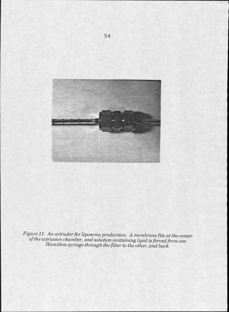
5 4
Figure 11. An extruder for liposome production. A membrane fits at the center of the extrusion chamber, and solution containing lipid is forced from one
Hamilton syringe through the filter to the other, and back.
5 5
In the case of DC CholesterolZPE liposomes, which were received as
premade liposomes, lipid labels were incorporated into the bilayer by first
drying down the appropriate volume of label in chloroform, and then by
adding liposome solution to the dried lipid and sonicating gently (30 seconds
at power level 3, 50% duty).
Extrusion, like sonication, is a procedure that can be accomplished in
less than an hour. Since extrusion does not introduce high energy to the
lipid solutions, it was desirable for SUV production in situations where
sensitive compounds such as peptides or proteins were included in the lipid
mixture.
Liposome yield can be determined in formulations containing known
quantities of phospholipid by phosphate determination. This method
assumes the even distribution of phospholipid and other contents in the
liposome, and assumes also that loss of material on the surface of the glass
test tube is equal for all liposome component materials.
Cationic liposome preparations, because of their tendency to come
out of solution, were used within a few hours of preparation. Loss of
material during production of DOPEZDOTAP liposomes (molar ratio 3:1)
was 25%, as determined by Dittmer and Wells phosphate determination [3].
Absolute amounts of starting materials and rehydration volumes were
usually adjusted to produce a final concentration of one mg IipidZml aqueous
solution, assuming 25% loss during manufacture.
Quality of liposomes was analyzed by viewing fluorescently labelled
liposomes with a fluorescence microscope. Liposomes prepared from

phospholipids such as phosphatidylcholine or phosphatidylserine appeared
as translucent solutions, colored when labels were included, and were not
individually visible under the fluorescence microscope. All cationic liposome
preparations contained a heterogeneous population of vesicles, differing
significantly in size from too small to view under 1250 x magnification
(indicated by an evenly fluorescent background) to vesicles with diameters
of approximately 500 nm.
Cell Culture
Cell cultures were established and maintained according to standard
culturing technique [4], supplemented with information on specific cell lines
from the American Type Culture Collection (ATCC) [5],
HeLa
The HeLa cell line was chosen for preliminary experiments because
of its hardiness, and because it does not express CFTR [6]. HeLa cells were
obtained from both CliffBond (Montana State University) and from the
At CC [5]. HeLa S3 is a subclone of the original HeLa line developed in
1955 from an human endothelial carcinoma. This fine has shown
outstanding growth characteristics, being tolerant of variant conditions and
quick to multiply. Characteristics of the line are well established and do not
change over time, so records of cell passage and morphology are not
necessary. HeLa cells are also adaptable to growth in suspension [7].
HeLa cells were grown in our lab in Dulbecco’s Modified Eagle’s medium
(DME) with 10% calf serum[8]. Antibiotics were not added to the medium.
5 6
Cell cultunng was carried out in a 37° incubator at approximately 5% CO2 .
Cells were passaged once to twice weekly, as needed, by trypsinization, and
subcultured at a dilution of approximately 1:5. Cultures were frozen for
cryostorage in liquid nitrogen by slow freezing in DME with serum,
supplemented with 5% dimethylsulfoxide. Periodic assays for mycoplasma
contamination were performed by the fluorescent DNA staining method [9].
HeLa cells grow well both on plastic substrates and on glass coated
with poly-L-lysine. Experiments were performed on both substrates, after
allowing the cells to attach and grow on the surface for at least 24 hours.
HeLa cells exhibit an epithelial type of morphology, defined for
culturing purposes as box-like, and not highly elongated. This morphology
was found to be detrimental to the analysis of fluorescent labelling by
microscopy, because the cells do not spread thinly over growth surfaces and
this made it is more difficult to interpret the intracellular location of
fluorescent labelling.
ElBTg
Expecting that their more fibroblastic (elongated) morphology would
ease this difficulty, the embryonic bovine tracheal line (EBTr) was grown
and studied. EBTr cells were obtained from Maria Temple (Montana State University).
Embryonic bovine tracheal (EBTr) cells are also easy to mAintain
and are grown in DME with 10% calf serum like the HeLa cells. EBTr cells
elongate dramatically after attachment to surfaces. Several drawbacks of
this line limited their usefulness for our purposes, though. Passage number
should be controlled for this line, as morphology tends to change after the
55th passage [10-11]. This line produces quantities of debris in the media
which make analysis of the samples under the microscope difficult.
Additionally, this line was found to be contaminated with mycoplasma. As
result, its use was discontinued.
RAW 264.7
EAW 264.7 cells are derived from a murine macrophage source,
transformed with Abelson leukemia virus [12,13]. They were purchased
from ATCC [5], grown in DME with 10% calf serum, and subcultured by
scraping from the flask surface and replating into fresh medium at a ratio
of 1:3.
This line was experimented with in an attempt to analyze
fluorescence patterns in a line capable of high levels of endocytosis. EAW
264.7 had been used in experiments to determine the internal pH of
endosomes, due to their vigorous endocytic activity [14]. It was
hypothesized that these cells would internalize label at a level
distinguishable from other lines, such as HeLa. Fluorescence patterns
exhibited by this line were, however, not visibly different from those
exhibited by HeLa cells after cationic liposome treatment.
'
CFG-SV40
CFG-SV40 is a cell line developed from the bronchial submucosal
glands of a Cystic Fibrosis patient, immortalized with a simian virus 40
promoter [15]. It was acquired in hopes that results in this line could more
5 8
easily be compared to CF experiments performed in vivo. Although cell
lines from sources similar to the affected tissues maybe more similar to
the tissues than other cell lines, caution should be exercised when
attempting to make such comparisons. It is clear that immortalized cells
do not behave identically to their tissues of origin [4].
CFG-SV40 is not available commercially or from ATCC, but was
supplied for experiments by its developer, Dr. Chopra of Wayne State
University. CFG-SV40 is a more challenging cell line to grow, requiring
defined medium with multiple growth factors and collagen coated growth
surfaces [15]. Recurring problems in culturing, including slow growth and
mycoplasma infection, led to the discontinuance of this line after just one
liposome experiment. In that experiment the cells proved highly sensitive
to the liposmes, dying during treatment.
Attempts at cryopreservation of this fine were unsuccessful. After
freezing, cells were no longer viable.
HT29
HT29 is derived from a human colon carcinoma, and expresses the
receptor for vasointestinal peptide (VIP), which was used for peptide-tagged
liposome/cell studies (discussed in detail later) [16-18]. This line does
express CFTR, making it less useful for studies of CFTR proteoliposomes,
but it exhibits other desirable characteristics. HT29 is vigorous, requiring
subculturing once to twice per week. It grows well in DME with 10% calf
serum. Its morphology is more epithelial than EBTr, but not as rounded as
59
HeLa. HT29 clings tenaciously to substrates, either plastic or untreated
glass, and it requires strong tiypsinization and scraping to remove.
Cationic liposome treatments, however, have proven disappointing
when performed on this cell line. HT29 appears to be extremely sensitive to
such treatments, resulting in massive cell death. To compound the
problem, HT29 autofluoresces, complicating analysis of fluorescence
exhibited in any experiment.
Liposome Treatments
Protocols for cell treatments were designed to loosely mimic protocols
available for transfection with DNAAiposome complexes [19-23]. Available
protocols came from commercial suppliers and noncommercial producers of
cationic liposome formulations. Commercial suppliers tend to be cautious
about supplying experimenters with the content of their liposome
formulations, so optimum concentrations of lipid and DNA (in those
experiments where it was used) were determined experimentally.
Frank Sorgi, from Leaf Huang’s laboratory at the University of
Pittsburgh, supplied a protocol for use of the non-commercial DC
Cholesterol formulation for transfection. This protocol suggested that
successful transfection with this reagent requires less lipid and DNAthan
the protocol developed by us for liposome/cell interaction studies [23].
Higher concentrations used by us did not show high levels of toxicity.
Incubation times are quite standard among suppliers of liposomes for
transfection. Commercial and non-commercial transfection protocols
recommend short (10-15 minute) preliminary incubations of liposomes and
6 0
DNA, followed by incubation with cells in buffer or medium without the
presence of serum for 30 minutes to 3 hours, and subsequent addition of
medium containing serum or, alternatively, replacement of liposome
solutions with medium containing serum after that time.
The experimental protocol for liposome/cell interaction studies
contained the same 10-15 minute preincubation of liposomes with DNA,
when used, followed by a 30 minute incubation with cells, without serum
present. Cells were then rinsed thoroughly in medium, and analyzed.
Interactions between liposomes or liposome/DNA complexes with
cells should occur over a short time frame, likely within the 30 minutes
from addition of liposomes to the cells. Either of the two mechanisms
proposed to be responsible for cell transfection should work within this time
frame.
Fusion of liposome membranes with plasma membrane maybe
triggered by contact, which would happen within minutes of liposome
introduction, or maybe triggered sometime after contact through
somewhat slower destabilizing events [24]. However, the tendency of
cationic liposomes prepared in our laboratoryto aggregate suggests
destabilization is likely to occur within 30 minutes.
Endocytosis is also a rapid process, and is thought to occur quickly
once material is at the plasma membrane. Times as short as 10 minutes
have been recorded for endocytosis and recycling of membrane components
to the plasma membrane [2,25-28].
6 1
Where metabolizable lipids and labels are concerned, longer
incubations may actually lead to mislocahzation or a decrease in visible
fluorescence after breakdown of liposome components.
A combination of theory and evidence from transfection protocols,
therefore, led to the adoption of the 30 minute incubation protocol used as
the standard method in liposome/cell membrane experiments. Lipid
concentrations used were determined empirically from toxicity studies
performed on HeLa cells with liposomes of DOPE/DOTAP, labelled with
fluorescent Hpid analogues.
Fluorescence Tehhnimies
A variety of fluorescence techniques are common for the physical
analysis of liposomes. Fluorescence measurements can be performed
quickly, and are highly sensitive. Fluorophores incorporated into Hposomal
membranes can be detected at very low concentrations (approximately I
mole percent), where they are unHkely to disturb the bilayer structure
significantly [29]. Fluorescence microscopy and resonance energy transfer
experiments have been performed in this study in an attempt to
understand the interactions between fluorescentiy-labeHed liposomes or
fluorescentiy-labelled proteoHposomes and cells.
At most concentrations, fluorecence emission intensity increases
with fluorescent analyte concentration. However, at high concentrations,
emission decreases due to absorption of energy by nearby excitable
molecules. This process is termed self-quenching.
6 2
The degree of quenching can indicate the relative concentration of
fluorophores in experiments involving liposomes. Decreases in fluorophore
quenching, measured with a fluorometer or estimated visually through a
microscope, indicates dilution of the solution. Such changes in quenching
efficiency can be related to certain processes which occur between
liposomes and cells, such as fusion of membranes and release of the
internal contents of a liposome to the cytosol [30-32].
Hydrophilic fluorophores can be concentrated in the lumen of a
liposome at concentrations where quenching occurs. Unentrapped
fluorophore is separated from the liposome preparation, and the liposomes
are then introduced to the cells. In the event of liposome rupture or leakage
of contents, the fluorescent compound is released, and the lowered
concentration can be detected as an increase in emission intensity from the
sample. Compounds used for this type of experiment are calcein,
fluorescein salts, and fluorescently labelled dextrans of varying molecular
weight. Consideration of hydrophilicity, emission wavelength and intensity,
and ability of a compound to leak out of a bilayer are all necessary when
choosing a label for a given experiment. In experiments described here,
calcein was the fluorophore of choice due to its low cost, reasonable
solubility at self-quenching concentrations, and high emission intensity at
wavelengths easily measured with available equipment.
An alternative to water soluble labels is lipid covalently modified with
a fluorescent group. Again, several options are available, and choice of an
appropriate label depends somewhat on the particulars of the experiment.
Lipids are covalently modified at one of two locations on the molecule:
6 3
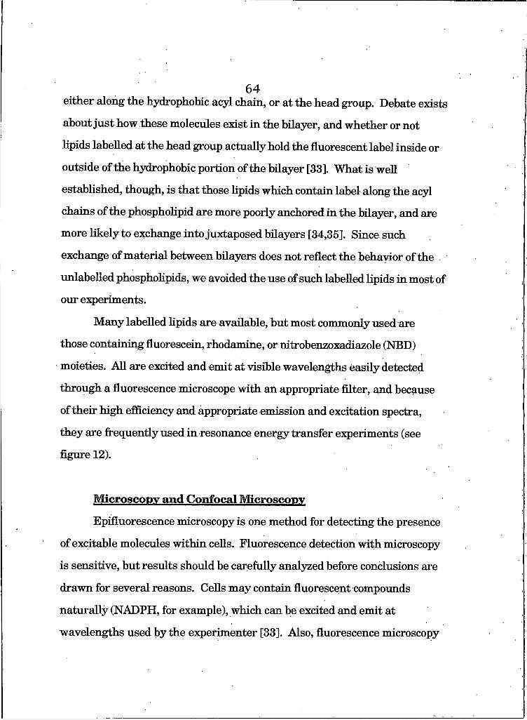
either along the hydrophobic acyl chain, or at the head group. Debate exists
about just how these molecules exist in the bilayer, and whether or not
lipids labelled at the head group actually hold the fluorescent label inside or
outside of the hydrophobic portion of the bilayer [33]. W hatis well
established, though, is that those lipids which contain label along the acyl
chains of the phospholipid are more poorly anchored in the bilayer, and are
more likely to exchange into juxtaposed bilayers [34,35]. Since such
exchange of material between bilayers does not reflect the behavior of the
unlabelled phospholipids, we avoided the use of such labelled lipids in most of
our experiments.
Many labelled lipids are available, but most commonly used are
those containing fluorescein, rhodamine, or nitrobenzoxadiazole (NBD)
moieties. All are excited and emit at visible wavelengths easily detected
through a fluorescence microscope with an appropriate filter, and because
of their high efficiency and appropriate emission and excitation spectra,
they are frequently used in resonance energy transfer experiments (see
figure 12).
Microscopy and Cmifneal Microscopy
Epifluorescence microscopy is one method for detecting the presence
of excitable molecules within cells. Fluorescence detection with microscopy
is sensitive, but results should be carefully analyzed before conclusions are
drawn for several reasons. Cells may contain fluorescent compounds
naturally (NADPH, for example), which can be excited and emit at
wavelengths used by the experimenter [33]. Also, fluorescence microscopy
64
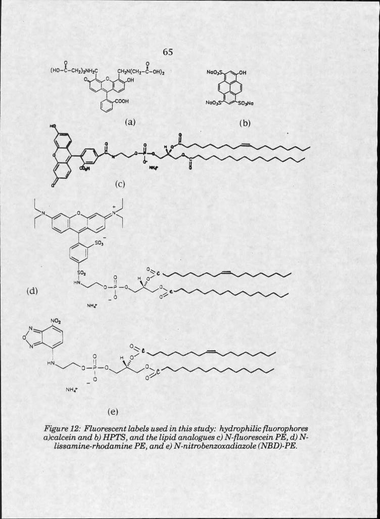
6 5
o — p —
NH/
H O— P — O,
NHV
(e)
Figure 12: Fluorescentlabels used in this study: hydrophilic fluorophores a)calcein and b) HPTS, and the lipid analogues c) N-fluorescein PE, d) N-
lissamine-rkodamine PE, and e) N-nitrobenzoxadiazole (NBD)-PE.
is inadequate to definitively identify the intracellular location of
fluorescence within the cell, because cells exist in three dimensions and are
excited by the instrument in all focal planes at one time. This can lead to
problematic interfering fluorescence from out of the plane of focus.
When interfering fluorescence is troublesome, the use of confocal
fluorescence microscopy can improve resolution [36-38]. Confocal
microscopy achieves high resolution by imaging emission from a narrow
plane of focus. Emission from molecules outside of the focal plane is
avoided by collection of emitted light from particular points, focussed
through a small aperture. Rasterizing the location of the excitation beam
across the sample is used to collect light from across the specimen but
within the narrow focal plane. Fast rasteiing devices in some instruments
allow confocal images to be viewed in real time. Algorithms are available,
such as NIH Image, to allow two dimensional images to be viewed and
saved [39].
Fluorescence microscopy experiments were performed on
mammalian cell lines grown in monolayer culture. Protocols were designed
to be somewhat similar to those used for cell transfection experiments with
IiposomeZDNA complexes. Cytotoxicity studies were performed initially to
determine optimal concentrations of liposomes for treating cells. Protocols
were then standardized, in order to make comparisons between differing cell
lines and liposome compositions possible.
Typically, fluorescence microscopy experiments were performed as
follows. Cells were seeded at appropriate densities to allow attachment and
growth to 50-80% confluency before introduction of liposomes. At least 24
6 6
6 7
hours of growth on the experimental surface was given to allow for thorough cell
adhesion. Media was removed, cells were rinsed twice with an equal volume of
buffer (10 mM HEPES, 150 mM N ad ) or serum-free medium, and liposnmp
solutions (at a concentration of I mg/ml) added. Experiments performed on
cover slips in 12-well dishes used 30 pg total lipid per well; experiments
performed in 8-well chamber slides used 6.2 pg lipid per well. Media without
serum, or HEPES buffered saline, was used to dilute liposomes to final volumes
of 400 pi, on the microscope cover slips, or 85 pi, in cell chambers. Incubations
were performed for 30 minutes, based on preliminary experiments which
indicated that visible labelling, but not toxicity, occurred in that amount of time.
After 30 minutes, liposome solutions were removed, and the cells were carefully,
but thoroughly rinsed twice with either HEPES buffer or serum-free medium (I
ml for cover slips and 350 pi for cell chambers). Cells were then ready to be
viewed with the microscope. A Leitz microscope, fitted with a number three
emission filter, was used in these experiments.
Resonance Energy Transfer
A subpopulation of fluorescent molecules are able to transfer their
energy to other fluorescent molecules when they exist in close proximity to one
another (high concentration) [40]. This nonradiative transfer effectively
quenches the fluorescence emission of the first molecule, and excites the second
to cause emission at a longer wavelength. This process, termed resonance
energy transfer (RET), can be used to measure dilution of fluorophores during
an experiment [40]. Such dilution may occur in transfer of fluorescently
labelled lipids from one membrane to another, or during the.
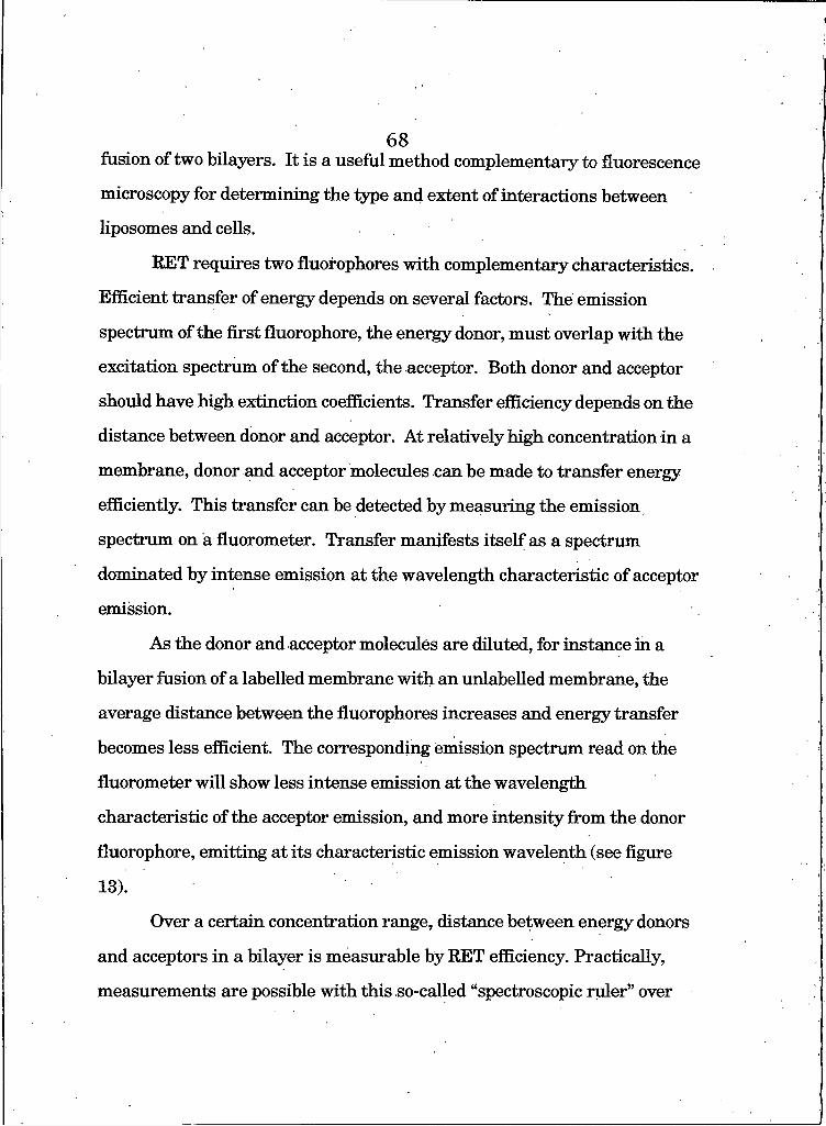
fusion of two bilayers. It is a useful method complementary to fluorescence
microscopy for determining the type and extent of interactions between
liposomes and cells.
RET requires two fluofophores with complementary characteristics.
Efficient transfer of energy depends on several factors. The emission
spectrum of the first fluorophore, the energy donor, must overlap with the
excitation spectrum of the second, the acceptor. Both donor and acceptor
should have high extinction coefficients. Transfer efficiency depends on the
distance between donor and acceptor. At relatively high concentration in a
membrane, donor and acceptor molecules can be made to transfer energy
efficiently. This transfer can be detected by measuring the emission
spectrum on a fluorometer. Transfer manifests itself as a spectrum
dominated by intense emission at the wavelength characteristic of acceptor
emission.
As the donor and acceptor molecules are diluted, for instance in a
bilayer fusion of a labelled membrane with an unlabelled membrane, the
average distance between the fluorophores increases and energy transfer
becomes less efficient. The corresponding emission spectrum read on the
fluorometer will show less intense emission at the wavelength
characteristic of the acceptor emission, and more intensify from the donor
fluorophore, emitting at its characteristic emission wavelenth (see figure
13).
Over a certain concentration range, distance between energy donors
and acceptors in a bilayer is measurable by RET efficiency. Practically,
measurements are possible with this so-called “spectroscopic ruler” over
6 8
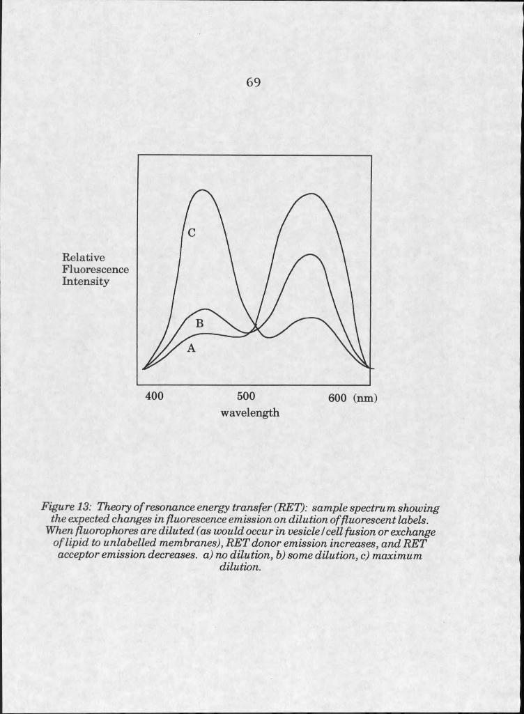
6 9
RelativeFluorescenceIntensity
400 500 600 (nm)wavelength
Figure 13: Theory of resonance energy transfer (RET): sample spectru m showing the expected changes in fluorescence emission on dilution of fluorescent labels.
When fluorophores are diluted (as would occur in vesicle I cell fusion or exchange of lipid to unlabelled membranes), RET donor emission increases, and RET acceptor emission decreases, a) no dilution, b) some dilution, c) maximum
dilution.
distances as great as ten nanometers [41]. This measurement requires
knowledge of total lipid content in the experimental sample, and the
mathematical relationship between fluorescence signal and distance breaks
down at extreme dilution [40].
In a typical liposome experiment, efficient RET can be attained at
molar fluorophore concentrations of 1-2 mole percent of phospholipid. A
theoretical maximum dilution (actually somewhat greater than dilution
possible by membrane dilution) can be approximated by disrupting all
membranes present in an experimental sample with detergent [40].
An RET experiment can be designed in one of two general ways,
distinguished by the locations of donor and acceptor molecules. Either I)
donor and acceptor can be in separate membrane populations, brought
together at the time of the experiment, or 2) donor and acceptor can be
incorporated into a single membrane, which is brought into the presence of
an unlabelled membrane at the time of the experiment [40].
There are advantages and disadvantages to both methods. The first
method allows the measurement of actual distances. Fusion of membranes
in this case is measured by the onset of RET. However, this method
requires introduction of donor and acceptor molecules into membranes at
defined concentrations, which is impossible for cell membranes. The second
method, requiring mixed donor and acceptor molecules, leads to a decrease
in RET as the membranes mix contents in a fusion event. Both labels can
easily be incorporated in known concentrations in the liposomes, and can
then be introduced to unmanipulated cells. However, because total lipid is
not defined in such an experiment, the decrease in RET measured cannot be
7 0
correlated to the amount of dilution; i.e. a linear relationship between RET
measured and label dilution is undefinable in this case.
A variety of phospholipid analogues containing fluorescent
headgroups capable of participating in RET are available commercially.
Traditional donor and acceptor pairs include a fluorescein group as donor
and a rhodamine group as acceptor. Fluorescein also has the capability of
serving as donor to eosin, a moiety available covalently linked to maleimide,
which is useful for labelling protein via reaction of the maleimide to
sulfhydryl groups on the polypeptide [42].
Certain substances are capable of interfering with RET, and controls
must be thorough. The effectiveness of RET is in part dependent on the
local environment of the fluorophores, so substances which interact closely
with the fluorophores, or with the bilayers, can interfere with the
measurement. Certain detergents, in particular, may interfere [40].
Particular caution must also be exercised when interpreting data
from RET experiments because mixing of membrane Upids without fusion of
the membranes may appear as fusion. These experiments are really a
measure of the dilution of membrane probes, not fusion itself. Use of lipids
labelled at the head group rather than along the acyl chain somewhat
decreases the odds of transfer between membranes [34].
Bilayer fusion studies performed to analyze the interactions between
cationic Uposomes and cultured ceUs in this study made use of one mole
percent of head group labelled fluorescein PE or NBD-PE as energy donors.
One mole percent of PE labelled at the head group with rhodamine was the
sole energy acceptor in all cases. Fluorescent labels were incorporated by
7 1

adding them into lipid solutions in chloroform, before drying. Analysis of
fluorescently labelled liposomes for label incorporation was performed by
viewing through a fluorescence microscope and by visualization of gross
solution properties with broad ultraviolet excitation over a 360 nm
ultraviolet light box.
Experimental protocols were designed to measure changes in RET
from as little as 20 seconds after introduction of liposomes to cells to up to
several hours. Experiments were performed with live mammalian cells,
grown up in monolayers and detached in some cases by trypsinization and
in other cases by physical removal from the flask surface. Cells were
transferred in media to fluorescence cuvettes. Experiments were
performed on two different available fluorometers: initially on a SPEX
Fluorolog 2 (courtesy Pat Callis, MSU Chemistry and Biochemistry), and
secondly, on a PTI instrument (courtesy Algirdas Jesaitis, MSU
Microbiology). Samples were stirred while measurements were taken with
the PTI ffuorimeter, but not with the SPEX instrument. Cells were
suspended in 2 ml serum free medium at an approximate density of 2.5 x
IO^ cells per ml. Liposomes were introduced to the cuvettes and spectra
taken over a wavelength range suitable for detection of fluorescence from
both RET donor and acceptor molecules. Spectra were taken periodically
over the time course designated for each particular experiment. An
approximation of infinite probe dilution was made by lysing all membranes
with the introduction of 0.1% Triton X-100 to the sample cuvette, and
recording a spectrum. Controls included experiments with each individual
7 2

label, with and without cells present, cells alone, and liposomes containing
each label lysed by Triton X-100.
7 3
ResultsLiposome Manufacture
Liposome formulations were initially inspected for evidence of
instability after manufacture, exhibited by cloudiness, or appearance of
precipitates, flocculant, or suspended aggregated lipid. Liposomes prepared
from naturally occurring phospholipids such as phosphatidylcholine (PC)
and phosphatidylseiine (PS) are stable over a period of days. However,
cationic liposomes composed of DOPE/DOTAP and pH sensitive liposomes
composed of oleic acid (OA) mixed with PE, in particular, showed some
tendency to destabilize, and so were analyzed in greater detail to determine
their properties.
pH sensitive liposomes were prepared from OA and PE [43] at
various ratios, all containing 1% fluorescent lipid label as a marker.
Solutions were sonicated and viewed over a 360 nm ultraviolet light box to
excite the fluorophores. OA/PE mole ratios of less than 3:7 exhibited the
most evenly distributed fluorescence. Ratios of OA/DOPE of 3:7 and
greater contained large fluorescent aggregates in the solution. Palmitoyl-
oleoyl-phosphatidylethanolamine (POPE) was used in place of DOPE, with
similar results (see figure 14).
In situations where liposomes were passed over Sephadex G 50-50
spin columns to remove small molecules from the extra-liposomal buffer,
OA/PE liposomes did not properly elute from the columns. In experiments
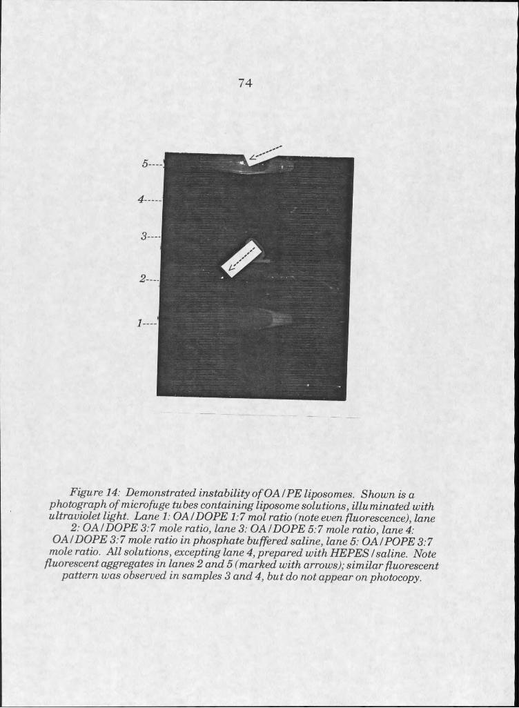
74
Figure 14: Demonstrated instability o f OAI FE liposomes. Shown is a photograph of microfuge tubes containing liposome solutions, illuminated with ultraviolet light. Lane I: OA /DOPE 1:7 mol ratio (note even fluorescence), lane
2: OAI DOPE 3:7 mole ratio, lane 3: OAt DOPE 5:7 mole ratio, lane 4:OA/DOPE 3:7 mole ratio in phosphate buffered saline, lane 5: GA!POPE 3:7
mole ratio. All solutions, excepting lane 4, prepared with HEPESI saline. Note fluorescent aggregates in lanes 2 and 5 (marked with arrows); similar fluorescent
pattern was observed in samples 3 and 4, but do not appear on photocopy.
using both calcein as an encapsulated label and rhodamine-PE as a
hydrophobic lipid label, calcein appeared to travel through the column,
leaving the lipid behind. This pattern did not occur when another
hydrophilic label, 8-hydroxypyrenetrisulfonic acid (HPTS), was Used in place
of the calcein. Pre-equilibration of the Sephadex column with unlabelled
phosphatidylcholine liposomes improved elution somewhat, so that some
liposome material eluted in the void volume.
Liposomes prepared from DOPEZDOTAP (3:1 molar ratio) also
exhibited some instability. In solutions containing calcein, DOPE/DOTAP
liposomes came out of solution in minutes after sonication. Attempts at
running solutions containing DOPE/DOTAP and calcein through Sephadex
G50-50 spin columns yielded no lipid, as determined by Dittmer and Wells
phosphorous determination on eluted fractions. HPTS, the alternate
hydrophilic label, also destabilized DOPE/DOTAP vesicles in a matter of
minutes. Likewise, a rhodamine-conjugated dextran destabilized
DOPE/DOTAP liposomes.
DOPE/DOTAP lipsome stability was not affected by ionic strength in
the range of 0-300 mOsm, in HEPES/NaCl buffers.
Microscopy
Results from microscopy experiments are qualitative. Analyses of
fluorescent labelling patterns in liposome-treated cells were performed
many times over, attempting to make conclusions based on a large amount
of evidence collected in separate experiments. It was hoped that a degree of
objectivity could be maintained by analyzing multiple experiments.
75

Cytotoxicity experiments were performed to determine reasonable
dosage levels for liposomes on HeLa cells. Toxicity studies initially used two
types of liposomes: DOPE/DOTAP in a 3:1 molar ratio, and DOPC/DOPG in
a 1:1 ratio, and covered the range of 25 to 200 ng lipid per well of a 12-well
culture dish. Incubations of varying length, from 15 minutes to 5 hours,
were used. Fluorescence labelling of the cells from the array of treatments
indicated that, for labelling to occur without gross cell damage and death, a
treatment of 25 to 50 pg, incubated for 30 minutes, was most appropriate.
Naturally occurring phospholipids composed of phosphatidylserine,
phosphatidylcholine, and phosphatidylglycerol applied in this dosage range
showed no toxicity.
Cell treatments with DOPE/DOTAP liposomes showed the most
promise of significant material transfer. This vehicle was used for the
majority of experiments performed.
DOPE/DOTAP was unstable in the presence of calcein and other
hydrophilic labels, so experiments were performed with 1-2 mole percent
. lipid tagged with fluorescein, rhodamine, or nitrobenzoxadiazole.
Experiments performed over the range of 0-30 minutes showed that
labelhng becomes visible under the fluorescence microscope within 15-25
minutes from the beginning of liposome treatment.
Standard experiments, involving half hour treatments with dosages
on the order of 30 pg/500 mm2 area, yielded reproducible results, with a few
exceptions. AU cells within the treatment area showed the same pattern of
labelling. The pattern of fluorescence exhibited by these ceUs was fine, but
punctate, IabeUing at or near the ceU membrane. No internalized label was
76
evident, either at internal membranes such as the nuclear envelope, or in
the cytosol. Distribution of small fluorescent aggregates at the plasma
membrane was even, covering the entire cell surface. Some label may have
been internal, but if so, the amount internalized was not distinguishable by
eye. As the plane of focus could be moved from one face of the cell to the
other, fluorescence at the membrane could be distinguished from cytosolic
portions of the cell which remained essentially unlabelled (see figure 15).
In some experiments, an attempt was made at inducing membrane
fusion with the aid of polyethylene glycol (PEG) treatment [44]. A 44%
solution of PEG was added to cells for I minute during the last stages of
liposome treatment to induce fusion. PEG solutions and liposomes were
then removed from the cells, cells were rinsed, and fresh media was added.
Fluorescence patterns exhibited by cells treated in this way were somewhat
different than those not receiving the PEG fusion treatment, in that there
was more diffuse fluorescent labelhng at the membranes of some cells
within the treated area. Such even labelling only occurred within small
populations of treated cells, however (see figures 16 and 17). PEG
treatment, in cells incubated for 24 hours after the experimental
procedures, inevitably caused large scale cell lysis.
To confirm that fluorescent labelling in DOPEZDOTAP experiments
was occurring at the cell surface only, without internalization, experiments
were performed utilizing a technique called “back exchange” with unlabelled
lipids [45]. Attempts at back exchange depend on the free exchange of
labelled for unlabelled lipids at the cell surface when a large excess of
unlabelled lipids is introduced to the cell surface. Experimentally, an excess
7 7
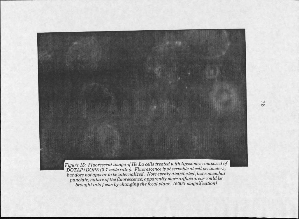
but does not appear to be internalized. Note evenly distributed, but somewhat punctate, nature of the fluorescence; apparently more diffuse areas could be
brought into focus by changing the focal plane. (500X magnification)
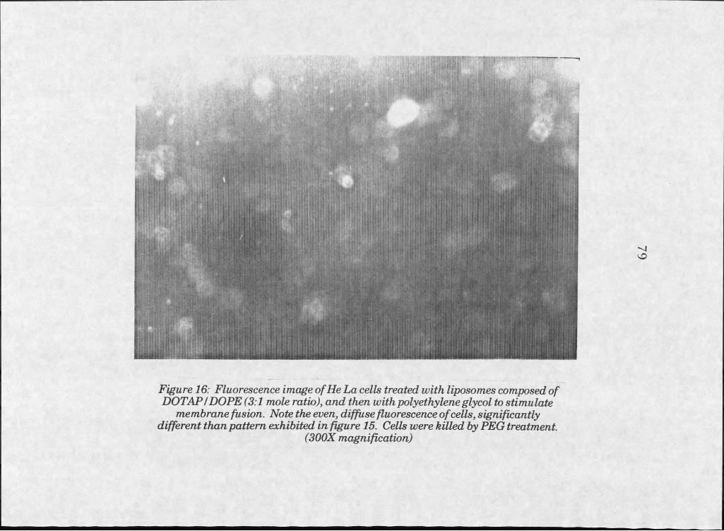
Figure 16: Fluorescence image of He La cells treated with liposomes composed of DOTAPI DOPE (3:1 mole ratio), and then with polyethyleneglycol to stimulate
membrane fusion. Note the even, diffuse fluorescence o f cells, significantly different than pattern exhibited in figure 15. Cells were killed by PEG treatment.
(300X magnification)

Figure 17: Samefield as shown in figure 15. Illumination with white light, forcomparison to fluorescent image.
of unlabelled liposomes are incubated with the cells after treatment with
labelled liposomes. In instances where liposomes are simply aggregating at
the cell surface, but are not tightly bound, exchange with unlabelled
liposomes occurs and the degree of visible labelling decrease over time.
Back exchange experiments were performed using unlabelled DOPS/DOPE
(1:1) or DOPEZDOTAP (3:1) Hposomes following the initial treatment with
labelled DOPEZDOTAP liposomes.
In these experiments, labelled DOPEZDOTAP treated cells back
exchanged with unlabelled Hposomes exhibited somewhat reduced cell
labelling, although ceU death was massive in experiments where back
exchange was performed with DOPEZDOTAP. Cells treated in this way
tended to retain label more at the junctions between ceUs than at more
exposed areas of plasma membrane. This may have occurred because
juxtaposed areas are less accessible to the introduced exchange Hposomes.
In experiments utilizing excess PSZPE for back exchange, results were
somewhat unclear but also suggested some exchange of lipids was
occurring.
Liposomes containing DC Cholesterol and DOPE IabeHed cells
similarly to DOPEZDOTAP Hposomes. Fluorescence was evenly distributed
across all plasma membranes, was fine but punctate, and did not appear to
exist in the internal portions of the cells.
pH sensitive Hposomes were prepared from OA and DOPE in molar
ratios of 2:7 and 1.5:7, which are stable at physiological pH but become
unstable when the pH is dropped below 6.0. Such Hposomes were used in an
attempt to deliver fluorescent labels to cell membranes by the standard 30-
8 1
minute liposome treatment. Experimental results with pH sensitive
liposomes varied, but in general were less suggestive of material transfer to
cells than experiments with DOPE/DOTAP and DC Cholesterol liposomes.
In some instances, labelling appeared in inappropriate places: on debris
within the cell chamber, and on the slide itself. Since glass slides were
coated with poly-lysine to aid in cell adhesion, an interaction may occur
between anionic pH sensitive liposomes and the poly-lysine.
Some experimentation with liposome formulations of DOPE/DOTAP
was done to further analyze the optimal combination of lipids for efficient
cell membrane labelling. Mixtures of DOPE/DOTAP and varying
proportions of DOPC were tested, and fluorescence patterns under the
microscope examined. DOPE/DOTAP at a 3:1 molar ratio, DOPE/DOTAP
at a 5:1 molar ratio, 3:3:1 DOPE/POPC/DOTAP, and 3:6:1
DOPE/POPC/DOTAP were tested. All gave similar results, except 5:1
DOPE/DOTAP, which was more prone to aggregation, and caused more
punctate labelling at the cell surface. Formulations containing PC were
more easily suspended in aqueous solutions after drying than pure
PE/DOTAP formulations, perhaps due to the fact that they carry less
charge density, which could reduce interactions with the surface of the test
tube.
Liposome/DNA complexes formed by incubation of premade cationic
liposomes with pUC19 were compared to cationic liposomes without DNA
to see if visible differences in fluorescence patterns after incubation with
cells were apparent. Labelling from liposomes versus liposomes complexed
with DNA could not be differentiated.
8 2
8 3
ConfnoaI microsnnpy
Confocal microscopy increased resolution in some experiments where
fluorescent labelling of cells was examined. DOPEZDOTAP liposomes and
DOPEZPOPC/DOTAP liposomes labelled with N-fluorescein-PE were used to
treat cells, which were then fixed with glutaraldehyde and viewed with a
confocal microscope. Images showed extensive labelling at the perimeter of
cells, but none internally (see figure 18).
Resonance Energy TVansfer
Resonance energy transfer experiments were performed under a
variety of different conditions to corroborate evidence gathered by
microscopy. Because microscopy experiments suggested a strong
interaction between liposomes composed of DOPE/DOTAP and cultured
HeLa cells, RET experiments began by analyzing that particular
interaction. The search was for evidence of fusion or transfer of lipid
components from labelled liposomes to the cells.
Experiments utilized various labels, including lipids with fluorescein,
nitrobenzoxadiazole, and rhodamine groups at the head group, and
nitrobenzoxadiazole at the sixth carbon in position two of the acyl chain on
PC. Initial concentrations of fluorescent resonance energy transfer pairs
incorporated into liposomal bilayers demonstrated transfer of energy when
excited at the donor excitation wavelength, showing emission at the
wavelength range characteristic for the RET acceptor molecule. Disruption
of the bilayers, performed by addition of Triton X-IOO to a final percentage
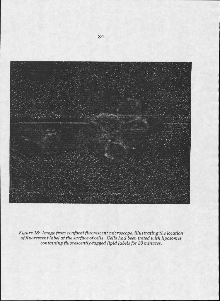
84
Figure 18: Image from confocal fluorescent microscope, illustrating the location of fluorescent label at the surface of cells. Cells had been treted with liposomes
containing fluorescently-tagged lipid labels for 30 minutes.
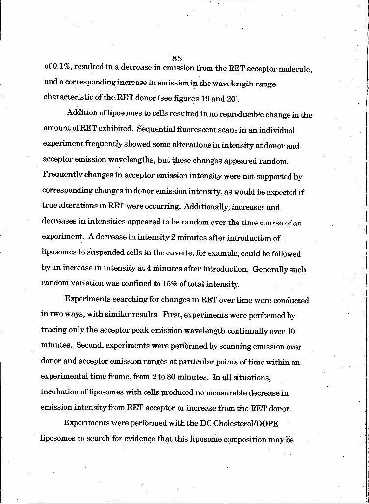
of 0.1%, resulted in a decrease in emission from the RET acceptor molecule,
and a corresponding increase in emission in the wavelength range
characteristic of the RET donor (see figures 19 and 20).
Addition of liposomes to cells resulted in no reproducible change in the
amount of RET exhibited. Sequential fluorescent scans in an individual
experiment frequently showed some alterations in intensify at donor and
acceptor emission wavelengths, but these changes appeared random.
Frequently changes in acceptor emission intensity were not supported by
corresponding changes in donor emission intensity, as would be expected if
true alterations in RET were occurring. Additionally, increases and
decreases in intensities appeared to be random over the time course of an
experiment. A decrease in intensity 2 minutes after introduction of
liposomes to suspended cells in the cuvette, for example, could be followed
by an increase in intensity at 4 minutes after introduction. Generally such
random variation was confined to 15% of total intensity.
Experiments searching for changes in RET over time were conducted
in two ways, with similar results. First, experiments were performed by
tracing only the acceptor peak emission wavelength continually over 10
minutes. Second, experiments were performed by scanning emission over
donor and acceptor emission ranges at particular points of time within an
experimental time frame, from 2 to 30 minutes. In all situations,
incubation of liposomes with cells produced no measurable decrease in
emission intensity from RET acceptor or increase from the RET donor.
Experiments were performed with the DC Cholesterol/DOPE
liposomes to search for evidence that this liposome composition maybe
85
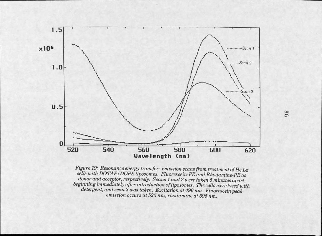
xIO6
I .
0 .
I .!
Scan I
Scan 2
Scan 3
5 2 0 5 4 0 5 6 0 5 8 0 6 0 0 6 2 0Wavelength (nn)
Figure 19: Resonance energy transfer: emission scans from treatment of He La cells with DOTAP/DOPE liposomes. Fluorescein-PE and Rhodamine-PE as donor and acceptor, respectively. Scans I and 2 were taken 5 minutes apart,
beginning immediately after introduction o f liposomes. The cells were lysed with detergent, and scan 3 was taken. Excitation at 496 nm. Fluorescein peak
emission occurs at 525 nm, rhodamine at 595 nm.
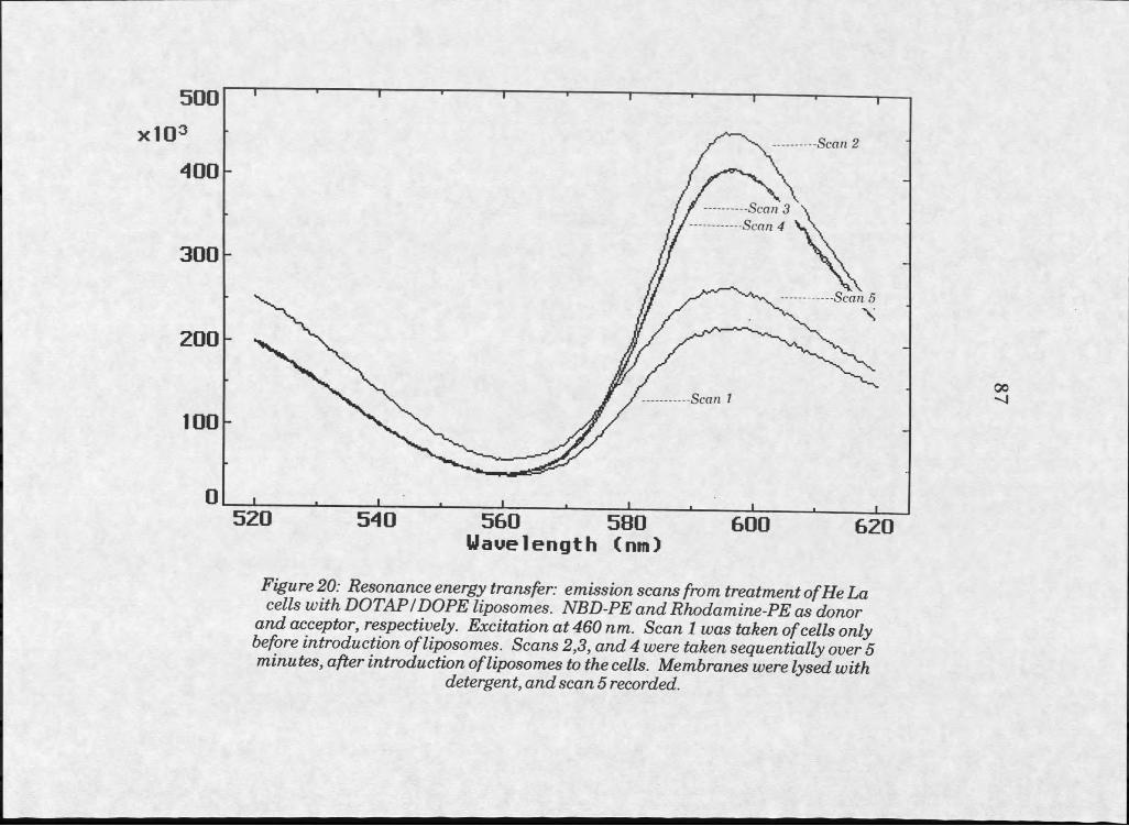
Scan 2
-Scan 3 Scan 4
Scan 5
5 6 0 5 8 0Wavelength Cnm)
Figure 20: Resonance energy transfer: emission scans from treatment o f He La cells with DOTAP/DOPE liposomes. NBD-PE and Rhodamine-PE as donor
and acceptor, respectively. Excitation at 460 nm. Scan I was taken of cells only before introduction of liposomes. Scans 2,3, and 4 were taken sequentially over 5 minutes, after introduction of liposomes to the cells. Membranes were lysed with
detergent, and scan 5 recorded.
capable of fusion or exchange of material with cell membranes. Microscopy
suggested that with DC Cholesterol/DOPE, as with DOPEZDOTAP, there
was potential for such an interaction. Resonance energy transfer
experiments revealed, as with DOPE/DOTAP liposomes, that no
measurable dilution of the probes occurred.
Since complexes of liposomes and DNA have been frequently used for
cell transfection, studies of liposomes in the presence of DNA were
performed and compared with liposomes alone, as they interacted with
suspended cells.
This group of experiments was designed to cover a wide time range, in
part to determine the possibility of RET changes over a longer time scale.
Although microscopy experiments suggested that interactions between
liposomes and cells occurred within 30 minutes, some RET experiments
were performed over longer time periods to search for more gradual
changes. Three sequential experiments included measurements of 0 to 90,
0 to 30, and 0 to 10 minutes after addition of liposomes to suspended HeLa
cells. No changes in intensity occurred after 10 minutes, although data
from these experiments suggested possible probe dilution on the time scale
of 0-10 minutes (data not shown). Repeated experiments, however, refuted
these preliminary results by showing no decreases in RET on this time
scale (see figure 21),
Variation in fluorescence intensities in replicate emission scans
caused some concern about the methods used. To dismiss the possibility
that domain formation during liposome/DNA complexation could be
interfering with results in liposome/DNA experiments, RET was measured
88
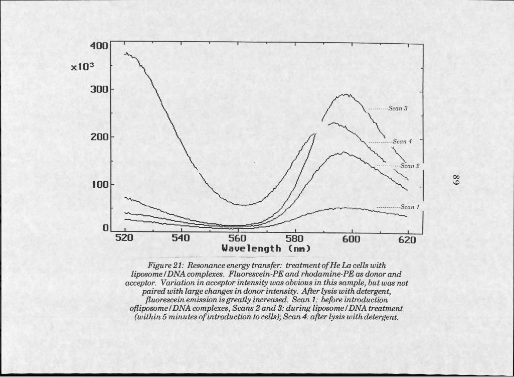
Scan 3
Scan 4
Scan 2
Scan I
5 6 0 5 8 0Wavelength (nm)
Figure 21: Resonance energy transfer: treatment o f He La cells with liposome IDNA complexes. Fluorescein-PE and rhodamine-PE as donor and
acceptor. Variation in acceptor intensity was obvious in this sample, but was not paired with large changes in donor intensity. After lysis with detergent, fluorescein emission is greatly increased. Scan I: before introduction
ofliposomeIDNA complexes, Scans 2 and 3: during liposome/ DNA treatment (within 5 minutes of introduction to cells); Scan 4: after lysis with detergent.
over the 10 minute incubation of liposomes with DNA No change in RET
was observed.
Changes in intensity With dilution of liposome solutions on adding
cells were measured, and found to be negligible. The effect of the detergent
Triton X-100 on the intensity of signals from individual fluorophores was
investigated, but also was found to be negligible.
Discussion
The evidence collected by microscopic examination of fluorescence
transfer from liposomes to cells suggested accumulation of visible amounts
of labelled vesicles at target cells occurring within 30 minutes. Gentle
washing was found inadequate to remove bound label, vigorous washing
detached the cells. Liposomes were indistinguishable from complexes; of
labelled liposomes and DNA commonly used for cell transfection.
Back exchange experiments suggested, although not strongly, that
some fluorescent label could be removed by incubation with a large excess
of unlabelled liposomes. However, the attraction between cationic lipids
and cell membranes may account for the poor back exchange with anionic
phospholipids. Back exchange with cationic liposomes damaged cells
extensively.
The fact that large doses or long incubations with cationic liposome
preparations is toxic does suggest that the interaction occurring does affect
cell physiology in some way. This affect could be due either to changes at
the membrane surface, or to changes brought about by internalization.
90
No evidence supporting internalization of fluorescent lipids was
obtained from fluorescence microscopy and confocal fluorescence
microscopy experiments. A small number of fluorescent vesicles, or
fluorescent molecules, could be internalized or incorporated into the cell
plasma membrane without detection by this method. Alternatively, the
physiological effects could be accounted for by the clear accumulation of
material at the surface of the plasma membrane.
Resonance energy transfer experiments suggested that the
interaction between liposomes and cell membranes does not involve
measurable lipid dilution, which would indicate liposome/cell membrane
fusion or exchange of material between bilayers. RET assays were
sensitive enough that dilution of the fluorescent probes by 10-20% should
have been measurable; such dilution did not occur over a period of up to 90
minutes after introduction of the liposomes to suspended cells. If liposome-
mediated transfection occurs by mechanisms similar to those examined
here, DNA delivery must occur at such a low rate that these isolated
internalization or fusion events are unmeasurable by fluorescence methods.
91
92
References Cited'
1. New, RRC., ed., Liposomes: a p ractica l approach . Oxford University Press, Oxford, 1990.
2. Huang, C., “Studies on phosphatidylcholine vesicles. Formation and physical characterization,” Biochem istry 8: 344-351, 1969.
3. Dittmer, J.C., and Wells, M.A., “Quantitative and qualitative analysis of lipids and Upid components,” M ethods EnzymoL 14: 482-530, 1969.
4. Freshney, RI., C ultnre of Animal Cells: AMamnal of Basic T echnique. Alan R Liss, Inc., New York, 1983.
5. American Type Culture Collection, 12301 Parklawn Drive, Rockville, MD 20852.
6. Puck, T.T., and Fisher, H.W., J . Exp. Med. 104: 427-434, 1956
7. Darnell, J.E., et a l, J . Exp. Med. HO: 445-450, 1959.
8. Morton, H.J., “A survey of ommercially available tissue culture media,” InV itro 6:89,1970.
9. Del Guidice, R.A., Hopps, H.E., in Mycoplasma infection of Cell C ultures. McGarrity, G.J., Murphy, D.G., and Nichols, W.W., Eds., (Plenum, New York) 57-70,1978.
10. Product Discription sheet, American Type Culture Collection, Rockville, MD
11. Raschke, W.C., Baird, S., Ralph, P., and Nakoinz, I., “Functional macrophage cell Unes transformed by Abelson leukemia virus,” Cell 15: 261-267, 1978.
12. Daleke, D.L., Hong, K., and Papahadjopoulos, D., “Endocytosis of liposomes by macrophages: binding, acidification, and leakage of liposomes monitored by a new fluorescence assay,” Biochim. Biophys.Acta 1024: 352-366, 1990.
13. Yoshimura, T., shono, M., Imai, K , and Hong, K , “Kinetic analysis of endocytosis and intracellular fate of liposomes in single macrophages,” J . Biochem. (Tokyo) 117(1): 34-41, 1995.
14. Product Description sheet, American Type Culture Collection, Rockville, MD
15. Chopra, D.P., Reddy, L., Gupta, S.K., Wan, L., Mathieu, P.A., Shoemaker, R.L., and Rhim, J.S., “Differentiation of immortalized epithelial cells derived from cystic fibrosis airway submucosal glands,” In V itro Cell. Dev. BioL 30A: 539-546, 1994.
16. Muller, J.M., El Battari, A., Ah-Kye, E., Luis, J., Ducret, F., Pichon,J., and Marvaldi, J., “Internalization of the vasoactive intestinal peptide (VIP) in a human adenocarcinoma cell line (HT29),” Em*. J . Biochem. 152: 107-114, 1985.
17. Goossens, J.F., Pommery, N., Lohez, M., Pommery, J., Helbecque, N., Cotelle, P ., Lhermitte, M., and Henichardt, J.P., “Antagonistic effect of a vasoactive intestinal peptide fragment, vasoactive intestinal peptide (1-11), on guinea pig trachea smooth muscle relaxation,” M olecular Pharm . 41:104-109, 1991.
18. Ichikawa, S., Sreedharan, S.P., Owen, R.L., Goetzl, E.J., “Immunochemical localization of type I VIP receptor and NK-l-type substance P receptor in rat lung,” Am. J . Physio. Lung Cell. Mol. PhysioL 268: L584-L588, 1995.
19. Guide to E ukaryotic Transfections w ith C ationir Tlnift Reagents. Life Technologies, Gaithersburg, MD
20. Schenbom5E., Oler5J., Goiffonj V., Balasubramaniamj R., Bennett, M.J., Aberle, AM., Nantz, M.H., and Malone, R.W., “Transfection: Tfx-50 Reagent: a new transfection reagent for eukaryotic cells,” P rom ega Notes Magazine, number 52: 2-7,1995.
21. Hawley-Nelson, P., Ciccarone, V., Gebeyehu, G., and Jessee J., “Lipofectamine Reagent: a new, higher efficiency polycationic liposome transfection reagent,” Focus 15(3): 73-79, 1993.
22. Buchberger, B., Femholz, E., Bantle, E., Weigert, M., Borowski, E., v.d. Eltz, H., and Hinzpeter, M., “DOSPER liposomal transfection reagent: a reagent with unique transfection properties,”Biochem ica No. 2, 7-10,1996.
23. Leaf Huang, personal communication.
93
24. Zimmerberg, J., Vogel, S.S., and Chmomordik, L.V., “Mechanisms of membrane fusion," A nnual Rev. Biophys. Biomol. S truct. 22: 433-466, 1993.
25. Watts, C., and Marsh, M., “Endocytosis: What goes in and how?” J . Cell Sci. 103: 1-8, 1992.
26. Lee, KD., Nir, S., and Papahadjopoulos, D., “Quantitative analysis of liposome-cell interactions in vitro: rate constants of binding and endocytosis with suspension and adherent J774 cells and human monocytes,” Biochem istry 32: 889-899, 1993.
27. Prince, L.S., Workman, KB. Jr., and Marchase, RB., “Rapid endocytosis of the cystic fibrosis transmembrane conductance regulator chloride channel,” Proc. NatL Acad. Sci. USA 91: 5192- 5196, 1994.
28. Straubinger, R.M., Papahadjopoulos, D., and Hong, K , “Endocytosis and intracellular fate of liposomes using pyranine as a probe,” Biochem istry 29: 4929-4939, 1990.
29. Hoekstra, D., and Duzgunes, N., “Lipid mixing assays to determine fusion in liposome systems,” M ethods EnzymoL 220: 15-32,1993.
30. Uster, P.S., and Pagano, R.E., “Resonance energy transfer microscopy: visual colocalization of fluorescent lipid probes in liposomes,” M ethods EnzymoL 171: 850-856, 1989.
31. Uster, P.S., iiIn situ resonance energy transfer microscopy: monitoring membrane fusion in living cells,” Methods EnzymoL 221: 239-246, 1993.
32. Shin, T.B., Leventis, R., and Silvius, J.R., “Partitioning of fluorescent phospholipid probes between different bilayer environments. Estimation of the free energy of interlipid hydrogen bonding,” Biochem istry 30: 7491-7497, 1991.
33. Salama, G., Lombardi, R., Elson, J., “Maps of optical action potentials and NADH fluorescence in intact working hearts,” Am. J . PhysioL 252: H384-394, 1987.
34. Stegmann, T., Schoen, P., Bron, R., Wey, J., Baroldus, I., Ortiz, A , Nieva, J.L., and Wilschut, J., “Evaluation of viral membrane fusion asays. Comparison of the octadecylrhodamine dequanching assay with the pyrene excimer assay,” Biochem istry 32: 11330-11337, 1993.
94
35. Malle, E., Schwengerer, E., Paltauf, £, and Hermetter, A., “Transfer of pyrene-labelled di acyl-, alkylacyl-, and alkenylacyl- glycerophospholipids from vesicles to human blood platelets,” Biochim .Biophys. A ctaB iom em br. 1189: 61-64, 1994.
36. Lichtman, J., “Confocal Microscopy,” ScL Am. 271: 40-45.
37. White, J.G., Amos, W.B., and Fordham, M., “An evaluation of confocal versus conventional imaging of biological structures by fluorescence light microscopy,” J . CellBioL 105: 41-48, 1987.
38. Chinoy, M.R., Fisher, A.B., and Shuman, H., “Confocal imaging of time-dependent internalization and localization of NBD-PC in intact ra t lungs,” Am. J . PhysioL Lung CellMoL Physiol. 266: L713- L721, 1994.
39. Available from the National Institutes of Health, at http//:www.zippy.nimh.gov
40. Duzgiines, N., and Hoekstra, D., “Lipid mixing assays to determine fusion in liposome systems,” M ethods EnzymoL 220: 15-32, 1993.
41. Uster, P.S., “In situ resonance energy transfer microscopy: monitoring membrane fusin in living cells,” M ethods EnzymoL 221,239-246, 1993.
42. Hfliigi and. R.P.. M olecular P robes H andbook of F luorescent PVnhes and R esearch Chemicals. 5th ed., Molecular probes, Eugene, OR 1992-1994.
43. Connor, J., Yatvin, M.B., and Huang, L., “pH-sensitive liposomes: acid-induced liposome fusion,” Proc. NatL Acad. ScL USA 81: 1715 1718, 1984.
44. Wu, J.R., and Lentz, B.R., “Mechanism of polyethylene glycol- induced lipid transfer between phosphatidylcholine large unilamellar vesicels: a fluorescent probe study,” Biochem istry 30: 6780-6787, 1991.
45. Struck, D.K, and Pagano, R.E., “Insertion of fluorescent phospholipids into the plasma membrane of a mammalian cell,” J . BioL Chem. 255: 5404-5410, 1980.
95
96
CHAPTER 4
INTERACTIONS OF PROTEOLIPOSOMES WITH CULTURED CELLS
In troduction and M otivation
Experiments conducted with liposomes described in chapter 3
indicated that the best possibility for successful protein delivery involved
cationic liposomes composed of DOTAP or DC Cholesterol and PE.
Microscopy and resonance energy transfer experiments did not suggest the
existence of significant transfer of liposomal components to cell membranes
after experimental incubations. However, the possibility of exchanging
integral membrane proteins from liposomes to cell membranes still required
investigation.
Protein transfer between phospholipid vesicles containing human
erythrocyte Band 3 protein and cells has been reported [1,2]. Protein
exchange was observed using liposomes made of naturally occurring
phospholipids, and potentially much more efficient delivery could be realized
with cationic liposomes, if efficiency of DNA delivery is analogous to protein
delivery. If adherence to cell surfaces is the limiting factor in transfer or
fusion efficiency, cationic liposomes would be better delivery vehicles than
97
anionic or zwitterionic phospholipid vesicles, which appear to have no
special attraction for cell surfaces.
Materials and Methods
Production of Proteoliposomes
Proteohposomes can be formed by two different methods,
distinguished by the concentration of detergent present and how it affects
bilayer formation. When detergents are in the presence of bilayer forming
lipids and proteins at concentrations well above their critical micellar
concentrations (CMC), the proteins and lipids are contained within micellar
structures. Proteoliposomes can form from such structures during dialysis
against detergent-free buffers. Detergent molecules are gradually dialyzed
out, and larger bilayer structures begin to form from lipids as they begin to
dominate. Proteins capable of integrating into a bilayer structure are
included as the bilayers form.
When detergents are present at concentrations below their CMC
from the outset, and lipids are introduced as preformed liposomes, proteins
can incorporate into the bilayer without wholesale disruption of the
liposome. Such a method of proteoliposome formation is called
“spontaneous insertion,” and has been documented for several proteins [3-
7]. Spontaneous insertions generally make use of membrane proteins in
solutions carrying as little detergent as possible without aggregation. Mild
detergents with high CMCs, such as octylglucoside, are ideal for such uses
because they dissociate from proteins readily once in the bilayer, and
98
because they can stabilize membrane proteins in aqueous solutions at
concentrations too low to disrupt phospholipid bilayers.
Proteoliposomes formed by the detergent dialysis method are
somewhat different from those formed by spontaneous incorporation into
preformed vesicles [5,8]. Detergent-dialysis liposomes are larger
structures, and because of the random formation of the bilayer around
proteins, they incorporate protein in a random orientation. In other words,
if a plasma membrane protein has distinct cytosolic and extracellular faces,
reconstituting the protein into a liposome by this method will result in
protein molecules facing both directions. Some will have their cytosolic
faces toward the lumen of the liposome, and some will have their cytosolic
faces toward the outside of the liposome.
Proteoliposomes formed by spontaneous incorporation into
preformed vesicles differ from detergent-dialysis liposomes because the
liposome bilayer is never totally disrupted. Such vesicles tend to
incorporate protein predominantly in one orientation [8]. Because
spontaneous incorporation requires insertion of the protein into intact Hpid
bilayers, hydrophobic interactions favor exclusion of large hydrophilic
domains, which won’t penetrate to the lumen of the liposome. Proteins with
distinct cytosolic and extracellular faces will tend to orient specifically with
their most hydrophilic domain remaining at the extraliposomal surface.
As control of orientation is critically important for introduction of
membrane proteins to cell membranes, utilization of the method of
spontaneous insertion was the most logical first choice for protein
incorporation.
Analysis of proteoliposome content and protein orientation can
involve liposome sizing (by visualization under the microscope, for rough
size determination), and quantitation of lipid or protein content by standard
methods (phorphorous determination [9], and Lowryprotein determination
[10]). Immunochemistry can be used to identify the directionality of
proteins in the membrane, since domains in the Hposome lumen will no
longer be reactive to their antibodies.
Spontaneous incorporation of membrane proteins into preformed
bilayers has been pubhshed only with stable phosphoHpid bilayers. Studies
on the incorporation of membrane proteins into bilayers formed from
materials used for transfection (cationic liposomes, pH sensitive liposomes,
etc.) have not been published. Because such materials form less stable
Hposome structures than standard phosphoHpids, disruption of these
bilayers by attempted insertion of proteins into the bilayer is plausible.
Our intention was to eventually incorporate purified CFTR into
liposomes made from materials conducive to fusing with cell membranes.
CFTR was reported by Bear et al. [11] to have been reconstituted into
liposomes by detergent dialysis, and a similar reconstitution experiment
could be used to incorporate CFTR into liposomes for our purposes.
Orientation of the protein could also be controlled, if necessary, by using
spontaneous reconstitution of integral membrane proteins into preformed
bilayers. If CFTR could be incorporated into the type of vesicle used for cell
transfection, delivery of CFTR to target ceUs might be accomplished using
that vehicle.
99
100
Band 3 as a Snurrogate Protein
Band 3 was chosen as a CFTR surrogate for this study not only
because it was the protein used by Newton and Huestis [1,2] for exchange
with cells, but also because it has other characteristics which make it
useful to study in lieu of CFTR. Band 3 is, like CFTR, an integral
membrane protein of considerable size. Human Band 3 has a predicted
molecular weight of over 100 kD [12], and although the sequence of rabbit
Band 3 used in these experiments is unknown, assumption that the
sequence is similar to known sequences can be justified because of
similarity among Band 3 protein sequences isolated from human, mouse,
rat, and rainbow trout species [13]. Band 3 is a membrane ion channel, like
CFTR, but is a prominent protein in red blood cell membranes. It is fairly
well characterized, and can be isolated from erythrocytes in a few days [14].
CFTR, on the other hand, is expressed in epithelial tissues, at low amounts,
and purification is much more difficult.
An additional benefit of using Band 3 for fluorescent studies is that it
contains a specific site which can be labelled with eosin-5-maleimide, a
fiuorophore which can act as a resonance energy transfer acceptor from
fluorescein moieties [15-17] (see figure 22). Band 3 can be selectively
labelled with this reagent under mild conditions, within a complex mixture of
proteins, and followed through purification by monitoring for the presence of
the label. In fluorescence experiments, the proximity of the protein to RET
donors can be monitored. Finally, rabbit Band 3 could be a useful protein in
prehminary in vivo protein transfer experiments in rabbits because it would
not be immunogenic.
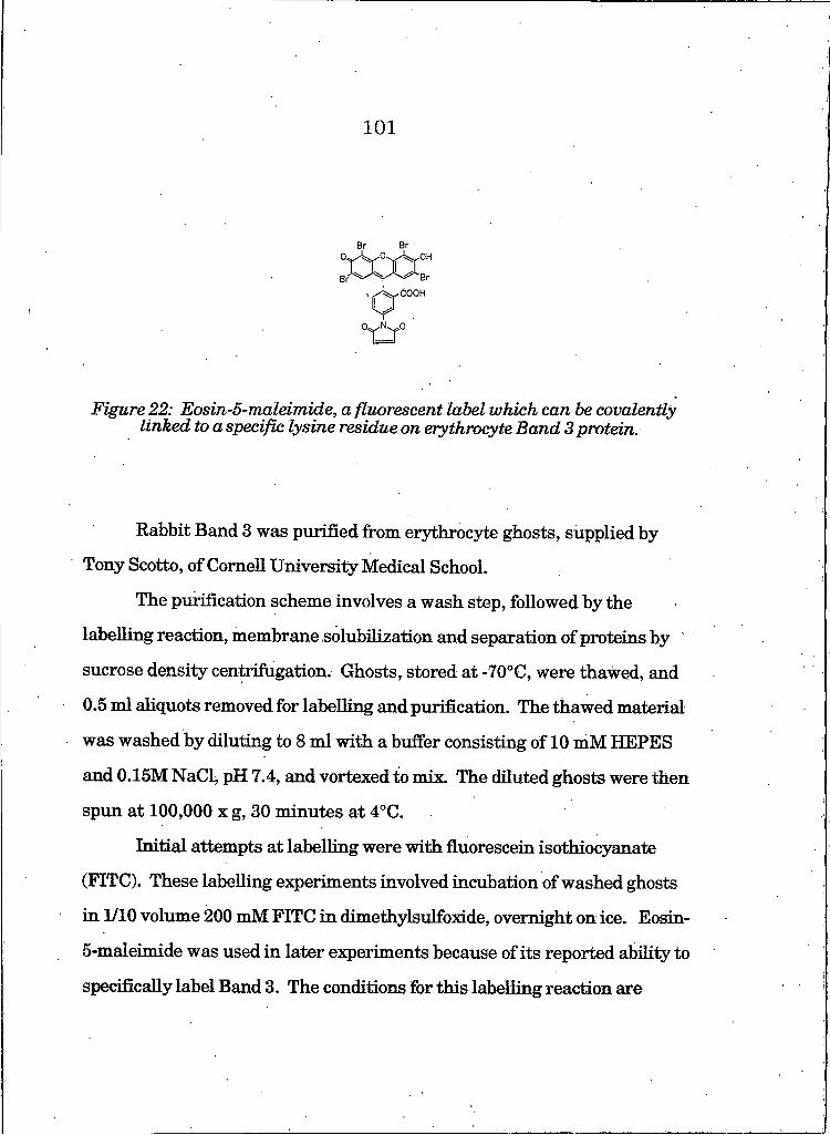
101
Figure 22: Eosin-5-maleimide, a fluorescent label which can be covalently linked to a specific lysine residue on erythrocyte Band 3 protein.
Rabbit Band 3 was purified from erythrocyte ghosts, supplied by
Tony Scotto, of Cornell University Medical School.
The purification scheme involves a wash step, followed by the
labelling reaction, membrane solubilization and separation of proteins by
sucrose density centrifugation. Ghosts, stored at -70°C, were thawed, and
0.5 ml aliquots removed for labelling and purification. The thawed material
was washed by diluting to 8 ml with a buffer consisting of 10 mM HEPES
and 0.15M NaCli pH 7.4, and vortexed to mix. The diluted ghosts were then
spun at 100,000 x g, 30 minutes at 4°C.
Initial attempts at labelling were with fluorescein isothiocyanate
(FITC). These labelling experiments involved incubation of washed ghosts
in 1/10 volume 200 mM FITC in dimethylsulfoxide, overnight on ice. Eosin-
5-maleimide was used in later experiments because of its reported ability to
specifically label Band 3. The conditions for this labelling reaction are
1 0 2
addition of 0.8 ml 0.5 mg/ml eosin-5-maleimide in buffer (10 mM HEPES,
150 mM NaCl) to 2 ml RBC ghosts, and room temperature incubation for
at least two hours in the dark [15]. The reaction may be stopped and
unreacted label removed by 2 washes in 40 to 50-fold excess volume of
buffer.
Pelleted membranes were solubilized by addition of solubilization
buffer containing 100 mM octylglucoside, 0.25 M sucrose, and IOmM
HEPES, pH 7.4. Thorough vortexing to disperse the pellet was followed by
an incubation of at least 10 minutes on ice. After sp in n in g at 100,000 x g, I
hour at 4°C, the supernatant was loaded onto a sucrose gradient.
Gradients were used to separate Band 3 from other solubilized
membrane proteins, primarily glycophoiin. 28 ml 10-80% sucrose
gradients were prepared, using 10 mM HEPES and 10 mM octylglucoside,
pH 7.4 for buffer, in polycarbonate tubes. Aliquots of supernatant, 600 gl
each, were loaded and the gradients spun 60-72 hours at 59K rpm in a
Sorvall Ti 60 rotor (375,000 x g). The suspected protein band was identified,
at approximately 50% sucrose, by visual identification of the pink band,
colored by the eosin-5-maleimide label on the protein.
Electrophoresis on 10% polyacrylamide gels and detection by silver
stain [18] were used to analyze for protein content. Quantitation of protein
in gradient fractions was by Lowry assay [10].
Reconstitution to form proteoliposomes was accomplished by mixing
gradient samples containing protein and liposomes, followed by dialysis to
remove detergent and sucrose. A microfuge tube was prepared for dialysis
by boring a hole through the cap with a hot cork borer. Samples of protein
103
and liposome solutions were mixed in the tube, which was then topped with
rinsed Spectracor dialysis tubing (12-14K molecular weight cutoff), and
capped. The tube was inverted, tapped to get all liquid in contact with the
membrane, and placed into buffer (held either with a styrofoam float or with
clamps) for dialysis. Samples were dialyzed against 400 ml 10 TnM
HEPES, 150 mM NaCl, pH 7.4 for 3 hours, changing buffer hourly.
Protein to lipid molar ratios covered the range from 1:100 to 1:1000.
Such ratios were considered appropriate for experiments where disruption
of the liposome bilayer was to be avoided, but fluorescent label needed to be
visible.
Proteoliposome solutions were then introduced to cultured cells at
total lipid concentrations equal to those used in liposome experiments.
Protocols were identical to the liposome experiments described in chapter 3,
as were analyses in both fluorescence microscopy and resonance energy
transfer experiments. Energy transfer was traced by incorporating
rhodamine labelled PE into liposomes, which is able to act as resonance
energy donor to the eosin group covalently linked to the Band 3 protein.
Results
SDS-PAGE analysis on fractions collected from sucrose gradients
from Band 3 purification revealed an isolated band of apparent molecular
weight 95 kD in fractions containing approximately 50% sucrose
(determined by refractive index) (see figure 22). Total protein content in

104
Figure 23: Electrophoresis of gradient fractions from Band 3 purificatwn protocol, with silver stain. Fractions at 45-55% sucrose contain a protein of
approximately 98 kDa (see arrow).
these fractions, determined by LowryAssay [10], ranged from 0.15 to 0.30
mg/ml. The presence of label in these bands, apparent by their p in k color,
suggested that eosin-5-maleimide had conjugated to Band 3 protein.
Attempts at dialyzing sucrose from gradient fractions before
introduction of liposomes were problematic. Precipitation of protein
occurred frequently at this step. Incorporation could be managed more
successfully by first adding protein in sucrose to preformed bposomes, and
then dialyzing against buffer containing no detergent.
Some experiments circumvented protein precipitation by introducing
additional detergent at the dialysis stage, to bring octylglucoside
concentration to well above its critical micellar concentration. Protein
solution was mixed with 1/4 volume 100 mM octylglucoside in HEPES
buffer to bring the final detergent concentration to 25-30 mM. Liposomes
were then added, and the mixture dialyzed against 400 ml detergent-free
buffer for 3 hours, changing buffer hourly.
Proteohposomes using DOTAP or DC Cholesterol as lipid
components frequently precipitated out of solution in minutes to hours after
introduction of protein. Proteoliposomes composed with these compounds
also appeared somewhat aggregated immediately after preparation when
viewed under the fluorescence microscope. Aggregation seemed to be the
result of protein incorporation. Controls using buffers and sucrose solutions
used in purification in the presence of liposomes (with protein the only
component missing) did not aggregate. However, the proteoliposomes did
maintain their ability to accumulate at cell membranes. Cell treatments
105
106were performed as soon as possible after liposome production (within 15
minutes) to minimize aggregation.
Analysis of DOPEZDOTAP proteoliposome treated monolayer cell
cultures by fluorescence microscopy revealed an interaction somewhat
similar, but importantly different, than that seen in liposome treated
cultures. As in liposome treated cells, interaction between the labelled
material (protein or lipid) and the cell membrane was maintained after
rinsing the cells. However, the labelling was more punctate, with larger
fluorescent areas distributed less evenly across the cell surface (see figure
24).
Similar results were seen when DC Cholesterol liposomes were used,
although this liposome formulation was less apt to precipitate on dialysis.
In some experiments, DC Cholesterol proteoliposomes were found to label
cells more evenly, and with finer patches of fluorescence, than
proteoliposomes containing DOTAP.
In both instances, labelling never occurred at the junctions between
closely apposed cells. This suggested that label was not internalized but
existed at the extracellular surface.
In contrast, proteoliposomes made of phosphatidylserine (DOPS) and
DOPE (1:1 molar ratio) produced no fluorescence in treated cultures.
Attempts to stimulate fusion with PEG in some cases resulted in
diffuse fluorescence in a small percentage of cells (see figure 25). However,
PEG treatment killed cells, as evidenced by their subsequent inability to
exclude the dye, Trypan Blue [26].
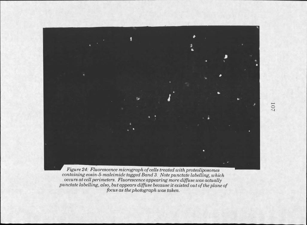
Figure 24: Fluorescence micrograph o f cells treated with proteoliposomes containing eosin-5-maleimide tagged Band 3. Note punctate labelling, which occurs at cell perimeters. Fluorescence appearing more diffuse was actually
punctate labelling, also, but appears diffuse because it existed out of the plane offocus as the photograph was taken.
107
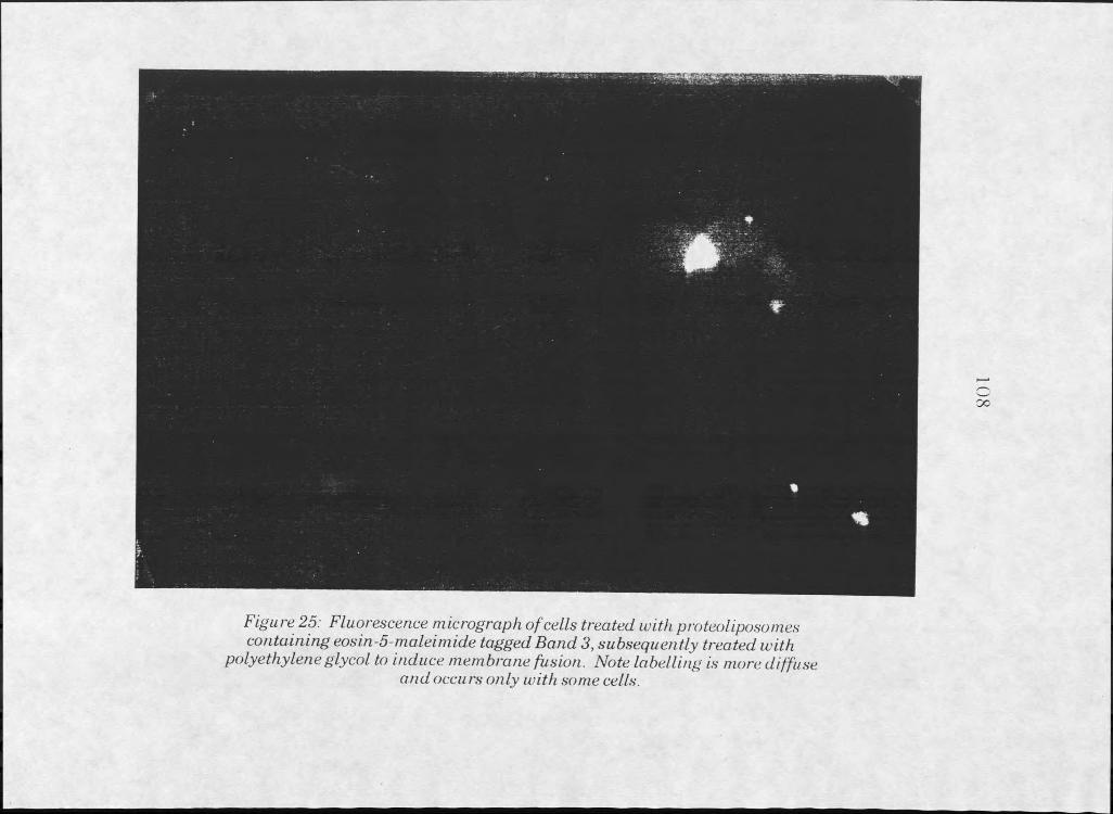
Figure 25: Fluorescence micrograph of cells treated with proteoliposomes cojitaining eosin-5-maleimide tagged Band 3, subsequently treated with
polyethyleneglycol to induce membrane fusion. Note labelling is more diffuseand occurs only with some cells.
109
Resonance energy transfer experiments were also carried out on
suspended cells exposed to solutions containing proteoliposomes. An
experiment to identify the peak emission of eosin-5-maleimide conjugated to
Band 3 (without the presence of cells) indicated that em iss io n peaked near
610 nm, when excited at 525 nm (where fluorescein would emit). Later,
experiments monitored emission at 610 nm to search for changes in
transfer to eosin-5-maleimide tagged Band 3 after proteoliposomes were
incubated with cells.
In all instances, experiments insignificant decreases in the amount of
energy transfer after introduction of the proteoliposome solutions to the cell
suspensions. Absolute intensities were low, however, and significance of the
signals measured was questionable. Signal to noise ratios were low,
perhaps due to the presence of cells or aggregates of cells passing before the
excitation beam. Noise is less significant on samples treated with protein in
detergent (see figure 25).
Discmssion
Investigation of the interactions of proteoliposomes with cultured
cells have contributed valuable information to studies of cationic liposome
interactions with cells. Cationic liposomes were able to deliver membrane
protein to the cell surface, but in the system studied, this was not adequate
for transfer of the protein to the target cell.
Cationic liposomes appear to be less stable in the presence of protein
than liposomes composed of neutral phospholipids. Addition of protein to
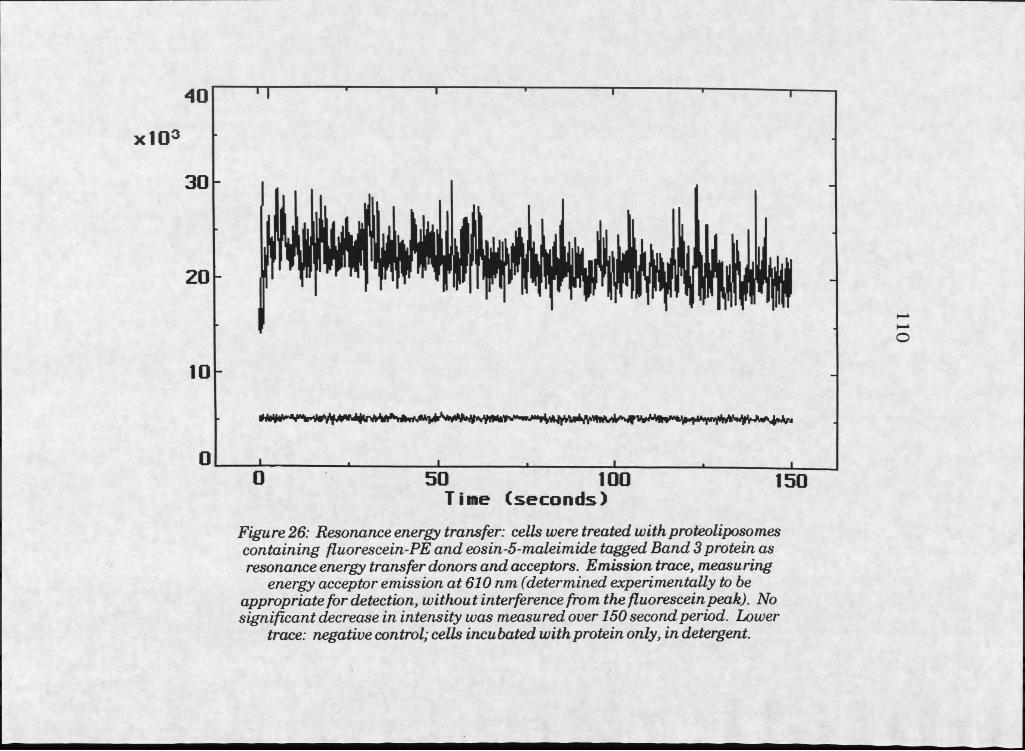
xIO3
3 0
40
20
10
0
Figure 26: Resonance energy transfer: cells weretreatedwithproteoliposom.es containing fluorescein-PE and eosin-5-maleimide tagged Band 3 protein as resonance energy transfer donors and acceptors. Emission trace, measuring
energy acceptor emission at 610nm (determined experimentally to be appropriate for detection, without interference from the fluorescein peak). No significant decrease in intensity was measured over 150 second period. Lower
trace: negative control; cells incubated with protein only, in detergent.
5 0 1 0 0Time (seconds)
1 5 0
no
I l l
the bilayer caused some aggregation of liposomes, and may have caused
destabilization of the liposome structure. Liposome structural integrity
may or may not be required for the delivery of material to the cell surface.
Data presented here indicates that aggregates of cationic lipid and protein
can be delivered to cell surfaces. However, transfer of protein to the target cell was not accomplished in this instance.
Ifmaintainance of Hposome structure is critical for successful
deHvery of membrane protein, development of carrier Hposomes wiH require
consideration of the added requirements for bilayer stability. Improved
stabiHty may require the use of stabilizing molecules, such as
phosphatidylchohne or phosphatidylserine, in liposomes.
Because deHvery to the cell surface with cationic liposomes was
inadequate for protein incorporation into ceU membranes, development of
an efficient deHvery vehicle may require fusogenic proteins to stimulate membrane fusion.
1 1 2
Refereiaces Cited
1. Newton, AC., Cook, S.L., and Huestis, W.H., “Transfer of band 3, the erythrocyte anion transporter, between phospholipid vesicles and cells,” Biochem isry 22(6): 6110-6117, 1983.
2. Newton, A.C., and Huestis, W.H., “Vesicle-to-cell protein transfer: insertion of band 3, the erythrocyte anion transporter, into lymphoid cells,” Biochem istry 27: 4655-4659, 1988.
3. Simon-Plas, F., Venema, K , Grouzis, J.P., Gibrat, R., Rigaud, J., Grignon, C., “Spontaneous insertion of plant plasma membrane (Hf)ATPase into a preformed bilayer,” J . Membr. Biol. 120: 51-58, 1991.
4. Scotto, AW., and Gompper, M.E., “Spontaneous incorporation of bacteriorhodopsin into large preformed vesicles,” Biochem istry 29: 7244-7251, 1990.
5. Scotto, AW., and Zakim, D., “Reconstitution of membrane proteins. Spontaneous incorporation of integral membraneproteins into preformed bilayers of pure phospholipid,” J . Biol. Chem. 263: 18500- 18506, 1988.
6. Zhang, F., Schmidt, W.G., Hou, Y., Williams, A.f., and Jacobson, K , “Spontaneous incorporation of the glycosyl-phosphatidylinositol- linked protein Thy-I into cell membranes,” Proc. Natl. Acad. Sci. USA 89: 5231-5235, 1992.
7. Engelman, D.M., and Steitz, T A , “The spontaneous insertion of proteins into and across membranes: the helical hairpin hypothesis,” Cell 23: 411-422, 1981.
8. Zakim, D., and Scotto, AW., “Spontaneous insertion of integral membrane proteins into preformed unilamellar vesicles,” Methods
- EnzymoL 171: 253-264, 1989.
9. Dittmer, J.C., and Wells, M.A, “Quantitative and qualitative analysis of lipids and lipid components,” M ethods Enzymol. 14: 482-530, 1969.
10. Lowry, O.H., Rosebrough, N.J., Farr, AL., and Randall, R.J., “Protein measurement with the Folin phenol reagent,” J . Biol. Chem. 193: 265-275, 1951.

113
11. Bear, C.E., Li, C., Kartner, N., Bridges, R J., Jensen, T.J., Ramjeesingh, M., and Riordan, J.R., “Purification and functional reconstitution of the cystic fibrosis transmembrane conductance regulator (CFTR)/' CeU 68: 809-818, 1992.
12. Lux, S.E., John, KM., Kopito, R.R., and Lodish, H.F., “Cloning and characterization of band 3, the human erythrocyte anion-exchange protein (AEl)," Proc. NatL Acad. ScL USA 86: 9089-9093, 1989. *5371*
13. SwissProt protein sequence data bank, at: gopher://gopher.nih.gov/77/gopherhb/indices/swiss/index.
14. Passow, H., “The band 3 proteins. An introduction,” Progress in CeU Res. 2:1-40, 1992.
15. Nig, E.A. and Cherry, R.J., “Influence of temperature and cholesterol on the rotational diffusion of Band 3 in the human erythrocyte membrane,” Biochem istry 18: 3457-3465, 1979.
16. Cobb, C.E., and Beth, A.H., “Identification of the eosinyl-5-maleimide reaction site on the human erythrocyte anion-exchange protein: overlap with the reaction sites of other chemical probes,” B iochem istry 29: 8283-8390, 1990.
17. Sherri Willard Argyres, personal communication (from Molecular Probes, Eugene, OR)
18. Oakley, B.R., Kirsch, D.r., and Morris, N.R., “A simplified ultrasensitive silver stain for detecting proteins in polyacrylamide gels,” Anal. Biochem. 105: 361-363, 1980.
19. Chao, A C., Dix, J.A., Sellers, M.C., and Verkman, AS., “Fluorescence measurement of chloride transport in monolayer cultured cells,” B iophys.J. 56: 1071-1081, 1989.
20. Chao, AC., Widdicombe, J.H., and Verkman, AS., “Chloride conductive and cotransport mechanisms in cultures of canine tracheal epithelial cells measured by an entrapped fluorescent indicator,” J . M em brane Biol. 113: 193-202, 1990.
21. Fong, P., Ulsley, N P., Widdicombe, J.H., and Verkman, AS., “Chloride transport in apical membrane vesicles from bovine tracheal epithelium: characterization using a fluorescent indicator,” J . M em brane Biol. 104: 233-239, 1988.
114
22. Verkman, AS., “Development and biological applications of chloride- sensitive fluorescent indicators,” Am. J . Physiol. 259: C375-C388, 1990.
23. Hlsley, N P., and Verkman, AS., “Membrane chloride transport measured using a chloride-sensitive fluorescent probe,” Biochem istry 26: 1215-1219, 1987.
24. Dunn, S.M.J., and Thuynsma, R.P., “Reconstitution of purified GABAa receptors: ligand binding and chloride transporting properties,” Biochem istry 33: 755-763, 1994.
25. Dunn, S.M.J., Shelman, R A , and Agay, M.W., “Fluorescence measurements of anion transport by the GABAa receptor in reconstituted membrane preparations,” Biochem istry 28: 2551- 2557, 1989,
26. Kaltenbach, J.P., Kaltenbach, M.H., and Lyons, W.B., “Nigrosin as a dye for differentiating live and dead ascites cells,” Exp. Cell Res. 15: 112-117, 1958.
1 1 5
- CHAPTEE 5
FUSION PROTEINS AND RECEPTOR MEDIATED PROCESSES
Introduction and Motivation
The problem of protein delivery via liposomal vectors is one of
efficiency. The results of the experiments presented previously would
suggest that the mechanism occurring in liposome-mediated transfection is
too inefficient for delivery of membrane proteins to cells. Evidence is
mounting that the mechanism responsible for transfection with cationic
liposomes is endocytosis and subsequent disruption of the endosomal
membrane [1-3]. Such a mechanism is not very well suited to delivery of
CFTR, or other membrane proteins, to the plasma membranes of affected
cells.
It may be possible to make use of the endosomal delivery route if the
amount of material delivered is large. In the case of CFTR, particularly,
introduction of protein into the endosomal membrane may result in
rerouting to the plasma membrane via recycling pathways. Still the
problem of efficiency persists, and it will be necessary to increase the
amount of material delivered to the endosomes, beyond what is currently
possible with available lipid reagents.
116Viruses are efficient delivery systems which can be observed and
imitated in order to produce more efficient delivery vehicles. Some viruses
deliver their DNA with the aid of particular proteins whose purpose it is to
cause fusion between the viral envelope and the cell membrane. Some
viruses, like Human Immunodeficiency Virus, dock and fuse directly with
plasma membranes, whereas others, like Influenza, fuse with endosomal
membranes after uptake of the viral particle [4-6].
Fusion proteins, and in some cases peptide domains within a fusion
protein, have been identified for many viral systems [4,7,8]. Approaches
for producing delivery vehicles based on these systems include use of
fusogenic peptides, proteins, or in some cases use of entire viral coats to
stimulate fusion and delivery of foreign materials to target cells [9-13]. The
greater the similarity to virus itself, the more likely such a vehicle will
efficiently deliver its contents, but there are drawbacks to using whole viral
envelopes or proteins. Viral proteins introduced in vivo are likely to
stimulate host immune responses which will be detrimental, either by
lowering infectivity of the vehicle, or by causing damaging inflammation.
After liposomes proved ineffective for the delivery of CFTR to cells,
the logical next step appeared to be investigation of systems with fusion
peptide or protein components. Identifying the particular viral system with
the most promise for efficient delivery was the first challenge. Influenza
virus envelopes, or virosomes, appeared to be an efficient delivery vehicle.
Fusion events between Influenza and host cells are well characterized, in
relation to other viruses, and the responsible fusion protein, hemagglutinin,
has a defined mechanism of action. Hemagglutinin acts by changing
117
conformation at lowered endosomal pH, resulting in disruption of endosomal
bilayer structure and delivery of viral contents to the cytosol [4,6].
Also pertinent to this study were investigations into the uptake of
liposomes mediated by receptor/ligand interactions. Multiple
receptor/ligand pairs have recently been reported to mediate endocytic
uptake of vesicles [14-17].
Increased uptake of foreign material by induced endocytosis may be
of use if destabilization in the endosome can be accomplished by another
method. UtQizing ligand/receptor protein interactions in conjunction with
pH sensitive liposomes could result in efficient delivery. Conjugation of
peptide or other ligands to liposomes could stimulate endocytosis of the
entire liposome through receptor mediated processes. After endocytosis,
acidification could result in disruption of the pH sensitive liposome and
efficient delivery of either hydrophfiic soluble contents, or hydrophobic
membrane components, to the cytosol or internal cellular membranes.
Steve Swain designed studies of a ligand and receptor system in order
to investigate the possibility of endosomal uptake of liposomes via this type
of protein mediated route. The specific ligand/receptor interaction used was
the vasointestinal peptide ligand (VIP) and its receptor. VIP was chosen
because a small portion of the peptide (11 amino acids) has been shown to
produce physiological changes in cells, as well as the whole peptide (28
amino acids) [18]. The VTP receptor is expressed by epithelial tissues,
including those in the lung which are candidates for eventual CFTR delivery,
and also by the immortalized cell line, HT29 [19]. It has been demonstrated
that VTP bound to receptor is rapidly internalized by this cell line [20].
1 1 8
Materials and Methods
TTiflinen a Virosomes
Initial experiments utilized viral coats from Influenza to see if
delivery by this process could deliver functional CFTR to cells. Influenza
strain A/PR/8/34 (HlNl) was supplied by the American Type Culture
Collection. This particular strain was chosen because it is thought to be
nonpathogenic. Large quantities of virus were prepared by growing virus in
the allantoic cavity of chick embryos. Eggs were infected at 8-10 days
after fertilization, by introduction of virus in a solution of nutrient broth.
Viral stock solution was at a titer of 10?-75 per ml. The solution containing
virus was introduced by first drilling a hole in the egg at the air chamber
(wide end) and then drilling a second hole over the location of the allantoic
cavity, opposite the eye spot. A 25 Ga x 5/8” needle was used to pierce the
membrane and introduce 0.1-0.2 ml viral inoculum. The holes were sealed
with paraffin, and eggs were incubated at 65-70% humidity, IOO0F, for 48
hours. Eggs were then cooled, to constrict blood vessels and reduce the
chance for allantoin contamination with blood. Virus was harvested from
the allantoin with a syringe after opening the egg at the wide end, and
visually identifying the allantoic cavity. A tthis stage, the allantoin
contains over 2 ml of clear yellow to pink fluid, and is readily distinguishable
from the other embryonic components. Positioning of the allantoic cavity is
opposite the developing embryo.
Embryos infected with virus diluted 1:100 were found to be
underdeveloped and nonviable, and were discarded. Embryos infected with
virus diluted 1:1000 were viable, and were the source of allantoic fluid for
virus isolation.
Allantoic fluid was spun 15 minutes at 10Q0 x g to remove
particulates. Supernatants were transferred to sterile polycarbonate
tubes, and spun 90 minutes at 75,000 x g, 4°C. Pellets were resuspended in
HNE buffer (10 mM HEPES, 150 mM NaCl, 0.1 mM EDTA) by forcing
them through a 22 Ga needle.
Virus samples were purified by sucrose density centrifugation on 28
ml, 20-60% weight/volume gradients in HNE buffer. 500 pi samples of
thawed virus were loaded onto the gradients, and spun at 165,000 xg, at
4°C, for at least 18 hours. One ml fractions were drawn from the top of the
gradients, and analyzed by SDS-PAGE and Coomassie Brilhant Blue stain.
Smnples of the viral stock solutions were used as markers to locate the
viral protein bands in the gel.
Virosomes were to be prepared by the procedure published by Bron,
et al. [21], but these experiments were not performed because results from
an identical study were published. Scheule, et al. reported the successful
transfer of CFTR protein to cultured epithelial cells using the Influenza
fusion protein, hemagglutinin [22].
VHP Receptor Studies
Peptide-conjugated liposomes were prepared to attempt receptor-
mediated endocytosis of liposomes. This system made use of a ligand and
119
receptor interaction which, in normal cells, stimulates receptor mediated
endocytosis. Coupled with the destabilizing properties of cationic liposome
materials, efficient delivery may be possible by this route, without
introduction of immunogenic proteins to the cell.
Liposomes which could bind the VIP receptor expressed at the
surface of HT29 cells were prepared by conjugating the VIP to a lipid
anchor. The 11-residue VEP fragment (HSDAVFTDNYT) was produced on
a peptide synthesizer (MSU peptide synthesis facility), with an additional
cysteine residue added at the carboxy-terminal end for conjugation to lipid.
Liposomes were formed from phosphatidylcholine, cholesterol, and
l,2-dilaureoyl-sn-glycero-3-phosphoethanolamine-N-[4-(p-
maleimidophenyDbutyrate] (MBP-PE) in a molar ratio of 10:3:1, to a final
concentration of approximately 10 mg/ml, by extrusion through a
membrane with pores of 50 nm diameter.
Lipids were dried from chloroform solutions by argon flow, then for 20
minutes in a rotovap, and finally for 15 additional minutes under vacuum in
a dessicator. lip id was rehydrated in PBS, pH 6.6, containing 10 mM
calcein. Liposome formation was conducted first by spinning on the
rotovap, with glass beads to stir the flask, until the solution was an even,
milky white. The lipid suspension was then subjected to five cycles of
freezing and thawing (freezing in liquid nitrogen and thawing at room
temperature), and finally was forced through an extrusion device fitted with
a 50 nm filter. The solution was passed through the filter 7-10 times.
Separation of the unencapsulated calcein was carried out by passing
the liposome suspension over a 10 cm x 7 mm Sepharose CIr4B column, in
1 2 0
1 2 1
PBS, pH 6.6. Liposomes eluted in the void volume, identified by the
passage of the first yellow calcein band through the column.
The peptide was covalently linked to a synthetic phospholipid, MBP-
PE, through reaction of the sulfhydryl on the cysteine with a maleimide
group on the phospholipid. The linkage reaction was performed after
formation of liposomes by extrusion, by overnight incubation, with stirring,
of I ml liposomes and 1.3 mg peptide in 240 |xl PBS, followed by removal of
excess peptide over a 10 cm x 7 mm Sepharose CL-4B column (as before).
Liposomal fractions were identified by observation of the elution of a yellow
band containing calcein, incorporated into the liposomes, from the column.
Peptide conjugated liposomes were then used to treat HT29 cells as
in liposome or proteoliposome experiments described earlier.
R esults
ITnfhxenza Virosomes
Virosome studies were cut short when a paper was published,
describing the successful transfer of CFTR protein to epithelial cells, using
liposomes containing influenza hemagglutinin [22],
V lP R eceptor studies
Experiments using the VIP liposomes and HT29 cells expressing the
VEP receptor resulted in clearly negative results. Although calcein
fluorescence was clearly present throughout the experimental procedure, no
distinguishing fluorescence appeared in treated cultures versus untreated
controls. Complicating matters, there was some nonspecific fluorescence in
1 2 2
controls. Complicating matters, there was some nonspecific fluorescence in
all cells, including the negative controls. Investigation into this problem
showed that the HT29 cells autofluoresce significantly. It did not appear
that calcein was delivered to cells in these experiments, although dim
fluorescence may have been obscured by the autofluorescence of the cells.
Follow up experiments, using fluorescein-PE, were performed
because labelhng we had observed with this label appeared bright, and
would be easily distinguishable from the diffuse, dim autofluorescence.
These experiments, however, produced no bright fluorescence in treated
cultures, suggesting that little or no interaction between the liposomes and
cell membranes was occuring.
Discussion
Better defined viral mimics, perhaps utilizing the purified influenza
hemagglutinin in liposomal bilayers with defined contents, may prove useful
for more efficient transfer of exogenous material to cells. Concerns exist
over safety and immunogenicity with the introduction of viral protein in
vivo, however.
The VIPATP receptor experiments were an attempt at imitating the
viral docking and endocytosis process, using nonimmunogenic materials to
stimulate the endocytosis. However, we collected no data indicating that
this occurred in our experiment.
However, other ligand/receptor pairs maybe more able to stimulate
uptake. Internalization of liposome contents through receptor mediated
123endocytosis has recently been reported with asialofetuin, transferrin, and
folate [14-17].
Our results did suggest that it is possible to form liposomes with
linked peptides, but that the interactions of such peptides with receptors on
target cells may be more complex than expected. Trial and error, and
diagnosis of what properties of the peptide are most likely to encourage a
natural ligand/receptor interaction, should lead to improved design and the
possible effectiveness of this method for macromolecule delivery.
124
References Cited
1. Daleke, D.L., HQng, K , and Papahadjopoulos, D., “Endocytosis of liposomes by macrophages: binding, acidification and leakage of liposomes monitored by a new fluorescence assay,” Biochim. Biophys. Acta 1024: 352-356, 1990.
2. Friend, D.S., Papahajopoulos, D., and Debs, R J., “Endocytosis and intracellular processing accompanying transfection mediated by cationic liposomes,” Biochim. Biophys. Acta Bio-membr. 1278: 41-51, 1996.
3. Wrobel, I., and Collins, D., “Fusion of cationic liposomes with mammalian cells occurs after endocytosis,” Biochim. Biophys. A cta Bio-membr. 1235: 296-304, 1995.
4. Schoch, C., and Blumenthal, R., “Role of the fusion peptide sequence in initial stages of influenza hemagglutinin-induced cell fusion,” J . Biol. Chem. 268: 9267-9274, 1993.
5. Stegmann, T., Hoekstra, D., Scherphof, G., and Wilschut, J ., “Kinetics of pH-dependent fusion between influenza virus and liposomes,” Biochemistry 24: 3107-3113, 1985.
6. Alford, D., Ellens, H., and Benz, J., “Fusion of influenza virus with sialic acid-bearing target membranes,” Biochem istry 33: 1977- 1987, 1994.
7. Glushakova, S.E., Omelyanenko, V.G., LUkashevitch, I.S>, Bogdanov, A A Jr., Moshinikova, AB., Kozytch, AT., Torchilin, V.P., “The fusion of artificial lipid membranes induced by the synthetic arenavirus fusion peptide,” Biochim. Biophys. Acta Bio-membr. 1110: 202-208, 1992.
8. Nieva, J.L., Nir, S., Muga, A , Goni, F.M., and Wilschut, J., “Interaction of the HIV-I fusion peptide with phospholipid vesicles: different structural requirements for fusion and leakage,” Biochem istry 33: 3201-3209, 1994.
9. Parent©, R A , Nir, S., and Szoka, F.C. Jr., “Mechanism of leakage of phospholipid vesicle contents induced by the peptide GALA,” Biochem istry 29: 8720-8728, 1990.
10 . Bron, R , OrtizvA., Dijkstra, J., Stegm aim, T., and Wilschut, J., “Preparation, properties, and applications of reconstituted influenza virus envelopes (virosomes),” M ethods Enz. 220: 313-331, 1993
11. Epand, R.M., Cheetham, J.J., Epand, R.F., Yeagle, P.L., Richardson, C.D., Rockwell, A., and Degrade, W.F., “Peptide models for the membrane destabilizing actions of viral fusion proteins,” Biopolymers 32: 309-314, 1992.
12. Harmsen, M.C., Wilschut, J., Scherphof, G., Hulstaert, C., and Hoekstra, D., “Reconstitution and fusogenic properties of Sendai virus envelopes,” E w . J . Biochem. 149: 591-599, 1985.
13. Cantell, K , and Hirvonen, S., “Preparation and assay of Sendai virus,” M ethods Enz. 78: 299-301, 1981.
14. Kara, T., Kuwasawa, H., Aramaki, Y., Takada, S., Koike, K , Ishidate, K , Kato, H., and Tsuchiya, S., “Effects of fusogenic and DNA-binding amphiphilic compounds on the receptor-mediated gene transfer into hepatic cells by asialofetuin-labelled liposomes,” Biochimu Biophys. A cta Bio-membr. 1278: 51-58, 1996.
15. Ferkol, R., Perales, J.C., Eckman, E., Kaetzel, C.S., Hanson, R.W., And Davis, P.B., “Gene transfer into the airway epithelium of animals by targetting the polymeric immunoglobulin receptor,” J . Clin. Invest. 95: 493-502, 1995.
16. Cheng, P.W., “R eceptor ligand-facilitated gene transfer: enhancem ent o f liposom e-m ediated gene, transfer and expression by transferrin ,” Htamu Gene Ther. 7: 275-282, 1996.
17. Lee, R.J., and Huang, L., “Folate-targetted, anionic liposome- entrapped polylysine-condensed DNA for tumor cell-specific gene transfer,” J . Biol. Chem. 271: 8481-8487, 1996.
18. Muller, J.M., El Battari, A , Ah-Kye, E., Luis, J., Ducret, F., Pichon,J., and Marvaldi, J., “Internalization of the vasoactive intestinal peptide (VIP) in a human adenocarcinoma cell line (HT29),” Eur. J . Biochem. 152: 107-114, 1985.
19. Goossens, J.F., Pommery, N., Lohez, M., Pommery, J., Helbecque, N., Cotelle, P., Lhermitte, M., and Henichardt, J.P., “Antagonistic effect of a vasoactive intestinal peptide fragment, vasoactive intestinal peptide (1-11), on guinea pig trachea smooth muscle relaxation,” M olecular P h arm . 41: 104-109, 1991.
125
20. Ichikawa, S., Sreedharan, S.P., Owen, R.L., Goetzl, E.J.,“Immunochemical localization of type I VIP receptor and NK-l-type substance P receptor in ra t lung,” Am. J. Physio. JLmag C eil Mol. PhysioL 268: L584-L588, 1995.
21. Bron, R , Ortiz, A., Dijkstra, J., Stegmann, T., and Wilschut, J.,Preparation, properties, and applications of reconstituted influenza
virus envelopes (virosomes),” M ethods EnzymoL 220: 313-331 1993.
22. Scheule, R.K, Bagley, R.G., Erickson, AL., Wang, KX., Fang, S.L., Vaccaro, D., O’riordan, C.R., Cheng, S.H., and Smith, AE., 6Deliveiy of purified, functional CFTR to epithelial cells in vitro using influenza hemagglutinin,”Am. J . Respir. Cell MoL Biol. 13: 330-343, 1995.
127 .
CHAPTER 6
ISOLATION OF CLATHRIN COATED VESICLES: THE ROLE OF VESICLE
TRAFFICKING
Introduction and Motivation
CFTR is present and functional in endosomes, and has been reported
to be routed through recycling pathways [1,2]. The mechanism of cationic
liposome delivery in transfection is also reported to involve endocytosis
[3,4]. The development of an immunochemical assay for endosomal CFTR
would simplify investigations of liposome-mediated protein delivery.
Quantitation of CFTR from immunoblots could be performed with a
densitometer.
Using this assay to improve understanding of CFTR?s trafficking
could aid design of vectors for protein delivery. Development of an
endosomal CFTR assay would allow quantitative determination of protein
delivery to endosomes. Quantitative determination of CFTR in endosomes
would allow data on the efficiency of one delivery vehicle versus another to
be obtained, and could be used to determine the optimal conditions for
delivering CFTR to cells by endocytosis.
Additionally, the ability to determine CFTR concentrations in
endosomes would enlighten other types of investigations. The assay would
be useful in studies of endocytosis and recycling. CFTR’s presence in
endosomes is documented, but not understood. It appears that CFTR does
not alter the pH of the endosome [5]. Understanding the role CFTR plays in
the endosome could be useful in the search for new methods of normalizing
ion transport in defective cells.
With the aid of an endosomal CFTR assay, methods for manipulating
endosomal traffic in order to increase CFTR concentrations at the plasma
membrane could be envisioned. Inhibition of endocytosis may result in an
increase in CFTR functioning at the plasma membranes in defective cells,
and may improve ion conductance.
Development of a quantitative assay for CFTR from endosomes first
required the successful colocalization of CFTR and clathrin, the marker
protein for coated endosomal vesicles, from endosomal vesicle-enriched cell
fractions.
1 2 8
Materials and Methods
Isolation of coated vesicles was based on a procedure published by
Pearse for isolation of coated vesicles from excised tissues [6]. The method
is based on purification in D20/Ficoll gradients, after homogenization of
cells by shearing.
Empirical evidence suggests Ficoll /D2O gradients are most
appropriate for vesicle isolations [7]. Ficoll, a polymer of sucrose with
epichlorohydrin, is useful in situations where high osmolality must be
129
avoided. High osmolality would lead to deformation or disruption of
endocytic vesicles. Deuterium oxide has a density much closer to that of
biological organelles than water. Use of these compounds makes it possible
to prepare gradients covering the necessary density range, without osmotic disruption of vesicles.
HT29 cells were used because they express CFTR [8]. Cells were
grown up in monolayer cultures to 4 x IO8. Trypsinization and physical
removal by scraping, or sometimes simply scraping, were used to remove
cells from the growth surface. HT29 cells adhere tenaciously to surfaces,
though, making removal by scraping difficult. Cells were washed, pelleted
at 800 x g, and resuspended in isolation buffer (10 mM HEPES, 150 mM
NaCl, I mM EGTA, 0.5 mM MgClg, 0.02% NaNg, and 0.2 mM
pheynylmethylsulfonyl fluoride (PMSF), pH 7.2). Plasma membranes were
then disrupted by shearing with a Tenbroeck tissue homogenizer (clearance
of0.004-0.006”), followed by at least 15 forceful passes through a 25 Ga
needle. Disruption was analyzed by viewing a sample of cell homogenate
with a microscope. Nuclei were pelleted by centrifugation for 30 minutes at
1000 x g. Supernatants were treated with 10 U/ml RNase in a 30 minute,
room temperature incubation. Samples were spun again for I hour at
55,000 x g. Pellets were collected by resuspending in a small volume of
isolation buffer, and loaded onto 10-90% gradients of Ficoll in D2O.
Crude coated vesicles, which run to the bottom of the gradient, were
analyzed by dot blot or SDS-PAGE and immunoblot with goat ariti-clathrin
antibody (Sigma product C8034). Secondary antibody was mouse anti
goat, conjugated to alkaline phosphatase (Sigma product A8062).
130
Detection was with nitro blue tetrazolium [9]. CIathrin bands a t an
apparent molecular weight of 180 kD.
CFTR was detected by incubation of blots with mouse anti-CFTR
antibody (Genzyme product 1660-01), detected again with alkaline
phosphatase conjugated secondary antibody (rabbit anti-mouse, Sigma
product M6024), and incubation in nitro blue tetrazolium [9]. Because of
concerns over sensitivity of direct methods, immunoprecipitation of CFTR
was also attempted.
Immunoprecipitation is a more sensitive method than SDS-PAGE on
vesicle isolates for detection of small amounts ofCFTR. This technique
was used to detect CFTR in crude coated vesicle preparations from HT29
cells. A protocol for immunoprecipitation with a monoclonal mouse anti
human CFTR antibody (Genzyme product 1660-01) was available from the
antibody supplier. This protocol describes the immunoprecipitation of
CFTR from whole, lysed T84 cells. The attempt to isolate CFTR from
clathrin vesicles would require greater sensitivity than the reported method,
because our vesicle preparation isolates a subpopulation of the total
expressed CFTR, most of which is in the plasma membrane.
Gradient fractions enriched for coated vesicles were
immunoprecipitated as follows. Three hundred fifty microliter aliquots were
added to anti-CFTR antibody (10 pi), and incubated on ice for 90 minutes.
Pansorbin Staph A cells (from Calbiochem) at I mg/ml were precoated with
polyclonal rabbit anti-mouse IgG by incubating equal volumes (100 pi each)
at 4°C for I hour. Unbound IgG was then removed by washing the cells
three times, each time mixing cells with a I ml portion of RIPA buffer (150
131
mM NaCl, 1.0% Triton X-100,1% sodium deoxycholate, 0.1% SDS, 50 mM
Tris-HCl, pH 8.0), and pelleting cells with a 30 second spin at 1000 x g.
The coated Staph A conjugated to IgG was taken up in 100 pi buffer,
and 20 pi aliquots were added to each experimental sample. After a 30
minute incubation at 4°C, Staph A cells were again pelleted, washed, and
repelleted three times with RIPA buffer, and a fourth time in HEPES
buffered saline, pH 7.4. The pellet was then mixed with 50 pi Protein
Kinase A buffer (50 mM Tris-HCl, 10 mM MgCl2, pH 7.5, with 100 pg/ml
bovine serum albumin); 10 pCi y[32P] ATP and 50 ng Protein Kinase A were
added, to label CFTR with radioactive phosphate.
After incubating 60 minutes at 30°C, the reaction was stopped by
addition of 0.5 ml RIPA buffer, followed by pelleting and washing twice with
the same buffer. The final pellet was resuspended in 6x SDS sample buffer,
and was loaded onto a 7% polyacrylamide gel. Samples were run at 30 mA
for 45 minutes, and the gel was dried. Autoradiography of CFTR was
carried out by exposing the dried gel to X-ray film for 12 hours.
Results
Western blots were then performed to identify clathrin in gradient
fractions from vesicle isolations. Fractions taken from the bottom of the
gradients contained an anti-clathnn immunoreactive protein of greater
than 180 kD. These fractions were used to attempt immunoprecipitation of
CFTR (see figure 2 7).
Immunoprecipitation (using anti-CFTR) on fractions thought to
contain clathrin revealed a band at approximately 170 kD, possibly CFTR.
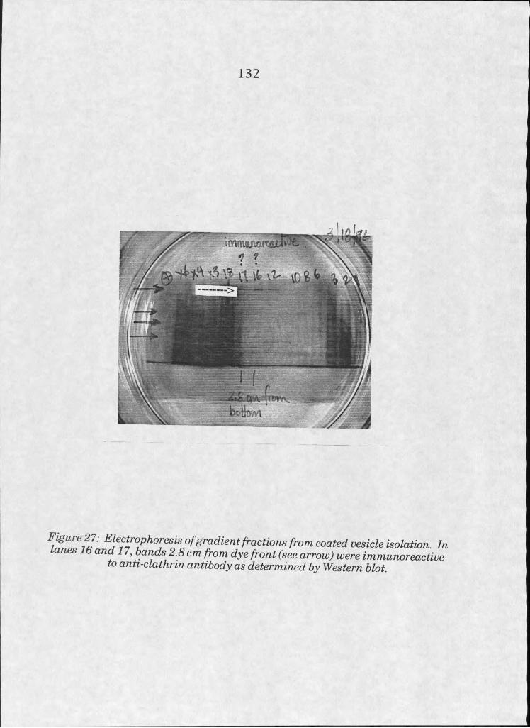
132
/rom cooW w%%cZe KoZafm^. /m ^ ^^ 2.* cm /m m ^ e (see OrrowJ were immwooreoedue
to anti-clathrin antibody as determined by Western blot.
133
Some nonspecific bands, or bands of lower molecular weight fragments of
CFTR, however, were also visible (see figure 28).
Discussion
CFTR is expressed at low levels, and endosomal CFTR is only a
subpopulation of all expressed CFTR, so studies of this protein in the
endosome require high sensitivity for detection. Development of a
semiquantitative assay, utilizing a densitometer to calculate the degree of
immunoreaction, may be possible, but will require either highly sensitive
methods or greater amounts of starting material.
Greater sensitivity could be achieved by improving cell
homogenization procedures. Trypsinization has been reported to make cell
homogenization more difficult, and this may have contributed to some
difficulty encountered disrupting the HT29 cells. More efficient cell
disruption could possibly be accomplished by using lower clearance
homogenizers, also. Microscopic identification of efficient cell disruption,
without destruction of internal membranes, is a skill developed by practice.
Many repeated attempts may be necessary to learn to identify optimal
conditions for cell disruption and isolation of intact internal vesicles.
Use of an alternate cell line which expresses CFTR may also
improve detection in the immunoassays. The T84 line has commonly been
used for CFTR studies; like HT29, it originates from colon epithelium [10].
T84 cells were grown for a short time for these experiments, but difficulty
maintaining the cultures prompted us to turn to experiments with HT29
cells.

134
v
t
Standards (kD)
180116-84
f7>"f
58 48.5
-
-26.6
28: Immunoprecipitation o f CFTR from fractions containing 180 kD protein suspected to be clathrin. Banding is faint, but present, in experimental
samples (see arrow). Exposure time 14 hours.
135
CFTR and clathnn were colocalized in cell fractions assumed to be
ennched for endosomes. Colocalization of these proteins corroborates other
reports of CFTR in endosomes. Development of a quantitative assay for
CFTR in endosomes, based on this procedure, may be possible by
densitometry, if signals from immunoblots are sufficiently strong. Such an
assay would be useful for varied studies of CFTR and vesicle trafficking of
membrane proteins.
136
Refegemces Cited
1. Biwersi, J., and Verkman, AS., “Functional CFTB in endosomal compartment of CFTB-expressing fibroblasts and T84 cells,” Am. J . PhysioL Cell PhysioL 266: C14-C156, 1994.
2. Bradbury, N A , Cohn, J.A., Venglarik, C.J., and Bridges, R.J., “Biochemical and biophysical identification of cystic fibrosis transmembrane conductance regulator chloride channels as components of endocytis clathiin-coated vesicles,” J . Biol. Chem. 269: 8296-8302, 1994.
3. Wrobel, I., and Collins, D., “Fusion of cationic liposomes with mammalian cells occurs after endocytosis,” Biochim. Biophys A cta Bio-membr. 1235: 296-304, 1995.
4. Friend, D.s., Papahadjopoulos, d., and Debs, R.J., “Endocytosis and intracellular processing accompanying transfection mediated by. cationic liposomes,” Biochim. Biophys. Acta Bio-membr. 1278: 41-50, 1996.
5. Root, K.V., Engelhardt, J.F., Post, M., Wilson, J.W., and van Dyke, R.W., “CFTR does not alter acidification of L cell endosomes,” Biochem. Biophys. Res. Comm. 205: 396-401, 1994.
6. Pearse, B.M.F., “Isolation of coated vesicles,” M ethods EnzymoL 98: 320-326, 1983.
7• C u rren t Protocols in P ro te in Science. Section 4.2.18, “GradientFractionation Using D2O,” 1995.
8. Wardle, C.J., and Harris, A., “Down-regulation of CFTR expression in an HT29-derived cell line by stable antisense RNA production,” Biochim. Biophys. Acta 1261(3): 417-423, 1995.
9. Harlow, E. and Lane, D., Antibodies: A laboratory m anual. Cold Spring Harbor Laboratory Press, Cold Spring Harbor, NY, 1988.
10. Dharmsathaphom, K , McRoberts, J.A., Mandel, KG., Tisdale, L.D., and Masui, H., “A human coloic tumor cell line that maintains vectorial electrolyte transport,” Am. J . PhysioL 246: G204-208, 1984.
137
CHAPTER 7
GENERAL CONCLUSIONS
Liposome preparations have become common for cell transfection in
vitro, and have recently been investigated for potential in vivo transfection
as well [I]. However, studies on the actual molecular interactions occurring
in such experiments are rare. This study was performed in order to make
up for this lack of information, and to find underlying principles involved in
liposome/cell interactions. Additionally, exploration of the feasibility of
using liposomes to carry integral membrane proteins to cells was
undertaken. An applied goal was to find a vehicle suitable for delivery of
CFTR, the protein deficient in individuals affected by cystic fibrosis, to the
plasma membranes of cells.
The study was subdivided into several more specific problems.
Optimization of expression and purification of CFTR was carried out. The
interactions between liposomes composed only of lipid or lipid analogues and
cells were investigated. This investigation then extended to liposomes
containing protein. The results suggested that these systems did not
transfer significant amounts of material to target cells. Studies of viral and
receptor/ligand interactions were initiated in attempts to increase the
138
chances of detecting protein or lipid transfer to cells. Finally, we focussed
on increasing the understanding of CFTR trafficking, as it relates to
endosomal delivery of exogenous material.
Expressinn aTnH Purification of CFTR
It was hoped that we would be capable of preparing enough pure
protein to incorporate into liposomes, and to show that our recombinant
CFTR was active in a liposome bilayer. CFTR was successfully expressed
in the Sf9 cell line, and was purified. However, the quantities of protein
produced were less than adequate for experiments we had proposed. The
purification scheme used (from Bear et al.) involves multiple steps, which
may account for significant protein loss during purification [2]. Also,
proteolysis contributed to low yields. Proteolysis would likelybe less
troublesome if the purification could be performed at reduced temperature.
However, the SDS used to solubilize CFTR tends to precipitate at low
temperatures. The Bear procedure, in our hands, produced protein of high
purity, but in disappointingly low yields. La addition, the ion channeling
activity of purified CFTR was not detected by SPQ fluorescence
dequenching assay.
In terac tions of Liposomes w ith Cultured Cells
Our goal in investigating liposome/cell interactions was to define the
mechanism of interaction, and to determine the likelihood that protein
transfer could be accomplished by that mechanism. We focussed on
cationic liposomes, because of their effectiveness at eukaryotic cell
139
transfection [3], and on pH sensitive liposomes, because their proposed
mechanism of action may allow transfer of membrane proteins to target
cell membranes via endocytosis [4]. The cell lines HeLa, HT29, and others
were observed after treatment with fluorescently labelled liposomes.
Microscopy and resonance energy transfer experiments were used in an
attempt to identity cellular uptake of fluorescent material, and lipid dilution
as would be expected from membrane fusion events.
No evidence of lipid dilution was obtained by resonance energy
transfer in any case investigated. However, cationic liposome treatment
resulted in accumulation of fluorescent lipid at the cell surfaces, as evident
by fluorescence microscopy. The interaction between liposomes and cell
surfaces was strong enough that fluorescence could not be washed away.
pH sensitive liposome treatment did not label cells at all. Cationic
liposomes were found to be prone to aggregation, visible by eye, in the
presence of several hydrophilic labels. Cationic liposome treatments, at the
concentrations used in our protocols, were not significantly toxic to He La
cells, but were very damaging to cultures of HT29 and CFG-SV40 cells.
It is possible that newly developed liposome formulations, such as
those containing polycationic Upid analogues, will be more effective for
incorporation of lipid or other materials into target cells [5]. Phospholipids
such as phosphatidylcholine or phosphatidylserine, introduced to stabilize
cationic liposomes, may increase their effectiveness for molecular dehvery.
Such formulations are expected to remain intact longer and under more
variable conditions.
140
Evidence we have gathered supports the theory that cationic
liposomes accumulate at cell surfaces by an electrostatic interaction. Our
data indicate that only a low level of true incorporation (by fusion with
plasma membranes or by internalization through endocytosis) occurs, even
in cell transfection experiments using complexes of cationic liposomes and
DNA If the behavior of IiposomeZDNA complexes and liposomes alone is
identical, then it appears that less than approximately 10% of the material
reaching the cell membrane actually fuses with the plasma membrane or is
internalized. Results presented here do not support the application of
cationic liposomes or pH sensitive liposomes for delivery of integral
membrane proteins to cells.
Interactions of Proteoliposomes with Ciiltiireil Cftlls
The interactions occurring between cationic proteohposomes and
HeLa cells was investigated. The feasibility of protein transfer from
cationic proteohposomes to cells was investigated.
Fluorescence microscopy and resonance energy transfer techniques
were employed to search for evidence of protein transfer to HeLa cells.
Proteohposomes containing fluorescently tagged erythrocyte Band 3
protein were formed by spontaneous incorporation and by detergent
dialysis. Aggregation of proteohposomes was a continual problem, but
occurred to a varying degree from batch to batch. Using freshly prepared
proteohposomes, in order to minimize aggregation, it was shown that label
could be transferred to cell surfaces, and could not be washed away. As in
liposome experiments, no evidence of label internalization was obtained.
141
Proteoliposomes tended to label cells less, and in a more punctate fashion,
than protein-free liposomes, probably due to aggregation.
More stable bilayers, or use of protein less apt to aggregate, may
result in finer, more even labelling. Evidence presented here suggests that
the positive charge of cationic proteoliposomes does result in association of
protein with target cells. DOPS/DOPE liposomes, DOPC liposomes, and
OAZPE liposomes by contrast did not label cell surfaces.
Results presented in this study do not support the hypothesis that
protein can be transferred to cells by cationic proteoliposomes. Disruption
of bilayer structure, or aggregation of proteoliposomes, resulted in large
aggregates of material adhering to cell surfaces. Transfer of protein, if it
occurs at all, is probably less than would be necessary, in the case of CFTR
introduction. Results do support the theory that positively charged
liposomes interact strongly with the plasma membrane (which carries a
net negative charge). Such an interaction may be responsible for low levels
of lipid/DNA complex uptake adequate for cell transfection.
V iral and R eceptor M ediated Processes
In an attempt to improve efficiency of protein delivery to target cells,
we investigated the potential of using viral envelope proteins and
receptor/ligand interactions to stimulate uptake.
Use of influenza viral envelopes as carriers of CFTR to cells was
reported by Scheule, et al., cutting short our study of this system [6]. As an
alternative, peptide-conjugated liposomes were studied for their potential to
stimulate endocytosis of liposomes. We used an 11 amino acid segment of

142
the peptide ligand vasointestinal peptide (VIP), which was covalently linked
to a lipid anchor, and introduced it as part of a labelled liposome to HT29
cells which express a receptor for VIP. This system proved inadequate for
detectable uptake. No fluorescence in treated cells was observed.
Other ligand and receptor systems should be studied for their
potential as delivery vehicle components. Others have reported on the use
of similar systems, stimulating receptor mediated endocytosis with folate,
asialofetuin, and transferrin conjugated to liposomes [7-9].
The reported use of influenza virosomes to deliver CFTR is
encouraging [6], but this system will not have direct therapeutic benefit.
Potential immunogenicity of the hemagglutinin limits its use to in vitro
situations. Less immunogenic protein mediators of membrane fusion or
endocytosis may be more realistically useful. The VIPATP receptor
system, as studied here, did not show potential for usefulness in delivery of
CFTR, however.
CFTR an d Vesicle fTraffjoking
We hoped to identify CFTR in endosomes by immunochemical
methods. In so doing, we hoped to provide evidence for understanding CFTR
recycling in order a) to enable additional possibilities for protein delivery
vehicle design, b) to improve understanding of CFTR function, and c)
examine the possibilities for increasing the concentration of CFTR at the
plasma membrane by inhibiting endocytosis.
Clathrin enriched cell fractions, which are assumed to contain
endosomal vesicles, were prepared from whole cell lysates. An
1 4 3
immunoreactive band approximately the same molecular weight as CFTR
was identified in vesicle enriched fractions by polyacrylamide gel
electrophoresis and Western blotting or immunoprecipitating with anti-
CFTR antibodies.
Putative CFTR bands were somewhat faint, and the positive result
reported here should be considered with caution. Improvements in the
detection of CFTR could be gained by increasing the concentration of CFTR
in experimental samples loaded onto electrophoresis gels. This would
require starting with a larger number of cells, experimenting with
alternative cell lines, or improving the cell homogenization process to
increase the number of lysed cells, without disrupting large numbers of
internal vesicles.
Results suggest that CFTR can be detected in vesicle enriched
fractions by immunochemical methods. This supports the assertion that
CFTR is present in endosomes, and suggests that development of
quantitative method for detection of CFTR in endosome enriched cell
fractions may be possible. Furthermore, it may eventually be possible to
monitor CFTR trafficking through endocytosis and recycling pathways with
such a method. Manipulation of endosomal traffic may affect endosomal or
plasma membrane CFTR concentrations, providing valuable insight into
CFTR function and control.
144
I.
References Cited!
Caplen, N .JA lton, E.W.F.W., Middleton, P.G., Bonn, J.R ,r S i f r Y - 0a0T x ^ f ham- SR., Jeffeiy, P .K , Hodson, M.E. CouteUe, C., Huang, L., Porteou^D^., WiUiamson, R., and geddes,D.M., Liposome-mediated CFTR gene transfer to the nasal 1995 ™ °f patients with cystic fibrosis,” N ature Med. I: 39-46,
2 .
3.
Bear, C.E., Li, c., Kartner, N., Bridges, R.J., Jensen, T.J., Ramjeesmgh, M., and Riordan, J.R., “Purification and functional reconstitution of the cystic fibrosis transmembrane conductance regulator (CFTR),” Cell 68: 809-818, 1992.
Legendre, J., and Szoka, F., “Delivery of plasmid DNA into mammalian cell lines using pH-sensitive liposomes,” P h an n . Res. 9: 1235-1241, 1992,
Connor, J., Yatvin, M.b., and Huang, L., “pH-sensitive liposomes: acid mduced liposome fusion,” Proc. Natl. Acad. Sci. USA 81: 1715-
5. Sells, M.A, Li, J., and Chemoff, J., “Delivery of protein into cells using polycationic liposomes,” BioTechniques 19: 72-78, 1995.
6. Scheule, R.K, Bagley, R.G., Erickson, AL., Wang, KX., Fang, S.L Vaccaro, D., O’nordan, C.R., Cheng, S.H., and Smith, A.E., “Deliveiy of purified, functional CFTR to epithelial cells in vitro using influenza hemagglutinin,”Am. J . Respir. CeU Mol. Biol. 13: 330-343, 1995.
7. Lee, R.J., and Huang, L., “Folate-targetted, anionic liposome- entrapped polylysine DNA for tumore cell-specific gene transfer ” J BioL Chem. 271: 8481-8487, 1996.
8. Ferkol, R., Perales, J.C., Eckman, E., Kaetzel, C.S., Hanson, R.W., and Davis, P B., Gene transfer into the airway epithelium of animals by targetting the polymeric immunoglobulin receptor ” J . Clin. Invest. 95: 493-502, 1995.
9. Kara, T. Aramaki, Y., Takada, S., Koike, K , Tsuchiya, S., “Receptor-transfer of pSV2CAT DNA to a human hepatoblastoma
cell line HepG2 using asialofetuin-labelled cationic liposomes ” Gene 159(2): 167-174, 1995.

145
APPENDICES
146
APPENDIX A:
A BRIEF OVERVIEW OF AO. LIPOSOME AND PROTEOLIPOSOME EXPERIMENTS
PERFORMED ON HELA CELLS

No Comoosition Dose fua) Time Buffer A nalysis R esults/C om m entsmicroscopy (M) or
energy transfer (RET)I POPC 10, 60, .200 12 hr. DME no M viewed 24 hr. after lipid removal
POPC/POPE (1:1 molar) serum, HBSS random damage-HBSS incubation caused cell deathPOPG 60 & 200 ug doses killed all cells
200 ug killed all cells
2 POPC 100 I, 2 HBSS M viewed 8 hr. after lipid removal.3 hr. encapsulated calcein as label
no fluorescence, no damage in liposome-treated wells
3 POPG 50, 100, 200 2.75 hr. HBSS M no toxicity observed+ calcium (brightfield)
4 DOTAP/POPE 25, 50, 75, 2.25 hr. PBS M liposomes precipitated( I OmM calcein) 100, 200 no toxicity, no fluorescence observed
5 DOTAP/POPE 50, 100, 200 3.5 hr. PBS, HBSS M liposomes precipitated(50 mM calcein) 500, 1000 18 hrs. after liposome removal, no fluorescence
no toxicity
6 DOTAP/POPE 750 3.0 hr. PBS M no toxicity(200 mM calcein) no fluorescence
7 DOTAP/POPE 50, 100, 200 2.5 hr. PBS M ATP-induced fusion attempted as(200 mM calcein) positive control
no fluorescence (control failed)
147
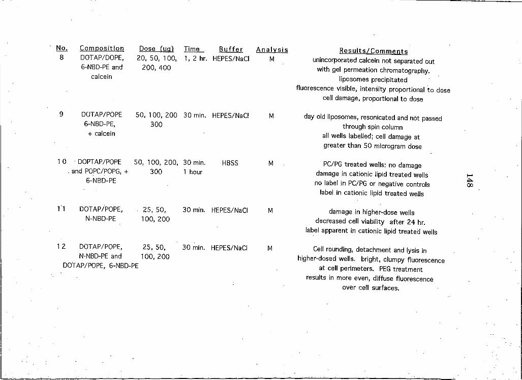
No. Composition 8 DOTAP/DOPE,
6-NBD-PE and calcein
Dose fug) Time B uffer A nalysis 20, 50, 100, I, 2 hr. HEPES/NaCI M
200,400
R esults/C om m ent s unincorporated calcein not separated out
with gel permeation chromatography, liposomes precipitated
fluorescence visible, intensity proportional to dose cell damage, proportional to dose
9 DOTAP/POPE 5 0 , 1 0 0 , 2 0 0 30 min. HEPES/NaCI M6-NBD-PE, 300
+ calcein
I 0 DOPTAP/POPE 50, 100, 200, 30 min. HBSS Mand POPC/POPG, + 300 I hour
6-NBD-PE
Tl DOTAP/POPE, . 25, 50, 30 min. HEPES/NaCI MN-NBD-PE 100, 200
I 2 DOTAP/POPE, 25, 50, 30 min. HEPES/NaCI MN-NBD-PE and 100, 200
DOTAP/POPE, 6-NBD-PE
day old liposomes, resonicated and not passed through spin column
all wells labelled; cell damage at greater than 50 microgram dose
PC/PG treated wells: no damage damage in cationic lipid treated wells no label in PC/PG or negative controls
label in cationic lipid treated wells
damage in higher-dose wells decreased cell viability after 24 hr.
label apparent in cationic lipid treated wells
Cell rounding, detachment and lysis in higher-dosed wells, bright, clumpy fluorescence
at cell perimeters. PEG treatment results in more even, diffuse fluorescence
over cell surfaces.
148
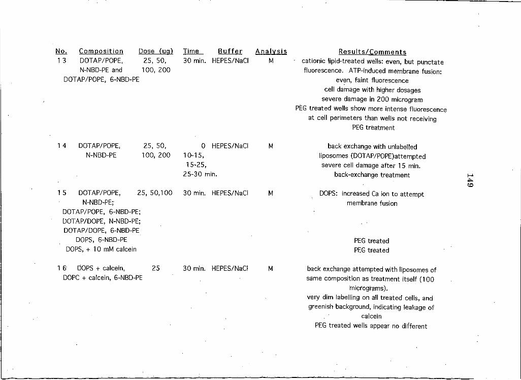
No. Corrmosition Dose fua) Time B ufferI 3 DOTAP/POPE, 25, 50, 30 min. HEPES/NaCI
N-NBD-PE and 100, 200DOTAP/POPE, 6-NBD-PE
I 4 DOTAP/POPE, 25, 50,N-NBD-PE 100, 200
010-15,15-25,
HEPES/NaCI
25-30 min.
15 DOTAP/POPE, 25, 50,100N-NBD-PE;
DOTAP/POPE, 6-NBD-PE; DOTAP/DOPE, N-NBD-PE; DOTAP/DOPE, 6-NBD-PE
DOPS, 6-NBD-PE DOPS, + 10 mM caIcein
30 min. HEPES/NaCI
I 6 DOPS + calcein, 25DOPC + calcein, 6-NBD-PE
30 min. HEPES/NaCI
A nalysisM
R esults/C om m ents■ cationic lipid-treated wells: even, but punctate
fluorescence. ATP-induced membrane fusion: even, faint fluorescence
cell damage with higher dosages severe damage in 200 microgram
PEG treated wells show more intense fluorescence at cell perimeters than wells not receiving
PEG treatment
M back exchange with unlabelled liposomes (DOTAP/POPE)attempted severe cell damage after 15 min.
back-exchange treatment
M DOPS: increased Ca ion to attempt membrane fusion
M
PEG treated PEG treated
back exchange attempted with liposomes of same composition as treatment itself (100
micrograms).very dim labelling on all treated cells, and greenish background, indicating leakage of
calceinPEG treated wells appear no different
149
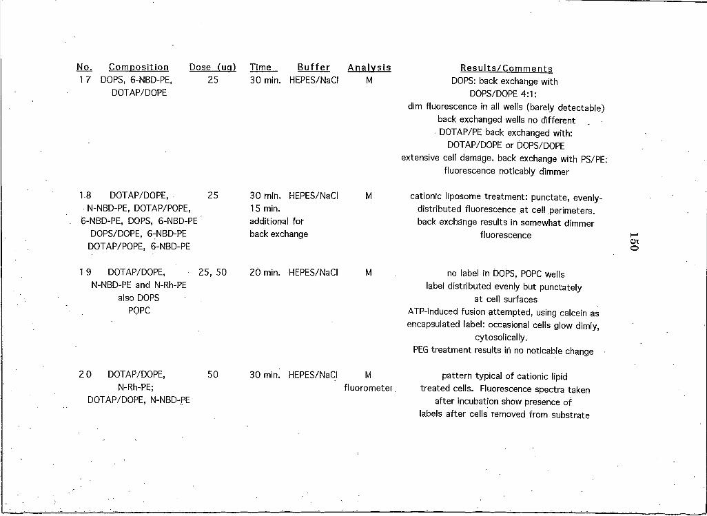
No. Composition Dose (ua) Time BufferI 7 DOPS1 6-NBD-PE,
DOTAP/DOPE
18 DOTAP/DOPE, N-NBD-PE1 DOTAP/POPE,
6-NBD-PE, DOPS1 6-NBD-PE DOPS/DOPE, 6-NBD-PE
DOTAP/POPE, 6-NBD-PE
I 9 DOTAP/DOPE, N-NBD-PE and N-Rh-PE
also DOPS POPC
2 O DOTAP/DOPE, N-Rh-PE;
DOTAP/DOPE, N-NBD-PE
25 30 min. HEPES/NaCI
25 30 min. HEPES/NaCI15 min. additional for back exchange
25, 50 20 min. HEPES/NaCI
50 30 mini HEPES/NaCI
A nalysisM
R esults/C om m ents DO PS: back exchange with
DOPS/DOPE 4:1:dim fluorescence in all wells (barely detectable)
back exchanged wells no different DOTAP/PE back exchanged with:
DOTAP/DOPE or DOPS/DOPE extensive cell damage, back exchange with PS/PE:
fluorescence noticably dimmer
M cationic liposome treatment: punctate, evenly- distributed fluorescence at cell perimeters, back exchange results in somewhat dimmer
fluorescence
M no label in DOPS, POPC wells label distributed evenly but punctately
at cell surfacesATP-induced fusion attempted, using calcein as encapsulated label: occasional cells glow dimly,
cytosolically.PEG treatment results in no noticable change
Mfluorometer.
pattern typical of cationic lipid treated cells. Fluorescence spectra taken
after incubation show presence of labels after cells removed from substrate
150
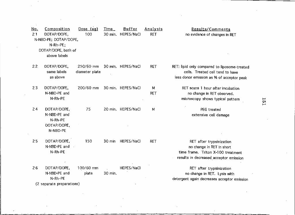
No. Composition Dose (ua) Time 21 DOTAP/DOPE, 100 30 min.
N-NBD-PE; DOTAP/DOPE,N-Rh-PE;
DOTAP/DOPE, both ofabove labels
2 2 DOTAP/DOPE, same labels
as above
2 3 DOTAP/DOPE, N-NBD-PE and
N-Rh-PE
2 4 DOTAP/DOPE, N-NBD-PE and
N-Rh-PE DOTAP/DOPE,
N-NBD-PE
2 5 DOTAP/DOPE, N-NBD-PE and
N-Rh-PE
250/60 mm 30 min. diameter plate
200/60 mm 30 min.
75 20 min.
150 30 min
2 6 DOTAP/DOPE, 100/60 mmN-NBD-PE and plate 30 min.
N-Rh-PE(2 separate preparations)
B ufferHEPES/NaCI
HEPES/NaCI
HEPES/NaCI
HEPES/NaCI
HEPES/NaCI
HEPES/NaCI
A nalysisRET
R esults/C om m ent s no evidence of changes in RET
RET RET: lipid only compared to liposome-treated cells. Treated cell tend to have
less donor emission as % of acceptor peak
MRET
RET scans I hour after incubation no change in RET observed,
microscopy shows typical pattern
M PEG treated extensive cell damage
RET RET after trypsinization no change in RET in short
time frame. Triton X-100 treatment results in decreased acceptor emission
RET after trypsinization no change in RET. Lysis with
detergent again decreases acceptor emission
151
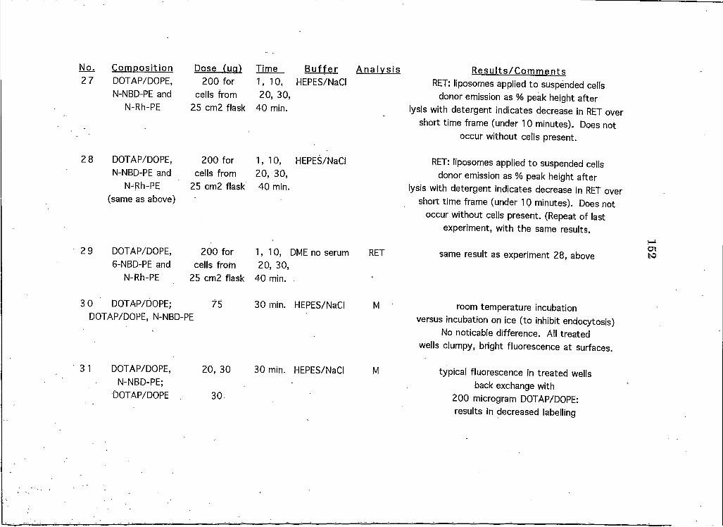
No. Composition 2 7 DOTAP/DOPE,
N-NBD-PE and N-Rh-PE
Dose (Ua) Time Buffer 200 for I, 10, HEPES/NaCI
cells from 20, 30,25 cm2 flask 40 min.
2 8 DOTAP/DOPE, 200 for 1 ,10 , HEPES/NaCI N-NBD-PE and cells from 20, 30,
N-Rh-PE 25 cm2 flask 40 min.(same as above)
29 DOTAP/DOPE, 6-NBD-PE and
N-Rh-PE
200 for 1 ,1 0 , DME no serum cells from 20, 30,
25 cm2 flask 40 min. .
3 0 DOTAP/DOPE; 75 30 min. HEPES/NaCIDOTAP/DOPE, N-NBD-PE
20, 303 1 DOTAP/DOPE, N-NBD-PE;
DOTAP/DOPE 30.
30 min. HEPES/NaCI
A nalysis R esults/C om m ents RET: liposomes applied to suspended cells
donor emission as % peak height after lysis with detergent indicates decrease in RET over
short time frame (under 10 minutes). Does not occur without cells present.
RET: liposomes applied to suspended cells donor emission as % peak height after
lysis with detergent indicates decrease in RET over short time frame (under 10 minutes). Does not
occur without cells present. (Repeat of last experiment, with the same results.
RET same result as experiment 28, above
M room temperature incubation versus incubation on ice (to inhibit endocytosis)
No noticable difference. All treated wells clumpy, bright fluorescence at surfaces.
M typical fluorescence in treated wells back exchange with
200 microgram DOTAP/DOPE: results in decreased labelling
152
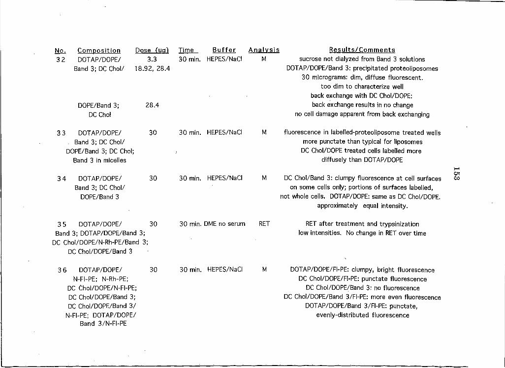
No. Composition 3 2 DOTAP/DOPE/
Band 3; DC Chol/
DOPE/Band 3; DC Chol
Dose (UcO 3.3
18.92, 28.4
28.4
Time B uffer A nalysis R esults/C om m ents30 min. HEPES/NaCI M sucrose not dialyzed from Band 3 solutions
DOTAP/DOPE/Band 3: precipitated proteoliposomes 30 micrograms: dim, diffuse fluorescent,
too dim to characterize well back exchange with DC Chol/DOPE: back exchange results in no change
no cell damage apparent from back exchanging
3 3 DOTAP/DOPE/ 30Band 3; DC Chol/
DOPE/Band 3; DC Chol;Band 3 in micelles
30 min. HEPES/NaCI M fluorescence in labelled-proteoliposome treated wellsmore punctate than typical for liposomes
DC Chol/DOPE treated cells labelled more diffusely than DOTAP/DOPE
3 4 DOTAP/DOPE/ Band 3; DC Chol/
DOPE/Band 3
30 30 min. HEPES/NaCI M DC Chol/Band 3: clumpy fluorescence at cell surfaceson some cells only; portions of surfaces labelled,
not whole cells. DOTAP/DOPE: same as DC Chol/DOPE. approximately equal intensity.
3 5 DOTAP/DOPE/ 30 30 min. DME no serum RETBand 3; DOTAP/DOPE/Band 3;
DC Chol/DOPE/N-Rh-PE/Band 3;DC Chol/DOPE/Band 3
RET after treatment and trypsinization low intensities. No change in RET over time
3 6 DOTAP/DOPE/ 30 30 min. HEPES/NaCIN-FI-PE; N-Rh-PE;
DC Chol/DOPE/N-FI-PE;DC Chol/DOPE/Band 3;DC Chol/DOPE/Band 3/
N-FI-PE; DOTAP/DOPE/Band 3/N-FI-PE
M DOTAP/DOPE/FI-PE: clumpy, bright fluorescenceDC Chol/DOPE/FI-PE: punctate fluorescence
DC Chol/DOPE/Band 3: no fluorescence DC Chol/DOPE/Band 3/FI-PE: more even fluorescence
DOTAP/DOPE/Band 3/FI-PE: punctate, evenly-distributed fluorescence
153
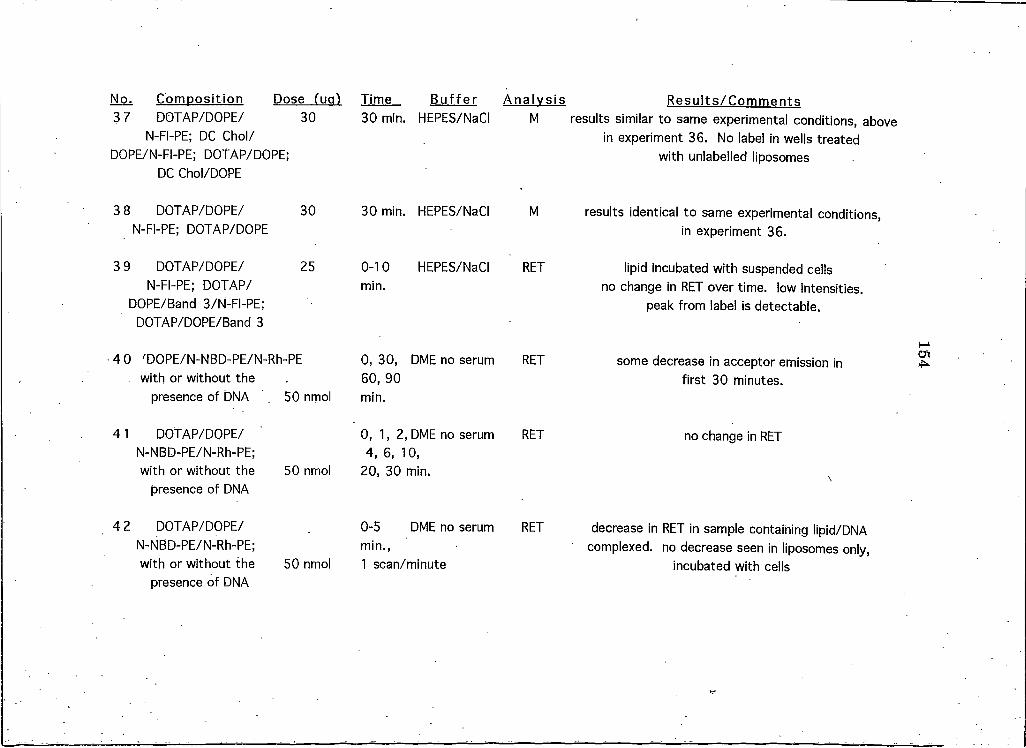
No. Composition Dose (ua) Time Buffer3 7 DOTAP/DOPE/
N-FI-PE; DC Chol/ DOPE/N-FI-PE; DOTAP/DOPE;
DC Chol/DOPE
3 8 DOTAP/DOPE/ N-FI-PE; DOTAP/DOPE
3 9 DOTAP/DOPE/ N-FI-PE; DOTAP/
DOPE/Band 3/N-FI-PE; DOTAP/DOPE/Band 3
30 30 min. HEPES/NaCI
30 30 min. HEPES/NaCI
25 0-10 HEPES/NaCImin.
4 0 'DOPE/N-NBD-PE/N-Rh-PE 0, 30, DME no serumwith or without the 60, 90
presence of DNA 50 nmol min.
4 1 DOTAP/DOPE/ N-NBD-PE/N-Rh-PE; with or without the 50 nmol
presence of DNA
0, 1 ,2 , DME no serum 4, 6, 10,
20, 30 min.
4 2 DOTAP/DOPE/ N-NBD-PE/N-Rh-PE; with or without the 50 nmol
presence of DNA
0-5 DME no serum min.,I scan/minute
A nalysis R esults/C om m entsM results similar to same experimental conditions, above
in experiment 36. No label in wells treated with unlabelled liposomes
M results identical to same experimental conditions,in experiment 36.
RET lipid incubated with suspended cellsno change in RET over time, low intensities,
peak from label is detectable.
RET some decrease in acceptor emission infirst 30 minutes.
RET no change in RET
\
RET decrease in RET in sample containing lipid/DNAcomplexed. no decrease seen in liposomes only,
incubated with cells
154
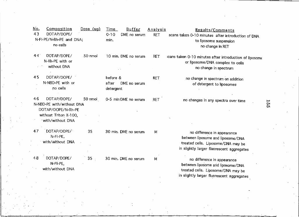
No. Composition Dose (iml 43 DOTAP/DOPE/ N-FI-PE/N-Rh-PE and DNA;
no cells
Time. Buffer A nalysis R esults/C om m ents0-10 DME no serum RET scans taken 0-10 minutes after introduction of DNAmin- to liposome suspension
no change in RET
4 4 DOTAP/DOPE/N-Rh-PE with or
without DNA
50 nmol 10 min. DME no serum RET scans taken 0-10 minutes after introduction of liposome or liposome/DNA complex to cells
no change in spectrum
4 5 DOTAP/DOPE/N-NBD-PE with or
no cells
before &after DME no serum detergent
RET no change in spectrum on addition of detergent to liposomes
4 6 DOTAP/DOPE/ 50 nmolN-NBD-PE with/without DNA
DOTAP/DOPE/N-Rh-PE without Triton X-100,• with/without DNA
0-5 min DME no serum RET no changes in any spectra over time
4 7 DOTAP/DOPE/N-FI-PE,
with/Without DNA
35 30 min. DME no serum M no difference in appearance between liposome and liposome/DNA treated cells. Liposome/DNA may be
in slightly larger fluorescent aggregates
4 8 DOTAP/DOPE/N-FI-PE,
with/without DNA
35 30 min. DME no serum M no difference in appearance between liposome arid liposome/DNA treated cells. Liposome/DNA may be
in slightly larger fluorescent aggregates
155
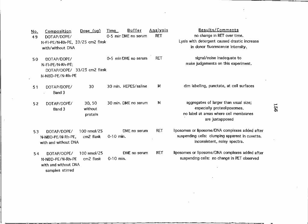
No.49
50
5 I
52
53
54
Comoosition Dose fuq) Time Buffer A nalvsis R esults/C om m entsDOTAP/DOPE/
N-FI-PE/N-Rh-PE, 33/25 cm2 flask with/without DNA
0-5 min DME no serum RET no change in RET over time.Lysis with detergent caused drastic increase
in donor fluorescence intensity.
DOTAP/DOPE/N-FI-PE/N-Rh-PE;
0-5 min DME no serum RET signal/noise inadequate to make judgements on this experiment.
DOTAP/DOPE/ 33/25 cm2 flask N-NBD-PE/N-Rh-PE
DOTAP/DOPE/ 30 30 min. HEPES/saline M dim labelling, punctate, at cell surfacesBand 3
DOTAP/DOPE/ 30, 50Band 3 without
protein
30 min. DME no serum M aggregates of larger than usual size; especially proteoliposomes.
no label at areas where cell membranes are juxtapposed
DOTAP/DOPE/ 10O nmol/25 N-NBD-PE/N-Rh-PE, cm2 flaskwith and without DNA
DME no serum 0-10 min.
RET liposomes or liposome/DNA complexes added after suspending cells: clumping apparent in cuvette,
inconsistent, noisy spectra.
DOTAP/DOPE/ 10O nmol/25 N-NBD-PE/N-Rh-PE cm2 flask with and without DNA
DME no serum 0-10 min.
RET liposomes or liposome/DNA complexes added after suspending cells: no change in RET observed
samples stirred
156
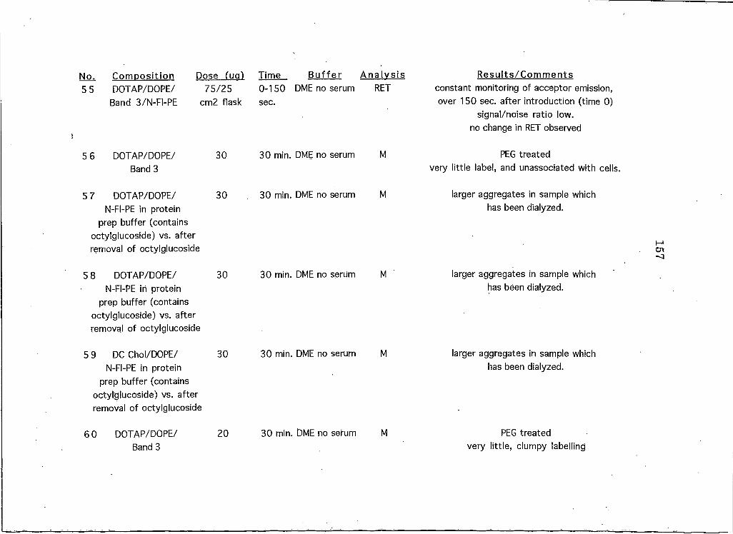
No. Composition Dose fug) Time B uffer A nalysis 5 5 DOTAP/DOPE/ 75/25 0-150 DME no serum RET
Band 3/N-FI-PE cm2 flask sec.
DOTAP/DOPE/ Band 3
30 min. DME no serum
7 DOTAP/DOPE/ N-FI-PE in protein
prep buffer (contains octylglucoside) vs. after removal of octylglucoside
30 min. DME no serum M
5 8 DOTAP/DOPE/ 30 30 min. DME no serum MN-FI-PE in protein
prep buffer (contains octylglucoside) vs. after removal of octylglucoside
5 9 DC Chol/DOPE/ 30 30 min. DME no serum MN-FI-PE in protein
prep buffer (contains octylglucoside) vs. after removal of octylglucoside
6 0 DOTAP/DOPE/ 20 30 min. DME no serum MBand 3
R esults/C om m ent s constant monitoring of acceptor emission, over 150 sec. after introduction (time 0)
signal/noise ratio low. no change in RET observed
PEG treatedvery little label, and unassociated with cells.
larger aggregates in sample which has been dialyzed.
larger aggregates in sample which has been dialyzed.
larger aggregates in sample which has been dialyzed.
PEG treatedvery little, clumpy labelling
157
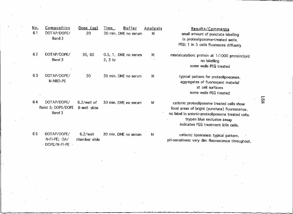
No.61
62
63
64
65
Comoosition DOTAP/DOPE/
Band 3
Dose fua) 20
Time Buffer30 min. DME no serum
A nalysisM
R esults/C om m ents small amount of punctate labelling in proteoliposome-treated wells.
PEG: I in 5 cells fluoresces diffusely
DOTAP/DOPE/ Band 3
30, 60 0.5, I , DME no serum 2, 3 hr
M miscalculation: protein at 1:1000 prpteindipid no labelling
some wells PEG treated
DOTAP/DOPE/N-NBD-PE
30 30 min. DME no serum M typical pattern for proteoliposomes. aggregates of fluorescent material
at cell surfaces some wells PEG treated
DOTAP/DOPE/ Band 3; DOPS/DOPE
Band 3
6.2/well of 8-well slide
30 min. DME no serum M cationic proteoliposome treated cells show local areas of bright (punctate) fluorescence,
no label in anionic-proteoliposome treated cells, trypan blue exclusion assay
indicates PEG treatment kills cells.
DOTAP/DOPE/ N-FI-PE; OA/ DOPE/N-Fi-PE ■
6.2/well chamber slide
30 min. DME no serum M cationic liposomes: typical pattern. pH-sensitives: very dim fluorescence throughout.
158

No. Comoosition Dose fua) Time Buffer A nalvsis; Re suits/C om m ent s66 DOTAP/DOPE/
N-FI-PE; OA/ DOPE/N-FI-PE
6.2/well chamber slide
30 min. DME no serum M excess label: not rinsed away properly, no label observed in pH-sensitive
liposome treated wells
67 OA/DOPE/ N-FI-PE; DOPS/ DOPE/N-FI-PE
6.2/well chamber slide
30 min. DME no serum M no labelling evident
68 OA/DOPE/ N-FI-PE; DOTAP/
DOPE/N-FI-PE
6.2/well chamber slide
30 min. DME no serum M OA/PE treated cells: label appears to be stuck to slide, between cells. Cationic liposome
treatments result in typical pattern of fluorescence
69 OA/DOPE/ N-FI-PE; DOTAP/
DOPE/N-FI-PE
6.2/well chamber slide
30 min. DME no serum M OA/PE treated cells: label appears to be stuck to slide, between cells. Cationic liposome
treatments result in typical pattern of fluorescence
70 DOTAP/DOPE/N-FI-PE
6.2/well chamber slide
30 min. DME no serum M typical pattern: treated wells show fine, punctate labelling at cell surfaces
71 DOTAP/DOPE/N-FI-PE
6.2/well chamber slide
30 min. DME no serum M viewed cells 5 hours after treatment ended for signs of damage: cell rounding, lysis apparent.
72 DOTAP/DOPE/N-FI-PE
6.2/well chamber slide
30 min. DME no serum M viewed cells 5 hours after treatment ended for signs of damage: cell rounding, lysis apparent.
159
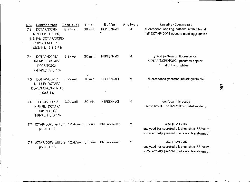
No. Comoosition Dose fua) Time B uffer A nalysis7 3 DOTAP/DOPE/ 6.2/well 30 min. HEPES/NaCI M
N-NBD-PEjI :3:1%,1:5:1%; DOTAP/DOPE/
POPC/N-NBD-PE,1:3:3:!%, 1:3:6:1%
Re suits/C om m ents fluorescent labelling pattern similar for all. 1:5 DOTAP/DOPE appears most aggregated
7 4 DOTAP/DOPE/ 6.2/well 30 min. HEPES/NaCI MN-FI-PE; DOTAP/
DOPE/POPC/N-FI-PE;! :3:3:1%
typical pattern of fluorescence. DOTAP/DOPE/POPC liposomes appear
slightly brighter
7 5 DOTAP/DOPE/ 6.2/well 30 min. HEPES/NaCI MN-FI-PE; DOTAP/
DOPE/POPC/N-FI-PE;I =3:3:1%
fluorescence patterns indistinguishable.
7 6 DOTAP/DOPE/ 6.2/well 30 min. HEPES/NaCI MN-FI-PE; DOTAP/
DOPE/POPC/N-FI-PE;! :3:3:_1%
confocal microscoysame result, no internalized label evident.
7 7 )OTAP/DOPE witl 6.2, 12.4/well 3 hours DME no serum MpSEAP DNA
also HT29 cellsanalyzed for secreted alk phos after 72 hours some activity present (cells are transformed)
7 8 )OTAP/DOPE witl 6.2, I 2.4/well 3 hours DME no serum MpSEAP DNA
also HT29 cellsanalyzed for secreted alk phos after 72 hours some activity present (cells are transformed)
160
1 6 1
APPENDIX B:
A SUMMARY OF ALL LIPOSOME EXPERIMENTS PERFORMED ON CELL
LINES OTHER THAN HELA
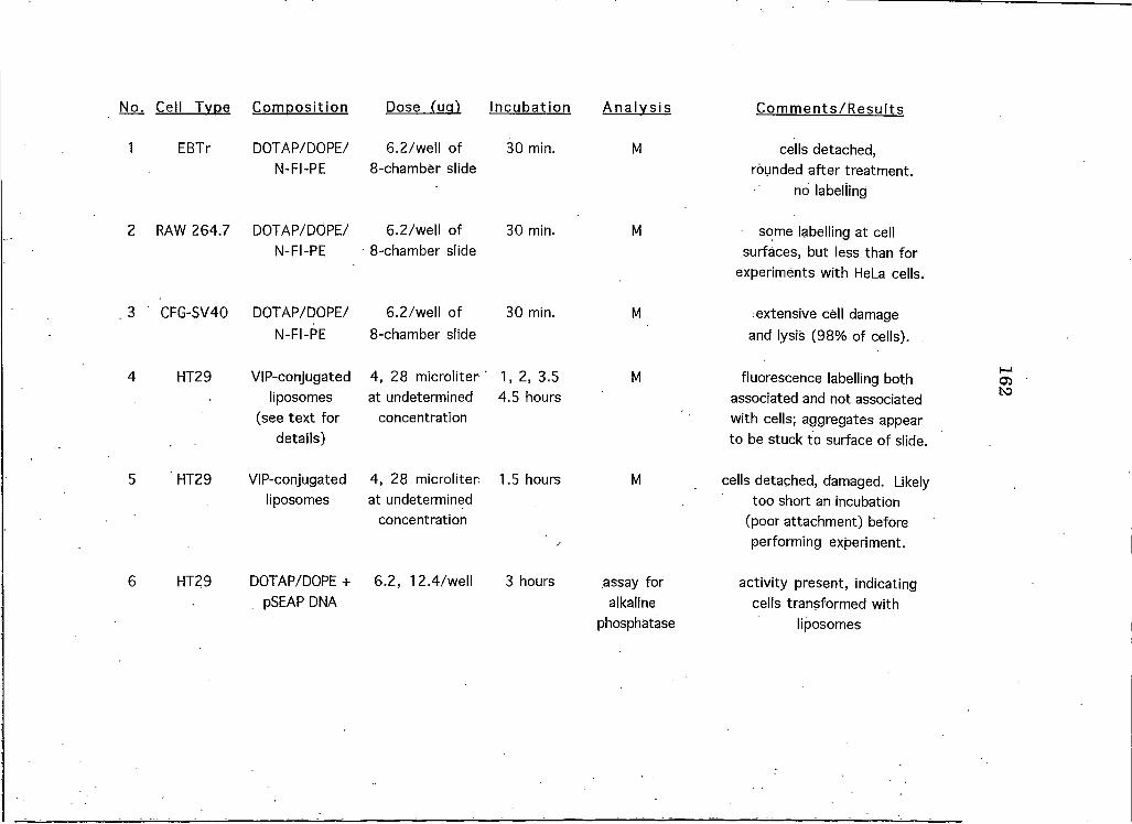
No^ Cell Tvoe ComDosition Dose fua) Incubation
I EBTr DOTAP/DOPE/ 6.2/well of 30 min.N-FI-PE 8-chamber slide
2 RAW 264.7 DOTAP/DOPE/ 6.2/well of 30 min.N-FI-PE 8-chamber slide
.3 CFG-SV40 DOTAP/DOPE/ 6.2/well of 30 min.N-FI-PE 8-chamber slide
4 HT29 VIP-conjugated 4 , 28 microliter 1 ,2 , 3.5liposomes at undetermined 4.5 hours
(see text for concentrationdetails)
5 HT29 VIP-conjugated 4, 28 microliter: 1.5 hoursliposomes at undetermined
concentration
6 HT29 DOTAP/DOPE + 6.2, 12.4/well 3 hourspSEAP DNA
A nalvsis C om m ents/R esults
M cells detached, rounded after treatment,
no labelling
M some labelling at cell surfaces, but less than for
experiments with HeLa cells.
M extensive cell damage and lysis (98% of cells).
M fluorescence labelling both associated and not associated with cells; aggregates appear to be stuck to surface of slide.
M cells detached, damaged. Likely too short an incubation
(poor attachment) before performing experiment.
assay for alkaline
phosphatase
activity present, indicating cells transformed with
liposomes
162
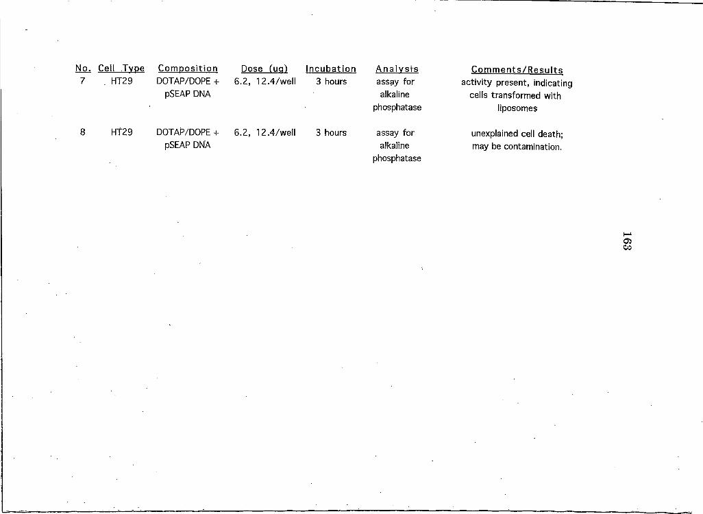
Dose fug) IncubationNo. Cell Type Composition 7 HT29 DOTAP/DOPE + 6.2, 12.4/well
pSEAP DNA
8 HT29 DOTAP/DOPE + 6.2, 12.4/wellpSEAP DNA
3 hours
3 hours
A nalysis assay for alkaline
phosphatase
C om m ents/R esults activity present, indicating
cells transformed with liposomes
assay for alkaline
phosphatase
unexplained cell death; may be contamination.
163

MONTANA STATE UNIVERSITY LIBRARIES
3 1762 10236758 6