transfusion-related acute lung injury: report of two casespmr.cuni.cz/file/5642/pmr2012a0031.pdf ·...
TRANSCRIPT

294)
© Charles University in Prague – Karolinum Press, Prague 2012
Prague Medical Report / Vol. 113 (2012) No. 4, p. 294–298
Transfusion-related Acute Lung Injury: Report of Two CasesČermáková Z.1,3, Kořístka M.2, Blahutová Š.2, Dvořáčková J.1, Brát R.1, Valkovský I.1, Hrdličková R.21Faculty of Medicine, University of Ostrava, Ostrava, Czech Republic;2Blood Centre, University Hospital Ostrava, Ostrava, Czech Republic;31st Department of Medicine – Department of Hematology, First Faculty of Medicine, Charles University in Prague and General University Hospital in Prague, Prague, Czech Republic
Rece ived J anuar y 30 , 2012 ; Accepted October 10 , 2012 .
Key words: Transfusion-related acute lung injury – Granulocyte antibodies – Blood products
Abstract: Transfusion-related acute lung injury (TRALI) is a severe life-threatening complication of blood transfusion, characterized by acute lung injury developing within 2–6 h of transfusion. However, TRALI is difficult to diagnose, and the initial report or suspicion of TRALI depends on close collaboration between clinical departments and transfusion centres. A total of 17 adverse post-transfusion reactions were reported to the Blood Centre of the University Hospital Ostrava as suspected TRALI between 2005 and 2010. We report two cases of serious TRALI with different pathogenetic mechanisms.
Mailing Address: Zuzana Čermáková, MD., PhD., Blood Centre, University Hospital Ostrava, 17. listopadu 1790, 708 52 Ostrava, Czech Republic; Phone: +420 597 374 420; e-mail: [email protected]
PMR 2012 04 2670.indd 294 12.11.12 7:10

295)
Transfusion-related Acute Lung Injury: Report of Two Cases
Prague Medical Report / Vol. 113 (2012) No. 4, p. 294–298
IntroductionTransfusion-related acute lung injury (TRALI) is a clinical syndrome characterized by dyspnoea, hypoxaemia, hypotension, bilateral infiltrates on chest radiography, and fever, during or within 6 h after transfusion (Kopko et al., 2002). TRALI is a rare complication of transfusion (1/5,000 transfusions); however 70% of affected patients require mechanical ventilatory support and 6–9% of cases are fatal (Bux, 2005). TRALI can be triggered by leukocyte antibodies (immune TRALI) or by the presence of substances (biologically-active lipids) that activate neutrophils (non-immune TRALI). Immune TRALI results in the presence of leukocyte antibodies in the donor plasma, particularly in the case of multiparous women, while leukocyte antibodies are less frequent in recipients. Leukocyte antibodies are directed against human leukocyte antigens (HLA) and/or human neutrophil antigens (HNA). HLA-specific antibodies were detected in 65% in cases that subsequently developed TRALI (Popovsky and Moore, 1985). Kopko et al. (2001) reported that HLA class II antibodies were associated with TRALI, and although the mechanism by which these antibodies induce TRALI remains unclear, the binding of HLA class II antibodies to monocytes and the subsequent release of cytokines and neutrophil activation is suggested to be involved (Kao et al., 2003; Kopko et al., 2003). Anti-HNA-1a (NA1), anti-HNA-1b (NA2), anti-HNA-2a (NB1) and anti-HNA-3a (5b) antibodies are the most frequently reported HNAs involved in TRALI, and anti-HNA-3a appears to trigger the most severe clinical manifestation (Norhagen et al., 1986; Zupanska et al., 1999; Davoren et al., 2003). Biologically-active substances released during the storage of blood products can cause non-immune TRALI. According to some authors, these substances increase the activity of neutrophil NADPH oxidase (Silliman et al., 2003). A neutrophil-priming model can also be applied to the pathogenesis of TRALI, whereby TRALI occurs as a result of two independent pathological processes (Kleinman et al., 2004); the patient’s clinical condition initially leads to endothelial cell activation and lung neutrophil sequestration, and this is followed by the administration of a blood product containing biologically-active mediators and/or granulocyte antibodies. Neutrophils are a key trigger of reactions. Alveolar lesions that lead to pulmonary oedema are the result of neutrophil enzyme release and the subsequent synthesis of highly toxic reactive oxygen species. This “double-strike” (two-hit hypothesis) mechanism has been demonstrated in animal models (Silliman et al., 1997; Chin-Yee et al., 1998).
Case IA 58-year-old female patient with negative transfusion anamnesis, received a postoperative transfusion with three units of erythrocytes and three plasma units, two from female and one from male donors. An adverse reaction was reported within 2 h after the administration of the last transfusion unit. The clinical picture involved fever, dyspnoea, hypoxaemia (saturation <80%) and bilateral infiltrates
PMR 2012 04 2670.indd 295 12.11.12 7:10
296)
Čermáková Z. et al.
Prague Medical Report / Vol. 113 (2012) No. 4, p. 294–298
on a chest radiograph. Laboratory analysis demonstrated strong positivity for granulocyte antibodies in post-transfusion, but not pre-transfusion samples, supporting the diagnosis of TRALI (Table 1).
Table 1 – Laboratory findings in case I
Pre-transfusion Post-transfusion
LCT negative strong positive
GAT negative strong positive
GIFT negative strong positive
LCT – lymphocytotoxic test; GAT – granulocyte agglutination test; GIFT – granulocyte immunofluorescence test
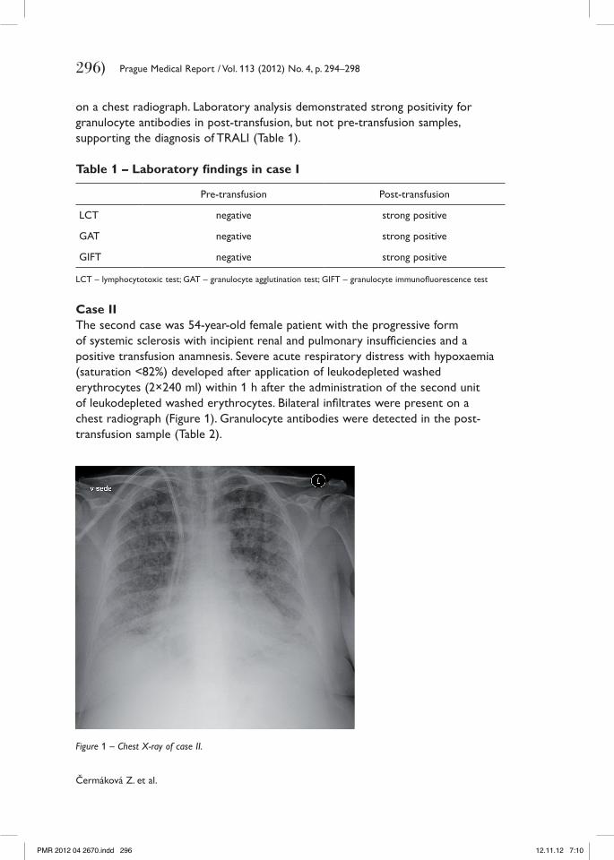
Case IIThe second case was 54-year-old female patient with the progressive form of systemic sclerosis with incipient renal and pulmonary insufficiencies and a positive transfusion anamnesis. Severe acute respiratory distress with hypoxaemia (saturation <82%) developed after application of leukodepleted washed erythrocytes (2×240 ml) within 1 h after the administration of the second unit of leukodepleted washed erythrocytes. Bilateral infiltrates were present on a chest radiograph (Figure 1). Granulocyte antibodies were detected in the post-transfusion sample (Table 2).
Figure 1 – Chest X-ray of case II.
PMR 2012 04 2670.indd 296 12.11.12 7:10
297)
Transfusion-related Acute Lung Injury: Report of Two Cases
Prague Medical Report / Vol. 113 (2012) No. 4, p. 294–298
Table 2 – Laboratory findings in case II
Pre-transfusion Post-transfusion
LCT negative negative
GAT negative negative
GIFT negative positive
LCT – lymphocytotoxic test; GAT – granulocyte agglutination test; GIFT – granulocyte immunofluorescence test
DiscussionThe first patient reported here developed immune (antibody-mediated) TRALI. In the female-donor samples investigated in an attempt to explain the post-transfusion reaction, were detected granulocyte antibodies. The strongly positive laboratory findings (LCT, GAT and GIFT) in post-transfusion sample supported the diagnosis. The Blood Centre of the University Hospital Ostrava prefers to use male-donor plasma; however, women are not totally excluded from the clinical plasma-donor program, because of concerns over the resulting significant reduction in accessible clinical plasma. This decision is supported by the literature, and Goldman et al. (2005) suggested that exclusion of multiparous female donors would result in a loss of donors of approximately 30%.
Neutrophils play a key role in the pathogenesis of TRALI; a wide range of substances have the capacity to activate neutrophils or to prime them, either directly or via activated pulmonary endothelium (Andreu, 2009). These activating substances occur in blood products. Endogenous substances are present in recipients undergoing surgery or with infections, or as a result of pro-inflammatory responses in the patient’s history (Densmore et al., 1999). The last of these may have been responsible for the increased risk of acute lung injury in the second patient, who had a progressive form of systemic sclerosis with incipient renal and pulmonary insufficiencies. We suggest that a neutrophil priming-agent-mediated TRALI reaction develops when neutrophil priming agents are not adequately removed by washing (Bux and Sachs, 2007), or if the cumulative potential of substances (including endogenous substances) that prime/activate neutrophils is strong enough to provoke a clinical and laboratory response (Silliman et al., 2005).
ConclusionTRALI is a serious life-threatening complication, and several different factors can play important roles in its development. Close cooperation between clinical departments and transfusion physicians is required to tailor transfusion therapy to respect the clinical condition of individual patients.
PMR 2012 04 2670.indd 297 12.11.12 7:10

298)
Čermáková Z. et al.
Prague Medical Report / Vol. 113 (2012) No. 4, p. 294–298
ReferencesAndreu, G. (2009) Transfusion-associated circulatory overload and transfusion-related acute lung injury:
diagnosis, pathophysiology, management and prevention. Vox Sang. 4, 63–71.Bux, J. (2005) Transfusion-related acute lung injury (TRALI): a serious adverse event of blood transfusion.
Vox Sang. 89, 1–10.Bux, J., Sachs, U. J. (2007) The pathogenesis of transfusion-related acute lung injury (TRALI). Br. J. Haematol.
136(6), 788–799.Chin-Yee, I., Keeney, M., Krueger, G., Dietz, G., Moses, G. (1998) Supernatant from stored red cells activates
neutrophils. Transfus. Med. 8, 49–56.Davoren, A., Curtis, B. R., Shulman, I. A. (2003) TRALI due to granulocyte-agglutinating human neutrophil
antigen-3a(5b) alloantibodies in donor plasma: a report of 2 fatalities. Transfusion 43, 641–645.Densmore, T. L., Goodnough, L. T., Ali, S., Dynis, M., Chaplin, H. (1999) Prevalence of HLA sensitization in
female apheresis donors. Transfusion 39, 103–106.Goldman, M., Webert, K. E., Arnold, D. M. (2005) Proceedings of a consensus conference: towards and
understanding of TRALI. Transfus. Med. Rev. 19, 2–31.Kao, G. S., Wood, I. C., Dorfman, D. M. (2003) Investigations into the role of anti-HLA class II antibodies in
TRALI. Transfusion 43, 185–191.Kleinman, S., Caulfield, T., Chan, P., Davenport, R., McFarlant, J., McPhedran, S., Meade, M., Morrison, D., Pinsent,
T., Robillard, P., Slinger, P. (2004) Toward an understanding of transfusion-related acute lung injury: statement of a consensus panel. Transfusion 44, 1774–1789.
Kopko, P. M., Popovsky, M. A., MacKenzie, M. R. (2001) HLA class II antibodies in transfusion-related acute lung injury. Transfusion 41, 1244–1248.
Kopko, P. M., Marshall, C. S., Mackenzie, M. R., Holland, P. V., Popovsky, M. A. (2002) Transfusion-related acute lung injury. Report of a clinical look-back investigation. JAMA 287, 1968–1971.
Kopko, P. M., Paglieroni, T. G., Popovsky, M. A. (2003) Transfusion-related acute lung injury: Correlation of antigen-antibody and monocyte activation in donor-recipient pairs. Transfusion 43, 177–184.
Norhagen, R., Conradi, M., Dromtorp, S. M. (1986) Pulmonary reaction associated with transfusion of plasma containing anti-5b. Vox Sang. 51, 102–107.
Popovsky, M. A., Moore, S. B. (1985) Diagnostic and pathogenic consideration in transfusion-related acute lung injury. Transfusion 25, 573–577.
Silliman, C. C., Paterson, A. J., Dickey, W. O., Stronceck, D. F., Popovsky, M. A., Caldwell, S. A. (1997) The association of biologically active lipids with the development of transfusion-related acute lung injury. Transfusion 37, 719–726.
Silliman, C. C., Boshkov, L. K., Mehdizadehkashi, Z., Elzi, D. J., Dickey, W. O., Podlosky, L., Clarke, G., Ambrus, D. R. (2003). Transfusion-related acute lung injury: epidemiology and prospective analysis of etiologic factors. Blood 101, 454–462.
Silliman, C. C., Ambruso, D. R., Boshkov, L. K. (2005) Transfusion-related acute lung injury. Blood 105, 2266–2273.
Zupanska, B., Uhrynowska, M., Konopka, L. (1999) Transfusion-related acute lung injury due to granulocyte-agglutinating antibody in a patient with paroxysmal nocturnal hemoglobinuria. Transfusion 39, 944–947.
PMR 2012 04 2670.indd 298 12.11.12 7:10