transepithelial transport of α-lipoic acid across human intestinal caco-2 cell monolayers
TRANSCRIPT
Transepithelial Transport of r-Lipoic Acid across HumanIntestinal Caco-2 Cell Monolayers
NAOKI TAKAISHI ,†,‡ KAZUTAKA YOSHIDA,† HIDEO SATSU,† AND
MAKOTO SHIMIZU * ,†
Graduate School of Agricultural and Life Sciences, The University of Tokyo, Tokyo 113-8657, Japan,and Unitika Ltd., Central Research Laboratories, Kyoto 611-0021, Japan
R-Lipoic acid (LA) is used in dietary supplements or food with antioxidative functions. The mechanismfor the intestinal absorption of R-lipoic acid was investigated in this study by using human intestinalCaco-2 cell monolayers. LA was rapidly transported across the Caco-2 cell monolayers, this transportbeing energy-dependent, suggesting transporter-mediated transport to be the mechanism involved.The LA transport was strongly dependent on the pH value, being accelerated in the acidic pH range.Furthermore, such monocarboxylic acids as benzoic acid and medium-chain fatty acids significantlyinhibited LA transport, suggesting that a proton-linked monocarboxylic acid transporter (MCT) wasinvolved in the intestinal transport of LA. The conversion of LA to the more antioxidative dihydrolipoicacid was also apparent during the transport process.
KEYWORDS: Caco-2; r-lipoic acid; dihydrolipoic acid; monocarboxylic acid transporter; intestinal
absorption; transepithelial transport
INTRODUCTION
Naturally occurringR-lipoic acid (LA) is an essential cofactorfor mitochondrial respiratory enzymes and exerts a powerfulantioxidative effect (1, 2). This effect is due to direct radicalscavenging, recycling of other antioxidants, accelerating glu-tathione (GSH) synthesis, and modulating the transcription factoractivity, especially that of NF-κB. Synthetic LA has long beenused as a therapeutic agent in the treatment of diabeticneuropathy (2, 3), and its recent use in dietary supplements orfoods with antioxidative functions has been markedly increasing.
The excretion and biotransformation of LA have beeninvestigated, following a single dose of14C-labeled LA to mice,rats, and dogs and of unlabeled LA to humans (2, 4, 5). Morethan 80% of the radioactivity given was renally excreted, and12 metabolites were identified. These results suggest that LAwould be efficiently absorbed in the intestines and metabolizedin the body.
There are two possible pathways for the intestinal transportof such low molecular weight compounds as LA: a transporter-mediated intracellular transport pathway and a paracellularpathway between the intercellular junctions. The former involvesenergy-dependent active transport, whereas the latter is passivediffusion. The intestinal absorption of such nutrients as glucose,amino acids, dipeptides, and water-soluble vitamins (6) is mainlyvia the transporter-mediated route, whereas the paracellularpathway plays an important role in the intestinal absorption of
such minerals as calcium (7). The paracellular transport ofnutrients such as glucose and amino acids has also been reported(8). It would be important to identify the mechanism for theintestinal transport of orally administered LA to control itsintestinal absorbability, although this mechanism remainsobscure.
Prasad et al. (9) and Balamurugan et al. (10) have reportedthat LA inhibited in a dose-dependent manner the uptake ofsuch soluble vitamins as biotin and pantothenic acid, which weretransported by a sodium-dependent multivitamin transporter(SMVT) in placental (9) or renal (10) epithelial cells. Althoughthis implies that LA would be transported by SMVT, there isno direct evidence for this.
We investigated in this study the mechanism for the intestinalabsorption of LA by using human intestinal Caco-2 monolayersas an in vitro model of the intestinal epithelium. Caco-2 cellsexpress a variety of nutrient transporters and are frequently usedto analyze transporter functions (11). The paracellular transportof a food-derived bioactive peptide has also been characterizedby using Caco-2 cell monolayers (12). We found from thepresent study that proton-dependent transporter-mediated trans-port was the main pathway for the transepithelial transport ofLA. The metabolic change of LA during the absorption processin Caco-2 cells was also studied.
MATERIALS AND METHODS
Materials. The human colon adenocarcinoma cell line, Caco-2, wasobtained from the American Type Culture Collection (Rockville, MD).Dulbecco’s modified Eagle’s medium (DMEM) was purchased fromKohjin Bio Co. (Saitama, Japan), and fetal bovine serum (FBS) was
* Corresponding author (telephone+81-3-5841-5127; fax+81-3-5841-8026; e-mail [email protected]).
† The University of Tokyo.‡ Unitika Ltd.
J. Agric. Food Chem. 2007, 55, 5253−5259 5253
10.1021/jf063624i CCC: $37.00 © 2007 American Chemical SocietyPublished on Web 05/31/2007
from Gemini Bio-Products (Woodland, CA). Penicillin-streptomycin(10000 units/mL and 10 mg/mL in 0.9% sodium chloride, respectively)and nonessential amino acids (NEAA) were purchased from Gibco(Gaithersburg, MD). The type-1 collagen solution was purchased fromNitta Gelatin (Osaka, Japan), Hanks’ balanced salt solution (HBSS)was from Sigma (St. Louis, MO), and phosphate-buffered saline (PBS)was from Nissui Pharmaceutical Co. (Tokyo, Japan).R-Lipoic acid,dihydrolipoic acid, and 1,3-bis(2-chloroethyl)-1-nitrosourea (BCNU)were purchased from Sigma (St. Louis, MO); sodium azide, biotin,and pantothenic acid were from Kanto Chemical Co. (Tokyo, Japan);and glycyl-sarcosine was from Bachem (Bubendorf, Switzerland).DL-Lactic acid, sodium benzoate, sodium acetate, sodium butyrate, sodiumhexanoate (caproate), and sodium octanoate (caprylate) were purchasedfrom Kanto Chemical Co. (Tokyo, Japan). All other chemicals usedwere of reagent grade.
Cell Culture. The Caco-2 cells were cultured at a density of 2×10⁵ cells in 100-mm plastic dishes with a culture medium containingDMEM, 10% FBS, 100µM NEAA, 100 units/mL of penicillin, and100 µg/mL of streptomycin. The cells were incubated at 37°C in ahumidified atmosphere of 5% CO2 in air, the culture medium beingrenewed on alternate days. When the cells had reached confluence (6-7days after seeding), they were passaged by trypsinization with 0.1%trypsin and 0.02% EDTA in PBS. All of the cells used in this studywere between passages 50 and 60. A transepithelial transport experimentwas performed by using Caco-2 cells that had been cultured at a densityof 2 × 10⁵ cells per well in 12-well Transwell inserts (12-mm diameterand 0.4-µm pore size; Corning Costar, NY) that had been precoatedwith collagen. The cell monolayers for all experiments were used after14 days of culture.
Measurement of the Transepithelial Electrical Resistance (TER).The integrity of the cell layer was evaluated by measuring TER withMillicell-ERS equipment (Millipore, MA). Monolayers with TER of>500 Ω/cm2 were used for the experiments. TER of the monolayerswas measured before and after an assay sample was added to the insert(apical side).
Transepithelial Transport Experiments. The apical side of Caco-2cell monolayers that had been cultured in the 12-well Transwell insertsand the basal chambers were washed twice with 0.5 mL of HBSS. Theinsert and basal chamber were, respectively, filled with 0.5 and 1.5mL of HBSS and then incubated at 37°C for 1 h. After HBSS in theinserts had been removed, 0.5 mL of 0.5 mM LA in HBSS (pH adjustedto 5.0-8.0) was added to the insert and then incubated at 37°C for 2h. The LA concentrations in the apical and basal solutions weremeasured by high-performance liquid chromatography (HPLC).
Competition Experiments on LA Transport. After the Caco-2 cellmonolayers had been washed and equilibrated with HBSS as justdescribed, HBSS in the inserts was removed. A 500-µL amount of 0.5mM LA in HBSS adjusted to pH 7.4 or 6.0 and containing an assaysample was then added to the insert and incubated at 37°C for 15min. The assay samples used were water-soluble vitamins (biotin andpantothenic acid), monocarboxylic acids (lactic acid, benzoic acid, aceticacid, butyric acid, hexanoic acid, and octanoic acid), and a peptide(glycyl-sarcosine). The concentration of each assay sample was 10 mM,except that biotin was 2 mM because of its low solubility. After theincubation, the basal solution was taken and the concentration of LAmeasured by HPLC.
Competition Experiment on Fluorescein Transport.This experi-ment was performed in a manner similar to that just described. Briefly,25 µM fluorescein, in the presence or absence of 0.5 mM LA, wasadded to the apical solution of the Caco-2 cell monolayers and thenincubated at 37°C for 15 min. The fluorescence of the basal solutionwas measured with a Fluoroskan Ascent CF fluorescence microplatereader (Labsystems, Helsinki, Finland), detecting the emission at 485nm with excitation at 544 nm.
HPLC Analysis of LA and Dihydrolipoic Acid (DHLA). AGulliver HPLC system (Jasco International Co., Japan) consisting of aPU-2080 Plus intelligent HPLC pump, a UV-1575 intelligent UV-visdetector, and an 807-IT integrator was used. Chromatographic separationwas performed in a C18 column (Cosmosil 5C18-MS-II, 4.6 × 150mm; Nacalai Tesque, Kyoto, Japan). The mobile phase was a 70%methanol solution containing 0.02% acetic acid at a flow rate of 1.0
mL/min. LA was monitored at UV 334 nm and quantified from itspeak area. DHLA was monitored by a Nanospace SI-2 electrochemicaldetector (ECD; Shiseido Co., Tokyo, Japan), the applied potential being900 mV versus Ag/AgCl.
Inhibition of Glutathione Reductase.Caco-2 cells were monolayer-cultured in a 12-well microplate for 2 weeks. The cells were washedwith HBSS (pH adjusted to 6.0) and then incubated in the same buffercontaining 100µM LA in the presence or absence of BCNU (0-100µM). The amount of DHLA in the culture solution after a 60-minincubation was measured by HPLC-ECD.
Statistical Analysis.Each result is expressed as the mean( standarddeviation (SD). A statistical analysis was conducted by Dunnett’s testto identify significant differences between the control and test groups.
RESULTS
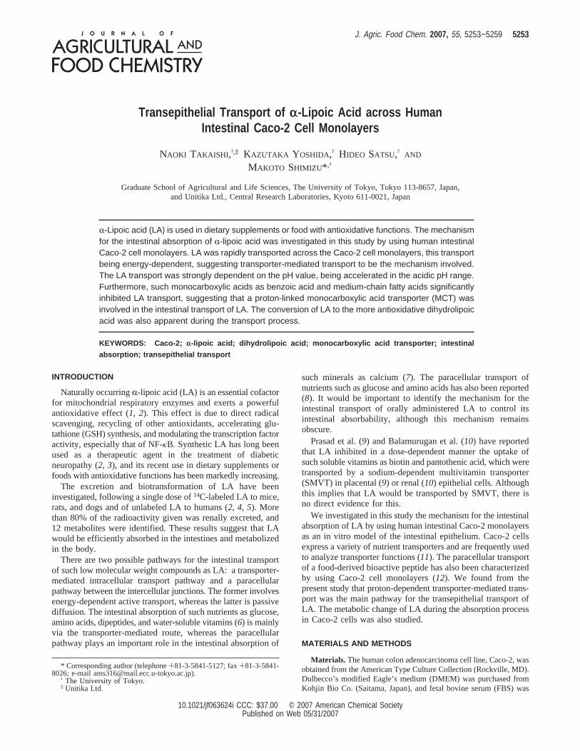
Transepithelial Transport of LA Was Energy-Dependent.After 15 min of incubation,>10% of LA added to the apicalsolution (at pH 7.4) had been transported to the basal chamber.This transport was strongly inhibited by pretreating the Caco-2cells with the metabolic inhibitor, sodium azide (Figure 1A),suggesting that the transport was via an energy-dependentpathway. To reveal whether this transport was mediated by themultivitamin transporter (SMVT), as has been suggested inprevious work (9, 10), the competitive effect of such SMVTsubstrates as biotin and pantothenic acid on the LA transportwas examined. The SMVT substrates generally had no effecton the transport of LA, except that pantothenic acid in the apicalsolution (pH 7.4) significantly decreased the transport of LA(Figure 1B). This suggests that SMVT was not principallyinvolved in the transepithelial transport of LA across the Caco-2cell monolayers.
Figure 1. Effect of NaN3 and soluble vitamins on the transepithelialtransport of R-lipoic acid across the Caco-2 cell monolayers. The amountof LA in the basal chamber was determined after incubation of the Caco-2cell monolayers at 37 °C for 15 min. The initial concentration of LA addedto the apical solution was 0.5 mM. The concentrations of sodium azide(A) and pantothenic acid (B) were 10 mM, whereas that of biotin (B) was2 mM because of its low solubility. The pH value was adjusted to 7.4 onboth sides. Data are expressed as a percentage of the control and arepresented as the mean ± SD (n ) 3). //, significantly lower than thecontrol value (P < 0.01).
5254 J. Agric. Food Chem., Vol. 55, No. 13, 2007 Takaishi et al.
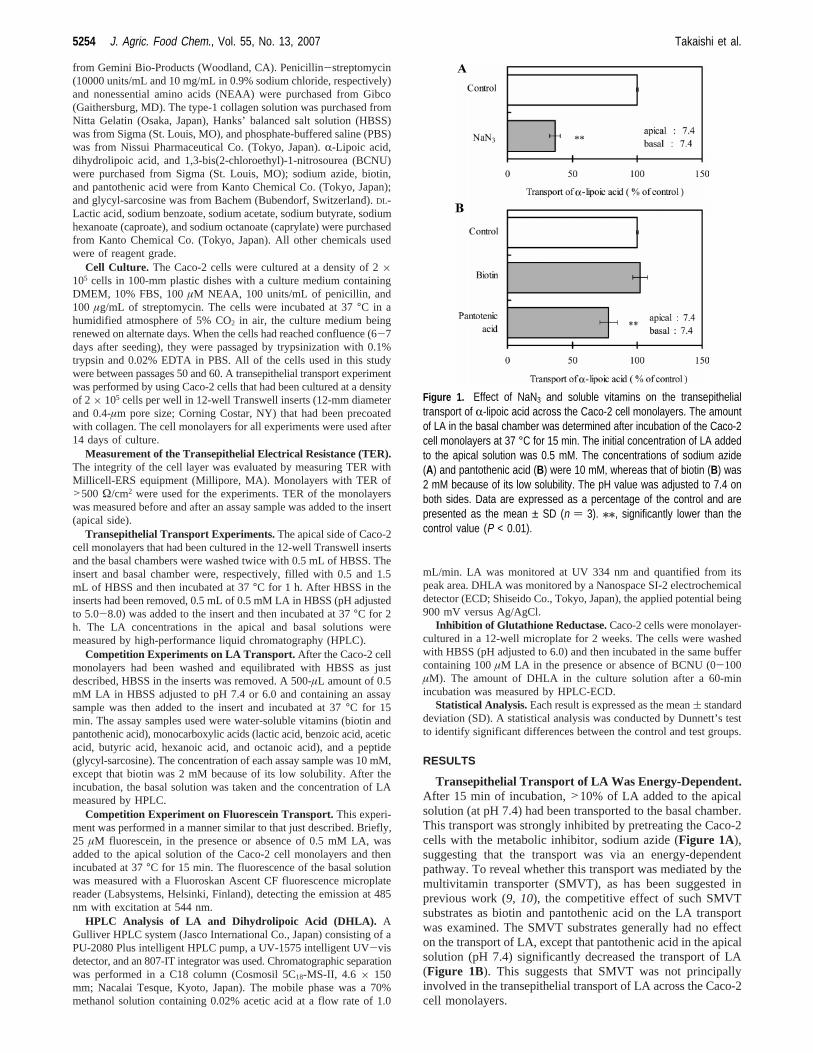
Transepithelial Transport of LA Was Proton-Dependent.The transepithelial transport of LA from the apical to basalchamber across the Caco-2 cells increased as a function of time.The transport of LA was also strongly pH-dependent (Figure2). When the apical solution was adjusted to the acidic pH range,the transport rate for LA was significantly higher than that atthe neutral pH level. At pH 5.0,>60% of LA added to theapical chamber had been transported to the basal solution within60 min. After a 120-min incubation, almost 80% of LA was inthe basal solution, indicating that the basal LA concentrationwas already higher than the apical LA concentration. In contrast,the transport of LA at an apical pH level of 8.0 was 30% lowerthan the control value (pH 7.4). These results suggest that aproton-linked transporter was involved in the LA transport.Addition of LA did not affect the monolayer integrity of Caco-2cells at any pH between 5 and 8, because no change in thetransepithelial electrical resistance was observed (data notshown).
Transepithelial Transport of LA Was Not Na+-Dependent.Because many transporters are dependent on the Na+ concentra-tion, the Na+ dependence of the LA transport was examined.LA (0.5 mM) was added to the apical side of the Caco-2 cellmonolayers in the presence of NaCl (0, 35, 70, or 140 mM)and incubated at 37°C for 60 min. The LA content in the basalsolution was then measured by HPLC. The transport rate ofLA from the apical to basal chamber across the Caco-2 cellmonolayers remained unchanged, irrespective of the Na+
concentration in the apical and basal solutions at pH 7.4 (Figure3A). This Na+ independence was also apparent when the apicalpH value was 6.0 (Figure 3B).
Peptide Transporter Was Not Involved in the Transportof LA. Because peptide transporter 1 (PepT1) expressed in theintestinal epithelium is proton-dependent, showing a highertransport rate in the acidic pH range (6), PepT1 may have beeninvolved in the LA transport. However, adding glycyl-sarcosine,a typical PepT1 substrate with high affinity, to the apical solutionhad no effect on the transport of LA (data not shown), indicatingthat PepT1 was not involved in the LA transport.
Transepithelial Transport of LA Was Inhibited by Mono-carboxylic Acids. The effect of monocarboxylic acids on theLA transport was then examined to learn whether the mono-carboxilic acid transporter (MCT), another proton-dependenttransporter, was involved in the LA transport.DL-Lactic acidand acetic acid, the good substrates of MCT-1, scarcely affectedthe transport of LA. On the other hand, benzoic acid, butyricacid, hexanoic acid, and octanoic acid all significantly inhibitedthe transport of LA, the inhibitory activity being dependent onthe carbon chain length (Figure 4). The effect of long-chainfatty acids, such as lauric acid, myristic acid, and palmitic acid,was not evaluated because they had low solubility.
LA Did Not Affect the Transport of Fluorescein, an MCTSubstrate.We have previously reported fluorescein as one ofthe MCT substrates (13, 14). However, the transepithelialtransport of fluorescein from the apical to basal chamber acrossthe Caco-2 cell monolayers was not inhibited by LA. Theaccumulation of fluorescein in the Caco-2 cells and the amountof fluorescein remaining in the apical solution were also notaffected by LA (data not shown).
Dihydrolipoic Acid Was Produced during Transport inthe Caco-2 Cells.An HPLC analysis using a UV-vis detectorcould detect only the peak of LA (Figure 5A). On the otherhand, an electrochemical detector made it possible to detect thepeaks of both LA and DHLA, demonstrating that DHLA wasproduced during the transport experiment (Figure 5B). DHLA
Figure 2. Effect of pH value on the transepithelial transport of R-lipoicacid across the Caco-2 cell monolayers. The amount of LA in the basalchamber was determined as a function of time after incubation of theCaco-2 cells at 37 °C. The initial concentration of LA added to the apicalsolution was 0.5 mM. The pH value of the apical solution was adjustedwithin the range of 5.0−8.0, and that of the basal solution was fixed at7.4. Data are expressed as a percentage of the total amount of LA andare presented as the mean ± SD (n ) 3).
Figure 3. Effect of Na+ on the transepithelial transport of R-lipoic acidacross the Caco-2 cell monolayers. The amount of LA in the basal chamberwas determined after incubation of the Caco-2 cells at 37 °C for 60 min.The initial concentration of LA added to the apical solution was 0.5 mM,and that of Na+ was from 0 to 140 mM on both sides. The pH value ofthe apical solution was adjusted to 7.4 (A) or 6.0 (B), and that on thebasal side was fixed at 7.4. Data are expressed as a percentage of thetotal amount of LA added to the apical chamber and are presented asthe mean ± SD (n ) 3).
Transepithelial Transport of R-Lipoic Acid J. Agric. Food Chem., Vol. 55, No. 13, 2007 5255

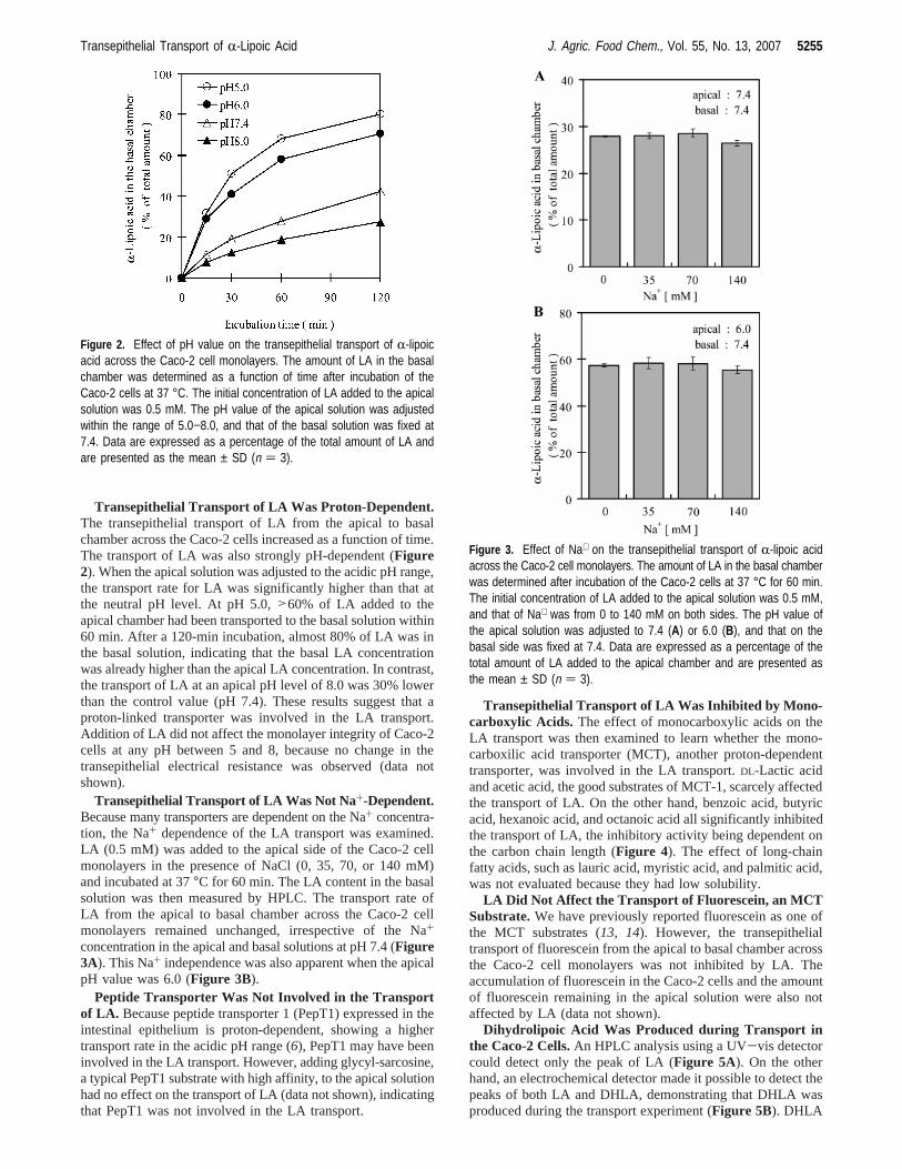
in the basal chamber increased as a function of time, reachingan amount corresponding to 7% of the total LA in 120 min(Figure 6A). DHLA was also detected on the apical side, theamount reaching 3% of the total LA after 30 min of incubation,although the amount of LA in the apical chamber remainedunchanged during the subsequent 90 min (Figure 6A). Theseresults suggest that about 10% of the total LA was convertedto DHLA in the Caco-2 cells, 30% of which was countertrans-ported to the apical side and 70% transported to the basal side(Figure 6A).
Glutathione Reductase Was Likely To Be Responsible forthe Production of DHLA. Constantinecsu et al. (15) havereported that LA is metabolized to DHLA in the erythrocyteby such reducing enzymes as glutathione reductase (GR) andthioredoxin reductases. Expression of several reducing enzymesincluding GR in Caco-2 cells has also been reported by Bakerand Baker (16). Involvement of the reducing enzymes in theDHLA production by Caco-2 cells was therefore studied. WhenCaco-2 cells were treated with BCNU, a specific GR inhibitor,
the production of DHLA in Caco-2 cells was markedly inhibited(Figure 7), suggesting that GR was responsible for DHLAproduction in Caco-2 cells.
DISCUSSION
The transepithelial transport of LA across the Caco-2 cellmonolayers was observed to be energy-dependent, suggestingtransporter-mediated transport to be the major mechanism forintestinal LA absorption. Because the involvement of SMVTin the intestinal transport of LA has been suggested in previousstudies (9, 10) and SMVT was expressed in Caco-2 cells (17),we had initially assumed that LA would be transported viaSMVT. However, the LA transport was proton-dependent, thetransport rate being increased by reducing the apical pH value(Figure 2). The transport of LA was also found to beindependent of the Na+ concentration (Figure 3) and notaffected by SMVT substrates (Figure 1B), indicating that SMVTwas not involved in the LA absorption.
Another possibility was the transport of LA via a peptidetransporter. The peptide transporter, PepT1, is proton-dependentand has a wide range of substrate specificity, recognizing di-and tripeptides (18). Other compounds such asâ-lactamantibiotics and synthetic peptide analogues have also beenreported to be PepT1 substrates (6). However, LA transportacross the Caco-2 cell monolayers was not affected by the PepT1substrate, glycyl-sarcosine (data not shown). Do¨ring et al. (19)have reported that octanoic acid, the structure of which
Figure 4. Effect of monocarboxylic acids on the transepithelial transportof R-lipoic acid across the Caco-2 cell monolayers. The amount of LA inthe basal chamber was determined after incubation of the Caco-2 cellsat 37 °C for 15 min. The initial concentration of LA added to the apicalsolution was 0.5 mM, and that of each competitor was 10 mM on theapical side. The pH value of the apical solution was adjusted to 7.4 (A)or 6.0 (B), and that on the basal side was fixed at 7.4. Data are expressedas a percentage of the total amount of LA and are presented as themean ± SD (n ) 3). //, significantly lower than the control value (P <0.01).
Figure 5. HPLC chromatograms of R-lipoic acid and dihydrolipoic acid.LA and DHLA in the basal chamber were monitored by using a UV−visdetector (A) and ECD (B). The mobile phase was a 50% methanol solutioncontaining 0.02% acetic acid to separate LA and DHLA, and the flowrate was 1.0 mL/min.
5256 J. Agric. Food Chem., Vol. 55, No. 13, 2007 Takaishi et al.
resembles that of LA, could not be a PepT1 substrate. Takingthese observations together, PepT1 is unlikely to have beeninvolved in the LA transport.
It is well-known that long-chain fatty acids (an alkyl chainof C10 or longer) are transported by fatty acid transporters(FATP). The FATP family consists of six members which
represent a group of evolutionarily conserved proteins that areinvolved in the cellular uptake and metabolism of long- andvery-long-chain fatty acids (20). Among them, FATP4 is locatedin the intestines and is expressed to a greater extent than thatof the other members (21). However, it has been reported thatFATP did not contribute to the transport of medium- and short-chain fatty acids with alkyl chains of shorter than C8 (21, 22).Considering the result that the LA transport was inhibited byoctanoic acid, it is unlikely that FATP would be involved inthe transport of LA in Caco-2 cells, although the inhibitory effecton LA transport of fatty acids with alkyl chains of longer thanC10 was not examined in the present study because of theirvery low solubility in HBSS.
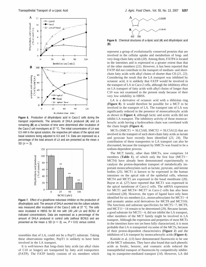
LA is a derivative of octanoic acid with a dithiolan ring(Figure 8). It would therefore be possible for a MCT to beinvolved in the transport of LA. The transport rate of LA wassignificantly reduced in the presence of monocarboxylic acidsas shown inFigure 4, although lactic and acetic acids did notinhibit LA transport. The inhibitory activity of those monocar-boxylic acids having a hydrocarbon chain was correlated withthe chain length (Figure 4).
MCTs (SMCT1) SLC5A8, SMCT2) SLC5A12) that areinvolved in the transport of such short-chain fatty acids as lactateand pyruvate have recently been identified (23, 24). Thecontribution of these transporters to LA transport can also bediscounted, because the transport by SMCTs was found to be asodium-dependent process.
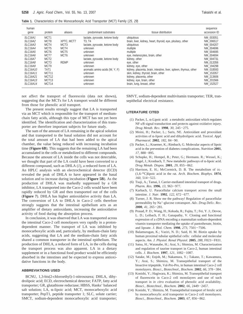
The MCT family, other than SMCTs, now comprises 14members (Table 1), of which only the first four (MCT1-MCT4) have already been demonstrated experimentally tocatalyze the proton-dependent transport of metabolically im-portant monocarboxylates such as lactate, pyruvate, and ketonebodies (25). MCT1 is known to be expressed in the humanintestines on the apical side of the epithelial cells, whereasMCT4 and MCT5 are expressed in the basal membrane (26).Buyse et al. (27) have reported that MCT1 was expressed inthe apical membrane of Caco-2 cells. The mRNA expressionfor MCT1 and MCT4-MCT7 in Caco-2 cells has also beenconfirmed (28). However, the types of ligand have only beenidentified for six members (i.e., monocarboxylates for MCT1-4and aromatic amino acid derivatives for MCT8 and MCT10).The functions and substrate specificities for MCT5-7, MCT9,and MCT11-14 remain to be determined (29). Because lactate,a good substrate for MCT1-4, did not inhibit the LA transport,other members of the MCT family might be involved in LAtransport. Although the expression and properties of most MCTsin the intestines have not yet been fully characterized, it is mostprobable that LA is transported via some of the MCTs, becauseof their proton-dependent characteristics (Figure 2) and theinhibition of LA transport by monocarboxylic acids (Figure 4).
Konishi et al. (13) have demonstrated fluorescein to be oneof the MCT substrates. They have also found that such phenolicacids as ferulic, benzoic, and coumaric acids reduced thetransepithelial transport of fluorescein by competitively inhibit-ing its transporter-mediated transport (14). However, LA did
Figure 6. Production of dihydrolipoic acid in Caco-2 cells during thetransport experiments. The amounts of DHLA produced (A) and LAremaining (B) as a function of time were determined after incubation ofthe Caco-2 cell monolayers at 37 °C. The initial concentration of LA was0.5 mM in the apical solution, the respective pH values of the apical andbasal solutions being adjusted to 6.0 and 7.4. Data are expressed as apercentage of the total amount of LA and are presented as the mean ±SD (n ) 3).
Figure 7. Effect of a glutathione reductase inhibitor on the production ofdihydroplipoic acid. The amount of DHLA secreted into the culture solutionwas measured after incubation of the Caco-2 cells at 37 °C. The cellswere incubated in HBSS for 60 min with 100 µM LA and BCNU ofindicated concentrations. Data are expressed as a percentage of theamount of DHLA produced in control cells (without BCNU) and arepresented as the mean ± SD (n ) 3). //, P < 0.01 versus control.
Figure 8. Chemical structures of R-lipoic acid (A) and dihydrolipoic acid(B).
Transepithelial Transport of R-Lipoic Acid J. Agric. Food Chem., Vol. 55, No. 13, 2007 5257
not affect the transport of fluorescein (data not shown),suggesting that the MCTs for LA transport would be differentfrom those for phenolic acid transport.
The present results strongly suggest that LA is transportedvia an MCT which is responsible for the transport of medium-chain fatty acids, although this type of MCT has not yet beenidentified. The identification and characterization of this trans-porter are therefore important subjects for future study.
The sum of the amount of LA remaining in the apical solutionand that transported to the basal solution did not account forthe total amount of LA that had been added to the apicalchamber, the value being reduced with increasing incubationtime (Figure 6B). This suggests that the remaining LA had beenaccumulated in the cells or metabolized to different compounds.Because the amount of LA inside the cells was not detectable,we thought that part of the LA could have been converted to adifferent compound, such as DHLA (1), a reduced form of LA.An HPLC analysis with an electrochemical detector (ECD)revealed the peak of DHLA to have appeared in the basalsolution and to increase during incubation (Figure 5B). As theproduction of DHLA was markedly suppressed by a GRinhibitor, LA transported into the Caco-2 cells would have beenrapidly reduced by GR and then transported out of the cells(Figure 7). DHLA has higher antioxidative activity than LA.The conversion of LA to DHLA in Caco-2 cells thereforestrongly suggests that the intestinal epithelium acts as anamplifier of dietary antioxidants, increasing the antioxidativeactivity of food during the absorption process.
In conclusion, it was observed that LA was transported acrossthe intestinal Caco-2 cell monolayers very rapidly in a proton-dependent manner. The transport of LA was inhibited bymonocarboxylic acids and, particularly, by medium-chain fattyacids, suggesting that LA and the medium-chain fatty acidsshared a common transporter in the intestinal epithelium. Theproduction of DHLA, a reduced form of LA, in the cells duringthe transport process was also apparent. LA in a dietarysupplement or in a functional food product would be efficientlyabsorbed in the intestines and be expected to express antioxi-dative functions in the body.
ABBREVIATIONS USED
BCNU, 1,3-bis(2-chloroethyl)-1-nitrosourea; DHLA, dihy-drolipoic acid; ECD, electrochemical detector; FATP, fatty acidtransporter; GR, glutathione reductase; HBSS, Hanks’ balancedsalt solution; LA,R-lipoic acid; MCT, monocarboxylic acidtransporter; PepT1, peptide transporter 1; SLC, solute carrier;SMCT, sodium-dependent monocarboxylic acid transporter;
SMVT, sodium-dependent multivitamin transporter; TER, tran-sepithelial electrical resistance.
LITERATURE CITED
(1) Packer, L.R-Lipoic acid: a metabolic antioxidant which regulatesNF-κB signal transduction and protects against oxidative injury.Drug Metab. ReV. 1998, 30, 245-275.
(2) Moini, H.; Packer, L.; Saris, NE. Antioxidant and prooxidantactivities ofR-lipoic acid and dihydrolipoic acid.Toxicol. Appl.Pharmacol.2002, 182, 84-90.
(3) Packer, L.; Kraemer, K.; Rimbach, G. Molecular aspects of lipoicacid in the prevention of diabetes complications.Nutrition 2001,17, 888-895.
(4) Schupke, H.; Hempel, R.; Peter, G.; Hermann. R.; Wessel, K.;Engel, J.; Kronbach, T. New metabolic pathways ofR-lipoic acid.Drug Metab. Dispos.2001, 29, 855-862.
(5) Harrison, E. H.; McCormick, D. B. The metabolism ofDL-(1,6-14C)lipoic acid in the rat.Arch. Biochem. Biophys.1974,160, 514-522.
(6) Tsuji, A.; Tamai, I. Carrier-mediated intestinal transport of drugs.Pharm. Res.1996, 13, 963-977.
(7) Karbach, U. Paracellular calcium transport across the smallintestine.J. Nutr. 1992, 122, 672-677.
(8) Turner, J. R. Show me the pathway! Regulation of paracellularpermeability by Na+-glucose cotransport.AdV. Drug DeliV. ReV.2000, 41, 265-281.
(9) Prasad, P. D.; Wang, H.; Kekuda, R.; Fujita, T.; Fei, Y. J.; Devoe,L. D.; Leibach, F. H.; Ganapathy, V. Cloning and functionalexpression of a cDNA encoding a mammalian sodium-dependentvitamin transporter mediating the uptake of pantothenate, biotin,and lipoate.J. Biol. Chem.1998, 273, 7501-7506.
(10) Balamurugan, K.; Vaziri, N. D.; Said, H. M. Biotin uptake byhuman proximal tubular epithelial cells: cellular and molecularaspects.Am. J. Physiol. Renal Physiol.2005, 288, F823-F831.
(11) Satsu, H.; Watanabe, H.; Arai, S.; Shimizu, M. Characterizationand regulation of taurine transport in Caco-2, human intestinalcells.J. Biochem.1997, 121, 1082-1087.
(12) Satake, M.; Enjoh, M.; Nakamura, Y.; Takano, T.; Kawamura,Y.; Arai, S.; Shimizu, M. Transepithelial transport of thebioactive tripeptide, Val-Pro-Pro, in human intestinal Caco-2 cellmonolayers.Biosci., Biotechnol., Biochem.2002, 66, 378-384.
(13) Konishi, Y.; Hagiwara, K.; Shimizu, M. Transepithelial transportof fluorescein in Caco-2 cell monolayers and use of suchtransport inin Vitro evaluation of phenolic acid availability.Biosci., Biotechnol., Biochem.2002, 66, 2449-2457.
(14) Konishi, Y.; Shimizu, M. Transepithelial transport of ferulic acidby monocarboxylic acid transporter in Caco-2 cell monolayers.Biosci., Biotechnol., Biochem.2003, 67, 856-862.
Table 1. Characteristics of the Monocarboxylic Acid Transporter (MCT) Family (25, 29)
humangene protein aliases predominant substrates tissue distribution
sequenceaccession ID
SLC16A1 MCT1 lactate, pyruvate, ketone body ubiquitous NM_003051SLC16A2 MCT8 XPTC, MCT7 T3, T4 brain, liver, kidney, heart, thyroid, eye, pituitary, other NM_006517SLC16A3 MCT4 MCT3 lactate, pyruvate, ketone body ubiquitous NM_004207SLC16A4 MCT5 MCT4 unknown multiple NM_004696SLC16A5 MCT6 MCT5 unknown multiple NM_004695SLC16A6 MCT7 MCT6 unknown eye, melanocytes, brain, other NM_004694SLC16A7 MCT2 lactate, pyruvate, ketone body kidney, other NM_004731SLC16A8 MCT3 REMP unknown eye, other NM_013356SLC16A9 MCT9 unknown kidney, eye, other NM_194298SLC16A10 TAT1 aromatic amino acids (W, Y, F) kidney, placenta, brain, intestine, liver, spleen, thymus, other NM_018593SLC16A11 MCT11 unknown skin, kidney, thyroid, brain, other NM_153357SLC16A21 MCT12 unknown kidney, placenta, other NM_213606SLC16A13 MCT13 unknown kidney, eye, brain, other NM_201566SLC16A14 MCT14 unknown brain, lung, breast, other NM_152527
5258 J. Agric. Food Chem., Vol. 55, No. 13, 2007 Takaishi et al.
(15) Constantinescu, A.; Pick, U.; Handelman, G. J.; Haramaki, N.;Han, D.; Podda, M.; Tritschler, H. J.; Packer, L. Reduction andtransport of lipoic acid by human erythrocytes.Biochem.Pharmacol.1995, 50, 253-61.
(16) Baker, S. S.; Baker, R. D. Antioxidant enzymes in the differenti-ated Caco-2 cell line.In Vitro Cell DeV. Biol. 1992, 28A, 643-647.
(17) Balamurugan, K.; Ortiz, A.; Said, H. M. Biotin uptake by humanintestinal and liver epithelial cells: role of the SMVT system.Am. J. Physiol. Gastrointest. LiVer Physiol.2003, 285, G73-G77.
(18) Fei, Y. J.; Kanai, Y.; Nussberger, S.; Ganapathy, V.; Leibach,F. H.; Romero, M. F.; Singh, S. K.; Boron, W. F.; Hediger, M.A. Expression cloning of a mammalian proton-coupled oligopep-tide transporter.Nature1994, 368, 563-566.
(19) Doring, F.; Will, J.; Amasheh, S.; Clauss, W.; Ahlbrecht, H.;Daniel, H. Minimal molecular determinants of substrates forrecognition by the intestinal peptide transporter.J. Biol. Chem.1998, 273, 23211-23218.
(20) Stahl, A. A current review of fatty acid transport proteins(SLC27).Pfluegers Arch.2004, 447, 722-727.
(21) Stahl, A.; Hirsch, D. J.; Gimeno, R. E.; Punreddy, S.; Ge, P.;Watson, N.; Patel, S.; Kotler, M.; Raimondi, A.; Tartaglia, L.A.; Lodish, H. F. Identification of the major intestinal fatty acidtransport protein.Mol. Cell 1999, 4, 299-308.
(22) Gimeno, R. E.; Ortegon, A. M.; Patel, S.; Punreddy, S.; Ge, P.;Sun, Y.; Lodish, H. F.; Stahl, A. Characterization of a heart-specific fatty acid transport protein.J. Biol. Chem.2003, 278,16039-16044.
(23) Martin, P. M.; Gopal, E.; Ananth, S.; Zhuang, L.; Itagaki, S.;Prasad, B. M.; Smith, S. B.; Prasad, P. D.; Ganapathy, V. Identityof SMCT1 (SLC5A8) as a neuron-specific Na+-coupled trans-porter for active uptake ofL-lactate and ketone bodies in thebrain.J. Neurochem.2006, 98, 279-288.
(24) Srinivas, S. R.; Gopal, E.; Zhuang, L.; Itagaki, S.; Martin, P.M.; Fei, Y. J.; Ganapathy, V.; Prasad, P. D. Cloning andfunctional identification of slc5a12 as a sodium-coupled low-affinity transporter for monocarboxylates (SMCT2).Biochem.J. 2005, 392, 655-664.
(25) Halestrap, A. P.; Meredith, D. The SLC16 gene familysfrommonocarboxylate transporters (MCTs) to aromatic amino acidtransporters and beyond.Pfluegers Arch.2004, 447, 619-628.
(26) Gill, R. K.; Saksena, S.; Alrefai, W. A.; Sarwar, Z.; Goldstein,J. L.; Carroll, R. E.; Ramaswamy, K.; Dudeja, P. K. Expressionand membrane localization of MCT isoforms along the lengthof the human intestine.Am. J. Physiol. Cell Physiol.2005, 289,C846-C852.
(27) Buyse, M.; Sitaraman, S. V.; Liu, X.; Bado, A.; Merlin, D.Luminal leptin enhances CD147/MCT-1-mediated uptake ofbutyrate in the human intestinal cell line Caco2-BBE.J. Biol.Chem.2002, 277, 28182-28190.
(28) Hadjiagapiou, C.; Schmidt, L.; Dudeja, P. K.; Layden, T. J.;Ramaswamy, K. Mechanism(s) of butyrate transport in Caco-2cells: role of monocarboxylate transporter 1.Am. J. Physiol.Gastrointest. LiVer Physiol.2000, 279, G775-G780.
(29) Friesema, E. C.; Jansen, J.; Milici, C.; Visser, T. J. Thyroidhormone transporters.Vitam. Horm.2005, 70, 137-167.
Received for review December 14, 2006. Revised manuscript receivedApril 17, 2007. Accepted April 18, 2007. This work was partiallysupported by Grant-in Aid for Scientific Research 15108002 and thaton the Priority Area “Transportsome“ from the Ministry of Education,Culture, Sports, Science, and Technology of Japan.
JF063624I
Transepithelial Transport of R-Lipoic Acid J. Agric. Food Chem., Vol. 55, No. 13, 2007 5259