transcrystallization at the interface of polyethylene …transcrystallization at the interface of...
TRANSCRIPT

Transcrystallization at the Interface of Polyethylene Single-Polymer
Composites
H.-J. Kestenbach*a, J. Loosb, J. Petermannc
aUniversidade Federal de São Carlos, Departamento de Engenharia de Materais,13565-905 São Carlos - SP, Brazil
bEindhoven University of Technology, Eindhoven Polymer Laboratories, 5600 MB Eindhoven, The Netherlands
cUniversität Dortmund, Lehrstuhl für Werkstoffkunde, 44221 Dortmund, Germany
Received: June 14, 1999; Revised: September 20, 1999
The phenomenon of transcrystallization was studied at the interface of UHMWPE fibersembedded in an HDPE matrix. It was hoped that epitaxial crystallization in such model compositescould eventually be used to improve adhesion between these high-strength fibers and the thermo-plastic matrix material. Matrix crystallization was induced and accompanied on a specially designedhot stage which made the crystallization front advance slowly along a thermal gradient. Transcrys-talline interfacial layers were observed without regard to temperature conditions, but with widelyvarying dimensions. Lamellar resolution within these layers was achieved by low voltage scanningelectron microscopy, and the very beginning of transcrystallization was observed in sample areaswhere UHMWPE fiber segments were only partially embedded into the HDPE matrix. Lamellaralignment on the fiber surface indicated that transcrystallization in this system was associated withepitaxial nucleation.
Keywords: polymer composites, polyethylene, UHMWPE, transcrystallinity, epitaxy, lowvoltage scanning electron microscopy
1. Introduction
The concept of single-polymer composites has beenknown for many years, based upon the idea that interfacialbonding should improve if the matrix and the reinforcementmaterial were made from different morphologies of thesame semicrystalline polymer1,2. In the case of polyethyl-ene (PE), oriented high-modulus PE fibers can be embed-ded in a non-oriented spherulitic PE matrix. Fabrication ofthe composite is rendered possible by the lower meltingtemperature of chain-folded lamellar crystals in the matrix,as compared to the higher melting temperature of extended-chain crystals in the fiber. More recently, a modified formof polyethylene single-polymer composites has been con-sidered where gel-spun UHMWPE (ultrahigh molecularweight polyethylene) fibers were embedded in a HDPE(high-density polyethylene) matrix3,4. UHMWPE fibers areespecially attractive as reinforcement material because oftheir very high tensile strength and elastic modulus values5.However, they are also known for rather poor interfacialbonding characteristics with respect to most of the usual
polymer matrix materials6-8. Already many years ago, tran-scrystallinity has been reported to be able to improve adhe-sion along fiber-matrix interfaces9,10. Its study maytherefore be useful for the development of PE single-poly-mer composites.The phenomenon of transcrystallinity wasfirst observed as a laboratory curiosity in the early fifties11.Today, many researchers believe that transcrystallinity mayimprove the mechanical properties of polymer composites,and some up-to-date review papers have appeared in therecent literature12,13. Transcrystallization requires hetero-geneous nucleation along the fiber surface to occur with asufficiently high density of nuclei so that interfacial crystalgrowth can only proceed in the perpendicular direction,leaving a layer of columnar crystals around the fiber9.However, the precise mechanisms by which such heteroge-neous nucleation occurs are not fully understood14,15. Inaddition, an improvement in interfacial bonding cannot beexpected to occur by preferred heterogeneous nucleationalone, but will depend upon the formation of a low-energyinterface between the fiber and the matrix. In the case ofpolyethylene single-polymer composites, cocrystallization
*e-mail: [email protected]
Materials Research, Vol. 2, No. 4, 261-269, 1999. © 1999

as well as epitaxial nucleation have been mentioned to leadto such low-energy interfaces, but no proofs weregiven1,16,17.It is believed that at least some of the questionsabout the role of transcrystallization in fiber compositescould be resolved if morphological observations of lamellardetail were able to reach the interface. Quite recently,improved equipment for low voltage scanning electronmicroscopy (LVSEM) has become available which permitslamellar resolution to be obtained from many polymersurfaces without the need for special sample preparationtechniques18,19. It is the purpose of the present work toreport on first results which LVSEM has given when ap-plied to the transcrystallization layer in a UHMWPE/HDPEcomposite.
2. ExperimentalComposite samples were prepared by embedding
Dyneema SK 65 high modulus UHMWPE fibers in amatrix of commercial HDPE (Lupolen 6021D). In order toassure access to the interface during scanning electronmicroscope observations after the crystallization experi-ment, UHMWPE fibers were embedded only partially intothe HDPE matrix. To this effect, one or more fibers weremanually extended at room temperature over the surface ofa small piece of HDPE film, previously pressed at 140 °Cbetween glass plates and supported on a microscope slide.After covering with another microscope slide, the sampleassembly was placed upon a hot plate maintained at 140 °C.After 5 min, a small pressure was applied to the glass coverwhich caused some fiber segments to sink into the moltenmatrix while other segments became embedded only par-tially, as evidenced by the formation and trapping of airbubbles between the molten matrix and the glass cover.After another waiting period of 5 min, the composite sam-ple assembly was transferred to a specially designed hotstage where the crystallization front could be observedwhile advancing slowly along a thermal gradient. As shownin Fig. 1, this rather simple stage consisted of two individualheating blocks which were separated by a small gap of 4.3mm width. Hot stage end temperatures T1 and T2 weremonitored by thermocouples embedded into the heatingblocks and were controlled to within 0.5 °C. For in situobservation of the crystallization process, the entire stageassembly with the composite sample bridging its gap wasplaced upon the specimen table of an ordinary opticaltransmission microscope. Phenacitin crystals (commercialcalibration powder with a melting temperature of 134.5 °C)were used to calibrate the temperature gradient within thesample which was established across the gap for any par-ticular choice of T1/T2 end temperatures, Fig. 2.
At the end of a typical crystallization experiment, sam-ples were cooled to room temperature and examined byordinary light in reflection, polarized light in transmission,and by scanning electron microscopy (SEM). A low voltage
field emission SEM from Hitachi (model S-4500) was usedto reveal lamellar detail during high-resolution observa-tions.
3. ResultsSome typical examples for in-situ observation of the
slowly advancing crystallization front by temperature gra-dient hot stage microscopy are shown in Figs. 3 and 4. Highcrystallization temperatures as well as low temperaturegradients were selected in order to reduce the rate of crys-tallization. For this purpose, temperature gradients across
262 Kestenbach et al. Materials Research
Figure 1. Schematic view of temperature gradient hot stage in (a), typicaltemperature gradient for T1 = 132 °C e T2 = 135 °C em (b).
Figure 2. Calibration of temperature gradient through the melting ofphenacitin crystals, with the lower temperature at the left and the highertemperature at the right hand side of the photograph. Melting front is at134.5 °C. Original magnification 80X.

the T1/T2 gap were varied between a minimum of 1 °C anda maximum of 5 °C. “Cold” T1 temperatures were selectedto range from 124 to 133 °C while “hot” T2 temperaturescovered the interval from 127 to 134 °C. Under suchconditions, incubation times of less than an hour wereobserved for the lower temperatures, while incubationtimes of up to 100 h occurred for the higher temperatures.Inmany cases, preferred nucleation as well as transcrystallinegrowth along the fiber-matrix interface could be detected
under the optical microscope, Figs. 3(a) and 3(b). However,and inspite of the favourable and closely controlled tem-perature regime, such observations were often masked byadverse crystallization kinetics which tended to favournucleation rates (very fast) over growth rates (very slow),Fig. 3(c). Thus, spherulitic crystallization within the matrixas well as transcrystallization along the interface occurredgenerally on a very fine scale.In order to reduce thespherulite nucleation rate in the HDPE matrix and to form
Vol. 2, No. 4, 1999 Polyethylene Single-Polymer Composites 263
Figure 4. Effect of matrix flow during sample preparation on subsequentcrystallization along temperature gradient. Well developed transcrys-talline layer in (a), melting of UHMWPE fiber in (b), transcrystallizationalong molten fiber remnant in (c). Original magnification 100X.
Figure 3. Crystallization front advancing along temperature gradient asviewed in situ under the hot stage optical microscope. Preferred nucleationon fiber surface in (a), transcrystallization along the fiber ahead of thehomogeneous crystallization front in (b), only general homogeneousmatrix crystallization in (c). Original magnification 100X.

well-developed transcrystalline layers, not only high crys-tallization temperatures but some form of matrix shearseemed to be necessary, Fig. 4. Such a situation was fre-quently observed at the outer sample portions where matrixflow had been induced in the molten state as the result ofthe pressure applied at 140 °C to produce fibre embedding,Fig. 4(a). In some instances, material flow in these areaswas sufficient to destroy the fiber, Fig. 4(b). In other cases,partial fiber melting left fiber remnants which generatedvery well developed transcrystallinity along their inter-faces, Fig. 4(c).
High-resolution scanning electron microscopy provedto be a valuable tool for identifying events of transcrys-tallinity even in those sample portions where optical mi-croscopy had failed to detect transcrystallization on a finerscale, Fig. 5. As an example, Fig. 5(a) once more presentsthe sample area previously shown in Fig. 4(a), but this timeafter crystallization had been completed by cooling to roomtemperature. Please note that, in comparison with Fig. 4(a),the micrograph of Fig. 5(a) has been rotated so that the fiberalignment is now the same as on the following scanningelectron micrographs. At the top of Fig. 5(a), matrix crys-tallization has occurred on too fine a scale for transcrystal-lization to be observed by optical microscopy. Throughlamellar resolution in the scanning electron microscope,however, clear evidence for transcrystallinity in this areawas obtained as shown in Figs. 5(b), 5(c) and 5(d). At arelatively low magnification, the formation of surface faultsin Fig. 5(b) indicated that the fiber ran close to the samplesurface in this area. At a higher magnification, Fig. 5(c),transcrystallization can be recognized by the characteristicpresence of parallel and closely-spaced crystal lamellae(viewed edge-on due to their dominantly vertical growthdirection above the fiber), as compared to the irregular andmore widely-spaced lamellar arrangement which wasfound to be typical for spherulitic growth at some distanceaway from the fiber, Fig. 5(d).
More detailed scanning electron microscopy observa-tions were carried out along the prominent, well-developedlayer of transcrystallization which appears at the lower partof Fig. 5(a). The same sample area is shown again in Fig.6(a), but this time as viewed in an optical reflection micro-scope. At first, lamellar detail at high magnification wasphotographed within the main area of transcrystallinitywhich has been marked by T1 and T2 on Fig. 6(a) and whichis characteristic for a region where the fiber was embeddedbelow the HDPE sample surface. Typical examples oflamellar resolution are presented in Fig. 6(b) which oncemore shows the closely-spaced lamellae within the tran-scrystalline layer, and in Fig. 6(c) where the transcrystallinelayer at the bottom half of the figure has met the outerportion of a spherulite which had been nucleated furtherabove. For a second area of observation, it can be seen fromFig. 6(a) that the fiber has surfaced along a segment marked
S which is still located within the same transcrystallizationlayer (see for example Fig. 5(a) and compare distances fromthe sample border). Region S was selected to investigatethe initial formation of transcrystallinity directly at thefiber-matrix interface, as shown in the following threefigures.
For better localization, the positions of two particularareas where lamellar detail again was photographed at highresolution are shown in Fig. 7. At successively highermagnification, transcrystalline lamellae with characteristicparallel growth and high-density packing are shown at firstfrom a "tangential" point of view in Fig. 8. It should benoted that the full fiber diameter is exposed in this area ascan be seen from Figs. 6(a) or 7(a), so that we can be sureabout this tangential perspective of view in Fig. 8. Sec-ondly, a “top-view” of the very first transcrystalline lamel-lae which have formed at the fiber-matrix interface ispresented at successively higher magnifications in Figs.9(a) and 9(b). It is important to recognize that these lamellaehave in fact formed from a thin layer of HDPE matrixmaterial which covered the UHMWPE fiber in this area,and not from incipient fiber surface melting. This fact maybe appreciated from Fig. 9(c) where a fiber segment whichwas located outside the HDPE film sample but whichexperienced the same temperature during the experimenthas been photographed at the same magnification as usedin Fig. 9(b).Finally, it should be noticed that the fiberdirection has been the same for the scanning electron mi-crographs of Figs. 6 to 9 where transcrystalline morphologyhas been presented at lamellar resolution. It can thus benoted that the lamellar alignment, although quite parallelwithin separate regions, is far from uniform and also farfrom being perpendicular to the fiber direction. This obser-vation which, in principle, could be taken as experimentalevidence against the presence of epitaxial growth will bediscussed in more detail below.
4. Discussion
4.1 Detection of transcrystallinity
Transcrystallization at the fiber-matrix interface hasbeen observed in many composite systems which employsemi-crystalline polymers as their matrix material1,2,9-17,20-30.It is frequently believed that it is the event of transcrystalli-zation at the interface which will improve mechanical prop-erties of the composite9,10,12,16,23,28, although some authorshave reported no improvements22,26 or even a decrease infiber-matrix bond strength due to the event of transcrys-tallinity24,25,27. In the particular case of UHMWPE/HDPEcomposites, transcrystallization was observed by some3,31
but not by other authors4.The detection of fine-scale transcrystallinity by the
low-voltage scanning electron microscopy technique couldperhaps explain the apparent absence of transcrystallinity
264 Kestenbach et al. Materials Research

Vol. 2, No. 4, 1999 Polyethylene Single-Polymer Composites 265
Figure 5. Detection of transcrystallization in the low voltage scanning electron microscope (LVSEM). Approximate sample locations for SEM observationmarked T (transcrystallinity) and S (spherulitic crystallization) on optical micrograph in (a), region T immediately above UHMWPE fiber observed atlow magnification by LVSEM in (b), central region T with lamellar resolution above fiber in (c), spherulitic matrix region S with lamellar resolutionaway from fiber in (d). Original magnification 100X in (a), 3.000X in (b), 15.000X in (c) and (d).

of HDPE along UHMWPE fibers reported recently in theliterature4. Usually, the width of transcrystalline layerswhich develop around fibers is of the same magnitude asthe spherulite diameter in the matrix3. In fact, identicalradial growth rates were found in polypropylene for matrixspherulites as well as for transcrystalline regions21. It istherefore to be expected that, in the case of very fine matrix
crystallization, transcrystallinity may not be detected byoptical microscopy as long as individual spherulites are notresolved. Such a situation has clearly been present in thefine-grained sample areas which were presented in Fig. 5,and probably also in Fig. 3.
4.2 Epitaxial growth vs. unoriented preferred nucleation
Transcrystallization is usually observed under the opti-cal microscope, where it is difficult to say something aboutits origin and its physical cause. Lamellar detail of trans-crystalline layers has recently been observed by transmis-sion electron microscopy after permanganic etching29. Inthat case, the preferred orientation (cross-hatched morphol-ogy) of individual iPP (isotactic polypropylene) lamellaewithin the transcrystalline regions was taken as proof forthe occurrence of epitaxial nucleation of iPP on polyimidefibers. The same argument can be used in the present casebecause a definite preferred orientation of the very firstHDPE lamellae which have nucleated on the fibre surfacecan be seen very clearly on Figs. 8 and 9. Before acceptingthe presence of epitaxial crystallization on this argument,
Figure 6. Lamellar resolution in well developed transcrystallizationlayer. Approximate sample locations for SEM observations marked T1, T2
and S in (a), lamellar detail within transcrystalline layer at T1 in (b),lamellar detail at the edge of transcrystalline layer at T2 in (c). Originalmagnification 180X in (a), 15.000X in (b) and (c).
Figure 7. Approximate sample locations for the observation of lamellardetail by LVSEM (see Figs. 8 and 9). As shown before in Fig. 6(a),UHMWPE fiber has surfaced along segment marked S in (a), samplelocations for high magnification SEM observation marked S1 and S2 in(b). Original magnification 300X in (a), 1500X in (b).
266 Kestenbach et al. Materials Research

however, some additional aspects about the geometry oflamellar nucleation and growth will be examined moreclosely.
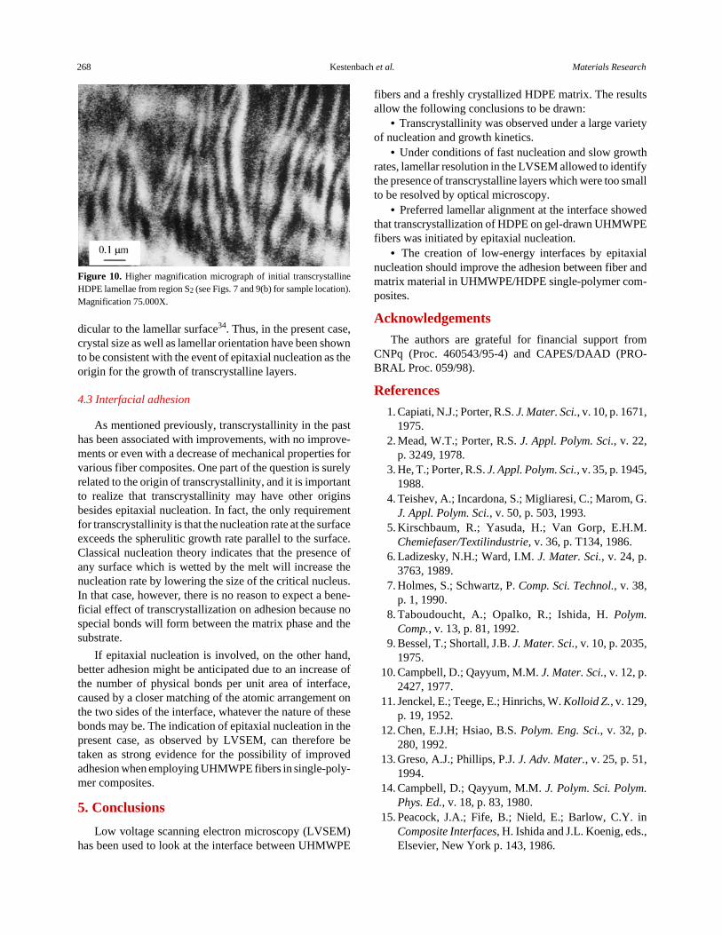
First, it has been argued that, for epitaxial nucleation tooccur, in addition to a close lattice match, the crystal sizeof the substrate may be of crucial importance. Thus, accord-ing to the “template model” of Greso and Philips32, thecrystal size of the substrate must be large enough to be ableto accommodate a critical secondary nucleus of the crystal-lizing polymer. This means, in the present case, that thelength of the crystalline segments in the UHMWPE fibersmust be equal to or larger than the lamellar thickness of theHDPE. Some of these UHMWPE crystalline segmentshave recently been observed by high resolution transmis-sion electron microscopy where irregular but extended-chain type crystal blocks were described with dimensionsof 40 to 70 nm in chain direction and 20 to 40 nm lateralwidth33. Thus, in the present case, substrate crystal dimen-sions in chain direction can be expected to be large enoughfor the epitaxial nucleation of HDPE lamellae whose thick-nesses are about 20 to 30 nm according to Fig. 10.
The second point refers to the particular orientationswhich the HDPE lamellae exhibit in Figs. 5, 6, 8 and 9, and
which do not seem to be perpendicular to the fiber axis. Ifthere is indeed epitaxial nucleation, it must be expected thatthe molecular direction in the HDPE lamellae matches themolecular direction in the UHMWPE substrate, i.e. thefiber axis. If this is so, it is important to realize that thelamellae should not form right angles with the fiber direc-tion because, at least in the case of solution-grown PElamellae whose crystallographic habits have been studiedin detail, chain-folded molecules are not in general perpen-
Figure 8. Tangential view of transcrystalline HDPE matrix lamellae fromregion S1 (see Fig. 7 for sample location). Magnification 7.500X in (a),15.000X in (b).
Figure 9. Top view of initial transcrystalline HDPE matrix lamellae fromregion S2 (see Fig. 7 for sample location). Magnification 7.500X in (a),15.000X in (b). For comparison, UHMWPE fiber surface without HDPEin (c). Magnification 15.000X.
Vol. 2, No. 4, 1999 Polyethylene Single-Polymer Composites 267
dicular to the lamellar surface34. Thus, in the present case,crystal size as well as lamellar orientation have been shownto be consistent with the event of epitaxial nucleation as theorigin for the growth of transcrystalline layers.
4.3 Interfacial adhesion
As mentioned previously, transcrystallinity in the pasthas been associated with improvements, with no improve-ments or even with a decrease of mechanical properties forvarious fiber composites. One part of the question is surelyrelated to the origin of transcrystallinity, and it is importantto realize that transcrystallinity may have other originsbesides epitaxial nucleation. In fact, the only requirementfor transcrystallinity is that the nucleation rate at the surfaceexceeds the spherulitic growth rate parallel to the surface.Classical nucleation theory indicates that the presence ofany surface which is wetted by the melt will increase thenucleation rate by lowering the size of the critical nucleus.In that case, however, there is no reason to expect a bene-ficial effect of transcrystallization on adhesion because nospecial bonds will form between the matrix phase and thesubstrate.
If epitaxial nucleation is involved, on the other hand,better adhesion might be anticipated due to an increase ofthe number of physical bonds per unit area of interface,caused by a closer matching of the atomic arrangement onthe two sides of the interface, whatever the nature of thesebonds may be. The indication of epitaxial nucleation in thepresent case, as observed by LVSEM, can therefore betaken as strong evidence for the possibility of improvedadhesion when employing UHMWPE fibers in single-poly-mer composites.
5. Conclusions
Low voltage scanning electron microscopy (LVSEM)has been used to look at the interface between UHMWPE
fibers and a freshly crystallized HDPE matrix. The resultsallow the following conclusions to be drawn:
• Transcrystallinity was observed under a large varietyof nucleation and growth kinetics.
• Under conditions of fast nucleation and slow growthrates, lamellar resolution in the LVSEM allowed to identifythe presence of transcrystalline layers which were too smallto be resolved by optical microscopy.
• Preferred lamellar alignment at the interface showedthat transcrystallization of HDPE on gel-drawn UHMWPEfibers was initiated by epitaxial nucleation.
• The creation of low-energy interfaces by epitaxialnucleation should improve the adhesion between fiber andmatrix material in UHMWPE/HDPE single-polymer com-posites.
Acknowledgements
The authors are grateful for financial support fromCNPq (Proc. 460543/95-4) and CAPES/DAAD (PRO-BRAL Proc. 059/98).
References
1. Capiati, N.J.; Porter, R.S. J. Mater. Sci., v. 10, p. 1671,1975.
2. Mead, W.T.; Porter, R.S. J. Appl. Polym. Sci., v. 22,p. 3249, 1978.
3. He, T.; Porter, R.S. J. Appl. Polym. Sci., v. 35, p. 1945,1988.
4. Teishev, A.; Incardona, S.; Migliaresi, C.; Marom, G.J. Appl. Polym. Sci., v. 50, p. 503, 1993.
5. Kirschbaum, R.; Yasuda, H.; Van Gorp, E.H.M.Chemiefaser/Textilindustrie, v. 36, p. T134, 1986.
6. Ladizesky, N.H.; Ward, I.M. J. Mater. Sci., v. 24, p.3763, 1989.
7. Holmes, S.; Schwartz, P. Comp. Sci. Technol., v. 38,p. 1, 1990.
8. Taboudoucht, A.; Opalko, R.; Ishida, H. Polym.Comp., v. 13, p. 81, 1992.
9. Bessel, T.; Shortall, J.B. J. Mater. Sci., v. 10, p. 2035,1975.
10. Campbell, D.; Qayyum, M.M. J. Mater. Sci., v. 12, p.2427, 1977.
11. Jenckel, E.; Teege, E.; Hinrichs, W. Kolloid Z., v. 129,p. 19, 1952.
12. Chen, E.J.H; Hsiao, B.S. Polym. Eng. Sci., v. 32, p.280, 1992.
13. Greso, A.J.; Phillips, P.J. J. Adv. Mater., v. 25, p. 51,1994.
14. Campbell, D.; Qayyum, M.M. J. Polym. Sci. Polym.Phys. Ed., v. 18, p. 83, 1980.
15. Peacock, J.A.; Fife, B.; Nield, E.; Barlow, C.Y. inComposite Interfaces, H. Ishida and J.L. Koenig, eds.,Elsevier, New York p. 143, 1986.
Figure 10. Higher magnification micrograph of initial transcrystallineHDPE lamellae from region S2 (see Figs. 7 and 9(b) for sample location).Magnification 75.000X.
268 Kestenbach et al. Materials Research

16. Ishida, H.; Bussi, P. Macromolecules, v. 24, p. 3569,1991.
17.Bashir, Z.; Odell, J.A. J. Mater. Sci. v. 28, p. 1081,1993.
18. Joy, D.C. J. Microscopy, v. 140, p. 283, 1985.19.Kestenbach, H.-J.; Nocite, N.C.P.S.; Gregório Filho,
R.; Loos, J; Petermann, J. Polímeros: Ciência e Tec-nologia, v. 7, p. 58, 1997. In Portuguese.
20.Chaterjee, A.M.; Price, F.B.; Newman, S. J. Polym.Sci. Polym. Phys. Ed., v. 13, p. 2369, 1975.
21.Chaterjee, A.M.; Price, F.B.; Newman, S. J. Polym.Sci. Polym. Phys. Ed., v. 13, p. 2385, 1975.
22.Masouka, M. Int. J. Adhesion Adhesives v. 1, p. 256,1981.
23.Burton, R.H.; Folkes, M.J. Plast. Rubber Process.Appl. v. 3, p. 129, 1983.
24.Huson, M.G.; McGill, W.J. J. Polym. Sci. Polym.Phys. Ed., v. 22, p. 121, 1985.
25. Folkes, M.J.; Wong, W.K. Polymer v. 28, p. 1309,1987.
26. Folkes, M.J.; Hardwick, S.T. J. Mater. Sci. Lett., v. 6,p. 656, 1987.
27. Folkes, M.J.; Hardwick, S.T. J. Mater. Sci. v. 25, p.2598, 1990.
28. Thomason, J.L.; Van Rooyen, A.A. J. Mater. Sci. v.27, p. 889, 1992.
29. Sukhanova, T.E.; Lednicky, F; Urban, J.; Balklgina,Y.G.; Mikhailov, G.M.; Kudryavtsev, V.V. J. Mater.Sci. v. 30, p. 2201, 1995.
30. Varga, J.; Karger-Kocsis, J. Polymer 36, 4877 (1995).31. Stern, T.; Wachtel, E.; Marom, G. J. Polym. Sci. B
(Polym. Phys.), v. 35, p. 2429, 1997.32. Greso, A.J.; Philips, P.J. Polymer v. 35, p. 3373, 1994.33. Chanzy, H.D.; Smith, P.; Revol, J.-F.; St.John Man-
ley, R. Polym. Comm. v. 28, p. 133, 1987.34. Bassett, D.C. Principles of Polymer Morphology,
Cambridge University Press, London, 1981, p. 49.
Vol. 2, No. 4, 1999 Polyethylene Single-Polymer Composites 269