transcriptome analysis of kaposi's sarcoma-associated
TRANSCRIPT

Transcriptome Analysis of Kaposi’s Sarcoma-Associated Herpesvirusduring De Novo Primary Infection of Human B and Endothelial Cells
Pravinkumar Purushothaman, Suhani Thakker, Subhash C. Verma
Department of Microbiology & Immunology, University of Nevada, Reno, School of Medicine, Center for Molecular Medicine, Reno, Nevada, USA
ABSTRACT
Kaposi’s sarcoma-associated herpesvirus (KSHV) infects many target cells (e.g., endothelial, epithelial, and B cells, keratinocytes,and monocytes) to establish lifelong latent infections. Viral latent-protein expression is critical in inducing and maintainingKSHV latency. Infected cells are programmed to retain the incoming viral genomes during primary infection. Immediately afterinfection, KSHV transcribes many lytic genes that modulate various cellular pathways to establish successful infection. Analysisof the virion particle showed that the virions contain viral mRNAs, microRNAs, and other noncoding RNAs that are transducedinto the target cells during infection, but their biological functions are largely unknown. We performed a comprehensive analy-sis of the KSHV virion packaged transcripts and the profiles of viral genes transcribed after de novo infections of various celltypes (human peripheral blood mononuclear cells [PBMCs], CD14� monocytes, and telomerase-immortalized vascular endo-thelial [TIVE] cells), from viral entry until latency establishment. A next-generation sequence analysis of the total transcriptomeshowed that several viral RNAs (polyadenylated nuclear RNA, open reading frame 58 [ORF58], ORF59, T0.7, and ORF17) wereabundantly present in the KSHV virions and effectively transduced into the target cells. Analysis of the transcription profiles ofeach viral gene showed specific expression patterns in different cell lines, with the majority of the genes, other than latent genes,silencing after 24 h postinfection. We differentiated the actively transcribing genes from the virion-transduced transcripts usinga nascent RNA capture approach (Click-iT chemistry), which identified transcription of a number of viral genes during primaryinfection. Treating the infected cells with phosphonoacetic acid (PAA) to block the activity of viral DNA polymerase confirmedthe involvement of lytic DNA replication during primary infection. To further understand the role of DNA replication duringprimary infection, we performed de novo PBMC infections with a recombinant ORF59-deleted KSHV virus, which showed sig-nificantly reduced numbers of viral copies in the latently infected cells. In summary, the transduced KSHV RNAs as well as theactively transcribed genes control critical processes of early infection to establish KSHV latency.
IMPORTANCE
Kaposi’s sarcoma-associated herpesvirus (KSHV) is the causative agent of multiple human malignancies in immunocompro-mised individuals. KSHV establishes a lifelong latency in the infected host, during which only a limited number of viral genes areexpressed. However, a fraction of latently infected cells undergo spontaneous reactivation to produce virions that infect the sur-rounding cells. These newly infected cells are primed early to retain the incoming viral genome and induce cell growth. KSHVtranscribes a variety of lytic proteins during de novo infections that modulate various cellular pathways to establish the latentinfection. Interestingly, a large number of viral proteins and RNA are encapsidated in the infectious virions and transduced intothe infected cells during a de novo infection. This study determined the kinetics of the viral gene expression during de novoKSHV infections and the functional role of the incoming viral transcripts in establishing latency.
Kaposi’s sarcoma-associated herpesvirus (KSHV), also called hu-man herpesvirus 8 (HHV8), is a double-stranded DNA virus that
causes Kaposi’s sarcoma, primary effusion lymphomas, and multi-centric Castleman’s disease (1–3). Like other herpesviruses, KSHVexhibits both latent and lytic modes of infection, persisting predom-inantly in the latent state in which only a subset of the viral proteinsare expressed, including the latency-associated nuclear antigen(LANA) protein (4–8). Although the expression of latent proteinsplays a critical role in inducing and maintaining KSHV latency, theinfected cells are primed early during the primary infection to retainthe viral genomes and induce tumors (9). During the primary infec-tion, KSHV undergoes a short lytic replication cycle that transcribesan array of viral genes, which have been shown to modulate variouspathways for establishing the latent infection (9). In addition, a smallfraction (1 to 5%) of the infected cells spontaneously undergo lyticreactivation to produce infectious virions, which is likely to be essen-tial for increasing the population of infected cells and inducing viralpathogenesis (10–13).
The infection of target cells with KSHV is a complex multistepprocess involving a variety of host cell surface receptors and mul-tiple viral glycoproteins. Irrespective of its mechanism of entry, fora successful infection, KSHV must overcome the obstacles it en-counters during the transportation of viral capsids from theplasma membrane into the nucleus. The main obstacles include
Received 30 August 2014 Accepted 23 December 2014
Accepted manuscript posted online 31 December 2014
Citation Purushothaman P, Thakker S, Verma SC. 2015. Transcriptome analysis ofKaposi’s sarcoma-associated herpesvirus during de novo primary infection ofhuman B and endothelial cells. J Virol 89:3093–3111. doi:10.1128/JVI.02507-14.
Editor: R. M. Longnecker
Address correspondence to Subhash C. Verma, [email protected].
Copyright © 2015, American Society for Microbiology. All Rights Reserved.
doi:10.1128/JVI.02507-14
March 2015 Volume 89 Number 6 jvi.asm.org 3093Journal of Virology
on March 15, 2018 by guest
http://jvi.asm.org/
Dow
nloaded from

apoptosis triggered by the virus’s binding and entry, autophagy,and the induction of various intrinsic, innate, and adaptive im-mune responses (14, 15). The mechanisms by which KSHV suc-cessfully circumvents these obstacles are beginning to be resolved.During de novo infections, KSHV generally establishes latency by24 h postinoculation (hpi) in cell culture systems (14, 16–20).However, very early, immediately after a de novo infection, KSHVundergoes a limited initial burst of lytic transcript accumulation(14). At this point, the viral gene expression shows a more com-plex pattern, wherein the latent and lytic genes are expressed con-currently, with an initial moderate proportion of lytic transcriptsfollowed by the onset of accumulation of more latent transcripts(14).
It has been shown that another gamma herpesvirus, Epstein-Barr virus (EBV), exhibits a similar pattern during early infection.The primary infection of B cells by EBV shows an expression oflytic genes (21–23). The expression of the lytic genes before thelatent genes during early infection suggests that this initial ex-pression of lytic genes is important for the successful establish-ment of EBV latency (21, 22). It has also been shown that EBVviral particles contain mRNAs and other nonstructural RNAsthat are transduced into the target cells early during infection(22). These mRNAs play crucial roles during infection to es-tablish the EBV latency (22). The detection of lytic transcriptsearly during de novo KSHV infections, followed by the latenttranscripts, suggests that a mechanism similar to that of EBVmay be occurring during the primary infection by KSHV. Ad-ditionally, the concurrent expression of open reading frame 73(ORF73) and ORF50 transcripts, detected early during KSHVinfection at 2 and 4 hpi (9), respectively, indicates that thesetranscripts might be transported into the newly infected cellduring the virus entry and are involved in altering the cellularenvironment prior to transcription from the incoming viralgenome.
Indeed, several recent studies have shown that herpesviruses,including KSHV particles, contain a variety of viral proteins (24,25) and diverse RNA species such as viral mRNA, noncodingRNA, viral and cellular microRNAs (miRNAs), and unusual smallRNA (usRNA) (26, 27). These RNAs are selectively packaged dur-ing virion budding and released into the target cells during de novoinfection, and they have been shown to be biologically functional(27). The KSHV ORF59 transcript is also present in the virionsand translated very early during primary infection (27). Recentstudies have similarly shown that several of the viral and cellularmiRNAs are selectively packaged into the virions and released intothe target cells during infection (26). However, the exact compo-sition of the virion transcripts in the KSHV particles and theirfunctions remain unclear (27). A recent study reported a highertransient expression of lytic genes at 24 hpi in comparison to la-tently infected cells. Moreover, the KSHV genome undergoesrapid chromatinization following infection, indicating that theinitial burst of lytic gene transcription likely originates from tran-scriptionally permissive chromatin rather than from incomingnaked DNA (28).
This study aimed to analyze the viral genes transcribed earlyduring the primary infection of peripheral blood mononuclearcells (PBMCs), CD14� and telomerase-immortalized vascular en-dothelial (TIVE) cells. To differentiate the RNAs present in thevirion and transcribed early during primary infection, noninter-nalized virions were removed from the cell surface by treating
FIG 1 Purification of KSHV virions. (A) Virions were purified from the cul-ture supernatant of approximately 9 � 108 reactivated TRExBCBL1-RTA cellsby a20 to 50% sucrose gradient ultracentrifugation. The three white bands asindicated represent A-type (empty), B-type (intermediate), and C-type (ma-ture) KSHV virus particles. (B) Western blotting of purified virions for thepresence of KSHV envelope glycoprotein K8.1 by immunoblotting with anti-K8.1 antibody. Total lysate from induced TRExBCBL1-RTA cells was used as apositive control. (C) Supernatants of uninduced or induced TRExBCBL1-RTA cells were used for real-time qPCR quantification of viral genome copyusing ORF73-specific primers along with ORF73 plasmid standards. Approx-imately 8.06 � 107 copies of viral DNA representing C-type virions weredetected.
Purushothaman et al.
3094 jvi.asm.org March 2015 Volume 89 Number 6Journal of Virology
on March 15, 2018 by guest
http://jvi.asm.org/
Dow
nloaded from
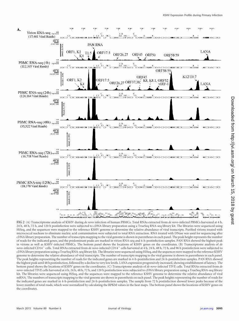
FIG 2 (A) Transcriptome analysis of KSHV during de novo infection of human PBMCs. Total RNAs extracted from de novo-infected PBMCs harvested at 4 h,24 h, 48 h, 72 h, and 120 h postinfection were subjected to cDNA library preparation using a TrueSeq RNA-seq library kit. The libraries were sequenced usingHiSeq, and the sequences were mapped to the reference KSHV genome to determine the relative abundance of viral transcripts. Purified virions treated withmicrococcal nuclease to eliminate nucleic acid contamination were subjected to total RNA extraction. RNA treated with DNase was used for sequencing aftercDNA library preparation. The number of transcripts mapping to the viral genome is shown in parentheses in each panel. The peak height represents the numberof reads for the indicated genes, and the predominant peaks are marked in virion RNA-seq and 4-h-postinfection samples. PAN RNA showed the highest peakin virions as well as KSHV-infected PBMCs. The bottom panel shows the locations of KSHV genes on the coordinates. (B) Transcriptome analysis of denovo-infected CD14� cells. Total RNAs extracted from de novo-infected CD14� cells harvested at 4 h, 24 h, 48 h, 72 h, and 96 h postinfection were subjected tocDNA library preparation using a TrueSeq RNA-seq library kit. The libraries were sequenced using HiSeq, and the sequences were mapped to the reference KSHVgenome to determine the relative abundance of viral transcripts. The number of transcripts mapping to the viral genome is shown in parentheses in each panel.The peak heights representing the number of reads for the indicated genes are marked in 4-h-postinfection and 24-h-postinfection samples. PAN RNA showedthe highest peak until 48 h postinfection, followed by a decline to very low levels. LANA expression progressively increased, showing establishment of latency. Thebottom panel shows the locations of KSHV genes on the coordinates. (C) Transcriptome analysis of de novo-infected TIVE cells. Total RNAs extracted from denovo-infected TIVE cells harvested at 4 h, 24 h, 48 h, 72 h, and 120 h postinfection were subjected to cDNA library preparation using a TrueSeq RNA-seq librarykit. The libraries were sequenced using HiSeq, and the sequences were mapped to the reference KSHV genome to determine the relative abundance of viralmRNA. The numbers of transcripts mapping to the viral genome are shown in parenthesis on each panel. The peak heights representing the number of reads forthe indicated genes are marked in 4-h-postinfection and 24-h-postinfection samples. The sample from 72 h postinfection showed lower peaks because of thelower number of total reads, which were normalized by calculating the RPKM values in the heat maps. The bottom panel shows the locations of KSHV genes onthe coordinates.
KSHV Expression Profile during Primary Infection
March 2015 Volume 89 Number 6 jvi.asm.org 3095Journal of Virology
on March 15, 2018 by guest
http://jvi.asm.org/
Dow
nloaded from

the cells with trypsin at 2 hpi, followed by capturing the na-scently transcribing RNA after 4 and 24 hpi. Also, the cells werecollected at 0, 4, 24, 48, 72, 96, and 120 hpi to extract total RNAfor transcriptomic profiling and real-time quantitative PCR(qPCR) analysis. The encapsulated RNA in the virions was de-termined by RNA-sequencing (RNA-seq) analysis as well as byreverse transcription-quantitative real-time PCR (RT-qPCR)for all of the viral genes. We captured newly synthesized RNAwith Click-iT technology during the de novo PBMC infectionsto identify the actively transcribing genes. The treatment ofKSHV-infected human PBMCs with phosphonoacetic acid (PAA)to block the viral DNA polymerase activity determined the in-volvement of lytic DNA replication during primary infection. Tounderstand the functional significance of the lytic gene transcrip-tion during KSHV infection and the establishment of latency, weinvestigated the role of ORF59, an abundantly packaged mRNA inthe virions that is transcribed during primary infection. The in-fectious recombinant KSHV BAC36WT and its ORF59 deletionmutant (BAC36�ORF59) were used for de novo PBMC infections.The infected cells showed that virions from the �ORF59 cells werenot able to attain as high a level of latent genomic copies as the
wild-type (WT) BAC36 virus, suggesting a defect in the lytic DNAreplication. Overall, our data show that a large number of viriontranscripts are transduced into the host cells along with the viralgenome during infection of the target cells, which helps in ampli-fying the viral genome copies and latency establishment.
MATERIALS AND METHODSCell culture, plasmids, and antibodies. The KSHV-positive cell lineTRExBCBL1-RTA, provided by Jae Jung (University of Southern Califor-nia), was cultured in RPMI 1640 medium supplemented with 10% fetalbovine serum (FBS), 2 mM L-glutamine, 5 U/ml penicillin, 5 �g/ml strep-tomycin, and 20 �g/ml hygromycin B. Cells (293L) harboring eitherBAC36WT or BAC36�ORF59 were cultured in Dulbecco modified Eaglemedium supplemented with 10% FBS, 2 mM L-glutamine, 5 U/ml peni-cillin, 5 �g/ml streptomycin, and 50 �g/ml hygromycin B. Telomerase-immortalized vascular endothelial (TIVE) cells, kindly provided by Erle S.Robertson, were cultured in endothelial basal medium-2 (Lonza) supple-mented with endothelial growth factor supplements (Lonza). HumanPBMCs (ReachBio, WA) were cultured in RPMI 1640 medium (HyClone)supplemented with 10% FBS, 2 mM L-glutamine, and penicillin-strepto-mycin (5 U/ml and 5 �g/ml, respectively). Human CD14� cells wereisolated from cord blood received from the Colorado Cord Blood Bank
FIG 2 continued
Purushothaman et al.
3096 jvi.asm.org March 2015 Volume 89 Number 6Journal of Virology
on March 15, 2018 by guest
http://jvi.asm.org/
Dow
nloaded from

(University of Colorado). Cells were maintained in Iscove modified Dul-becco medium (HyClone), supplemented with 20% heat-inactivated FBS(HyClone), 50 ng/ml macrophage colony-stimulating factor (M-CSF), 50ng/ml stem cell factor, 50 ng/ml granulocyte CSF (G-CSF), 50 ng/ml GM-CSF, and 50 ng/ml interleukin-3 (R&D Systems) at a density of 1 � 106
cells/ml on a low-cell-binding plate (Nunc Hydrocell). Protocols to ob-tain blood-associated cells were approved by the Institutional ReviewBoard and Office of Human Research Protection at the University ofNevada, Reno. The plasmid pA3F-LANA, carrying a Flag-tagged ORF73,has been previously described (29, 30). Commercially available mouseanti-K8.1 and rat anti-LANA antibodies (Advanced Biotechnologies,Inc.), were used in this study.
KSHV virion purification. KSHV virions were purified as previouslydescribed (26, 27). Approximately 100 million TRExBCBL1-RTA cellswere induced with doxycycline (1 �g/ml) for 5 days, after which the cul-ture supernatant was collected, centrifuged at 4,000 rpm for 10 min, andfiltered through a 0.45-�m filter to remove cellular debris before concen-trating the virus by centrifugation at 25,000 rpm for 2 h at 4°C. Theconcentrated virions were resuspended in 1 ml phosphate-buffered saline(PBS), layered onto a 30 to 50% sucrose gradient, and centrifuged at70,000 � g for 2 h. The purified virions formed a white opaque ring, whichwas collected in a syringe by puncturing the tube with the needle at the
band site. The collected band was diluted with PBS before centrifuging for2 h at 25,000 � g to pellet the virions.
Western blotting. The purified virus particles were resuspended in100 �l of lysis buffer (50 mM Tris-HCl [pH 7.5], 150 mM NaCl, 1 mMEDTA, 0.25% sodium deoxycholate, and 1% NP-40) supplemented withprotease inhibitors (1 mM phenylmethylsulfonyl fluoride, 10 �g/ml pep-statin, 10 �g/ml leupeptin, and 10 �g/ml aprotinin). The lysates werecentrifuged at high speed and mixed with protein sample buffer, and theprotein was resolved by 4 to 15% SDS-PAGE and Western blotted usingstandard protocols (Bio-Rad Laboratories). The KSHV-encoded struc-tural glycoprotein K8.1 was detected using mouse anti-K8.1 antibody fol-lowed by incubation with infrared dye-tagged (IR680 and IR800) second-ary antibodies and scanning with an Odyssey infrared scanner (Li-CorBiosciences, Lincoln, NE).
KSHV de novo infection. Approximately 8 � 107 human PBMCswere infected with KSHV purified from the induced TRExBCBL1-RTAcells. Two hours after infection at 37°C in the presence of 8 �g/ml Poly-brene, the cells were washed once with 0.005% trypsin in PBS and threetimes with PBS to remove the loosely bound virions, after which they wereresuspended and maintained in RPMI 1680 medium until harvesting. Thede novo KSHV-infected PBMCs (�5 � 106) were collected at different
FIG 2 continued
KSHV Expression Profile during Primary Infection
March 2015 Volume 89 Number 6 jvi.asm.org 3097Journal of Virology
on March 15, 2018 by guest
http://jvi.asm.org/
Dow
nloaded from

time points, washed twice with PBS, and processed separately to isolatethe DNA and RNA.
Indirect immunofluorescence microscopy. At 120 hpi, KSHV-in-fected PBMCs were washed with PBS, spread evenly on coverslips, and airdried. The cells were fixed for 10 min at room temperature with 4% para-formaldehyde and permeabilized with 0.2% Triton X-100 in PBS for 10min, also at room temperature. The cells were blocked by incubating inPBS containing 0.4% fish skin gelatin and 0.05% Triton X-100. The fixedcells were then incubated with primary rat anti-LANA antibody for 1 h atroom temperature, washed with PBS, incubated with Alexa Fluor 488secondary antibody (Molecular Probes) for 45 min at room temperature,and washed with PBS. The nuclear stain TO-PRO-3 (Molecular Probes)was used for counterstaining the nucleus. Images were obtained using alaser scanning confocal microscope (Carl Zeiss, Inc.).
Flow cytometry. PBMCs de novo infected with KSHV were harvestedat 120 hpi and fixed in Streck tissue fixative (STF) (Streck Laboratories)for 30 min. The fixed cells were washed twice in 1� PBS, permeabilizedwith 0.2% Triton X-100, blocked with 0.4% fish skin gelatin, and incu-bated with fluorescently tagged mouse anti-CD19 (Alexa Fluor 488),mouse anti-CD3 (Alexa Fluor 488; Rockland Immunochemicals, Inc.),and rat anti-LANA (Advanced Biotechnologies, Inc.), which was detectedwith a secondary antibody conjugated to Alexa Fluor 555 (Rockland Im-munochemicals, Inc.). The data were acquired on a FACSCalibur flowcytometer equipped with CellQuest Pro software and analyzed usingFlowJo software.
Viral genome extraction and quantification. De novo-infected cellswere collected by centrifugation (�2 � 106 cells per sample) and washedtwice with PBS before extracting the total DNA using a modified Hirt lysismethod (31). The PCR primers used for the KSHV genome quantificationwere selected from the ORF73 gene as previously described (9, 32). Two-fold serial dilutions of the pA3F-LANA plasmid were used as the templatein qPCRs to produce a standard curve for the quantifications. The ex-tracted total DNA was resuspended in 50 �l sterile water, and a 5-�laliquot of the DNA was used for the qPCR amplification of the KSHV-ORF73-specific sequence. The viral DNA copy numbers were calculatedwith reference to the standard curve.
RNA preparation and sequencing. The RNA-seq of the KSHV virionsand de novo-infected PBMCs was performed on a HiSeq next-generationsequencer (Illumina, Inc.). Total RNA was isolated from the purifiedKSHV virions after treating them with micrococcal nuclease (NEB) for 30min at 37°C and terminating the reaction with EGTA. The total RNA wasisolated with TRIzol reagent per the manufacturer’s instructions (LifeTechnologies), and viral DNA was eliminated by treating with DNase I(GE Health Care Life Sciences), which was subsequently inactivated. Thetotal RNA from the de novo-infected PBMCs was prepared using an Illus-tra RNAspin minikit with an in-column DNase treatment per the manu-facturer’s instructions (GE Health Care Life Sciences). The concentrationand purity of the extracted RNA were determined with a NanoDrop 2000cspectrophotometer (NanoDrop Technologies). A TrueSeq RNA samplepreparation kit v2 (Illumina, Inc.) was used to prepare cDNA libraries forthe RNA-seq according to the manufacturer’s instructions. The fragmentsizes and purity of the mature libraries were confirmed by analyzing on aBioanalyzer 2100 (Agilent Technologies). The quantities of the librariesrequired for RNA-seq were determined by real-time qPCR using a KAPAlibrary quantification kit for the Illumina platform (Kapa Biosystems).The libraries were sequenced using HiSeq (Illumina), and the sequenceswere mapped to the KSHV reference genome (accession numberNC_009333) using the RNA-seq analysis tool in the CLC Genomic Work-bench 7 (CLC Bio) software. The relative expression of the viral genes atdifferent time points was determined based on RPKM (reads per kilobaseof exon per million mapped reads) values, which were also similarly usedto generate heat maps showing the relative expression of those genes atdifferent time points utilizing the hierarchical clustering feature of theCLC Genomic Workbench 7 software.
Nascent RNA capture by Click-iT technology. To identify the newlysynthesized RNA during the de novo infections, a Click-iT RNA capturetechnology was used as described by the manufacturer (Life technologies,Inc.). Briefly, 8 � 107 human PBMCs were infected with KSHV purified
FIG 3 Transcriptome analysis of KSHV in de novo-infected PBMCs, TIVE cells,and CD14� cells. RPKM values, calculated based the number of reads for eachgene, were used for analyzing relative expression of KSHV genes as heat maps.Hierarchal clustering of genes was performed using CLC Workbench 7.0. Increas-ing RPKM values are represented from blue to red. (A) KSHV transcriptome fromde novo-infected human PBMCs. (B) KSHV transcriptome from de novo-infectedhuman TIVE cells. (C) KSHV transcriptome from de novo-infected humanCD14� cells.
Purushothaman et al.
3098 jvi.asm.org March 2015 Volume 89 Number 6Journal of Virology
on March 15, 2018 by guest
http://jvi.asm.org/
Dow
nloaded from

TABLE 1 mRNAs detected in purified TRExBCBL1-RTA virions by RNA-seq analysisa
No. mRNARPKMvalue
Totalgenereads Function
Transcription 4 h afterde novo infection ofhuman PBMCs
Source orreference
1 ORF4 2,633.55 101 Complement control protein (KCP), regulates complement activation This study2 ORF6 3,804.2 300 Single-stranded DNA binding protein essential for lytic DNA replication This study3 ORF7 2,332.60 113 Probably DNA packaging This study4 ORF8 4,432.43 261 Virion envelope-associated glycoprotein This study5 ORF9 5,318.55 375 KSHV DNA polymerase This study6 ORF10 6,446.37 188 Specific suppressor of interferon signaling This study7 ORF11 6,275.82 191 Viral lytic protein Yes This study8 K2 7,218.64 103 K2 (viral interleukin-6), regulates cellular proliferation Yes 279 K3 6,004.86 135 E3 ubiquitin ligases that reduce surface expression in infected cells This study10 ORF70 5,568.35 131 Homolog of viral thymidylate synthase This study11 K4.2 4,632.04 59 This study12 T1.5 10,245.93 358 This study13 K6 10,027.11 67 vMIP-IA 2714 PAN RNA 159,519.90 3,986 Late gene expression, lytic DNA replication, immune modulation 2715 ORF17 13,534.71 504 Yes 2716 ORF18 2,840.02 51 Late gene regulation This study17 ORF19 3,134.66 120 This study18 ORF21 3,140.50 127 Thymidine kinase This study19 ORF22 1,729.56 88 Glycoprotein H This study20 ORF23 2,305.84 65 Predicted glycoprotein This study21 ORF24 2,823.83 148 Essential for DNA replication This study22 ORF25 3,338.78 320 Major capsid protein This study23 ORF26 3,239.66 69 Minor capsid protein This study24 ORF27 6,270.22 127 Glycoprotein Yes This study25 ORF29 3,163.27 390 Packaging protein This study26 ORF34 2,233.92 51 This study27 ORF35 5,613.67 59 This study28 ORF36 4,907.44 152 Serine protein kinase Yes This study29 ORF37 2,625.62 89 Alkaline exonuclease Sox protein Yes This study30 ORF39 2,149.70 60 Glycoprotein M This study31 ORF40 2,904.36 144 Helicase/primase This study32 ORF44 2,676.778 147 Helicase This study33 ORF45 6,972.31 198 Virion-associated tegument protein regulating IFN function Yes This study34 ORF46 7,969.30 142 Uracil deglycosylase This study35 ORF48 3,101.60 87 Activates JNK/p38 This study36 ORF50 4,389.71 309 Replication transcriptional activator protein This study37 K8 4,072.23 89 bZIP Yes 2738 K8.1 3,918.32 71 Glycoprotein Yes This study39 ORF52 6,095.17 56 Tegument protein Yes This study40 ORF54 9,659.02 199 dUTPase/immune modulation 2741 ORF55 6,364.41 101 Tegument protein This study42 ORF56 3,830.11 225 DNA replication This study43 ORF57 3,533.39 121 Lytic replication protein homologous to EBV SM protein, mRNA
splicingYes This study
44 vIRF-1 7,407.08 232 Immune modulation Yes This study45 vIRF-4 2,324.47 153 Facilitates lytic replication by targeting cellular IRF4 and Myc gene Yes This study46 vIRF-3 2,302.58 96 Immune modulation This study47 vIRF-2 2,429.94 122 Type I interferon signaling This study48 ORF58 16,775.11 418 Yes 2749 ORF59 26,201.13 724 Processivity factor Yes 2750 ORF60 4,601.25 98 Ribonucleoprotein reductase This study51 ORF61 3,659.74 202 Ribonucleoprotein reductase This study52 ORF63 2,891.99 187 NLR homolog This study53 ORF64 3,897.02 715 Deubiquitinase Yes This study54 ORF65 5,629.25 67 Capsid protein This study55 ORF66 5,646.64 169 Capsid protein This study56 ORF67 4,753.85 90 Nuclear egress complex This study57 ORF68 5,372.34 175 Glycoprotein This study58 ORF69 6,353.81 134 BRLF2 nuclear egress Yes This study
(Continued on following page)
KSHV Expression Profile during Primary Infection
March 2015 Volume 89 Number 6 jvi.asm.org 3099Journal of Virology
on March 15, 2018 by guest
http://jvi.asm.org/
Dow
nloaded from

from the induced TRExBCBL1-RTA cells as described above. At 4 hourspostinfection, the cells were incubated for 1 h with ethylene uridine (EdU)(33) ribonucleotide homologs containing an alkyne-reactive group. Ad-ditionally, a de novo infection of KSHV was performed in the presence ofphosphonoacetic acid (PAA), an inhibitor of viral DNA polymerase andlytic DNA replication. The PBMCs were pretreated with 0.5 mM PAA for1 h prior to the infection, with the treatment continuing until harvestingat 24 hpi, similarly to a previously described method (34, 35). The denovo-infected PBMCs were collected 4 and 24 hpi and washed twice withPBS before isolating total RNA with an Illustra RNAspin minikit per themanufacturer’s instructions (GE Health Care). Biotin azide was “clickedon” to the isolated RNA, and the newly synthesized RNA was capturedusing streptavidin magnetic beads. Dimethyl sulfoxide (DMSO) insteadof biotin azide was added as control for the click reaction. The capturedRNAs were further utilized for preparing cDNA libraries with a TrueSeqRNA sample preparation kit v2 (Illumina, Inc.) and for the RNA-seqanalysis on a HiSeq Illumina sequencer. Relative numbers of copies of thenewly synthesized transcripts were determined by the ratios of the biotin-enriched (clicked) versus control, DMSO-enriched (nonclicked) copies ofthe transcripts identified by RNA sequence analysis.
qPCR. The real-time quantitative PCRs (qPCRs) were performed in a96-well plate in a total volume of 20 �l that included 10 �l of SYBR green PCR2� master mix (Applied Biosystems) and 0.5 �M each KSHV ORF-specificprimer. Primers for the human �-actin and GAPDH (glyceraldehyde-3-phosphate dehydrogenase) housekeeping genes were included for normaliz-ing the threshold cycle (CT) values. Purified genomic DNA samples or thevirion cDNA samples and KSHV-infected PBMCs were amplified on an ABIStepOnePlus real-time PCR machine (Applied Biosystems). As a standardpractice, a no-reverse-transcription (no-RT) control reaction was includedfor all of the prepared RNA samples to ensure their purity.
Microarray data accession number. The RNA-seq data were depos-ited in the NCBI Gene Expression Omnibus (GEO) database, and theaccession number for the complete data set is GSE62344.
RESULTSPurification of the KSHV virions. We isolated the KSHV particlesfrom doxycycline-induced (1 �g/ml) TRExBCBL1-RTA cells andpurified them on a 20 to 50% sucrose gradient. The three whitebands observed on the sucrose gradient (Fig. 1A) were consistentwith previous reports (26, 36), representing the A-type (emptyvirus particles), B-type (intermediate virus particles), and C-type(mature virions) KSHV particles. The lower band representing themature C-type KSHV virion particles was collected with a syringeand diluted before pelleting the virions by ultracentrifugation.These purified virions were tested for the presence of the KSHVenvelope glycoprotein K8.1 by immunoblotting with anti-K8.1antibody (Fig. 1B). The virion DNA was extracted to quantify thenumber of viral copies in a real-time qPCR assay using ORF73-specific primers (Fig. 1C), which showed approximately 8.0 � 107
virions/ml. This fraction of mature virions, which was confirmed
by both analyses, was further used for de novo infections of theindicated target cells.
KSHV viral transcripts are abundantly present during theprimary infection of human PBMCs, CD14�, and TIVE cells. Todetermine the profiles of the viral transcripts expressed during theprimary infection of B and endothelial cells (natural target cells ofKSHV), purified virions were used for the de novo infection ofhuman PBMCs, CD14� monocytes, and endothelial (TIVE) cells.An RNA-seq analysis was performed on total RNA extracted fromthese infected cells at different time points (4, 24, 48, 72, and 120hpi). Furthermore, 10 pM concentrations of the quantified librar-ies were sequenced and mapped to the reference KSHV genome(NC_009333). The transcriptome analysis revealed that several ofthe KSHV lytic and latent transcripts accumulated as early as 4 hpi(Fig. 2A to C and 3). Apart from the ORF50 replication and trans-activator protein (RTA), a number of other lytic transcripts in-volved in immune modulation, lytic DNA replication, and nucleicacid processing/synthesis were detected in significant amountsearly during the infection. These included polyadenylated nuclear(PAN) RNA, ORF58/59, kaposin B, K2, K4, K6, ORF11, ORF17,ORF45, ORF27, ORF37, ORF57, ORF64, ORF65, ORF73, and therecently identified T0.7. Among these, PAN RNA was the mostabundant as determined by the relative peak height (Fig. 2A to C)and RPKM values (Fig. 3) in all the three tested cell types. Al-though the overall patterns of the viral gene expressions were sim-ilar, i.e., with a majority of the genes being expressed at 24 hpi in allthe three tested cell types, CD14� monocytes showed completesilencing of viral gene expressions, other than for LANA, after 24hpi (Fig. 2A to C and 3). However, the human PBMCs and TIVEcells showed detectable levels of PAN RNA and ORF58/59 tran-scripts along with LANA after 48 hpi (Fig. 2A to C and 3). Thissuggests cell type-specific viral gene expression patterns duringprimary de novo infection and latency establishment. The relativeabundance of many of the viral transcripts, including ORF1, K2,K4, T1.5, PAN RNA, ORF17.5, ORF26, ORF27, ORF37 ORF38,ORF45, K8, K8.1, ORF52, vIRF-1, ORF58/59, K12/T0.7, andLANA, showed an increase in PBMCs at 4 hpi and 24 hpi com-pared with their abundance in the virions (Fig. 2A). However, theCD14� monocyte cells showed increases in the abundance offewer genes compared to those in the PBMCs (Fig. 2B). Impor-tantly, LANA was the only gene abundantly detected after 72 hpiin these CD14� cells, representing a true latency model of KSHVinfection. The endothelial (TIVE) cells showed detectable levels ofviral transcripts, but their relative abundance increased onlyslightly at 24 hpi (Fig. 2C). The heat maps generated based on theRPKM values showed an increase in their copies from 4 hpi to 24hpi, suggesting an active transcription, and these included PAN
TABLE 1 mRNAs detected in purified TRExBCBL1-RTA virions by RNA-seq analysisa
No. mRNARPKMvalue
Totalgenereads Function
Transcription 4 h afterde novo infection ofhuman PBMCs
Source orreference
59 T0.7 9,310.93 151 This study60 ORF72 3,118.46 56 vCyclin This study61 ORF73 23,623.23 1,858 Latency-associated nuclear antigen This study62 K14 3,116.41 59 vOX2 This study63 ORF74 4,481.89 107 vGPCR This study64 ORF75 3,810.57 344 Tegument protein vFGARAT Yes This studya Total gene reads above 50 and a qPCR relative fold change above 10 were considered significant.
Purushothaman et al.
3100 jvi.asm.org March 2015 Volume 89 Number 6Journal of Virology
on March 15, 2018 by guest
http://jvi.asm.org/
Dow
nloaded from

FIG 4 Real-time qPCR validation of de novo-infected PBMCs. Real-time qPCR analysis was performed on the cDNA prepared from total RNA harvested at 4 h,24 h, 48 h, 96 h, and 120 h postinfection of human PBMCs. A 96-well format qPCR plate representing each of the KSHV-specific ORF primers was used for theqPCR analysis. Fold changes of individual KSHV transcripts at various time points are plotted. The lytic genes are shown in red.
KSHV Expression Profile during Primary Infection
March 2015 Volume 89 Number 6 jvi.asm.org 3101Journal of Virology
on March 15, 2018 by guest
http://jvi.asm.org/
Dow
nloaded from

RNA, ORF73, ORF45, ORF58, T0.7, Kaposin B, ORF59, K4, K6,ORF17, ORF65, ORF27, K8.1, ORF11, ORF37, ORF38, ORF57,ORF69, ORF52, ORF54, and T1.5 (Fig. 3). The viral DNA poly-merase ORF9, along with other lytic DNA replication proteins,was detected, but their expression levels did not increase (Fig. 3).Also, there were several other detectable transcripts (K1, ORF4,ORF10, K6, ORF43, ORF44, ORF67, ORF68, and K14), but theirabundance did not increase, and the RPKM values remained un-changed during the time course tested, suggested that these geneswere not transcribed but were present due to their transductionthrough the virion particles during infection.
Earlier studies reported that immediately after de novo infec-tion, the herpesviruses express genes involved in lytic DNA repli-cation (21, 28, 37). Our RNA-seq analysis detected the transcriptsof proteins involved in viral DNA replication as early as 4 hpi, andthese included ORF59 (processivity factor), ORF9 (DNA poly-merase), ORF6 (single-stranded DNA binding protein), ORF56(DNA replication protein), ORF40 (primase-associated factor),and ORF54 (dUTPase), along with ORF60 and -61 (ribonucleo-protein reductase), ORF70 (thymidylate synthase), and ORF37(alkaline exonuclease). Apart from these transcripts, several othertranscripts, encoding the viral structural proteins ORF8 (glyco-protein B), K8.1 (glycoprotein), and ORF64 and -75 (tegumentprotein), were also detected in our transcription profiling data.Moreover, many of the noncoding RNAs, including PAN RNA, T0.7,and T1.5 (OriLyt transcript), were abundantly detected in the RNA-seq analysis. RNA-seq profiling of the virion encapsidated RNA, asrelative abundance, showed packaging of a large number of viral tran-scripts, which are brought into the target cells by the KSHV virionduring the de novo infection (Fig. 2 and Table 1).
In agreement with a previous report, our results showed theconcurrent expression of ORF50 and ORF73 transcripts duringearly primary infection (Fig. 2A to C and 3). However, the expres-sion of ORF50 decreased after 24 h, whereas the expression ofORF73 increased exponentially (Fig. 3). The KSHV latency-asso-ciated protein ORF73 is the predominant protein expressed dur-ing latency and is responsible for immune evasion, latent DNAreplication, and genome maintenance, whereas the ORF50-en-coded protein RTA activates lytic cycle-specific KSHV genes in acascaded manner to facilitate the lytic replication process (38–42).In a cell culture system, the overexpression of RTA is capable oftriggering the lytic DNA replication (38, 42). Our data detecting
the transcripts of ORF50 and other lytic genes along with ORF73during early infection suggest that KSHV may enter into a DNAreplicative phase before establishing latent infection.
Real-time qPCR validation of the RNA sequencing analysis.To further confirm the results of the transcriptome analysis by anindependent method, we performed a real-time qPCR analysis onde novo-infected PBMCs harvested at different time points (4, 24,48, 96, and 120 hpi). A custom qPCR array of the KSHV genes ina 96-well format (Bar Harbor Biotechnology) was used for thequantification of the viral mRNA levels. The fold changes for theindividual KSHV genes were calculated for the various timespostinfection using the ��CT method and plotted separately (Fig.4). The viral genes showed expression kinetics similar to thoseseen with the RNA-seq analysis. As first shown by the RNA-seqanalysis, even at 4 hpi, several lytic transcripts (ORF58/59, ORF11,ORF8, K8, K8.1, ORF17, ORF22, ORF57, ORF 45, ORF27, ORF37, and ORF64) were detected at high levels, and the expression ofthese transcripts was gradually depleted after 24 hpi (Fig. 4). Inaddition, latent transcripts (ORF73, K12, and ORF72) were con-sistently detected at significant levels during early infection (Fig.4). As reported previously (9), the real-time qPCR analysisshowed the concurrent expression of ORF50 and ORF73 at 4 hpi,and ORF50 showed a much higher fold increase during early in-fection than ORF 73. However, the expression of the ORF73 tran-script increased exponentially until 96 hpi. Additionally, the non-coding PAN RNA and recently identified T0.7 transcripts werealso detected abundantly at 4 hpi (Fig. 4). The qPCR validation ofthe RNA-seq analysis suggested that the limited lytic gene accu-mulation observed during primary infection might have been in-fluenced by the RTA-mediated gene expression as well as by thetransduction of viral transcripts with the virions during the denovo infection.
Detection of actively transcribing genes during de novo in-fection. The transcriptome analysis of de novo-infected cells dur-ing early infection revealed a significantly higher expression ofseveral genes which are expressed during the lytic replication cycle,including ORF50, ORF6, ORF9, ORF8, ORF7, ORF10, ORF11,ORF22, ORF27, ORF31, ORF37, ORF40/41, ORF45, ORF54,ORF55, ORF56, ORF58, ORF59, and ORF69 (Fig. 2A to C and 3).In addition, many of the immediate early unique KSHV genes, suchas K7, K8, K3, K5, K9, and K12, as well as late K8.1, were also detected.Many of these transcripts have crucial regulatory roles in immune
FIG 5 Viral genes transcribed at 4 hpi and 24 hpi, identified by a nascent RNA capture approach. Human PBMCs infected with KSHV virions were incubatedwith EdU (alkyne) at 4 hpi and 24 hpi to label the newly transcribing RNA. Total RNA extracted from these cells was subjected to click reaction with biotin-azide(Invitrogen, Inc.). DMSO instead of biotin-azide was used in control click reactions. Newly synthesized, biotin-labeled RNA was enriched by streptavidinmagnetic beads as per the manufacturer’s instructions. These enriched RNAs were subjected to RNA-seq analysis by HiSeq, and the sequences were mapped tothe KSHV reference genome (NC_009333) using CLC Bio Workbench 7.5. The relative numbers of copies of the newly synthesized viral RNA were determinedby ratios of specific (biotin-labeled) to nonspecific (DMSO) sequence reads.
Purushothaman et al.
3102 jvi.asm.org March 2015 Volume 89 Number 6Journal of Virology
on March 15, 2018 by guest
http://jvi.asm.org/
Dow
nloaded from

evasion and nucleic acid processing and synthesis, which are associ-ated with lytic DNA replication. The real-time qPCR analysis of theviral transcripts fully corroborated the RNA-seq analysis (Fig. 4).
To further identify the actively transcribing genes during denovo infection, we used an approach to capture the nascent tran-scribed RNA from the KSHV-infected PBMCs by labeling themwith EdU and isolating them by the Click-It approach. The se-quencing of newly synthesized RNA captured from the KSHV-infected PBMCs showed peaks of many viral genes at 4 hpi, andthese included ORF2, K3, T1.5, PAN RNA, ORF26, ORF29,ORF36, ORF37, ORF40, ORF50, K8.1, ORF55, vIRF-2, ORF58/59, ORF60, ORF63, ORF64, ORF67, T0.7, ORF72, ORF73, andORF74 (Fig. 5). Importantly, the majority of these were showedincreased expression in the graphs of relative abundance as well asin the heat maps (Fig. 2, 3, and 5). Interestingly, the majority of theactive transcription was limited to ORF73, detected by the se-
quence reads, at 24 hpi as suggested for the beginning of latencyestablishment (Fig. 5). We also confirmed the transcription dur-ing de novo infection by treating the cells with actinomycin D toblock active transcription. Comparison of the transcript levels at 4hpi in the untreated actinomycin D-treated cells revealed profilessimilar to those for the data for nascent RNA capture of genetranscription at 4 hpi (data not shown). As expected, the ORF50gene showed a moderate level of de novo transcription within 4 hpi(Fig. 5). Interestingly, not all the genes required for lytic DNAreplication showed active transcription during de novo infection,but they were detected in RNA-seq analysis, suggesting that thosegenes were transduced with the virions. Not surprisingly, theORF50 gene transcripts and those of genes regulated by ORF50decreased after 24 hpi. Also, a gradual increase in ORF73 tran-scripts by 24 hpi may aid in suppressing the expression of otherviral genes to promote the establishment of latency.
FIG 6 KSHV genome copies exponentially increase after infection. (A) Approximately 8 � 107 human PBMCs were infected with KSHV isolated fromreactivated TRExBCBL1-RTA, with a multiplicity of infection (MOI) of 10. De novo-infected PBMCs were harvested at 4 h, 24 h, 48 h, 72 h, 96 h, and 120 hpostinfection, followed by extraction of total DNA using a modified Hirt lysis method. This DNA was used to analyze the genome copy number by real-timeqPCR using ORF73-specific primers and ORF73 plasmid as a standard. (B) Indirect immunofluorescence assay (IFA) for LANA on de novo-infected PBMCs.KSHV-infected PBMCs at 120 h postinfection were subjected to IFA with rat anti-LANA antibody followed by detection with Alexa Fluor 488 secondaryantibody. Nuclear stain TO-PRO-3 was used for staining the nuclei. LANA is shown in green, and nuclear stain TO-PRO 3 is shown in blue. (C and D) Flowcytometry analysis of de novo-infected PBMCs. KSHV-infected PBMCs at 120 h postinfection were subjected to flow cytometry analysis to detect the percentagesof B and T lymphocytes infected with KSHV. Mouse anti-CD19 was used for the detection of B lymphocytes, and mouse anti-CD3 was used for the detection ofT lymphocytes. Rat anti-LANA was used to gate KSHV-positive cells. Data were acquired on a FACSCalibur equipped with CellQuest Pro software and analyzedusing FlowJo software. (E) PAA treatment reduces the expression of late genes. Untreated or PAA-pretreated human PBMCs were infected with KSHV virions(MOI of 10) for 4 h or 24 h. Total RNAs extracted at 4 hpi and 24 hpi were subjected to detection of selected viral transcripts by real-time qPCR, which showedsignificant reduction in ORF65 (late gene) expression with PAA treatment. (F) PAA treatment reduces viral genome copies in KSHV-infected human PBMCs.Untreated or PAA-treated human PBMCs infected with KSHV virions (MOI of 10) were subjected to genome copy analysis. Viral genome copies at different timepostinfection were determined relative to the copies at 4 hpi.
KSHV Expression Profile during Primary Infection
March 2015 Volume 89 Number 6 jvi.asm.org 3103Journal of Virology
on March 15, 2018 by guest
http://jvi.asm.org/
Dow
nloaded from

KSHV genome copy numbers increase exponentially after denovo infection. To investigate whether the detection of lytic reac-tivation genes leads to lytic DNA replication and genome ampli-fication, we analyzed the KSHV genome copy number during thede novo infection of PBMCs. Total DNAs from different timepoints (0, 4, 24, 48, 72, 96, and 120 hpi) were subjected to a real-time qPCR analysis to analyze the relative numbers of viral copiesin the infected cells. We used ORF73-specific primers and ORF73plasmid standards to obtain the copy numbers of the KSHV ge-nome. The relative number of KSHV genome copies was calcu-lated in the real-time qPCR assay by amplifying the ORF73 geneand normalizing it against GAPDH. KSHV DNA was detected asearly as 4 hpi, increased exponentially up to 48 hpi, and thenslightly decreased over time after establishing latency (Fig. 6A).This exponential increase in the genome copies confirms genomereplication during early infection until the establishment of la-tency. The punctate immunolocalization of LANA in the de novo-infected PBMCs (120 hpi) clearly demonstrated the establishmentof latency in these cells (Fig. 6B). A flow cytometry analysis of denovo-infected PBMCs using anti-LANA and B- and T-lymphocytemarkers gated for LANA expression showed that the populationsof both the B (�55.94%) and T (�24.75%) lymphocytes weresuccessfully infected with KSHV (Fig. 6C and D).
Since the number of viral genome copies increased about6-fold at 48 hpi compared to 4 hpi (the noninternalized virionswere removed at 2 hpi by treating the cells with trypsin), wewanted to determine whether the virus had undergone latent or
lytic modes of DNA synthesis to amplify the genome copies. Tothis end, we treated the cells with a viral DNA polymerase inhibi-tor, phosphonoacetic acid (PAA), to block the lytic DNA replica-tion. Cells pretreated with PAA or untreated were infected withKSHV virions for 4 h and 24 h for the transcriptome analysis.Analysis of selected latent, immediate early, and early genesshowed almost no effect of PAA on the transcription of latent(ORF73) and immediate early (ORF50) genes at 4 hpi (Fig. 6E).However, the expression of a late gene (ORF65) was significantlyreduced with PAA at both 4 and 24 hpi (Fig. 6D). We also deter-mined the number of genome copies during de novo infection ofPBMCs in the presence of PAA. The relative number of genomecopies showed a significant reduction in viral genome amplifica-tion in cells treated with PAA (Fig. 6F). Since there was still a slightincrease in the viral genome copies at 24 hpi, we speculate thatanother mechanism besides the lytic DNA replication takes placeduring de novo infection of PBMCs.
KSHV virions package the viral transcripts. To analyze theRNA composition in the encapsidated virions, total RNA ex-tracted from the purified C-type KSHV virions was used for theRNA-seq analysis. After confirming the purity of the RNA, thetotal RNA from two independent virus purifications was used toconstruct cDNA libraries. After quantifying, 10 pM concentra-tions of the mature library samples were used for the RNA-seqanalysis. In comparison with previous reports (26, 27), the tran-scriptome analysis with the CLC Genomic Workbench 7 softwareusing KSHV as the reference genome detected a larger number of
FIG 7 RNA-seq and real-time qPCR validation of the KSHV virion transcriptome. (A) RNA-seq analysis of virions purified from induced TRExBCBL1-RTA. (B)RNA-seq analysis of induced TRExBCBL1-RTA. Arrows indicate the most abundantly expressed KSHV genes. (C) Specificity of RNA encapsidation by KSHVvirion. Graphs show the ratio of virion-encapsidated mRNA to that in induced TRExBCBL1-RTA cells. The ratio was calculated by dividing the number ofvirion-packaged transcripts copies by the number of copies expressed during lytic reactivation.
Purushothaman et al.
3104 jvi.asm.org March 2015 Volume 89 Number 6Journal of Virology
on March 15, 2018 by guest
http://jvi.asm.org/
Dow
nloaded from
KSHV mRNAs in the virions. A previous DNA microarray studyidentified 11 KSHV mRNAs in the virions (27), but in this study,we identified more than 60 KSHV-specific transcripts, includingmRNAs for latent genes (Table 1). The mRNAs with 50 or morespecific gene reads and a 10-fold higher abundance in the qPCRassays were considered significant (Fig. 7A and Table 1). Themost abundant transcripts detected in the virions included PANRNA, K7, K14, K12, ORF58/59, and the recently identified T0.7,with PAN RNA being the most abundant (Fig. 7A). In addition,many lytic-specific mRNAs were also present in the virions, suchas ORF50, ORF8, ORF9, ORF21, ORF22, ORF31, ORF40/41,ORF45, ORF54, ORF55, ORF69, and ORF75. Moreover, many ofthe immediate early and early unique KSHV K genes (K7, K8, K3,K5, and K9) and the late K8.1 gene were also detected. Interest-ingly, in contrast to the previous report (27), a significant amountof latent-specific ORF73 mRNA was detected in the virions(Fig. 7A). Both this and the previous studies suggest that viriontranscripts are released into the target cells immediately after de
novo infection (26, 27). This may be the reason for the immediateconcurrent expression of ORF50 and ORF73 in the target cells (9).It has been shown that proteins encoded by these transcripts havesignificant regulatory roles in various cellular pathways, includingcell signaling, immune modulation, and apoptosis, that could crit-ically affect the phenotype of the newly infected cell and its mi-croenvironment (14, 15, 43, 44) to provide the necessary factorsrequired for a successful infection.
Virion-packaged transcript specificity. To further clarify theRNA-seq results, we performed a real-time qPCR analysis oncDNA prepared from the same source of total RNA (extractedfrom purified C-type KSHV virions) used in the sequencing anal-ysis. A custom qPCR array representing each of the KSHV-specificORF primers was used for the analysis. The uninfected PBMCsused as a reference control did not amplify, and a no-RT controlwas used to ensure the quality of the RNA. The qPCR data showedresults similar to those of the RNA-seq analysis, confirming thepresence of viral transcripts in the virions.
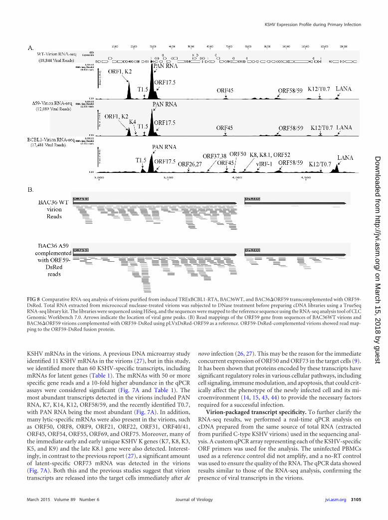
FIG 8 Comparative RNA-seq analysis of virions purified from induced TRExBCBL1-RTA, BAC36WT, and BAC36�ORF59 transcomplemented with ORF59-DsRed. Total RNA extracted from micrococcal nuclease-treated virions was subjected to DNase treatment before preparing cDNA libraries using a TrueSeqRNA-seq library kit. The libraries were sequenced using HiSeq, and the sequences were mapped to the reference sequence using the RNA-seq analysis tool of CLCGenomic Workbench 7.0. Arrows indicate the location of viral gene peaks. (B) Read mappings of the ORF59 gene from sequences of BAC36WT virions andBAC36�ORF59 virions complemented with ORF59-DsRed using pLVxDsRed-ORF59 as a reference. ORF59-DsRed-complemented virions showed read map-ping to the ORF59-DsRed fusion protein.
KSHV Expression Profile during Primary Infection
March 2015 Volume 89 Number 6 jvi.asm.org 3105Journal of Virology
on March 15, 2018 by guest
http://jvi.asm.org/
Dow
nloaded from

To determine whether there is a specific mechanism of RNAencapsidation during virion assembly, we compared the abun-dance of viral transcripts in the virions with the amounts presentduring the lytic reactivation of the cells. An earlier study indicatedthat RNA encapsidation by the KSHV virion could be a specificevent (27), and a recent report on KSHV virion miRNA also sug-gested that transcript encapsidation might be a specific process(26). To confirm this, we performed transcriptome (Fig. 7B) andreal-time qPCR analyses of the viral genes in the cDNA extractedfrom induced BCBL1 cells for comparison with the transcriptspresent in the purified virions (Fig. 7A and B). Many of the tran-scripts present in the virions were expressed in abundance duringlytic reactivation in the TRExBCBL1-RTA cells. To understandthe specificity of RNA encapsidation in the virion, we calculatedthe ratio of virion transcripts to the transcript expression levelspresent during reactivation in the induced TRExBCBL1-RTAcells. Several of the transcripts, including PAN RNA, T0.7, DR1,ORF58, and ORF59, that were detected in high abundance in thevirions were highly expressed during reactivation, and thus theratios of encapsidated to mRNA transcripts in the induced cellswere lower. We therefore concluded that they were packaged sim-ply due to their abundance and not because of any specificity.However, some of the transcripts, including ORF73, ORF31,ORF40, ORF50, ORF56, ORF49, and ORF64, showed significantlyhigher ratios (�0.5) of virion transcripts to their abundance dur-ing reactivation, suggesting the involvement of a specific mecha-nism in their encapsidation. The viral genes with a ratio of virionspackaged to induced cells above 0.5 were considered significant
and are marked by asterisks in Fig. 7C. Many of the virion-encap-sidated latent- and lytic-specific transcripts that were detectedearly during de novo infection showed a significantly higher ratio;the ratio for the latent transcript ORF73 was 1.0843, whereas theratio for the lytic transcript ORF50 was 2.6103. Taken together,these data suggest that KSHV may selectively encapsidate the la-tent and lytic transcripts required during early infection for prim-ing the cells to successfully establish latency.
Early ORF59 expression is required for KSHV de novo infec-tion and genome amplification. Our transcriptome data showeda high expression of ORF59 during the primary early infection ofboth B and endothelial cells, which led us to believe that ORF59may be required during the early events of KSHV infection andlatency establishment. To examine this, we used a recombinantKSHV with a stop codon in the ORF59 gene (BAC36�ORF59)(45) for de novo infection. Knowing that the ORF59-deletedBAC36 cannot produce virion particles, we generated a 293L cellline stably expressing the ORF59 gene fused with DsRed to com-plement the ORF59 in BAC36�ORF59. These bacterial artifi-cial chromosomes (BACs), WT in 293L and BAC36�ORF59 inORF59-DsRed-complemented 293L, were transfected and se-lected with hygromycin to obtain pure cell populations maintain-ing the BACs. These stable cells were induced with sodium bu-tyrate (NaB) and tetradecanoyl phorbol acetate to produce virionparticles, and the C-type virions were purified by sucrose densitygradient centrifugation for DNA extraction and infection studies.
To determine the composition of the KSHV virion-encapsi-dated RNA in BAC36WT and BAC36�ORF59, we performed an
TABLE 2 RPKM and gene reads of viral genes packaged in BAC36WT and �ORF59-BAC36 virions
No. mRNA
BAC36WT virion BAC36�59 virion
FunctionRPKM Gene reads RPKM Gene reads
1 K1 4,106.397 65 3,545.13 36 Glycoprotein2 ORF11 9,104.682 210 5,136.19 76 Viral lytic protein3 K2 264,559.9 3,066 1800,100.6 1,339 K2 (viral interleukin-6), regulates cellular proliferation4 K3 13,800.78 252 1,621.957 19 E3 ubiquitin ligases that reduce surface expression in infected cells5 K4 14,151.28 76 17,124.46 59 vMIP-II6 T1.5 2,572.319 73 3,735.02 687 K5 9,016.62 131 4,291.56 40 E3 ubiquitin ligase8 K6 10,134.38 55 2,010.55 7 vMIP-IA9 PAN RNA 456,221 9,259 554,384.1 7,218 Late gene expression, lytic DNA replication, immune modulation10 ORF16 5,125.818 51 2,506.66 16 Bcl2 homolog11 ORF17 61,895.31 1,872 40,406.45 784 Protease12 ORF18 12,752.6 186 13,786.64 129 Late gene regulation13 ORF45 5,332.74 123 4,798.29 71 Virion-associated tegument protein regulating interferon function14 ORF54 4,601.55 77 94.15 1 dUTPase/immune modulation15 ORF57 16,862.17 469 4,203.24 75 Lytic replication protein homologous to EBV SM protein, mRNA splicing16 vIRF-4 6,565.60 351 10,321.76 354 Facilitates lytic replication by targeting cellular IRF4 and Myc gene17 vIRF-3 8,888.84 301 11,738.21 255 Immune modulation18 vIRF-2 2,305.14 94 2,493.35 77 Type I interferon signaling19 ORF58 167,550.29 339 17,252.55 22420 ORF59 9,980.75 224 14,863.18 214 Processivity factor21 ORF60 3,005.99 52 2,793.37 31 Ribonucleoprotein reductase22 ORF61 1,672.91 75 1,599.45 46 Ribonucleoprotein reductase23 ORF64 899.21 134 1,234.312 118 Deubiquitinase24 ORF65 8,585.93 83 8,384.85 52 Capsid protein25 ORF66 3,085.30 75 3,077.94 48 Capsid protein26 T0.7 10,097.21 125 18,224.4 15427 ORF73 2,081.99 133 1,445.63 68 Latency-associated nuclear antigen28 ORF50 192.39 11 545.28 20 Replication and transcription activator
Purushothaman et al.
3106 jvi.asm.org March 2015 Volume 89 Number 6Journal of Virology
on March 15, 2018 by guest
http://jvi.asm.org/
Dow
nloaded from
RNA-seq analysis on the virions from these two cell lines. Unsur-prisingly, the patterns of the viral mRNA transcripts were similar(Fig. 8A and Table 2). We also compared the virion mRNA pat-terns from the BACs with those of the TRExBCBL1-RTA virions,which showed a slightly different packaging of the viral mRNA butsimilar levels of PAN RNA (Fig. 8A). Interestingly, the virionsproduced in the 293L cells complemented with ORF59-DsRedand containing BAC36�ORF59 also encapsidated ORF59, whichwas confirmed by mapping the sequence reads with ORF59-DsRed (Fig. 8B). The sequence reads from BAC36WT mappedonly to the ORF59 regions, confirming that BAC36WT packagedthe parental ORF59 gene (Fig. 8B), as expected. Interestingly, thetotal number of virions produced by BAC36�ORF59 was threelogs lower than that produced by BAC36WT (Fig. 9B), even withthe ORF59-DsRed complementation, which is reflected in the C-type band intensity in the BAC36�ORF59 tube (Fig. 9A).
We normalized the number of virions used for the infection ofhuman PBMCs by extracting the virion DNA and quantifying it ina real-time qPCR assay. Cells were collected at 4, 24, 48, 72, 96, and120 hpi and analyzed for viral gene expression and the number ofKSHV genome copies during early infection. Our results showed
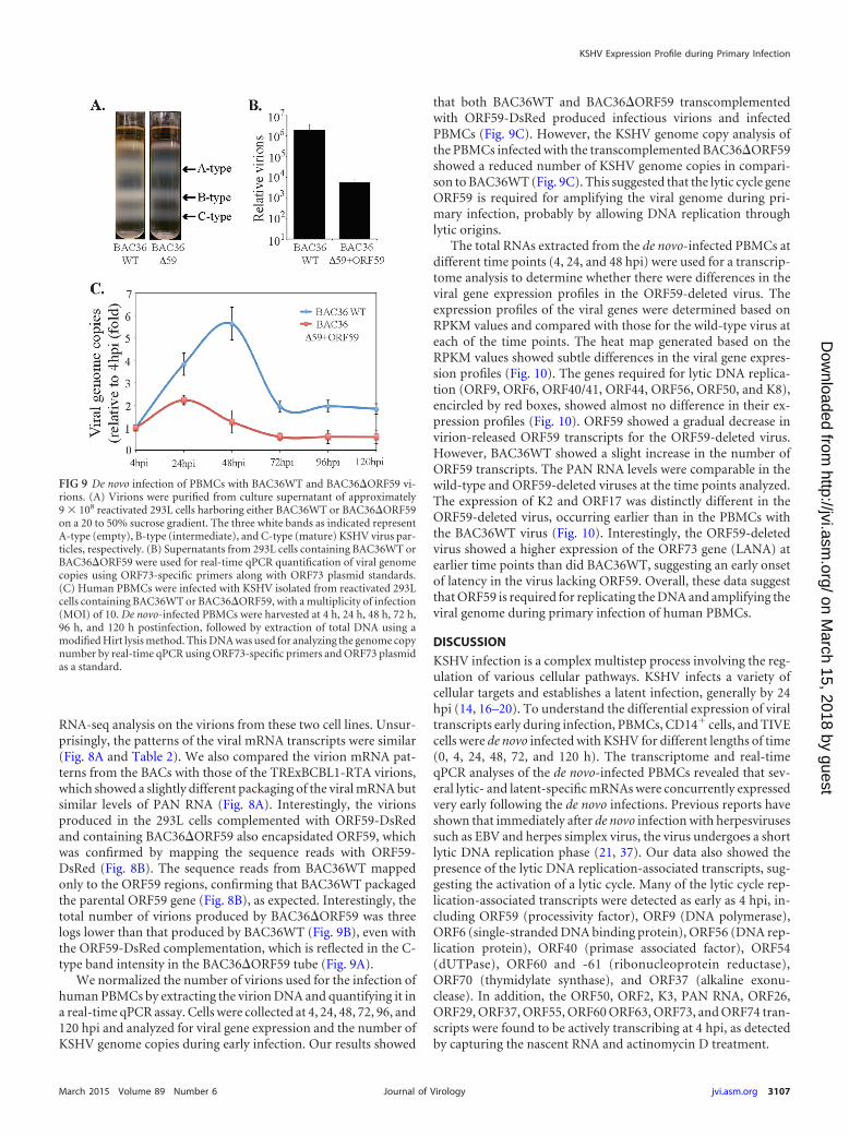
that both BAC36WT and BAC36�ORF59 transcomplementedwith ORF59-DsRed produced infectious virions and infectedPBMCs (Fig. 9C). However, the KSHV genome copy analysis ofthe PBMCs infected with the transcomplemented BAC36�ORF59showed a reduced number of KSHV genome copies in compari-son to BAC36WT (Fig. 9C). This suggested that the lytic cycle geneORF59 is required for amplifying the viral genome during pri-mary infection, probably by allowing DNA replication throughlytic origins.
The total RNAs extracted from the de novo-infected PBMCs atdifferent time points (4, 24, and 48 hpi) were used for a transcrip-tome analysis to determine whether there were differences in theviral gene expression profiles in the ORF59-deleted virus. Theexpression profiles of the viral genes were determined based onRPKM values and compared with those for the wild-type virus ateach of the time points. The heat map generated based on theRPKM values showed subtle differences in the viral gene expres-sion profiles (Fig. 10). The genes required for lytic DNA replica-tion (ORF9, ORF6, ORF40/41, ORF44, ORF56, ORF50, and K8),encircled by red boxes, showed almost no difference in their ex-pression profiles (Fig. 10). ORF59 showed a gradual decrease invirion-released ORF59 transcripts for the ORF59-deleted virus.However, BAC36WT showed a slight increase in the number ofORF59 transcripts. The PAN RNA levels were comparable in thewild-type and ORF59-deleted viruses at the time points analyzed.The expression of K2 and ORF17 was distinctly different in theORF59-deleted virus, occurring earlier than in the PBMCs withthe BAC36WT virus (Fig. 10). Interestingly, the ORF59-deletedvirus showed a higher expression of the ORF73 gene (LANA) atearlier time points than did BAC36WT, suggesting an early onsetof latency in the virus lacking ORF59. Overall, these data suggestthat ORF59 is required for replicating the DNA and amplifying theviral genome during primary infection of human PBMCs.
DISCUSSION
KSHV infection is a complex multistep process involving the reg-ulation of various cellular pathways. KSHV infects a variety ofcellular targets and establishes a latent infection, generally by 24hpi (14, 16–20). To understand the differential expression of viraltranscripts early during infection, PBMCs, CD14� cells, and TIVEcells were de novo infected with KSHV for different lengths of time(0, 4, 24, 48, 72, and 120 h). The transcriptome and real-timeqPCR analyses of the de novo-infected PBMCs revealed that sev-eral lytic- and latent-specific mRNAs were concurrently expressedvery early following the de novo infections. Previous reports haveshown that immediately after de novo infection with herpesvirusessuch as EBV and herpes simplex virus, the virus undergoes a shortlytic DNA replication phase (21, 37). Our data also showed thepresence of the lytic DNA replication-associated transcripts, sug-gesting the activation of a lytic cycle. Many of the lytic cycle rep-lication-associated transcripts were detected as early as 4 hpi, in-cluding ORF59 (processivity factor), ORF9 (DNA polymerase),ORF6 (single-stranded DNA binding protein), ORF56 (DNA rep-lication protein), ORF40 (primase associated factor), ORF54(dUTPase), ORF60 and -61 (ribonucleoprotein reductase),ORF70 (thymidylate synthase), and ORF37 (alkaline exonu-clease). In addition, the ORF50, ORF2, K3, PAN RNA, ORF26,ORF29, ORF37, ORF55, ORF60 ORF63, ORF73, and ORF74 tran-scripts were found to be actively transcribing at 4 hpi, as detectedby capturing the nascent RNA and actinomycin D treatment.
FIG 9 De novo infection of PBMCs with BAC36WT and BAC36�ORF59 vi-rions. (A) Virions were purified from culture supernatant of approximately9 � 108 reactivated 293L cells harboring either BAC36WT or BAC36�ORF59on a 20 to 50% sucrose gradient. The three white bands as indicated representA-type (empty), B-type (intermediate), and C-type (mature) KSHV virus par-ticles, respectively. (B) Supernatants from 293L cells containing BAC36WT orBAC36�ORF59 were used for real-time qPCR quantification of viral genomecopies using ORF73-specific primers along with ORF73 plasmid standards.(C) Human PBMCs were infected with KSHV isolated from reactivated 293Lcells containing BAC36WT or BAC36�ORF59, with a multiplicity of infection(MOI) of 10. De novo-infected PBMCs were harvested at 4 h, 24 h, 48 h, 72 h,96 h, and 120 h postinfection, followed by extraction of total DNA using amodified Hirt lysis method. This DNA was used for analyzing the genome copynumber by real-time qPCR using ORF73-specific primers and ORF73 plasmidas a standard.
KSHV Expression Profile during Primary Infection
March 2015 Volume 89 Number 6 jvi.asm.org 3107Journal of Virology
on March 15, 2018 by guest
http://jvi.asm.org/
Dow
nloaded from

Apart from these lytic transcripts, the expression of severalother transcripts encoding structural proteins, such as K8.1 (gly-coprotein) and ORFs 65 (tegument protein), were also detectedearly during infection. Moreover, many of the noncoding RNAs,including PAN RNA, T0.7, and T1.5 (OriLyt transcript), werealso expressed during early infection. PAN RNA has beenshown to modulate viral and cellular transcription as well as toactivate the KSHV lytic cycle by interacting with the ORF50promoter and viral genome (46, 47). Furthermore, there were alarge number of viral transcripts whose expression did not in-crease from their levels at 4 hpi, suggesting that those weretransduced with the virions.
The detection of PAN RNA in the virion and during infectionsuggests that PAN RNA could be playing major role in triggeringthe lytic cycle cascade. The KSHV ORF50-encoded immediateearly protein RTA is known to activate the cascade of lytic genesfor facilitating the lytic replication process (38–42). Studies on theprimary infection of B cells by EBV showed an accumulation oflytic transcripts during infection, indicating the requirement of ashort lytic replication prior to the establishment of latency (21,23). Earlier studies have shown that the KSHV virion also containsseveral KSHV lytic proteins, including ORF50, ORF8, ORF25,ORF26, K12, ORF62, ORF63, and ORF75, that are brought intothe target cells during de novo infection (24). The ORF50 proteinaccompanying the virion may also be important for orchestratingthe activation of RTA-responsive genes early during infection toinduce a short lytic cycle. A significant amount of the concurrentexpression of ORF50 and the KSHV latent protein ORF73 wasdetected as early as 4 hpi. Similar observations were reported in aprevious study based on microarray and real-time qPCR analyses,but the possibility of lytic DNA replication was not proposed dueto the lack of detection of all of the proteins required for lytic DNAreplication (9).
A recent study on histone modifications of the KSHV ge-nome during de novo infection demonstrated that there is adistinct pattern of activating the histone H3K4me3 mark acrossthe KSHV genome, which is gradually replaced by the repres-sive H3K27me3 mark by the LANA-mediated displacement ofsoluble sp100 (48). Additionally, in comparison to the repres-sive H3K27me3 mark and the rest of the genome, a LANAChIP-seq analysis of TRExBCBL1-RTA and TIVE-LTC showeda clear association of H3K4me3 mark and RNA polymerase IIactivation on the latent gene promoters (49). Our resultsshowed that immediately after the de novo infection of PBMCs,the viral genome increased exponentially up to 48 hpi, followedby a slight decrease at 72 hpi. The accumulation of ORF50 andother RTA-regulated lytic cycle genes during early infection issuggestive of the involvement of lytic DNA replication in ge-nome amplification. Treating the cells with a late gene expres-sion inhibitor, PAA, led to a significantly reduced number ofviral genome copies, further substantiating the involvement of
FIG 10 Expression profiles of viral genes in BAC36WT and BAC36�ORF59 atdifferent times postinfection. Normalized expression (RPKM values) of viralgenes in BAC36WT and BAC36�ORF59 at different times postinfection wasused for generating hierarchal clustering. (A) Relative expression of KSHV
genes at 4 h postinfection of PBMCs with BAC36WT and BAC36�ORF59virions. (B) Relative expression of KSHV genes at 24 h postinfection of PBMCswith BAC36WT and BAC36�ORF59 virions. (C) Relative expression of KSHVgenes at 48 h postinfection of PBMCs with BAC36WT and BAC36�ORF59virions. The red boxes encircle KSHV genes required for lytic DNA replication.The latency-associated gene (LANA), which showed early expression in cellsinfected with BAC36�ORF59, is indicated with a green arrow.
Purushothaman et al.
3108 jvi.asm.org March 2015 Volume 89 Number 6Journal of Virology
on March 15, 2018 by guest
http://jvi.asm.org/
Dow
nloaded from

lytic DNA replication in genome copy amplification in humanPBMCs.
Discriminating whether the transcripts detected at 4 hpi weredue to the virion-transduced mRNA or active transcription, RNA-seq analysis of the newly synthesized transcripts at 4 hpi of humanPBMCs showed transcription of only a limited number of genesduring primary infection. However, a large number of viraltranscripts are detected at 4 hpi as well as in the virion particles,which confirms that those transcripts are introduced into theinfected cells along with the virions. Detection of actively syn-thesizing mRNA in KSHV-infected PBMCs at 24 hpi showedtranscription of primarily the ORF73 gene, which confirmedthat the viral genome is chromatinized and epigenetically mod-ified for restricted gene expression by 24 hpi. An earlier studyreported the viral gene transcription of ORF59 during primaryinfection (27), and here we provide a comprehensive list of thegenes transcribed during a de novo infection. The transcriptionof the viral genes as early as 4 hpi suggests that the viral DNAsentering the target cells are capable of transcribing genes beforethe assembly of the epigenetic histone marks. Not surprisingly,the latent protein ORF73 gene showed active transcription at 4hpi, suggesting that LANA begins transcription as early as 4 hpiand accumulates over time to help establish latency. The im-mediate early protein ORF50 undergoes a moderate level oftranscription immediately after infection, which may contrib-ute in triggering the transcription of the lytic cascade to com-plete the DNA replication. The KSHV virions have also beenshown to encapsidate a small quantity of the RTA protein,which may also be important in triggering the lytic gene expres-sion cascade (24, 27).
It has been shown that both KSHV and EBV encapsidate bio-logically functional virion transcripts that are transported into thetarget cells during de novo infections (22, 26, 27). A previous studyusing a DNA microarray and real-time qPCR identified 11 of thevirus transcripts packaged into the virion (27). In this study, usingRNA-seq and transcriptome analyses, we detected additionalKSHV latent and lytic cycle transcripts encapsidated in the virionsthat were also transported into the targets cells. These data werevalidated by a real-time qPCR analysis using KSHV gene-specificprimer arrays, which showed consistent results. The transcrip-tome analysis identified a variety of virion transcripts in highabundance, including PAN RNA, K7, K14, K12, ORF 58/59, andthe recently identified T1.5 and T0.7. Additionally, many lytic-specific mRNAs (ORF50, ORF8, ORF9, ORF21, ORF22, ORF31,ORF 40/41, ORF45, ORF54, ORF55, ORF69, and ORF75) werepresent in the virion in moderate amounts. A number of imme-diate early and early unique KSHV K genes (K7, K8, K3, K5, K9,and late K8.1) were also detected in the virion, in addition to asignificant amount of latent-specific mRNAs (ORF73, ORF72,and K2).
The mechanism of RNA encapsidation by KSHV is currentlyunknown. Reports show that, similarly to the herpes simplex vi-rus, the RNA packaging and encapsidation by KSHV could bespecific events (26, 27). In addition, the specific encapsidation ofmiRNA by the KSHV virion has recently been demonstrated(26). However, both the previous study and this study deter-mined that the majority of mRNAs detected in the KSHV viri-ons were present at very high levels during lytic reactivation (9,27). This suggests that these transcripts might have been ran-domly packaged into the virions simply due to their higher
abundance. Nonetheless, the qPCR analysis to determine thespecificity of the virion versus the reactivated TRExBCBL1-RTA showed that the ORF31, ORF40, ORF50, ORF56, ORF49,ORF64, and ORF73 transcripts had significantly higher pro-portions of encapsidated mRNA than did the PAN RNA,ORF58, ORF59, T0.7, and DR1 transcripts, suggesting a spe-cific packaging mechanism for those transcripts.
Several of these highly expressed KSHV lytic genes have beenshown to have regulatory roles in immune modulation, antiapo-ptosis, and lytic DNA replication, indicating that their expressionmay prime the host cell for retaining the incoming viral DNAduring the initial infection. This is evident from the de novo infec-tion of PBMCs with virions produced from the BAC36�ORF59virus. Although significantly fewer virions were produced fromBAC36�ORF59 than from BAC36WT, a similar number of viri-ons were used to compare the viral genome copy numbers andexpression profiles. The lower number of virion copies in BAC36�ORF59 complemented with ORF59-DsRed may have been dueto a comparably lower expression of ORF59 in those cells. Thetranscriptome analysis of the de novo-infected PBMCs showedthe differential expression of only a few latent- and lytic-spe-cific genes (PAN RNA, T0.7, K2, K4, ORF17, ORF18, ORF58,ORF45, and ORF73), confirming that the incoming DNA istranscription competent.
The virions from both the BAC36WT- and ORF59-comple-mented cells showed the packaging of ORF59 transcripts. How-ever, the BAC36�ORF59 virus genome in de novo-infectedPBMCs failed to show an appreciable increase of copy number incomparison to BAC36WT. This indicates that the extended ex-pression of ORF59 during early infection is probably required forsynthesizing viral DNA through lytic DNA replication to in-crease the copy number. The exact mechanism requiring theinitial transient accumulation of lytic-specific transcripts forthe establishment of latency currently remains elusive. To fullyunderstand the early events of KSHV infection, i.e., latencyestablishment and genome amplification, further experimentsidentifying the viral proteins expressed and the mechanism ofviral DNA replication during primary infection in various cellsare ongoing.
ACKNOWLEDGMENTS
We thank Erle S. Robertson at the University of Pennsylvania for provid-ing the cell lines and LANA expression plasmids.
This work was supported by public health grants from the NIH(CA174459 and AI105000) to S.C.V. S.T. was supported by the MickHitchcock Fellowship at the University of Nevada, Reno.
REFERENCES1. Moore PS, Chang Y. 2010. Why do viruses cause cancer? Highlights of the
first century of human tumour virology. Nat Rev Cancer 10:878 – 889.http://dx.doi.org/10.1038/nrc2961.
2. Moore PS, Chang Y. 2003. Kaposi’s sarcoma-associated herpesvirus im-munoevasion and tumorigenesis: two sides of the same coin? Annu RevMicrobiol 57:609–639. http://dx.doi.org/10.1146/annurev.micro.57.030502.090824.
3. Verma SC, Robertson ES. 2003. Molecular biology and pathogenesis ofKaposi sarcoma-associated herpesvirus. FEMS Microbiol Lett 222:155–163. http://dx.doi.org/10.1016/S0378-1097(03)00261-1.
4. Fakhari FD, Dittmer DP. 2002. Charting latency transcripts in Kaposi’ssarcoma-associated herpesvirus by whole-genome real-time quantitativePCR. J Virol 76:6213–6223. http://dx.doi.org/10.1128/JVI.76.12.6213-6223.2002.
5. Jenner RG, Alba MM, Boshoff C, Kellam P. 2001. Kaposi’s sarcoma-
KSHV Expression Profile during Primary Infection
March 2015 Volume 89 Number 6 jvi.asm.org 3109Journal of Virology
on March 15, 2018 by guest
http://jvi.asm.org/
Dow
nloaded from

associated herpesvirus latent and lytic gene expression as revealed byDNA arrays. J Virol 75:891–902. http://dx.doi.org/10.1128/JVI.75.2.891-902.2001.
6. Verma SC, Lan K, Robertson E. 2007. Structure and function of latency-associated nuclear antigen. Curr Top Microbiol Immunol 312:101–136.http://dx.doi.org/10.1007/978-3-540-34344-8_4.
7. Zhong W, Wang H, Herndier B, Ganem D. 1996. Restricted expressionof Kaposi sarcoma-associated herpesvirus (human herpesvirus 8) genes inKaposi sarcoma. Proc Natl Acad Sci U S A 93:6641– 6646. http://dx.doi.org/10.1073/pnas.93.13.6641.
8. Ballestas ME, Chatis PA, Kaye KM. 1999. Efficient persistence of extrach-romosomal KSHV DNA mediated by latency-associated nuclear antigen. Sci-ence 284:641–644. http://dx.doi.org/10.1126/science.284.5414.641.
9. Krishnan HH, Naranatt PP, Smith MS, Zeng L, Bloomer C, Chandran B.2004. Concurrent expression of latent and a limited number of lytic geneswith immune modulation and antiapoptotic function by Kaposi’s sarcoma-associated herpesvirus early during infection of primary endothelial and fi-broblast cells and subsequent decline of lytic gene expression. J Virol 78:3601–3620. http://dx.doi.org/10.1128/JVI.78.7.3601-3620.2004.
10. Cannon JS, Ciufo D, Hawkins AL, Griffin CA, Borowitz MJ, HaywardGS, Ambinder RF. 2000. A new primary effusion lymphoma-derived cellline yields a highly infectious Kaposi’s sarcoma herpesvirus-containingsupernatant. J Virol 74:10187–10193. http://dx.doi.org/10.1128/JVI.74.21.10187-10193.2000.
11. Grundhoff A, Ganem D. 2004. Inefficient establishment of KSHV latencysuggests an additional role for continued lytic replication in Kaposi sar-coma pathogenesis. J Clin Invest 113:124 –136. http://dx.doi.org/10.1172/JCI200417803.
12. Renne R, Zhong W, Herndier B, McGrath M, Abbey N, Kedes D,Ganem D. 1996. Lytic growth of Kaposi’s sarcoma-associated herpesvirus(human herpesvirus 8) in culture. Nat Med 2:342–346. http://dx.doi.org/10.1038/nm0396-342.
13. Wang CY, Sugden B. 2004. New viruses shake old paradigms. J ClinInvest 113:21–23. http://dx.doi.org/10.1172/JCI20662.
14. Chandran B. 2010. Early events in Kaposi’s sarcoma-associated herpesvi-rus infection of target cells. J Virol 84:2188 –2199. http://dx.doi.org/10.1128/JVI.01334-09.
15. Chakraborty S, Veettil MV, Chandran B. 2012. Kaposi’s sarcoma asso-ciated herpesvirus entry into target cells. Front Microbiol 3:6. http://dx.doi.org/10.3389/fmicb.2012.00006.
16. Akula SM, Naranatt PP, Walia NS, Wang FZ, Fegley B, Chandran B.2003. Kaposi’s sarcoma-associated herpesvirus (human herpesvirus 8) in-fection of human fibroblast cells occurs through endocytosis. J Virol 77:7978 –7990. http://dx.doi.org/10.1128/JVI.77.14.7978-7990.2003.
17. Akula SM, Pramod NP, Wang FZ, Chandran B. 2002. Integrinalpha3beta1 (CD 49c/29) is a cellular receptor for Kaposi’s sarcoma-associated herpesvirus (KSHV/HHV-8) entry into the target cells. Cell108:407– 419. http://dx.doi.org/10.1016/S0092-8674(02)00628-1.
18. Bechtel JT, Liang Y, Hvidding J, Ganem D. 2003. Host range of Kaposi’ssarcoma-associated herpesvirus in cultured cells. J Virol 77:6474 – 6481.http://dx.doi.org/10.1128/JVI.77.11.6474-6481.2003.
19. Lagunoff M, Bechtel J, Venetsanakos E, Roy AM, Abbey N, Herndier B,McMahon M, Ganem D. 2002. De novo infection and serial transmissionof Kaposi’s sarcoma-associated herpesvirus in cultured endothelial cells. JVirol 76:2440 –2448. http://dx.doi.org/10.1128/jvi.76.5.2440-2448.2002.
20. Chakraborty S, Veettil MV, Bottero V, Chandran B. 2012. Kaposi’ssarcoma-associated herpesvirus interacts with EphrinA2 receptor to am-plify signaling essential for productive infection. Proc Natl Acad Sci U S A109:E1163–E1172. http://dx.doi.org/10.1073/pnas.1119592109.
21. Halder SMM, Verma SC, Kumar P, Yi F, Robertson ES. 2009. Earlyevents associated with infection of Epstein-Barr virus infection of pri-mary B-cells. PLoS One 4:e7214. http://dx.doi.org/10.1371/journal.pone.0007214.
22. Jochum SRR, Moosmann A, Hammerschmidt W, Zeidler R. 2012. RNAs inEpstein-Barr virions control early steps of infection. Proc Natl Acad Sci U S A109:E1396–E1404. http://dx.doi.org/10.1073/pnas.1115906109.
23. Wen W, Iwakiri D, Yamamoto K, Maruo S, Kanda T, Takada K. 2007.Epstein-Barr virus BZLF1 gene, a switch from latency to lytic infection, isexpressed as an immediate-early gene after primary infection of B lym-phocytes. J Virol 81:1037–1042. http://dx.doi.org/10.1128/JVI.01416-06.
24. Bechtel JT, Winant RC, Ganem D. 2005. Host and viral proteins in thevirion of Kaposi’s sarcoma-associated herpesvirus. J Virol 79:4952– 4964.http://dx.doi.org/10.1128/JVI.79.8.4952-4964.2005.
25. Zhu FX, Chong JM, Wu L, Yuan Y. 2005. Virion proteins of Kaposi’ssarcoma-associated herpesvirus. J Virol 79:800 – 811. http://dx.doi.org/10.1128/JVI.79.2.800-811.2005.
26. Lin X, Li X, Liang D, Lan K. 2012. MicroRNAs and unusual small RNAsdiscovered in Kaposi’s sarcoma-associated herpesvirus virions. J Virol 86:12717–12730. http://dx.doi.org/10.1128/JVI.01473-12.
27. Bechtel J, Grundhoff A, Ganem D. 2005. RNAs in the virion of Kaposi’ssarcoma-associated herpesvirus. J Virol 79:10138 –10146. http://dx.doi.org/10.1128/JVI.79.16.10138-10146.2005.
28. Toth Z, Brulois K, Lee HR, Izumiya Y, Tepper C, Kung HJ, Jung JU.2013. Biphasic euchromatin-to-heterochromatin transition on the KSHVgenome following de novo infection. PLoS Pathog 9:e1003813. http://dx.doi.org/10.1371/journal.ppat.1003813.
29. Verma SC, Bajaj BG, Cai Q, Si H, Seelhammer T, Robertson ES. 2006.Latency-associated nuclear antigen of Kaposi’s sarcoma-associated her-pesvirus recruits uracil DNA glycosylase 2 at the terminal repeats and isimportant for latent persistence of the virus. J Virol 80:11178 –11190. http://dx.doi.org/10.1128/JVI.01334-06.
30. Verma SC, Choudhuri T, Kaul R, Robertson ES. 2006. Latency-associated nuclear antigen (LANA) of Kaposi’s sarcoma-associated her-pesvirus interacts with origin recognition complexes at the LANA bindingsequence within the terminal repeats. J Virol 80:2243–2256. http://dx.doi.org/10.1128/JVI.80.5.2243-2256.2006.
31. Hirt B. 1967. Selective extraction of polyoma DNA from infected mousecell cultures. J Mol Biol 26:365–369. http://dx.doi.org/10.1016/0022-2836(67)90307-5.
32. Lallemand F, Desire N, Rozenbaum W, Nicolas JC, Marechal V. 2000.Quantitative analysis of human herpesvirus 8 viral load using a real-timePCR assay. J Clin Microbiol 38:1404 –1408.
33. Bates I, Bedu-Addo G, Jarrett RF, Schulz T, Wallace S, Armstrong A,Sheldon J, Rutherford T. 2001. B-lymphotropic viruses in a novel tropicalsplenic lymphoma. Br J Haematol 112:161–166. http://dx.doi.org/10.1046/j.1365-2141.2001.02517.x.
34. Medveczky MM, Horvath E, Lund T, Medveczky PG. 1997. In vitro anti-viral drug sensitivity of the Kaposi’s sarcoma-associated herpesvirus. AIDS11:1327–1332. http://dx.doi.org/10.1097/00002030-199711000-00006.
35. Chang PJ, Boonsiri J, Wang SS, Chen LY, Miller G. 2010. Binding ofRBP-Jkappa (CSL) protein to the promoter of the Kaposi’s sarcoma-associated herpesvirus ORF47 (gL) gene is a critical but not sufficientdeterminant of transactivation by ORF50 protein. Virology 398:38 – 48.http://dx.doi.org/10.1016/j.virol.2009.11.022.
36. Nealon K, Newcomb WW, Pray TR, Craik CS, Brown JC, Kedes DH.2001. Lytic replication of Kaposi’s sarcoma-associated herpesvirus resultsin the formation of multiple capsid species: isolation and molecular char-acterization of A, B, and C capsids from a gammaherpesvirus. J Virol75:2866 –2878. http://dx.doi.org/10.1128/JVI.75.6.2866-2878.2001.
37. Roizman B, Knipe DM. 2001. Herpes simplex viruses and their replica-tion, p 2399 –2459. In Knipe DM, Howley PM, et al. (ed), Fields virology,4th ed. Lippincott, Williams & Wilkins, Philadelphia, PA.
38. Guito J, Lukac DM. 2012. KSHV Rta promoter specification and viralreactivation. Front Microbiol 3:30. http://dx.doi.org/10.3389/fmicb.2012.00030.
39. Dourmishev LA, Dourmishev AL, Palmeri D, Schwartz RA, Lukac DM.2003. Molecular genetics of Kaposi’s sarcoma-associated herpesvirus (hu-man herpesvirus-8) epidemiology and pathogenesis. Microbiol Mol BiolRev 67:175–212. http://dx.doi.org/10.1128/MMBR.67.2.175-212.2003.
40. Sun R, Lin SF, Staskus K, Gradoville L, Grogan E, Haase A, Miller G.1999. Kinetics of Kaposi’s sarcoma-associated herpesvirus gene expres-sion. J Virol 73:2232–2242.
41. West JT, Wood C. 2003. The role of Kaposi’s sarcoma-associated herpes-virus/human herpesvirus-8 regulator of transcription activation (RTA) incontrol of gene expression. Oncogene 22:5150 –5163. http://dx.doi.org/10.1038/sj.onc.1206555.
42. Nakamura H, Lu M, Gwack Y, Souvlis J, Zeichner SL, Jung JU. 2003.Global changes in Kaposi’s sarcoma-associated virus gene expressionpatterns following expression of a tetracycline-inducible Rta transac-tivator. J Virol 77:4205– 4220. http://dx.doi.org/10.1128/JVI.77.7.4205-4220.2003.
43. Chan SR, Bloomer C, Chandran B. 1998. Identification and character-ization of human herpesvirus-8 lytic cycle-associated ORF 59 protein andthe encoding cDNA by monoclonal antibody. Virology 240:118 –126.http://dx.doi.org/10.1006/viro.1997.8911.
44. Unal A, Pray TR, Lagunoff M, Pennington MW, Ganem D, Craik CS.
Purushothaman et al.
3110 jvi.asm.org March 2015 Volume 89 Number 6Journal of Virology
on March 15, 2018 by guest
http://jvi.asm.org/
Dow
nloaded from

1997. The protease and the assembly protein of Kaposi’s sarcoma-associated herpesvirus (human herpesvirus 8). J Virol 71:7030 –7038.
45. McDowell ME, Purushothaman P, Rossetto CC, Pari GS, Verma SC.2013. Phosphorylation of Kaposi’s sarcoma-associated herpesvirus pro-cessivity factor ORF59 by a viral kinase modulates its ability to associatewith RTA and oriLyt. J Virol 87:8038 – 8052. http://dx.doi.org/10.1128/JVI.03460-12.
46. Rossetto CC, Pari G. 2012. KSHV PAN RNA associates with demethy-lases UTX and JMJD3 to activate lytic replication through a physical in-teraction with the virus genome. PLoS Pathog 8:e1002680. http://dx.doi.org/10.1371/journal.ppat.1002680.
47. Rossetto CC, Pari GS. 2011. Kaposi’s sarcoma-associated herpesvirus
noncoding polyadenylated nuclear RNA interacts with virus- and hostcell-encoded proteins and suppresses expression of genes involved inimmune modulation. J Virol 85:13290 –13297. http://dx.doi.org/10.1128/JVI.05886-11.
48. Gunther T, Schreiner S, Dobner T, Tessmer U, Grundhoff A. 2014.Influence of ND10 components on epigenetic determinants of earlyKSHV latency establishment. PLoS Pathog 10:e1004274. http://dx.doi.org/10.1371/journal.ppat.1004274.
49. Hu J, Yang Y, Turner PC, Jain V, McIntyre LM, Renne R. 2014. LANAbinds to multiple active viral and cellular promoters and associates withthe H3K4methyltransferase hSET1 complex. PLoS Pathog 10:e1004240.http://dx.doi.org/10.1371/journal.ppat.1004240.
KSHV Expression Profile during Primary Infection
March 2015 Volume 89 Number 6 jvi.asm.org 3111Journal of Virology
on March 15, 2018 by guest
http://jvi.asm.org/
Dow
nloaded from