transcription factor
TRANSCRIPT
MOLECULAR AND CELLULAR BIOLOGY, Jan. 1987, p. 403-409 Vol. 7, No. 10270-7306/87/010403-07$02.00/0Copyright © 1987, American Society for Microbiology
Isolation of a Saccharomyces cerevisiae Centromere DNA-BindingProtein, Its Human Homolog, and Its Possible Role as a
Transcription FactorRICHARD J. BRAM AND ROGER D. KORNBERG*
Department of Cell Biology, Stanford University School of Medicine, Stanford, California 94305
Received 7 April 1986/Accepted 22 September 1986
A protein that binds specifically to Saccharomyces cerevisiae centromere DNA element I was purified on thebasis of a nitrocellulose filter-binding assay. This protein, termed centromere-binding protein 1 (CP1), was heatstable and renaturable from sodium dodecyl sulfate (SDS), and assays of eluates from SDS gels indicated amolecular weight of 57,000 to 64,000. An activity with similar specificity and stability was detected in humanlymphocyte extracts, and analysis in SDS gels revealed a molecular weight of 39,000 to 49,000. CP1-bindingsites occurred not only at centromeres but also near many transcription units, for example, adjacent to bindingsites for the GAL4-positive regulatory protein upstream of the GAL2 gene in S. cerevisiae and adjacent to theTATA element of the adenovirus major late promoter. A factor (termed USF) that binds to the latter site andstimulates transcription has been isolated from HeLa cells by others.
Centromeres are specialized regions of eucaryotic chro-mosomes that form sites of attachment for spindlemicrotubules in mitosis and meiosis (13, 35, 36). Little isknown of the mechanism of spindle attachment or themolecules involved. We report here on the isolation of acentromere DNA-binding protein from Saccharomyces cere-visiae and its counterpart from human cells.
Cloning and deletion analysis of yeast centromeres re-vealed three contiguous conserved sequences essential to orimportant for centromere function: centromere DNA ele-ment I (CDEI), an 8-base-pair (bp) conserved sequence;CDEII, an apparently random, adenine-plus-thymine (A +T)-rich stretch of approximately 90 bp; and CDEIII, a 25-bpconserved sequence (11, 12, 17, 18, 20, 21, 24, 29, 32, 33, 39).Nuclease digestion studies of chromatin containing cen-tromere regions CEN3 and CENII revealed an unusualconfiguration: 250 bp, including the three conserved DNAelements, are especially protected from digestion (4). Crudefractions from S. cerevisiae bind naked DNA containingcentromere sequences (5), but no resolution of the proteinsinvolved has been described.A centromere-specific DNA-binding activity was discov-
ered during recent studies of the GAL4-positive regulatoryprotein (7). DNase I footprints with a yeast nuclear extractshowed three 30-bp regions of protection upstream of theGAL2 gene: two regions located 55 bp apart, and the thirdlocated 25 bp further upstream. The first two regions wereattributed to GAL4 protein binding, and the third wasattributed to another activity for the following reasons. (i)Protection of the first two regions was abolished by compe-tition with a GAL4-binding oligonucleotide, while protectionof the third region was unaffected. (ii) The first two regionswere homologous with a consensus GAL4-binding sequence,while the third region was altogether different, showing astriking homology to many CDEIs. During studies of proteinbinding to the third region (see below), a further homologycame to light, suggesting a relationship to transcription.
* Corresponding author.
MATERIALS AND METHODS
Plasmid DNAs. Plasmid pG2p contains the 5' half of theGAL2 gene (7). Plasmids p181, p113, p200, p201, p203, p202,and p182 containing HindlIl fragments of CENI, CEN4,CEN7, CENII, CEN14, CEN15, and 2,um-STB inYRpJ41ARS1 were kindly provided by P. Hieter (20).Yeast strains and media. BY2 (6) is a protease-deficient
strain (pep4-3) carrying the GAL4 gene on a high-copy-number plasmid. YNN 267 (a ura3-52 Ahis3-200 ade2-101lys2-801 met- Agal4-537) was a gift from M. Johnston. BJ926(a trpl prcl -126 pep4-3 prbl-1122 can1la his] prcl-126pep4-3 prbl-1122 can]) was kindly provided by E. Jones.Cells were grown in YP medium (1% yeast extract, 2%Bacto-Peptone) containing 2% glucose to an A6w of 5.
Nitrocellulose filter-binding assay and DNase I footprints.Plasmid pG2p DNA was linearized by cleavage with EcoRI,and the recessed 3' ends were filled in with the largefragment of DNA polymerase I in the presence of [a-32P]dATP and cold TTP. In some experiments, a 0.28-kilobase (kb) fragment containing the two GAL4-bindingsites and the centromere-binding protein 1 (CP1)-binding siteprepared by cleavage of pG2p with Hindlll and HpaI andagarose gel electrophoresis was used for greater sensitivity.A palindromic oligonucleotide (Fig. 1) was synthesized,purified, and self-annealed (6). Details of the nitrocellulosefilter-binding and DNase I footprint experiments were pre-viously described (6). Other probes for filter binding wereprepared by excision of CEN-containing fragments withHindIII, labeling, and isolation as described above. Probesfor DNase I footprint experiments were prepared by cleav-age with BglII (CEN7 and CEN15) or HindlIl (CENJ I) afterlabeling as described above. Polymerase I was inactivatedby heating at 65°C for 20 min. The labeled DNAs werecleaved with XbaI (CEN15 and CENJ I) or Hindlll (CEN7),and CEN-containing fragments were isolated by agarose gelelectrophoresis.
Yeast cell extracts and protein fractionation. Crude extractsand nuclear extracts were prepared as described previously
403
on March 31, 2018 by guest
http://mcb.asm
.org/D
ownloaded from
404 BRAM AND KORNBERG
GAL2CEN1CEN4CEN7
2 CEN1 1CEN11L
c] CEN1 1 RCEN14CEN15YRP14
2gSTB I
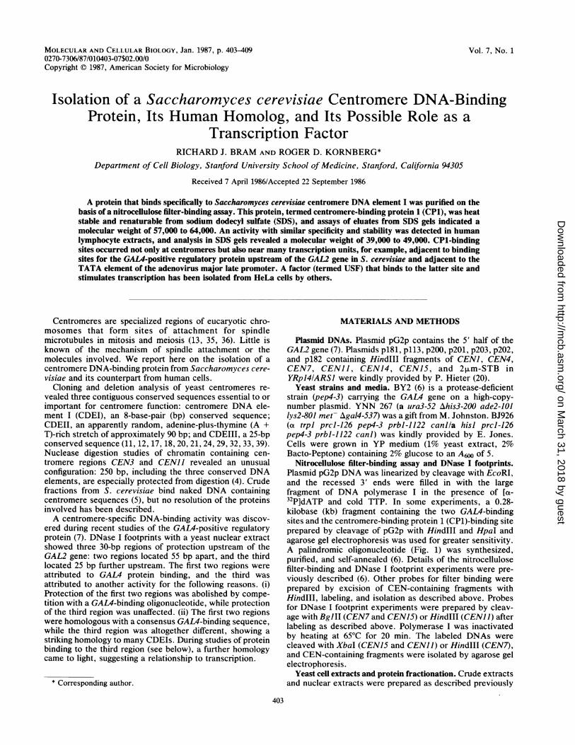
FIG. 1. S32P-labeled Ivector (YRPfrom strainfractions inthrough nitrcfilter in the al1) minus thalabsence of ththe absence
(6, 7). BJ92with cold v
Tris (pH 7..mM pheny,uM leupeplof the cell Ivolume of
pulses of a bead beater (Biospec Products, Bartlesville,Okla.). The cell lysate was centrifuged in a Sorvall SS34rotor at 10,000 rpm for 10 min at 4°C, and the supernatantwas made isotonic by addition of 0.1 volume of 250 mMN-2-hydroxyethylpiperazine-N'-2-ethanesulfonic acid (pH7.5)-S50 mM MgClr-1 mM EDTA-5 mM dithiothreitol-500mM KCl. The mixture was centrifuged in a Beckman Ti60
AN rotor at 55,000 rpm for 3 h at 0°C, and the supernatant (14 mlwith 9.5 mg of protein per ml) was loaded at 0.7 ml/min on a
30-ml phosphocellulose column (diameter, 2.5 cm) equili-0 5 10 15 20 25 30 60 brated with buffer A50 as described previously (6). ThePercent Specific Binding column was washed with 80 ml each of buffer A50 and buffer
,pecific protein binding to yeast centromere DNAs. A250, and CP1 activity was eluted with 50 ml of buffer A400.)NA fragments containing centromere, 2,um-STB, or Peak fractions were pooled, adjusted to 70% of saturation'141ARSI) sequences were treated with crude extract with solid ammonium sulfate, and centrifuged. The precipi-YNN267 (5 ,ug of protein) or with phosphocellulose tate was resuspended in buffer A50, dialyzed twice againstthe case of CEN15 (0.6 ,ug of protein) and filtered 100 volumes of buffer A50 for 3 h at 4°C, and stored at
)cellulose. Percent specific binding is 32P bound to the -800C.
bsence of a competing, synthetic oligonucleotide (Table Human lymphocyte extract. MICH cells (an Epstein-Barr
tbound in its presence, all divided by 3P bound in thete oligonucleotide and multiplied by 100%. 32P bound in virus-immortalized human B lymphocyte line) were kindlyof oligonucleotide was at least 1,400 cpm in all cases. provided by J. Ways and D. Goldfarb. Cells were grown in
suspension in 200 ml of RPMI 1640 plus 5% fetal bovineserum at 37°C to a density of 5 x 10⁵ to 10 x 105/ml,harvested by centrifugation, washed wth 10 ml of buffer A50,
'6 cells were harvested by centrifugation, washed and lysed by adding 2.7 ml (9 pellet volumes) of distilledvater, and suspended with an amount of 10 mM water, followed by 20 strokes of a Dounce homogenizer.5)-1.5 mM MgCl2-15 mM KCl-0.1 mM EDTA-1 Buffer A500 (0.1 volume) was added, and nuclei were
Imethylsulfonyl fluoride-2 ,uM pepstatin A-0.6 collected by centrifugation at 3,000 rpm for 10 min at 0°C.tin-0.5 mM dithiothreitol (9) equal to the volume The pellet was suspended in 2.7 ml of buffer A50, and solidpellet. The suspension was treated with an equal ammonium sulfate was added to 0.3 M. After gentle mixingglass beads (diameter, 0.45 mm) and eight 30-s at 4°C for 20 min, the solution was clarified by centrifugation
TABLE 1. Homology between DNA sequence upstream of the GAL2 gene and sequences of yeast centromere DNA elements PaNo. of matches/22 bp to:
Genetic element Sequence CDEI Symmetrizedconsensus GAL2
CENI AGTCTTGTCACATGACATAATA 17 13
CEN3 AAATAAGTCACATGATGATATT 20 16
CEN4 CAAAAGGTCACATGCTTATAAT 16 14
CEN6 CTTTTCATCACGTGCTATAAAA 17 14
CEN7 AAATATATCACGTGTTATATTT 20 19
CENJO AACTTAATCACGTGTTAAATAA 19 15
CENII TCATAAGTCACATGATAAAAAC 19 14
CEN14 TAGTTAGTCACGTGCAGCTTTT 15 14
CENIS AATAATATCACGTGAACTTATT 17 16
CEN16 AAATAGATCACATGATATATTT 21 20
GAL2 AAATGGGTCACGTGATCTATAT 19 22
CDEI consensus AA TTTA TCAC GTGATA AT TAA A A T TT
Symmetrized GAL2 A AT G ATCACGTGA CCCATATA G GTTA
Synthetic oligonucleotide AATTAAAATAGATCACGTGATCTATTTTa GAL2 sequence is from Bram et al. (7). Centromere sequences are from Hieter et al. (20) for CENI, 7, 10, 14, 15, and 16; Fitzgerald-Hayes et al. (18) for CEN3
and 11; and Panzeri and Philippsen (33) for CEN6. CDEI was defined by Hieter et al. (20) as the sequence Pu-T-C-A-C-Pu-T-G near the center of the consensusshown above. Matches to the symmetrized GAL2 sequence are overlined.
MOL. CELL. BIOL.
on March 31, 2018 by guest
http://mcb.asm
.org/D
ownloaded from

S. CEREVISIAE CENTROMERE DNA-BINDING PROTEIN 405
TABLE 2. Partial list of sequences homologous to the GAL2-associated CP1-binding siteGenetic element Sequencea Location or source (reference)
GAL2
Yeast CENII-MET14
Yeast CBP2
Yeast Arg-tRNA 3a
Yeast TRPl-ARSI
Yeast ADR2
Bovine 1.706 satellite DNA
Adenovirus major late promoter
Mouse middle repetitiveLTRb-like sequence
Human KpnI repeat
Drosophila melanogaster transposableelement MDG3
D. melanogaster transposable P
Trypanosome kinetoplast DNAminicircle
Xenopus laevis histone H2A1
Human Alu sequences 5'of globin genes
Alu sequence 5' of humana-globin gene
Gross passage A murineleukemia virus LTR
Mouse adenovirus strain Flinverted terminal repeat
HTLVIII LTRs
Bovine papilloma virus
A TGGGTCACGTGACCcATT TT
CACTAATTTCACGTGATC
ACAC GCCCT CACG TGAGTGAAT TT
AGTT ACCAT CACG TGCCGCTCTAT
ACTA TTGAG CACG TGAGT ATAC GT
GGAATGTTC CACGTGAAGCTATCT
TGCACTGAT CACG TGACT GATC AT
GGTGTAGGC CACG TGACC GGGTGT
ATAAAGTCT CACGTGGTTTGCAAC
CAAT GAGAT CACG TGGAC ACAGGA
ATACAATTT CACG TGTCT CTTT TA
AAAT TAATT CACG TGCCGAAGTGT
TACACAAAT CACG TGCTATTTTCG
ATCAGAGCT CACG TGATC ACAT GG
CAGG CAGAT CACCTGAGGTCAAGA
TGGG CAGAT CATGAGGTCAAGAGA
ATTG TGAAT CACG TGAATAAAAGA
CCCGGGTTT CACG TGGTG CGTC AG
GCATTTCAT CACG TGGCC CGAG AG
CCTC TAAAT CACG TGGCATTTTAAa Matches to the symmetrized GAL2-associated sequence are overlined.b LTR, Long terminal repeat.
(7)
510 bp 3' of CDEIII (18)
285 bp 5' of AUG (25)
475 bp 5' of tRNA start (42)
51 bp 5' of TRPJ AUG (41)
734 bp 5' of mRNA start (43)
23 bp prototype (34)
60 bp 5' of mRNA start (37)
No. 38 of Wirth et al. (44)
No. 52 of Digiovanni et al. (15)
No. 111 at 3' end, no. 78 at 5' end ofBayev et al. (3)
No. 195 of O'Hare and Rubin (31)
No. 746 of Barrois et al. (2)
71 bp 3' of stop codon 200-300 bp 3'of H4 gene (27)
55 in Alu upstream of Gy and 8 (16)
in Alu sequence 1.5 kb 5' ofE AUG (1)
92 bp from 3' end of provirus (14)
No. 88 of Temple et al. (40)
176 bp 5' of mRNA start also 365 bp5' of 3' end (28)
No. 4922 of Chen et al. (10)
in a Sorvall AH650 rotor at 50,000 rpm for 1 h at 0°C. Thesupernatant was frozen in liquid N2 and stored at -80°C.Recovery of CP1 activity from SDS-polyacrylamide gels.
Partially purified yeast CP1 (60 ,ug of peak phosphocellulosefractions) or crude extracts containing human CP1 (270 ,ugafter dialysis against buffer A50) were subjected to electro-phoresis in 1.5-mm-thick sodium dodecyl sulfate (SDS)-10%polyacrylamide gels (23) calibrated with SDS-polyacryl-amide gel electrophoresis low-molecular-weight standards(Bio-Rad Laboratories, Richmond, Calif.). Proteins wereeluted from gel slices as described before (19), precipitated,and renatured by resuspension in 5 ,ul of 6 M guanidinehydrochloride-30 mM Tris (pH 8), followed by dilution with250 ,ul of 50 mM Tris (pH 7.5)-0.1 mM EDTA-1 mMdithiothreitol-0.1 mg of bovine serum albumin per ml-0.15M NaCl-20% glycerol. Approximately 75% of the yeast and
>50% of the human CP1 activity was recovered after incu-bation of the diluted protein at 22°C for 1 h.
RESULTSHomology of a protein-binding sequence upstream of the
GAL2 gene to CDEI. The protein-binding sequence locatedupstream of the GAL2 gene, 25 bp beyond a pair of GAL4-binding sites, was dyad symmetric at 18 of 24 bp, with thecentral 8 bp forming the perfect palindrome T-C-A-C-G-T-G-A. This GAL2-associated sequence was homologous at 19of 22 bp with a consensus sequence (Table 1) of 10 CDEIs(compared with 15 to 21 correct matches to the consensus ofthe CDEI sequences themselves). Comparison with theGAL2-associated sequence revealed a previously unrecog-nized dyad symmetry of CDEI sequences (with 20 of 22 bp ofdyad-symmetric sequence in the CDEI of CEN7).
VOL. 7, 1987
on March 31, 2018 by guest
http://mcb.asm
.org/D
ownloaded from

406 BRAM AND KORNBERG
6
EC,0
2
6
E0
CfO0
A
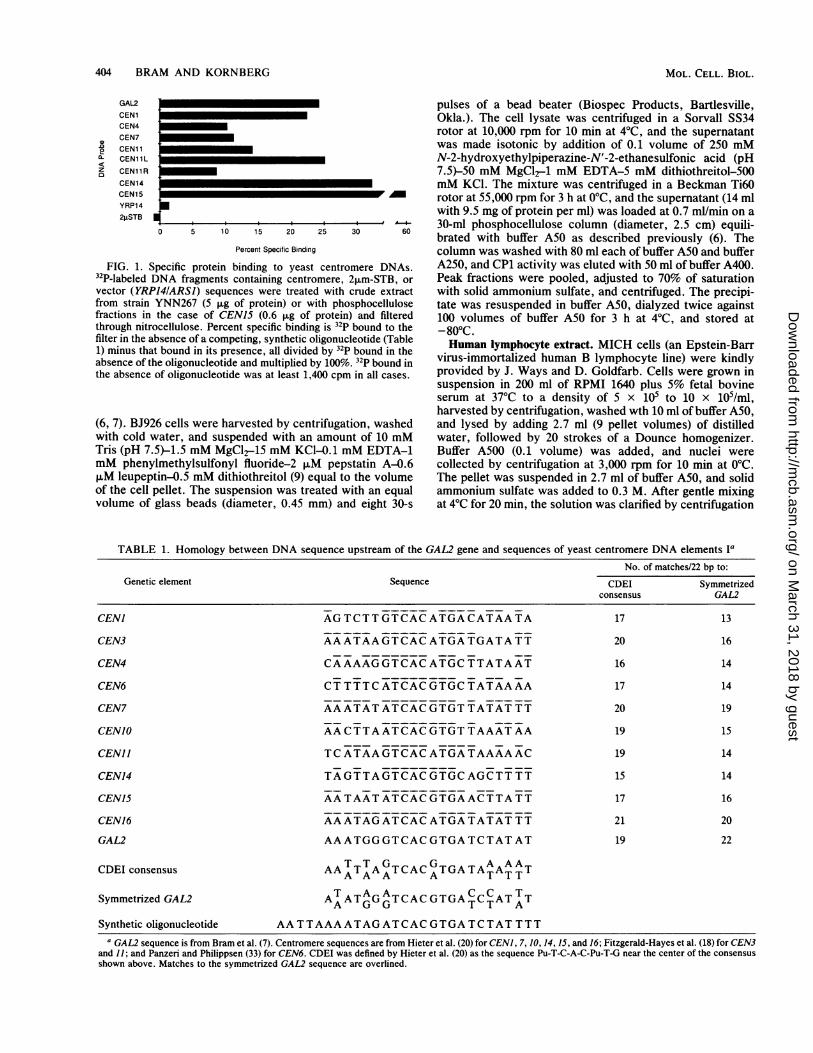
20 210157 1293 82 72 64 57 1 45 41 3 4 31 28 25 23kD
B
2902 12- 130 105 87 74 54 55 49 43 39 35 32 2Z 26 24 22
kD
FIG. 2. Distribution of CEN-binding activity in SDS gels of (A) yeast phosphocellulose fractions and (B) human lymphocyte nuclearextract. 32P-labeled pG2p DNA was incubated with 5 ,ul of renatured protein from each gel slice and filtered through nitrocellulose asdescribed. 32p counts per minute bound to filters are plotted for slices from the top of the gel at the left. Major peaks of binding activity inpanels A and B were abolished by adding a competing, synthetic oligonucleotide.
Specific protein binding to CEN sequences. Protein bindingto GAL2-associated and CEN sequences was detected innitrocellulose filter-binding assays. 32P-labeled plasmidDNAs (1 to 2 ng) containing these sequences were mixedwith protein fractions and carrier DNA (1 to 2 ,ug) andpassed through nitrocellulose. Binding specific for theGAL2-associated sequence was determined from the differ-ence between label bound in the presence and absence of a50-fold molar excess (4 ng) of synthetic oligonucleotidecontaining the GAL2 sequence. In a typical experiment withlabeled plasmid containing the GAL2 upstream region andcrude yeast extract (5 ,g of protein), 35% of the label boundin the presence, and 65% bound in the absence of competingoligonucleotide. All six CEN DNAs tested showed bindingspecific for the GAL2-associated sequence, whereas DNAscontaining chromosome replication or stabilization elements(ARSJ, ARS14, and 2tLm-STB, a cis-acting locus requiredfor stable maintenance of the 2,um plasmid) showed nospecific binding (Fig. 1). The variation in the percentage ofspecific binding among the CEN DNAs was due to variationin the length and sequence composition of the restrictionfragments used. This is illustrated for a 2.9-kb HindIllfragment containing CENJ I and 0.8-kb (CENJIL, containingCENJI) and 2.1-kb (CENJIR) products of cleaving thisfragment with XbaI, which showed 14.1, 25.1, and 8.6%specific binding, respectively (Fig. 1). The increase in per-cent specific binding upon shortening the CENJI -containingfragment may be attributed to the removal of nonspecific
binding to flanking DNA. The effect would have beengreater, were it not for some specific binding to the flankingDNA (CENJIR), possibly to a sequence between CENIIand the METJ4 gene identical with the central eight residuesof the GAL2-associated sequence (Table 2).
Purification and properties of CEN-binding protein. CEN-binding activity was heat stable and renaturable from SDS(Heating a crude extract for 10 min at 100°C resulted in a23% loss of activity and in the precipitation of 98% of thetotal protein; removal of SDS by acetone precipitation andrenaturation from guanidine hydrochloride resulted in 76%recovery of activity). CEN-binding activity was also insen-sitive to RNase treatment (36% loss of activity upon treat-ment with 1 ,g of RNase A per RI for 40 min at 37°C), but itwas completely destroyed by proteinase K.Crude extracts were fractionated on phosphocellulose
(160-fold enrichment of CEN-binding activity in 54% yield)or they were heated, centrifuged, and fractionated on Affi-Gel Blue (875-fold enrichment of CEN-binding activity in22% yield). Peak phosphocellulose or Affi-Gel Blue fractionswere subjected to SDS-polyacrylamide gel electrophoresis,and eluates of gel slices were treated with acetone andguanidine hydrochloride for renaturation of CEN-bindingactivity. Nitrocellulose filter-binding assays revealed a singlepeak of activity in the 57- to 64-kilodalton region of the gel(Fig. 2A).DNAse I footprint of CEN-binding protein on CEN DNAs.
End-labeled DNA fragments containing CEN sequences
MOL. CELL. BIOL.
on March 31, 2018 by guest
http://mcb.asm
.org/D
ownloaded from

S. CEREVISIAE CENTROMERE DNA-BINDING PROTEIN 407
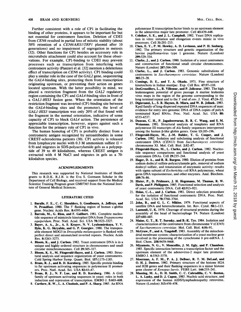
were mixed with peak phosphocellulose fracti(briefly with DNase I, and subjected to polyacelectrophoresis and autoradiography. CEN7, Cland CEN15 (not shown) showed protected regi25 bp centered on CDEI but no protection ofIII. The size and location of the protected i
similar to those previously found for the GAlsequence (7).CEN-binding activity in human cells. Fitzger
al. (17) noted a homology of 12 of 14 bp betwe4the bovine 1.706 satellite DNA (a 23-bp repeatirThe region of homology included the sequencethe CEN-binding interaction described above.associated binding sequence was identical wiltype bovine sequence (Table 2) at 11 of 11 cerprompted us to investigate the occurrence ofactivity in mammalian cells. The nitrocelluloseassay described above was applied to extractslymphocyte nuclei with a labeled HindIII-Hpament containing the GAL2 upstream regionDNA-binding activity specific for the GAL2-aquence was detected (31% of label bound in the6% bound in the presence of oligonucleotide clGAL2-associated sequence with 5 ,ug of proteirotides containing unrelated sequences had no
CEN7
1o0I
tI
C
III F
O
-
to ma m
III I
11
am
a a
--B gI 11-
FIG. 3. DNase I footprints of CEN-binding actiand CENII DNAs. 32P-labeled CEN DNA fragmeibated with (right lanes) or without (left lanes) peak phfractions (12 ,ug of protein) and processed as descritThe correspondence between points in the CEN Dpositions of DNA bands was determined fron4X174/HaeIII fragments (not shown). Abbreviatiortides; I, CDEI; II, CDEII; III, CDEIII.
ons, digestedrylamide gelEN]I (Fig. 3)ions of aboutCDEs II andregions werer2-associated
activity was heat stable (43% recovery of activity aftertreatment at 100°C for 10 min) and renaturable from SDS.Upon electrophoresis in SDS-polyacrylamide gels and elu-tion from gel slices, a single peak of activity in the 39- to49-kilodalton region of the gel was obtained (Fig. 2B).
DISCUSSION
ald-Hayes et Although the polypeptides from yeast responsible foren CDEI and binding six CEN sequences and a GAL2-associated se-ng structure). quence were not purified to homogeneity, several lines of,s involved in evidence suggested that the various binding activities wereThe GAL2- identical. First, the various DNA sequences were highly
th the proto- homologous, and an oligonucleotide containing one se-itral bp. This quence competed for binding with all the others. Second,CEN-binding DNase I footprints showed that the location of proteinfilter-binding binding to all the sequences was the same. Finally, thefrom human binding activity migrated as a single band in an SDS-
zI DNA frag- polyacrylamide gel. We refer to what is, in all likelihood, al. Significant single DNA-binding polypeptide as CP1.,ssociated se- The striking features of CP1 are the occurrence ofabsence, and noncentromeric binding sites and the apparent conserva-ontaining the tion of the protein through evolution. In addition to ai; oligonucle- GAL2-associated site, a computer-assisted search of a DNAeffect). The sequence data bank revealed several possible binding se-
quences in yeast (Table 2). These sequences were homolo-gous at 8 or more of the 12 residues containing the central
CEN1 1 palindrome C-A-C-G-T-G and were located upstream of- _ various transcription units. The abundance of CP1 in S.NHscerevisiae was in keeping with its action at sites in addition
to the few associated with centromeres. The amount ofDNA* w specifically bound by crude extracts led to an estimate ofE @ approximately 500 copies of CP1 per cell. This is probably a
low estimate that does not take into account the proteinassociated with cellular DNA, since the -extracts were pre-pared under conditions similar to those used to measurebinding, and the complex was quite stable under theseconditions (half-life for dissociation, ca. 20 min).
Yeast CP1 and the CP1 activity identified in humanlymphocyte extracts shared both specificity for CDEI se-quences and unusual stability and capacity for renaturation.These findings, together with the homology between yeastCDEIs and a bovine satellite known to occur in centromeres(22, 38), lead us to suggest that the yeast and human proteins
-E perform similar functions. An indication of a possible func-tion that would fit with the occurrence of noncentromericsites comes from recent reports (8, 26, 37) of a transcriptionfactor from HeLa cells, termed USF, whose recognition
_ _ sequence in adenovirus DNA is identical at 13 out of 14central bp with the GAL2-associated CP1-binding sequence(Table 2). USF not only recognizes the same DNA sequence
_- as CP1, but is heat stable as well, and it seems likely thatUSF is analogous to CP1 in lymphocyte extracts. USFstimulates transcription from the adenovirus major latepromoter 10- to 20-fold in vitro, and its binding appears to becooperative with that of a factor that recognizes the TATA
- ~ sequence. CP1 may play a similar role in S. cerevisiae,assisting the binding of GAL4 protein, microtubule proteins,and the like. Preliminary experiments indeed suggested that
nts were incu- CP1 facilitates GAL4-binding to the GAL2 upstream regiontosphocellulose (Bram, unpublished data). The occurrence of a CP1-bindingoed in the text site in this region but not upstream of other galactose-)NA maps and inducible genes may be explained by the low level of GAL4n patterns of protein in cells and the advantage of transcribing the GAL2ns: nt, nucleo- gene, which encodes a galactose permease, before the other
GAL genes.
VOL. 7, 1987
on March 31, 2018 by guest
http://mcb.asm
.org/D
ownloaded from
408 BRAM AND KORNBERG
Further consistent with a role of CP1 in facilitating thebinding of other proteins, it appears to be important for butnot essential for centromere function. Deletion of CDEIfrom CEN6 resulted in partial loss of mitotic stability (about50% retention of a CEN6/ARSJITRPI plasmid after 10generations) and no impairment of segregation in meiosis(32). Other functions for CP1 besides an accessory role inmicrotubule attachment could also account for these obser-vations. For example, CP1-binding to CDEI may preventprocesses such as transcription from interfering withcentromere activity (Panzeri et al. [32] mention a deleteriouseffect of transcription on CEN6 activity). CP1 binding couldplay a similar role in the case of the GAL2 gene, sequesteringthe GAL4-binding sites, protecting them from transcriptionoriginating upstream, or preventing their action on geneslocated upstream. With the latter possibility in mind, weplaced a restriction fragment from the GAL2 regulatoryregion containing the CP1 and GALA-binding sites in front ofa GALJ-HIS3 fusion gene. When the orientation of therestriction fragment was inverted (CP1-binding site betweenthe GAL4-binding sites and the promoter), the level ofGALI-HIS3 transcription was only 20% of that found withthe fragment in the normal orientation, indicative of somecapacity of CP1 to block GAL4 action. The persistence ofappreciable transcription, however, argues against such afunction for the primary role of CP1 in vivo.The human homolog of CP1 is probably distinct from a
centromeric antigen recognized by autoantibodies in someCREST-scleroderma patients (30). CP1 activity is releasedfrom lymphocyte nuclei with 0.3 M ammonium sulfate (I =0.9) and migrates in SDS-polyacrylamide gels as a polypep-tide of 39 to 49 kilodaltons, whereas CREST antigen isextracted with 4 M NaCl and migrates in gels as a 70-kilodalton species.
ACKNOWLEDGMENTS
This research was supported by National Institutes of Healthgrants to R.D.K. R.J.B. is the Eva S. Gutmann Scholar in theDepartment of Cell Biology, and received support from the MedicalScientist Training Program grant GM07365 from the National Insti-tute of General Medical Sciences.
LITERATURE CITED
1. Baralle, F. E., C. C. Shoulders, S. Goodbourn, A. Jeffreys, andN. Proudfoot. 1980. The 5' flanking region of human r-globingene. Nucleic Acids Res. 8:4393-4404.
2. Barrois, M., G. Riou, and F. Galibert. 1981. Complete nucleo-tide sequence of minicircle kinetoplast DNA from Trypanosomaequiperdum. Proc. Natl. Acad. Sci. USA 78:3323-3327.
3. Bayev, A. A., Jr., A. S. Krayev, N. V. Lyubomirskaya, Y. V.Ilyin, K. G. Skryabin, and G. P. Georgiev. 1980. The transpos-able element MDG3 in Drosophila melanogaster is flanked withperfect direct and mismatched inverted repeats. Nucleic AcidsRes. 8:3263-3273.
4. Bloom, K., and J. Carbon. 1982. Yeast centromere DNA is in aunique and highly ordered structure in chromosomes and smallcircular minichromosomes. Cell 29:305-317.
5. Bloom, K. S., M. Fitzgerald-Hayes, and J. Carbon. 1983. Struc-tural analysis and sequence organization of yeast centromeres.Cold Spring Harbor Symp. Quant. Biol. 157:1175-1185.
6. Bram, R. J., and R. D. Kornberg. 1985. Specific protein bindingto far upstream activating sequences in polymerase II promot-ers.. Proc. Natl. Acad. Sci. USA 82:43-47.
7. Bram, R. J., N. F. Lue, and R. D. Kornberg. 1986. A GALfamily of upstream activating sequences in yeast: roles in bothinduction and repression of transcription. EMBO J. 5:603-608.
8. Carthew, R. W., L. A. Chodosh, and P. A. Sharp. 1985. An RNA
polymerase II transcription factor binds to an upstream elementin the adenovirus major late promoter. Cell 43:439-448.
9. Celniker, S. E., and J. L. Campbell. 1982. Yeast DNA replica-tion in vitro: initiation and elongation events mimic in vivoprocesses. Cell 31:201-213.
10. Chen, E. Y., P. M. Howley, A. D. Levinson, and P. H. Seeburg.1982. The primary structure and genetic organization of thebovine papillomavirus type 1 genome. Nature (London)299:529-534.
11. Clarke, J., and J. Carbon. 1980. Isolation of a yeast centromereand construction of functional small circular chromosomes.Nature (London) 287:504-509.
12. Clarke, L., and J. Carbon. 1983. Genomic substitutions ofcentromeres in Saccharomyces cerevisiae. Nature (London)305:23-28.
13. Comings, D. E., and T. A. Okada. 1971. Fine structure ofkinetochora in Indian muntjac. Exp. Cell Res. 67:97-110.
14. DesGroseillers, L., R. Villemur, and P. Jolicoeur. 1983. The highleukemogenic potential of gross passage A murine leukemiavirus maps in the region of the genome corresponding to thelong terminal repeat and to the 3' end of env. J. Virol. 47:24-32.
15. Digiovanni, L., S. R. Haynes, R. Misra, and W. R. Jelinek. 1983.KpnI family of long-dispersed repeated DNA sequences of man:evidence for entry into genomic DNA of DNA copies of polyAterminated KpnI RNAs. Proc. Natl. Acad. Sci. USA 80:6533-6537.
16. Duncan, C. H., P. Jagadeeswaran, R. R. C. Wang, and S. M.Weissman. 1981. Structural analysis of templates and RNApolymerase III transcripts of Alu family sequences interspersedamong the human P-like globin genes. Gene 13:185-1%.
17. Fitzgerald-Hayes, M., J.-M. Buhler, T. G. Cooper, and J.Carbon. 1982. Isolation and subcloning analysis of functionalcentromere DNA (CENII) from Saccharomyces cerevisiaechromosome XI. Mol. Cell. Biol. 2:82-87.
18. Fitzgerald-Hayes, M., L. Clarke, and J. Carbon. 1982. Nucleo-tide sequence comparisons and functional analysis of yeastcentromere DNAs. Cell 29:235-244.
19. Hager, D. A., and R. R. Burgess. 1980. Elution of proteins fromsodium dodecyl sulfate-polyacrylamide gels, removal of sodiumdodecyl sulfate, and renaturation of enzymatic activity: resultswith sigma subunit of Escherichia coli RNA polymerase, wheatgerm DNA topoisomerase, and other enzymes. Anal. Biochem.109:76-86.
20. Hieter, P., D. Pridmore, J. H. Hegemann, M. Thomas, R. W.Davis, and P. Philippsen. 1985. Functional selection and analysisof yeast centromeric DNA. Cell 42:913-921.
21. Hsiao, C.-L., and J. Carbon. 1981. Direct selection procedurefor the isolation of functional centromeric DNA. Proc. Natl.Acad. Sci. USA 78:3760-3764.
22. John, B., and G. L. C. Miklos. 1979. Functional aspects ofsatellite DNA and heterochromatin. Int. Rev. Cytol. 58:1-113.
23. Laemmli, U. K. 1970. Cleavage of structural proteins during theassembly of the head of bacteriophage T4. Nature (London)227:680-685.
24. Maine, G. T., R. T. Surosky, and B.-K. Tye. 1984. Isolation andcharacterization of the centromere from chromosome V (CEN5)of Saccharomyces cerevisiae. Mol. Cell. Biol. 4:86-91.
25. McGraw, P., and A. Tzagoloff. 1983. Assembly of the mitochon-drial membrane system: characterization of a yeast nuclear geneinvolved in the processing of the cytochrome b pre-mRNA. J.Biol. Chem. 258:9459-9468.
26. Miyamoto, N. G., V. Moncoilin, J. M. Egly, and P. Chambon.1985. Specific interaction between a transcription factor and theupstream element of the adenovirus-2 major late promoter.EMBO J. 4:3563-3570.
27. Moorman, A. F. M., P. A. J. DeBoer, R. T. M. DeLaaf, and0. H. J. Destree. 1982. Primary structure of the histone H2Aand H2B genes and their flanking sequences in a minor histonegene cluster of Xenopus laevis. FEBS Lett. 144:235-241.
28. Muesing, M. A., D. H. Smith, C. C. Cabradilla, C. V. Benton,L. A. Lasky, and D. J. Capon. 1985. Nucleic acid structure, andexpression of the human AIDS/lymphadenopathy retrovirus.Nature (London) 313:450-458.
MOL. CELL. BIOL.
on March 31, 2018 by guest
http://mcb.asm
.org/D
ownloaded from

S. CEREVISIAE CENTROMERE DNA-BINDING PROTEIN 409
29. Neitz, M., and J. Carbon. 1985. Identification and characteriza-tion of the centromere from chromosome XIV in Saccharomy-ces cerevisiae. Mol. Cell. Biol. 5:2887-2893.
30. Nishikai, M., Y. Okano, H. Yamashita, and M. Watanabe. 1984.Characterization of centromere (kinetochore) antigen reactivewith sera of patients with a scleroderma variant (CRESTsyndrome). Ann. Rheum. Dis. 43:819-824.
31. O'Hare, K., and G. M. Rubin. 1983. Structures of P transpos-able elements and their sites of insertion and excision in theDrosophila melanogaster genome. Cell 34:25-35.
32. Panzeri, L., L. Landonio, A. Stotz, and P. Philippsen. 1985. Roleof conserved sequence elements in yeast centromere DNA.EMBO J. 4:1867-1874.
33. Panzeri, L., and P. Philippsen. 1982. Centromeric DNA fromchromosome VI in Saccharomyces cerevisiae. EMBO J. 1:1605-1611.
34. Pech, M., R. E. Streeck, and H. G. Zachau. 1979. Patchworkstructure of a bovine satellite DNA. Cell 18:883-893.
35. Ris, H., and P. L. Witt. 1981. Structure of the mammaliankinetochore. Chromosoma 82:153-170.
36. Roos, U. P. 1973. Light and election microscopy of rat kangaroocells in mitosis. Chromosoma 41:195-200.
37. Sawadogo, M., and R. G. Roeder. 1985. Interaction of a gene-specific transcription factor with the adenovirus major late
promoter upstream of the TATA box region. Cell 43:165-175.38. Singer, M. 1982. Highly repeated sequences in mammalian
genomes. Int. Rev. Cytol. 76:67-112.39. Stinchcomb, D. T., C. Mann, and R. W. L. Davis. 1982.
Centromeric DNA from Saccharomyces cerevisiae. J. Mol.Biol. 158:157-179.
40. Temple, M., G. Antoine, H. Delius, S. Stahl, and E.-L.Winnacker. 1981. Replication of mouse adenovirus strain FlDNA. Virology 109:1-12.
41. Tschumper, G., and J. Carbon. 1980. Sequence of a yeast DNAfragment containing a chromosomal replicator and the TRP1gene. Gene 10:157-166.
42. Villanueva, J., P. Bull, P. Valenzuela, and A. Vanegas. 1984.Nucleotide sequence of a yeast Arg-tRNA-3A gene and itstranscription in a homologous in vitro system. FEBS Lett.167:165-169.
43. Williamson, V. M., D. Cox, E. T. Young, D. W. Russell, and M.Smith. 1983. Characterization of transposable element-associated mutations that alter yeast alcohol dehydrogenase II
expression. Mol. Cell. Biol. 3:20-31.44. Wirth, T., K. Gloeggler, T. Baumruker, M. Schmidt, and I.
Horuk. 1983. Family of middle repetitive DNA sequences in themouse genome with structural features of solitary retroviral longterminal repeats. Proc. Natl. Acad. Sci. USA 80:3327-3330.
VOL. 7, 1987
on March 31, 2018 by guest
http://mcb.asm
.org/D
ownloaded from