increase of transcription factor eb (tfeb) and lysosomes ...anatomy.khu.ac.kr/research/increase of...
TRANSCRIPT
Neuroscience 313 (2016) 10–22
INCREASE OF TRANSCRIPTION FACTOR EB (TFEB) AND LYSOSOMESIN RAT DRG NEURONS AND THEIR TRANSPORTATION TO THECENTRAL NERVE TERMINAL IN DORSAL HORN AFTER NERVE INJURY
J. JUNG, a,dy N. UESUGI, b,dy N. Y. JEONG, cy B. S. PARK, a
H. KONISHI b,d AND H. KIYAMA b,d*
aDepartment of Anatomy and Neurobiology, School of
Medicine, Kyung Hee University, Heogi-Dong 1,
Dongdaemun-Gu, Seoul 130-701, Republic of Korea
bDepartment of Functional Anatomy & Neuroscience, Nagoya
University, Graduate School of Medicine, 65 Tsurumaicho,
Showa-ku, Nagoya 466-8550, Japan
cDepartment of Anatomy and Cell Biology and Mitochondria,
Hub Regulation Center, Dong-A University College of Medicine,
3-1 Dongdaesin-dong, Seo-gu, Busan 602-714, Republic of Korea
dCore Research for Evolutional Science and Technology (CREST)
of the Japan Science and Technology Agency (JST), Saitama, Japan
Abstract—In the spinal dorsal horn (DH), nerve injury
activates microglia and induces neuropathic pain. Several
studies clarified an involvement of adenosine triphosphate
(ATP) in the microglial activation. However, the origin of
ATP together with the release mechanism is unclear. Recent
in vitro study revealed that an ATP marker, quinacrine, in
lysosomes was released from neurite terminal of dorsal
root ganglion (DRG) neurons to extracellular space via lyso-
somal exocytosis. Here, we demonstrate a possibility that
the lysosomal ingredient including ATP released from DRG
neurons by lysosomal-exocytosis is an additional source
of the glial activation in DH after nerve injury. After rat L5
spinal nerve ligation (SNL), mRNA for transcription factor
EB (TFEB), a transcription factor controlling lysosomal
activation and exocytosis, was induced in the DRG. Simulta-
neously both lysosomal protein, LAMP1- and vesicular
nuclear transporter (VNUT)-positive vesicles were increased
in L5 DRG neurons and ipsilateral DH. The quinacrine stain-
ing in DH was increased and co-localized with LAMP1
immunoreactivity after nerve injury. In DH, LAMP1-positive
vesicles were also co-localized with a peripheral nerve
marker, Isolectin B4 (IB4) lectin. Injection of the adenovirus
encoding mCherry-LAMP1 into DRG showed that mCherry-
positive lysosomes are transported to the central nerve
terminal in DH. These findings suggest that activation of
http://dx.doi.org/10.1016/j.neuroscience.2015.11.0280306-4522/� 2015 IBRO. Published by Elsevier Ltd. All rights reserved.
*Correspondence to: H. Kiyama, Department of Functional Anatomy& Neuroscience, Nagoya University, Graduate School of Medicine,65 Tsurumaicho, Showa-ku, Nagoya 466-8550, Japan. Tel: +81-52-744-2015; fax: +81-52-744-2020.
E-mail address: [email protected] (H. Kiyama).y These authors contributed equally to this work.
Abbreviations: DH, dorsal horn; DRG, dorsal root ganglion; IB4,Isolectin B4; Iba1, ionized calcium binding adapter molecule 1; IHC,immunohistochemistry; Neu-1, neuramidase-1; PB, phosphate buffer;PBS, phosphate-buffered saline; PFA, paraformaldehyde; RT, roomtemperature; SNL, spinal nerve ligation; TFEB, transcription factor EB;VNUT, vesicular nuclear transporter; VP, adenovirus particle.
10
lysosome synthesis including ATP packaging in DRG, the
central transportation of the lysosome, and subsequent its
exocytosis from the central nerve terminal of DRG neurons
in response to nerve injury could be a partial mechanism
for activation of microglia in DH. This lysosome-mediated
microglia activation mechanism may provide another clue
to control nociception and pain. � 2015 IBRO. Published
by Elsevier Ltd. All rights reserved.
Key words: lysosomal exocytosis, dorsal horn, spinal nerve
injury, dorsal root ganglion neurons, microglia.
INTRODUCTION
Adenosine triphosphate (ATP) was established as the
source of free energy involved in biochemical pathways.
However, ATP is now recognized as a key molecule
both of an intracellular energy source and an
intercellular signaling (Fields and Stevens, 2000). It has
been demonstrated that ATP is released from axons
in vitro (Holton, 1959; Soeda et al., 1997; Vizi et al.,
1997; Jung et al., 2013) and involved in synaptic transmis-
sion (Engelman and Mac Dermott, 2004), and currently
the role of ATP is established as a transmitter of relevant
purinergic signaling (Pankratov et al., 2002; Zhang et al.,
2003). ATP in the central nervous system (CNS) could be
a crucial neurotransmitter because ATP-mediated activa-
tion of P2X and P2Y receptors in postsynaptic neuron,
microglia, and astrocyte can trigger significant Ca2+ entry
into cytoplasm and influence many metabolisms within
cells (Lalo et al., 1998; Abbracchio et al., 2009). A recent
study revealed that microglia express several ATP recep-
tors after nerve injury and that released ATP contributed
to the activation of microglia (Inoue et al., 2007). In addi-
tion, ATP-induced microglial activation in the spinal dorsal
horn (DH) caused the neuropathic pain (Tsuda et al.,
2003; Maeda et al., 2010). Thus, the regulation of ATP
release in DH may be crucial for the treatment of neuro-
pathic pain associated with microglial activation. How-
ever, the mechanisms underlying ATP release to the
extracellular space in DH are poorly understood.
Proposed pathways of ATP release from cells are
vesicular exocytosis and diffusion via transmembrane
pores (Pankratov et al., 2006; Imura et al., 2013). Previ-
ous reports have shown that ATP was stored in presynap-
tic terminals and it released mainly through vesicular
exocytosis (Morel and Meunier, 1981; Iijima, 1983;

J. Jung et al. / Neuroscience 313 (2016) 10–22 11
Terrian et al., 1989; Sawynok et al., 1993). In our previous
in vitro study, we suggested that ATP in lysosomes could
be released from primary cultured dorsal root ganglion
(DRG) neurons via lysosomal exocytosis (Jung et al.,
2013). Lysosomes are acidified, enzyme-containing intra-
cellular organelles to break down phagocytosed materi-
als, cell debris and waste materials (Holtzman, 1989).
Recently, it was identified that lysosomes have an addi-
tional property for regulatory exocytosis (secretory lyso-
somes) (Blott and Griffiths, 2002). The exocytotic
process of mature lysosomes can be triggered by an
increase of intracellular free Ca2+ (Andrews, 2000; Blott
and Griffiths, 2002). Then, a microtubule-dependent step
provides the movement of exocytic lysosomes toward the
plasma membrane (Andrews, 2000). The lysosomal exo-
cytosis also can be triggered by chemicals that cause
alkalinization of lysosomes (Sundler, 1997). The lysoso-
mal ingredients such as enzymes, degraded molecules,
ions and ATP, which are all released by lysosomal exocy-
tosis, could be mediators for the activation of microglia.
In the present study, we addressed a possibility that
lysosome is exocytosed from central nerve terminal of
DRG neurons into DH after peripheral nerve injury (Kim
and Chung, 1992; Tsuda et al., 2003). We here demon-
strated that peripheral nerve injury induced the transcription
factor EB (TFEB) mRNA expression in DRG and subse-
quent increase of vesicular-nuclear-transporter (VNUT)-
positive lysosomal vesicles in both DRG neurons and DH
suggesting up-regulation of lysosomal synthesis and ATP
packaging in DRG neurons. In addition the transportation
of lysosomes from DRG to DH was also confirmed by inject-
ing adenovirus encoding fluorescence-labeled lysosomal
protein. The results suggested that lysosomal exocytosis
in DH after peripheral nerve injury may be partly associated
with microglial activation in DH.
EXPERIMENTAL PROCEDURES
Materials
The primary antibodies against LAMP1 (Sana Cruz
Biotechnology, Sana Cruz, CA, USA, Cat# sc8098,
RRID: AB_2134494; Cat# sc71489, RRID: AB 2265605)
were used for immunostaining or Western blotting.
Phospho-p44/42 MAPK (p-ERK1/2, Cat# 9101S, RRID:
AB 331046) and p44/42 MAPK (ERK1/2, Cat# 9102S,
RRID: AB 10695746) were obtained from Cell signaling
(Beverly, USA). VNUT and ionized calcium binding
adapter molecule 1 (Iba-1) were obtained from MBL
Co., Ltd (Woburn, MA, USA, Cat# BMP079, RRID: AB
10597575) and Wako (Osaka, Japan, Cat# 019-19741,
RRID: AB_839504), respectively. IB4 and all other
reagents were purchased from Sigma–Aldrich (St. Luis,
MO, USA).
Animals and surgical procedure for nerve injury
All animal experiments have been carried out in
accordance with EU Directive 2010/63/EU for animal
experiments. Also all animal experiments have been
carried out in accordance with the University Animal
Committee ‘‘Guidelines for the Care and Use of
Laboratory Animals”, and were approved by the Nagoya
University Institutional Animal Care and Use Committee.
All possible efforts were made to minimize suffering. Adult
male Wistar rats (7 weeks rats) were obtained from SLC
(Hamamatsu, Japan). Animals were housed with a 12-h
light/dark cycle (8:00/20:00) at a constant room
temperature (RT) of 23± 2 �C and humidity of 45�65%.
After induction of anesthesia by intraperitoneal (i.p.)
injection of pentobarbital (40 mg/kg), the L5 nerve was
tightly ligated with a 5-0 silk and cut distal to the ligature
as previously described by (Tozaki-Saitoh et al. (2008).
Tissue processing
Seven days after L5 spinal nerve ligation (SNL), the L5
spinal cord were collected and stored as appropriate for
each experiment. For immunohistochemistry (IHC),
tissues were perfused with 4% paraformaldehyde (PFA)
in 0.1 M phosphate buffer (PB) and then excised. The
samples were post-fixed by the same solution overnight
at 4 �C and treated by with 30% sucrose in 0.1-M PB for
3 days. They were then embedded in OCT and frozen
immediately on dry ice. Frozen sections (16 lm) were
cut from tissues and put on the polylysine-coated slide
glasses (Fisher Scientific, Houston, TX, USA).
For the analysis of protein and RNA, the spinal cord
was isolated from animals immediately after sacrifice.
The spinal cord was separated into DHs at the L5
segment. Each sample was homogenized in a modified
radioimmunoprecipitation assay buffer (RIPA; 50 mmol/L
Tris–HCl pH 7.4, 150 mmol/L NaCl, 0.5% deoxycholic
acid, 0.5% Triton X-100, 1 mmol/L phenylmethylsulfonyl
fluoride, 1 mmol/L sodium o-vanadate, and protease
inhibitor cocktail [Roche Molecular Biochemicals, Nutley,
NJ, USA]). Protein concentrations were quantified by a
Bradford assay following standard protocols. Total RNA
was extracted from each sample by the conventional acid
guanidine iso-thiocyanate/phenol/chloroform extraction
(AGPC) method (Chomczynski and Sacchi, 2006).
Immunohistochemistry
Before staining, sample slides were fixed in 4% PFA for
10 min. Following three washes in phosphate-buffered
saline (PBS), the samples were permeabilized in PBS
containing 0.3% Triton X-100 (PBST) and blocked with
5% bovine serum albumin (BSA) and 5% fetal bovine
serum overnight at 4 �C. Primary antibodies for each
molecule were placed on the slides, which were
incubated for 16 h at 4 �C. Following three washes in
PBS, slides were incubated for 2 h at RT with Alexa
Fluor 594 (Invitrogen, Carlsbad, CA, USA), Alexa Fluor
488 (Invitrogen, USA) or Alex Flour 647 (Invitrogen,
USA) secondary antibodies. The slides were then
washed three times with PBS, slide-mounted and
subsequently cover-slipped. The immunolabeling was
analyzed using an LSM700 imaging system (Carl Zeiss,
Oberkochen, Germany).
Western blotting
Western blotting analysis was performed with the tissues
described above. Protein extracts were separated
using 10% sodium dodecyl sulfate polyacrylamide gel

12 J. Jung et al. / Neuroscience 313 (2016) 10–22
electrophoresis and transferred to a nitrocellulose
membrane (GE Healthcare, Waukesha, WI, USA). The
membrane was blocked with 5% non-fat milk in Tris-
buffered saline (TBS) containing 0.05% Tween 20
(TBST) overnight at 4 �C and incubated with the
following primary antibodies for 1 h at RT; mouse anti-
beta actin (1:5000), goat anti-LAMP1 (1:1000), p-
ERK1/2 (1:1000) and ERK1/2 (1:1000). After three 10-
min washes in TBST, the blots were incubated for 1 h at
RT with the appropriate horseradish peroxidase-
conjugated secondary antibodies (1:2000; Invitrogen,
USA), developed in enhanced chemiluminescence
reaction (Amersham; Buckinghamshire, England) for
1 min and film-detected (AGFA, Mortsel, Belgium).
Semi-quantitative reverse transcriptase polymerasechain reaction (RT-PCR)
RT-PCR analysis was performed with the tissues
described above. In brief, isolated RNA was obtained
from L4–L6 spinal cord and then reverse-transcribed with
random 6-mer oligo dT using SuperScript II (Invitrogen
Corporation, USA). RT-PCR was performed using the
specific primers as follows: TFEB, forward, 50-GGTGCAGT
CCTACCTGGAGA-30; reverse, 50-CTTTCTTCTGCCGTTC
CTTG-30, Neu-1, forward, 50-TCGGCTCCGTAGACACTTT-
30; reverse, 50-CGTGGTCATCACTGAGGAGA-30, VNUT,
forward, 50-GCT TCATCACTG TCACCACA-30; reverse, 50-CCAGGACAAGGTCTTTCTCA-30, LAMP1, forward, 50-AACCCCAGTGTGTCCAAGTA-30; reverse, 50-GCTGACAAA
GATGTGCTCCT-30, GAPDH, forward, 50-CAGCAATG
CATCCTGCACC-30; reverse, 50-TGGACTGTGGTCAT
GAGCCC-3’. RT-PCR was performed using 30 PCR
cycles depending on the target gene, with annealing
temperatures of 60 �C. PCR products were visualized by
1% agarose gel electrophoresis followed by GelRed
(Biotium, Hayward, CA, USA) staining.
ATP staining
For ATP staining in vivo, we used quinacrine
dihydrochloride as a fluorescent dye. Quinacrine
staining was performed as described in previous report
(Lee et al., 2013). Briefly, ATP staining was performed
by i.p. injection with quinacrine (50 mg/kg) in PBS to rats
three times per day (Belai and Burnstock, 1994). Slide
preparation was performed as described above.
Adenovirus production and its delivery in vivo
Cosmid pAxCALNLmLAMP1-mCherry and pAxCAWCre
were constructed by the insertion of mouse LAMP1-
mCherry or Cre recombinase sequence into the SwaI
cloning site of pAxCALNLw or pAxCAwt respectively.
Using a Cell Phect transfection kit (GE Healthcare, Cat#
27-9268-01), recombinant adenovirus of AxCALNLmLAMP1-
mCherry and AxCAWCre was generated by transfecting 293
cells with DNA-TPC (Takara Shuzo, Japan) and
pAxCALNLmLAMP1-mCherry or pAxCAWCre. When
these two viral vectors co-infected cells, Cre recombinase
expressed under the CAG promoter activates the stuffer
PolyA through the Cre/LoxP system. Recombinant
adenovirus particle titer (VP) was determined spectro-
photometrically. Each adenoviral vector was used at the
concentration of 1.5� 1010 VP/lL (AxCALNLmLAMP1-
mCherry) or 5.0� 109 VP/lL (AxCAWCre). The viral
solutions were injected into L5 DRG (total, 1.0 lL)concurrently when L5 nerve was injured.
Statistical analysis
Differences between groups were statistically analyzed
using an analysis of variance followed by Bonferroni’s
post hoc test. Data were assessed as mean ± SEM.
Values were considered to be statistically significant
at p< 0.01 after the analysis of four independent
experiments.
RESULT
Increase of LAMP1 expression and TFEB mRNAin DRG neurons after nerve injury
To confirm whether the nerve injury increases lysosomal
vesicles in DRG neurons in vivo as shown in the
previous in vitro result (Jung et al., 2013), we immunos-
tained injured L5 DRG with anti-LAMP1 antibody. Activat-
ing transcription factor 3 (ATF3) was used for a neuron-
injury marker (Takeda et al., 2000; Tsujino et al., 2000;
Ohba et al., 2003). A significant increase of LAMP1
immunoreactivity was observed in the ATF3-positive DRG
neurons (Fig. 1A–C). Next, we examined a possibility that
the ATP stores are associated with lysosomal vesicles in
a DRG neuron using quinacrine staining and immunohisto-
chemistry with anti-LAMP1 antibody. Fig. 1D shows clear
merged signals of LAMP1 and quinacrine in a DRG neuron
cell body. These data indicate that ATP could be localized
in lysosomal vesicles in DRG neurons in vivo as well as
DRG in vitro (Jung et al., 2013). The increase of LAMP1
mRNA in in vivo DRG neurons was validated independently
by semi-quantitative RT-PCR analysis.
Recent studies have reported that TFEB activates
lysosomal exocytosis (Medina et al., 2011; Settembre
et al., 2011), but neuramidase-1 (Neu-1) negatively regu-
lates lysosomal exocytosis (Yogalingam et al., 2008). To
confirm induction of lysosomal synthesis and exocytosis
in injured DRG neurons, we performed RT-PCR analysis
with specific primers for LAMP1, TFEB and Neu-1
mRNAs. We found that LAMP1 and TFEB mRNA levels
were significantly increased in the ipsilateral DRG neu-
rons compared with contralateral DRG neurons, but no
alteration of Neu-1 mRNA expression level in the injured
DRG (Fig. 2A, B). These results indicate that mRNAs
for TFEB and LAMP1 as well as the number of lysosomes
are concomitantly increased in the injured DRG neurons,
suggesting that induced TFEB expression activates lyso-
somal synthesis and possibly lysosomal exocytosis in
DRG neurons after nerve injury.
Increase of VNUT expression in DRG neurons afternerve injury and co-localization with LAMP1
Previous studies have revealed that VNUT plays a crucial
role in transporting ATP into vesicles in the several cell
types (Shehab et al., 2004; Sawada et al., 2008; Jung

Fig. 1. LAMP1 activation in the injured DRG neuronal cell bodies. (A) Confocal images showed ATF3 (green) activation in the injured DRG neurons.
Scale bar = 200 lm. (B) High magnification images showed LAMP1 (red) activation in ATF3 (green)-positive DRG neuronal cell bodies (arrows).
Scale bar = 50 lm. (C) Quantitative analysis of immunofluorescent staining against LAMP1 and ATF3. The number of LAMP1-activated ATF3
neuronal cell bodies in DH (200 lm � 200 lm) was calculated. **p< 0.01 (n= 4). (D) Quinacrine staining (green) and immunostaining (red) with
anti-LAMP1 antibody in a DRG neuron. Scale bar = 5 lm. Arrows show the co-localization of LAMP1-immunoreactivity and quinacrine. (For
interpretation of the references to colour in this figure legend, the reader is referred to the web version of this article.)
Fig. 2. Alteration of LAMP1 and TFEB mRNAs expressions in the
injured DRG. (A) RT-PCR analysis of LAMP1, TFEB and Neu-1
mRNA in the total RNA extract from the rat DRG on day 7 after
peripheral nerve injury. Neuramidase 1 (Neu-1) is a negative
regulator for lysosomal exocytosis (Yogalingam et al., 2008). GAPDH
is loading control. Cont, contralateral; Ipsi, ipsilateral. (B) Quantifica-
tion of the relative intensity of bands derived by RT-PCR. **p< 0.01
(n= 3).
J. Jung et al. / Neuroscience 313 (2016) 10–22 13
et al., 2013). We therefore immunostained rat DRG sections
with anti-VNUT and anti-LAMP1 antibodies. The VNUT
immunoreactivity was significantly increased in the injured
side compared with uninjured side of L5 DRG (Fig. 3A, B).
The increased VNUT immunoreactivities were also over-
lapped with LAMP1-positive profiles in the injured side
(Fig. 3C). RT-PCR analysis also indicated the increased
mRNA expression level of VNUT in the injured DRG neurons
compared with the control DRG neurons (Fig. 3D, E). Thus,
these findings suggest lysosomal vesicles are capable of
storing ATP through VNUT in DRG neurons.
Increase of LAMP1 immunoreactivity in the ipsilateralDH after nerve injury
To determine the alteration of lysosomal localization in
the spinal DH after sciatic nerve injury, we performed

Fig. 3. Increases of VNUT immunoreactivity and its mRNA in nerve injured DRG. (A) Confocal images showed that the vesicular nucleotide
transporter (VNUT, green) immunoreactivity was increased in injured DRG neurons compared with uninjured DRG neurons after L5 SNL (upper left
panel). Scale bar = 200 lm. DIC indicated that amounts of contralateral DRG neuron cell bodies are similar to ipsilateral DRG. DIC, differential
interference contrast. (B) Quantitative analysis showed that nerve injury induced an increase in the relative signal intensity of VNUT-positive profiles
in DRGs (200 lm � 200 lm). **p< 0.01 (n= 4). (C) Co-localization of VNUT and LAMP1 in ipsilateral (ipsi) DRG neuron (Upper panel). Lower
panel is higher magnification images of upper panel. Arrows indicate apparent VNUT (green) staining in LAMP1 (red)-positive vesicle in DRG
neuron. Scale bar = 30 lm (upper panel), 15 lm (lower panel). Arrows indicate VNUT (green) activation in LAMP1 (red)-positive DRG neuronal cell
bodies. (D) Alterations in VNUT mRNA expression in DRG neurons after L5 SNL (n= 4). (E) Quantification of the relative intensity of mRNA
expression of VNUT in DRG after nerve injury (n= 4). (For interpretation of the references to colour in this figure legend, the reader is referred to
the web version of this article.)
14 J. Jung et al. / Neuroscience 313 (2016) 10–22
immunostaining using sections from the L5 level of spinal
cord with anti-LAMP1 antibody. We found that LAMP1
immunoreactivity was significantly increased in the
ipsilateral side of spinal DH (Fig. 4A). Previous studies
have reported that spinal nerve injury induces microglial
activation in the injured side of spinal DH (Watkins and
Maier, 2003; Tsuda et al., 2005). We confirmed the
microglial activation and increase in the lysosome-
increased ipsilateral DH using Iba-1 antibody (Fig. 4A).
In addition, the increase of LAMP1 immunoreactivity
was confirmed by Western blot analysis. LAMP1
expression in DH at day 1 after nerve injury was
significantly increased and the increase was maintained
at day 7 after nerve injury (Fig. 4B, C). The increase of

Fig. 4. Increase of LAMP1 immunoreactivity in DH after peripheral nerve injury. (A) Immunohistochemistry revealed increases in LAMP1 (red) level
in the ipsilateral spinal horn (L5) 7 days after peripheral nerve injury. Iba-1 (green) indicated the increase of microglia in DH of injured side. Scale
bar = 200 lm. (B) Increase of LAMP1 protein was demonstrated by Western Blotting. pERK level was demonstrated as a positive control. Protein
extracts (10 lg) from spinal dorsal horn were analyzed by Western blotting. ERK1/2 was loading control. (C) Quantitative analysis of Western
blotting for LAMP1 expression from three independent experiments. **p< 0.01 (n= 4). (D) Simultaneous demonstration of LAMP1 (red) and Iba1
(green). LAMP1 immunoreactivity is co-localized mainly out of Iba-1 in the ipsilateral side of DH after nerve injury. The right hand side photo
demonstrates higher magnification of the white square region seen in left. Scale bar = 50 lm (left) and 10 lm (right). (For interpretation of the
references to colour in this figure legend, the reader is referred to the web version of this article.)
J. Jung et al. / Neuroscience 313 (2016) 10–22 15
LAMP1 in DH after nerve injury had continued for 3 weeks
(data not shown). A previous study showed that pERK1/2
activation occurs in the spinal DH after nerve injury
(Zhang et al., 2007b). We used pERK1/2 as a positive
control and ERK1/2 as a loading control. To assess
whether the increase of LAMP1 immunoreactivity is in
microglia or not, we performed simultaneous visualization
of LAMP1 and Iba-1 immunoreactivities. Many large
LAMP1-positive puncta were observed in ipsilateral DH.
Many of LAMP1 immunoreactivities did not co-localize
with Iba-1 immunostaining in the ipsilateral side of DH
(Fig. 4D). These results indicate that the increased large
lysosomes mainly exist out of microglia after SNL.
Next, we accessed whether the ATP stores are
associated with lysosomal vesicles in DH using
quinacrine staining and immunohistochemistry with anti-
LAMP1 antibody. On day 6 after SNL, rats were injected
with quinacrine and, the next day, spinal cords were
sampled for analyses. In L5 spinal cord sections,
quinacrine staining was apparently more detectable in
the ipsilateral DH compared to the contralateral side of
the DH after SNL (Fig. 5A). In addition, the most
prominent quinacrine-positive staining was found in the
laminas I and II of the ipsilateral DH (Fig. 5A arrows).
Low basal constitutive quinacrine staining was found in
the contralateral region of DH as well as in cell bodies
of motor neurons in the ventral horn (Fig. 5A).
In superficial layers of DH, the quinacrine staining was
co-labeled with LAMP1 (Fig. 5B). High-magnification
images also showed that the most of intense quinacrine
staining co-localized with LAMP1 (the lowest panel in
Fig. 5B). Although all LAMP1 immunoreactivities were
not co-localized with the quinacrine staining, most of
quinacrine staining showed co-localization with LAMP1

Fig. 5. Co-localization of LAMP1 immunoreactivity and quinacrine staining in the spinal dorsal horn. (A) Confocal images showing a ATP marker
quinacrine (white) in the L5 spinal cord on day 7 after peripheral nerve injury, Scale bar = 500 lm. Higher magnification images (lower panel)
showed accumulation of quinacrine staining in the laminae II (arrows) of the ipsilateral dorsal horn. Scale bar = 200 lm. (B) LAMP1
immunoreactivity (red) and quinacrine staining (green) were co-localized in the ipsilateral dorsal horn of the spinal cord 7 days after peripheral nerve
injury (arrows in the bottom column). Quin, quinacrine. Scale bar = 50 lm. 3 panels in the bottom are higher magnification images of middle 3
panels. Scale bar = 20 lm. (C) Quantitative analysis of immunofluorescent staining of LAMP1 (n= 4). The number of LAMP1-positive profiles in
DH (200 lm � 200 lm) was calculated. **p< 0.01 (n= 4). (D) Quantitative analysis of quinacrine fluorescence staining. The number of quinacrine-
positive profiles in DH (200 lm � 200 lm) was calculated. **p< 0.01 (n= 5). (E) Percentage of quinacrine/LAMP1 double-positive signals in the
total LAMP1-positive signals. Ipsi: ipsilateral, Cont: contralateral. (For interpretation of the references to colour in this figure legend, the reader is
referred to the web version of this article.)
16 J. Jung et al. / Neuroscience 313 (2016) 10–22
immunoreactivity, suggesting that there exist various
types of lysosomes with abundant to less ATP. A
quantitative analysis indicated that LAMP1-positive
signals (Fig. 5C) and quinacrine-positive signals
(Fig. 5D) were significantly increased in the ipsilateral
DH compared with the contralateral side after SNL.
Percentage of quinacrine/LAMP1 double-positive signals
in the total of LAMP1-positive signals indicated no
significant difference between the ipsilateral and
contralateral side of DH after SNL (Fig. 5E). Taken
together, these results suggested that the number of
ATP containing vesicles is increased in the injured side
of DH after nerve injury and the increased ATP storage
would be lysosomal vesicles in DH.
Increase of VNUT immunoreactivity in the injured DHand co-localization with LAMP1 immunoreactivity
Since a higher rate of co-localization of VNUT and LAMP1
immunoreactivities was observed in DRG neurons after
nerve injury, we also examined their co-localization in
DH. The increases of mRNAs for LAMP1 and VNUT in
the ipsilateral side of DH were identified 7 days after
nerve injury by RT-PCR (Fig. 6A, B). Next, we
immunostained both VNUT and LAMP1 in the spinal
cord sections. The VNUT immunostaining was
significantly increased in the ipsilateral side compared
with contralateral side of spinal DH (Fig. 6C), and VNUT
immunoreactivities were co-localized with LAMP1-
positive profiles significantly in the ipsilateral side
compared with the contralateral side of DH (Fig. 6D, E).
These findings suggest that lysosomal vesicles in DH
are capable of storing ATP through VNUT.
Co-localizations of quinacrine/IB4 in the nociceptivecentral afferent terminals in vivo
We previously reported that lysosomal activation is
increased in the process terminal of primary cultured
DRG neurons (Jung et al., 2013). We therefore examined
whether the LAMP1-positive vesicles locate in the central
terminals from DRG neurons. To examine a possibility
that the increased LAMP1 immunoreactivity seen in DH
after nerve injury is in the central branch of DRG neurons,
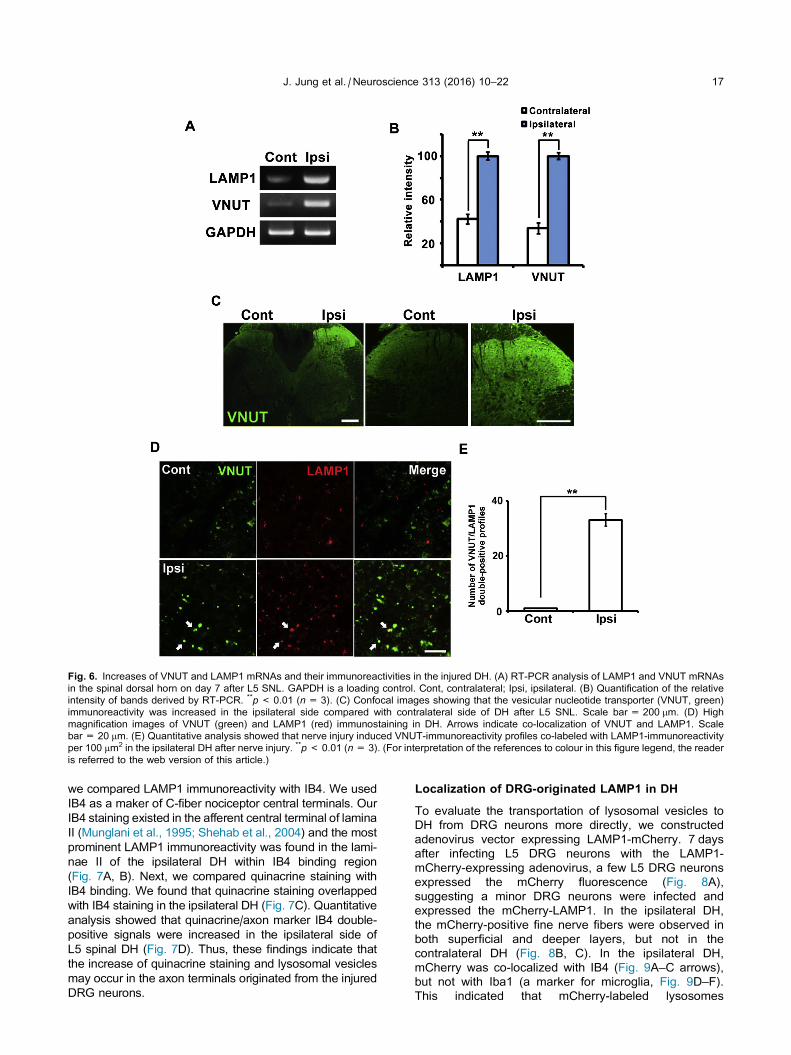
Fig. 6. Increases of VNUT and LAMP1 mRNAs and their immunoreactivities in the injured DH. (A) RT-PCR analysis of LAMP1 and VNUT mRNAs
in the spinal dorsal horn on day 7 after L5 SNL. GAPDH is a loading control. Cont, contralateral; Ipsi, ipsilateral. (B) Quantification of the relative
intensity of bands derived by RT-PCR. **p< 0.01 (n= 3). (C) Confocal images showing that the vesicular nucleotide transporter (VNUT, green)
immunoreactivity was increased in the ipsilateral side compared with contralateral side of DH after L5 SNL. Scale bar = 200 lm. (D) High
magnification images of VNUT (green) and LAMP1 (red) immunostaining in DH. Arrows indicate co-localization of VNUT and LAMP1. Scale
bar = 20 lm. (E) Quantitative analysis showed that nerve injury induced VNUT-immunoreactivity profiles co-labeled with LAMP1-immunoreactivity
per 100 lm2 in the ipsilateral DH after nerve injury. **p< 0.01 (n= 3). (For interpretation of the references to colour in this figure legend, the reader
is referred to the web version of this article.)
J. Jung et al. / Neuroscience 313 (2016) 10–22 17
we compared LAMP1 immunoreactivity with IB4. We used
IB4 as a maker of C-fiber nociceptor central terminals. Our
IB4 staining existed in the afferent central terminal of lamina
II (Munglani et al., 1995; Shehab et al., 2004) and the most
prominent LAMP1 immunoreactivity was found in the lami-
nae II of the ipsilateral DH within IB4 binding region
(Fig. 7A, B). Next, we compared quinacrine staining with
IB4 binding. We found that quinacrine staining overlapped
with IB4 staining in the ipsilateral DH (Fig. 7C). Quantitative
analysis showed that quinacrine/axon marker IB4 double-
positive signals were increased in the ipsilateral side of
L5 spinal DH (Fig. 7D). Thus, these findings indicate that
the increase of quinacrine staining and lysosomal vesicles
may occur in the axon terminals originated from the injured
DRG neurons.
Localization of DRG-originated LAMP1 in DH
To evaluate the transportation of lysosomal vesicles to
DH from DRG neurons more directly, we constructed
adenovirus vector expressing LAMP1-mCherry. 7 days
after infecting L5 DRG neurons with the LAMP1-
mCherry-expressing adenovirus, a few L5 DRG neurons
expressed the mCherry fluorescence (Fig. 8A),
suggesting a minor DRG neurons were infected and
expressed the mCherry-LAMP1. In the ipsilateral DH,
the mCherry-positive fine nerve fibers were observed in
both superficial and deeper layers, but not in the
contralateral DH (Fig. 8B, C). In the ipsilateral DH,
mCherry was co-localized with IB4 (Fig. 9A–C arrows),
but not with Iba1 (a marker for microglia, Fig. 9D–F).
This indicated that mCherry-labeled lysosomes

Fig. 7. Intense LAMP1 immunoreactivity localizes in IB4 enriched layers. (A) IB4 (green) and LAMP1 (red) immunoreactivity in DH. Scale
bar = 200 lm. (B) Higher magnification images of (A). Scale bar = 200 lm. (C) quinacrine-positive puncta (green) is colocalized wit IB4 (red) in the
ipsilateral DH (arrows). Scale bar = 10 lm. (D) Quantitative analysis showed that nerve injury induced the increase of IB4-positive quinacrine
puncta per 100 lm2 in the ipsilateral dorsal horn after nerve injury. **p< 0.01 (n= 4). (For interpretation of the references to colour in this figure
legend, the reader is referred to the web version of this article.)
18 J. Jung et al. / Neuroscience 313 (2016) 10–22
synthesized in IB4-positive DRG neurons were transported
to DH. Obviously the mCherry-labeled lysosomes were
considered as DRG-originated lysosomes. Collectively,
these findings support an idea that the increase of
quinacrine staining and lysosomal vesicles in DH after
nerve injury would be partly due to the transportation of
ATP containing lysosomes from DRG neurons.
DISCUSSION
In this study, we demonstrated that nerve injury-activated
lysosomal biosynthesis in DRG neurons and VNUT-
positive lysosomes were transported to the primary
afferent nerve terminal in DH (Fig. 10). We found that
the injury of peripheral process of DRG neurons induced
the expression of TFEB mRNA (Fig. 2A, B). TFEB is a
master transcriptional regulator of lysosomal biogenesis,
and the TFEB transcriptionally induces both the docking
and fusion of lysosomes with the plasma membrane by
regulating the expression of certain genes (Medina
et al., 2011; Settembre et al., 2011, 2013). This suggests
that the increase of lysosomal vesicles in DRG neurons
could be due to the induction of TFEB in DRG neurons.
Intriguingly, together with the increase of lysosomes in
DRG neurons, VNUT mRNA was induced in the same
DRG neurons, and prominent co-localization of VNUT
and LAMP1 was observed on vesicles. This would sug-
gest that ATP storage in lysosomes is activated in
nerve-injured neurons for some reason.
What are the functional consequences of the
lysosome increase in injured DRG neurons? One most
likely explanation would be an involvement of lysosomes
in membrane repair at injured peripheral nerve process
(Settembre et al., 2013; Appelqvist et al., 2013). Previous

Fig. 8. Vesicles expressing LAMP1-mCherry in DRG neurons are
anterogradely transported to dorsal horn. (A) A representative image
of L5 DRG which is injected the adeno-virus encoding LAMP1-
mCherry. Arrows indicate the neurons expressing LAMP1-mCherry in
DRG. (B) Contralateral dorsal horn of L5 spinal cord (Cont) 7 days
after peripheral nerve injury and adeno-virus injection. (C) Ipsilateral
side (Ipsi) of identical section with (B). Many LAMP1-mCherry-
positive fibers are seen in the ipsilateral dorsal horn. Scale bars
indicate 100 lm.
J. Jung et al. / Neuroscience 313 (2016) 10–22 19
studies have reported that lysosomal vesicles are
involved in the process of membrane repair through
Ca2+-dependent synaptotagmin VII in the damaged cell
by a way to fuse with the plasma membrane
(Gerasimenko et al., 2001; Mc Neil, 2002; Idone et al.,
2008; Settembre et al., 2013; Appelqvist et al., 2013).
The present nerve injury might induce the influx of Ca2+
into DRG neurons and subsequently induces the number
of lysosomal vesicles and lysosomal exocytosis under the
activation of TFEB. The increased fusion of lysosomes
with plasma membrane could function to repair the injured
neurite of DRG neurons. The second possibility would be
an intracellular clearance of molecules and organelles by
lysosomal activation and exocytosis. Under pathological
conditions, TFEB-mediated activation of lysosomal bio-
genesis and exocytosis would be advantageous for
removing intracellular toxic materials and injured orga-
nelle to outside of cell membrane (Medina et al., 2011;
Settembre et al., 2013). Nerve injury elicits various types
of stresses including oxidative stress and ER stress in
neurons and these stresses would damage organelle
and induce accumulation of unfolded proteins. For the
removal of those unnecessary or even toxic contents,
lysosomal degradation and clearance including autop-
hagy would be necessary in nerve-injured neurons.
Although both the lysosome-mediated membrane repair
and intracellular clearance are critical in injured peripheral
process and soma of DRG neurons, it is likely that the
increased lysosome plays some role in the central pro-
cess as well. DRG neurons have both peripheral and cen-
tral processes and it appears that no directional
preferences to be transported exist for some neurotrans-
mitters such as substance P and organelle such as mito-
chondria at least (Harmar and Keen, 1982). In fact, the
present adenovirus-mediated expression of mCherry-
LAMP1 clearly demonstrated the transport of lysosomes
in the central branch of DRG neurons. Thus, in response
to nerve injury, ATP-containing lysosomes would be deliv-
ered to both central and peripheral terminals of DRG neu-
rons and whereby the ingredients including ATP might be
released.
Recently lysosomes are considered as one of storage
vesicles for ATP in neuronal and glial cells, and the
lysosomal exocytosis possibly releases ATP together
with other ingredients into the extracellular space
(Zhang et al., 2007a; Pryazhnikov and Khiroug, 2008;
Shin et al., 2012; Jung et al., 2013). The present co-
localization study of LAMP1 and quinacrine in the same
vesicle suggested that the increased lysosomal vesicles
in the ipsilateral DH after nerve injury also contain ATP
(Fig. 5). Because the existence of ATP in lysosomes
and ATP release by lysosomal exocytosis were demon-
strated in cultured DRG neurons (Jung et al., 2013), it is
likely that the lysosomal exocytosis is a mechanism
underlying ATP release in DH after nerve injury. Substan-
tial increase of lysosomal protein, LAMP1, was demon-
strated in the injured DH by immunohistochemistry and
Western blot analysis for LAMP1 (Figs. 4 and 5). Thus,
these findings suggest that the lysosomal ATP may be
associated with nociceptive pathophysiology because
the activation of ATP containing lysosomes occurred in
the area where the central terminals of nociceptive c-
fiber from DRG exist (Figs. 7–9).
Several types of axons and processes including the
central process of DRG neurons locate in DH. Because
quinacrine- and LAMP1-positive vesicles were highly
increased in DRG neurons after nerve injury as shown
in Figs. 1–3 and our previous study (Jung et al., 2013),
it is very likely that ATP containing lysosomes is trans-
ported to DH in response to peripheral nerve injury. To
clarify this possibility, peripheral nerve marker IB4 was
used and LAMP1-positive structures existed jointly with
IB4 immunoreactivity in the ipsilateral DH with adenoviral
infection (Figs. 8 and 9). In addition, we showed the
increase of VNUT expression in the ipsilateral DH and
the injured DRG neurons and its expression in lysosomal
vesicles in vivo (Figs. 3 and 6) as shown in our previous

Fig. 9. Vesicles expressing LAMP1-mCherry are transported in IB4-positive fibers. Higher magnification images showing LAMP1-mCherry (red)
containing fibers in the dorsal horn of L5 spinal cord (A, D). These are co-stained with IB4 (green in B) or Iba1 (green in E) respectively. (C) The
merged images of (A) and (B). (F) The merged image of (D) and (E). Arrows in (A–C) indicate the LAMP1-mCherry and IB4 double-positive fibers.
None of the LAMP1-mCherry-positive structure was colocalized with Iba1-positive cells. Scale bars indicate 25 lm. (For interpretation of the
references to colour in this figure legend, the reader is referred to the web version of this article.)
Fig. 10. Schematic demonstration of the present results. Red large dots, lysosomes; green small dots, ATP. (For interpretation of the references to
colour in this figure legend, the reader is referred to the web version of this article.)
20 J. Jung et al. / Neuroscience 313 (2016) 10–22
in vitro study (Jung et al., 2013). These findings together
with our previous study (Jung et al., 2013) would support
our hypothesis that peripheral nerve injury induces the
transportation of ATP-containing lysosomes to the central
branch of DRG neurons and suggest a possibility that
ATP is released from axon terminals of DRG neurons
through lysosomal exocytosis in addition to releases from
astrocytes and microglia (Zhang et al., 2007b; Dou et al.,
2012).
CONCLUSION
We demonstrated the transcriptional activation of
lysosomal exocytosis activator TFEB in DRG neurons in
response to nerve injury and prominent increase of
lysosomes was also observed in both DRG and DH of
the spinal cord where DRG neurons project. Since it
becomes evident that the transportation of lysosomes
from DRG to DH through the central branch of primary

J. Jung et al. / Neuroscience 313 (2016) 10–22 21
afferent neurons, it is likely that ATP in addition to some
additional ingredients are released by the lysosomal
exocytosis at the central terminal of DRG neurons.
Although we could not demonstrate direct release of
ATP from the primary afferent nerve terminal in DH via
lysosomal exocytosis, previous in vitro study supported
the possibility (Jung et al., 2013). Thus, a regulation of
lysosomal exocytosis at the axon terminals of DRG neu-
rons may provide an alternative clue to control nocicep-
tion and pain.
Acknowledgments—This study was partially supported by
CREST, JST, Japan and a Grant-in-Aid for Scientific Research
from the Ministry of Education, Science, Sports and Culture of
Japan (MEXT) and Basic Science Research Program through
the National Research Foundation of Korea (NRF) funded by
the Ministry of Science, ICT and Future Planning (J. Jung,
2015R1A2A2A01002735; N. Y. Jeong, 2015R1C1A1A02036863).
We are grateful to Profs. Inoue K and Tsuda M (Kyushu Univ.,
Japan) for their kind advice, Ms. Y. Tabata and N. Tawarayama
for their technical assistance, and Ms. A. Asano for secretarial
assistance.
REFERENCES
Abbracchio MP, Burnstock G, Verkhratsky A, Zimmermann H (2009)
Purinergic signalling in the nervous system: an overview. Trends
Neurosci 32:19–29.
Andrews NW (2000) Regulated secretion of conventional lysosomes.
Trends Cell Biol 10:316–321.
Appelqvist H, Wster P, Kagedal K, Ollinger K (2013) The lysosome:
from waste bag to potential therapeutic target. J Mol Cell Biol
5:214–226.
Belai A, Burnstock G (1994) Evidence for coexistence of ATP and
nitric oxide in non-adrenergic, non-cholinergic (NANC) inhibitory
neurons in the rat ileum, colon and anococcygeus muscle. Cell
Tissue Res 278:197–200.
Blott EJ, Griffiths GM (2002) Secretory lysosomes. Nat Rev Mol Cell
Biol 3:122–131.
Chomczynski P, Sacchi N (2006) The single-step method of RNA
isolation by acid guanidinium thiocyanate-phenol-chloroform
extraction: twenty-something years on. Nat Protoc 1:581–585.
Dou Y, Wu HJ, Li HQ, Qin S, Wang YE, Li J, Lou HF, Chen Z, Li XM,
Luo QM, Duan S (2012) Microglial migration mediated by ATP-
induced ATP release from lysosomes. Cell Res 22:1022–1033.
Engelman HS, Mac Dermott AB (2004) Presynaptic ionotropic
receptors and control of transmitter release. Nat Rev Neurosci
5:135–145.
Fields RD, Stevens B (2000) ATP: an extracellular signaling molecule
between neurons and glia. Trends Neurosci 23:625–633.
Gerasimenko JV, Gerasimenko OV, Petersen OH (2001) Membrane
repair: Ca(2+)-elicited lysosomal exocytosis. Curr Biol 11:
R971–R974.
Harmar A, Keen P (1982) Synthesis, and central and peripheral
axonal transport of substance P in a dorsal root ganglion-nerve
preparation in vitro. Brain Res 231:379–385.
Holton P (1959) The liberation of adenosine triphosphate on
antidromic stimulation of sensory nerves. J Physiol 145:494–504.
Holtzman E (1989) Autophagy and related phenomena. In: Siekevitz
P, editor. Lysosomes. New York: Plenum Press. p. 243–316.
Idone V, Tam C, Andrews NW (2008) Two-way traffic on the road to
plasma membrane repair. Trends Cell Biol 18:552–559.
Iijima T (1983) Quinacrine-induced degeneration of non-adrenergic,
non-cholinergic autonomic nerves in the rat anococcygeus
muscle. Cell Tissue Res 230:639–648.
Imura Y, Morizawa Y, Komatsu R, Shibata K, Shinozaki Y, Kasai H,
Moriishi K, Moriyama Y, Koizumi S (2013) Microglia release ATP
by exocytosis. Glia 61:1320–1330.
Inoue K, Tsuda M, Tozaki-Saitoh H (2007) Modification of
neuropathic pain sensation through microglial ATP receptors.
Purinergic Signal 3:311–316.
Jung J, Shin YH, Konishi H, Lee SJ, Kiyama H (2013) Possible ATP
release through lysosomal exocytosis from primary sensory
neurons. Biochem Biophys Res Commun 430:488–493.
Kim SH, Chung JM (1992) An experimental model for peripheral
neuropathy produced by segmental spinal nerve ligation in the rat.
Pain 50:355–363.
Lalo U, Voitenko N, Kostyuk P (1998) Iono- and metabo-tropically
induced purinergic calcium signalling in rat neocortical neurons.
Brain Res 799:285–291.
Lee SJ, Rao AS, Shin YH, Chung HJ, Huh Y, Ahn KH, Jung J (2013)
A novel method using an acedan-based Zn(DPA) probe to
monitor ATP localization in an in vivo system. J Mol Histol
44:241–247.
Maeda M, Tsuda M, Tozaki-Saitoh H, Inoue K, Kiyama H (2010)
Nerve injury-activated microglia engulf myelinated axons in a
P2Y12 signaling-dependent manner in the dorsal horn. Glia
58:1838–1846.
Mc Neil PL (2002) Repairing a torn cell surface: make way,
lysosomes to the rescue. J Cell Sci 115:873–879.
Medina DL, Fraldi A, Bouche V, Annunziata F, Mansueto G,
Spampanato C, Puri C, Pignata A, Martina JA, Sardiello M,
Palmieri M, Polishchuk R, Puertollano R, Ballabio A (2011)
Transcriptional activation of lysosomal exocytosis promotes
cellular clearance. Dev Cell 21:421–430.
Morel N, Meunier FM (1981) Simultaneous release of acetylcholine
and ATP from stimulated cholinergic synaptosomes. J
Neurochem 36:1766–1773.
Munglani R, Bond A, Smith GD, Smith GD, Harrison SM, Elliot PJ,
Birch PJ, Hunt SP (1995) Changes in neuronal markers in a
mononeuropathic rat model relationship between neuropeptide Y,
pre-emptive drug treatment and long-term mechanical
hyperalgesia. Pain 63:21–31.
Ohba N, Maeda M, Nakagomi S, Muraoka M, Kiyama H (2003)
Biphasic expression of activating transcription factor-3 in neurons
after cerebral infarction. Brain Res Mol Brain Res 115:147–156.
Pankratov Y, Lalo U, Krishtal O, Verkhratsky A (2002) Ionotropic P2X
purinoreceptors mediate synaptic transmission in rat pyramidal neurons
of layer II/III of somato-sensory cortex. J Physiol 542:529–536.
Pankratov Y, Lalo U, Verkhratsky A, North RA (2006) Vesicular
release of ATP at central synapses. Pflugers Arch 452:589–597.
Pryazhnikov E, Khiroug L (2008) Sub-micromolar increase in [Ca(2
+)](i) triggers delayed exocytosis of ATP in cultured astrocytes.
Glia 56:38–49.
Sawada K, Echigo N, Juge N, Miyaji T, Otsuka M, Omote H,
Yamamoto A, Moriyama Y (2008) Identification of a vesicular
nucleotide transporter. Proc Natl Acad Sci USA 105:5683–5686.
Sawynok J, Downie JW, Reid AR, Cahill CM, White TD (1993) ATP
release from dorsal spinal cord synaptosomes: characterization
and neuronal origin. Brain Res 610:32–38.
Settembre C, Di Malta C, Polito VA, Garcia Arencibia M, Vetrini F,
Erdin S, et al. (2011) TFEB links autophagy to lysosomal
biogenesis. Science 332:1429–1433.
Settembre C, Fraldi A, Medina DL, Ballabio A (2013) Signals from the
lysosome: a control centre for cellular clearance and energy
metabolism. Nat Rev Mol Cell Biol 14:283–296.
Shehab SA, Spike RC, Todd AJ (2004) Do central terminals of intact
myelinated primary afferents sprout into the superficial dorsal
horn of rat spinal cord after injury to a neighboring peripheral
nerve? J Comp Neurol 474:427–437.
Shin YH, Lee SJ, Jung J (2012) Secretion of ATP from Schwann cells
through lysosomal exocytosis during Wallerian degeneration.
Biochem Biophys Res Commun 429:163–167.
Soeda H, Tatsumi H, Katayama Y (1997) Neurotransmitter release
from growth cones of rat dorsal root ganglion neurons in culture.
Neuroscience 77:1187–1199.

22 J. Jung et al. / Neuroscience 313 (2016) 10–22
Sundler R (1997) Lysosomal and cytosolic pH as regulators of
exocytosis in mouse macrophages. Acta Physiol Scand
161:553–556.
Takeda M, Kato H, Takamiya A, Yoshida A, Kiyama H (2000) Injury-
specific expression of activating transcription factor-3 in retinal
ganglion cells and its colocalized expression with phosphorylated
c-Jun. Invest Ophthalmol Vis Sci 41:2412–2421.
Terrian DM, Hernandez PG, Rea MA, Peters RI (1989) ATP release,
adenosine formation, and modulation of dynorphin and glutamic
acid release by adenosine analogues in rat hippocampal mossy
fiber synaptosomes. J Neurochem 53:1390–1399.
Tozaki-Saitoh H, Tsuda M, Miyata H, Ueda K, Kohsaka S, Inoue K
(2008) P2Y12 receptors in spinal microglia are required for neuropathic
pain after peripheral nerve injury. J Neurosci 28:4949–4956.
Tsuda M, Shigemoto-Mogami Y, Koizumi SMizokoshi A, Kohsaka S,
Salter MW, Inoue K (2003) P2X4 receptors induced in spinal microglia
gate tactile allodynia after nerve injury. Nature 424:778–783.
Tsuda M, Inoue K, Salter MW (2005) Neuropathic pain and spinal
microglia: a big problem from molecules in ‘‘small” glia. Trends
Neurosci 28:101–107.
Tsujino H, Kondo E, Fukuoka T, Dai Y, Tokunaga A, Miki K,
Yonenobu K, Ochi T, Noguchi K (2000) Activating transcription
factor 3 (ATF3) induction by axotomy in sensory and
motoneurons: a novel neuronal marker of nerve injury. Mol Cell
Neurosci 15:170–182.
Vizi ES, Liang SD, Sperlagh B, Kittel A, Juranyi Z (1997) Studies on
the release and extracellular metabolism of endogenous ATP in
rat superior cervical ganglion: support for neurotransmitter role of
ATP. Neuroscience 79:893–903.
Watkins LR, Maier SF (2003) Glia: a novel drug discovery target for
clinical pain. Nat Rev Drug Discov 2:973–985.
Yogalingam G, Bonten EJ, van de Vlekkert D, Hu H, Moshiach S,
Connell SA, d’Azzo A (2008) Neuraminidase 1 is a negative
regulator of lysosomal exocytosis. Dev Cell 15:74–86.
Zhang JM, Wang HK, Ye CQ, Ge W, Chen Y, Jiang ZL, Wu CP, Poo
MM, Duan S (2003) ATP released by astrocytes mediates
glutamatergic activity-dependent heterosynaptic suppression.
Neuron 40:971–982.
Zhang X, Chen Y, Wang C, Huang LY (2007a) Neuronal somatic ATP
release triggers neuron-satellite glial cell communication in dorsal
root ganglia. Proc Natl Acad Sci USA 104:9864–9869.
Zhang Z, Chen G, Zhou W, Song A, Xu T, Luo Q, Wang W, Gu XS,
Duan S (2007b) Regulated ATP release from astrocytes through
lysosome exocytosis. Nat Cell Biol 9:945–953.
(Accepted 13 November 2015)(Available online 19 November 2015)