transcription, chromatin, and lcor
TRANSCRIPT

Jiao Yang and John H. WhiteVeronique Bourdeau, Sylvie Mader, Xiang Kourelis, Luz E. Tavera-Mendoza, Zhi Li,Bastien, Liqun Tang, Mark Verway, Maria Ana Palijan, Isabelle Fernandes, Yolande LCoRCofactor of Nuclear Receptor Coregulator Function of Histone Deacetylase 6 as aEpigenetics:Transcription, Chromatin, and
doi: 10.1074/jbc.M109.045526 originally published online September 10, 20092009, 284:30264-30274.J. Biol. Chem.
10.1074/jbc.M109.045526Access the most updated version of this article at doi:
.JBC Affinity SitesFind articles, minireviews, Reflections and Classics on similar topics on the
Alerts:
When a correction for this article is posted•
When this article is cited•
to choose from all of JBC's e-mail alertsClick here
http://www.jbc.org/content/284/44/30264.full.html#ref-list-1
This article cites 52 references, 18 of which can be accessed free at
by guest on Novem
ber 18, 2013http://w
ww
.jbc.org/D
ownloaded from
by guest on N
ovember 18, 2013
http://ww
w.jbc.org/
Dow
nloaded from

Function of Histone Deacetylase 6 as a Cofactor of NuclearReceptor Coregulator LCoR*
Received for publication, July 16, 2009 Published, JBC Papers in Press, September 10, 2009, DOI 10.1074/jbc.M109.045526
Ana Palijan‡1, Isabelle Fernandes‡2, Yolande Bastien‡, Liqun Tang‡, Mark Verway‡, Maria Kourelis‡,Luz E. Tavera-Mendoza§3, Zhi Li‡, Veronique Bourdeau¶, Sylvie Mader¶4, Xiang Jiao Yang�5, and John H. White‡�4,6
From the Departments of ‡Physiology and �Medicine, McGill University, Montreal, Quebec H3G 1Y6, Canada, the §Department ofAdult Oncology, Dana-Farber Cancer Institute, Harvard Medical School, Boston, Massachusetts 02115, and the ¶Institute forResearch in Immunology and Cancer and Biochemistry Department, University of Montreal, Montreal, Quebec H3C 3J7, Canada
Ligand-dependent corepressor LCoR was identified as aprotein that interacts with the estrogen receptor � (ER�) ligandbinding domain in a hormone-dependent manner. LCoR alsointeracts directly with histone deacetylase 3 (HDAC3) andHDAC6. Notably, HDAC6 has emerged as a marker of breastcancer prognosis. However, although HDAC3 is nuclear, HDAC6is cytoplasmic in many cells. We found that HDAC6 is partiallynuclear in estrogen-responsive MCF7 cells, colocalizes withLCoR, represses transactivation of estrogen-inducible reportergenes, andaugments corepressionbyLCoR. In contrast, no repres-sionwasobserveduponHDAC6expression inCOS7cells,where itis exclusively cytoplasmic. LCoR binds to HDAC6 in vitro via acentral domain, and repression by LCoR mutants lacking thisdomain was attenuated. Kinetic chromatin immunoprecipitationassays revealed hormone-dependent recruitment of LCoR to pro-moters of ER�-induced target genes in synchrony with ER�.HDAC6wasalso recruited to thesepromoters, andrepeat chroma-tin immunoprecipitation experiments confirmed the corecruit-ment of LCoR with ER� and with HDAC6. Remarkably, however,although we find evidence for corecruitment of LCoR and ER� ongenes repressed by the receptor, LCoR andHDAC6 failed to coim-munoprecipitate, suggesting that they are part of distinct com-plexes on these genes. Although small interfering RNA-mediatedknockdownofLCoRorHDAC6augmentedexpressionof anestro-gen-sensitive reportergene inMCF7cells, unexpectedly their abla-tion led to reducedexpressionof someendogenous estrogen targetgenes. Taken together, these data establish that HDAC6 can func-tion as a cofactor of LCoRbut suggest that theymay act in enhanceexpressing some target genes.
Nuclear receptors are ligand-regulated transcription factorswhose activities are controlled by a variety of lipophilic extra-
cellular signals, including steroid and thyroid hormones,metabolites of vitamins A (retinoids) and D (1, 2). DNA-boundnuclear receptors regulate transcription by recruiting com-plexes of coregulatory proteins, classified as coactivators orcorepressors depending on whether they act to stimulate orrepress transcription (2–4). Many coactivators interact withreceptors through signature LXXLL motifs, known as NRboxes, which are oriented within a hydrophobic pocket of ago-nist-bound receptor ligand binding domains (5). Several coac-tivators or their associated cofactors possess histone acetyl-transferase activity, which essentially caps positively chargedlysine residues and loosens their association with DNA, facili-tating chromatin remodeling and subsequent access of thetranscriptional machinery to promoters.Nuclear receptor corepressors NCoR7 and SMRT were iso-
lated as factors that interacted with hormone-free but not hor-mone-bound thyroid and retinoid receptors (6, 7). They bind toreceptor ligand binding domains through extended LXXX-IXXX(L/I) motifs known as CoRNR boxes (8, 9) and recruitmultiprotein complexes implicated in transcriptional repres-sion andhistone deacetylation (2–4, 10–13).Hormone bindinginduces a conformational change in ligand binding domainsthat leads to dissociation of NCoR or SMRT. Both corepressorsare components of several different complexes containing dis-tinct combinations of ancillary proteins and class I or class IIhistone deacetylases (HDACs), suggesting that their functiondepends on cell type, combinations of transcription factorsbound to specific promoters, and phase of the cell cycle.We identified a ligand-dependent corepressor, LCoR, as an
NR box-containing protein that interacted with the ligandbinding domains of agonist-bound receptors and repressedhormone-dependent transactivation when overexpressed (14).Although LCoR interacts with nuclear receptors in essentially
* This work was supported by Canadian Institutes of Health Research GrantMT-11704 (to J. H. W.) and a grant from the Canadian Cancer Society(018362 to X. J. Y.).
1 Supported by studentships from the Canadian Institutes of Health Researchand the Montreal Centre for Experimental Therapeutics in Cancer.
2 Supported by postdoctoral fellowships from l’Association pour la Recher-che sur le Cancer and the Canadian Institutes of Health Research.
3 Supported by Fonds de Recherche en Sante du Quebec.4 Chercheurs Boursier National of the Fonds de Recherche en Sante du
Quebec.5 Holder of a Canadian Institutes of Health Research scholarship.6 To whom correspondence should be addressed: Dept. of Physiology, McGill
University, 3655 Drummond St., Montreal, Quebec H3G Y6, Canada. Tel:514-398-8498; Fax: 514-398-7452; E-mail: [email protected].
7 The abbreviations used are: NCoR, nuclear receptor corepressor; ADORA1,adenosine A1 receptor; BMP7, bone morphogenetic protein 7; ChIP, chro-matin immunoprecipitation; CYP26B1, cytochrome P450, family 26, sub-family b, polypeptide 1; E2, estradiol; ER�, estrogen receptor �; GREB1,gene regulated by estrogen in breast cancer protein; HDAC, histonedeacetylase; IGFBP4, insulin-like growth factor binding protein 4; KRT4,keratin 4; LCoR, ligand-dependent corepressor; p300, E1A-binding proteinp300; pS2, trefoil factor 1; SGK3, serum/glucocorticoid regulated kinasefamily, member 3; SMRT, silencing mediator for retinoid and thyroid-hormone receptors; TSA, trichostatin A; siRNA, small interfering RNA;DMEM, Dulbecco’s modified Eagle’s medium; FBS, fetal bovine serum;HA, hemagglutinin; PBS, phosphate-buffered saline; ERE, estrogenresponse element; GST, glutathione S-transferase; NRIP1, nuclearreceptor interacting protein 1.
THE JOURNAL OF BIOLOGICAL CHEMISTRY VOL. 284, NO. 44, pp. 30264 –30274, October 30, 2009© 2009 by The American Society for Biochemistry and Molecular Biology, Inc. Printed in the U.S.A.
30264 JOURNAL OF BIOLOGICAL CHEMISTRY VOLUME 284 • NUMBER 44 • OCTOBER 30, 2009
by guest on Novem
ber 18, 2013http://w
ww
.jbc.org/D
ownloaded from

the same manner as coactivators, it recruits both HDACs andC-terminal-binding proteins corepressors. LCoR interactsdirectly with class I HDAC3 and class II HDAC6 in vitro andcoimmunoprecipitates with the two proteins from MCF7 cellextracts (14). AlthoughHDAC3, like LCoR, is a nuclear protein,the interaction of LCoR with HDAC6 is remarkable as HDAC6is cytoplasmic in many cells (15). Indeed, HDAC6 has beenshown to function as a tubulin deacetylase (16, 17) through anassociation controlled by a tetradecapeptide motif (18). How-ever, a portion ofHDAC6 can be nuclear in some cells. Notably,experiments performed in breast cancer cells have revealed thatHDAC6 is an estrogen target gene (19) and that HDAC6 pro-tein is present in the nuclei of normal breast epithelial cells butis cytoplasmic in adjacent malignant cells (20, 21). Moreover,these studies found that HDAC6 expression levels correlatewith better prognosis and response to endocrine therapy inbreast cancer (19–21).Based on the above, we examined the subcellular localization
of HDAC6 in estrogen-responsive MCF7 breast cancer cellsand its potential role as an LCoR cofactor.We find thatHDAC6is partially nuclear in MCF7 cells and that LCoR and HDAC6are recruited together during ER�-dependent gene regulationin MCF7 cells. Remarkably, however, although ablation ofLCoR or HDAC6 enhanced estrogen-dependent stimulation ofa reporter gene, the effect was not reproduced on endogenousER� target genes. Rather, the results suggested that the twoproteins can act to enhance expression of specific estrogen-regulated genes.
EXPERIMENTAL PROCEDURES
Antibodies—A rabbit polyclonal antipeptide antibody wasraised against LCoR amino acids 20–36 (QDPSQPNSTKNQS-LPKA) fused to keyhole limpet hemocyanin and purified over apeptide affinity column (Bethyl Laboratories, MontgomeryTX). Goat polyclonal HDAC3 (sc-8138), goat polyclonalHDAC6 (sc-5253), rabbit polyclonal HDAC6 (sc-11420), rabbitpolyclonal ER� (sc-543), protein A-agarose (sc-2001), and pro-tein G PLUS-agarose (sc-2002) were from Santa Cruz Biotech-nology (Santa Cruz, CA). Rabbit polyclonal p300 (ab3425) wasfrom Abcam Inc. (Cambridge, MA). Cy3-donkey polyclonal�-goat (705-165-147) and Cy2-goat polyclonal �-rabbit (711-225-152), Cy3-donkey polyclonal �-rabbit (711-165-152), andCy2-donkey polyclonal �-mouse (715-225-150) were pur-chased from Jackson ImmunoResearch (West Grove, PA).Mouse monoclonal �-FLAG M2 (F3165) and �-FLAG M2horseradish peroxidase (HRP)-conjugate (A8592), monoclonal�-rabbit HRP conjugate (A2074), and rabbit polyclonal �-goatHRP conjugate (A5420) were from Sigma.Recombinant Plasmids—PSG5/LCoR, FLAG-HDAC6/pcDNA3,
HA-HDAC3/pCDNA3.1, and FLAG-LCoR/pcDNA3.1 havebeen described (14). FLAG-LCoR�HDAC6/pcDNA3.1 wasmade using the QuikChange mutagenesis kit (200518, Strat-agene, La Jolla, CA) as per the manufacturer’s instructions.Primers were designed to delete amino acids 203–319 fromLCoR. The new construct was sequenced to confirm properdeletion, and a Western blot was performed to show the equallevel of expression when compared with wild-type LCoR.
Cell Culture and Transfections—All cells were culturedunder the recommended conditions. For immunocytochem-istry, COS7 cells grown on collagen IV-treated microscopeslides in 6-well plates in DMEM supplemented with 10% FBSwere transfected in medium without serum with 12.5 �l ofLipofectamine 2000 (Invitrogen) containing 1 �g each ofpSG5/LCoR and HA-FLAG-HDAC6/pcDNA3. Medium wasreplaced 24 h after transfection, and cells were prepared forimmunocytochemistry after 48 h as described below. Foranalysis of the effects of HDAC3 or -6 on LCoR corepression,COS7 cells (60–70% confluent) grown in DMEM withoutphenol red supplemented with 10% FBS on 6-well plateswere transfected in minimal medium without serum withLipofectamine 2000 (5 �l) with 100 ng of ER� expressionvector, 250 ng of ERE3-TATA-pXP2 reporter plasmid, and250 ng of internal control vector pCMV-�gal. Quantities ofexpression vectors (LCoR/pSG5, HA-HDAC3/pCDNA3.1,FLAG-HDAC6/pcDNA3, FLAG-LCoR/pcDNA3.1, andFLAG-LCoR�HDAC6/pcDNA3.1) used are indicated in thefigures or corresponding figure legends. Medium was replaced18 h after transfection with medium containing charcoal-stripped serum and estradiol (10 nM) for 30 h. MCF7 cellsgrown in 6-well plates were transfected similarly. MCF7 cellswere also grown in 24-well plates and were transfected using aon-fifth scale. TrichostatinA (TSA) and trapoxinwere added to500 and 50 nM, respectively, as indicated. Cells were harvestedin 250�l of reporter lysis buffer (Promega). Note that the trans-fection conditions above were chosen because the amounts ofHDAC and LCoR expression vectors used led to selectiverepression of ER�-dependent transactivation without affectingexpression of the �-galactosidase internal control.Immunocytochemistry—Cells were cultivated on collagen
IV-treated microscope slides in 6-well plates, fixed with 2%paraformaldehyde for 15 min at room temperature, washed(3�) with 1� PBS, and permeabilized with 0.2% Triton X-100,5% bovine serum albumin, 10% horse serum in PBS.MCF7 cellswere then incubated with �-LCoR (1:500) and goat polyclonalantibodies against HDAC6 or Bmi1 (1:50) in buffer B (0.2%TritonX-100, 5% bovine serum albumin in PBS) for 1 h at roomtemperature. Cells were washed (3�) with PBS and incubatedwith goat anti-rabbit-Cy2 and donkey anti-goat Cy3 (1:300) inbuffer B for 1 h at room temperature. Transiently transfectedCOS7 cells were incubated with �-LCoR (1:500) and anti-FLAG (1:300) to detect FLAG-HDAC6.Cells werewashed (3�)with PBS and incubated with Cy3-donkey polyclonal �-rabbit(1:300) and Cy2-donkey polyclonal �-mouse (1:400) in bufferB for 1 h at room temperature. Slides were mounted withImmuno-Fluore mounting medium (ICN, Aurora, OH) andvisualized using a Zeiss LSM 510 confocal microscope at 63�magnification.Western Blotting—The following primary antibodies were
used: LCoR (GenWay Biotech, 18-003-44018), and FLAG(Santa Cruz Biotechnology, sc-807). The following secondaryantibody was purchased from Santa Cruz Biotechnology: goatanti-rabbit (sc-2004). A Western blot was performed as previ-ously described (22) using MCF7 cells extracts. Cells weregrown in 10 cm dishes (70% confluent) and transiently trans-
Function of HDAC6 as an LCoR cofactor
OCTOBER 30, 2009 • VOLUME 284 • NUMBER 44 JOURNAL OF BIOLOGICAL CHEMISTRY 30265
by guest on Novem
ber 18, 2013http://w
ww
.jbc.org/D
ownloaded from

fected with 500 ng of FLAG-tagged LCoR. 30h later, cells wereharvested.Chromatin Immunoprecipitation (ChIP) and ReChIP Assays—
ChIP and reChIP assays were performed as previously de-scribed (23) in MCF7 cells. Cells were grown in 10-cm dishes(70% confluent) and transiently transfected with 500 ng ofFLAG-tagged LCoR. After the transfection, cells were starvedfor 2 days in DMEM-phenol free and FBS-free media andtreated with 2.5 �M �-amanitin (Sigma, A2263) for 2 h beforehormone treatment to properly synchronize cells. Cells werecollected, and cofactor recruitment was evaluated on promoterregions containing EREs of estrogen target genes. Immunopre-cipitations were performed with the following antibodies: ER�(sc-543), HDAC6 (Upstate, 07-732), FLAG (OctA-Probe,sc-807), and p300 (sc-8981). Protein A-agarose (sc-2001) wasused for the immunoprecipitation, and normal rabbit IgG (sc-2027) was used for background control. Primer sequences usedare were following: pS2 promoter 5�-CTCTCACTATGA-ATCACTTCTGCAG-3� (forward) and 5�-AGATAACATTT-GCCtAAGGAGGCC-3� (reverse), non-targeting pS2 5�-CAG-CCCCCAAGAACTTCCAG-3� (forward) and 5�-TGAG-CAGGTTTGCAGCACACTT-3� (reverse), ADORA1 pro-moter 5�-CAGAAGCTCTGTTGGGCATG-3� (forward) and5�-ATCGGGCTTTGACGTGGT-3� (reverse), ADORA1non-targeting 5�-TAGAATCCACTAGTCCACCTTCT-3�(forward) and 5�-TCACTTGCTGCTTACTACTTACCC-TTC-3� (reverse), IGFBP4 promoter 5�-CTTTCTTGCTGC-AAAGTCCC-3� (forward) and 5�-ATGGCCTTCCATGCT-ACAAG-3� (reverse), IGFBP4 non-targeting 5�-GCCAGG-GACCGGTATAAAG-3� (forward) and 5�-GACGTAGCG-GGGGAAGTTAG-3� (reverse), NRIP1 promoter 5�-GATG-CAGATTGGCTGACAGA-3� (forward) and 5�-CCCACCC-CCAATTTCTATCT-3� (reverse), NRIP1 non-targeting 5�-GCGAGGGGAGGGACTGGG-3� (forward) and 5�-ATGT-CTGCGAGGCTGACTTT-3� (reverse), BMP7 promoter 5�-TGCAGACGACGAAAAATCAG-3� (forward) and 5�-AGGGG-TGGGAGGTTTAGATG-3� (reverse), and BMP7 non-targeting5�-CGCTATCAGTCACCCCATTT-3� (forward) and 5�-CGA-AAAGGCTTTGAGATTGC-3� (reverse).siRNA Knockdowns—siRNAs were purchased from Thermo
Scientific Dharmacon (Lafayette, CO). The following ON-TAR-GETplus SMART pool siRNA were used: LCoR (L-026303-00),HDAC6 (L-003499-00), and non-targeting (D-001818-10).siRNAs were resuspended per the manufacturer’s instructions.Transfections were done in 6-well plates as described previously.Lipofectamine 2000 (10 �l) was used as the transfection reagent.DMEM phenol-free with 10% stripped FBS was added 12 h aftertransfection. For Western blot analysis, cells were collected 48 hafter transfection. Luciferase reporter assays after siRNA knock-downswereperformedas follows. 100ngofER�expressionvectorand250ngofERE3-TATA-pXP2vectorwere transfectedwith thecorresponding siRNA.DMEMphenol-freewith 10% strippedFBSwas added12hafter transfection. Estradiol (10nM)was added36hafter transfection, and cells were collected 24 h later. Luciferaseactivity was measured as previously described.RNA Isolation, cDNA Synthesis, and Quantitative Real-
time-PCR—Cells were grown in 100-mm dishes. Medium wasreplaced with charcoal-stripped medium containing ligand.
Total RNAwas extracted with TRIZOL reagent. cDNA synthe-sis was performed with iScript cDNA synthesis kit (Bio-Rad)according to the instructions of the manufacturer. A Mini-Opticon real-time PCR system with iQ SYBR Green Supermix(Bio-Rad) were used for quantitative real-time-PCR expressionanalysis of target genes. The program was: 1) incubation at94 °C for 60 s, 2) incubation at 95 °C for 20 s, 3) incubation at60 °C for 30 s (decreasing temperature by 1° per cycle), 4) incuba-tion at 72 °C for 30 s, 5) plate reading, 6) repetition from step 2five more times, 7) incubation at 95 °C for 20 s, 8) incubation at57.5 °C for 30 s, 9) incubation at 72 °C for 30 s, 10) plate reading,11) repetition from step 7 thirty-five more times, 12) meltingcurve formed, and end. Results were normalized to �-actinmRNA expression. The following primers were used: pS25�-ACCATGGAGACAAGGTGAT-3� (forward) and pS25�-AAATTCACACTCCTCTTCTG-3� (reverse), GREB1 5�-CCA-CAAAGGGTGGTCTCCAGAA-3� (forward) and GREB15�-CACTGGCTTGGCCTTGCATATT-3� (reverse), SGK35�-CAAAAGAAGATTCCACCACCA-3� (forward) and SGK3 5�-TGTCAAAGTTTCTGATATCATCTC-3� (reverse),CYP26B1 5�-ACATCCACCGCAACAAGC-3� (forward) and CYP26B15�-GGATCTTGGGCAGGTAACTCT-3� (reverse), BMP7 5�-GGTCATGAGCTTCGTCAACC-3� (forward) and BMP7 5�-GCAGGAAGAGATCCGATTCC-3� (reverse), KRT4 5�-GCC-GACAATGACTTTGTGGT-3� (forward) and KRT4 5�-CCT-CCAACTCCACCTTGTTC-3� (reverse), and �-actin 5�-GGCA-TGGGTCAGAAGGATTCC-3� (forward) and �-actin 5�-GCT-GGGGTGTTGAAGGTCTC-3� (reverse), ADORA1 5�-GAC-CTACTTCCACACCTGCCTCA-3� (forward) andADORA1 5�-CCAGCCAAACATAGGGGTCAGT-3� (reverse), IGFBP4 5�-GGGGGCAAGATGAAGGTCAAT-3� (forward) and IGFBP45�-CGGTCCACACACCAGCACTT-3� (reverse) and NRIP15�-GTGATTCCAGGATGGTTTGG-3� (forward) and NRIP15�-ATGGTTTTAATAAAGGTTAAGGATGC-3� (reverse).
RESULTS
Colocalization of HDAC6 with LCoR in MCF7 Cells—Ourprevious results showed that endogenous LCoR coimmunopre-cipitated with endogenous HDAC3 and -6 from extracts ofMCF7 cells (14). However, as HDAC6 is cytoplasmic in manycells, we further investigated the colocalization of LCoR andHDAC6 in MCF7 cells by immunocytochemistry. As expected(14), LCoR was almost exclusively nuclear, as was HDAC3, andthere was a marked colocalization of the two proteins (Fig. 1A).Moreover, a substantial portion of HDAC6 was nuclear inMCF7 cells, and there was a clear colocalization of nuclearHDAC6with LCoR (Fig. 1A), substantiating the possibility thatthe two proteins function together.Cell-specific Repression of Hormone-dependent Transactiva-
tion by HDAC6—The capacity of HDAC6 to function as a(cell-specific) cofactor in LCoR-dependent corepression ofestrogen-dependent transactivation was further analyzed intransiently transfected COS7 and MCF7 cells. COS7 cellswere chosen for comparison because HDAC6 remains cyto-plasmic even when overexpressed in transient expressionexperiments (Fig. 1B). Coexpression of HDAC6 with LCoRin COS7 cells had no effect on LCoR-dependent corepres-sion (Fig. 1C). As a control for the repressive effects of
Function of HDAC6 as an LCoR cofactor
30266 JOURNAL OF BIOLOGICAL CHEMISTRY VOLUME 284 • NUMBER 44 • OCTOBER 30, 2009
by guest on Novem
ber 18, 2013http://w
ww
.jbc.org/D
ownloaded from
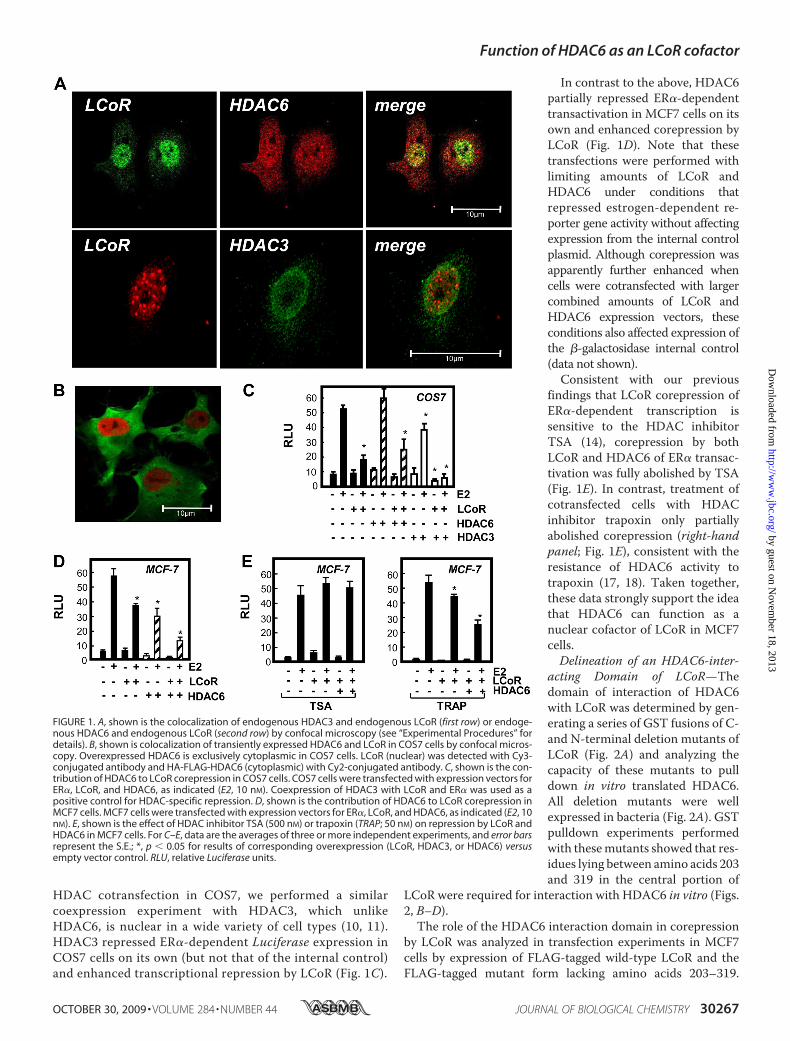
HDAC cotransfection in COS7, we performed a similarcoexpression experiment with HDAC3, which unlikeHDAC6, is nuclear in a wide variety of cell types (10, 11).HDAC3 repressed ER�-dependent Luciferase expression inCOS7 cells on its own (but not that of the internal control)and enhanced transcriptional repression by LCoR (Fig. 1C).
In contrast to the above, HDAC6partially repressed ER�-dependenttransactivation in MCF7 cells on itsown and enhanced corepression byLCoR (Fig. 1D). Note that thesetransfections were performed withlimiting amounts of LCoR andHDAC6 under conditions thatrepressed estrogen-dependent re-porter gene activity without affectingexpression from the internal controlplasmid. Although corepression wasapparently further enhanced whencells were cotransfected with largercombined amounts of LCoR andHDAC6 expression vectors, theseconditions also affected expression ofthe �-galactosidase internal control(data not shown).Consistent with our previous
findings that LCoR corepression ofER�-dependent transcription issensitive to the HDAC inhibitorTSA (14), corepression by bothLCoR and HDAC6 of ER� transac-tivation was fully abolished by TSA(Fig. 1E). In contrast, treatment ofcotransfected cells with HDACinhibitor trapoxin only partiallyabolished corepression (right-handpanel; Fig. 1E), consistent with theresistance of HDAC6 activity totrapoxin (17, 18). Taken together,these data strongly support the ideathat HDAC6 can function as anuclear cofactor of LCoR in MCF7cells.Delineation of an HDAC6-inter-
acting Domain of LCoR—Thedomain of interaction of HDAC6with LCoR was determined by gen-erating a series of GST fusions of C-and N-terminal deletion mutants ofLCoR (Fig. 2A) and analyzing thecapacity of these mutants to pulldown in vitro translated HDAC6.All deletion mutants were wellexpressed in bacteria (Fig. 2A). GSTpulldown experiments performedwith thesemutants showed that res-idues lying between amino acids 203and 319 in the central portion of
LCoR were required for interaction with HDAC6 in vitro (Figs.2, B–D).
The role of the HDAC6 interaction domain in corepressionby LCoR was analyzed in transfection experiments in MCF7cells by expression of FLAG-tagged wild-type LCoR and theFLAG-tagged mutant form lacking amino acids 203–319.
FIGURE 1. A, shown is the colocalization of endogenous HDAC3 and endogenous LCoR (first row) or endoge-nous HDAC6 and endogenous LCoR (second row) by confocal microscopy (see “Experimental Procedures” fordetails). B, shown is colocalization of transiently expressed HDAC6 and LCoR in COS7 cells by confocal micros-copy. Overexpressed HDAC6 is exclusively cytoplasmic in COS7 cells. LCoR (nuclear) was detected with Cy3-conjugated antibody and HA-FLAG-HDAC6 (cytoplasmic) with Cy2-conjugated antibody. C, shown is the con-tribution of HDAC6 to LCoR corepression in COS7 cells. COS7 cells were transfected with expression vectors forER�, LCoR, and HDAC6, as indicated (E2, 10 nM). Coexpression of HDAC3 with LCoR and ER� was used as apositive control for HDAC-specific repression. D, shown is the contribution of HDAC6 to LCoR corepression inMCF7 cells. MCF7 cells were transfected with expression vectors for ER�, LCoR, and HDAC6, as indicated (E2, 10nM). E, shown is the effect of HDAC inhibitor TSA (500 nM) or trapoxin (TRAP; 50 nM) on repression by LCoR andHDAC6 in MCF7 cells. For C–E, data are the averages of three or more independent experiments, and error barsrepresent the S.E.; *, p � 0.05 for results of corresponding overexpression (LCoR, HDAC3, or HDAC6) versusempty vector control. RLU, relative Luciferase units.
Function of HDAC6 as an LCoR cofactor
OCTOBER 30, 2009 • VOLUME 284 • NUMBER 44 JOURNAL OF BIOLOGICAL CHEMISTRY 30267
by guest on Novem
ber 18, 2013http://w
ww
.jbc.org/D
ownloaded from

Reporter gene experiments showed that corepression by thewild-type and mutant forms of LCoR was similar at low con-centrations. However, the mutant exhibited no dose-depend-ent increase in corepression (Fig. 3A).Western analysis with ananti-FLAG antibody showed that the tagged proteins wereexpressed at similar levels (Fig. 3B). Moreover, the deletionmutant could be detected with an antibody against LCoR (Fig.3B). To verify that the LCoR mutant lacking the HDAC6domain is still an active protein, a dominant-negative experi-ment was performed where constant levels of LCoR werecotransfected with greater amounts of the mutant form (Fig.3C). The coexpression of themutant LCoR reduced the repres-sion observed with the wild-type protein, hence showing com-petition between the two forms of LCoR.Hormone-dependent Association of LCoR and HDAC6 with
Estrogen-responsive Promoters in Vivo—To further substanti-ate the role of HDAC6 as a cofactor of LCoR in transcriptionalregulation in MCF7 cells, we performed chromatin immuno-precipitation (ChIP) assays to analyze the recruitment of LCoRand HDAC6 to ER binding regions of estrogen-inducible pro-moters of the pS2, insulin-like growth factor-binding protein 4(IGFBP4), adenosine A1 receptor (ADORA1), and nuclearreceptor interacting protein 1 (NRIP1) genes in vivo. As we lack
an antibody that reliably immuno-precipitates endogenous LCoR, weanalyzed recruitment of transientlyexpressed tagged LCoR to the pS2promoter with an anti-FLAG anti-body. Rapid (15 min) estradiol-de-pendent recruitment of ER� wasobserved to the ERE region of thepS2 promoter but not to non-targetsequences (Fig. 4A). The kinetics ofER� recruitment under these con-ditions is entirely consistent withdata reported by other groups (24,25). The anti-FLAG antibody con-sistently detected recruitment oftagged LCoR by 30 min of estradioltreatment to the region of thepS2 ERE but not non-target DNA(Fig. 4A).The recruitment of HDAC6 to
the pS2 promoter followed a similar,but not identical pattern to that ofLCoR; for example, unlike LCoR,HDAC6 did not dissociate from thepS2 promoter after 60 min of estra-diol treatment (Fig. 4A). As othershave shown that HDAC6 can func-tion as a regulator of the histoneacetyltransferase p300 (26), we ana-lyzed p300 recruitment to pS2 andfound that it was recruited rapidlybut lacked the clear cyclical patternof LCoR. Overlapping patterns ofcorecruitment of ER�, LCoR, andHDAC6 were also observed to ER
binding regions of promoters of genes encoding IGFBP4,ADORA, and NRIP1 (25) (Figs. 4, B–D). Note that we consis-tently observed binding of ER� and NRIP1 promoters in theabsence of estradiol (Figs. 4, C and D), a phenomenon that hasbeen observed by others on estrogen-inducible promoters (25).We also analyzed binding of ER�, LCoR, and HDAC6 to reg-
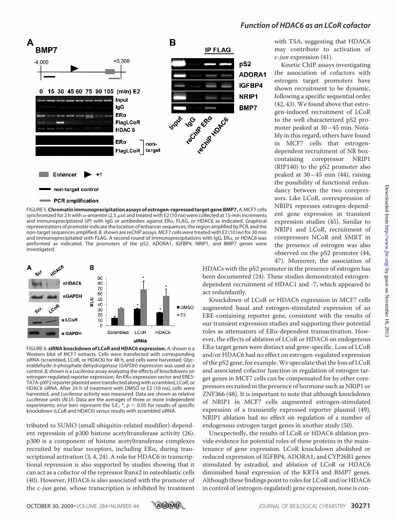
ulatory regions of the gene encoding bone morphogenic pro-tein 7 (BMP7), previously identified as being down-regulated byestrogen (27). A peak of LCoR recruitment to the BMP7 pro-moter occurred 30 min after the addition of estradiol (Fig. 5A),similar to the kinetics of recruitment to estrogen-induciblegenes. However, we observed a largely estrogen-independentassociation of HDAC6 with the BMP7 promoter.The binding of p300 to promoters complicates interpretation
of experiments, as HDAC6 could be recruited to the promotersin association with either p300 or LCoR. Therefore, to deter-mine whether LCoR and HDAC6 are corecruited to promotersin vivo, we performed a series of reChIP experiments on allpromoters analyzed. Experiments were performed from ex-tracts of MCF7 cells treated with estradiol for 30 min, a timecorresponding to peak LCoR recruitment to all promoters ana-lyzed. Extracts were immunoprecipitated with an anti-FLAGantibody followed by a second round of immunoprecipitation
FIGURE 2. Delineation of the domain of interaction of LCoR with HDAC6. A, shown are the C- and N-terminalmutants of LCoR fused to GST used in this study. The results of GST pulldown assays with in vitro-translatedHDAC6 are summarized in the middle with SDS-PAGE analyses confirming the expression of GST-LCoR fusions.Note that the C-terminal deletion mutant truncated at 169 was expressed at similar levels to other mutants (notshown). B and C, shown are GST pulldown analyses of the interaction of in vitro-translated HDAC6 with C- andN-terminal LCoR mutants presented in A. The 146-kDa band corresponding to HDAC6 is indicated. D, shown isa schematic representation of the domain structure of LCoR based on present results and those of Fernandeset al. (14). The NR box (LXXLL motif) required for interaction with nuclear receptors is indicated as are the twomotifs (1 and 2) required for interaction with C-terminal-binding protein corepressors. The central domainrequired for interaction with HDAC6 is indicated as is the C-terminal helix-turn-helix (HTH) domain.
Function of HDAC6 as an LCoR cofactor
30268 JOURNAL OF BIOLOGICAL CHEMISTRY VOLUME 284 • NUMBER 44 • OCTOBER 30, 2009
by guest on Novem
ber 18, 2013http://w
ww
.jbc.org/D
ownloaded from

with either anti-ER� or anti-HDAC6 antibodies. The observedcoimmunoprecipitation of LCoR with ER� or HDAC6 con-firms their coassociation with the ERE regions of estrogen-in-ducible pS2, ADORA1, IGFBP4, and NRIP1 promoters (Fig.5B). Remarkably, however, although ER� and LCoR wererecruited together on the BMP7 promoter, we found no evi-dence for corecruitment of LCoR andHDAC6 (Fig. 5B), a resultthat was reproduced several times. Note that all reChIP exper-iments presented in Fig. 5 were performed on the same sets ofextracts. Thus, although LCoR andHDAC6 are present on TheBMP7 promoter, they appear to be associated with distinctcomplexes.Effects of Ablation of LCoR or HDAC6 Expression in MCF7
Cells on Estrogen-regulatedGene Transcription—Todeterminethe functional significance of association of LCoR and HDAC6with ER� target genes, we performed knockdown experimentswith siRNAs targeting LCoR or HDAC6 (Fig. 6A). Knockdownof LCoR or HDAC6 augmented both basal and hormone-stim-ulated expression from an estrogen-sensitive reporter gene.Essentially identical results were obtained in several independ-ent sets of siRNA transfections. Note that the elevated Lucifer-ase expression seen in the absence of estradiol is consistentwiththe dose-dependent inhibition of basal expression from estro-gen-sensitive promoter/reporters observed uponLCoRoverex-pression (14). These data suggest that LCoR and HDAC6 canfunction as attenuators of (hormone-regulated) expression ofestrogen target genes.The effects of LCoR and HDAC6 ablation in MCF7 cells on
regulation of endogenous estrogen target genes were alsoexamined (Fig. 7). Genes analyzed included those tested in
ChIP assays in Fig. 5 along with sev-eral other direct target genes identi-fied in recent microarray studies inMCF7 cells (28). Knockdowns gen-erally led to unexpected and gene-specific changes in gene expression.In contrast to data obtained inrepeated experiments with an estro-gen-sensitive reporter gene (Fig. 6),ablation of either LCoR or HDAC6expression did not augment estro-gen-stimulated expression of any ofthe genes tested (Fig. 7). Rather,knockdown of LCoR either did notaffect expression (pS2, NRIP1,GREB1, SGK3; Figs. 7, A,D, F,G) ormarkedly reduced estrogen-in-duced transcription of the IGFBP4,ADORA1, and CYP26B1 genes(Figs. 7, B, C, and E). In addition,combined knockdown of LCoR andHDAC6 led to reduced estrogen-dependent stimulation of theGREB1 gene even though individualknockdowns had no substantialeffect on hormone-regulated ex-pression of these genes (Figs. 7B). Asimilar effect of double knockdown
was observed on the SGK3 gene (Fig. 7C), although it did notquite reach statistical significance. These effects, seen in multi-ple biological replicates, are in striking contrast to the enhancedreporter gene expression seen above after LCoR or HDAC6knockdown and are not consistent with the two proteins serv-ing corepressor functions on the genes affected. These resultssuggest an unexpected role of LCoR in activation of a subset ofestrogen-stimulated genes. In contrast, combined ablation ofLCoR and HDAC6 augmented estrogen-stimulated expressionof the NRIP1 gene (Fig. 7D).Consistent with previous reports (29–33) estrogen
repressed expression of the BMP7 gene (Fig. 7H), and thegene encoding the ras-associated protein keratin 4 (Fig. 7I).Although ablation of LCoR had no substantial effect onBMP7mRNA levels, knockdown of HDAC6 or together withLCoR reduced BMP7 expression in the absence of estrogen(Fig. 7H). In addition, knockdown of LCoR or HDAC6 indi-vidually or together markedly reduced basal expression ofKRT4 but had no substantial effects on estrogen-repressedexpression of the gene.Taken together, the colocalization, direct association, and
corecruitment of LCoR and HDAC6 along with results ofknockdown of LCoR or HDAC6 on expression of an estrogen-sensitive reporter gene are consistent withHDAC6 functioningas a cofactor of LCoR in transcriptional corepression. However,analysis of the effects of knockdowns on endogenous estrogentarget genes suggest that the two proteins function independ-ently on some genes and reveal a potential roles of both LCoRand HDAC6 in enhancing expression of specific genes.
FIGURE 3. Analysis of the function of the HDAC6-interacting domain of LCoR. A, shown are dose-responsecurves of FLAG-LCoR or FLAG-LCoR�HDAC6 in MCF7 cells treated with E2 (10 nM). MCF7 cells were transfectedwith expression vectors for ER� and FLAG-LCoR or FLAG-LCoR�HDAC6 (as indicated). Data are shown asrelative Luciferase units (RLU). B, Western blot of MCF7 extracts expressing FLAG-LCoR or FLAG-LCoR�HDAC6probed for LCoR (first row) or FLAG (second row). Glyceraldehyde-3-phosphate dehydrogenase (GAPDH) wasused as the loading control (third row). C, shown is a dominant negative experiment in MCF7 cells treated withE2 (10 nM). Cells were transfected with expression vectors for ER� and FLAG-LCoR alone (200 ng) or ER� andFLAG-LCoR (200 ng) along with increasing amounts of FLAG-LCoR�HDAC6 (200, 400, and 800 ng) as indicated.Data are shown as relative Luciferase units (RLU). Data are the averages three or more independent experi-ments; error bars represent the S.E.; *, p � 0.05 for results of corresponding expression (FLAG-LCoR or FLAG-LCoR�HDAC6) versus empty vector control.
Function of HDAC6 as an LCoR cofactor
OCTOBER 30, 2009 • VOLUME 284 • NUMBER 44 JOURNAL OF BIOLOGICAL CHEMISTRY 30269
by guest on Novem
ber 18, 2013http://w
ww
.jbc.org/D
ownloaded from

DISCUSSION
This study has analyzed the recruitment of corepressor LCoRand associated HDAC6 to estrogen-regulated genes in MCF7cells. Both proteins are widely expressed in adult organisms;HDAC6 is present in mouse oocytes and zygotes (34), andLCoR is expressed as early as the two-cell stage of mouseembryonic development (14). HDAC6 likely plays numerousbiochemical roles during development and in the adult. Cyto-plasmic HDAC6 is best known for its function as a tubulindeacetylase (16). Remarkably, however, HDAC6 knock-outmice are viable and exhibit hyperacetylated tubulin in mosttissues while demonstrating apparently normal development(35), suggesting that other HDACs can substitute for somecytoplasmic and nuclear functions of HDAC6.Evidence accumulated to date has suggested that HDAC6
can either suppress or promote tumorigenesis and that its pre-cise functionmay depend on its subcellular localization. Immu-nohistochemical analyses showed that a portion ofHDAC6wasnuclear in normal breast epithelial cells but entirely cytoplas-mic in adjacent breast tumor cells, suggesting that nuclearlocalization of HDAC6 is at least partly dependent on the stateof differentiation of cells (20). This notion is supported by the
observation that transfer of a por-tion of HDAC6 from the cytoplasmto the nucleus accompanied theinduced differentiation and cellcycle arrest of the mouse B16 mela-noma line (36). MCF7 cells expressboth ER� and the progesteronereceptor and are estrogen-depend-ent for growth, consistentwith a rel-atively well differentiated pheno-type. HDAC6 expression is inducedby estradiol in MCF7 and otherbreast cancer cells, and its level ofexpression correlates with a betterprognosis and response to endo-crine therapy (19–21). In addition,patients with ER-positive breasttumors who received tamoxifen asadjuvant therapy for two years havea better prognosis and survival ratewhen tumors expressed HDAC6(19). Moreover, inhibition ofHDAC6 enhanced heat shock pro-tein 90-mediated maturation ofmatrix metalloproteinase-2, whichwas associated with increasedbreast cancer cell invasion in an invitro model (34). However, otherstudies have shown that cytoplas-mic HDAC6 may enhance cellmotility and, thus, metastases andthat inhibition of the tubulin acety-lation activity ofHDAC6 inmultiplemyeloma may have therapeuticpotential (37, 38).Our previous work showed that
LCoR interacted with HDAC6 in vitro and coimmunoprecipi-tated with HDAC6 fromMCF7 breast cancer cell extracts (14).However, given several studies showing the cytoplasmic loca-tion and function of HDAC6 (39) as well as its emergence as aprognostic marker of breast cancer, we were interested inexamining its potential function as an LCoR cofactor moreclosely. We found that a substantial portion of HDAC6 wasnuclear in MCF7 cells. Its function as an LCoR cofactor wassupported by the finding that its coexpression with ER�repressed estradiol-dependent transactivation in reporter geneassays and that it augmented the repressive effect of coex-pressed LCoR. This effect was cell-specific as HDAC6 wasentirely cytoplasmic when expressed in COS7 cells and did notenhance corepression by overexpressed LCoR. In contrast,HDAC3, which is a class I HDAC and a nuclear protein,strongly repressed hormone-dependent transcription in COS7cells. Given its estrogen-dependent expression (19–21), ourresults raise the possibility that HDAC6 may function withLCoR on some genes as part of a feedback loop to regulateestrogen-dependent gene regulation in breast cancer cells.The notion that HDAC6 can function in transcriptional
repression is supported by studies showing that HDAC6 con-
FIGURE 4. Kinetic ChIP assays of estrogen-induced target genes. MCF7 cells synchronized for 2 h with�-amanitin (2.5 �M) and treated with E2 (10 nM) were collected at 15-min increments and immunoprecipitatedwith IgG or antibodies against ER�, FLAG, HDAC6, or p300 as indicated. A–D, shown are kinetic ChIP assays onthe pS2, IGFBP4, ADORA1, and NRIP1 promoters. Graphical representations of promoters indicate the locationof the ERE sequence, the ER binding region amplified by PCR, and non-target sequences analyzed. Note thatthe region of the pS2 promoter amplified lies immediately adjacent to the ERE and is identical to that amplifiedby others (24, 28) in analysis of estrogen regulation of the promoter.
Function of HDAC6 as an LCoR cofactor
30270 JOURNAL OF BIOLOGICAL CHEMISTRY VOLUME 284 • NUMBER 44 • OCTOBER 30, 2009
by guest on Novem
ber 18, 2013http://w
ww
.jbc.org/D
ownloaded from
tributed to SUMO (small ubiquitin-related modifier)-depend-ent repression of p300 histone acetyltransferase activity (26).p300 is a component of histone acetyltransferase complexesrecruited by nuclear receptors, including ER�, during tran-scriptional activation (3, 4, 24). A role for HDAC6 in transcrip-tional repression is also supported by studies showing that itcan act as a cofactor of the repressor Runx2 in osteoblastic cells(40). However, HDAC6 is also associated with the promoter ofthe c-jun gene, whose transcription is inhibited by treatment
with TSA, suggesting that HDAC6may contribute to activation ofc-jun expression (41).Kinetic ChIP assays investigating
the association of cofactors withestrogen target promoters haveshown recruitment to be dynamic,following a specific sequential order(42, 43).We found above that estro-gen-induced recruitment of LCoRto the well characterized pS2 pro-moter peaked at 30–45 min. Nota-bly in this regard, others have foundin MCF7 cells that estrogen-dependent recruitment of NR box-containing corepressor NRIP1(RIP140) to the pS2 promoter alsopeaked at 30–45 min (44), raisingthe possibility of functional redun-dancy between the two corepres-sors. Like LCoR, overexpression ofNRIP1 represses estrogen-depend-ent gene expression in transientexpression studies (45). Similar toNRIP1 and LCoR, recruitment ofcorepressors NCoR and SMRT inthe presence of estrogen was alsoobserved on the pS2 promoter (44,47). Moreover, the association of
HDACs with the pS2 promoter in the presence of estrogen hasbeen documented (24). These studies demonstrated estrogen-dependent recruitment of HDAC1 and -7, which appeared toact redundantly.Knockdown of LCoR or HDAC6 expression in MCF7 cells
augmented basal and estrogen-stimulated expression of anERE-containing reporter gene, consistent with the results ofour transient expression studies and supporting their potentialroles as attenuators of ER�-dependent transactivation. How-ever, the effects of ablation of LCoR or HDAC6 on endogenousER� target genes were distinct and gene-specific. Loss of LCoRand/or HDAC6 had no effect on estrogen-regulated expressionof the pS2 gene, for example.We speculate that the loss of LCoRand associated cofactor function in regulation of estrogen tar-get genes in MCF7 cells can be compensated for by other core-pressors recruited in the presence of hormone such asNRIP1 orZNF366 (48). It is important to note that although knockdownof NRIP1 in MCF7 cells augmented estrogen-stimulatedexpression of a transiently expressed reporter plasmid (49),NRIP1 ablation had no effect on regulation of a number ofendogenous estrogen target genes in another study (50).Unexpectedly, the results of LCoR or HDAC6 ablation pro-
vide evidence for potential roles of these proteins in the main-tenance of gene expression. LCoR knockdown abolished orreduced expression of IGFBP4, ADORA1, and CYP26B1 genesstimulated by estradiol, and ablation of LCoR or HDAC6diminished basal expression of the KRT4 and BMP7 genes.Although these findings point to roles for LCoR and/orHDAC6in control of (estrogen-regulated) gene expression, none is con-
FIGURE 5. Chromatin immunoprecipitation assays of estrogen-repressed target gene BMP7. A, MCF7 cellssynchronized for 2 h with �-amanitin (2.5 �M) and treated with E2 (10 nM) were collected at 15-min incrementsand immunoprecipitated (IP) with IgG or antibodies against ER�, FLAG, or HDAC6 as indicated. Graphicalrepresentations of promoter indicate the location of enhancer sequences, the region amplified by PCR, and thenon-target sequences amplified. B, shown are reChIP assays. MCF7 cells were treated with E2 (10 nM) for 30 minand immunoprecipitated with FLAG. A second round of immunoprecipitations with IgG, ER�, or HDAC6 wasperformed as indicated. The promoters of the pS2, ADORA1, IGFBP4, NRIP1, and BMP7 genes wereinvestigated.
FIGURE 6. siRNA knockdown of LCoR and HDAC6 expression. A, shown is aWestern blot of MCF7 extracts. Cells were transfected with correspondingsiRNA (scrambled, LCoR, or HDAC6) for 48 h, and cells were harvested. Glyc-eraldehyde-3-phosphate dehydrogenase (GAPDH) expression was used as acontrol. B, shown is a Luciferase assay analyzing the effects of knockdowns onestrogen-regulated reporter expression. An ER� expression vector and ERE3-TATA-pXP2 reporter plasmid were transfected along with scrambled, LCoR, orHDAC6 siRNA. After 24 h of treatment with DMSO or E2 (10 nM), cells wereharvested, and Luciferase activity was measured. Data are shown as relativeLuciferase units (RLU). Data are the averages of three or more independentexperiments; error bars represent the S.E.; *, p � 0.05 for results of specificknockdown (LCoR and HDAC6) versus results with scrambled siRNA.
Function of HDAC6 as an LCoR cofactor
OCTOBER 30, 2009 • VOLUME 284 • NUMBER 44 JOURNAL OF BIOLOGICAL CHEMISTRY 30271
by guest on Novem
ber 18, 2013http://w
ww
.jbc.org/D
ownloaded from

FIGURE 7. Effects of LCoR and HDAC6 ablation in MCF7 cells on regulation of endogenous estrogen target genes. Cells were transfected with corre-sponding siRNA (scrambled, LCoR, HDAC6, or LCoR and HDAC6) for 36 h, then treated with vehicle (DMSO) or E2 (10 nM) for 24 h. Quantitative real-time-PCR wasperformed on pS2 (A) IGFBP4 (B), ADORA1 (C), NRIP1 (D), CYP26B1 (E), and GREB1 (F), SGK3 (G), BMP7 (H), KRT4 (I) genes. �-Actin was used as internal control.Results are shown as -fold induction. Data are the averages three or more independent experiments; error bars represent the S.E.; *, p � 0.05 for results ofspecific knockdown (LCoR, HDAC6, or LCoR and HDAC6) versus results with scrambled siRNA.
Function of HDAC6 as an LCoR cofactor
30272 JOURNAL OF BIOLOGICAL CHEMISTRY VOLUME 284 • NUMBER 44 • OCTOBER 30, 2009
by guest on Novem
ber 18, 2013http://w
ww
.jbc.org/D
ownloaded from

sistent with their function as corepressors on the genesaffected. The effects of LCoR ablation on endogenous estrogen-regulated gene expression are also in contrast to observations inthe accompanyingmanuscript that LCoR knockdown generallyaugmented progesterone receptor-stimulated expression ofendogenous target genes (51).It is important to note that, although the nature of the effects
of LCoR or HDAC6 ablation on endogenous gene regulationwas unexpected, the results are consistent with data in the lit-erature on roles in gene activation of factors generally associ-ated with gene repression (52). For example, knockdown ofNRIP1 inhumanembryonal carcinomacells diminished ligand-dependent activation of a subset of retinoic acid-inducible tar-get genes (46). In addition, a number of studies have shown thatpharmacological inhibition of HDAC activity leads to activa-tion and repression of roughly equal numbers of genes, provid-ing evidence for a role of HDACs in both gene activation andrepression (52). Recruitment of LCoR and HDAC6 to someestrogen-regulated promoters may be necessary for direct orindirect regulation of post-translational modifications of non-histone proteins associated with the dynamics of gene activa-tion (52).In summary, our results provide evidence that HDAC6 can
function as a cofactor of LCoR and show that LCoR andHDAC6 are corecruited to promoters regulated by estradiol.Although transient expression experiments suggest that LCoRand HDAC6 can function as corepressors, results of geneknockdown experiments indicate that the proteins individuallyor together are required for maintenance of expression of asubset of estrogen target genes.
Acknowledgments—We are grateful to Dr. M. Yoshida (RIKEN) fortrapoxin and to Jacynthe Laliberte for technical assistance with con-focal microscopy. A special thank you goes to Drs. Myles Brown (Har-vard Medical School) for providing the locations of EREs-and ER-regulated enhancers.
REFERENCES1. Chawla, A., Repa, J. J., Evans, R. M., and Mangelsdorf, D. J. (2001) Science
294, 1866–18702. McKenna, N. J., and O’Malley, B. W. (2002) Cell 108, 465–4743. Glass, C. K., and Rosenfeld, M. G. (2000) Genes Dev. 14, 121–1414. Rosenfeld, M. G., and Glass, C. K. (2001) J. Biol. Chem. 276, 36865–368685. Renaud, J. P., and Moras, D. (2000) Cell. Mol. Life Sci. 57, 1748–17696. Chen, J. D., and Evans, R. M. (1995) Nature 377, 454–4577. Horlein, A. J., Naar, A.M., Heinzel, T., Torchia, J., Gloss, B., Kurokawa, R.,
Ryan, A., Kamei, Y., Soderstrom, M., Glass, C. K., and Rosenfeld, M. G.(1995) Nature 377, 397–404
8. Perissi, V., Staszewski, L. M., McInerney, E. M., Kurokawa, R., Krones, A.,Rose, D. W., Lambert, M. H., Milburn, M. V., Glass, C. K., and Rosenfeld,M. G. (1999) Genes Dev. 13, 3198–3208
9. Hu, X., and Lazar, M. A. (1999) Nature 402, 93–9610. Alland, L., Muhle, R., Hou, H., Jr., Potes, J., Chin, L., Schreiber-Agus, N.,
and DePinho, R. A. (1997) Nature 387, 49–5511. Hassig, C. A., Fleischer, T. C., Billin, A. N., Schreiber, S. L., and Ayer, D. E.
(1997) Cell 89, 341–34712. Heinzel, T., Lavinsky, R.M.,Mullen, T.M., Soderstrom,M., Laherty, C.D.,
Torchia, J., Yang, W. M., Brard, G., Ngo, S. D., Davie, J. R., Seto, E., Eisen-man, R. N., Rose, D. W., Glass, C. K., and Rosenfeld, M. G. (1997) Nature387, 43–48
13. Nagy, L., Kao, H. Y., Chakravarti, D., Lin, R. J., Hassig, C. A., Ayer, D. E.,
Schreiber, S. L., and Evans, R. M. (1997) Cell 89, 373–38014. Fernandes, I., Bastien, Y., Wai, T., Nygard, K., Lin, R., Cormier, O., Lee,
H. S., Eng, F., Bertos, N. R., Pelletier, N., Mader, S., Han, V. K., Yang, X. J.,and White, J. H. (2003)Mol. Cell 11, 139–150
15. Yang, X. J., and Seto, E. (2003) Curr. Opin. Genet. Dev. 13, 143–15316. Hubbert, C., Guardiola, A., Shao, R., Kawaguchi, Y., Ito, A., Nixon, A.,
Yoshida, M., Wang, X. F., and Yao, T. P. (2002) Nature 417, 455–45817. Matsuyama, A., Shimazu, T., Sumida, Y., Saito, A., Yoshimatsu, Y., Sei-
gneurin-Berny, D., Osada, H., Komatsu, Y., Nishino, N., Khochbin, S.,Horinouchi, S., and Yoshida, M. (2002) EMBO J. 21, 6820–6831
18. Bertos, N. R., Gilquin, B., Chan, G. K., Yen, T. J., Khochbin, S., and Yang,X. J. (2004) J. Biol. Chem. 279, 48246–48254
19. Saji, S., Kawakami, M., Hayashi, S., Yoshida, N., Hirose, M., Horiguchi, S.,Itoh, A., Funata, N., Schreiber, S. L., Yoshida, M., and Toi, M. (2005)Oncogene 24, 4531–4539
20. Zhang, Z., Yamashita, H., Toyama, T., Sugiura, H., Omoto, Y., Ando, Y.,Mita, K., Hamaguchi, M., Hayashi, S., and Iwase, H. (2004) Clin. CancerRes. 10, 6962–6968
21. Yoshida, N., Omoto, Y., Inoue, A., Eguchi, H., Kobayashi, Y., Kurosumi,M., Saji, S., Suemasu, K., Okazaki, T., Nakachi, K., Fujita, T., and Hayashi,S.-i. (2004) Cancer Sci. 95, 496–502
22. Tavera-Mendoza, L. E., Quach, T. D., Dabbas, B., Hudon, J., Liao, X.,Palijan, A., Gleason, J. L., and White, J. H. (2008) Proc. Natl. Acad. Sci.U.S.A. 105, 8250–8255
23. Tavera-Mendoza, L., Wang, T. T., Lallemant, B., Zhang, R., Nagai, Y.,Bourdeau, V., Ramirez-Calderon, M., Desbarats, J., Mader, S., andWhite,J. H. (2006) EMBO Rep 7, 180–185
24. Metivier, R., Penot, G., Hubner, M. R., Reid, G., Brand, H., Kos, M., andGannon, F. (2003) Cell 115, 751–763
25. Bourdeau, V., Deschenes, J., Metivier, R., Nagai, Y., Nguyen, D.,Bretschneider, N., Gannon, F., White, J. H., and Mader, S. (2004) Mol.Endocrinol. 18, 1411–1427
26. Girdwood, D., Bumpass, D., Vaughan, O. A., Thain, A., Anderson, L. A.,Snowden, A. W., Garcia-Wilson, E., Perkins, N. D., and Hay, R. T. (2003)Mol. Cell 11, 1043–1054
27. Chang, E. C., Frasor, J., Komm, B., and Katzenellenbogen, B. S. (2006)Endocrinology 147, 4831–4842
28. Bourdeau, V., Deschenes, J., Laperriere, D., Aid, M., White, J. H., andMader, S. (2008) Nucleic Acids Res. 36, 76–93
29. Alarmo, E. L., Parssinen, J., Ketolainen, J.M., Savinainen, K., Karhu, R., andKallioniemi, A. (2009) Cancer Lett. 275, 35–43
30. Hsu, M. Y., Rovinsky, S. A., Lai, C. Y., Qasem, S., Liu, X., How, J., En-gelhardt, J. F., and Murphy, G. F. (2008) Lab. Invest. 88, 842–855
31. Motoyama, K., Tanaka, F., Kosaka, Y., Mimori, K., Uetake, H., Inoue, H.,Sugihara, K., and Mori, M. (2008) Ann. Surg. Oncol. 15, 1530–1537
32. Kusumegi, T., Tanaka, J., Kawano, M., Yonemoto, J., Tohyama, C., andSone, H. (2004) J. Biochem. Mol. Toxicol. 18, 1–11
33. Zhu, P., Baek, S. H., Bourk, E. M., Ohgi, K. A., Garcia-Bassets, I., Sanjo, H.,Akira, S., Kotol, P. F., Glass, C. K., Rosenfeld,M.G., and Rose, D.W. (2006)Cell 124, 615–629
34. Verdel, A., Seigneurin-Berny, D., Faure, A. K., Eddahbi, M., Khochbin, S.,and Nonchev, S. (2003) Zygote 11, 323–328
35. Zhang, Y., Kwon, S., Yamaguchi, T., Cubizolles, F., Rousseaux, S., Kneissel,M., Cao, C., Li, N., Cheng, H. L., Chua, K., Lombard, D., Mizeracki, A.,Matthias, G., Alt, F. W., Khochbin, S., and Matthias, P. (2008) Mol. Cell.Biol. 28, 1688–1701
36. Verdel, A., Curtet, S., Brocard, M. P., Rousseaux, S., Lemercier, C., Yo-shida, M., and Khochbin, S. (2000) Curr. Biol. 10, 747–749
37. Rodriguez-Gonzalez, A., Lin, T., Ikeda, A. K., Simms-Waldrip, T., Fu, C.,and Sakamoto, K. M. (2008) Cancer Res. 68, 2557–2560
38. Yang, Y., Rao, R., Shen, J., Tang, Y., Fiskus, W., Nechtman, J., Atadja, P.,and Bhalla, K. (2008) Cancer Res. 68, 4833–4842
39. Boyault, C., Sadoul, K., Pabion, M., and Khochbin, S. (2007)Oncogene 26,5468–5476
40. Westendorf, J. J., Zaidi, S. K., Cascino, J. E., Kahler, R., van Wijnen, A. J.,Lian, J. B., Yoshida, M., Stein, G. S., and Li, X. (2002) Mol. Cell. Biol. 22,7982–7992
41. Hazzalin, C. A., and Mahadevan, L. C. (2005) PLoS Biol. 3, e393
Function of HDAC6 as an LCoR cofactor
OCTOBER 30, 2009 • VOLUME 284 • NUMBER 44 JOURNAL OF BIOLOGICAL CHEMISTRY 30273
by guest on Novem
ber 18, 2013http://w
ww
.jbc.org/D
ownloaded from

42. Lemaire, V., Lee, C. F., Lei, J., Metivier, R., and Glass, L. (2006) Phys. Rev.Lett. 96, 198102
43. Metivier, R., Reid, G., and Gannon, F. (2006) EMBO Rep. 7, 161–16744. Perissi, V., Scafoglio, C., Zhang, J., Ohgi, K. A., Rose, D. W., Glass, C. K.,
and Rosenfeld, M. G. (2008)Mol. Cell 29, 755–76645. White, J. H., Fernandes, I., Mader, S., and Yang, X. J. (2004) Vitam. Horm.
68, 123–14346. Heim, K. C., White, K. A., Deng, D., Tomlinson, C. R., Moore, J. H., Free-
mantle, S. J., and Spinella, M. J. (2007)Mol. Cancer 6, 5747. Naughton, C., MacLeod, K., Kuske, B., Clarke, R., Cameron, D. A., and
Langdon, S. P. (2007)Mol. Endocrinol. 21, 2615–262648. Lopez-Garcia, J., Periyasamy, M., Thomas, R. S., Christian, M., Leao, M.,
Jat, P., Kindle, K. B., Heery, D.M., Parker,M.G., Buluwela, L., Kamalati, T.,and Ali, S. (2006) Nucleic Acids Res. 34, 6126–6136
49. White, K.A., Yore,M.M.,Deng,D., and Spinella,M. J. (2005) J. Biol. Chem.280, 7829–7835
50. Carroll, J. S., Meyer, C. A., Song, J., Li, W., Geistlinger, T. R., Eeckhoute, J.,Brodsky, A. S., Keeton, E. K., Fertuck, K. C., Hall, G. F., Wang, Q.,Bekiranov, S., Sementchenko, V., Fox, E. A., Silver, P. A., Gingeras, T. R.,Liu, X. S., and Brown, M. (2006) Nat. Genet. 38, 1289–1297
51. Palijan, A., Fernandes, I., Verway, M., Kourelis, M., Bastien, Y., Tavera-Mendoza, L. E., Sacheli, A., Bourdeau, V., Mader, S., and White, J. (2009)J. Biol. Chem. 284, 30275–30287
52. Smith, C. L. (2008) BioEssays 30, 15–24
Function of HDAC6 as an LCoR cofactor
30274 JOURNAL OF BIOLOGICAL CHEMISTRY VOLUME 284 • NUMBER 44 • OCTOBER 30, 2009
by guest on Novem
ber 18, 2013http://w
ww
.jbc.org/D
ownloaded from