tramesan, a novel polysaccharide from trametes …...research article tramesan, a novel...
TRANSCRIPT

RESEARCH ARTICLE
Tramesan, a novel polysaccharide from
Trametes versicolor. Structural characterization
and biological effects
Marzia Scarpari1☯, Massimo Reverberi1☯*, Alessia Parroni1☯, Valeria Scala2☯,
Corrado Fanelli1☯, Chiara Pietricola1☯, Slaven Zjalic3☯, Vittoria Maresca4☯,
Agostino Tafuri5☯, Maria R. Ricciardi5☯, Roberto Licchetta5☯, Simone Mirabilii5☯,
Aris Sveronis6‡, Paola Cescutti6‡, Roberto Rizzo6‡
1 Sapienza University, Dept. of Environmental Biology, P.le Aldo Moro 5, Roma, Italy, 2 Research Unit for
Plant Pathology, Council for Agricultural Research and Economics, Rome, Italy, Roma, Italy, 3 Department of
Ecology, Agronomy and Aquaculture, University of Zadar, HR, Zadar, 4 IFO-S- Gallicano, Roma, Italy,
5 Department of Clinical and Molecular Medicine, Hematology, "Sant’Andrea" University Hospital Sapienza,
University of Rome Roma, 6 Dept Life Sciences, Univ. Trieste, Trieste, Italy
☯ These authors contributed equally to this work.
‡ These authors also contributed equally to this work.
Abstract
Mushrooms represent a formidable source of bioactive compounds. Some of these may be
considered as biological response modifiers; these include compounds with a specific bio-
logical function: antibiotics (e.g. plectasin), immune system stimulator (e,g, lentinan), antitu-
mor agents (e.g. krestin, PSK) and hypolipidemic agents (e.g. lovastatin) inter alia. In this
study, we focused on the Chinese medicinal mushroom “yun zhi”, Trametes versicolor, tradi-
tionally used for (cit.) “replenish essence and qi (vital energy)”. Previous studies indicated
the potential activity of extracts from culture filtrate of asexual mycelia of T. versicolor in con-
trolling the growth and secondary metabolism (e.g. mycotoxins) of plant pathogenic fungi.
The quest of active principles produced by T. versicolor, allowed us characterising an exo-
polysaccharide released in its culture filtrate and naming it Tramesan. Herein we evaluate
the biological activity of Tramesan in different organisms: plants, mammals and plant patho-
genic fungi. We suggest that the bioactivity of Tramesan relies mostly on its ability to act as
pro antioxidant molecule regardless the biological system on which it was applied.
Introduction
Polysaccharides alone or conjugated to proteins are biopolymers exhibiting a variety of biolog-
ical functions; from a structural point of view, this is due to the huge amount of different
chemical and conformational motifs they can possess using different i) sugar composition, ii)
glycosidic linkages and iii) branching. Actually, only 8x10³ tripeptides arise using 20 amino
acids, but 6x10⁶ structurally different trisaccharides can be arranged using 20 different mono-
saccharides. Cells have evolved receptors with particular domains for carbohydrates [1] and
PLOS ONE | https://doi.org/10.1371/journal.pone.0171412 August 22, 2017 1 / 22
a1111111111
a1111111111
a1111111111
a1111111111
a1111111111
OPENACCESS
Citation: Scarpari M, Reverberi M, Parroni A, Scala
V, Fanelli C, Pietricola C, et al. (2017) Tramesan, a
novel polysaccharide from Trametes versicolor.
Structural characterization and biological effects.
PLoS ONE 12(8): e0171412. https://doi.org/
10.1371/journal.pone.0171412
Editor: Sabrina Sarrocco, Universita degli Studi di
Pisa, ITALY
Received: October 21, 2016
Accepted: June 21, 2017
Published: August 22, 2017
Copyright: © 2017 Scarpari et al. This is an open
access article distributed under the terms of the
Creative Commons Attribution License, which
permits unrestricted use, distribution, and
reproduction in any medium, provided the original
author and source are credited.
Data Availability Statement: All relevant data are
within the paper and its Supporting Information
files.
Funding: This study was partly funded by external
contributions provided by Fondazione Benefica
Kathleen Foreman Casali, Ministero dell’Istruzione,
dell’Università e della Ricerca, award number 9837
05/05/2009. The funders had no role in study
design, data collection and analysis, decision to
publish, or preparation of the manuscript.
polysaccharides can be recognized as such or as glycoproteins moieties. Indeed, the density
and number of glycan epitopes can modulate the ligand/receptor interactions [2] and ulti-
mately drive the fate of host-pathogen challenge [3].
In host-fungal interactions, glucans, cell wall-bound as well as secreted ones, are gaining
momentum within scientific community since the incidence of fungal infections has been
steadily rising in the past decades, in plant crops as well as in humans [4,5]. Among various
polysaccharides, β-glucans of different sizes and branching patterns display significant and
variable immune potency [6] and a significant number of studies on fungal β-glucans and
their host receptors (such as Toll Like Receptors, TLR, and Dectin-1) have been performed [6].
Most fungal pathogens (e.g. Candida albicans, Aspergillus fumigatus) as well as non-toxic
(edible) fungi (e.g. Lentinula edodes) display a species-specific pattern of cell wall glycans [7].
Two groups of glucans are present in fungi, α- and β-glucans. Some fungi have α-1,4-linked
glucose units in their cell walls either to interconnect the linear α-1,3-glucan chains or in an
alternated way with α-1,3-linked glucose units (e.g. nigeran in Aspergillus niger) [8]. The most
abundant β-glucan in the fungal cell wall is a β-1,3-glucan which account for between 65% and
90% of the whole β-glucan content [9] together with the cell wall linked water insoluble β-
1,4-linked poly-N-acetyl-D-glucosamine (chitin) [10]. These glucans may act as molecular/
pathogen associated molecular patterns (MAMP or PAMP). Hosts have evolved an array of
pattern recognition receptors (PRRs) able to sense the different molecular moieties that com-
pose fungal cell walls. PRRs for fungal PAMPs include Toll-like receptors (TLRs), C-type lectin
receptors (CLRs) and galectin family proteins [5,10,11].
Although poorly studied, glycans are important in cell-to-cell communication even in
fungi. The most studied issue within fungus/fungus interactions is related to self/non-self-rec-
ognition between vegetative mycelia. This mechanism relies on het gene complex and ulti-
mately leads fungus to programmed cell death under vegetative incompatibility circumstances
[12,13]. Notably, TOL receptor in Neurospora crassa possesses a LRR domain, similar to plant
and humans PRR, which allows fungus to distinguish self from non-self [12].
Host cell may react similarly towards fungal glucans. After having recognised them as non-
self/MAMP, host cells activate defence strategies such as the cell antioxidant system [5,12].
Examples may be provided for mammals, plants and fungi. In mammals, dectin-1, assisted by
TLRs, recognizes fungal ß-glucans and triggers ROS formation and activation of the master
regulator of the antioxidant cellular defence Nrf-2. The downstream pathways may lead to
cell death or antimicrobial immunity [14,15]. As an example in plants, Cerk1, together with
CEBiP, recognizes fungal chitin and activates defence responses [16,17]. Fungi induce forma-
tion of ROS with a consequent alteration of cell redox balance. Recently, the close link between
ROS perception and defence activation was described for the tomato WRKY transcriptional
factor SlDRW1 required for disease resistance against Botrytis cinerea and tolerance to oxida-
tive stress [18]. In fungi, the semi-purified fraction of lentinan, a ß-glucan of the basidiomycete
Lentinula edodes, modulates oxidant/antioxidant balance in Aspergillus sect. Flavi by manipu-
lating the expression of the oxidative stress-related transcription factor ApyapA [19,20]. In
turn, several features of A. parasiticus are modified by recognition of lentinan: morphology,
growth and secondary metabolism [19]. The lectins recognition is required for coiling around
the prey mycelium and formation of helix shaped hyphae in mycoparasitism. In Hypocreavirens the sucrose transporter is induced in the early stages of root colonization [21]. Thus,
fungal glucans acting as elicitors may trigger several responses in different hosts. Amongst
these, fungal polysaccharides may elicit the activation of cell redox balancers such as the oxida-
tive stress-related transcription factors Yap-1 whose mechanism of action was firstly described
in Saccharomyces cerevisiae [20].
Biological effects of an exopolysaccharide of Trametes versicolor
PLOS ONE | https://doi.org/10.1371/journal.pone.0171412 August 22, 2017 2 / 22
Competing interests: The authors have declared
that no competing interests exist.
Despite the increasing biological relevance of fungal polysaccharides, the majority of glu-
cans investigated are derived from fungal extracts [6] obtained applying poor purification pro-
cedures which hinder result standardization and render the characterization of their biological
activity, confused and, in some cases, contradictory.
In this study, we describe the identification of a biological active saccharidic fraction in the
filtrate of the medicinal mushroom T. versicolor and the partial chemical characterization of
the polysaccharide therein contained (henceforth referred as Tramesan). Its ability to trigger
an antioxidant response in different biological systems (human cell lines, plants, and fungi) is
reported.
Materials and methods
Fungal strain and growth conditions
Trametes versicolor strain C used in this study was registered at CABI biosciences (UK) and
deposited in the culture collection of Department of Environmental Biology of Sapienza Uni-
versity of Rome as ITEM 117. T. versicolor C was grown for 7 days in potato dextrose broth
(PDB, Himedia) and incubated at 25˚C under shaken conditions (100 rpm). The liquid culture
was homogenized, in sterile condition in Waring blender 8012. After homogenization, an ali-
quot (5% v/v) of the fungal culture was inoculated in 500 mL of PDB in 1L-Erlenmeyer flasks
and incubated for 14 days at 25˚C under rotary shaken conditions (100 rpm). The mycelia
were then separated from the culture filtrates by subsequent filtrations with different size filters
(Whatmann) to eliminate all the mycelia. Mycelia-purified culture filtrate was lyophilized and
utilized for subsequent analyses. The isolates were kept in Potato Dextrose Agar at 4˚C and the
cultures were sub-cultured every 30 days.
Purification of polysaccharide fractions
A suspension of the lyophilized T. versicolor culture filtrate (1g in 30 mL of H2O) was filtered
to separate insoluble from soluble part. The remaining solution was cooled and precipitated
with 4 volumes of cold ethanol. The precipitate was recovered by centrifugation at 1200 xg (20
min at 4˚C), dissolved in 8 mL of 10 mM phosphate buffer pH 7.5 and treated with pronase E
(Sigma Aldrich) at 37˚C for 16 hr. After dialysis (12000 Da membrane Cut Off), the sample
was recovered by lyophilisation (yield = 60 mg/L).
The polysaccharide fraction was separated by low-pressure size exclusion chromatography
on a Sephacryl S-300 column (fractionation domain: 1–400 kDa for dextrans; gel bed volume:
1.6 id x 90 cm), using 50 mM NaNO3 as eluent at a flow rate of 6 mL/h. About 30 mg of the
sample was dissolved in 1.9 mL of eluent and centrifuged before being loaded onto the col-
umn. Fractions of 2 mL were collected at 20 min intervals. Elution was monitored using a
refractive index detector (WGE Dr. Bures, LabService Analytica), connected to a paper
recorder and interfaced with a computer via PicoLog software. Polysaccharide molecular mass
(MM) was evaluated by high performance size exclusion chromatography (HP-SEC) on an
Agilent Technologies 1200 series HPLC equipped with three columns in series (Tosoh Biosci-
ence, TSKgel G3000PW, G5000PW and G6000PW, i.d. 7.5 mm, length 30 cm) kept at 40˚C
with a thermostat (Waters Millipore). Calibration of the chromatographic system was per-
formed using pullulan standards exhibiting molecular masses in the range 1.66x10⁶–5.90x10³
(Polymer Laboratories, Germany and Sigma for pullulan with MM = 1.66x10⁶). The calibra-
tion curve is reported in Figure A in S1 File. Elution was performed with 0.15 M NaCl, with a
flow rate of 0.5 mL/min and monitored using a refractive index detector (Knauer, Labservice
Analytica), interfaced with a computer via Agilent software.
Biological effects of an exopolysaccharide of Trametes versicolor
PLOS ONE | https://doi.org/10.1371/journal.pone.0171412 August 22, 2017 3 / 22

Composition analysis of Tramesan
In order to determine the composition in terms of neutral monosaccharides, the polysaccha-
ride fraction was hydrolysed with 2 M trifluoroacetic acid (TFA) at 125˚C for 1 h and the
monosaccharides obtained were derivatised to alditol acetates [22]. The products were sepa-
rated by gas–liquid chromatography (GLC) on a Perkin-Elmer Autosystem XL gas chromato-
graph equipped with a flame ionisation detector, an SP2330 capillary column (Supelco, 30 m),
using He as carrier gas. The temperature program applied was 200–245˚C at 4˚C/min. Identifi-
cation of the monosaccharides in Tramesan was achieved by comparing the retention times of
each peak with those of standard sugars, previously derivatized in the same way. Quantifica-
tion was obtained using inositol as internal standard.
Linkage analysis of Tramesan
The glycosidic linkage position for each sugar residue was achieved by methylation analysis.
The polysaccharide was permethylated according to the protocol by Harris [23], hydrolysed
with 2 M TFA at 125˚C for 1 h, and the products were then derivatized to alditol acetates [22],
thus obtaining a mixture of partially methylated alditol acetates (PMAA). The alditols are
substituted with a methyl group on the hydroxyls groups that were originally not engaged in
linkages while acetyl esters are formed on the hydroxyl functions that were originally engaged
in glycosidic linkages and in ring formation. The elution time in the GLC chromatogram led
to the identification of the sugar type, and integration of the peak areas, after correcting by the
effective carbon response factors [24], gave quantitative information. GLC-MS mass spectra
identified the ring size and the position of glycosidic bonds. The PMAA were analysed by GLC
using the same instrument and settings as for the composition analyses but with the tempera-
ture program: 150–250˚C at 4˚C/min. GLC-MS analyses were carried out on an Agilent Tech-
nologies 7890A gas chromatograph coupled to an Agilent Technologies 5975C VL MSD.
Partial hydrolysis of Tramesan and characterisation of the products
Since the glycosidic linkages of 6-deoxy-sugars are more labile compared to those engaged by
other monosaccharides, the polysaccharide was subjected to mild acid treatment in order to
produce oligosaccharides that are suitable to electro-spray ionization mass spectrometry
(ESI-MS). The mass spectra gave information on the monosaccharide composition and frag-
mentation of the parent ions revealed the position of the 6-deoxy sugars thanks to the different
molecular mass of 6-deoxy-sugars and hexoses. 2 mg of Tramesan were treated with 0.5 M
TFA at 100˚C for 2 h. The hydrolysate was dried under a N2 flow, dissolved in 50% aqueous
methanol—11 mM NH4OAc and subjected to ESI-MS experiments using a Bruker Esquire
4000 ion trap mass spectrometer connected to a syringe pump for the injection of the samples.
The instrument was calibrated using a tune mixture provided by Bruker. In order to increase
the detection sensitivity, oligosaccharides were reduced with NaBH4 [25], to label the reducing
end and subsequently permethylated [26], dissolved in a 1:1 chloroform: methanol mixture,
11mM NH4OAc and subjected to ESI-MS and MS2 analyses. Samples were injected at 180 μL/
h and detection was performed in the positive ion mode. After having verified that the hydro-
lysis conditions produced the desired oligosaccharides, the partial hydrolysis reaction was
repeated on 10 mg of Tramesan, the hydrolysate was rotovaporated to dryness under reduced
pressure at 45˚C to eliminate residual TFA, taken to pH = 7.2, rotovaporated to dryness again,
dissolved in 1.9 mL of 50 mM NaNO3, centrifuged and separated by size exclusion chromatog-
raphy on a Bio Gel P2 column (fractionation domain: 100–1800 Da; gel bed volume: 1.6 cm i.
d. × 90 cm), using 50 mM NaNO3 as eluent at a flow rate of 6 mL/h and the same system set up
reported above. Fractions of 1.5 mL were collected at 15 min intervals and those belonging to
Biological effects of an exopolysaccharide of Trametes versicolor
PLOS ONE | https://doi.org/10.1371/journal.pone.0171412 August 22, 2017 4 / 22
the same peak were pooled together, and desalted on a Bioline preparative chromatographic
system equipped with a Superdex G30 column (fractionation domain: up to 10 kDa; gel bed
volume: 90 cm x 1.0 cm i.d., flow rate 1.5 mL/min) previously equilibrated in H2O. Elution of
each peak was monitored with a Refractive Index detector (Knauer, LabService Analytica).
The purified oligosaccharides were analysed by NMR spectroscopy.
NMR spectroscopy
When necessary, polysaccharide fractions were de-O acetylated with 10 mM NaOH at room
temperature for 5 h, under N2 flow. Samples were exchanged two times with 99.9% D2O by
lyophilisation and then dissolved in 0.6 mL of 99.96% D2O. Spectra were recorded on a 500
MHz VARIAN spectrometer operating at 50˚C for polysaccharides solution and at 25˚C for
oligosaccharide solution. 2D experiments were performed using standard VARIAN pulse
sequences and pulsed field gradients for coherence selection when appropriate. HSQC spectra
were recorded using 140 Hz (for directly attached 1H–13C correlations). TOCSY spectra were
acquired using 120 ms spin-lock time and 1.2 s relaxation time. NOESY experiments were
recorded with 200 ms mixing time and 1.5 s relaxation time. Chemical shifts are expressed
in ppm using acetone as internal reference (2.225 ppm for 1H and 31.07 ppm for 13C). NMR
spectra were processed using MestreNova software.
Assay of Tramesan in fungi
A. flavus (Speare) NRRL 3357and A. parasiticus (Speare) NRRL 2999, both producers of afla-
toxin B1 (AFL B1), were grown on PDA at 30˚C for 7 days and from these cultures a suspen-
sion of 100 conidia, of each strain, independently, in 10 μL of sterilised distilled water was
inoculated in 190 μL of PDB in presence or absence of Tramesan 0.38 μM, using 96-wells
microplates. The cultures were incubated at 30˚C for 3 days. The assay allowed us to test all the
fractions in minimal amount and to generate hundreds of replications in a very short time
(aflatoxin microtiter-based bioassay). Different cultures were independently filtrated with
Millipore filters (0.22 μm). Then, the mycelia were lyophilized and weighted. The aflatoxin
B1 was extracted adding, for each condition (control and treated with 0.19 and 0.38 μM of
Tramesan), chloroform/methanol (2:1, v/v). The mixture was vortexed for 1 min, centrifuged
and then the lower phases was drawn off. The extraction was repeated twice and the samples
were concentrated under a N2 stream, re-dissolved in 50 μL of acetonitrile/water/acetic acid
(20:79:1 v/v) and quantified by triple quad LC/MS 6420 (Agilent) with a method reported by
Sulyok et al. [27], with minor modifications. Such modifications regarded mainly the use of
Mycospin (Romer Labs) for the cleaning up of the samples prior analysis. The amount of afla-
toxin B1 was evaluated by using an ISTD-normalised method in MassHunter workstation soft-
ware, quantitative analysis version B.07.00. Aflatoxin B1-13C-d3 (Clearsynth) at 2 μM final
concentration was used as ISTD. Aflatoxin B1 amount was expressed in ppb.
For the gene expression evaluation, total RNA from the mycelia of A. flavus and A. para-siticus strains was extracted, as reported by Scala et al. [28], 7 days after inoculation in PDB
cultures amended or not (control) with Tramesan 0.38 μM and used to develop reverse-tran-
scriptase quantitative PCR (RT-qPCR) assays for AfyapA (XM_002382086.1), ApyapA(DQ104418.2), (yapA primers:; for 5’- GGTTGTTTGAGCCGTTGAGT-3’; rev 5’- ACGGCCTCAATAACAACGAC-3’), sod1 (AFLA_099000; for 5’- AGTCGGTAAGGCAAACTGGG-3’;
rev 5’- GAATTCGCCAGGACCAGACA -3’). RT-PCR were performed using SensiFAST™SYBR1 No-ROX™ (Bioline, Italy) at 95˚C for 2 min; 30 cycles of 95˚C for 15 s, 54–58˚C
(according to primer selected) for 30 s and 72˚C for 15 s. The specificity of the reaction was
verified by melt curve analysis and the efficiency of each primer was checked using the
Biological effects of an exopolysaccharide of Trametes versicolor
PLOS ONE | https://doi.org/10.1371/journal.pone.0171412 August 22, 2017 5 / 22

standard curve method. Primers with slopes between −3.1 and −3.6, and reaction efficiencies
between 90 and 110% were selected for the analysis. Gene expression in the fungal strains was
calculated by using the 2-ΔΔCt method, i.e. by normalizing transcript levels of the gene of inter-
est (GOI) onto the transcript of a housekeeping gene β-tubulin for A. flavus (HF937107.1; for
5’- GCTGGAGCGTATGAACGTCT-3’; rev 5’- GTACCAGGCAGAACGAGGAC-3’) and A.
parasiticus (L49386.1; for 5’- TCACCTGCTCTGCCATCTTG-3’; rev 5’- TGTTGTTGGGGATCCACTCG-3’) as reported in Reverberi et al., 2011 [29] and onto their value in the untreated
control (no Tramesan added). The housekeeping gene β-tubulin proved as being the most sta-
ble after analysis with the Normfinder algorithm (https://moma.dk/normfinder-software). The
software for relative expression quantification provided with the Line GeneK thermocycler
(Bioer, PRC) was used.
Assay of Tramesan in planta
Parastagonospora nodorum (Berk.) was isolated from naturally infected durum wheat leaves
(cv. Ciccio) cultivated in Italy. The strain ITEM 17131 was registered and conserved in the
Culture Collection Agro Food Important Toxigenic Fungi-Item, Institute of Science of Food
Production (ISPA), National Research Council (CNR), Via Amendola, 122/O, 70126 Bari,
Italy (http://server.ispa.cnr.it/ITEM/Collection/). For in planta analysis, a growth in phytotron
was used. Temperature, humidity and light were regulated in the chamber and notably,
T = 20˚C; humidity: 80%; light: 18 h of light with 150 μmol of photon/m2s, from 6.00 am to
22.00 pm). The plots were positioned in a rotary floor, so every plants were submitted to same
conditions. Only a susceptible durum wheat variety (Svevo) was used in these tests. Kernels
were disinfected by sodium hypochlorite (1%) during 10 min with permanent agitation of 150
rpm, and then rinsed three times with sterile distilled water during 5 min with permanent agi-
tation of 150 rpm. The kernels germinated in vitro in water medium (0.5%). After, they were
incubated at 20˚C in dark during 24 h; 4˚C in dark during 48 h; 20˚C in dark during 24 h. The
kernels were transferred in a two time autoclaved (20 min at 121˚C) soil mixture (20 L of soil /
5 L of perlite), in pots of 0.5 L. The plants were irrigated three time a week, twice with 1 L of
osmotic water. Plant leaves were sprayed with a solution of Tramesan 0.38 μM (100 mL per 64
plots) 48 hours prior pathogen inoculation. This latter occurred by spraying a picnidiospore
suspension of P. nodorum on wheat second “real” leaf after flag leaf emergence (BBCH39).
Visual identification of the disease and microscopic identification of P. nodorum pathogen
were performed. In the phytotron, the SNB infection was visually assessed every 7–10 days
post inoculation (dpi) as disease severity on flag leaf and as severity and incidence of the dis-
ease on ear using the Liu’ scale [30]. To assess fungal growth, we calculated fungal DNA pres-
ent into plant tissues. Total DNA extracted from wheat leaves and seeds according to Farber
method with minor modifications [19] and used for developing a specific SYBR green qPCR
method by designing primers (for_ TGGGTACGCTTTTGATCTCC; rev_ AACGAGGTGGTTCAGGTCAC) in the β-tubulin of P. nodorum (NCBI Gene Bank Ac. No. AY786332) as reported by
Iori et al. [31]. For gene expression analysis, aliquots of 25 mg of lyophilized wheat leaves were
powdered in liquid nitrogen and treated for RNA extraction. RNA extraction was performed
with the TRI REAGENT method (Sigma-Aldrich, USA) and following manufacturer’ instruc-
tions and cDNA obtained using first Strand cDNA synthesis SUPER SCRIPT II for RT-PCR
(Invitrogen, USA) kit. Real-time PCR was performed as described by Nobili et al. [32], using
SensiFAST™ SYBR1 No-ROX ™ (Bioline, Italy) at 95˚C for 2 min; 30 cycles of 95˚C for 15 s,
54–58˚C (according to primer selected) for 30 s and 72˚C for 15 s. The specificity of the
reaction was verified by melt curve analysis and the efficiency of each primer was checked
using the standard curve method. Primers with slopes between −3.1 and −3.6, and reaction
Biological effects of an exopolysaccharide of Trametes versicolor
PLOS ONE | https://doi.org/10.1371/journal.pone.0171412 August 22, 2017 6 / 22
efficiencies between 90 and 110% were selected for the analysis. The housekeeping gene β-
tubulin of T. turgidum susp. durum (AJ971820.1; for 5’- GCTGCTGTATTGCAGTTGGC-3’;
rev 5’- AAGGAATCCCTGCAGACCAG-3’), proved as being the most stable after analysis with
the Normfinder algorithm (https://moma.dk/normfinder-software), was used as a reference
for data normalization. The relative expression, as 2− ΔΔ Ct values, of PR9 wheat gene PR9(EU264058.1; for 5’-CAAGGTGAACTCGTGATGGA-3’; rev 5’-TTGAGGATTCAACCGTCGTT-3’), was evaluated by using as calibrator the Ct values of this gene in the infected, but
not treated with Tramesan, samples (control).
Assay of Tramesan on mammalian cell lines
The analysis were carried out on murine melanoma B16-F10 stabilized cells, grown in DMEM
and incubated at 37˚C with 5% CO2 for different times. An amount of cells/well (1x10⁵) were
plated in 12-wells plate and incubated o/n. The day after, the cells were treated with Tramesan
0.38 μM and incubated for 48 h. Subsequently, the cells were centrifuged for 7 min at 1000
rpm and the pellet was washed with PBS. The pellet was then resuspended in 50 μL of 1M
NaOH and incubated 1 h at 60˚C. The quantity of extracted melanin was determined at 405
nm, with a Perkin-Elmer Lambda 25 UV/Vis spectrometer. Melanin content was calculated by
interpolating the results with a standard curve, generated by absorbance of known concentra-
tions of synthetic melanin and corrected for the number of cells. Three determinations were
performed in duplicate, the results were expressed as μg of total melanin/number of cells, and
values were reported as percentage of control. At the same time, experiments were carried out
treating the melanoma cells (about 4 x 10⁴ cells for well) with Tramesan 0.38 μM. The mela-
noma cells were then incubated at 37˚C for 24 and 48 h and the cell counts were performed
with light microscopy at these time points. For evaluating gene expression, an amount of
B16-F10 stabilized cells (3x10⁵) was plated and incubated. After 24 h of incubation, the cells
were treated with Tramesan 0.75 μM and after 6 h from the treatment, the cells were harvested.
Before RT-PCR analysis, the cells were washed with PBS and total RNA was isolated using
RNeasy Minikit (Qiagen). Subsequently, cDNA was synthesized using oligo-dT primers and
ImProm-IITM reverse transcriptase (Promega) according to the manufacturer’s instructions.
RT-PCR was carried out in 15 μL (total volume) with SYBR green PCR Master Mix (Bio-Rad)
and 200 nM of each primer. The sequences of primers were forward and reverse: β-actin 5’-GACAGGATGCAGAAGGAGATTACT-3’ and 5’-TGATCCACATCTGCTGGAAGGT-3’; nrf-2
for 5’ -CGCTGGAAAAAGAAGTGG- 3’ and rev 5’-AGTGACTGACTGATGGCAGC- 3’. The
housekeeping gene β-actin proved as being the most stable after analysis with the Normfinder
algorithm (https://moma.dk/normfinder-software). RT-PCR reactions were carried out in
triplicate using the Real Time Detection System (iQ5 Bio-Rad) equipped with ICYCLER IQ5
optical system software version 2.0 (Bio-Rad). The condition of thermal cycling were: initial
denaturation step at 95˚C for 3 min, followed by 40 cycles at 95˚C for 10 sec and 60˚C for 30
sec.
Results
Bio-based purification assays
In a previous study, we assessed that culture filtrates of T. versicolor inhibited aflatoxin synthe-
sis by enhancing the antioxidant capacity of A. flavus [33]. Notably, aflatoxin synthesis, as well
as other secondary metabolites in pathogenic fungi, is controlled by the cell redox status viaAP-1 like factors [20,33,34]. Thus, we demonstrated that bioactive compounds present in the
culture filtrate of T. versicolor enhanced Ap1-like gene expression in A. flavus that, in turn,
switched off toxin synthesis [32,35,36].
Biological effects of an exopolysaccharide of Trametes versicolor
PLOS ONE | https://doi.org/10.1371/journal.pone.0171412 August 22, 2017 7 / 22
In order to reduce volumes, and mainly the time needed to verifying aflatoxin inhibition,
we set a more handy assay using 96-wells microplates. This is based on the ability of A. flavusto grow and produce toxins in a small volume, as reported in materials and methods section.
Firstly, we separated the main components of a fraction of T. versicolor culture filtrate particu-
larly active in inhibiting aflatoxins (fraction A), [36]. Fungal secretome is essentially composed
of polysaccharides, proteins (glycosylated and not, with enzymatic or different activities), poly-
phenols and small metabolites [37–40]. In relation to this, considering the fraction A as essen-
tially free from polyphenols, small metabolites and small peptides [36], we separated fractions
enriched with polysaccharides or proteins. A scheme of the procedure is present in Figure B in
S1 File. Thus, we originated six fraction (from B to G) and we tested them as dry pellets added
to fungal medium (1% w/v) with our microtiter-based bioassay (Table 1). In this screening, we
included also a commercially available “extract” of T. versicolor, used as diet supplementation
(C-TV).
Fraction C proved to be the most efficient in inhibiting aflatoxin synthesis (up to 90%)
without a significant fungi static effect (data not shown). Indeed, other fractions had a lower
aflatoxin-inhibitory effect whilst preserving a certain degree (up to 50%) of fungal growth inhi-
bition (data not shown). It is possible that for these fractions a certain amount of polysaccha-
ride co-precipitated with proteins that, in turn, could present a slight antimicrobial ability. In
relation to this, aflatoxin inhibition may be related also to fungal growth reduction.
The aflatoxin microtiter-based bioassay allowed us to identify fraction C as the one active
limiting consistently mycotoxin synthesis by A. flavus.
Purification and characterization of fraction C
Fraction C was analysed by 1H-NMR and the spectrum showed the typical pattern of sacchari-
dic molecules (data not shown) indicating that its major component was a polysaccharide. A
scheme of fraction C fractionation and characterization is reported in Figure C in S1 File In
addition, fractions obtained from different T. versicolor cultivations resulted to contain the
same polysaccharide as confirmed by NMR analysis. The polysaccharide produced by T. versi-color strain C was compared with that present in a T. versicolor commercial powder by means
of composition analysis. The data (Table A in S1 File) indicated that the two samples are very
different: Fraction C contained Fuc, Man, Gal and Glc, while the commercial powder con-
tained mainly Man and Glc, a small amount of Gal, but no Fuc. Therefore, these preliminary
data suggested that T. versicolor strain C produced a novel polysaccharide, not present in the
commercial sample.
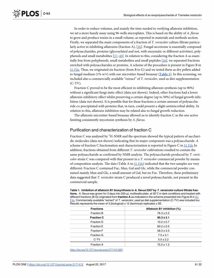
Table 1. Inhibition of aflatoxin B1 biosynthesis in A. flavus 3357 by T. versicolor culture filtrate frac-
tions. A. flavus was grown for 3 days into 200-μL multiwells plate, at 30˚C in dark conditions and treated with
different fractions (B-G) originated from fraction A as indicated in the scheme presented into Figure B in S1
File. Commercially available “extract” of T. versicolor, used as diet supplementation (C-TV) was included too.
Results represents the mean of 3 (biological) x 12 (technical) replicates ± SE.
Fractions Aflatoxin B1 inhibition (%)
Fraction B 76.3 ± 2.2
Fraction C 90.3 ± 3.1
Fraction D 10.2 ± 0.7
Fraction E 60.2 ± 0.5
Fraction F 58.3 ± 3.5
Fraction G 7.5 ± 3.1
C-TV 5.0 ± 2.2
Fraction A 75.2 ± 1.2
https://doi.org/10.1371/journal.pone.0171412.t001
Biological effects of an exopolysaccharide of Trametes versicolor
PLOS ONE | https://doi.org/10.1371/journal.pone.0171412 August 22, 2017 8 / 22
Fraction C was subjected to size exclusion chromatography and three fractions (referred as
CI, CII and CIII see Figure D in S1 File) were obtained. They were tested using aflatoxin inhi-
bition in A. flavus as indicator of biological activity (as described above): fraction CI did not
show any effect while fractions CII and CIII presented a significant bioactivity (Figure F in S1
File). These fractions were analysed by 1H NMR spectroscopy (Figure G in S1 File) which, a
part the 1.3 and 4.2 ppm peaks, due to free lactate often present as contaminant from hands,
showed that fractions CII and CIII were structurally identical. Therefore, further 2D NMR
experiments were carried out only on fraction CIII, taking advantage of its lower molecular
mass. In fact, high-pressure size exclusion chromatography using pullulan standards, (Figure
A in S1 File) showed that fraction CII had a molecular mass of about 23000 Da and fraction
CIII of about 6000 Da. Compositional analysis of fraction CIII showed Fuc:Man:Gal:Glc in the
molar ratios 1.00:2.25:2.22:0.26. Methylation analysis for glycosidic linkages determination
showed a rather complex pattern reported in Table 2. The presence of 2,6-Gal and 2,6-Hex indi-
cated the occurrence of two branch sites as also confirmed by the presence of terminal non-reduc-
ing sugars, t-Fuc and t-Man. The sum of all terminal monosaccharides (t-Fuc + t-Man) is 1.01, in
very good agreement with the sum of all branched monosaccharides (2,6-Gal + 2,6-Hex), equal to
0.94. The most important finding is the presence of fucose, an uncommon component of fungal
polysaccharides found only in Mucorales and in some basidiomycetes [41].
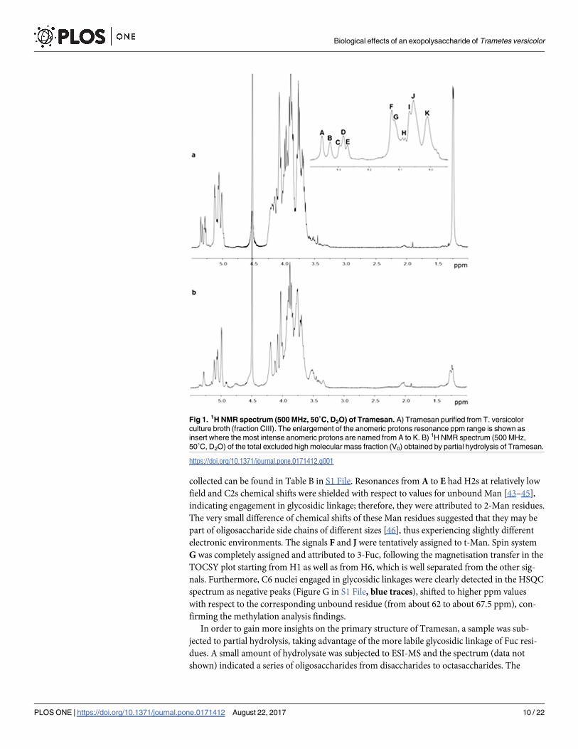
Since the 1H NMR spectrum indicated the presence of O-acetyl groups (Figure F in S1 File)
which increase the complexity of the spectrum [42], fraction CIII was de-O-acetylated for fur-
ther NMR analysis. The 1H NMR spectrum (Fig 1A) is typical of a polysaccharide: the anome-
ric proton region, between 5.5–4.8 ppm, (inset of Fig 1A) exhibited a complex pattern,
reflecting the glycosidic linkage data and suggesting the absence of regular repeating units.
The resonance at 1.25 ppm was attributed to the methyl groups of 6-deoxy residues, confirm-
ing the presence of Fuc.
After inspection of the 1H NMR spectrum and of the COSY plot, the most intense anomeric
protons were named from A to K (inset of Fig 1A) in order of decreasing chemical shift, and
TOCSY, NOESY and HSQC experiments were recorded. Heavy overlapping of most peaks
prevented complete chemical shifts assignments; nevertheless, some useful information could
be gained.
Resonances belonging to H1 and H2 of each spin system were easily identified from the
COSY plot. Taking advantage of the small JH1-H2 coupling constant of Man, which results in
the absence of magnetisation transfer from H2 to H3 in the TOCSY plot, this monosaccharide
was easily identified with signals A-E, F, and J. Resonances of carbon nuclei linked to the
respective protons were then assigned from the HSQC plot (Figure G in S1 File) and the data
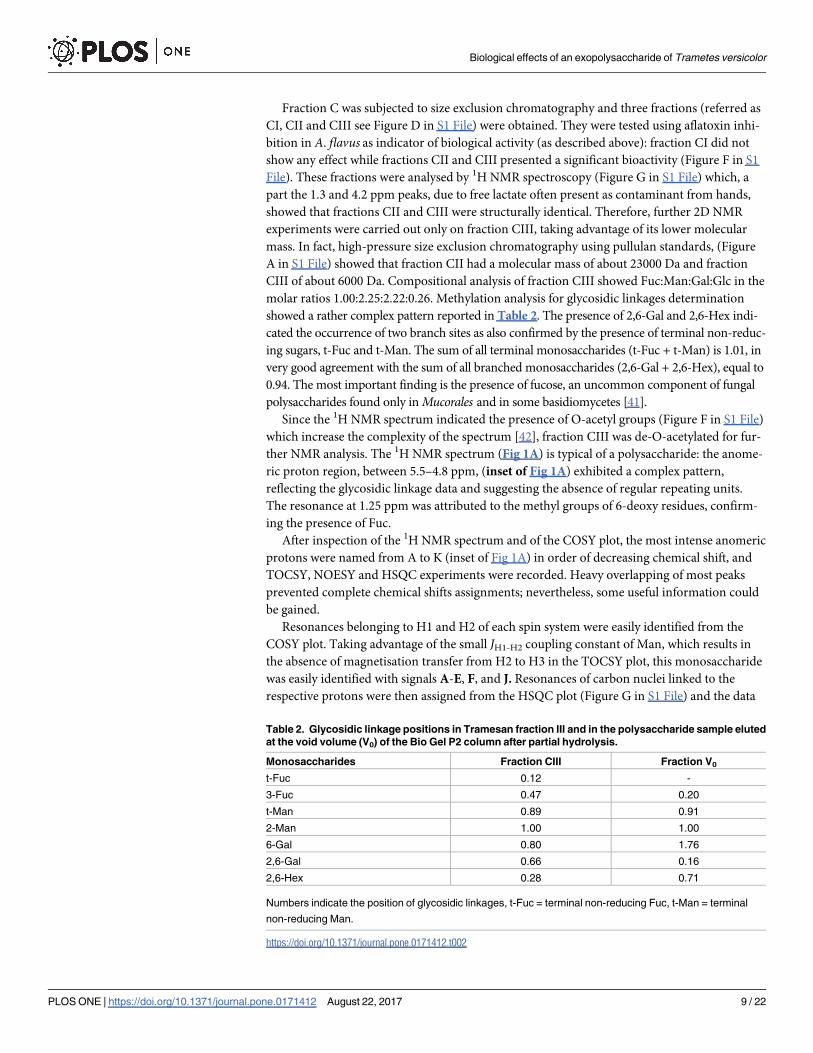
Table 2. Glycosidic linkage positions in Tramesan fraction III and in the polysaccharide sample eluted
at the void volume (V0) of the Bio Gel P2 column after partial hydrolysis.
Monosaccharides Fraction CIII Fraction V0
t-Fuc 0.12 -
3-Fuc 0.47 0.20
t-Man 0.89 0.91
2-Man 1.00 1.00
6-Gal 0.80 1.76
2,6-Gal 0.66 0.16
2,6-Hex 0.28 0.71
Numbers indicate the position of glycosidic linkages, t-Fuc = terminal non-reducing Fuc, t-Man = terminal
non-reducing Man.
https://doi.org/10.1371/journal.pone.0171412.t002
Biological effects of an exopolysaccharide of Trametes versicolor
PLOS ONE | https://doi.org/10.1371/journal.pone.0171412 August 22, 2017 9 / 22
collected can be found in Table B in S1 File. Resonances from A to E had H2s at relatively low
field and C2s chemical shifts were shielded with respect to values for unbound Man [43–45],
indicating engagement in glycosidic linkage; therefore, they were attributed to 2-Man residues.
The very small difference of chemical shifts of these Man residues suggested that they may be
part of oligosaccharide side chains of different sizes [46], thus experiencing slightly different
electronic environments. The signals F and J were tentatively assigned to t-Man. Spin system
G was completely assigned and attributed to 3-Fuc, following the magnetisation transfer in the
TOCSY plot starting from H1 as well as from H6, which is well separated from the other sig-
nals. Furthermore, C6 nuclei engaged in glycosidic linkages were clearly detected in the HSQC
spectrum as negative peaks (Figure G in S1 File, blue traces), shifted to higher ppm values
with respect to the corresponding unbound residue (from about 62 to about 67.5 ppm), con-
firming the methylation analysis findings.
In order to gain more insights on the primary structure of Tramesan, a sample was sub-
jected to partial hydrolysis, taking advantage of the more labile glycosidic linkage of Fuc resi-
dues. A small amount of hydrolysate was subjected to ESI-MS and the spectrum (data not
shown) indicated a series of oligosaccharides from disaccharides to octasaccharides. The
Fig 1. 1H NMR spectrum (500 MHz, 50˚C, D2O) of Tramesan. A) Tramesan purified from T. versicolor
culture broth (fraction CIII). The enlargement of the anomeric protons resonance ppm range is shown as
insert where the most intense anomeric protons are named from A to K. B) 1H NMR spectrum (500 MHz,
50˚C, D2O) of the total excluded high molecular mass fraction (V0) obtained by partial hydrolysis of Tramesan.
https://doi.org/10.1371/journal.pone.0171412.g001
Biological effects of an exopolysaccharide of Trametes versicolor
PLOS ONE | https://doi.org/10.1371/journal.pone.0171412 August 22, 2017 10 / 22
sample mixture was then reduced with sodium borohydride and permethylated. ESI-MS (Fig
2A) showed pairs of (M + Na)+ parent ions corresponding to oligosaccharides, differing for
the presence of one Fuc residue in place of one Hex residue. MS2 (Fig 2B and 2C) of the penta-
saccharides gave a good fragmentation pattern establishing the exclusive reducing terminal
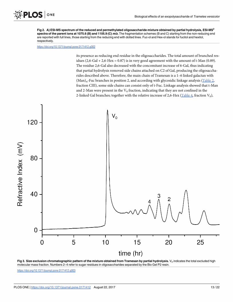
position of the Fuc residues. The hydrolysate was separated by size exclusion chromatography
on a Bio Gel P2 column (Fig 3); fractions belonging to same peak were pooled together and
subjected to NMR spectroscopy.
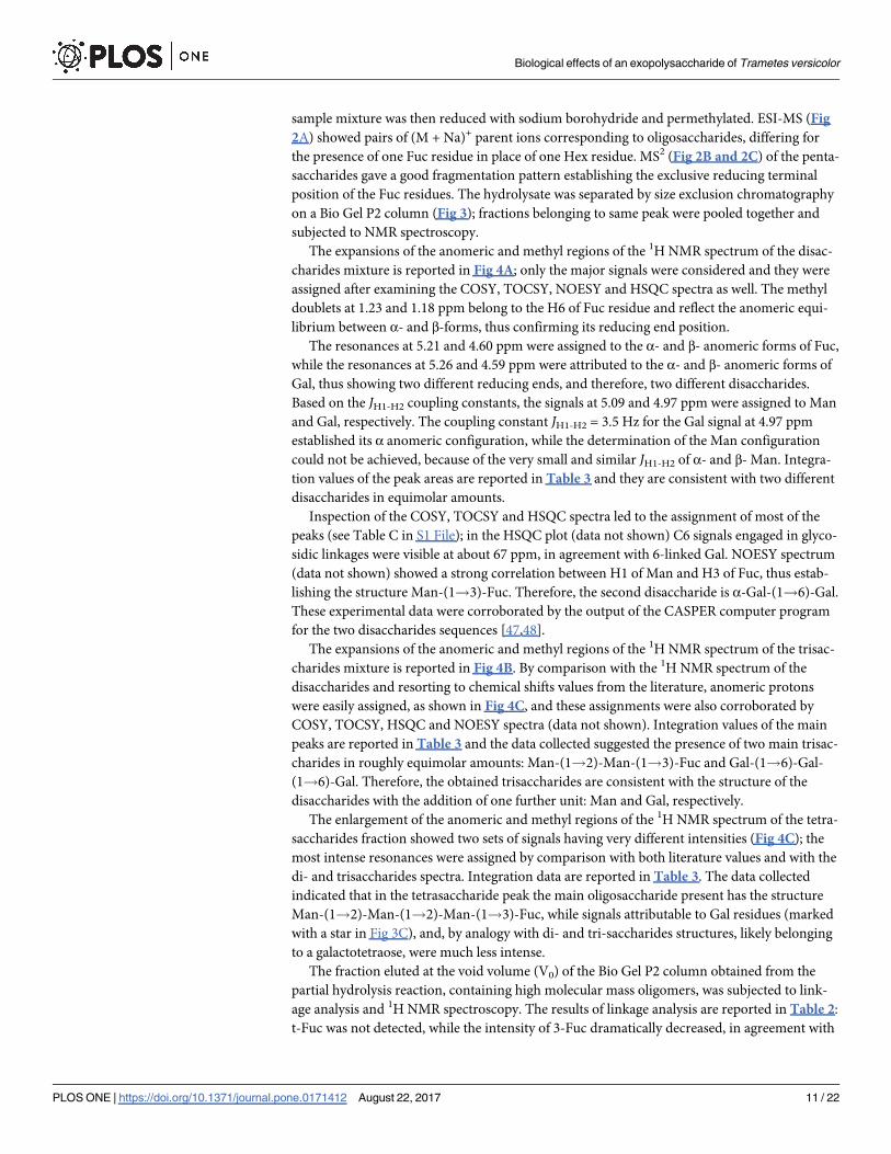
The expansions of the anomeric and methyl regions of the 1H NMR spectrum of the disac-
charides mixture is reported in Fig 4A; only the major signals were considered and they were
assigned after examining the COSY, TOCSY, NOESY and HSQC spectra as well. The methyl
doublets at 1.23 and 1.18 ppm belong to the H6 of Fuc residue and reflect the anomeric equi-
librium between α- and β-forms, thus confirming its reducing end position.
The resonances at 5.21 and 4.60 ppm were assigned to the α- and β- anomeric forms of Fuc,
while the resonances at 5.26 and 4.59 ppm were attributed to the α- and β- anomeric forms of
Gal, thus showing two different reducing ends, and therefore, two different disaccharides.
Based on the JH1-H2 coupling constants, the signals at 5.09 and 4.97 ppm were assigned to Man
and Gal, respectively. The coupling constant JH1-H2 = 3.5 Hz for the Gal signal at 4.97 ppm
established its α anomeric configuration, while the determination of the Man configuration
could not be achieved, because of the very small and similar JH1-H2 of α- and β- Man. Integra-
tion values of the peak areas are reported in Table 3 and they are consistent with two different
disaccharides in equimolar amounts.
Inspection of the COSY, TOCSY and HSQC spectra led to the assignment of most of the
peaks (see Table C in S1 File); in the HSQC plot (data not shown) C6 signals engaged in glyco-
sidic linkages were visible at about 67 ppm, in agreement with 6-linked Gal. NOESY spectrum
(data not shown) showed a strong correlation between H1 of Man and H3 of Fuc, thus estab-
lishing the structure Man-(1!3)-Fuc. Therefore, the second disaccharide is α-Gal-(1!6)-Gal.
These experimental data were corroborated by the output of the CASPER computer program
for the two disaccharides sequences [47,48].
The expansions of the anomeric and methyl regions of the 1H NMR spectrum of the trisac-
charides mixture is reported in Fig 4B. By comparison with the 1H NMR spectrum of the
disaccharides and resorting to chemical shifts values from the literature, anomeric protons
were easily assigned, as shown in Fig 4C, and these assignments were also corroborated by
COSY, TOCSY, HSQC and NOESY spectra (data not shown). Integration values of the main
peaks are reported in Table 3 and the data collected suggested the presence of two main trisac-
charides in roughly equimolar amounts: Man-(1!2)-Man-(1!3)-Fuc and Gal-(1!6)-Gal-
(1!6)-Gal. Therefore, the obtained trisaccharides are consistent with the structure of the
disaccharides with the addition of one further unit: Man and Gal, respectively.
The enlargement of the anomeric and methyl regions of the 1H NMR spectrum of the tetra-
saccharides fraction showed two sets of signals having very different intensities (Fig 4C); the
most intense resonances were assigned by comparison with both literature values and with the
di- and trisaccharides spectra. Integration data are reported in Table 3. The data collected
indicated that in the tetrasaccharide peak the main oligosaccharide present has the structure
Man-(1!2)-Man-(1!2)-Man-(1!3)-Fuc, while signals attributable to Gal residues (marked
with a star in Fig 3C), and, by analogy with di- and tri-saccharides structures, likely belonging
to a galactotetraose, were much less intense.
The fraction eluted at the void volume (V0) of the Bio Gel P2 column obtained from the
partial hydrolysis reaction, containing high molecular mass oligomers, was subjected to link-
age analysis and 1H NMR spectroscopy. The results of linkage analysis are reported in Table 2:
t-Fuc was not detected, while the intensity of 3-Fuc dramatically decreased, in agreement with
Biological effects of an exopolysaccharide of Trametes versicolor
PLOS ONE | https://doi.org/10.1371/journal.pone.0171412 August 22, 2017 11 / 22

Biological effects of an exopolysaccharide of Trametes versicolor
PLOS ONE | https://doi.org/10.1371/journal.pone.0171412 August 22, 2017 12 / 22
its presence as reducing end residue in the oligosaccharides. The total amount of branched res-
idues (2,6-Gal + 2,6-Hex = 0.87) is in very good agreement with the amount of t-Man (0.89).
The residue 2,6-Gal also decreased with the concomitant increase of 6-Gal, thus indicating
that partial hydrolysis removed side chains attached on C2 of Gal, producing the oligosaccha-
rides described above. Therefore, the main chain of Tramesan is a 1–6 linked galactan with
(Man)n-Fuc branches in position 2, and according with glycosidic linkage analysis (Table 2,
fraction CIII), some side chains can consist only of t-Fuc. Linkage analysis showed that t-Man
and 2-Man were present in the V0 fraction, indicating that they are not confined in the
2-linked Gal branches; together with the relative increase of 2,6-Hex (Table 4, fraction V0),
Fig 2. A) ESI-MS spectrum of the reduced and permethylated oligosaccharide mixture obtained by partial hydrolysis, ESI-MS2
spectra of the parent ions at 1075.6 (B) and 1105.9 (C) m/z. The fragmentation schemes (B and C) starting from the non-reducing end
are reported with full lines, those starting from the reducing end with dotted lines. Fuc-ol and Hex-ol stands for fucitol and hexitol,
respectively.
https://doi.org/10.1371/journal.pone.0171412.g002
Fig 3. Size exclusion chromatographic pattern of the mixture obtained from Tramesan by partial hydrolysis. V0 indicates the total excluded high
molecular mass fraction. Numbers 2–4 refer to sugar residues in oligosaccharides separated by the Bio Gel P2 resin.
https://doi.org/10.1371/journal.pone.0171412.g003
Biological effects of an exopolysaccharide of Trametes versicolor
PLOS ONE | https://doi.org/10.1371/journal.pone.0171412 August 22, 2017 13 / 22

Biological effects of an exopolysaccharide of Trametes versicolor
PLOS ONE | https://doi.org/10.1371/journal.pone.0171412 August 22, 2017 14 / 22
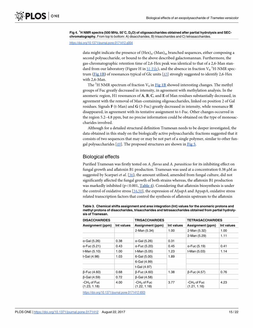
data might indicate the presence of (Hex)n-(Man)m branched sequences, either composing a
second polysaccharide, or bound to the above described galactomannan. Furthermore, the
gas-chromatographic retention time of 2,6-Hex peak was identical to that of a 2,6-Man stan-
dard from our laboratory (Figure H in S1 File), and the absence in fraction V01H-NMR spec-
trum (Fig 1B) of resonances typical of Glc units [43] strongly suggested to identify 2,6-Hex
with 2,6-Man.
The 1H NMR spectrum of fraction V0 in Fig 1B showed interesting changes. The methyl
groups of Fuc greatly decreased in intensity, in agreement with methylation analysis. In the
anomeric region, H1 resonances of A, B, C, and E of Man residues substantially decreased, in
agreement with the removal of Man-containing oligosaccharides, linked on position 2 of Gal
residues. Signals F (t-Man) and G (3-Fuc) greatly decreased in intensity, while resonance H
disappeared, in agreement with its tentative assignment to t-Fuc. Other changes occurred in
the region 5.2–4.8 ppm, but no precise information could be obtained on the type of monosac-
charides involved.
Although for a detailed structural definition Tramesan needs to be deeper investigated, the
data obtained in this study on the biologically active polysaccharidic fractions suggested that it
consists of two sequences that may or may be not part of a single polymer, similar to other fun-
gal polysaccharides [49]. The proposed structures are shown in Fig 5.
Biological effects
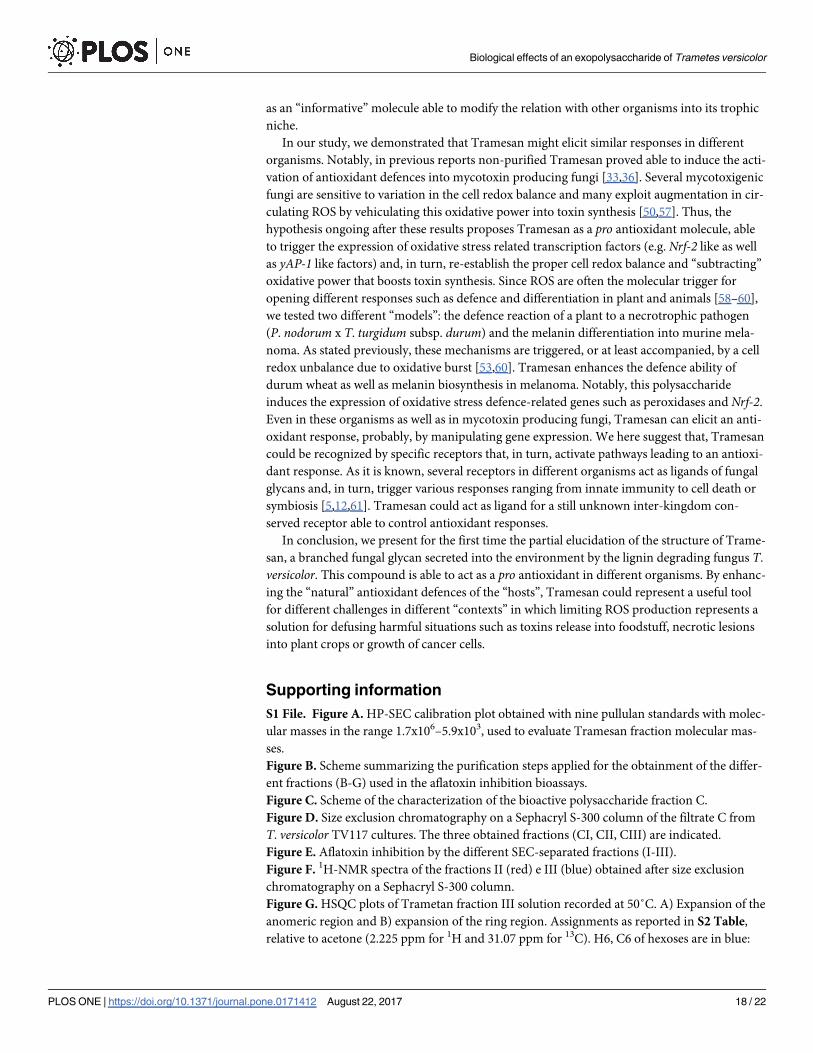
Purified Tramesan was firstly tested on A. flavus and A. parasiticus for its inhibiting effect on
fungal growth and aflatoxin B1 production. Tramesan was used at a concentration 0.38 μM as
suggested by Scarpari et al. [36]; the amount utilised, amended from fungal culture, did not
significantly affected the fungal growth of both strains whereas, the aflatoxin B1 production
was markedly inhibited (p<0.001, Table 4). Considering that aflatoxin biosynthesis is under
the control of oxidative stress [34,50], the expression of AfyapA and ApyapA, oxidative stress
related transcription factors that control the synthesis of aflatoxin upstream to the aflatoxin
Fig 4. 1H NMR spectra (500 MHz, 50˚C, D2O) of oligosaccharides obtained after partial hydrolysis and SEC-
chromatography. From top to bottom: A) disaccharides, B) trisaccharides and C) tetrasaccharides.
https://doi.org/10.1371/journal.pone.0171412.g004
Table 3. Chemical shifts assignment and area integration (Int) values for the anomeric protons and
methyl protons of disaccharides, trisaccharides and tetrasaccharides obtained from partial hydroly-
sis of Tramesan.
DISACCHARIDES TRISACCHARIDES TETRASACCHARIDES
Assignment (ppm) Int values Assignment (ppm) Int values Assignment (ppm) Int values
2-Man (5.34) 1.00 2-Man (5.32) 1.00
2-Man (5.29) 1.11
α-Gal (5.26) 0.38 α-Gal (5.26) 0.31
α-Fuc (5.21) 0.43 α-Fuc (5.20) 0.45 α-Fuc (5.19) 0.41
t-Man (5.10) 1.00 t-Man (5.05) 1.23 t-Man (5.03) 1.14
t-Gal (4.98) 1.03 6-Gal (5.00) 1.89
6-Gal (4.99)
t-Gal (4.97)
β-Fuc (4.60) 0.68 β-Fuc (4.60) 1.38 β-Fuc (4.57) 0.76
β-Gal (4.59) 0.72 β-Gal (4.58)
-CH3 of Fuc
(1.23, 1.18)
4.00 -CH3 of Fuc
(1.22, 1.18)
3.77 -CH3 of Fuc
(1.21, 1.16)
4.23
https://doi.org/10.1371/journal.pone.0171412.t003
Biological effects of an exopolysaccharide of Trametes versicolor
PLOS ONE | https://doi.org/10.1371/journal.pone.0171412 August 22, 2017 15 / 22

regulator AflR [20,34] and of the superoxide dismutase encoding sod1 in A. flavus and A. para-siticus, respectively, was monitored. It was observed that Tramesan significantly triggered the
expression of AfyapA and of sod1 (Table 4).
To check if Tramesan could elicit antioxidant response in other biological systems, thus act-
ing as a pro antioxidant molecule, we tested its effect in hampering the necrotrophic progres-
sion of a foliar pathogen of durum wheat, Parastagonospora nodorum. This pathogen produces
a necrotrophic effector that also act by increasing ROS production at the interface for facilitat-
ing tissue degeneration and cell death [51]. Thus, we pre-treated durum wheat leaves in green-house with a suspension of Tramesan 48 h before inoculating the pathogen. Concomitantly, we
tested the antifungal activity of Tramesan on pure P. nodorum culture. Wheat plants treated
with Tramesan resulted more protected from P. nodorum infection compared to untreated
ones; interestingly, Tramesan had no appreciable effect on fungal growth under in vitro condi-
tions (data not shown), whereas in planta limited its growth. We here suggest that Tramesan
enhanced some ROS-scavenging ability (e.g. PR9) of wheat plant leaves (Table 5) disabling
necrotrophic weapons for triggering PCD and causing disease.
To validate the widespread ability of Tramesan to behave as a pro antioxidant molecule, we
also tested its effect on a murine cell line of melanoma (B16). In this case, the expected effect is
a limitation in cell growth since cancer cells express high level of intrinsic oxidative stress that
normally boosts their division [52]. Notably, we aimed at checking if Tramesan was able to
enhance the ROS scavenging ability of these cutaneous murine cells. In relation to this, we
quantified the amount of an antioxidant molecule (melanin) and tested the expression of Nrf-2
Table 4. Biological activity of Tramesan on A. flavus and A. parasiticus. In vitro culture of A. flavus and A. parasitucus under aflatoxin permissive condi-
tions (PDB, 30˚C) were treated or not (control) with 0.38 μM Tramesan and incubated for 7 days. Aflatoxin B1 production, evaluated by LC-MS/MS, and fungal
growth, evaluated by weighting dried mycelia, in Tramesan-treated cultures, were normalised for non-treated ones and the percentage of inhibition calculated
consequently. Mycelia was used to evaluate the expression, calculated by 2-ΔΔCt method in RT-PCR, of the oxidative stress related transcription factors
AfyapA and ApyapA and the superoxide dismutase encoding gene sod1 in A. flavus and A. parasiticus, respectively.
Fungal
species
treatment Aflatoxin B1
(ppb)
% of Aflatoxin B1 inhibition compared to
untreated control
Fungal growth (mg/mL
d.w.)
Ap-1 like sod1
A. flavus control 125.2 ± 2.5 95.2 5.1 ± 0.6 2.1 ± 0.3 25.2 ± 3.2
Tramesan 0.38
µM
6.02 ± 0.2 5.2 ± 0.5
A. parasiticus control 185.5 ± 7.2 98.7 4.5 ± 0.2 2.5 ± 0.2 22.3 ± 4.1
Tramesan 0.38
µM
2.4 ± 0.5 4.4 ± 0.8
https://doi.org/10.1371/journal.pone.0171412.t004
Fig 5. Proposed structure for Tramesan. Scheme 1 and 2 can be part of the same polysaccharide or forming repeating
units of different polymers.
https://doi.org/10.1371/journal.pone.0171412.g005
Biological effects of an exopolysaccharide of Trametes versicolor
PLOS ONE | https://doi.org/10.1371/journal.pone.0171412 August 22, 2017 16 / 22

mRNA, since Nrf-2 protects melanocytes against the harmful ROS effects [53]. Cells treatment
with 0.38 μM Tramesan increased melanin content of about two-folds whilst enhancing Nrf-2expression and consistently reducing cell growth (Table 6).
Discussion
Fungal glycans may act as eliciting molecules in host cells. In addition, some fungi produce
and secrete glycans to be recognised [54]. Recently, some oligomers of chitin were found to be
actively released in the interface between fungal symbionts and their host to “facilitate” recog-
nition processes and repress host defence [21,55]. Apart lignin degrading enzymology, few
reports actually concern the biology of T. versicolor. In a previous study, the semi-purified cul-
ture filtrate of T. versicolor containing molecules with a supposed molecular mass (MM) >3.0
kDa showed a particular ability in enhancing antioxidant activity in A. flavus, dramatically
inhibiting the biosynthesis of aflatoxins [36]. In relation to this, we exploited the ability of the
semi-purified culture filtrate of T. versicolor to inhibit aflatoxin biosynthesis as a fast and reli-
able method to detect active principles present into the MM>3.0 kDa fraction (fraction A in
this study). We found that this mushroom produces an exo-polysaccharide that is not simply
“leaked” by the complex fungal cell wall, but is secreted into the environment with a still
unknown biosynthetic and transportation pathways and, overall, with an unknown biological
function. In the present study, we provide some hints for elucidating the latter. Tramesan is a
branched fucose-enriched fungal polysaccharide of about 23 kDa with a probable “repetitive”
scheme of monosaccharide sequence in the linear (α-1,6-Gal)n backbone as well as in the lat-
eral chain Man-(1!2)-Man-(1!3)-Fuc. It could be suggested that host cells may recognize
fungi from their saccharidic “barcode” composed by a scheme of repetitive units in which the
single unit acts–de facto–as signalling molecule. This was recently confirmed for signal factors
in fungal mycorrhizae that are composing lipochitooligosaccharides [56] and chitooligosac-
charides [54]. These fungal complex glucans may elicit calcium waves in different systems and
Tramesan, when administered to murine melanoma cell culture, displays this effect too (data
from our laboratories). Thus, what we suggest here is that Tramesan may act for T. versicolor
Table 5. Effect of Tramesan on wheat leaves infected with P. nodorum. Durum wheat leaves of an Italian commercial variety were sprayed with a solu-
tion of 0.38μM Tramesan and after 48 h, inoculated with 105 conidia of P. nodorum; other plants were inoculated with the pathogen but not pre-treated with
Tramesan (Infected control). Disease severity was quantified by using Liu’ scale (necrotic spot extension) whereas relative expression of the peroxidase-
encoding, PR-9 gene, was calculated using the 2-ΔΔCt method. Fungal growth was assessed by qPCR using P. nodorum specie-specific primers.
Durum wheat leaves Necrotic spot Fungal growth PR-9 (peroxidase)
rated by Liu’ scale1 qPCR (ng fungal DNA/μg total DNA) mRNA relative expression (2-ΔΔCt)
Infected control 4 80.1 ± 2.2 1
Tramesan 0 5.0 ± 0.5 64.2 ± 2.1
1Liu et al., 2004 [30]
https://doi.org/10.1371/journal.pone.0171412.t005
Table 6. Effect of Tramesan on murine cell lines of melanoma (B16). Murine cell lines of melanoma (B16) were treated with 0.38 μM Tramesan or
untreated (control). The amount of melanin was calculated by spectrophotometer (Abs405), normalised for untreated samples and the percentage of its
increase was evaluated and compared to untreated samples. Cell growth was calculated by counting the number of viable cells. The results are the
mean ± SD of 5 separate experiments Relative expression of the Nrf-2 gene normalised on untreated cells was calculated using the 2-ΔΔCt method.
Melanin Cell growth Nrf-2
Percentage of increase compared to untreated samples (%) Percentage of reduction of cell number (%) mRNA relative expression
(2-ΔΔCt)
Tramesan 97.5 ± 4.3 83.7 ± 2.2 9.5 ± 0.2
https://doi.org/10.1371/journal.pone.0171412.t006
Biological effects of an exopolysaccharide of Trametes versicolor
PLOS ONE | https://doi.org/10.1371/journal.pone.0171412 August 22, 2017 17 / 22
as an “informative” molecule able to modify the relation with other organisms into its trophic
niche.
In our study, we demonstrated that Tramesan might elicit similar responses in different
organisms. Notably, in previous reports non-purified Tramesan proved able to induce the acti-
vation of antioxidant defences into mycotoxin producing fungi [33,36]. Several mycotoxigenic
fungi are sensitive to variation in the cell redox balance and many exploit augmentation in cir-
culating ROS by vehiculating this oxidative power into toxin synthesis [50,57]. Thus, the
hypothesis ongoing after these results proposes Tramesan as a pro antioxidant molecule, able
to trigger the expression of oxidative stress related transcription factors (e.g. Nrf-2 like as well
as yAP-1 like factors) and, in turn, re-establish the proper cell redox balance and “subtracting”
oxidative power that boosts toxin synthesis. Since ROS are often the molecular trigger for
opening different responses such as defence and differentiation in plant and animals [58–60],
we tested two different “models”: the defence reaction of a plant to a necrotrophic pathogen
(P. nodorum x T. turgidum subsp. durum) and the melanin differentiation into murine mela-
noma. As stated previously, these mechanisms are triggered, or at least accompanied, by a cell
redox unbalance due to oxidative burst [53,60]. Tramesan enhances the defence ability of
durum wheat as well as melanin biosynthesis in melanoma. Notably, this polysaccharide
induces the expression of oxidative stress defence-related genes such as peroxidases and Nrf-2.
Even in these organisms as well as in mycotoxin producing fungi, Tramesan can elicit an anti-
oxidant response, probably, by manipulating gene expression. We here suggest that, Tramesan
could be recognized by specific receptors that, in turn, activate pathways leading to an antioxi-
dant response. As it is known, several receptors in different organisms act as ligands of fungal
glycans and, in turn, trigger various responses ranging from innate immunity to cell death or
symbiosis [5,12,61]. Tramesan could act as ligand for a still unknown inter-kingdom con-
served receptor able to control antioxidant responses.
In conclusion, we present for the first time the partial elucidation of the structure of Trame-
san, a branched fungal glycan secreted into the environment by the lignin degrading fungus T.
versicolor. This compound is able to act as a pro antioxidant in different organisms. By enhanc-
ing the “natural” antioxidant defences of the “hosts”, Tramesan could represent a useful tool
for different challenges in different “contexts” in which limiting ROS production represents a
solution for defusing harmful situations such as toxins release into foodstuff, necrotic lesions
into plant crops or growth of cancer cells.
Supporting information
S1 File. Figure A. HP-SEC calibration plot obtained with nine pullulan standards with molec-
ular masses in the range 1.7x10⁶–5.9x10³, used to evaluate Tramesan fraction molecular mas-
ses.
Figure B. Scheme summarizing the purification steps applied for the obtainment of the differ-
ent fractions (B-G) used in the aflatoxin inhibition bioassays.
Figure C. Scheme of the characterization of the bioactive polysaccharide fraction C.
Figure D. Size exclusion chromatography on a Sephacryl S-300 column of the filtrate C from
T. versicolor TV117 cultures. The three obtained fractions (CI, CII, CIII) are indicated.
Figure E. Aflatoxin inhibition by the different SEC-separated fractions (I-III).
Figure F. 1H-NMR spectra of the fractions II (red) e III (blue) obtained after size exclusion
chromatography on a Sephacryl S-300 column.
Figure G. HSQC plots of Trametan fraction III solution recorded at 50˚C. A) Expansion of the
anomeric region and B) expansion of the ring region. Assignments as reported in S2 Table,
relative to acetone (2.225 ppm for 1H and 31.07 ppm for 13C). H6, C6 of hexoses are in blue:
Biological effects of an exopolysaccharide of Trametes versicolor
PLOS ONE | https://doi.org/10.1371/journal.pone.0171412 August 22, 2017 18 / 22
those belonging to 6-linked hexoses resonate at about 67 ppm, those that are not linked at
about 62 ppm.
Figure H. Comparison of GLC elution profiles between PMAA derivatives of Tramesan and a
sample containing 2,6-Man. The peaks of interest are marked with asterisks.
Table A. Composition analysis of the fraction C containing the polysaccharide produced by T.
versicolor Tv117 (PLS 117) and of a Trametes versicolor commercial powder sample (CP).
Results are expressed in weight %.
Table B. 1H and 13C chemical shift assignments of Tramesan, referred to acetone (2.225 ppm
for 1H and 31.07 ppm for 13C).
Table C. 1H and 13C chemical shift assignments of the disaccharides obtained from partial
hydrolysis of Tramesan. Signals are referred to acetone (2.225 ppm for 1H and 31.07 ppm for13C).
(DOCX)
Acknowledgments
Tramesan was patented (N˚ 0001418693) in Italy with the commercial name of Trametano.
We are grateful to Dr. Anna Adele Fabbri for her invaluable and constant advices. We are
grateful to Ms V. Farina and Dr. A. Iori for aiding in the execution of experiments on plants.
Author Contributions
Conceptualization: Massimo Reverberi, Valeria Scala, Corrado Fanelli, Slaven Zjalic, Agos-
tino Tafuri, Maria R. Ricciardi, Paola Cescutti, Roberto Rizzo.
Data curation: Marzia Scarpari, Alessia Parroni, Chiara Pietricola, Aris Sveronis, Paola Ces-
cutti, Roberto Rizzo.
Formal analysis: Agostino Tafuri, Aris Sveronis, Paola Cescutti, Roberto Rizzo.
Funding acquisition: Corrado Fanelli, Agostino Tafuri.
Investigation: Massimo Reverberi, Valeria Scala, Agostino Tafuri, Paola Cescutti.
Methodology: Marzia Scarpari, Alessia Parroni, Chiara Pietricola, Vittoria Maresca, Maria R.
Ricciardi, Roberto Licchetta, Simone Mirabilii, Aris Sveronis, Paola Cescutti.
Project administration: Corrado Fanelli, Agostino Tafuri.
Resources: Agostino Tafuri.
Software: Paola Cescutti.
Supervision: Massimo Reverberi, Slaven Zjalic, Paola Cescutti.
Validation: Marzia Scarpari, Alessia Parroni, Agostino Tafuri, Paola Cescutti.
Visualization: Paola Cescutti, Roberto Rizzo.
Writing – original draft: Massimo Reverberi, Valeria Scala, Corrado Fanelli, Maria R. Ric-
ciardi, Paola Cescutti, Roberto Rizzo.
Writing – review & editing: Massimo Reverberi, Paola Cescutti, Roberto Rizzo.
References1. Meng X, Liang H, Luo L. Antitumor polysaccharides from mushrooms: a review on the structural charac-
teristics, antitumor mechanisms and immunomodulating activities. Carbohydr Res. 2016; 424: 30–41
https://doi.org/10.1016/j.carres.2016.02.008 PMID: 26974354
Biological effects of an exopolysaccharide of Trametes versicolor
PLOS ONE | https://doi.org/10.1371/journal.pone.0171412 August 22, 2017 19 / 22

2. Dam TK, Brewer CF. Effects of clustered epitopes in multivalent ligand–receptor interactions. Biochem.
2008; 47: 8470–8476.
3. Nurnberger T, Brunner F, Kemmerling B, Piater, L. Innate immunity in plants and animals: striking simi-
larities and obvious differences. Immun Rev. 2004; 198: 249–266.
4. Savary S, Ficke A, Aubertot J.-N, Hollier C. Crop losses due to diseases and their implications for global
food production losses and food security. Food Security (Online) 2012; 19
5. Cottier F, Pavelka N. Complexity and dynamics of host-fungal interactions. Immunol Res. 2012; 53(1–
3):127–35 https://doi.org/10.1007/s12026-012-8265-y PMID: 22415250
6. Chan G.CF, Chan WK, Sze DMY. The effects of β-glucan on human immune and cancer cells. J Hema-
tol Oncol. 2009; 2: 25–35. https://doi.org/10.1186/1756-8722-2-25 PMID: 19515245
7. Nobuyuki S, Akifumi S, Hidemitsu K, Yoshio O. Chemical structure of the cell-wall mannan of Candida
albicans serotype A and its difference in yeast and hyphal forms. Biochem J. 2007; 404: 365–372.
https://doi.org/10.1042/BJ20070081 PMID: 17331070
8. Ruiz-Herrera J. Fungal Cell Wall: Structure, Synthesis, and Assembly, Second Edition— CRC Press
Book. 1991
9. Bowman SM, Free SJ. The structure and synthesis of the fungal cell wall. BioEssays 2006; 28 (8): 799–
808. https://doi.org/10.1002/bies.20441 PMID: 16927300
10. Fesel PH, Zuccaro A. β-glucan: Crucial component of the fungal cell wall and elusive MAMP in plants.
Fungal Genet Biol. 2016; 90: 53–60. https://doi.org/10.1016/j.fgb.2015.12.004 PMID: 26688467
11. Newman MA, Sundelin T, Nielsen JT and Erbs G. MAMP (microbe-associated molecular pattern) trig-
gered immunity in plants. Front Plant Sci. 2013; 4:139 https://doi.org/10.3389/fpls.2013.00139 PMID:
23720666
12. Glass NL, Dementhon K, Non-self recognition and programmed cell death in filamentous fungi. Curr
Opin Microbiol. 2006; 9(6): 553–558. https://doi.org/10.1016/j.mib.2006.09.001 PMID: 17035076
13. Hall C, Welch J, Kowbel DJ, Glass NL. Evolution and diversity of a fungal self/nonself recognition locus.
PLoS ONE 2010; 5(11) e14055. https://doi.org/10.1371/journal.pone.0014055 PMID: 21124910
14. Reid DM, Gow NA, Brown GD. Pattern recognition: recent insights from Dectin-1. Curr Opin Immunol.
2009; 21(1): 30–37. https://doi.org/10.1016/j.coi.2009.01.003 PMID: 19223162
15. Vijayan V, Baumgart-Vogt E, Naidu S, Qian G, Immenschuh S. Bruton’s tyrosine kinase is required for
TLR-dependent heme oxygenase-1 gene activation via Nrf2 in macrophages. J Immunol. 2011; 187
(2):817–27. https://doi.org/10.4049/jimmunol.1003631 PMID: 21677132
16. Lai Z, Vinod K, Zheng Z, Fan B, Chen Z. Roles of Arabidopsis WRKY3 and WRKY4 transcription factors
in plant responses to pathogens. BMC Plant Biol. 2008; 8: 68. https://doi.org/10.1186/1471-2229-8-68
PMID: 18570649
17. Shimizu T, Nakano T, Takamizawa D, Desaki Y, Ishii-Minami N, Nishizawa Y, et al. Two LysM receptor
molecules, CEBiP and OsCERK1, cooperatively regulate chitin elicitor signaling in rice. Plant J. 2010;
64: 204–214. https://doi.org/10.1111/j.1365-313X.2010.04324.x PMID: 21070404
18. Liu B, Hong YB, Zhang YF, Li XH, Huang L, Zhang HJ, Li DY, et al. Tomato WRKY transcriptional factor
SlDRW1 is required for disease resistance against Botrytis cinerea and tolerance to oxidative stress,
Plant Sci. 2014, 227: 145–156. https://doi.org/10.1016/j.plantsci.2014.08.001 PMID: 25219316
19. Reverberi M, Fabbri AA, Zjalic S, Ricelli A, Fanelli C. Antioxidant enzymes stimulation in Aspergillus
parasiticus by Lentinula edodes inhibits aflatoxin production. Appl Microbiol Biotechnol. 2005; 69: 207–
215. https://doi.org/10.1007/s00253-005-1979-1 PMID: 15838675
20. Reverberi M, Zjalic S, Ricelli A, Punelli F, Camera E, Fabbri AA, et al. Modulation of antioxidant defence
in Aspergillus parasiticus is involved in aflatoxin biosynthesis: a role of the ApyapA gene. Eukaryot Cell
2008; 7: 988–1000. https://doi.org/10.1128/EC.00228-07 PMID: 18441122
21. Druzhinina I, Seidl-Seiboth V, Herrera-Estrella A, Horwitz BA, Kenerley CM, Monte E, et al. Tricho-
derma: the genomics of opportunistic success. Nat Rev Microbiol. 2011; 9(10):749–59. https://doi.org/
10.1038/nrmicro2637 PMID: 21921934
22. Albersheim P, Nevins DJ, English PD, Karr A. A method for the analysis of sugars in plant cell wall poly-
saccharides by gas-liquid chromatography. Carbohydr. Res. 1967; 5: 340–345.
23. Harris PJ, Henry RJ, Blakeney AB, Stone BA. An improved procedure for the methylation analysis of oli-
gosaccharides and polysaccharides. Carbohydr. Res. 1984; 127: 59–73. PMID: 6713442
24. Sweet DP, Shapiro RH, Albersheim P. Quantitative analysis by various g.l.c. response-factor theories
for partially methylated and partially ethylated alditol acetates. Carbohydr. Res. 1975; 40:217–25.
25. Dolfi S, Sveronis A, Silipo A, Rizzo R, Cescutti P. A novel rhamno-mannan exopolysaccharide isolated
from biofilms of Burkholderia multivorans C1576. Carbohydr. Res. 2015; 411: 42–48. https://doi.org/10.
1016/j.carres.2015.04.012 PMID: 25974852
Biological effects of an exopolysaccharide of Trametes versicolor
PLOS ONE | https://doi.org/10.1371/journal.pone.0171412 August 22, 2017 20 / 22
26. Dell A. Preparation and desorption mass spectrometry of permethyl and peracetyl derivatives of oligo-
saccharides. Method. Enzymol. 1990; 193: 647–660.
27. Sulyok M, Berthiller F, Krska R, Schuhmacher R. Development and validation of a liquid chromatogra-
phy/tandem mass spectrometric method for the determination of 39 mycotoxins in wheat and maize.
Rapid Commun. Mass Sp. 2006; 20:2649–2659.
28. Scala V, Camera E, Ludovici M. Dall’Asta C, Cirlini M, Giorni P, et al. Fusarium verticillioides and maize
interaction in vitro: relation between oxylipin cross-talk and fumonisin synthesis. World Mycotox J.
2013; 6: 343–3514.
29. Reverberi M, Zjalic S, Ricelli A, Di Meo C. Scarpari M, Fanelli C, et al. Mushrooms versus fungi: natural
compounds from Lentinula edodes inhibit aflatoxin biosynthesis by Aspergillus parasiticus. World Myco-
tox J. 2011; 4(3): 217–224.
30. Liu ZH, Faris JD, Meinhardt SW, Ali S, Rasmussen JB, Friesen TL. Genetic and Physical Mapping of a
Gene Conditioning Sensitivity in Wheat to a Partially Purified Host-Selective Toxin Produced by Stago-
nospora nodorum. Phytopathol. 2004; 94(10): 1056–1060.
31. Iori A, Scala V, Cesare D, Pinzari F, D’Egidio MG, Fanelli C, et al. Hyperspectral and molecular analysis
of Stagonospora nodorum blotch disease in durum wheat Eur J Plant Pathol. 2015; 141(4): 689–702.
32. Nobili C, D’angeli S, Altamura MM, Scala V, Fabbri AA, Reverberi M, et al. ROS and 9-oxylipins are cor-
related with deoxynivalenol accumulation in the germinating caryopses of Triticum aestivum after
Fusarium graminearum infection. Eur J Plant Pathol. 2014; 139(2): 423–438.
33. Zjalic S, Reverberi M, Ricelli A, Granito VM, Fanelli C, Fabbri AA. Trametes versicolor: a possible tool
for aflatoxin control. Int J Food Microbiol. 2006; 107:243–249 https://doi.org/10.1016/j.ijfoodmicro.2005.
10.003 PMID: 16337299
34. Roze LV, Hong S-Y, Linz JE. Aflatoxin biosynthesis: current frontiers. Annu Rev Food Sci Tech. 2013;
4: 293–311.
35. Zaccaria M, Ludovici M, Sanzani SM, Ippolito A, Cigliano RA, Sanseverino W, et al. Menadione-induced
oxidative stress re-shapes the oxylipin profile of Aspergillus flavus and its Lifestyle. Toxins 2015; 7:
4315–4329. https://doi.org/10.3390/toxins7104315 PMID: 26512693
36. Scarpari M, Parroni A, Zaccaria M, Fattorini L, Bello C, Fabbri AA, et al. Trametes versicolor bioactive
compounds stimulate Aspergillus flavus antioxidant system and inhibit aflatoxin synthesis, Plant Bio-
syst. 2016; 150(4): 653–659.
37. Krijger J-J, Thon MR, Deising HB and Wirsel SGR. Compositions of fungal secretomes indicate a
greater impact of phylogenetic history than lifestyle adaptation. BMC Genomics 2014; 15:722. https://
doi.org/10.1186/1471-2164-15-722 PMID: 25159997
38. Gander JE, Beachy J, Unkefer CJ, Tonn SJ. Toward understanding the structure, biosynthesis, and
Function of a membrane-bound fungal glycopeptide. In Fungal Polysaccharides, Paul A. Sanfdfor and
Kazuo Matsuda Eds., ACS Symposium Series Vol. 126. 1980; Cpt4: 49–79.
39. Rizzo J, Oliveira DL, Joffe LS, Hu G, Gazos-Lopes F, Fonseca FL, et al. Role of the Apt1 protein in poly-
saccharide secretion by Cryptococcus neoformans. Eukaryot. Cell. 2014; 13(6):715–26. https://doi.org/
10.1128/EC.00273-13 PMID: 24337112
40. Fernando MDM, Wijesundera RLC, Soysa SSBDP, de Silva ED, Nanayakkara CM. Antioxidant poten-
tial and content of the polyphenolic secondary metabolites of white rot macrofungi; Flavodon flavus
(Klotzsch.) and Xylaria feejeensis (Berk.). Plant Sci. J. 2016; 1 (1): 1–6.
41. Liao SF, Liang CH, Ho MY, Hsu TL, Tsai TI, Hsieh YS, et al. Immunization of fucose-containing polysac-
charides from Reishi mushroom induces antibodies to tumor-associated Globo H-series epitopes. Proc
Natl Acad Sci USA 2013; 110(34):13809–13814. https://doi.org/10.1073/pnas.1312457110 PMID:
23908400
42. Cescutti P, Cuzzi B, Liut G, Segonds C, Di Bonaventura G, Rizzo R. A novel highly charged exopolysac-
charide produced by two strains of Stenotrophomonas maltophilia recovered from patients with cystic
fibrosis. Carbohydr Res. 2011; 346: 1916–1923. https://doi.org/10.1016/j.carres.2011.05.007 PMID:
21636078
43. Bock K, Thøgersen H. Nuclear Magnetic resonance Spectroscopy in the study of mono- and oligosac-
charides. Ann Rep NMR Spectr. 1982; 13: 1–57.
44. Bock K, Pedersen C. Carbon-13 Nuclear Magnetic Resonance Spectroscopy of Monosaccharides. Adv
Carbohydr Chem Biochem. 1983; 41: 27–66.
45. Bock K, Pedersen C, Pedersen H. Carbon-13 nuclear magnetic resonance data for oligosaccharides.
Adv Carbohydr Chem Biochem. 1984; 42: 193–225.
46. Shibata N, Suzuki A, Kobayashi H and Okawa Y. Chemical structure of the cell-wall mannan of Candida
albicans serotype A and its difference in yeast and hyphal forms. Biochem J. 2007; 404: 365–372.
https://doi.org/10.1042/BJ20070081 PMID: 17331070
Biological effects of an exopolysaccharide of Trametes versicolor
PLOS ONE | https://doi.org/10.1371/journal.pone.0171412 August 22, 2017 21 / 22
47. Lundborg M, Widmalm G. Structure Analysis of Glycans by NMR Chemical Shift Prediction. Anal
Chem. 2011; 83:1514–1517. https://doi.org/10.1021/ac1032534 PMID: 21280662
48. Jansson P-E, Stenutz R, Widmalm G. Sequence determination of oligosaccharides and regular poly-
saccharides using NMR spectroscopy and a novel Web-based version of the computer program CAS-
PER. Carbohydr Res. 2006; 341:1003–1010. https://doi.org/10.1016/j.carres.2006.02.034 PMID:
16564037
49. Leal JA, Prieto A, Bernabe M., Hawksworth DL. An assessment of fungal wall heteromannans as a phy-
logenetically informative character in ascomycetes. FEMS Microbiol Rev. 2010; 34:986–1014. https://
doi.org/10.1111/j.1574-6976.2010.00225.x PMID: 20491931
50. Roze LV, Laivenieks M, Hong S-Y, Wee J, Wong SS, Vanos B, et al. Aflatoxin biosynthesis is a novel
source of reactive oxygen species—a potential redox signal to initiate resistance to oxidative stress?
Toxins 2015; 7: 1411–1430. https://doi.org/10.3390/toxins7051411 PMID: 25928133
51. Vincent D, Du Fall LA, Livk A, Mathesius U, Lipscombe RJ, et al. A functional genomics approach to dis-
sect the mode of action of the Stagonospora nodorum effector protein SnToxA in wheat. Mol Plant
Pathol. 2012; 13(5):467–82. https://doi.org/10.1111/j.1364-3703.2011.00763.x PMID: 22111512
52. Liou GY, Storz P. Reactive oxygen species in cancer. Free Radic Res. 2010; 44(5):479–96. https://doi.
org/10.3109/10715761003667554 PMID: 20370557
53. Gęgotek A, Skrzydlewska E. The role of transcription factor Nrf2 in skin cells metabolism. Arch Derma-
tol Res. 2015; 307(5):385–396. https://doi.org/10.1007/s00403-015-1554-2 PMID: 25708189
54. Genre A, Chabaud M, Balzergue C, Puech-Pagès V, Novero M, Rey T, et al. Short-chain chitin oligo-
mers from arbuscular mycorrhizal fungi trigger nuclear Ca2+ spiking in Medicago truncagula roots and
their production is enhanced by strigolactone. New Phytol. 2013; 198: 190–202. https://doi.org/10.1111/
nph.12146 PMID: 23384011
55. Sun J, Miller JB, Granqvist E, Wiley-Kalil A, Gobbato E, Maillet F, et al. Activation of symbiosis signaling
by arbuscular mycorrhizal fungi in legumes and rice. Plant Cell 2015; 27(3): 823–838. https://doi.org/
10.1105/tpc.114.131326 PMID: 25724637
56. Denarie J, Debelle F, Prome JC. Rhizobium lipo-chitooligosaccharide nodulation factors: signaling mol-
ecules mediating recognition and morphogenesis. Ann Rev Biochem. 1996; 65: 503–535. https://doi.
org/10.1146/annurev.bi.65.070196.002443 PMID: 8811188
57. Sanzani SM, De Girolamo A, Schena L, Solfrizzo M, Ippolito A, Visconti A. Control of Penicillium expan-
sum and patulin accumulation on apples by quercetin and umbelliferone. Eur Food Res Technol. 2009;
228: 381. https://doi.org/10.1007/s00217-008-0944-5
58. Lushchak VI. Adaptive response to oxidative stress: Bacteria, fungi, plants and animals. Comp Biochem
Physiol C Toxicol Pharmacol. 2011; 153(2):175–190. https://doi.org/10.1016/j.cbpc.2010.10.004 PMID:
20959147
59. Scandalios JG. The rise of ROS. Trends Biochem Sci. 2002; 27:483–486 PMID: 12217524
60. Torres MA. ROS in biotic interactions. Physiol Plant. 2010; 138:414–429. https://doi.org/10.1111/j.
1399-3054.2009.01326.x PMID: 20002601
61. Limpens E, Franken C, Smit P, Willemse J, Bisseling T, Geurts R. LysM domain receptor kinases regu-
lating rhizobial Nod factor-induced infection. Science 2003; 302: 630–633. https://doi.org/10.1126/
science.1090074 PMID: 12947035
Biological effects of an exopolysaccharide of Trametes versicolor
PLOS ONE | https://doi.org/10.1371/journal.pone.0171412 August 22, 2017 22 / 22