training - genesys spine · training. amniotic membrane ©2011 mimedx group, ... • control...
TRANSCRIPT

Training

Amniotic Membrane

Do Not Duplicate, Disseminate or Distribute©2011 MiMedx Group, All rights reserved.
• History shows human amniotic membrane has been used in a clinical setting since the early 1900s.
• In vivo studies show the barrier properties of amniotic membrane help reduce scar tissue formation and scar attachment to the dura1
• Studies show amniotic membrane enhances the wound healing process.
– It is non-immunogenic
– It reduces inflammation
– It reduces scar tissue
– Contains essential growth factors
Why Amnion?
1Tao H and H. Fan. Implantation of Amniotic Membrane to Reduce Postlaminectomy Epidural Adhesion, Eur Spine J. 2009 Aug; 18(8): 1202-12

Do Not Duplicate, Disseminate or Distribute©2011 MiMedx Group, All rights reserved.
Collagen types: IV, V and VII
Presence of Growth Factors1*:• Epidermal Growth Factor (EGF)• Transforming Growth Factor
Beta (TGF-β)• Fibroblast Growth Factors (FGFs) • Platelet Derived Growth Factors
(PDGF) A&B
Anatomy and Physiology
1Hopkinson A. et al Proteomic Analysis of Amniotic Membrane Prepared for Human Transplantation: Characterization of Proteins and Clinical Implications. Journ Proteome Res 2006, 5, 2226-2235.
*Confirmed by HPLC tests of sterilized AmnioFix
Amnion
Chorion

Do Not Duplicate, Disseminate or Distribute©2011 MiMedx Group, All rights reserved.
H&E Stain of Amnion after Purion® Process
Do Not Duplicate, Disseminate or Distribute©2011 MiMedx Group, All rights reserved.
• Efficacy
• Applications
• Safety
Do Not Duplicate, Disseminate or Distribute©2011 MiMedx Group, All rights reserved.
• Efficacy
• Applications
• Safety

Do Not Duplicate, Disseminate or Distribute©2011 MiMedx Group, All rights reserved.
Efficacy
1. Overview of Healing Process
2. Anti- Inflammatory Properties
3. Reduction of Scar Tissue Formation
4. Enhance Healing Process

Do Not Duplicate, Disseminate or Distribute©2011 MiMedx Group, All rights reserved.
Step 1: Inflammatory Phase (Immediate to 2-5 days)
• Hemostasis
• Inflammation
Healing Process
Step 2: Proliferative Phase (2 days to 3 weeks)
• Granulation- Fibroblasts lay bed of collagen
• Contraction- wound edges pull together to reduce defect, Epithelialization
Reduction in Inflammation
Provides Collagen and growth factors
Step 3: Remodeling Phase ( 3 weeks to 2 years)
• New Collagen forms which increases tensile strength to wound

Do Not Duplicate, Disseminate or Distribute©2011 MiMedx Group, All rights reserved.
“Amniotic membrane reduces inflammation through entrapment of inflammatory cells”1
Amniotic membrane contains anti-inflammatory growth factors that can enhance wound healing.2
“HAM (Human Amniotic Membrane) cells express various anti-angiogenic and anti inflammatory proteins such as interleukin (IL)-1… and IL-10”3
1 Baradaran-Raffi et al Amniotic Membrane Transplantation. Iran J Ophthalmic Res 2007;2 (1):58-75.2John T. et al Human amniotic membrane transplantation: Past, present, and future. Ophthalmology
Clinic of North Am. 16 (2003) 43-653 Toda A. et al The Potential of Amniotic Membrane/Amnion-Derived Cells for Regeneration of Various
Tissues. J. Pharmacol Sci 105 (2007), 215-228
Anti-Inflammatory Properties

Do Not Duplicate, Disseminate or Distribute©2011 MiMedx Group, All rights reserved.
Reduction of Scar Tissue Formation and Enhancing Healing Process
Contains Collagen types, IV, V, and VII
• May inhibit fibrosis when used as a anatomic barrier.1
• Epidermal Growth Factor (EGF), Transforming Growth Factors-β (TGF-β), and Fibroblast Growth Factors (FGFs).
1John T. et al Human amniotic membrane transplantation: Past, present, and future. Ophthalmology Clinic of North Am. 16 (2003) 43-65

Do Not Duplicate, Disseminate or Distribute©2011 MiMedx Group, All rights reserved.
• Efficacy
• Applications
• Safety

Do Not Duplicate, Disseminate or Distribute©2011 MiMedx Group, All rights reserved.
Applications
• Posterior– Laminectomy
– Posterior Decompression
• Anterior– ACDF

Do Not Duplicate, Disseminate or Distribute©2011 MiMedx Group, All rights reserved.
Posterior Laminectomy Open or MAST Approach

Do Not Duplicate, Disseminate or Distribute©2011 MiMedx Group, All rights reserved.
Posterior Spinal Approach

Do Not Duplicate, Disseminate or Distribute©2011 MiMedx Group, All rights reserved.
How to Apply on Posterior Approach
Dry Application:1. Cut the AmnioFix to the appropriate size, larger is better.2. Ensure all excess blood /fluid is removed from the surgical site.3. Lay the AmnioFix so you can read the SB on the affected area, dura or exiting
nerve root.4. The AmnioFix will become tacky to the site. If surgeon needs to reposition
just add a couple drops of sterile saline or water. 5. To make the tissue more malleable, add a couple drops of saline to hydrate
the graft. The AmnioFix may be wrapped around corners and edges.6. Do not suction near the AmnioFix once it is place.7. Complete surgical procedure and close.
AmnioFix may be applied dry or wet, preferably dry

Do Not Duplicate, Disseminate or Distribute©2011 MiMedx Group, All rights reserved.
Reasons for using AmnioFix on a Laminectomy or Posterior decompression1. Reduce scar tissue or “fibrosis” formation near or on dura
– Fibrosis may lead to adhesions and could contribute to post-op pain
2. Reduce inflammation in the surgical site
3. “Enhances Healing”– Provides a scaffold for native connective tissues to penetrate, leaving a dissection
plane of native tissue
1 and 3 should aid if surgeon needed to do a revision

Do Not Duplicate, Disseminate or Distribute©2011 MiMedx Group, All rights reserved.

Do Not Duplicate, Disseminate or Distribute©2011 MiMedx Group, All rights reserved.
Model
• Laminectomy Model- Rat (32)
Groups
• Human Amnion (16)
• Control (16)
Timepoints
8 weeks
Outcome Measurement
Epidural fibrosis
• Histological analysis to demonstrate a separation of epideral fibrosis from dura
• Adhesion tenacity via a nerve pull-out
• Pain
Preclinical Study of Human Allograft Amniotic Membrane as a Barrier to Epidural Fibrosis in the Early Wound of a Postlaminectomy Rat Model
SAS 2011 Poster

Do Not Duplicate, Disseminate or Distribute©2011 MiMedx Group, All rights reserved.
Results
• Clear demarcated borders of the amniotic barrier separating the epidural fibrosis from the dura while control group with no barrier demonstrated epidural scar directly on the dura with visual obstruction of the dura
• Axial pull-out force was 50% reduced with the amniotic barrier group
• Barrier group demonstrated a significantly greater tolerance to pain (30% difference)
Preclinical Study of Human Allograft Amniotic Membrane as a Barrier to Epidural Fibrosis in the Early Wound of a Postlaminectomy Rat Model
SAS 2011 Poster

Do Not Duplicate, Disseminate or Distribute©2011 MiMedx Group, All rights reserved.
How to Apply on Anterior Approach
Dry Application:1. Cut the AmnioFix to the appropriate size, larger is better.2. Ensure all excess blood /fluid is removed from the surgical site.3. Lay the AmnioFix so you can read the SB on the affected area, dura or exiting
nerve root.4. The AmnioFix will become tacky to the site. If surgeon needs to reposition
just add a couple drops of sterile saline or water. 5. To make the tissue more malleable, add a couple drops of saline to hydrate
the graft. The AmnioFix may be wrapped around corners and edges.6. Surgeon may choose to tack the tissue down, but does not have to7. Do not suction near the AmnioFix once it is place.8. Complete surgical procedure and close.
AmnioFix may be applied dry or wet, preferably dry

Do Not Duplicate, Disseminate or Distribute©2011 MiMedx Group, All rights reserved.
Reasons for using AmnioFix on a ACDF
1. Provides a gliding surface for the esophagus and trachea to move over the plate
– Slower resorption time of the AmnioFix Anterior than Posterior grafts
2. Reduce scar tissue or “fibrosis” formation onto the plate– Anterior fibrosis may contribute to dysphasia
3. Reduce inflammation in the surgical site
4. “Enhances Healing”– Provides a scaffold for native connective tissues to penetrate, leaving a dissection plane of
native tissue
2 and 4 should aid if surgeon needed to do a revision

Do Not Duplicate, Disseminate or Distribute©2011 MiMedx Group, All rights reserved.
• APS-5160or
• APS-5230
– Posterior Laminectomy– Micro-disc– TLIF– PLIF– Over a Dural Repair
• AAS-5330– TLIF– PLIF– Single Level Posterior Fusion– Over a Dural Repair
Sizes and Potential Applications3cm
3cm
16mm
2cm
3cm

Do Not Duplicate, Disseminate or Distribute©2011 MiMedx Group, All rights reserved.
• AAS-5440
• AAS-5460
– Single Level ACDF
– Single Level Anterior Lumbar Fusion
– Multilevel ACDF
Sizes and Potential Applications
6cm
4cm
4cm square

Do Not Duplicate, Disseminate or Distribute©2011 MiMedx Group, All rights reserved.
• Orientation of the Tissue Graft
• There is an up and a down. The surgeon should be able to read the SB embossment when placed on surgical site
Handling Considerations

Do Not Duplicate, Disseminate or Distribute©2011 MiMedx Group, All rights reserved.
• Depends on the anatomic location
• Depends on the patient
• Depends on the AmnioFix configuration
• Usually 3-4 weeks
• Scar tissue typically forms up to 2-3 weeks
Resorption/ Remodeling of AmnioFix

Do Not Duplicate, Disseminate or Distribute©2011 MiMedx Group, All rights reserved.
• Efficacy
• Applications
• Safety

Do Not Duplicate, Disseminate or Distribute©2011 MiMedx Group, All rights reserved.
Safety
• What is the Regulatory Status of AmnioFix?
• How do we get and process Amniotic Tissue?
• Is there a possible host/patient reaction like other tissues?
• How and how long has been amniotic tissue been used?

Do Not Duplicate, Disseminate or Distribute©2011 MiMedx Group, All rights reserved.
• AmnioFix products are regulated by the FDA under Section 361 of the PHS Act as tissue products (HCT/Ps).*
• This regulatory pathway allows for homologous use (i.e. the tissue performs a similar function in the recipient) in patients requiring wound healing and tissue protection from scar adhesions.
• FDA 510(k) clearance and approval are not needed.
*Surgical Biologics has pipeline products that will be regulated outside of Section 361 of the PHS Act.
Regulatory Status

Do Not Duplicate, Disseminate or Distribute©2011 MiMedx Group, All rights reserved.
Licensure and Accreditation• Compliant with FDA, AATB, State and Local Regulations.• Registered with the FDA as a Tissue Establishment.• Accredited by the American Association of Tissue Banks (AATB) for the
recovery, processing and distribution of amnion-based products.• Licensed in the states of New York, Maryland and California.• Copies of pertinent licensure and registration are available.
Regulatory Status• Products regulated by the FDA as 361 tissue products (HCT/Ps).
Regulatory Compliance

Do Not Duplicate, Disseminate or Distribute©2011 MiMedx Group, All rights reserved.
• MiMedx Group acquired Surgical Biologics January 5, 2011– Surgical Biologics is now a MiMedx Group company– MiMedx tissues, AmnioFix will focus on Spinal applications
• Control largest amnion supply network in the nation– Unequalled ability to meet current product demand – Well-positioned for product growth– Vertically integrated
• Operate a state-of-the-art processing facility in Atlanta, GA– All products are processed, packaged and quality checked before final release – in
one facility– Product chain-of-custody is maintained throughout process
• Maintain a strong product pipeline– Poised to introduce more sophisticated amnion-based tissues and products– Directed indications to optimize therapeutic benefits of amniotic membrane
About Surgical Biologics and MiMedx

Do Not Duplicate, Disseminate or Distribute©2011 MiMedx Group, All rights reserved.
• Prospective donors are referred from OB/GYN physicians.
• Only scheduled cesarean section births are used for transplant.
• Obviously, all live donors
• Surgical Biologics attends each donation.
• Nearly 100% of donations are from the Atlanta area.
The Donation Process

Do Not Duplicate, Disseminate or Distribute©2011 MiMedx Group, All rights reserved.
The Purion® process is validated to ensure safety, promoting surgical confidence.
• Effective decontamination step to reduce microbial contamination
• Terminal sterilization using E-Beam, which does not harm the product or matrix structure
• Products are presented dry for a simple application; unique embossment ensures proper orientation
• Five year shelf life
Purion processed Tissues are easy…
…to store…to ship…for the clinician to handle …to orient prior to placement at the surgical site
The Tissue Process

Do Not Duplicate, Disseminate or Distribute©2011 MiMedx Group, All rights reserved.
• The Purion process – provides increased surgical
confidence.
– yields a reliable graft which ensures patient safety.
– has been validated for effective bioburden reduction.
• The Purion process– has been specifically developed for
the unique characteristics of amniotic membrane.
– minimal graft manipulation maintains structural integrity.
Why use the Purion® Process?

Do Not Duplicate, Disseminate or Distribute©2011 MiMedx Group, All rights reserved.
Immunoprivileged Tissue
• No Immune response to the tissue
• Lack of HLA-A, B, C antigens or β2 microglobulin
• Immunosuppressive cytokines IL-4, IL-10, TGF
Anatomy and Physiology

Do Not Duplicate, Disseminate or Distribute©2011 MiMedx Group, All rights reserved.
• Amniotic membrane tissues are immunopriviledgedand have negligable antigens on the tissue
Mothers and Babies can have different blood types but don’t react to eachother. Typically the only difficulty could be during the birth where the bloods could mix
Immunoprivileged Characteristics
Do Not Duplicate, Disseminate or Distribute©2011 MiMedx Group, All rights reserved.
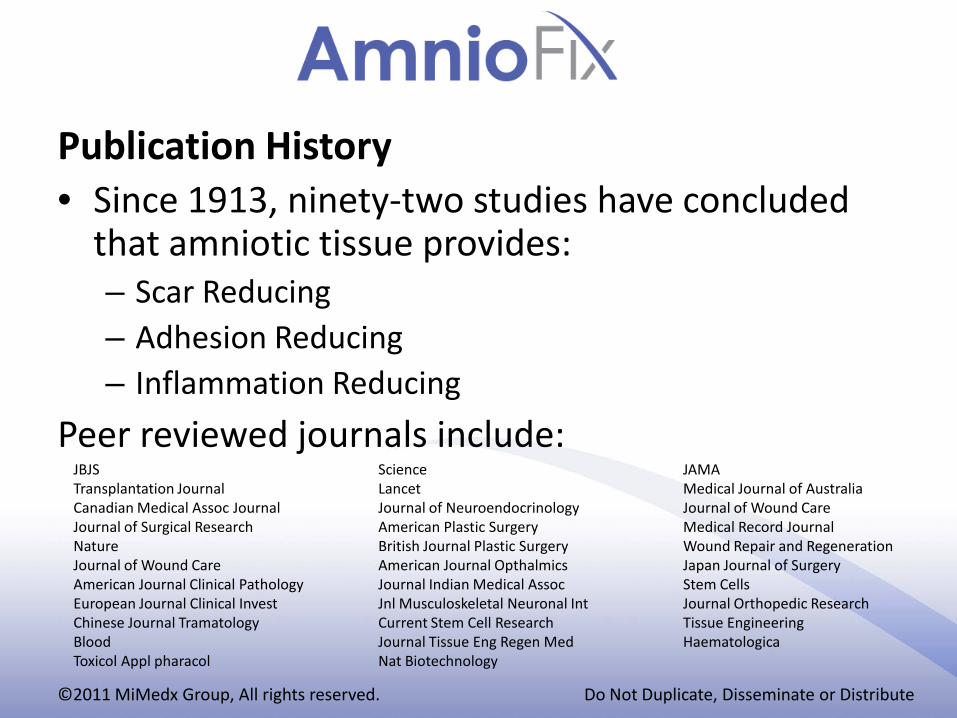
Publication History• Since 1913, ninety-two studies have concluded
that amniotic tissue provides:– Scar Reducing– Adhesion Reducing– Inflammation Reducing
Peer reviewed journals include: JBJSTransplantation JournalCanadian Medical Assoc JournalJournal of Surgical ResearchNatureJournal of Wound CareAmerican Journal Clinical PathologyEuropean Journal Clinical InvestChinese Journal TramatologyBloodToxicol Appl pharacol
ScienceLancetJournal of NeuroendocrinologyAmerican Plastic SurgeryBritish Journal Plastic SurgeryAmerican Journal OpthalmicsJournal Indian Medical AssocJnl Musculoskeletal Neuronal IntCurrent Stem Cell ResearchJournal Tissue Eng Regen MedNat Biotechnology
JAMAMedical Journal of AustraliaJournal of Wound CareMedical Record JournalWound Repair and RegenerationJapan Journal of SurgeryStem CellsJournal Orthopedic ResearchTissue EngineeringHaematologica

Do Not Duplicate, Disseminate or Distribute©2011 MiMedx Group, All rights reserved.
• Amnion is used to treat pterygium and chemical burns.
• Amnion closely mimics the natural properties of conjunctiva tissue.
• Amnion helps comfort the surgical site and guide healing.
Supports the anti-inflammatory and scar reduction properties of AmnioFix.
Implant History
Over 30 Thousand implants in Ophthalmology Clinical Application

Do Not Duplicate, Disseminate or Distribute©2011 MiMedx Group, All rights reserved.
• AmnioFix is terminally sterilized using e-beam radiation.
– Target radiation level is <2.7 Mrads
• AmnioFix has a 5 year shelf-life
Sterilization, Shelf Life and Safety

Do Not Duplicate, Disseminate or Distribute©2011 MiMedx Group, All rights reserved.
• Temperature Restrictions• 32°F to 100°F
• Distributor Tissue Tracking
• Hospital Tissue Tracking
• Graft Preparation• Double Foil Pouch
• Dry
Handling Considerations

Do Not Duplicate, Disseminate or Distribute©2011 MiMedx Group, All rights reserved.
Competition
• NuTech- NuShield
• AF Cell/Alphatec-AmnioShield
• BioD/ US Spine, Amedica- BioDfence

Do Not Duplicate, Disseminate or Distribute©2011 MiMedx Group, All rights reserved.
Bio D-logics– Single Layer Amnion
• Thin, Difficult to place• Provided Hydrated• Cross-linked with Gluteraldehyde
– Distributed by Amedica/US Spine, plus other regional distributors
NuTechOur Tissue, Purion Processed
AF Cell/ AlphatecOur Tissue(most recent) there were previous suppliers that had more difficult to handle Amniotic tissue (BioD)
Competition

Do Not Duplicate, Disseminate or Distribute©2011 MiMedx Group, All rights reserved.
Freeze Dried-• Goes through a freeze process, ice crystal formation,
damage collagen structureCross-linked-• Uses a chemical to make stronger• Changes the structure of the collagen molecule• Residual chemical agents (Gluderaldehyde)Dried-• Minimal damage to collagen structure• No residual chemicals to damage tissue
Dried vs. Freeze Dried vs. Cross-linked

Do Not Duplicate, Disseminate or Distribute©2011 MiMedx Group, All rights reserved.
“The hypertrophic epidural scarring occurred in these three cases despite the presence of autologous fat grafts”1
“This study suggests that the use of free fat grafts during lumbar disc surgery was clinically ineffective”2
1 Martin-Ferrer S. Failure of autologous fat grafts to prevent postoperative epidural fibrosis in surgery of the lumbar spine. Neurosurgery. 1989 May;24(5):718-21.
2 Gorgulu A. The effect of epidural free fat graft on the outcome of lumber disc surgery. Neurosurg Rev. 2004 Jul;27(3):181-4.
Autologous Free Fat Grafts

Key Points

Do Not Duplicate, Disseminate or Distribute©2011 MiMedx Group, All rights reserved.
EffectiveContains growth factors unique to placental tissue:
Collagen Types IV, V, and VII
– Enhance wound healing
– Reduces Inflammation
– Barrier Protection
– Reduces Scar Tissue Formation
– Non-immunogenic
Key Selling Points

Do Not Duplicate, Disseminate or Distribute©2011 MiMedx Group, All rights reserved.
AmnioFix is not a single layer of Amnion, it is a composite graft
Purion Process® protects the scaffold leaving the collagen matrix intact, terminally sterilized
Dehydrated– Simple application– Embossed to facilitate ease of orientation– Doesn’t damage the collagen– 5 year shelf life
AmnioFix is not cross-linked, leaving no residual Gluderaldehyde
Key Competitive Points

Do Not Duplicate, Disseminate or Distribute©2011 MiMedx Group, All rights reserved.
Offering Dimensions
• APS-5160 16mm disc
• APS-5230 2x3cm
• AAS-5330 3x3cm
• AAS-5440 4x4cm
• AAS-5460 4x6cm
Sizes and Potential Applications

Do Not Duplicate, Disseminate or Distribute©2011 MiMedx Group, All rights reserved.
Frequently Asked QuestionsQ: What is Amniotic Tissue (or Amniotic Membrane)?
A: The amniotic membrane is the innermost layer of the placenta which lines the amniotic cavity. The membrane itself is made up of layers of tissue containing specialized cells. These cells allow the membrane to provide specific functions which aid in healing.
Q: What are the specialized cells and growth factors that make Amniotic Membrane a good barrier and wound healing agent?
A: The amniotic membrane has a structure or Extra Cellular Matrix (ECM) that is constructed of collagen types IV, V and VII, plus specialized proteins (fibrillin, fibronectin and laminins). In addition, amniotic membranes have growth factors that signal to elicit a specific cellular response. In vivo studies show that the properties of amniotic membrane help reduce scar tissue formation and scar attachment to the dura.1
Q: What growth factors are present in Amniotic Tissue?
Epidermal Growth Factor (EGF)
Transforming Growth Factor Beta (TGF-β)
Fibroblast Growth Factor (FGF)
Platelet Derived Growth Factor (PDGF)

Do Not Duplicate, Disseminate or Distribute©2011 MiMedx Group, All rights reserved.
Q: How does AmnioFix both reduce scar formation and heal at the same time?
A: AmnioFix reduces scar tissue formation by down regulating or inhibiting expression of TGF-b receptors in migrating fibroblast cells. These migrating cells are now stimulated by other growth factors where granulation tissue is minimized and a native connective tissue matrix is generated at the wound site. AmnioFix also reduces inflammation by entrapment of T lymphocytes and the expression of IL-4 and IL-10 (interleukins).
Q: What kind of publications are there on Amniotic Tissue?
A: There are over 90 publications on Amniotic Tissue. Publications on ophthalmic, wound care, spine and orthopedic uses of the tissue describing the anti-inflammatory properties, scar reduction and wound healing.
Q: How safe is AmnioFix?
A: MiMedx uses the patent pending Purion® process for the processing of amniotic membrane tissue. The process technology and donor screening follow strict guidelines as set forth by both the Food and Drug Administration (FDA) and the American Association of Tissue Banks (AATB). Eligible donors are all living mothers which have given full consent and must have delivered a live birth via cesarean section. Serologic blood tests are then performed to rule out the potential for infectious disease transmission. The complete process concludes with validation by an outside source to ensure that the procedural process results in a safe and effective implant.

Do Not Duplicate, Disseminate or Distribute©2011 MiMedx Group, All rights reserved.
Q: Are there differences between the tissues being used for different applications?
A: Yes, there are difference between the tissues for each of the applications. Each tissue has been specifically processed for the application it is promoted for.
Q: What is the “SB” that I see on the tissue?
A: “SB” stands for Surgical Biologics, the founder and processor of the amniotic tissue and Wholly owned subsidiary of MiMedx Group, Inc.
Q: Is amniotic tissue permanent or resorbable?
A: AmnioFix is resorbable and will resorb depending on the specific application, patient, and location it is placed, typically 4-6 weeks.
Q: Do I need to suture the Amniotic Tissue?
A: Suturing is not required.

Do Not Duplicate, Disseminate or Distribute©2011 MiMedx Group, All rights reserved.
Q: What is the technique to apply AmnioFix?
A: 1. Cut the AmnioFix to the appropriate size, if clinically necessary.
2. Ensure all excess blood/fluid is removed from the surgical site.
3. Lay the AmnioFix so the SB can be read on the desired surgical site. (“SB” side up)
4. The AmnioFix will become tacky to the site. If surgeon needs to reposition, just add a couple drops of sterile saline or water.
5. Suture the AmnioFix (optional based on surgeon preference)
6. To make the AmnioFix more malleable, add a couple drops of saline to hydrate the graft. The AmnioFix may be contoured to the underlying anatomy.
7. Do not suction near the AmnioFix.
8. Complete surgical procedure and close.
Q: Is this product freeze dried? I heard the freeze dried process destroys cell structure.
A: No, this product is not freeze dried.
Q: How is this more effective than the liquid versions on the market?
A: The injectable liquid versions on the market are in early stages of development. We
are working on an injectable that we think will clinically relevant soon. There is room on
the market for both treatments.

Forms

Forms

Forms

Forms

Forms

Forms