traditional acupuncture meets modern nanotechnology...
TRANSCRIPT
Review ArticleTraditional Acupuncture Meets Modern Nanotechnology:Opportunities and Perspectives
He Zhang,1,2 Gang Han ,3 and Gerhard Litscher 1,2
1Department of Respiration, Guang’anmen Hospital, China Academy of Chinese Medical Sciences, Beijing 100053, China2TCM Research Center Graz, Research Unit of Biomedical Engineering in Anesthesia and Intensive Care Medicine and Research Unitfor Complementary and Integrative Laser Medicine, Medical University of Graz, 8036 Graz, Austria3Department of Biochemistry and Molecular Pharmacology, University of Massachusetts Medical School, Worcester, MA 01605, USA
Correspondence should be addressed to Gang Han; [email protected] Gerhard Litscher; [email protected]
Received 25 March 2019; Accepted 23 June 2019; Published 16 July 2019
Academic Editor: Min Li
Copyright © 2019 HeZhang et al.This is an open access article distributed under theCreative CommonsAttribution License, whichpermits unrestricted use, distribution, and reproduction in any medium, provided the original work is properly cited.
Acupuncture is an ancient method in traditional Chinese medicine (TCM). Usually acupuncture needles are inserted into the bodyto achieve therapeutic effects. However, there are still some challenges to achieve consensuses. What is the essence or anatomy ofacupuncture meridians? How does acupuncture work? How to improve acupuncture clinical therapeutic effect? These questionsmay be addressed by highlighting recent developments in innovative nanotechnology. The aim of this review is to elucidate thepossible applications and future potential of nanotechnology in acupuncture. Nanoparticles are promising for imaging and it maygain a better understanding of the essence of meridian. Nanotechnology enables nanochips/nanosensors providing new solutionsin detection reactive molecules in vivo and in real time. The connections and changing of these molecules with needle stimulationwill allow insight into the mechanisms of acupuncture. Acupuncture combined with nano-TCM could provide a great potentialin some type of characteristic acupuncture therapies improvement. By virtue of nanotechnology, the acupuncture needles couldbe innovated as multifunction toolbox. Acupuncture needles could be considered as a method for controlled drug delivery. Thenanoparticulated photothermal, magnetothermal, photodynamic agents could also be filled on the surface of needle.
1. Introduction
Acupuncture is an ancient typical therapy in traditionalChinese medicine (TCM), in which acupuncture needles areinserted into the body to achieve desired therapeutic effects[1]. Acupuncture is believed to originate in China.Thousandsof years have witnessed the remarkable clinical applicationsof acupuncture. According to theWorld Health Organization(WHO), acupuncture has been shown to be an effective alter-native or complementary treatment to 28 diseases, symptoms,or conditions [2]. In daily practices, the clinical applicationsof acupuncture spread far beyond these diseases. Moreover,acupuncture is suitable for the large majority, regardless ofphysical condition, with little or no side effect. Despite thelong history and widespread use of acupuncture, there arestill some challenges in achieving a consensus. The efficacy
of acupuncture remains a topic of discussion. Some studies,including multicenter randomized control trials (RCT), havesuggested that acupuncture is no more than a theatricalplacebo [3–5]. Besides, how acupuncture works as well asthe molecular mechanisms behind it is still unclear. Themeridian theory has a solid role in guiding the clinicalpractice of acupuncture and is a fundamental of TCMscience.However, as of now, the anatomy of meridians has not beenexplicated in detail [6]. Acupuncture also has the featureof TCM treatment based on syndrome differentiation. Byselecting different acupoints and changing the manipulationof the system, a unique, but different, clinical outcomeresults each time. Therefore, it is becoming increasinglynecessary to standardize acupuncture treatment [7]. Thesechallenges are consequently the key roadblocks hinderingthe advancement of acupuncture. Thus, new and modern
HindawiEvidence-Based Complementary and Alternative MedicineVolume 2019, Article ID 2146167, 9 pageshttps://doi.org/10.1155/2019/2146167
2 Evidence-Based Complementary and Alternative Medicine
Imaging
Sensing
Tip Sensor part
Time
Bioactive molecules
Therapy
Nano TCM
Drugs
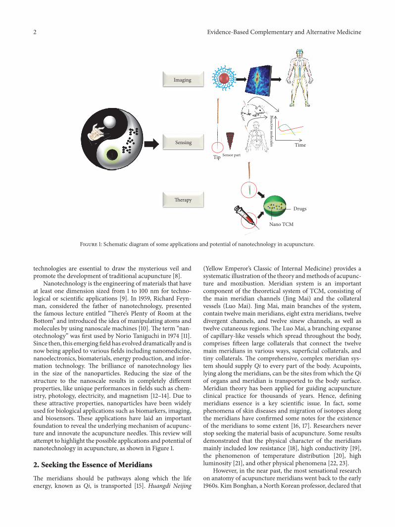
Figure 1: Schematic diagram of some applications and potential of nanotechnology in acupuncture.
technologies are essential to draw the mysterious veil andpromote the development of traditional acupuncture [8].
Nanotechnology is the engineering of materials that haveat least one dimension sized from 1 to 100 nm for techno-logical or scientific applications [9]. In 1959, Richard Feyn-man, considered the father of nanotechnology, presentedthe famous lecture entitled “There’s Plenty of Room at theBottom” and introduced the idea of manipulating atoms andmolecules by using nanoscale machines [10]. The term “nan-otechnology” was first used by Norio Taniguchi in 1974 [11].Since then, this emerging field has evolved dramatically and isnow being applied to various fields including nanomedicine,nanoelectronics, biomaterials, energy production, and infor-mation technology. The brilliance of nanotechnology liesin the size of the nanoparticles. Reducing the size of thestructure to the nanoscale results in completely differentproperties, like unique performances in fields such as chem-istry, photology, electricity, and magnetism [12–14]. Due tothese attractive properties, nanoparticles have been widelyused for biological applications such as biomarkers, imaging,and biosensors. These applications have laid an importantfoundation to reveal the underlying mechanism of acupunc-ture and innovate the acupuncture needles. This review willattempt to highlight the possible applications and potential ofnanotechnology in acupuncture, as shown in Figure 1.
2. Seeking the Essence of Meridians
The meridians should be pathways along which the lifeenergy, known as Qi, is transported [15]. Huangdi Neijing
(Yellow Emperor’s Classic of Internal Medicine) provides asystematic illustration of the theory andmethods of acupunc-ture and moxibustion. Meridian system is an importantcomponent of the theoretical system of TCM, consisting ofthe main meridian channels (Jing Mai) and the collateralvessels (Luo Mai). Jing Mai, main branches of the system,contain twelve main meridians, eight extra meridians, twelvedivergent channels, and twelve sinew channels, as well astwelve cutaneous regions. The Luo Mai, a branching expanseof capillary-like vessels which spread throughout the body,comprises fifteen large collaterals that connect the twelvemain meridians in various ways, superficial collaterals, andtiny collaterals. The comprehensive, complex meridian sys-tem should supply Qi to every part of the body. Acupoints,lying along the meridians, can be the sites from which the Qiof organs and meridian is transported to the body surface.Meridian theory has been applied for guiding acupunctureclinical practice for thousands of years. Hence, definingmeridians essence is a key scientific issue. In fact, somephenomena of skin diseases and migration of isotopes alongthe meridians have confirmed some notes for the existenceof the meridians to some extent [16, 17]. Researchers neverstop seeking the material basis of acupuncture. Some resultsdemonstrated that the physical character of the meridiansmainly included low resistance [18], high conductivity [19],the phenomenon of temperature distribution [20], highluminosity [21], and other physical phenomena [22, 23].
However, in the near past, the most sensational researchon anatomy of acupuncture meridians went back to the early1960s. Kim Bonghan, a North Korean professor, declared that
Evidence-Based Complementary and Alternative Medicine 3
their research team found the anatomic and physiologicalbasis of the meridians [24]. A new system was proposed,constituting nodes like anatomical structures at the acupointsand the tube-like structure connected to the nodes in the skin.The nodes and the tubes were named Bonghan corpusclesand the Bonghan ducts. Bonghan liquor was discovered, aliquid circulating in the Bonghan ducts, which is abundantin DNA, RNA, hyaluronic acid, more than 19 amino acids,and 16 free mono nucleotides [24]. Besides, according toKim’s reports, the Bonghan system comprised various bio-chemicals related to immune activities and “Sanals” withself-regenerating properties [25]. According to Sanal theory,the amount of DNA contained in one Sanal is nearly equalto that contained in one chromosome [26]. Kim’s resultscreated a great sensation at that time. However, unfortunatelythere were several attempts to reproduce Kim’s results butwithout success [26]. Thus, Kim was thought to cheat in hisresearch and apparently he did not describe the materialsand methods in detail. Around 40 years later, the storydramatically reversed and Kim’s discoveries were confirmedby a Korean researcher Kwang-sup Soh. Kim’s team holds theviews that the threadlike microscopic anatomical structureas primo vascular system (PVS) is served as an extension ofacupuncture meridians [25]. Bonghan duct, corpuscle, andSanals were renamed: primo vessel (PV), primo node (PN),and primo-microcells, respectively [24, 27].
The generation of diverse type nanoparticles clearlyexhibited the importance of nanoparticles in biological imag-ing applications [8, 28, 29]. Apart from Trypan blue sprayingtechnique, fluorescent nanoparticles, gold nanospheres, andquantum dots were adopted to visualize the novel system.Hollow gold nanospheres (HGNs) can be adjusted to generatean optical contrast to the intralymphatic PVS (IL-PVS)because of the porous nature of the PV’s external wall [30].As previously stated, the diameter of the pore was smaller(∼1 𝜇m) than the external diameter (2–5 𝜇m) in rabbits,suggesting that lymph inflow into PVs was relatively easierthan outflow [31]. Eric Carlson et al. [30] selected two rangesof the HGNs size, which were 50–70 nm and 100–125 nm.The HGNs colors were turquoise and green for the 50–70nm and 100– 125 nm, respectively. The two different sizesof HGNs were injected into lumar lymphatic nodes (LLN).When the HGN-contrasted PV became visible, it was stillneed to further confirm it as an IL-PV. The result showedboth the LLN and LV darkened after HGNs injection, butLVs remain partially translucent allowing visualization of IL-PVS. The turquoise-green-colored HGNs seemed to provideexcellent optical contrast for the IL-PVS in rats. Kwang-supSoh holds the view that the structure of PVS was very similarwith the connective tissues around the PVS so that it couldnot be discriminated in the conventional histology. However,the fluorescence nanoparticles were preferentially absorbedby the primo vessels compared with surrounding tissues. Inorder to elucidate the functional and the medical aspects ofthe PVS, fluorescent nanoparticles could be used as contrastagents. Several kinds of fluorescence nanoparticles have beendeveloped by using silica loading fluorescence dyes, includingthe rhodamine B isothiocyanate [32–34], and a mixed dyeof commercial Pelikan ink and rhodamine B [35] to observe
the PVS in the abdominal wall, epineurium along the sciaticnerve, the subcutaneous layer of skin, and even the fourthventricle and spinal cord.These nanoparticles were detectablewith MRI [36]. In order to evaluate the feasibility of usingnanoparticles as a contrast agent during MRI or CT imagingof PV in the brain or the spinal cord, the nanoparticles wereinjected into the lateral ventricles of rats. The nanoparticles,flowing from lateral ventricles to the fourth ventricle andthe central canal of the spinal cord, were absorbed by theprimo vessel floating in the cerebrospinal fluid (CSF). Theflorescence of the nanoparticles was measured and calibratedby using a reference suspension. The results showed thatthe nanoparticles could be potential as a contrast agent toobserve PV in the brain or the spinal cord. The magneticfluorescent nanoparticles with proper surface modificationsare not only nontoxic and biocompatible but also targetableto the specific area under external magnetic field [37, 38].Johng et al. observed that magnetic fluorescent nanoparticleflew along the livermeridian, where the route was same as theclassical books recorded, from the acupoint Taichong (LR 3)to the acupoint Yinbao (LR 9)where amagnetwas attached tothe skin surface [39]. In addition, IL-PVS, a novel threadlikestructure inside the lymphatic vessel, could be imaged invivo by the magnetic fluorescent nanoparticle [40, 41]. Thenanoparticles were injected into two lumbar lymph nodesand a magnet was placed on the lymphatic vessels whichwere connected to the nodes. However, injection into lymphnode to visualize PV and PN is not always recommendedas bleeding is not avoidable. The best method to verify theexistence of PV and PN is to directly inject some dyesor nanoparticles into lymphatic vessels, not lymph nodeof the rabbit. In the case of rat, actually, the size of thelymph vessel is not enough to be controlled in situ andin vivo for visualization. It further has to be mentionedthat nanoparticles could be spread into fibrin fibers whichfrequently are formed inside lymph vessel owing to evenslight bleeding.
In terms of the external magnetic field, the injectednanoparticles were attracted and stayed inside the lymphaticvessel rather than flowing away with the lymphocytes. Thus,nanoparticles were taken up by the threadlike structures.According to Soh’s research, we can surely gain a revelationthat there are still somenovel structureswhich are not noticedor recognized in numerous conventional histological studies.Soh’s team believed that PVS is an extension of acupuncturemeridians. The PVS is a previously unknown system indeed,but the question what the relationship between PVS andmeridians is still needs future exploration. To a certain extent,the difference of distribution, position features, identification,and origin of PVS are still inconsistent with classic acupunc-ture meridian theory [42].
Within this article, the PVS are discussed as exampleand to highlight the possible applications and potential ofnanotechnology in acupuncture. Maybe the PVS is lack ofmainstream acceptance, but the research methods couldprovide reference. In this review, the PVS are describedaccording to the published articles without overstating it.We clearly want to point out that the relationship betweenPVS and meridians still needs future exploration. This is in
4 Evidence-Based Complementary and Alternative Medicine
accordance with the article of Kang et al. [43]. These authorsalso reported on identifying PVSwith nanoparticles; howeverthey also stated that the possible existence of PVS is still notknown to many scientists [43].
3. Detecting the BiologicalMechanisms of Acupuncture
Another debate of acupuncture in the medical communityis how the acupuncture works and what its underlyingmolecular mechanisms are. Based on TCM theory, diseaseis caused by deficiencies, excess, or imbalances of Qi. Thus,stimulating the specific acupoints could regulate correspond-ing organs or meridians and subsequently restore the stateof balance. Much modern scientific work has been done toseek the mechanism of acupuncture. Unfortunately, thereis no unified theory of acupuncture mechanism. Variousmodels and hypotheses for different clinical applicationshave been proposed. In summary, mechanisms of acupunc-ture treatment are included in neurobiological mechanismby which acupuncture may trigger a somatic autonomicreflex, changing the levels of neurotransmitters, affecting thehypothalamus pituitary axis [44–46] and immunomodula-tory effects [47], and modulating neuro-endocrine-immune(NEI) network [48].
Acupuncture needles are vital tools in the delivery ofacupuncture, coming in different diameters and lengths tobe used on the different areas of the body. The earliestacupuncture needles were named bian stones made of sharpedged stones. In ancient times, people lived in a chillyoutdoor environment.The heated sharp stones were found asinstruments of healing by scratching or pricking the certainsite of body [49]. Since then, the theories of acupuncture,such as description of the meridians, acupoints location, andneedling techniques, have emerged gradually. The materialsof acupuncture needles also experienced great developmentduring thousands of years.
Early acupuncture needles were made from the bambooand bone. With the emergence of metallurgy, iron, copper,bronze, silver, and gold needles were made. However, com-pared with other materials, stainless steel needles are moreeconomical and practical in clinical practice. Due to theproperty of good conductivity, acupuncture needles can beconsidered as a modern biosensor or other innovative tools.
Nanotechnology enables nanosensors providing newsolutions in physical, chemical, and biological sensing andlargely increases detection sensitivity, specificity, and porta-bility. It laid a foundation for detection reactive molecules invivo and in real time. Furthermore, it supports an excellenttool for observing and studying the biological effect andbiological function of these reactive molecules. The con-nections of these molecules with the needle stimulation atthe central and peripheral level will absolutely allow insightover the mechanisms of acupuncture treatment. The use ofcarbon nanotubes (CNTs) for sensing applications is promis-ing [50]. It has already been reported that CNTs are usedto measure and acquire pH, temperature, and biomedicalinformation [51–53]. In order to monitor the molecular eventoccurring in acupoint and gain access to the local responses
of acupuncture stimulation, a unique nanosensing platformby modifying carbon nanotubes (CNTs) on the tip surface ofacupuncture needle has been developed. The CNT-modifiedacupuncture needle (CNT/AN) monitored serotonin (5-HT) level in vivo via electrochemistry in acupoint Zusanli(ST 36) [54]. The 5-HT was pumped into the acupointST 36 to make a proof-of-concept experiment. The resultsindicated that the CNT/AN was quite stable and able todetect the neurotransmitters in vivo, showing a great potentialfor better understanding the mechanism of acupuncturetreatment. However, in fact, the effects of acupuncture onthe healing process are through a holistic pathway, producinga series of physiological reactions, not only depending onone simple bioactive molecule or one signaling pathwaychanging. Thanks to the nanotechnology and Omics-basedtechniques, it may give answers to the acupuncture complexmechanisms. Omics-based techniques refer to screen oftargets from nucleic acids to proteins and metabolites andtheir heterogeneous interactions. As the development of thetechnology of nanobiochips and omics, it will be possible tointegrate relatively high spatial (different body regions andtissues) and temporal resolution data to reveal the molecularsignaling pathways that flow from the tip of the needleto the disease or injury site in the near future [55]. Theinformation obtained from acupoint, the target organ, as wellas systemically, can be enriched with the temporal resolutiondata to construct a systems biology network. This networkwill provide a holistic view of how acupuncture works andits potential mechanism. Raman spectroscopy is commonlyused in chemistry to provide a structure fingerprint basedon vibrations in molecules [56]. It is also considered as aqualitatively or semiquantitatively analytical tool with thefunction of ultrasensitive detection because of the design andfabrication of nanostructure with surface-enhanced Ramanscattering (SERS) activity.With themerits of beingminimallyinvasive [57], the acupuncture needle was used as a tool,coated on the gold nanoshells (GNSs) and polystyrene, whichcould absorb SERS-active nanomaterials to become a SERS-active needle [58]. When inserted into the body, the SERS-active needle can detect interstitial fluids. And when SERS-active needle was pulled out, analytes in the diffused fluidsat different depths were taken out in the meantime. TheSERS-active needle presented an approach on depth profilesof target molecules in tissues. A novel SERS-active needlehas the function of detection glucose in vivo by integratingglucose oxidase (GOx, signal convertor), 4-mercaptobenzoicacid (4-MBA, signal reporter), and microporous polystyrene[59]. This method could be a universal strategy to SERSdetection of small biomolecules in vivo with the suitableintegrating enzymes and corresponding reporters on SERS-active needles.
An important aspect of acupuncture treatment is thatacupuncture needle must be manually manipulated to get theDeqi response after insertion into the body.Deqi is consideredas an important component in the process of achievingtherapeutic effectiveness in the acupuncture treatment. TheDeqi feeling of patients is individual difference, which canbe described as suan (aching or soreness), ma (numbness ortingling), zhang (fullness or pressure), or zhong (heaviness)
Evidence-Based Complementary and Alternative Medicine 5
around the acupuncture point or along the meridians [60].When Deqi occurs, the practitioner, meanwhile, perceives asensation often called “needle grasp.” However, the underly-ing mechanisms of acupuncture manipulation and the bio-logical effect of needle grasp have remained unresolved. Thehypothesis of the needle grasp mechanism might involve thewinding connective tissue during the process of needle man-ual manipulation, which might deliver a stronger mechanicalsignal into the tissue [61]. A research has been conducted thatused silicon carbide sandpapers with different grit numbersto manipulate the needle grasp force by changing surfaceroughness. The atomic force microscope (AFM) images wereobtained by using Nanostation II�. AFM, as one of theforemost imaging tools, can measure andmanipulate mattersat the nanoscale by using a high-resolution scanning probe.In order to observe and verify the scratches on the surfaceof the needle analytically, the shape and depth of the scratchwere analyzed. The result showed that surface roughnessof the acupuncture needle could enhance the analgesiceffect of acupuncture therapy, which partially supports themechanical signaling theory through the winding connectivetissues in the process of acupuncture manipulation [62].To get the Deqi response, acupuncture needles are manu-ally manipulated after insertion into the body. The typicalacupuncturemanipulation ismade up by rapid twisting (backand forth or opposite direction) and lifting-thrusting (up anddown motion) of the needle [63]. The stimulation amountand intensity are significant factors for different amount andintensity of simulation cause different physiological effects.However, these acupuncture manipulations are merely basedon the patient’s feelings and there is a lack of a quantitative,objective standard. Some efforts have been made to attemptto solve the problem. The force sensor (Nano-17 titanium)was attached to the acupuncture needle, and various rotationfrequencies or lifting-thrusting movements were measuredafter acupuncture needle inserting in phantom tissue [64, 65].While maybe it would be better to use the nanosensor whichwas connected to the tip of acupuncture needle; in that case,the real force and the amount of stimulation inside the tissuecould bemeasured. Or even the acupuncture needle tip couldbe connected tomultisensors, by which the force, the amountof stimulation, and therapy-induced biochemical progressioncould be monitored in real time and in vivo.
4. Enhancing the Therapeutic Effect
Acupuncture, as one of the complementary and alternativetherapies, has attracted much attention to its therapeuticeffect. How to enhance the clinical therapeutic effect andmeanwhile reduce the discomfort induced by acupunctureneedles are challenging and considerable. Clinically, variousneedle parameters such as diameter, depth of insertion, andnumber of needles used at a time are important factorsfor improving acupuncture performance. Some studies sug-gested that applying thick needles or deeper insertion couldincrease stimulus intensity and ultimately achieve the desiredlevel of Deqi [66]. Thus, based on these parameters, a novelclass of acupuncture needles, porous acupuncture needles(PANs) with hierarchical micro-/nanoscale conical pores
upon the surface, have been fabricated [67]. The surface areaof PANs was approximately 20 times greater than conven-tional acupuncture needles. The conventional acupunctureneedles and PANs were inserted into the receptive field ofthe lumbar spinal dorsal horn neuron in alcohol dependenceWistar rats. The neuronal response to acupuncture stimula-tion, tremor activity, and Cocaine induced locomotor activitywere investigated. Furthermore, the comparison of the dif-ferent needles was made via measurement of spontaneouspain sensation induced by acupuncture needle insertion.The results demonstrated the higher efficacy of PANs inpsychiatric treatment over conventional needles. Further, thePANs showed reduced pain sensation levels compared withthe same interfacial surface area of conventional acupunctureneedles. The fabrication of these novel PANs could be goodnews for the people fearing pain.
Acupoint injection therapy is an innovation in TCMwhere herbal extract or liquid drugwas injected into acupointfor treatment or prevention [68]. Bee venom (BV), consistingof various biologically active compounds, is one of the mostcommonly encountered animal venoms [69]. BV injectionexerts a remarkable effect in chronic diseases such as shoulderpain and inflammation, with the function of stimulatingan acupoint as well as inducing allergic reactions in thehuman body [70–72]. Although the therapeutic utility ofBV injection has been demonstrated, its safety profile isstill somewhat limited. A systemic or local allergic response,accompanying fever, tonic pain, and edema, could occurafter the BV injection [73]. To enhance the target or deliveryefficiency of the drug, thus reducing the side effect of BVinjection, the nanoparticle-based drug delivery systems seemto be a promising strategy. Jeong et al. [74] investigated theBV loaded into biodegradable poly(d,l-lactide-co-glycolide)nanoparticles (BV-PLGA-NPs) on formalin-induced pain.BV-PLGA-NPs were injected into the acupoint Zusanli (ST36) at 0.5, 1, 2, 6, 12, 24, and 48 h before plantar injection offormalin. BV-PLGA-NPs exhibited the same analgesic effectas typical BV injection at the time points of 0.5, 1, and 2 h butprovided a more prolonged effect than typical BV acupunc-ture treatment. Compared with classical acupuncture, acu-point injection therapy is easily administered, standardized,and timesaving [75]. However, at the moment the type ofdrugs for acupoints injection such as BV, placental extract,and Chinese medicine injections are limited [76]. Thanksto nanotechnology, a new concept, nano-traditional Chinesemedicine (nano-TCM) has been proposed. Nano-TCM refersto bioactive ingredients, bioactive parts, medicinal materials,or complex prescription to be approximately 100 nm in size oreven the size within 1000 nm processed by nanotechnology[77]. Nano-TCM may be an important direction towardsTCM’s modernization and internationalization. The activityof some Chinese medicines processed into nanodrugs hasbeen greatly improved, which could significantly increasetheir bioavailability and targeting, decrease the using time,and largely reduce the toxic side effects [78–80]. Acupuncturecombined with nano-TCM could offer a great potential insome type of characteristic acupuncture therapies improve-ment. As the development of nano-TCM, it would offer moreoptions for acupoint injection thus expanding the range of
6 Evidence-Based Complementary and Alternative Medicine
the clinical applications. Acupoint external application is aspecific therapeutic method that drugs, especially traditionalChinese herbs, are made into plaster to be applied on certainacupoints. The mechanism of this therapy relies on per-cutaneous absorption. Compared with traditional Chinesemedicine, nano-TCM exhibits stronger penetrating abilitydue to small particle size and large selection of adsorptioncapacity, which could penetrate the skin barrier. After acertain modification of nanodrug carriers, it could promotesustained release of active constituent of the TCM, thusprolonging the curative effect [81].
Acupoint catgut embedding therapy is referred to infixseveral surgical chromic catgut sutures into the acupoints orsubcutaneous tissue with a specialized needle under asepticprecautions. It is widely used in obesity, perimenopausalsyndrome, chronic urticaria, depressive neurosis, and refrac-tory insomnia [82]. Currently, the commonly used threadsin clinical practice could be divided into medical catgutthread, absorbable surgical suture, and medicated suture[83]. In most cases, this therapy is considered safe, butsome individual could be allergic to catgut. Inspired bythe application of nanomaterial in medicine, a nanosilverthread is applied to acupoint catgut embedding therapy. Thenanosilver with the antibacterial effect is better than theconventional catgut in lower inflammatory response [84].
5. Conclusion and Perspectives
Fluorescent magnetic nanoparticles allow the application ofmagnetic properties together with the ability of fluorescenceto visible, for example, PVS. Although there are still somedifferences betweenPVS and acupuncturemedians, nanopar-ticles give a new resolution to explore the basis of acupuncturemedians.Nanochips or nanosensors are promising for detect-ing and monitoring the changing of acupuncture inducedbioactive molecules and the intensity and amount of stim-ulation. Under the circumstances, it will help to fill the gapin knowledge about the underlying molecular mechanismsof acupuncture treatment and gain the quantitative, objectiveacupuncture manipulation simulation data. Accordingly, thestandard of acupuncture manipulation could be set up andconducive to attracting more and more people to adoptacupuncture therapy.
The meridians and acupoints are dynamic processes ofreceiving stimuli and regulating functional activities of thebody. The molecular imaging is intended to visually displaybioactive molecule level in physiology or monitoring thepathological dynamic process of the bioactive molecule levelin vivo or in situ. Molecular imaging based on nanotechnol-ogy, especially the multimodal imaging, could be a powerfulapproach that provides more reliable and accurate detectionof dynamic changing of meridians and acupoints.
By virtue of nanotechnology, the acupuncture needlescould be innovated as multifunction toolbox. It could realizethe idea of not only controlled drug delivery but alsowarming and invigorating the acupoint in the original basis.Microneedles, which were first conceptualized for controlleddrug delivery, emphasize drug and vaccine delivery to theskin and deliver molecules into cells and nuclei. Acupuncture
needles could be considered as a method for controlled drugdelivery using microneedle for reference. The acupunctureneedles are fabricated with nanoscale conical pores upon thesurface of needles in which drugs could be filled, or justcoated the drug on the surface of the needles. In that case,it can not only have the therapeutic effects of acupuncturestimulation but also bring the drug to the lesions directly.
Disclosure
The original data (references) can be found at the institutionof the first author (Dr. He Zhang) and at the institution of thecorresponding author (Prof. Gerhard Litscher).
Conflicts of Interest
The authors declare no conflicts of interest regarding thepublication of this paper.
Acknowledgments
The first author would like to thank Prof. Zhiguo Zhou, Prof.Hong Yang, and Dr. Kai Huang for their valuable help withthe manuscript. The work was supported by the AustrianFederal Ministry of Education, Science and Research (projecttitle “Sino-Austrian TCM Research on Lifestyle-RelatedDiseases: Innovative Acupuncture Research” (2016–2019);project leaderG. Litscher).Themanuscript has been preparedin advance of a 3-month research stay of the first author Mrs.He Zhang, Ph.D., MM, at Medical University of Graz whichwas supported by Eurasia-Pacific Uninet. Professor GerhardLitscher is also Visiting Professor at the China Academy ofChinese Medical Sciences in Beijing, China.
References
[1] C. A. Vincent and P. H. Richardson, “The evaluation of thera-peutic acupuncture: concepts and methods,” PAIN, vol. 24, no.1, pp. 1–13, 1986.
[2] World Health Organization, Acupuncture: Review And Analysisof Reports on Controlled Clinical Trials, World Health Organi-zation, 2002.
[3] J. Vas, J. M. Aranda,M.Modesto et al., “Acupuncture in patientswith acute low back pain: A multicentre randomised controlledclinical trial,” PAIN, vol. 153, no. 9, pp. 1883–1889, 2012.
[4] E. A. Macklin, P. M. Wayne, L. A. Kalish et al., “Stop Hyper-tension with the acupuncture research program (SHARP),”Hypertension, vol. 48, no. 5, pp. 838–845, 2006.
[5] E. Ernst, “Acupuncture—a critical analysis,” Journal of InternalMedicine, vol. 259, no. 2, pp. 125–137, 2006.
[6] N. Maurer, H. Nissel, M. Egerbacher, E. Gornik, P. Schuller,andH. Traxler, “Anatomical evidence of acupuncturemeridiansin the human extracellular matrix: results from a macro-scopic and microscopic interdisciplinary multicenter study onhuman corpses,” Evidence-Based Complementary and Alterna-tive Medicine, vol. 2019, Article ID 6976892, 8 pages, 2019.
[7] S. H. Hong, F. Wu, S. S. Ding et al., “Current status ofstandardization of acupuncture and moxibustion in China,”QJM:An International Journal ofMedicine, vol. 107, no. 3, ArticleID hct240, pp. 173–178, 2014.
Evidence-Based Complementary and Alternative Medicine 7
[8] G. Litscher, X. Y. Gao, L.Wang, and B. Zhu,High-Tech Acupunc-ture and Integrative Laser Medicine, Pabst Science Publisher,Lengerich, Germany, 2012.
[9] M. N. Rittner and T. Abraham, “Nanostructured materials: anoverview and commercial analysis,” JOM: The Journal of TheMinerals, Metals & Materials Society (TMS), vol. 50, no. 1, pp.37-38, 1998.
[10] R. P. Feynman, “There’s plenty of room at the bottom,” Engineer-ing and Science, vol. 23, no. 2, pp. 22–36, 1960.
[11] N. Taniguchi, “On the basic concept of nanotechnology,” inProceedings of the ICPE Tokyo, vol. 2, pp. 18–23, Japan Societyof Precision Engineering, 1974.
[12] Y. L. Hewakuruppu, L. A. Dombrovsky, C. Chen et al.,“Plasmonic “pump–probe” method to study semi-transparentnanofluids,” Applied Optics, vol. 52, no. 24, pp. 6041–6050, 2013.
[13] R. Taylor, S. Coulombe, T. Otanicar et al., “Small particles, bigimpacts: a review of the diverse applications of nanofluids,”Journal of Applied Physics, vol. 113, no. 1, Article ID 011301, 2013.
[14] A. H. Lu, E. L. Salabas, and F. Schuth, “Magnetic nanoparticles:synthesis, protection, functionalization, and application,”Ange-wandte Chemie International Edition, vol. 46, no. 8, pp. 1222–1244, 2007.
[15] S. Novella, “What Is Traditional ChineseMedicine?” Society forScience-Based Medicine, 2015.
[16] D. Z. Li, “Investigation the exist of meridians through 93 casesshin disease,” Journal of CapitalMedical University, pp. 235–240,1980.
[17] J. B. Meng, H. H. Gao, B. Q. Chang et al., “A study of themigration channels along 12 meridians in healthy volunteersimaged by gamma camera with isotope,” Zhen Ci Yan Jiu, vol.14, supplement 4, pp. 1–6, 1989.
[18] C. X. Falk, S. Birch, S. K. Avants, Y. Tsau, and A. Margolin,“Preliminary results of a new method for locating auricu-lar acupuncture points,” Acupuncture & Electro-TherapeuticsResearch, vol. 25, no. 3-4, pp. 165–177, 2000.
[19] M. S. Lee, S.-Y. Jeong, Y.-H. Lee, D.-M. Jeong, Y.-G. Eo, and S.-B. Ko, “Differences in electrical conduction properties betweenmeridians and non-meridians,” American Journal of ChineseMedicine, vol. 33, no. 5, pp. 723–728, 2005.
[20] J. Li, Q. Wang, H. Liang et al., “Biophysical characteristics ofmeridians and acupoints: a systematic review,” Evidence-BasedComplementary and Alternative Medicine, vol. 2012, Article ID793841, 6 pages, 2012.
[21] Z. Q. Yan, Y. Q. Shi, Y. Z. Wang et al., “Research on thebiophysical features of strong luminescence phenomena in the14 regularmeridians of human body,”Acupuncture Research, no.8, pp. 389–394, 1989.
[22] C. Zhong, L. Bai, R. Dai et al., “Modulatory effects of acupunc-ture on resting-state networks: a functional MRI study combin-ing independent component analysis and multivariate grangercausality analysis,” Journal of Magnetic Resonance Imaging, vol.35, no. 3, pp. 572–581, 2012.
[23] M. A. Krevsky, E. S. Zinina, Y. Koshurinov et al., “Microwavepropagation on acupuncture channels,” Acupuncture & Electro-Therapeutics Research, vol. 31, no. 1-2, pp. 1–12, 2006.
[24] K.-S. Soh, K. A. Kang, and Y. H. Ryu, “50 Years of bong-hantheory and 10 years of primo vascular system,” Evidence-BasedComplementary and Alternative Medicine, vol. 2013, Article ID587827, 12 pages, 2013.
[25] B. H. Kim, “Sanal and hematopoiesis,” Journal of Jo SunMedicine, vol. 108, pp. 1–6, 1965.
[26] V. Vodyanoy, O. Pustovyy, L. Globa, and I. Sorokulova, “Primo-vascular system as presented by Bong Han Kim,” Evidence-Based Complementary and Alternative Medicine, vol. 2015,Article ID 361974, 17 pages, 2015.
[27] K.-S. Soh, “Bonghan circulatory system as an extension ofacupuncture meridians,” Journal of Acupuncture and MeridianStudies, vol. 2, no. 2, pp. 93–106, 2009.
[28] M. Ferrari, “Cancer nanotechnology: opportunities and chal-lenges,” Nature Reviews Cancer, vol. 5, no. 3, pp. 161–171, 2005.
[29] M. L. Forrest and G. S. Kwon, “Clinical developments in drugdelivery nanotechnology,”AdvancedDrugDelivery Reviews, vol.60, no. 8, pp. 861-862, 2008.
[30] E. Carlson, G. Perez-Abadia, S. Adams, J. Z. Zhang, K. A.Kang, and C. Maldonado, “A novel technique for visualizingthe intralymphatic primo vascular system by using hollowgold nanospheres,” JAMS Journal of Acupuncture and MeridianStudies, vol. 8, no. 6, pp. 294–300, 2015.
[31] M.-S. Kim, S.-W. Oh, J.-H. Lim, and S.-W. Han, “Phase contrastx-raymicroscopy study of rabbit primo-vessels,”Applied PhysicsLetters, vol. 97, no. 21, Article ID 213703, 2010.
[32] H. S. Jang, J. Yoon, H. J. Gil et al., “Observation of a flowing ductin the abdominal wall by using nanoparticles,” PLoS ONE, vol.11, no. 3, Article ID e0150423, 2016.
[33] Z.-F. Jia, B.-C. Lee, K.-H. Eom et al., “Fluorescent nanoparticlesfor observing primo vascular system along sciatic nerve,” JAMSJournal of Acupuncture and Meridian Studies, vol. 3, no. 3, pp.150–155, 2010.
[34] J. Lim, J. H. Jung, S. Lee et al., “Estimating the density offluorescent nanoparticles in the primo vessels in the fourthventricle and the spinal cord of a rat,” Journal of BiomedicalOptics, vol. 16, no. 11, pp. 116010–1160107, 2011.
[35] B.-C. Lee, V. Ogay, K. W. Kim, Y. Lee, J.-K. Lee, and K.-S. Soh,“Acupuncture muscle channel in the subcutaneous layer of ratskin,” JAMS Journal of Acupuncture and Meridian Studies, vol.1, no. 1, pp. 13–19, 2008.
[36] J. Kwon, S. Hwang, H. Jin et al., “Body distribution of inhaledfluorescent magnetic nanoparticles in the mice,” Journal ofOccupational Health, vol. 50, no. 1, pp. 1–6, 2008.
[37] B. Ramaswamy, S. D. Kulkarni, P. S. Villar et al., “Movement ofmagnetic nanoparticles in brain tissue: mechanisms and impacton normal neuronal function,” Nanomedicine: Nanotechnology,Biology and Medicine, vol. 11, no. 7, pp. 1821–1829, 2015.
[38] E. X. Wu, H. Tang, K. K. Wong, and J. Wang, “Mapping cyclicchange of regional myocardial blood volume using steady-statesusceptibility effect of iron oxide nanoparticles,” Journal ofMagnetic Resonance Imaging, vol. 19, no. 1, pp. 50–58, 2004.
[39] H. M. Johng, C. H. Lee, J. Yoo et al., “Nanoparticles for tracingacupuncture meridians and Bonghan ducts,” in Proceedingsof the World Congress on Medical Physics and BiomedicalEngineering ’06, pp. 3584–3586, Springer, Berlin, Germany,2007.
[40] J. S. Yoo, H. Johng, T. Yoon et al., “In vivo fluorescence imagingof threadlike tissues (Bonghan ducts) inside lymphatic vesselswith nanoparticles,” Current Applied Physics, vol. 7, no. 4, pp.342–348, 2007.
[41] H.-M. Johng, J. S. Yoo, T.-J. Yoon et al., “Use of magneticnanoparticles to visualize threadlike structures inside lymphaticvessels of rats,” Evidence-Based Complementary and AlternativeMedicine, vol. 4, no. 1, pp. 77–82, 2007.
[42] D.-J. Cai, J. Chen, Y. Zhuang et al., “Review and comment onthe relationship between primo vascular system andmeridians,”
8 Evidence-Based Complementary and Alternative Medicine
Evidence-Based Complementary and Alternative Medicine, vol.2013, Article ID 279176, 7 pages, 2013.
[43] K. A. Kang, C. Maldonado, and V. Vodyanoy, “Technical chal-lenges in current primo vascular system research and potentialsolutions,” JAMS Journal of Acupuncture and Meridian Studies,vol. 9, no. 6, pp. 297–306, 2016.
[44] G. A. Ulett, S. Han, and J.-S. Han, “Electroacupuncture: mech-anisms and clinical application,” Biological Psychiatry, vol. 44,no. 2, pp. 129–138, 1998.
[45] Z. H. Cho, S. C. Hwang, E. K. Wong et al., “Neural substrates,experimental evidences and functional hypothesis of acupunc-ture mechanisms,” Acta Neurologica Scandinavica, vol. 113, no.6, pp. 370–377, 2006.
[46] Y. Fukazawa, T. Maeda, and S. Kishioka, “The pharmacologicalmechanisms of electroacupuncture,”Current Opinion in Investi-gational Drugs (London, England: 2000), vol. 10, no. 1, pp. 62–69,2009.
[47] M. T. Cabyoglu, N. Ergene, and U. Tan, “The mechanism ofacupuncture and clinical applications,” International Journal ofNeuroscience, vol. 116, no. 2, pp. 115–125, 2006.
[48] D. Shasha, H. Shouhai,W. Chao et al., “Acupuncturemodulates-the neuro-endocrine-immune network,”QJM: An InternationalJournal of Medicine, Article ID hct196, 2013.
[49] K. W. Ma, “Acupuncture: its place in the history of chinesemedicine,” Acupuncture in Medicine, vol. 18, no. 2, pp. 88–99,2000.
[50] N. Sinha, J. Ma, and J. T. W. Yeow, “Carbon nanotube-basedsensors,” Journal of Nanoscience and Nanotechnology, vol. 6, no.3, pp. 573–590, 2006.
[51] N. Ferrer-Anglada, M. Kaempgen, and S. Roth, “Transparentand flexible carbon nanotube/polypyrrole and carbon nan-otube/polyaniline pH sensors,” Physica Status Solidi (b) – BasicSolid State Physics, vol. 243, no. 13, pp. 3519–3523, 2006.
[52] P. R. Bandaru, “Electrical properties and applications of carbonnanotube structures,” Journal of Nanoscience and Nanotechnol-ogy, vol. 7, no. 4-5, pp. 1239–1267, 2007.
[53] S. Yang, J. Luo, Q. Zhou, and H. Wang, “Pharmacokinetics,metabolism and toxicity of carbon nanotubes for biomedicalpurposes,”Theranostics, vol. 2, no. 3, pp. 271–282, 2012.
[54] Y. T. Li, L. N. Tang, Y. Ning et al., “In vivo monitoring ofSerotonin by nanomaterial functionalized acupuncture needle,”Scientific Reports, vol. 6, 2016.
[55] C. Nardini, S. Carrara, Y. Liu et al., “i-Needle: detecting thebiological mechanisms of acupuncture,” Science, vol. 346, pp.S21–S22, 2014, EPFL-ARTICLE-204677.
[56] P. R. Graves and D. J. Gardiner, Practical Raman Spectroscopy,1989.
[57] N. Goldman, M. Chen, T. Fujita et al., “Adenosine A1 receptorsmediate local anti-nociceptive effects of acupuncture,” NatureNeuroscience, vol. 13, no. 7, pp. 883–888, 2010.
[58] J. Dong, Q. Chen, C. Rong, D. Li, and Y. Rao, “Minimallyinvasive surface-enhanced raman scattering detection withdepth profiles based on a surface-enhanced raman scattering-active acupuncture needle,”Analytical Chemistry, vol. 83, no. 16,pp. 6191–6195, 2011.
[59] J. Dong, Q. Tao, M. Guo, T. Yan, and W. Qian, “Glucose-responsive multifunctional acupuncture needle: a universalSERS detection strategy of small biomolecules in vivo,” Analyt-ical Methods, vol. 4, no. 11, pp. 3879–3883, 2012.
[60] J. Kong, R. Gollub, T. Huang et al., “Acupuncture de qi, fromqualitative history to quantitative measurement,” The Journal
of Alternative and Complementary Medicine, vol. 13, no. 10, pp.1059–1070, 2007.
[61] H. M. Langevin, D. L. Churchill, andM. J. Cipolla, “Mechanicalsignaling through connective tissue: a mechanism for thetherapeutic effect of acupuncture,” The FASEB Journal, vol. 15,no. 12, pp. 2275–2282, 2001.
[62] P. A. Mackereth and P. Maycock, Needling Techniques forAcupuncturists: Basic Principles and Techniques, 2012.
[63] S. Kwon, Y. Lee, H.-J. Park, and D.-H. Hahm, “Coarse needlesurface potentiates analgesic effect elicited by acupuncture withtwirling manipulation in rats with nociceptive pain,” BMCComplementary and Alternative Medicine, vol. 17, no. 1, article1, 2017.
[64] Y. J. Han, S. Y. Yi, Y. J. Lee et al., “Quantification of theparameters of twisting–rotating acupuncture manipulationusing a needle force measurement system,” Integrative MedicineResearch, vol. 4, no. 2, pp. 57–65, 2015.
[65] S. Y. Lee, Y. N. Son, I. Choi, K. Shin, K. Kim, and S. Lee,“Quantitative study of acupuncture manipulation of lifting-thrusting using an needle insertion-measurement system inphantom tissue,” Journal of Korean Medicine, vol. 35, no. 4, pp.74–82, 2014.
[66] W. Hsu, X. Shen, J. Yang et al., “Effects of acupuncture appliedto sanyinjiao with different stimuli on uterine contraction andmicrocirculation in rats with dysmenorrhea of cold coagulationsyndrome,” Evidence-Based Complementary and AlternativeMedicine, vol. 2014, Article ID 328657, 8 pages, 2014.
[67] S. In, Y. S. Gwak, H. R. Kim et al., “Hierarchical micro/nano-porous acupuncture needles offering enhanced therapeuticproperties,” Scientific Reports, vol. 6, 2016.
[68] D. Litscher and G. Litscher, “The history of liquid ear acupunc-ture and the current scientific state of the art,” Journal ofPharmacopuncture, vol. 19, no. 2, pp. 109–113, 2016.
[69] J. H. Park, B. K. Yim, J. Lee, S. Lee, T. Kim, and C. Gao, “Riskassociated with bee venom therapy: a systematic review andmeta-analysis,” PLoS ONE, vol. 10, no. 5, Article ID e0126971,2015.
[70] Y.-B. Kwon, J.-D. Lee, H.-J. Lee et al., “Bee venom injection intoan acupuncture point reduces arthritis associated edema andnociceptive responses,” PAIN, vol. 90, no. 3, pp. 271–280, 2001.
[71] Y. Kwon, J. Kim, J. Yoon et al., “The analgesic efficacy ofbee venom acupuncture for knee osteoarthritis: a comparativestudy with needle acupuncture,” American Journal of ChineseMedicine, vol. 29, no. 02, pp. 187–199, 2001.
[72] S. M. Lim and S. H. Lee, “Effectiveness of bee venom acupunc-ture in alleviating post-stroke shoulder pain: a systematicreview and meta-analysis,” Journal of Integrative Medicine, vol.13, no. 4, pp. 241–247, 2015.
[73] W. R. Lariviere and R. Melzack, “The bee venom test: a newtonic-pain test,” PAIN, vol. 66, no. 2-3, pp. 271–277, 1996.
[74] I. Jeong, B. Kim, H. Lee et al., “Prolonged analgesic effect ofPLGA-encapsulated bee venom on formalin-induced pain inrats,” International Journal of Pharmaceutics, vol. 380, no. 1-2,pp. 62–66, 2009.
[75] M.W. Strudwick, R. C.Hinks, and S. T. B. Choy, “Point injectionas an alternative acupuncture technique—an exploratory studyof responses in healthy subjects,” Acupuncture in Medicine, vol.25, no. 4, pp. 166–174, 2007.
[76] K. M. Park and T. H. Cho, “Therapeutic effect of acupuncturepoint injection with placental extract in knee osteoarthritis,”Journal of Integrative Medicine, vol. 15, no. 2, pp. 135–141, 2017.
Evidence-Based Complementary and Alternative Medicine 9
[77] Y. Xiangliang, X. Huibi, W. Jizhou et al., “Application of nano-technology in the research of traditional chinese medicine,”Journal-Huazhong University of Science and Technology ChineseEdition, vol. 28, no. 12, pp. 104-105, 2000.
[78] S. M. Musthaba, S. Ahmad, A. Ahuja, J. Ali, and S. Baboota,“Nano approaches to enhance pharmacokinetic and pharma-codynamic activity of plant origin drugs,” Current MolecularPharmacology, vol. 5, no. 3, pp. 344–352, 2009.
[79] Y. Tsai, W. Jan, C. Chien, W. Lee, L. Lin, and T. Tsai, “Opti-mised nano-formulation on the bioavailability of hydrophobicpolyphenol, curcumin, in freely-moving rats,” Food Chemistry,vol. 127, no. 3, pp. 918–925, 2011.
[80] W. Xu, F. J. Xing, K. Dong et al., “Application of traditional Chi-nese medicine preparation in targeting drug delivery system,”Drug Delivery, vol. 22, no. 3, pp. 258–265, 2015.
[81] Y. Huang, Y. Zhao, F. Liu, and S. Liu, “Nano traditional chinesemedicine: current progresses and future challenges,” CurrentDrug Targets, vol. 16, no. 13, pp. 1548–1562, 2015.
[82] C.-Y. Huang, M.-Y. Choong, and T.-S. Li, “Treatment of obesityby catgut embedding: an evidence-based systematic analysis,”Acupuncture in Medicine, vol. 30, no. 3, pp. 233-234, 2012.
[83] Y. X. Xu, J. Y. Cai, L. Q. Liang, G. Z. Chen, and X. L. Xu, “Theapplication of nanotechnology in acupoint catgut embeddingtherapy,”Materials Science Forum, vol. 694, pp. 68–72, 2011.
[84] X. Y. Ren, S. L. Zhou, and H. J. Li, “Study on the differencebetween acupoint embedded silver nanowire and intestinalcatgut in infrared thermography,” Journal of Beijing Universityof Traditional Chinese Medicine (Clinical Medicine), vol. 16, no.2, pp. 20-21, 2009.
Stem Cells International
Hindawiwww.hindawi.com Volume 2018
Hindawiwww.hindawi.com Volume 2018
MEDIATORSINFLAMMATION
of
EndocrinologyInternational Journal of
Hindawiwww.hindawi.com Volume 2018
Hindawiwww.hindawi.com Volume 2018
Disease Markers
Hindawiwww.hindawi.com Volume 2018
BioMed Research International
OncologyJournal of
Hindawiwww.hindawi.com Volume 2013
Hindawiwww.hindawi.com Volume 2018
Oxidative Medicine and Cellular Longevity
Hindawiwww.hindawi.com Volume 2018
PPAR Research
Hindawi Publishing Corporation http://www.hindawi.com Volume 2013Hindawiwww.hindawi.com
The Scientific World Journal
Volume 2018
Immunology ResearchHindawiwww.hindawi.com Volume 2018
Journal of
ObesityJournal of
Hindawiwww.hindawi.com Volume 2018
Hindawiwww.hindawi.com Volume 2018
Computational and Mathematical Methods in Medicine
Hindawiwww.hindawi.com Volume 2018
Behavioural Neurology
OphthalmologyJournal of
Hindawiwww.hindawi.com Volume 2018
Diabetes ResearchJournal of
Hindawiwww.hindawi.com Volume 2018
Hindawiwww.hindawi.com Volume 2018
Research and TreatmentAIDS
Hindawiwww.hindawi.com Volume 2018
Gastroenterology Research and Practice
Hindawiwww.hindawi.com Volume 2018
Parkinson’s Disease
Evidence-Based Complementary andAlternative Medicine
Volume 2018Hindawiwww.hindawi.com
Submit your manuscripts atwww.hindawi.com