tr-020 bioassay of dapsone for possible carcinogenicity (cas … · of dapsone for possible...
TRANSCRIPT

National Cancer Institute CARCINOGENESIS Technical Report Series No. 20 1977
BIOASSAY OF
DAPSONE
FOR POSSIBLE CARCINOGENICITY
CAS No. 80-08-0
NCI-CG-TR-20
U.S. DEPARTMENT OF HEALTH, EDUCATION, AND WELFARE Public Health Service National Institutes of Health


BIOASSAY OF
DAPSONE
FOR POSSIBLE CARCINOGENICITY
Carcinogen Bioassay and Program Resources Branch Carcinogenesis Program
Division of Cancer Cause and Prevention National Cancer Institute
National Institutes of Health Bethesda, Maryland 20014
DHEW Publication No. (NIH) 77-820


BIOASSAY OF
DAPSONE
FOR POSSIBLE CARCINOGENICITY
Carcinogenesis Program
Division of Cancer Cause and Prevention
National Cancer Institute
National Institutes of Health
CONTRIBUTORS; This report presents the results of the bioassay
of dapsone for possible carcinogenicity, conducted for the
Carcinogen Bioassay and Program Resources Branch, Carcinogenesis
Program, Division of Cancer Cause and Prevention, National Cancer
Institute (NCI), Bethesda, Maryland. The bioassay was conducted
by Southern Research Institute, Birmingham, Alabama, initially
under direct contract to NCI and currently under a subcontract to
Tracor Jitco, Inc., prime contractor for the NCI carcinogenesis
bioassay program.
The experimental design and doses were determined by Drs. D. P. 1 1 2
Griswold , J. D. Prejean , E. K. Weisburger , and J. Η.
Weisburger2>3. ^s^ j
# Belzer^ and Mr. I. Brown-'- were responsible
for the care and feeding of the laboratory animals. Data
management and retrieval were performed by Ms. C. A. Dominick^-.
Histopathologic examinations were performed by Drs. S. D.
Kosankel and J. C. Peckham-*-, and the diagnoses included in this
report represent their interpretation. Pathologists at NCI and
Tracor Jitco have reviewed selected slides and concur with the
overall pathologic evaluation of the study.
Animal pathology tables and survival tables were compiled at EG&G
Mason Research Institute^. The statistical analyses were per
formed by Dr. J. R. Joiner-*, using methods selected for the
bioassay program by Dr. J. J. Gart^. Chemicals used in this
bioassay were analyzed under the direction of Dr. E. Murrill^,
and the analytical results were reviewed by Dr. C. W. Jameson^.
This report was prepared at Tracor Jitco under the direction of
NCI. Those responsible for the report at Tracor Jitco were Dr.
iii

Marshall Steinberg5, Director of the Bioassay Program; Drs. J. F. Robens5 and R. W. Fogleman5, toxicologists; Ms. L· A. Waitz5, bioscience writer; and Dr· E. W. Gunberg5, technical editor, assisted by Ms. Y. E. Presley5 and Ms. P. J. Graboske5.
The statistical analysis was reviewed by a member or members of the Mathematical Statistics and Applied Mathematics Section of NCI (Dr. John J. Gart, Mr. Jun-mo Nam, Dr. Hugh. M. Pettigrew, and Dr. Robert E. Tarone served as reviewers on an alternating basis).
The following other scientists at the National Cancer Institute were responsible for evaluating the bioassay experiment, interpreting the results, and reporting the findings:
Dr. Kenneth C. Chu Dr. Cipriano Cueto, Jr. Dr. J. Fielding Douglas Dr. Dawn G. Goodman Dr. Richard A. Griesemer Dr. Thomas W. Orme Dr. Robert A. Squire8
Dr. Jerrold M. Ward
^•Southern Research Institute, 2000 Ninth Avenue South, Birmingham, Alabama.
^Carcinogenesis Program, Division of Cancer Cause and Prevention, National Cancer Institute, National Institutes of Health, Bethesda, Maryland.
^Now with the Naylor Dana Institute for Disease Prevention, American Health Foundation, Hammond House Road, Valhalla, New York.
^EG&G Mason Research Institute, 1530 East Jefferson Street, Rockville, Maryland.
5Tracor Jitco, Inc., 1776 East Jefferson Street, Rockville, Maryland.
iv

^Mathematical Statistics and Applied Mathematics Section, Biometry Branch, Field Studies and Statistics, Division of Cancer Cause and Prevention, National Cancer Institute, National Institutes of Health, Bethesda, Maryland.
^Midwest Research Institute, 425 Volker Boulevard, Kansas City, Missouri.
"Mow with the Division of Comparative Medicine, Johns Hopkins University, School of Medicine, Traylor Building, Baltimore, Maryland.


SUMMARY
A bioassay of dapsone, 4,4'-sulfonyldianiline, for possible carcinogenicity was conducted by administering the test material in Eeed to Fischer 344 rats and B6C3F1 mice.
Groups of 35 rats and 35 mice of each sex were administered dapsone at one of two doses, either 600 or 1,200 ppm for rats and either 500 or 1,000 ppm for mice. The rats and mice were treated for 78 weeks; the rats were then observed for 26-28 weeks, the mice for 28-30 weeks. Matched controls consisted of groups of 15 untreated rats and 14 untreated mice of each sex; pooled controls, used for statistical evaluation, consisted of the matched controls combined with 30 male and 30 female untreated rats and 29 male and 29 female untreated mice from similarly performed bioassays of two other test chemicals. All surviving rats were killed at 104-106 weeks, all surviving mice at 106-108 weeks.
Treated rats and mice had lower mean body weights than the corresponding controls; when treatment was discontinued at week 78, both species showed some increase in body weight. Survival among rats was unaffected by treatment with dapsone; adequate numbers of animals survived for meaningful statistical analyses of the Incidences of tumors. Dapsone did not adversely affect the survival of mice, as shown by the test for positive dose-related trend. Suppurative bronchopneumonia was found in some mice in all matched-control and treated groups. Sevetal control males died early in the study, while survival of the other groups of mice was not affected until week 75.
Among rats, mesenchymal tumors of the abdominal organs or peritoneal tissues occurred in 13/35 low-dose males and 22/33 high-dose males. None occurred among control males or among control or treated females. The most commonly occurring tumors were fibroma, fibrosarcoma, or sarcoma, NOS (not otherwise specified), of the spleen and the peritoneum. In male rats, these mesenchymal tumors of the spleen occurred in a statistically significant incidence in both treated groups
vii

(low-dose 6/34, Ρ = 0.006; high-dose 14/32, Ρ < 0.001) when
compared with pooled controls. In the peritoneum, the incidences
of these mesenchymal tumors were significant in both treated
groups (low-dose 5/35, Ρ = 0.014; high-dose 6/33, Ρ = 0.005) when
compared with the pooled controls. No tumors related to
treatment were found in female rats.
Among the mice, there were no tumors that could clearly be
related to treatment.
It is concluded that under the conditions of this bioassay,
dapsone was not carcinogenic for female Fischer 344 rats or
B6C3F1 mice of either sex. Dapsone was carcinogenic
(sarcomagenic) for male Fischer 344 rats, causing mesenchymal
tumors in the spleen and the peritoneum.
viii

TABLE OF CONTENTS
Page
I. Introduction 1
II. Materials and Methods 3
A. Chemical 3
Β. Dietary Preparation 3
C. Animals 4
D. Animal Maintenance 4
E. Subchronic Studies 7
F. Designs of Chronic Studies 8
G· Clinical and Pathologic Examinations 11
H· Data Recording and Statistical Analyses 12
III. Results - Rats 19
A. Body Weights and Clinical Signs (Rats) 19
B. Survival (Rats) 19
C. Pathology (Rats) 22
D. Statistical Analyses of Results (Rats) 26
IV. Results - Mice 31
A. Body Weights and Clinical Signs (Mice) 31
B. Survival (Mice) 31
C. Pathology (Mice) 34
D. Statistical Analyses of Results (Mice) 35
V. Discussion ; 39
VI. Bibliography 43
APPENDIXES
Appendix A Summary of the Incidence of Neoplasms in
Rats Fed Dapsone in the Diet 45
Table Al Summary of the Incidence of Neoplasms in
Male Rats Fed Dapsone in the Diet 47
Table A2 Summary of the Incidence of Neoplasms in
Female Rats Fed Dapsone in the Diet 50
ix

Page
Appendix Β Summary of the Incidence of Neoplasms in
Mice Fed Dapsone in the Diet 53
Table B1 Summary of the Incidence of Neoplasms in
Male Mice Fed Dapsone in the Diet 55
Table B2 Summary of the Incidence of Neoplasms in
Female Mice Fed Dapsone in the Diet 58
Appendix C Summary of the Incidence of Nonneoplastic
Lesions in Rats Fed Dapsone in the Diet 61
Table C1 Summary of the Incidence of Nonneoplastic
Lesions in Male Rats Fed Dapsone
in the Diet 63
Table C2 Summary of the Incidence of Nonneoplastic
Lesions in Female Rats Fed Dapsone
in the Diet 66
Appendix D Summary of the Incidence of Nonneoplastic
Lesions in the Mice Fed Dapsone
in the Diet 69
Table D1 Summary of the Incidence of Nonneoplastic
Lesions in the Male Mice Fed Dapsone
in the Diet 71
Table D2 Summary of the Incidence of Nonneoplastic
Lesions in Female Mice Fed Dapsone
in the Diet 74
Appendix Ε Analyses of the Incidence of Primary Tumors
in Rats Fed Dapsone in the Diet 77
Table E1 Analyses of the Incidence of Primary Tumors
in Male Rats Fed Dapsone in the Diet 79
Table E2 Analyses of the Incidence of Primary Tumors
in Female Rats Fed Dapsone in the Diet 88
χ

Page
Appendix F Analyses of the Incidence of Primary Tumors in Mice Fed Dapsone in the Diet 91
Table Fl Analyses of the Incidence of Primary Tumors in Male Mice Fed Dapsone in the Diet 93
Table F2 Analyses of the Incidence of Primary Tumors
in Female Mice Fed Dapsone in the Diet 96
TABLES
Table 1
Table 2
Design of Dapsone Chronic Feeding
Studies in Rats
Design of Dapsone Chronic Feeding
9
Studies in Mice « 10
FIGURES
Figure 1
Figure 2
Growth Curves for Rats Fed Dapsone in the Diet
Survival Curves for Rats Fed Dapsone in the Diet
20
21
Figure 3 Growth Curves for Mice Fed Dapsone in the Diet 32
Figure 4 Survival Curves for Mice Fed Dapsone in the Diet 33
xi

I. INTRODUCTION
Dapsone (CAS 80-08-0; NCI C01718) is the parent chemical of the
sulfone drugs, and the major therapeutic agent in this group for
the treatment of leprosy (Weinstein, 1975). It is also
administered to treat dermatitis herpetiformis and malaria, and
is used in combination with radiotherapy in the treatment of
gynecologic neoplasms (Graham et al., 1968; Graham et al. , 1969;
Rollo, 1975). The mechanism of action of the sulfones is not
known, although some evidence suggests that they act similarly to
the sulfonamides, which inhibit bacterial utilization of
p-aminobenzoic acid (Weinstein, 1975). Dapsone is also sold for
use as an accelerator in epoxy resins (RSA Corp., 1977). Dapsone
was selected for screening by the carcinogenesis program in an
attempt to evaluate the carcinogenicity of certain drugs that are
used extensively and for prolonged periods in humans.
1


II. MATERIALS AND METHODS
A. Chemical
Dapsone is the generic name for 4,4'-sulfonyldianiline. It was
obtained in a single batch (Lot No. 870-5) from the RSA
Corporation, Ardsley, New York. This lot, according to the RSA
Corporation, met USP grade specifications, indicating a purity of
> 99%. Nonaqueous titration of the amine functions performed at
the Midwest Research Institute indicated 100.0 + 0.4%. The
melting point was 177.5-179°C (literature: 178.5°C). Elemental
analyses (C, Η, Ν, S) were correct for C]^2^12^2^2^> *-he molecular
formula of 4,4'-sulfonyldianiline· Identity was confirmed by
nuclear magnetic resonance and infrared spectra, which were in
agreement with the structure and matched the spectra found in the
literature.
The chemical was stored at 5°C in plastic bottles enclosed in
sealed plastic bags.
B. Dietary Preparation
Test diets were formulated every 2 weeks using finely ground
Wayne® Lab Blox (Allied Mills, Inc., Chicago, 111.) to which was
added the required amount of dapsone for each dietary concen
tration· A given amount of the test chemical was first hand
3

mixed with a small amount of feed. This mixture was then added
to a larger quantity of feed to give the desired concentration of
the chemical, and mixed mechanically in a twin-shell blender for
not less than 10 minutes to assure homogeneity of the mixture·
Formulated diets were stored in plastic bags at room temperature
until used·
C. Animals
Fischer 344 rats and B6C3F1 mice, used in the chronic studies,
were obtained through contracts of the Division of Cancer
Treatment, National Cancer Institute. The rats were obtained
from Charles River Breeding Laboratories, Inc., Wilmington,
Massachusetts, and the mice were obtained in mixed shipments
from A. R. Schmidt, Madison, Wisconsin, and from Charles River
Laboratories. On arrival at the laboratory, all animals were
quarantined for an acclimation period (rats for 5 days, mice for
4 days) and were then assigned to control and treated groups·
D. Animal Maintenance
All animals were housed in temperature- and humidity-controlled
rooms. The temperature range was 20-24°C and the relative
humidity was maintained at 40-60%. Room air was changed 15
times per hour and passed through both incoming and exhaust
fiberglass roughing filters. In addition to natural light,
4

illumination was provided by fluorescent light for 9 hours per
day. Food and water were supplied daily and were available ad
libitum.
Rats were housed five per cage and mice seven per cage in solid-
bottom stainless steel cages (Hahn Roofing and Sheet Metal Co·,
Birmingham, Ala.). The bottoms of the rat cages were lined with
Iso-Dri(R)w hardwood chips (Carworth, Edison, N. J.), and cage tops
were covered with disposable filter bonnets beginning at week 25;
mouse cages were provided with Sterolit® clay bedding (Englehard
Mineral and Chemical Co., New York, N. Y.). Bedding was replaced
once per week; cages, water bottles, feeders, and racks were
sanitized once per week.
The rats and mice were housed in separate rooms. Control animals
were housed with respective treated animals. Animals treated
with dapsone were maintained in the same rooms as animals of the
same species being treated with the following chemicals:
RATS
Feed Studies
4-acetyl-N-((cyclohexylamino)carbonyl)benzenesulfonamide (acetohexamide) (CAS 968-81-0)
anthranilic acid (CAS 118-92-3) l-butyl-3-(p-tolylsulfonyl)urea (tolbutamide) (CAS 64-77-7) 4-chloro-N-((propylamino)carbonyl)benzenesulfonamide
(chlorpropamide) (CAS 94-20-2) 5-(4-chlorophenyl)-6-ethyl-2,4-pyrimidinediamine
(pyrimethamine) (CAS 58-14-0)
5

2,6-diamino-3-(phenylazo)pyridine hydrochloride (CAS 136-40-3)
L-tryptophan (CAS 73-22-3)
N-9H-fluoren-2-ylacetamide (CAS 53-96-3)
N-(p-toluenesulfonyl)-N'-hexamethyleniminourea
(tolazamide) (CAS 1156-19-0)
1-phenethylbiguanide hydrochloride (phenformin) (CAS 114-86-3)
pyrazinecarboxamide (pyrazinamide) (CAS 98-96-4)
4,4'-thiodianiline (CAS 139-65-1)
ethionamide (CAS 536-33-4)
MICE
Feed Studies
4-acetyl-N-((cyclohexylamino)carbonyl)benzenesulfonamide
(acetohexamide) (CAS 968-81-0)
anthranilic acid (CAS 118-92-3)
l-butyl-3-(p-tolylsulfonyl)urea (tolbutamide) (CAS 64-77-7)
4-chloro-N-((propylamino)carbonyl)benzenesulfonamide
(chlorpropamide) (CAS 94-20-2)
5-(4-chlorophenyl)-6-ethyl-2,4-pyrimidinediamine
(pyrimethamine) (CAS 58-14-0)
2,6-diamino-3-(phenylazo)pyridine hydrochloride (CAS 136-40-3)
L-tryptophan (CAS 73-22-3)
N-9H-fluoren-2-ylacetamide (CAS 53-96-3)
N-(p-toluenesulfonyl)-N'-hexamethyleniminourea
(tolazamide) (CAS 1156-19-0)
1-phenethylbiguanide hydrochloride (phenformin) (CAS 114-86-3)
pyrazinecarboxamide (pyrazinamide) (CAS 98-96-4)
4,4'-thiodianiline (CAS 139-65-1)
ethionamide (CAS 536-33-4)
Gavage Studies
cholesterol (ρ-(bis(2-chloroethyl)amino)phenyl)acetate
(phenesterin) (CAS 3546-10-9)
estradiol bis((p-(bis(2-chloroethyl)amino)phenyl)acetate)
(estradiol mustard) (CAS 22966-79-6)
Intraperitoneal Injection Studies
4'-(9-acridinylamino)methansulfon-m-aniside monohydrochloride
(MAAM) (NSC 141549)
acronine (CAS 7008-42-6)
5-azacytidine (CAS 320-67-2)
beta-2'-deoxy-6-thioguanosine monohydrate (beta-TGDR)
(CAS 789-61-7)
6

1,4-butanediol dimethanesulfonate (busulfan) (CAS 55-98-1) emetine dihydrochloride tetrahydrate (CAS 316-42-7) 3,3' -iminobis-1-propanol dimethanesulfonate (ester) hydrochloride (CAS 3458-22-8)
(+)-4,4'-(1-methyl-l,2-ethanediyl)bis-2,6-piperazinedione (ICRF-159) (CAS 21416-87-5)
N,3-bis(2-chloroethyl)tetrahydro-2H-l,3,2-oxazaphosphorin~2amine-2-oxide (isophosphamide) (CAS 3778-73-2)
N-(2-chloroethyl)-N-(l-methyl-2-phenoxyethyl)benzylamine hydrochloride (phenoxybenzamine) (CAS 63-92-3)
N-(l-methylethyl)-4-((2-methylhydrazino)methyl)benzamide monohydrochloride (procarbazine) (CAS 366-70-1)
tris(l-aziridinyl)phosphine sulfide (thio-TEPA) (CAS 52-24-4) 2,4,6»tris(dimethylamino)-s-triazine (CAS 645-05-6)
E. Subchronic Studies
Subchronic feeding studies were conducted to estimate the maximum
tolerated doses of dapsone, on the basis of which low and high
concentrations (hereinafter referred to as f!low doses11 and "high
doses") were determined for administration in the chronic
studies. In these subchronic studies, dapsone was added to the
animal feed in concentrations of 1,200, 3,000, 6,000, 15,000, or
30,000 ppm for rats and 2,000, 5,000, 10,000, or 50,000 ppm for
mice. The treated and control groups each consisted of five male
animals. The chemical was provided in the feed to the treated
groups 7 days per week for 45 days, followed by a 45-day period
of observation.
All rats receiving 30,000 ppm died before termination of the
study, as did 4/5 rats receiving 15,000 or 6,000 ppm, and 1/5
rats receiving 3,000 ppm. At 1,200 ppm there were no deaths;

weight gains in this group were depressed throughout both the
treatment and the observation periods, but were within 15% of the
control weights. The low and high doses for the chronic studies
in rats were set at 600 and 1,200 ppm·
Because deaths occurred in all treated groups of mice, a second
study was conducted using twofold increasing concentrations
ranging from 120 to 2,000 ppm. In this study, one animal
receiving 2^000 ppm died during week 2. Body weights of all
treated animals, and particularly those of the high-dose groups,
were depressed during the treatment period, but were similar to
those of the controls by the end of the observation period. The
low and high doses for the chronic studies in mice were set at
500 anc 1,000 ppm,
F. Designs of Chronic Studies
The designs of the chronic studies are shown in tables 1 and 2.
Since the numbers of animals in the matched-control groups were
small, pooled-control groups also were used for statistical
comparisons. Matched controls from the current studies on
dapsone were combined with matched conLrols from studies
performed on anthranilic acid (CAS 118-92-3) and 4,4'-thiodi
aniline (CAS 139-65-1). The pooled controls for statistical
tests using rats consisted «*f 45 males and 4-5 females; using
8

Table 1. Design of Dapsone Chronic Feeding Studies in Rats
Sex and Initial Dapsone Time on Study Treatment No. of in Diet Treated Untreated Group Animalsa (ppm)b (weeks) (weeks)
Male
Matched-Control 15 0 0 106
Low-Dose 35 600 78 27
High-Dose 35 1,200 78 26
Female
Matched-Control 15 0 0 106
Low-Dose 35 600 78 27
High-Dose 35 1,200 78 27
aAll animals were 34 days of age when placed on study.
^Animals were fed diets containing dapsone 5 days per week, and control diets 2 days per week.

Table 2. Design of Dapsone Chronic Feeding Studies in Mice
Sex and Initial Dapsone Time on Study Treatment No. of in Diet Treated Untreated Group Animalsa (ppm)b (weeks) (weeks)
Male
Matched-Control 14 0 0 106
Low-Dose 35 500 78 29
High-Dose 35 1,000 78 29
Female
Matched-Control 14 0 0 108
Low-Dose 35 500 78 30
High-Dose 35 1,000 78 29
aMale animals were 34 days of age and females 38 days of age when placed on study.
bAnimals were fed diets containing dapsone 5 days per week, and
control diets 2 days per week.
10
mice, 43 males and 43 females. The studies on chemicals other
than dapsone were also conducted at Southern Research Institute
and overlapped the dapsone study by at least 7 months for rats
and at least 10 months for mice. The matched-control groups for
the different test chemicals were of the same strain and from the
same supplier, and they were examined by the same pathologists.
G. Clinical and Pathologic Examinations
All animals were observed twice daily for signs of toxicity, and
palpated for masses at each weighing. Rats and mice were weighed
individually each week for 8 weeks and every 2 weeks for the
remainder of the study. Animals that were moribund at the time
of clinical examination were killed and necropsied.
The pathologic evaluation consisted of gross and microscopic
examination of major tissues, major organs, and all gross lesions
from killed animals and from animals found dead. The following
tissues were examined microscopically: skin, muscle, lungs and
bronchi, trachea, bone and bone marrow, spleen, lymph nodes,
thymus, heart, salivary gland, liver, gallbladder and bile duct
(mice), pancreas, esophagus, stomach, small intestine, large
intestine, kidney, urinary bladder, pituitary, adrenals thyroid,
parathyroid, mammary gland, prostate or uterus, testis or ovary,
brain and sensory organs. Peripheral blood smears were prepared
11

from each animal. Occasionally, additional tissues were also
examined microscopically. The different tissues were preserved
in 10% buffered formalin, embedded in paraffin, sectioned, and
stained with hematoxylin and eosin. Special staining techniques
were utilized when indicated for more definitive diagnosis.
A few tissues from some animals were not examined, particularly
from those animals that died early. Also, some animals were
missing, cannibalized, or judged to be in such an advanced state
of autolysis as to preclude histopathologic evaluation. Thus,
the number of animals from which particular organs or tissues
were examined microscopically varies, and does not necessarily
represent the number of animals that were placed on study in each
group.
H. Data Recording and Statistical Analyses
Pertinent data on this experiment have been recorded in an auto
matic data processing system, the Carcinogenesis Bioassay Data
System (Linhart et al. , 1974). The data elements include descrip
tive information on the chemicals, animals, experimental design,
clinical observations, survival, body weight, and individual
pathologic results, as recommended by the International Union
Against Cancer (Berenblum, 1969). Data tables were generated for
verification of data transcription and for statistical review.
12

These data were analyzed using the statistical techniques
described in this section. Those analyses of the experimental
results that bear on the possibility of carcinogenicity are
discussed in the statistical narrative sections.
Probabilities of survival were estimated by the product-limit
procedure of Kaplan and Meier (1958) and are presented in this
report in the form of graphs. Animals were statistically
censored as of the time that they died of other than natural
causes or were found to be missing; animals dying from natural
causes were not statistically censored. Statistical analyses for
a possible dose-related effect on survival used the method of Cox
(1972) for testing two groups for equality and Tarone's (1975)
extensions of Cox's methods for testing for a dose-related trend·
One-tailed Ρ values have been reported for all tests except the
departure from linearity test, which is only reported when its
two-tailed Ρ value is less than 0.05.
The incidence of neoplastic or nonneoplastic lesions has been
given as the ratio of the number of animals bearing such lesions
at a specific anatomic site (numerator) to the number of animals
in which that site is examined (denominator). In most instances,
the denominators included only those animals for which that site
was examined histologically. However, when macroscopic examin
ation was required to detect lesions prior to histologic sampling
13

(e.g., skin or mammary tumors), or when lesions could have
appeared at multiple sites (e.g., lymphomas), the denominators
consist of the numbers of animals necropsied.
The purpose of the statistical analyses of tumor incidence is to
determine whether animals receiving the test chemical developed a
significantly higher proportion of tumors than did the control
animals. As a part of these analyses, the one-tailed Fisher
exact test (Cox, 1970) was used to compare the tumor incidence of
a control group with that of a group of treated animals at each
dose level. When results for a number of treated groups (k) are
compared simultaneously with those for a control group, a
correction to ensure an overall significance level of 0.05 may be
made. The Bonferroni inequality (Miller, 1966) requires that the
Ρ value for any comparison be less than or equal to 0.05/k. In
cases where this correction was used, it is discussed in the
narrative section. It is not, however, presented in the tables,
where the Fisher exact Ρ values are shown.
The Cochran-Armitage test for linear trend in proportions, with
continuity correction (Armitage, 1971), was also used. Under the
assumption of a linear trend, this test determines if the slope
of the dose-response curve is different from zero at the one-
tailed 0.05 level of significance. Unless otherwise noted, the
direction of the significant trend is a positive dose relation
14

ship. This method also provides a two-tailed test of departure
from linear trend.
A time-adjusted analysis was applied when numerous early deaths
resulted from causes that were not associated with the formation
of tumors. In this analysis, deaths that occurred before the
first tumor was observed were excluded by basing the statistical
tests on animals that survived at least 52 weeks, unless a tumor
was found at the anatomic site of interest before week 52. When
such an early tumor was found, comparisons were based exclusively
on animals that survived at least as long as the animal in which
the first tumor was found. Once this reduced set of data was
obtained, the standard procedures for analyses of the incidence
of tumors (Fisher exact tests, Cochran-Armitage tests, etc.) were
followed.
When appropriate, life-table methods were used to analyze the
incidence of tumors. Curves of the proportions surviving without
an observed tumor were computed as in Saffiotti et al. (1972).
The week during which animals died naturally or were sacrificed
was entered as the time point of tumor observation. Cox's
methods of comparing these curves were used for two groups;
Tarone's extension to testing for linear trend was used for three
groups. The statistical tests for the incidence of tumors which
used life-table methods were one-tailed and, unless otherwise
15

noted, in the direction of a positive dose relationship. Signifi
cant departures from linearity (P < 0.05, two-tailed test) were
also noted.
The approximate 95 percent confidence interval for the relative
risk of each treated group compared to its control was calculated
from the exact interval on the odds ratio (Gart, 1971). The
e r e srelative risk is defined as P^/Pc w ^ Pt *- the true binomial
probability of the incidence of a specific type of tumor in a
treated group of animals and pc is the true probability of the
spontaneous incidence of the same type of tumor in a control
group. The hypothesis of equality between the true proportion of
a specific tumor in a treated group and the proportion in a
control group corresponds to a relative risk of unity. Values in
excess of unity represent the condition of a larger proportion in
the treated group than in the control.
The lower and upper limits of the confidence interval of the
relative risk have been included in the tables of statistical
analyses. The interpretation of the limits is that in
approximately 95% of a large number of identical experiments, the
true ratio of the risk in a treated group of animals to that in a
control group would be within the interval calculated from the
experiment. When the lower limit of the confidence interval is
greater than one, it can be inferred that a statistically
16

significant result (a P < 0*025 one-tailed test when the control
incidence is not zero, Ρ < 0·050 when the control incidence is
zero) has occurred· When the lower limit is less than unity, but
the upper limit is greater than unity, the lower limit indicates
the absence of a significant result while the upper limit
indicates that there is a theoretical possibility of the
induction of tumors by the test chemical, which could not be
detected under the conditions of this test·
17


III. RESULTS - RATS
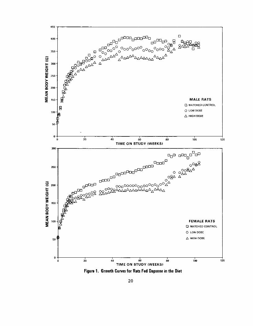
A. Body Weights and Clinical Signs (Rats)
Among males, decreased body weight gains occurred in the high-
dose animals after about 20 weeks on study; animals at the low
dose also had depressed body weights as compared with the matched
controls after approximately 40 weeks· Body weights in both
high- and low-dose female rats were lower than those in the
control group after aproximately 12 weeks· Following withdrawal
of the chemical at week 78, the body weights of these groups
increased (figure 1)·
B. Survival (Rats)
Kaplan and Meier curves estimating the probabilities of survival
for male and female rats fed dapsone in the diet at the doses of
this experiment, together with those of the controls, are shown
in figure 2· In both sexes, the Tarone test results are not
significant at the 0.05 level for positive dose-related trend in
mortality over the period. In male rats, 51% of both treated
groups and 73% of the controls lived to the end of the study·
The overall survival rate was higher in females than in males,
with more than 80% of the treated and control female rats living
to termination of the study. Sufficient animals of both sexes
19
450
400
s X
3 5 0 -
3 0 0
D
_,DD o ° O,
UJ
δ >
s 2 5 0 -
2 0 0 -
< LU
150
1008
MALE RATS
D MATCHED CONTROL
Ο LOW DOSE
Δ HIGH DOSE
5 0
300
20 40 60
TIME ON STUDY (WEEKS) 80 100 120
D
2 5 0 -
2Ι
Ο
Ω Ο GO
< LU
Έ
200 Η
1 5 0 -
100 A
,Ο*ΔΔΔΔ^
D DOG
FEMALE RATS
D MATCHED CONTROL
Ο LOW DOSE
50 s Δ HIGH DOSE
20 40 60 80
TIME ON STUDY (WEEKS)
Figure 1. Growth Curves for Rats Fed Dapsone in the Diet
100 120
2 0

0 6 0 -
< 04°H m Ο QC a. 0 30
D
Ο
Δ
MALE RATS
MATCHED CONTROL
LOW DOSE
HIGH DOSE
30 45 60 75 90 TIME ON STUDY (WEEKS)
105 120
0 90
λ ""Ι-Α ; (ID
0 80
J
>
0 7 0 -
* 0 60
0 )
LL
o > 0 50
BIL
< 0 40 OQ Ο QC
°" 0 30 FEMALE RATS D MATCHED CONTROL
Ο LOW DOSE
Δ HIGH DOSE
0 20
0 10
105 120
TIME ON STUDY (WEEKS)
Figure 2. Survival Curves for Rats Fed Dapsone in the Diet
21

were available for meaningful statistical analyses of the
incidences of late-developing tumors.
C. Pathology (Rats)
Histopathologic findings on neoplasms in rats are summarized in
Appendix A, tables Al and A2; findings on nonneoplastic lesions
are summarized in Appendix C, tables CI and C2.
A variety of neoplasms occurred in both the control and treated
groups. With the exception of malignant lymphomas and
mesenchymal tumors of the abdominal cavity and viscera in treated
male rats, the neoplasms listed in Appendix A either occurred
with approximately equal frequency in treated and matched-control
rats, or occurred in insufficient numbers for accurate evaluation
of dose relationships.
Of the low-dose male rats, 4/35 (11%) had lesions classified as
malignant lymphomas of the histiocytic type. Similar lympho
reticular neoplasms have been observed previously in the Fischer
344 rat independent of any treatment. In some areas, the histio
cytes of the lymphomas had marked pleomorphism including spindle-
cell shapes with various degrees of invasion into adjacent
tissues and the peritoneal cavity. In these areas, the malignant
histiocytes could not be differentiated from spindle cells of
mesenchymal tumors arising from the spleen and peritoneum.
22

Mesenchymal lesions which occurred only in chemical-treated male
rats had the following tissue distribution and incidences:
Tissue-Lesion
Spleen sarcoma, NOS fibroma fibrosarcoma fibrosis metaplasia, osseous
Pancreas fibroma metaplasia, osseous
Abdominal cavity fibroma
Peritoneum sarcoma, NOS fibroma fibrosarcoma fibrosis metaplasia, osseous
Peritoneal cavity metaplasia, osseous
Mesentery metaplasia, osseous
Multiple organs sarcoma, NOS fibrosarcoma metaplasia, osseous
Number of Rats Examined
Not otherwise specified.
Matched Control
JA
JA
JA
JA
JA
JA
JA
14
Low Dose
Ih. 6 (18%)
3 (9%)
33 1 (3%)
35 1 (3%)
35 4 (11%) 1 (3%) 1 (3%) 2 (6%) 2 (6%)
35 1 (3%)
35 1 (3%)
_35
1 (3%)
35
High Dose
32 3 (9%) 10 (31%) 3 (9%) 3 (9%) 6 (19%)
32
1 (3%)
11
33 3 (9%)
3 (9%)
1 (3%)
11
11
33 1 (3%) 1 (3%)
33
23

The group frequencies for animals with lesions of the abdominal
organs or peritoneal tissues were as follows: mesenchymal
tumors, low-dose males 13/35 (37%) and high-dose males 22/33
(67%); splenic and peritoneal fibrosis, low-dose males 4/35 (11%)
and high-dose males 3/33 (9%); and osseous metaplasia, low-dose
males 3/35 (9%) and high-dose males 7/33 (21%).
The mesenchymal tumors were classified as fibromas, fibro
sarcomas, and sarcomas, NOS· They were spindle-cell neoplasms
which varied in morphology and differentiation and had various
degrees of collagen formation. The fibromas had well-differen
tiated fibroblasts with ample collagen, whereas the fibrosarcomas
were more pleomorphic, more anaplastic, and more variable in the
amount of collagen deposited. The poorly differentiated
spindle-cell tumors with little or no collagen production were
classified as sarcomas, NOS. Nonneoplastic proliferation of the
connective tissues, either fibrosis or fibroplasia, showed mature
collagen formation. Osseous metaplasia was associated with
proliferation of connective tissues, and was most often seen in
rats with neoplastic lesions. These osseous lesions were
well-differentiated, localized, and noninvasive. They did not
metastasize.
In addition to the neoplastic and nonneoplastic proliferative
lesions of the connective tissues of the abdomen, a number of
24

other degenerative, proliferative, and inflammatory changes were
also encountered in animals of the treated and control groups.
These nonneoplastic lesions are commonly seen in aged rats.
The spleen appeared to be a primary site affected by dapsone.
Both neoplastic and nonneoplastic proliferative lesions were
often present in the same spleen* These proliferative
mesenchymal lesions ranged from focal areas of fibrosis,
fibroplasia, and fibromas with deposits of mature collagen to
poorly differentiated fibrosarcomas and sarcomas of unspecified
type. In some rats, lesions became disseminated throughout the
peritoneal cavity. The formation of well-differentiated
membranous bone and osseous metaplasia are believed to be
secondary to the neoplastic proliferation of the connective
tissues caused by chemical treatment.
In the judgment of the pathologists, a dose-related increase
occurred in the number and malignancy of abdominal tumors found
in male Fischer 344 rats. Under the conditions of this study,
dapsone was carcinogenic (sarcomagenic) for male but not female
Fischer 344 rats.
D. Statistical Analyses of Results (Rats)
Tables El and E2 in Appendix Ε contain the statistical analyses
of the incidences of those specific primary tumors that were
25

observed in at least 5% of one or more treated groups of either
sex.
In male rats, the Cochran-Armitage test results for positive
dose-related trend in proportions of fibroma of the spleen are
significant when the pooled-control group (P < 0·001) or the
matched-control group (P = 0.012) is used. The Fisher exact test
shows that the incidence in the high-dose group is significantly
higher than that in either the pooled controls (P < 0.001) or the
matched controls (P = 0.016), and the incidence in the low-dose
group is significantly higher than that in the pooled controls
(P = 0.006). The lower limits of the 95% confidence interval of
these relative risks have values greater than one. The
statistical conclusion is that the occurrence of fibroma of the
spleen in male rats is associated with dapsone at the doses of
this experiment. No such tumor was observed in female rats.
Fibrosarcomas of the spleen were found exclusively in the high-
dose males. The Cochran-Armitage test shows probability levels
of 0.023 when the pooled controls are used. The Fisher exact
test results are not significant. The spontaneous rate of this
tumor in the male Fischer 344 historical-control rats compiled to
date by this laboratory is 0/235. No such tumor was observed in
female rats.
26

The analyses of the incidence of sarcoma, NOS, of the spleen in
male rats show significant Cochran-Armitage test results (P =
0.023) when the pooled-control group is used. The Fisher exact
test results are not significant. The incidence observed in the
male historical-control rats compiled to date by this laboratory
is 0/235. No such tumor was observed in females. When the
incidences of fibroma, fibrosarcoma, and sarcoma, NOS, of the
spleen in male rats are grouped for statistical analyses, all
test results are significant (P j< 0.006), indicating a dose
association of this combination of tumors with the chemical.
Malignant lymphomas were observed exclusively in the low-dose
male rats. The Cochran-Armitage test results for linearity are
not significant, but an indicated departure from linear trend is
observed when the pooled-control group (P = 0.003) or the
matched-control group (P = 0.022) is used, since the proportion
in the low-dose group is greater than that in the high-dose
group. The Fisher exact test results show a P value of 0.035
when the incidence in the low-dose group is compared with that in
the pooled-control group, but this is above the 0.025 level
required by the Bonferroni inequality criterion for significance.
No such tumors were observed in female rats.
In male rats, the Cochran-Armitage tests on the proportions of
sarcoma, NOS, of the peritoneum are not significant. The Fisher
27

exact test shows a Ρ value of 0.035 when the incidence in the
low-dose group is compared with that in the pooled-control group,
and the lower limit of the 95% confidence interval for the
relative risk of the low-dose group versus the pooled-control
group has a value greater than one. This positive finding is
accentuated by the zero incidence of this tumor in the 235
Fischer 344 male historical-control rats from similar bioassays
conducted by this laboratory. However, the probability level of
0.035 is above the Bonferroni criterion of 0.025, and therefore,
the significance of the result of the analysis of this specific
tumor is questionable. No such tumors were observed in female
rats.
Fibrosarcomas of the peritoneum of male rats were observed in
both treated groups, and the incidence in the high-dose group was
three times that in the low-dose group. The Cochran-Armitage
test shows a probability level of 0.037 when the pooled-control
group is used, but the Fisher exact test shows that the
incidences in the treated groups are not significantly higher
than the incidence in either of the control groups. This tumor
did not occur in any of the 235 Fischer 344 male historical-
control rats used by this laboratory. When the incidences of
fibrosarcoma or of sarcoma, NOS, of the peritoneum in male rats
are grouped for statistical analysis, increased significance (P <
28

0.014) is observed for both Fisher exact test results and for the
test of linear trend using the pooled controls. The statistical
conclusion is that the combination of these tumors and the
chemical are dose related.
Negative results are observed in the incidence of mammary tumors
in female rats, where the incidence in the pooled controls
exceeds the incidences in the treated groups. There is no other
incidence of tumors at any specific site in either male or female
rats for which the statistical test results are significant in
the positive direction.
29


IV. RESULTS - MICE
A. Body Weights and Clinical Signs (Mice)
Mean body weights of both high- and low-dose male mice were lower
than those of the matched controls from initiation of the study
to about week 70 (figure 3). Among the females, the mean body
weights of the matched controls were unusually high, approaching
50g, and it is difficult to assess the significance of the lower
mean body weights among the treated animals.
When treatment was stopped at week 78, body weights increased.
At this time, mean body weights of the controls decreased to less
than those of the male treated groups, and were equal to those of
the female treated groups.
B. Survival (Mice)
Kaplan and Meier curves estimating the probabilities of survival
for male and female mice fed dapsone in the diet at the doses of
this experiment, together with those of the controls, are shown
in figure 4. In both sexes, the Tarone test results for positive
dose-related trend in mortality over the period are not signifi
cant. In male mice, 73% of the high-dose mice and 67% of the
low-dose mice, but only 8% of the controls, survived to the end
of the study. Early deaths in the male controls were not tumor
31

MALE MICE
D MATCHED CONTROL
Ο LOW DOSE
Δ HIGH DOSE
40 50 60 70 80 90 100 110
TIME ON STUDY (WEEKS)
FEMALE MICE
D MATCHED CONTROL
Ο LOW DOSE
Δ HIGH DOSE
TIME ON STUDY (WEEKS)
Figure 3, Growth Curves for Mice Fed Dapsone in the Diet
32

ι οο a
', Δ
Δ
o - 0--, Δ
(SO
DC D CO LL
Ο >•
<0Q Ο QC 0
40
30
MALE MICE
Π MATCHED CONTROL
Ο LOW DOSE
Δ HIGH DOSE
45 60 75 90
TIME ON STUDY (WEEKS)
1 οο Β
D CO
m <CD
ο QCft
0 40
0 30- FEMALE MICE
D MATCHED CONTROL
Ο LOW DOSE
Λ HIGH DOSE
Figure 4.
45 60 75 90
TIME ON STUDY (WEEKS)
Survival Curves for Mice Fed Dapsone in the Diet
13b
33

associated, since there was no incidence of tumors in this group.
The median time on study was 69 weeks in the controls. Fewer
females than males survived, with only 23% of the high-dose
group, 31% of the low-dose group, and 43% of the controls living
to termination of the study. The median time on study was 92
weeks in the treated groups and 102 weeks in the controls. The
severe early mortality rate in the female mice may have
suppressed the incidence of late-developing tumors.
C. Pathology (Mice)
Histopathologic findings on neoplasms in mice are summarized in
Appendix B, tables Bl and B2; findings on nonneoplastic lesions
are summarized in Appendix D, tables Dl and D2.
A variety of neoplasms occurred in both matched-control and
treated groups. The neoplasms listed in Appendix Β occurred with
approximately equal frequency in treated and control mice, or
appeared in insignificant numbers. No malignant tumors occurred
in male mice of this study, and more neoplasms occurred in
control females than in treated females. No tumor metastases
were recorded in these mice.
In addition to the neoplastic lesions, a number of degenerative,
proliferative, and inflammatory changes were encountered in
34

animals of both the treated and control groups (Appendix D ) .
These nonneoplastic lesions are commonly seen in aged mice;
however, the suppurative lesions involving the lungs were
associated with increased mortalities or decreased life spans in
the treated and control groups of mice. The incidence of
suppurative bronchopneumonia in the male mice was as follows:
matched controls 6/11 (55%), low-dose group 5/33 (15%), and
high-dose group 9/32 (29%). In the female mice the incidence was
as follows: matched controls 9/13 (69%), low-dose group 22/30
(73%), and high-dose group 21/35 (60%). The reduction in life
spans was caused by respiratory disease.
In the judgment of the pathologists, feeding the mice dapsone in
the diet for 18 months resulted in no increase in the incidence
of tumors. Under the conditions of this study, dapsone was not
carcinogenic for B6C3F1 mice.
D. Statistical Analyses of Results (Mice)
Tables Fl and F2 in Appendix F contain the statistical analyses
of the incidences of those specific primary tumors that were
observed in at least 5% of one or more treated groups of either
sex.
In male mice, although the results of the Cochran-Armitage test
35

for positive dose-related linear trend in the incidence of
alveolar/bronchiolar adenoma of the lung are not significant, the
Fisher exact test shows a Ρ value of 0.031 when the incidence in
the low-dose group is compared with that in the pooled-control
group. This probability level, however, is below the Bonferroni
criterion of 0.025 required by the multiple comparisons.
Therefore, association between this incidence of tumors and
dapsone is not confirmed, since the incidence in the high-dose
group is not statistically significant. No such tumor was
observed in female mice.
There is no other incidence of tumors at any specific site in
either male or female mice for which the statistical test results
are significant in the positive direction. When lymphoma and
leukemia in female mice were combined for analysis, the incidence
of leukemia was not included in table F2, since the proportion in
each of the treated groups is less than 5%. A list of the
incidences of each type of tumor is provided in Appendix B,
tables Bl and B2.
In all but one of the 95% confidence intervals, shown in the
tables, the lower limit has a value less than one; this indicates
the negative aspects of the results. It should also be noted
that each of these intervals has an upper limit greater than one,
36

indicating the theoretical possibility of the induction of tumors
by dapsone, which could not be detected under the conditions of
this test.
37


V. DISCUSSION
In this bioassay, dapsone was toxic to both the treated rats and
treated mice, since the mean body weights of these animals were
lower than those of the corresponding matched controls throughout
most of the study. However, the body weights of control female
mice were unusually high. When the drug was discontinued at week
78, treated animals of both species showed varying increases in
body weight.
Dapsone did not affect survival of rats; adequate numbers of
animals survived for meaningful statistical analyses of the
incidences of tumors. Dapsone also did not affect the survival
of mice, as shown by the test for positive dose-related trend,
but suppurative bronchopneumonia was found in some animals in all
matched-control and treated groups. Several control males died
early in the study, while survival of the other groups of mice
was not affected until week 75.
Among rats, mesenchymal tumors of the abdominal organs or
peritoneal tissues occurred in 13/35 low-dose males and 22/33
high-dose males. None occurred among control males or among
control or treated females* The most commonly occurring tumors
were fibroma, fibrosarcoma, or sarcoma, NOS, of the spleen and
the peritoneum. In male rats, these mesenchymal tumors of the
39

spleen occurred in a statistically significant incidence in both
treated groups (low-dose 6/34, Ρ = 0.006; high-dose 14/32, Ρ <
0.001) when compared with pooled controls. In the peritoneum,
the incidences of these mesenchymal tumors were significant in
both treated groups (low-dose 5/35, Ρ = 0.014; high-dose 6/33,
Ρ = 0.005) when compared with the pooled controls. No tumors
related to treatment were found in female rats.
Among the mice, there were no tumors that could clearly be
related to treatment. In the males, alveolar/bronchiolar adenoma
was observed in 5/33 low-dose animals. Although significant (P =
0.031) when compared with pooled controls, the tumors cannot
clearly be associated with treatment, because of the low
incidence in the low-dose group, and because only one tumor was
found in the high-dose group» Similar tumors were not observed
in female mice.
In studies of the toxicity of dapsone in animals, Dhar and
Mukherji (1971) administered daily intraperitoneal injections at
a dose of 20 mg/kg/week (chosen to simulate the human dose) for
22 weeks. In this study, there were minor changes in
hematological values, but none in the histology of the liver or
spleen. Higher doses for shorter periods of time caused anemia
and enlargement and degeneration of the spleen and liver.
40

Bergel (1973) reported that in a feeding study with Wistar rats,
tumors were found in 8/15 animals living more than 17 months, but
none were found in the 15 controls. Five of the eight animals
had tumors similar to those found in significant incidences in
rats in the present bioassay; i.e., mesenchymal tumors of the
peritoneum and organs of the abdominal cavity.
Dapsone inhibits the oxidation of pyruvate in the citric acid
cycle in rats and mice, and this is believed to be related to the
toxicity of the chemical (Wu and Dubois, 1970). It is
metabolized to the monohydroxylamine by N-oxidation; this occurs
to a greater extent in man than in rats (Israili et al. , 1973).
The monohydroxylamine metabolite is excreted in the urine in the
form of various conjugates.
It is concluded that under the conditions of this bioassay,
dapsone was not carcinogenic for female Fischer 344 rats or
B6C3F1 mice of either sex. Dapsone was carcinogenic
(sarcomagenic) for male Fischer 344 rats, causing mesenchymal
tumors in the spleen and the peritoneum.
41


VI. BIBLIOGRAPHY
Armitage, Ρ·, Statistical Methods in Medical Research, John Wiley
& Sons, Inc., New York, 1971, pp. 362-365.
Berenblum, I., ed., Carcinogenicity Testing: A Report of the
Panel of Carcinogenicity of the Cancer Research Commission
-2JL the UICC, Vol. _2, International Union Against Cancer,
Geneva, 1969.
Cox, D. R. , Regression models and life tables. J _ IU_ Statist.
S£c _ t 3A: 187-220, 1972.
Cox, D. R. , Analysis of Binary Data, Methuen & Co., Ltd., London,
1970, pp. 48-52.
Dtiar, D. C. and Mukherji, Α., Effect of long-term administration
of 4,4'-diamino diphenyl sulphone (DDS) in white rats.
Indian J. Exp. Biol. 9:388-390, 1971.
Gart, J. J., The comparison of proportions: a review of
significance tests, confidence limits and adjustments for
stratification. Rev. Int. Stat. Inst. 39^148-169, 1971.
Graham, J. and Graham, R. , Cancer treatment with sulfones. Surg.
Gynec. Obstet. 127:103-107, 1969.
Graham, J., Graham, R. , and Hirabayashi, K. , Recurrent cancer of
the cervix uteri. Surg. Gynec. Obstet. 126:799-804, 1968.
Israili, Z. H. , Cucinell, S. Α., Vaught, J., Davis, Ε. , Lesser,
J. Μ. , and Dayton, P. G., Studies of the metabolism of
dapsone in man and experimental animals: formation of
N-hydroxy metabolites. J_ Pharmacol. Expertl. Therap. 187
(1):138-151, 1973.
Kaplan, E. L. and Meier, P., Nonparametric estimation from
incomplete observations. J^ Amer. Statist. Assoc. 53:
457-481, 1958.
Linhart, M. S. , Cooper, J. Α., Martin, R. L. , Page, N. P., and
Peters, J. Α., Carcinogenesis bioassay data system, Comp.
and Biomed. Res. 7:230-248, 1974.
43

Miller, R· G., Jr. , Simultaneous Statistical Inference,
McGraw-Hill Book Co., New York, 1966, pp. 6-10.
Rollo, I· M., Drugs used in the chemotherapy of malaria, chapter
52 in Goodman, L. S. and Gilman, Α., eds., The
Pharmacological Basis of Therapeutics, Macmillan Publishing
Co., Inc., New York, 1975, pp. 1045-1069.
RSA Corporation, personal communication, Ardsley, Ν. Υ., 1977.
Saffiotti, U., Montesano5 R. , Sellakumar, A. R., Cefis, F., and
Kaufman, D. G. , Respiratory tract carcinogenesis in hamsters
induced by different numbers of administrations of benzo (a)
pyrene and ferric oxide. Cancer Res. 32:1073-1081, 1972.
Tarone, R. E., Tests for trend in life table analysis.
Biometrika 62:679-682, 1975.
Weinstein, L. , antimicrobial agents, drugs used in the
chemotherapy of tuberculosis and leprosy, chapter 60 in
Goodman, L. S. and Gilman, A. eds., The Pharmacological
Basis of Therapeutics, Macmillan Publishing Co., Inc., New
York, 1975, pp. 1201-1223.
Wu, D· L. and Dubois, Κ. P. , Effect of DDS on endogenous
respiration and the oxidation of several substrates by rat
diaphragm. Arch, int. Pharmacodyn. 183:36-45, 1970.
44

APPENDIX A
SUMMARY OF THE INCIDENCE OF NEOPLASMS IN
RATS FED DAPSONE IN THE DIET
45


TABLE A1.
SUMMARY OF THE INCIDENCE OF NEOPLASMS IN MALE RATS FED DAPSONE INTHEDIET
HIGH DOSE
35 33 33
(33) 1 (3%)
(31)
(33)
(32) 3 (9%) 10 (31%) 3 (9%)
(32) 1 (3%)
(32)
ANIMALS INITIALLY IN STUDY ANIMALS NECROPSIED ANIMALS EXAMINED HISTOPATHOLOGICALLY
INTEGUHENTABY SYS1EB
*SUBCUT TISSUE BASAL-CELL CARCINOMA
EESPIRATCBY SYSTEM
#LUNGALVEOLAR/BRONCHIOLAR CARCINOMA OSTEOSARCOMA, METASTATIC
HEMATOPOIETIC SYSTEM
•MULTIPLE ORGANS MALIG.LYMPHOMA, HISTIOCYTIC TYPE UNDIFFERENTIATED LEUKEMIA
#SPLEEN SARCOMA, NOS FIBBCMA FIBEOSARCCMA
CIRCULATORY SYSTEM
NONE
DIGESTIVE SYSTEM
#LIVER HEMANGIOSAfiCOMA
#PANCREAS
CONTROL LOU DOSE
15 35 35
14 35
(14) (35)
(14) (34) 1 (3%)
1 (7%)
(35) 4 (11%)
1 (756)
(14) (34)
6 (18%)
(14) (35)
(14) (33)
# NUMBER OF ANIMALS WITH TISSUE EXAMINED MICROSCOPICALLY * NUMBER OF ANIMALS NECROPSIED
47

TABLE A1. MALE RATS: NEOPLASMS (CONTINUED)
CONTROL LOW DOSE HIGH DOSE
URINARY SYSTEM
NONE
ENDOCRINE SYSTEM
#PITUITARY CHROMOPHOBECHROMOPHOBE
ADENOMA CARCINOMA
(14) 1 (7%)
(28) 2 (7%)
(28) 2 (7%) 2 (7%)
iTHYROID FOLIICULAR-CELL CARCINOMA C-CELL ADENOMA C-CELL CARCINOMA
#PANCREATIC ISLETS ISLET-CELL ADENOMA
(14)
1 (7%)
(14)
(33) 1 (3%) 1 (3%)
1 0%)
(33) 1 (3%)
(28)
1 (4%)
(32)
REPRODUCTIVE SYSTEM
#TESTISINTERSTITIAL-CELL TUMOR
(14) 14 (100%)
(35) 29 (83%)
(31) 28 (90%)
NERVOUS SYSTEM
#BRAINSARCOMA, NOS
(14) 2 (14%)
(32) (32)
SPECIAL SENSE ORGANS
NONE
MUSCULOSKELETAL SYSTEM
NONE
BODY CAVITIES
• ABDOMINAL CAVITYFIBBCMA
(14) (35) (33)
i NUMBER OF ANIMALS WITH TISSUE EXAMINED* NUMBER OF ANIMALS NECROFSIED
MICROSCOPICALLY
48

TABLE A1. MALE RATS: NEOPLASMS (CONTINUED)
CONTROL LOW DOSE HIGH DOSE
•PERIICNEUM (14) (35) ( 3 3 )
SARCOMA, NOS 4 (11%) 3 (9%) FIBROMA 1 (3%) FIBROSARCOMA 1 (3%) 3 (9%)
ALL OTHJER SYSTEMS
•MULTIPLE ORGANS (35) (33) SARCOMA, NOS 1 (3%) FIBROSARCOMA 1 (3%)
ANIMAL DISPOSITION SUHMAhi
ANIMALS INITIALLY IN STUDY 15 35 35 NATURAL DEATHS 10 10 MORIBUND SACRIFICE 7 7 SCHEDULED SACRIFICE ACCIDENTALLY KILLED TERMINAL SACRIFICE 11 18 18 ANIMAL MISSING
d INCLUDES AUT0LY2ED ANIMALS
TUMOR SUMMARY
TOTAL ANIMALS WITH PRIMARY TUTORS* 14 32 32 TOTAL PRIMARY TUMORS 19 54 59
TOTAL ANIMALS WITH BENIGN TUMORS 14 31 30 TOTAL BENIGN TUMORS 15 42 40
TOTAL ANIMALS WITH MALIGNANT TUMORS 12 16 TOTAL MALIGNANT TUMORS 12 19
TOTAL ANIMALS WITH SECONDARY TUMORS# 1 TOTAL SECONDARY TUMORS 1
TOTAL ANIMALS WITH TUMORS UNCERTAINBENIGN OR MALIGNANT
TOTAL UNCERTAIN TUMORS
TOTAL ANIMALS WITH TUMORS UNCERTAINPRIMARY OR METASTATIC
TOTAL UNCERTAIN TUMORS
* PRIMARY TUMORS: ALL TUMORS EXCEPT SECONDARY TUMORS # SECONDARY TUMORS: METASTATIC TOMORS OR TUMORS INVASIVE INTO AN ADJACENT ORGAN
49

TABLE A2.
SUMMARY OF THE INCIDENCE OF NEOPLASMS IN FEMALE RATS FEODAPSONEINTHEDIET
CONTROL LO» DCSE HIGH DOSE
ANIMALS INITIALLY IN STUDY 15 35 35 ANIMALS SECROPSIED 15 34 35 ANIMALS EXAMINED HISTOPATHOLOGICALLI 15 34 35
INTEGUMENTARY SYSTEM
•SUBCUT TISSUE (15) (34) (35) SQUAMOUS CELL PAPILLOMA 1 (3%)
RESPIRATORY SYSTEM
NONE
HEMATOPOIETIC SYSTEM
NONE
CIRCULATORY SYSTEM
NONE
DIGESTIVE SYSTEM
NONE
URINARY SYSTEM
tKIDNEY (15) (34) (34) ADENOCARCINOMA, NOS 1 (3%)
#URINARY BLADDER (15) (3D (33) TRANSITIONAL-CELL CARCINOMA 1 (3%)
ENDOCBINE SYSTEM
•PITUITARY (15) (24) (30)
._£-I4flSL 5-iHSL IJLJIZII # NUMBER OF ANIMALS WITH TISSUE EXAMINED MICROSCOPICALLY * NUMEER OF ANIMALS NECROPSIED
50

TABLE A2. FEMALE RATS: NEOPLASMS (CONTINUED)
CONTROL LOW DOSE HIGH DOSE
#THYFOIE (15) (32) (34) ADENOCARCINOMA, NOS 1 (3%) C-CELL ADENOMA 1 (3%)
HEPBOEUCTIVE SYSTEM
•MAMMARY GLAND (15) (34) (35) ADENOMA, NCS 1 (3%) FIBROADENOMA 1 (7%) 2 (6%) 1 (3%)
#UTERUS (14) (33) (34) ADENOCARCINOMA, NOS 1 (7%) ENDOMETRIAL STROMAL POLYP 3 (21%) 8 (24%) 2 (6%)
NERVOUS SYSTEM
NCNE
SPECIAL SENSE ORGANS
NONE
MUSCULOSKELETAL SYSTEM
NCNE
BODY C&VITIIS
NCNE
ALL OTHER SXSTEMS
• NUMBER OF ANIMALS WITH TISSUE EXAMINED MICROSCOPICALLY * NUMBER OF ANIMALS NECROPSIED
51

TABLE A2. FEMALE RATS: NEOPLASMS (CONTINUED)
CONTROL LOW DOSE HIGH DOSE
ANIMAL DISPOSITION SUMMARY
ANIMALS INITIALLY IN STUDYNATURAL DEATHSMORIBUND SACRIFICESCHEDULED SACRIFICE ACCIDENTALLY KILLED TERMINAL SACRIFICEANIMAL MISSING
15
15
35 4
31
35 2 4
29
3 INCLUDES AUTOLYZED ANIMALS
TUMOR SUMMARY
TOTAL ANIMALS WITH ERIMARY TUMORS* 8 15 14 TOTAL PRIMARY TUMORS 11 20 15
TOTAL ANIMALS WITH BENIGN TUMORS 7 14 13 TOTAL BENIGN TUMORS 10 18 14
TOTAL ANIMALS WITH MALIGNANT TUMORS 1 2 1 TOTAL MALIGNANT TUMORS 1 2 1
TOTAL ANIMALS WITH SECONDARY TUMORSt TOTAL SECONDARY TUMORS
TOTAL ANIMALS WITH TUMORS UNCERTAINBENIGN OR MALIGNANT TOTAL UNCERTAIN TUMORS
TOTAL ANIMALS WITH TUMORS UNCERTAINPRIMARY OR METASTATIC
TOTAL UNCERTAIN TUMORS
* PRIMARY TUMORS: ALL TUMORS EXCEPT SECONDARY TUMORS # SECONDARY TUMORS: METASTATIC TUMORS OR TUMORS INVASIVE INTO AN ADJACENT CRGAN
52

APPENDIX B
SUMMARY OF THE INCIDENCE OF NEOPLASMS IN
MICE FED DAPSONE IN THE DIET
53


TABLE B1.
SUMMARY OF THE INCIDENCE OF NEOPLASMS IN MALE MICE FEDDAPSONEINTHEDIET
CONTROL LOW DOSE HIGH DOSE
ANIMALS INITIALLY IN STUDY 14 35 34 ANIMALS MISSING 1 1 1 ANIMALS NECROPSIED 11 33 33 ANIMALS EXAMINED HXSTOPATHOLOGICALLY 11 33 32
INTEGUMENTARY SYSTEM
•SKIN (11) (33) (33) FIBROMA 1 (3%)
BESPIRATCRY SYSTEM
#LUNG (11) (33) (32) ALVEOLAR/BRONCHIOLAR ADENOMA 5 (15%) 1 (3%)
HEMATOPOIETIC SYSTEM
NONE
CIRCULATORY SYSTEM
NONE
DIGESTIVE SYSTEM
«LIVEB (11) (33) (31) HEPATOCELLULAR ADENOMA 6 (18%) 2 (6%)
URINARY SYSTEM
NONE
ENDOCRINE SYSTEM
•PITUITARY (7) (28) (16) 11611
# NUMBER OF ANIMALS WITH TISSUE EXAMINED MICROSCOPICALLY * NUMBER OF ANIMALS NECROPSIED
55

TABLE B1. MALE MICE: NEOPLASMS (CONTINUED)
#THYBOID FOLLICULAR-CELL ADENOMA
REPRODUCTIVE SYSTEM
NCNE
NERVOUS SYSTEM
NCNE
SPECIAL SENSE ORGANS
•EYE/LACRIMAL GLAND ADENCMA, NOS
MUSCULOSKELETAL SYSTEM
NONE
BODY CAVITIES
NONE
ALL OTHER SYSTEMS
NONE
ANIMAL DISPOSITION SUMMARY
ANIMAiS INITIALLY IN STUDY NATURAL DEATHS MORIBUND SACRIFICE SCHEDULED SACRIFICE ACCIDENTALLY KILLED TERMINAL SACRIFICE ANIMAL MISSING
INCLUDES AuTOkXZ^
CONTROL LOW DOSE HIGH DOSE
(8) (30) (24) 1 (3%)
(11) (33) (33) 1 (3%)
14 35 12 6 4
3 7
1 24 22 1 1
* NUMBER OF ANIMALS WITH TISSUE EXAMINED MICROSCOPICALLY • NUMBER OF ANIMALS SECROPSIED
56

TABLE B1. MALE MICE: NEOPLASMS (CONTINUED)
CONTROL LOW DOSE HIGH DOSE
TOMOR SUMMARY
TOTAL ANIMALS WITH PRIMARY TUMORS* 11 TOTAL PRIMARY TUMORS 13
TOTAL ANIMALS WITH BENIGN TUMORS 11 TOTAL BENIGN TUMORS 13
TOTAL ANIMALS MITH MALIGNANT TUMORS TOTAL MALIGNANT TUMORS
TOTAL ANIMALS WITH SECONDARY TUMORS# TOTAL SECONDARY TUMORS
TOTAL ANIMALS WITH TUMORS UNCERTAINBENIGN OR MALIGNANT TOTAL UNCERTAIN TUMORS
TOTAL ANIMALS HITH TUMORS UNCERTAINPRIMARY OR METASTATIC TOTAL UNCERTAIN TUMORS
• PRIMARY TUMORS: ALL TUMORS EXCEPT SECONDARY TUMORS # SECONDARY TUMORS; METASTATIC TUMORS OR TUMORS INVASIVE INTO AN ADJACENT ORGAN
57

TABLE B2.
SUMMARY OF THE INCIDENCE OF NEOPLASMS IN FEMALE MICE FED DAPSONE IN THE DIET
CONTROL LOW DOSE HIGH DOSE
ANIMALS INITIALLY IN STUDY 14 35 36 ANIMALS HISSING 3 ANIMALS NECROPSIED 13 31 35 ANIMALS EXAMINED HISTOPATHOLOGICALLY 13 31 35
INTEGUMENTARY SYSTEM
*SUBCUT TISSUE (13) (31) (35) FIBRCMA 1 (8%) FIBROSARCOMA 1 (8%) 1 (3%)
RESPIRATORY SYSTEM
#LUNG (13) (30) (35) ALVEOLAR/BRONCHIOLAR CARCINOMA 1 (8%)
HEMATOPOIETIC SYSTEM
•MULTIPLE ORGANS (13) (31) (35) MALIG.LYMPHOMA, LYMPHOCYTIC TYPE 1 (ΘΧ) MALIG.LYMPHOMA, HISTIOCYTIC TYPE 1 (3%)
•PANCREATIC L.NODE (6) (1) MALIG.LYMPHOMA, HISTIOCYTIC TYPE 1 (17%)
•LIVER (13) (30) (35) GRANULOCYTIC LEUKEMIA 1 (8X)
CIRCULATORY SYSTEM
NONE
DIGESTIVE SYSTEM
NONE
URINARY SYSTEM
# NUMBER OF ANIMALS WITH TISSUE EXAMINED MICROSCOPICALLY * NUMBER OF ANIMALS NECROPSIED
58
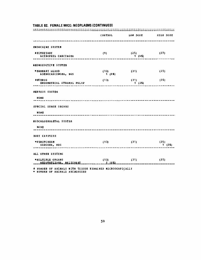
TABLE B2. FEMALE MICE: NEOPLASMS (CONTINUED)
CONTROL LOW DOSE HIGH DOSE
ENDOCRINE SYSTEM
•PITUITARY (9) (25) (27) ACIDOPHIL CARCINOMA 1 («)
REPRODUCTIVE SYSTEM
•MAMMARY GLAND (13) (31) (35) ADENOCARCINOMA, NOS 1 (8*)
#UTERUS (13) (31) (35) ENDOMETRIAL STROMAL POLYP 1 (3%)
NERVOUS SYSTEM
NONE
SPECIAL SENSE ORGANS
NONE
MUSCULOSKELETAL SYSTEM
NCNE
BODY CAVITIES
•PERITONEUM (13) (31) (35) SARCOMA, NOS 1 (3%)
ALL OTHER SYSTEMS
•MULTIPLE ORGANS (13) (31) (35)
# NUMBER OF ANIMALS WITH TISSUE EXAMINED MICROSCOPICALLY • NUMBER OF ANIMALS NECROPSIED
59

TABLE B2. FEMALE MICE: NEOPLASMS (CONTINUED)
ANIMAL DISPOSITION SUMMARY
ANIMALS INITIALLY IN STUDYNATURAL DEATHSMORIEUND SACRIFICESCHEDULED SACRIFICE ACCIDENTALLY KILLEDTERMINAL SACRIFICEANIMAL MISSING
d INCLUDES AUTOLYZED ANIMALS
TUMOR SUMMARY
TOTAL ANIMALS WITH fRIMARY TUMORS*TOTAL PRIMARY TUMORS
TOTAL ANIMALS WITH BENIGN TUMORSTOTAL BENIGN TUMORS
TOTAL ANIMALS WITH MALIGNANT TUttOHSTOTAL MALIGNANT TUMORS
TOTAL ANIMALS WITH SECONDARY TUMORS* TOTAL SECONDARY TUMORS
TOTAL ANIMALS WITH TUMORS UNCERTAINBENIGN OR MALIGNANT
TOTAL UNCERTAIN TUMORS
TOTAL ANIMALS WITH TUMORS UNCERTAINPRIMARY OR METASTATIC TOTAL UNCERTAIN TUMORS
CONTROL
14 4
4
6
6 7
1 1
5 6
LOS DOSE HIGH DOSE
35 36 14 8
9 17
2 7 11 3
4 2 4 2
1 1
3 2 3 2
* PRIMARY TUMORS: ALL TUMORS EXCEPT SECONDARY TUMORS t SECONDARY TUMORSi METASTATIC TUMORS OR TDMORS INVASIVE INTO AN ADJACENT ORGAN
60

APPENDIX C
SUMMARY OF THE INCIDENCE OF NONNEOPLASTIC LESIONS
IN RATS FED DAPSONE IN THE DIET
61


TABLE CI.
SUMMARY OF THE INCIDENCE OF NONNEOPLASTIC LESIONS IN MALE RATS FEDDAPSONEINTHEDIET
CONTROL LOW DOSE
ANIMALS INITIALLY IN STUDY 15 35 ANIMALS NECROPSIED 14 35 ANIMALS EXAMINED HISTOPATHOLOGICALLY 14 35
INTEGUMENTARY SYSTEM
NONE
RESPIRATORY SYSTEM
«TRACHEA (14) (35) INFLAMMATION, SUPPURATIVE 1 (7%) INFLAMMATION, ACUTE/CHRONIC INFLAMMATION, CHRONIC 1 (7%) 1 (3%) INFLAMMATION, CHRONIC SUPPURATIV 1 (7%) 6 (17%)
«LUNG/BRONCHIOLE (14) (34) HYPERPLASIA, LYMPHOID 1 (7%) 1 (3%)
#LUNG (14) (34) INFLAHHATION, INTERSTITIAL INFLAMMATION, CHRONIC SUPPORATIV 1 (3%) BRONCHOPNEUMONIA CHRONIC SUPPURA
HEMATOPOIETIC SYSTEM
«BONE HARROW (13) (32) ATROPHY, NOS 5 (38%) 3 (9%)
«SPLEEN (14) (34) CONGESTION, NOS 1 (3%) HEMORRHAGE 3 (9%) FIBROSIS 3 (9%) NECROSIS, NOS DEPOSIT, NOS 1 (3%) ATROPHY, NOS 1 (3%) METAPLASIA, OSSEOUS HEMATOPOIESIS 1 (3%)
CIRCULATORY SYSTEM
« NUMBER OF ANIMALS WITH TISSUE EXAMINED MICROSCOPICALLY • NUHBEB OF ANIMALS NECROPSIED
63
HIGH
35 33 33
(31) 7 1 1 1
(31) 2
(31) 1
DOSE
(23%) (3%) (3%) (3%)
(6%)
(3%)
1 (3%)
(28) 2
(32)
3 3 1 3
6 2
(7%)
(9%) (9%) (3%) (9%)
(19%) (6%)

TABLE CI. MALE RATS: NONNEOPLASTIC LESIONS (CONTINUED)
CONTROL LOW DOSE HIGH DOSE
DIGESTIVE SYSTEM
#LIVER (14) (35) (32) NECROSIS, FOCAL 1 (3%) CYTOPLASMIC VACUOLIZATION 1 (3%) HEMATOPOIESIS 1 (3%)
#LIVER/CENTRILOBULAR (14) (35) (32) NECROSIS, NOS 2 (6%) 1 (3%) NECRCSIS, DIFFUSE 1 (3*)
•BILE DUCT (14) (35) (33) HYPERPLASIA, NOS 1 (3%)
tPANCREAS (14) (33) (32) METAPLASIA, OSSEOUS 1 (3%)
#PANCREATIC ACINUS (14) (33) (32) ATROPHY, NOS 1 (7%)
URINARY SYSTEM
#KIDNEY (14) (35) (32) INFLAMMATION, INTERSTITIAL 5 (16%) INFLAMMATION, CHRONIC 14 (100%) 22 (63%) 15 (47%)
ENDOCRINE SYSTEM
#ADRENAL CORTEX (14) (35) (32) HYPERPLASIA, NOS 1 (3X)
#THYROID (14) (33) (28) HYPERPLASIA, FOLLICULAR-CELL 1 (4%)
REPRODUCTIVE SYSTEM
•MAMMARY GLAND (14) (35) (33) CYST, NOS 1 (3%)
#PROSTATE (13) (31) (27) INFLAMMATION, SUPPURATIVE 1 (4%)
•SEMINAL VESICLE (14) (35) (33) _ -IMIAMMATION, SUPfUSATiV? 1 13})
# NUMBER OF ANIMALS WITH TISSUE EXAMINED MICROSCOPICALLY • NUMBER OF ANIMALS NECfiOPSIED
64

TABLE C1. MALE RATS: NONNEOPLASTIC LESIONS (CONTINUED)
CONTROL LOW DOSE HIGH DOSE
#TESTIS (35) (31) LYMPHOCYTIC INFILTRATE 1 (3X) 2 (6%) NECROSIS, FAT 1 (7%)
NERVOUS SYSTEM
NONE
SPECIAL SENSE ORGANS
•EYE/CRYSTALLINE LENS (14) (35) (33) MLNERALIZATION 1 (7%)
MUSCULOSKELETAL SYSTEM
NONE
BODY CAVITIES
•PERITONEUM (14) (35) (33) INFLAMMATION, CHRONIC 1 (3%) FIBROSIS 2 (6S) METAPLASIA, OSSEOUS 2 (6X) 1 (3X)
•PERITONEAL CAVITY (14) (35) (33) METAPLASIA, OSSEOUS 1 (3X)
•MESENTERY (14) (35) (33) METAPLASIA, OSSEOUS 1 (3X)
ALL OTHER SYSTEMS
•MULTIPLE ORGANS d<0 (35) (33) METAPLASIA, OSSEOUS 1 (3%)
SPECIAL UORPHOLOGY SUMMARY
NO LESION REPORTED
# NUMBER OF ANIMALS WITH TISSUE EXAMINED MICROSCOPICALLY • NUMBER OF ANIMALS NECROPSIED
65

TABLE C2.
SUMMARY OF THE INCIDENCE OF NONNEOPLASTIC LESIONS IN FEMALE RATS FEDDAPSONEINTHEDIET
CONTROL LCW DCSE
ANIMALS INITIALLY IN STUDY 15 35 ANIMALS KECBOPSIED 15 34 ANIMALS EXAMINED HISTOPATHOLOGICALLY 15 34
INTEGUMENTARY SYSTEM
NCNE
RESPIRAICRY SYSTEM
tTRACHEA LYMPHOCYTIC INFILTBATE INFLAMMATION, SUPPUBATIVE INFLAMMATION, CHRONIC INFLAMMATION, CHRONIC SUPPURATIV
(15)
2 (13%)
(34)
1
3
(3%)
(9%)
#LUNG/BRONCHIOLE HYPERPLASIA, LYMPHOID
(15) 2 (13%)
(33) 1 (3%)
«LUNG INFLAMMATION, INTERSTITIAL BRONCHOPNEUMONIA CHfiONIC SUPPURA
(15) (33)
HEMATOPOIETIC SYSTEM
«BONE MARROW (14) (33) ATROPHY, NOS 8 (57%) 20 (61%)
«SPLEEN (15) (33) HEMATOPOIESIS 1 (7%) 1 (3%)
CIRCOLATOBY SYSTEM
NONE
DIGESTIVE SYSTEM
«LIVER/PERIPORTAL (15) (34)
L.1Z4L # NUMBER OF ANIMALS WITH TISSUE EXAMINED MICROSCOPICALLY * NUMBER OF ANIMALS ΝECBOPSIED
HIGH DOSE
35 35 35
(34) 1 (3%) 2 (6%) 1 (3%)
(35) 2 (6%)
(35) 4 (11%) 1 (3%)
(3 5) 22 (6 3%)
(34)
(34)
66

TABLE C2. FEMALE RATS: NONNEOPLASTIC LESIONS (CONTINUED)
HIGH DOSE
(34) 1 (3%)
(34) 4 (12%) 1 (3%)
(34)
(35)
(34) 2 (6%) 1 (3%)
(34) 7 (21%) 4 (12%)
(34) 1 (3%) 1 (3%)
(34)
(34)
(34)
J-ilSL.
#£ANCREATIC ACINUS ATROPHY, NOS
URINARY SYSTEM
#KIDNEY INFLAMMATION, CHRONIC INFLAMMATION, CHRONIC
ENDOCRINE SYSTEM
#ADRENAL ANGIECTASIS
REPRODUCTIVE SYSTEM
•MAMMABY GLAND CYST# NOS
#UTERUS EDEMA, NOS PYCMETfiA
#UTERU£/ENDOMETRIUM
SUPPURATIV
INFLAMMATION, INFLAMMATION, HYPERPLASIA, HYPERPLASIA,
#OVARY/OVIDUCT INFLAMMATION, INFLAMMATION,
#OVARY CYST, NOS INFLAMMATION,
#OVARY/MEDULLA HYPERPLASIA,
NERVOUS SYSTEM
#BRAIN MALACIA
SUPPURATIVE CHRONIC SUPPURATIV NOS CYSTIC
SUPPURATIVE CHRONIC SUPPURATIV
SUPPURATIVE
NOS
CONTROL
(15) 1 (7%)
(15) 13 (87%)
(15)
(15)
(14)
(14) 7 (50%) 1 (7%) 6 (43%)
(14)
(14)
(14) 1 (7%)
(15)
LOW DOSE
(33)
(34) 16 (47%)
(34) 1 (3%)
(34) 6 (18%)
(33)
11 (33%)
2 (6%)
(33)
(33) 3 (9%) 1 (3%)
(33)
(31)
# NUMBER OF ANIMALS WITH TISSUE EXAMINED MICROSCOPICALLY * NUMBER OF ANIMALS NECROFSIED
67

TABLE C2. FEMALE RATS: NONNEOPLASTIC LESIONS (CONTINUED)
CONTROL LOW DOSE HIGH DOSE
SPECIAL SENSE ORGANS
•EYE/CBYSTALLINE LENS (15) (34) (35) MINERALIZATION 1 OX)
MUSCULOSKELETAL SYSTEM
NONE
BODY CAVITIES
•PERITCNEUM (15) (34) (35) INFLAMMATION# CHBONIC 1 (3%)
•MESENTERY (15) (34) (35) NECROSIS, FAT 1 (3%)
ALL OTHER SYSTEMS
NONE
SPECIAL BORPHOLOGY SUMMARY
NO LESION REPORTED AUTOLYSIS/NO NECROPSY
# NUMBER OF ANIMALS WITH TISSUE EXAMINED MICROSCOPICALLY * NUMBER OF ANIMALS NECROPSIED
68

APPENDIX D
SUMMARY OF THE INCIDENCE OF NONNEOPLASTIC LESIONS
IN MICE FED DAPSONE IN THE DIET
69


TABLE D1.
SUMMARY OF THE INCIDENCE OF NONNEOPLASTIC LESIONS IN MALE MICE FED DAPSONE IN THE DIET
CONTROL LOW DCSE HIGH DOSE
ANIMALS INITIALLY IN STUDY 14 35 34 ANIMALS HISSING 1 1 1 ANIMALS &ECRCPSIED 11 33 33 ANIMALS EXAMINED HISTOPATHOLOGICALLY 11 33 32
INTEGUMENTARY SYSTEM
•SKIN (11) (33) (33) INFLAMMATION, SUPPURATIVE 1 (3%) FIBROSIS 1 (3%)
RESPIRATORY SYSTEM
#LUNG/ERONCHIOLE 1) (33) (32) HYPERPLASIA, PLASMA CELL 1 (3%) HYPERPLASIA, LYMPHOID 2 (6%)
#LUNG 1) (33) (32) INFLAMMATION, INTERSTITIAL 4 (13%) INFLAMMATION, SUPPURATIVE 1 (3%) BRONCHOPNEUMONIA SUPPURATIVE 5 (15%) 4 (13%) ABSCESS, NOS 2 (18%) BRCNCHOENEUMONIA CHRONIC SUPPURA 6 (55%) 5 (16%)
HEMATOPOIETIC SYSTEM
«SPLEEN (9) (33) (32) CONTRACTURE 1 (3%) HEMORRHAGE 2 (6%) HYPERPLASIA, LYMPHOID 1 (3%) 1 (3%) HEMATOPOIESIS 2 (6%) 2 (6%)
•MESENTERIC L. NODE (1) (7) (6) CONGESTION, NOS 1 (14%) 3 (50%) HEMORRHAGE 1 (17%) HYPERPLASIA, LYMPHOID 2 (29%) 1 (17%) HEMATOPOIESIS 1 (14%)
CIRCULATOHY SiSTEB
•MYOCARDIUM (11) (33) (32)
* NOHBEH OF ANIMALS WITH TISSUE EXAMINED MICROSCOPICALLY * NUMBER OF ANIMALS NECBOPSIED
71

TABLE D1. MALE MICE: NONNEOPLASTIC LESIONS (CONTINUED)
CONTROL LOW DOSE HIGH DOSE
INFLAMMATION, CHRONIC 1 (3%)
FIBROSIS, DIFFUSE 1 (3%)
DIGESTIVE SYSTEM
#LIVER (33) (31) (U) FIBROSIS, FOCAL 1 13%) NECROSIS, NOS 1 (3%) HYPERPLASIA, NODULAR 2 (6%) ANGIECTASIS 1 (3%) HYPEEPLASIA, LYMPHOID 1 (3%)
«LIVrR/CENTRILOBULAR f Ί l > (33) (31) NECROSIS, NOS 1 (3%) 1 (3%)
•BILE DUCT (11) (33) (33) CYST, NOS 1 (3%)
«ESOPHAGUS (6) (33) (30) ABSCESS, NOS 1 (3%)
«DUODENUM (6) .(3 3) (32) HYPERPLASIA, ADENOMATOUS 1 (3%)
URINARY SYSTEM
«URINARY BLADDER (9) (32) (32) MUCOCELE 1 (3%)
ENDOCRINE SYSTEM
NONE
REPRODUCTIVE SYSTEM
NONE
NERVOUS SYSTEM
NONE
SPECIAL SENSE ORGANS
« NUMBER OF ANIMALS WITH TISSUE EXAMINED MICROSCOPICALLY • NUMBER OF ANIMALS NECBOPSIED
72

TABLE D1. MALE MICE: NONNEOPLASTIC LESIONS (CONTINUED)
CONTROL LOW DOSE HIGH DOSE
MUSCULOSKELETAL SYSTEM
NCNE
BODY CAVITIES
NCNE
ALL OTHER SYSTEMS
NONE
SPECIAL KORPHOLOGY SUMMARY
NO LESION REPORTED 5 13 10 ANIMAL MISSING/NO NECROPSY 1 1 1 ACCIDENTAL DEATH 1 NECROPSY PERF/NO HISTO PERFORMED 1 AUTOLYSIS/NO NECROPSY 2
73

TABLE D2.
SUMMARY OF THE INCIDENCE OF NONNEOPLASTIC LESIONS IN FEMALE MICE FEDDAPSONE INTHEDIET
CONTROL LOW DOSE HIGH DOSE
ANIMALS INITIALLY IN STUDY 14 35 36 ANIMALS MISSING 3 ANIMALS NECROPSIED 13 31 35 ANIMALS EXAMINED HISTOPATHOLOGICALLY 13 31 35
INTEGUMENTARY SYSTEM
NONE
RESPIRATORY SYSTEM
#TRACHEA (13) (30) (35) INFLAMMATION, SUPPURATIVE 1 (8%) 1 (3%) 3 (9%) INFLAMMATION, CHRONIC SUPPURATIV 1 (3%)
#LUNG (13) (30) (35) INFLAMMATION# INTERSTITIAL 1 (3%) PNEUMONIA, LIPID 1 (3%) BRCNCHOENEUMONIA SUPPURATIVE 9 (69%) 16 (5 3%) 16 (46%) ABSCESS, NOS 2 (7%) BRONCHOPNEUMONIA CHRONIC SUPPURA 6 (20%) 5 (14%) INFLAMMATION, FOCAL GRANULOMATOU 1 (3%) CHOLESTEROL DEPOSIT 1 (3%)
HEMATOPOIETIC SYSTEM
•SPLEEN (13) (28) (34) HYPERPLASIA, LYMPHOID 2 (7%) HEMATOPOIESIS 3 (23%) 5 (18%) 3 (9%)
•PULMONARY LYMPH NODE (6) (1) INFLAMMATION, SUPPURATIVE 1 (17%)
#ABDOMINAL LYMPH NODE (6) (1) HEMATOPOIESIS 1 (17%)
•PANCREATIC L.NODE (6) (1) HYPERPLASIA, LYMPHOID 2 (33%)
•MESENTERIC L. NODE (6) (1)
# NUMBER OF ANIMALS WITH TISSUE EXAMINED MICROSCOPICALLY * NUMBER OF ANIMALS NECROPSIED
74

TABLE D2. FEMALE MICE: NONNEOPLASTIC LESIONS (CONTINUED)
CONTROL LOW DOSE
CIRCULA1CRY SYSTEM
NONE
DIGESTIVE SYSTEM
#LIVER (13) (30) ANGIECTASIS 1 (3%) HEMATOPOIESIS 2 (7%)
#LIVER/CENTRILOBULAR (13) (30) NECROSIS, NOS 1 (8%) 1 (3%)
•BILE DUCT (13) (31) CYST, NOS 1 (8%)
#PANCREATIC ACINUS (13) (29) ATROPHY, NOS 1 (8%)
URINABY SYSTEM
#KIDNEY (13) (31) AMYLOIDOSIS 1 (8%)
ENDOCRINE SYSTEM
NONE
REPfiCDUCIIVE SYSTEM
#UTERUS (13) (31) ABSCESS, NOS 1 (3%)
•UTERUS/ENDOMETRIUM (13) (31) INFLAMMATION, SUPPURATIVE 3 (23%) 3 (10%) INFLAMMATION, CHRONIC SUPPURATIV 1 (3%) HYPERPLASIA, CYSTIC 5 (38%) 16 (52%)
#OVARY (13) (30) ABSCESS, NOS 1 (8%) INFLAMMATION, CHRONIC SUPPURATIV 1 (8%)
NERVOUS SYSTEM
NONE
HIGH DOSE
(35)
(35)
(35)
(34)
(35)
(35)
(35) 2 (6%)
17 (49%)
(35)
75

TABLE D2. FEMALE MICE: NONNEOPLASTIC LESIONS (CONTINUED)
CONTROL LOW DOSE HIGH DOSE
SPECIAL SENSE ORGANS
NONE
MUSCULOSKELETAL SYSTEM
NONE
BODY CAVITIES
•MESENTERYMINERALIZATIONNECRCSIS, NOS
(13) 1 (8%) 1 (8%)
(31) (35)
ALL OTHEB SYSTEMS
NONE
SPECIAL £CRFHOLCGY SUMMARY
NO LESION REPORTEDANIMAL MISSING/NO NECROPSYACCIIENTAL DEATHAUTOLYSIS/NO NECROPSY
# NUMBER OF ANIMALS iITH TISSUE EXAMINED• NUMBER OF ANIMALS NECBOPSIED
1
MICROSCOPICALLY
3 3
1
5
1
76

APPENDIX E
ANALYSES OF THE INCIDENCE OF PRIMARY TUMORS
IN RATS FED DAPSONE IN THE DIET
77


Table El. Analyses of the Incidence of Primary Tumors in Male Rats Fed Dapsone in the Dieta
Topography: Morphology
Matched
Control
Hematopoietic System:
Malignant Lymphoma*3
0/14 (0.00)
Ρ Values0 >
d N.S.
Departure from Linear Trende
Ρ = 0.022
Relative Risk (Matched Control)f
Lower Limit
Upper Limit
VD Relative Risk (Pooled Control)
f
Lower Limit
Upper Limit
Weeks to First Observed Tumor —
Pooled
Control
Low
Dose
High
Dose
0/44 (0.00) 4/35 (0.11) 0/33 (0.00)
N.S. Ρ = 0.035** N.S.
Ρ = 0.003
Infinite
0.400
Infinite
Infinite
1.170
Infinite
— 71 —

Table El. Analyses of the Incidence of Primary Tumors in Male Rats Fed Dapsone in the Dieta
(continued)
Topography: Morphology Matched
Control
Pooled
Control Low Dose
High
Dose
HematopoieticLymphoma or
System: Leukemia 1/14 (0.07) 1/44 (0.02) 4/35 (0.11) 0/33 (0.00)
Ρ Values 0 > d N.S. N.S. N.S. N.S.
Departure from Linear Trende
Ρ = 0.015
Relative Risk (Matched Control)f
Lower Limit
Upper Limit
1.600
0.183
76.389
0.000
0.000
7.860
oo Relative Risk (Pooled Control)f
Lower Limit
Upper Limit
5.029
0.525
239.834
0.000
0.000
24.636
Weeks to First Observed Tumor 106 — 71

Table El. Analyses of the Incidence of Primary Tumors in Male Rats Fed Dapsone in the Dieta
(continued)
Topography: Morphology
Matched
Control
Pooled
Control
Low
Dose
High
Dose
Spleen: Fibroma*5
0/14 (0.00) 0/43 (0.00) 6/34 (0.18) 10/32 (0.31)
Ρ Values0 >
d Ρ 0.012 Ρ < 0.001 Ρ = 0.006** Ρ < 0.001**
Ρ 0.016*
Relative Risk (Matched Control)
Lower Limit
Upper Limit
f Infinite
0.710
Infinite
Infinite
1.406
Infinite
0 0
Relative Risk (Pooled Control)f
Lower Limit
Upper Limit
Infinite
2.041
Infinite
Infinite
4.044
Infinite
Weeks to First Observed Tumor __ 69 91
Spleen: Fibrosarcoma*5 0/14 (0.00) 0/43 (0.00) 0/34 (0.00) 3/32 (0.09)
Ρ Valuesc >
d N.S. Ρ - 0.023 N.S. N.S.
Relative Risk (Matched Control)
Lower Limit
Upper Limit
f Infinite
0.281
Infinite
Relative Risk (Pooled Control)f
Lower Limit
Upper Limit
Infinite
0.815
Infinite
Weeks to First Observed Tumor — — — 95
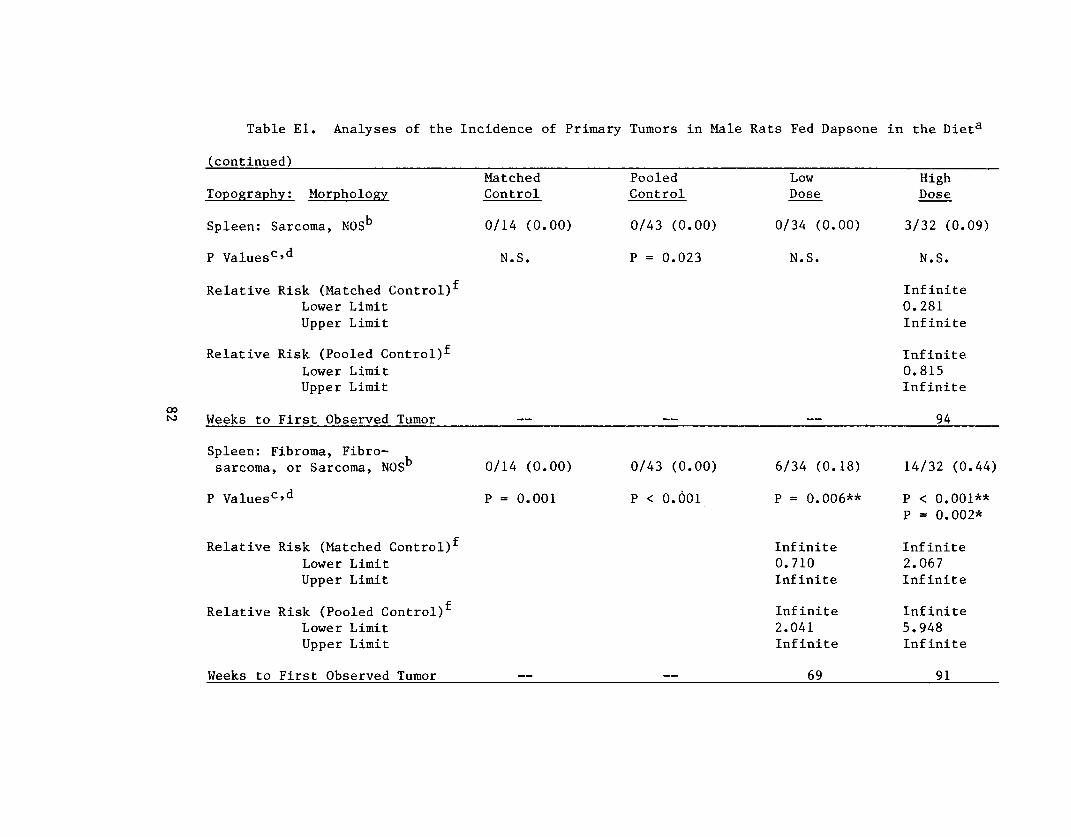
Table El· Analyses of the Incidence of Primary Tumors in Male Rats Fed Dapsone in the Dieta
(continued)
Topography; Morphology
Matched
Control
Spleen: Sarcoma, NOSb
0/14 (0.00)
Ρ Values0 >
d N.S.
Relative Risk (Matched Control)f
Lower Limit
Upper Limit
Relative Risk (Pooled Control)f
Lower Limit
Upper Limit
00
N> Weeks to First Observed Tumor
Spleen: Fibroma, Fibro
sarcoma, or Sarcoma, NOS 0/14 (0.00)
Ρ Values°>d
Ρ = 0.001
Relative Risk (Matched Control)f
Lower Limit
Upper Limit
Relative Risk (Pooled Control)f
Lower Limit
Upper Limit
Weeks to First Observed Tumor
Pooled
Control
Low
Dose
High
Dose
0/43 (0.00) 0/34 (0.00) 3/32 (0.09)
Ρ = 0.023 N.S. N.S.
Infinite
0.281
Infinite
Infinite
0.815
Infinite
94
0/43 (0.00) 6/34 (0.18) 14/32 (0.44)
Ρ < 0.001 Ρ = 0.006** Ρ < 0.001**
Ρ = 0.002*
Infinite
0.710
Infinite
Infinite
2.067
Infinite
Infinite
2.041
Infinite
Infinite
5.948
Infinite
69 91
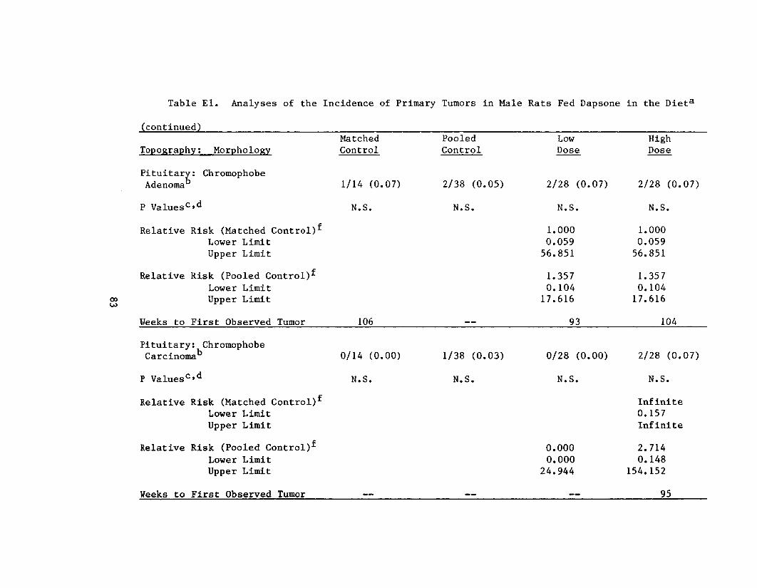
idence of Primary
(continued)
Topography; Morphology Matched
Control
Pituitary :Adenoma
Chromophobe 1/14 (0.07)
Ρ Values0 >d N.S.
Relative Risk (Matched Control)f
Lower Limit
Upper Limit
00
u>
Relative Risk (Pooled Control)f
Lower Limit
Upper Limit
Weeks to First Observed Tumor 106
Pitui tary :Carcinoma
Chromophobe 0/14 (0.00)
Ρ Values0 >d N.S.
Relative Risk (Matched Control)f
Lower Limit
Upper Limit
Relative Risk (Pooled Control)f
Lower Limit
Upper Limit
Weeks to First Observed Tumor
Tumors in Male
Pooled
Control
2/38 (0.05)
N.S.
1/38 (0.03)
N.S.
Rats Fed Dapsone
Low
Dose
2/28 (0.07)
N.S.
1.000
0.059
56.851
1.357
0.104
17.616
93
0/28 (0.00)
N.S.
0.000
0.000
24.944
in the Dieta
High
Dose
2/28 (0.07)
N.S.
1.000
0.059
56.851
1.357
0.104
17.616
104
2/28 (0.07)
N.S.
Infinite
0.157
Infinite
2.714
0.148
154.152
95

Table El· Analyses of the Incidence of Primary Tumors in Male Rats Fed Dapsone in the Dieta
(continued)
Topography: Morphology
Matched
Control
Pituitary: Chromophobe
Adenoma or Carcinoma 1/14 (0.07)
Ρ Valuesc >
d N.S.
Relative Risk (Matched Control)f
Lower Limit
Upper Limit
o°
Relative Risk (Pooled Control)f
Lower Limit
Upper Limit
Weeks to First Observed Tumor 106
Thyroid: C-cell Carcinomab
1/14 (0.07)
Ρ Values0 >
d N.S.
Relative Risk (Matched Control)f
Lower Limit
Upper Limit
Relative Risk (Pooled Control)f
Lower Limit
Upper Limit
Weeks to First Observed Tumor 106
Pooled
Control
3/38 (0.08)
N.S.
—_
2/44 (0.05)
N.S.
—
Low Dose
2/28 (0.07)
N.S.
1.000
0.059
56.851
0.905
0.079
7.325
93
1/33 (0.03)
N.S.
0.424
0.006
32.328
0.667
0.012
12.197
105
High
Dose
4/28 (0.14)
N.S.
2.000
0.229
94.585
1.810
0.329
11.364
95
1/28 (0.04)
N.S.
0.500
0.007
37.877
0.786
0.014
14.261
104

(continued)
Topography: Morphology
Thyroid: C-cell Adenoma or
Carcinoma
Ρ Values0>
d
Relative Risk (Matched Control)f
Lower Limit
Upper Limit
Relative Risk (Pooled Control)f
Lower Limit
Upper Limit oo Ln
Weeks to First Observed Tumor
Testis: Interstitial-cell
Tumorb
Ρ Values0>
d
Relative Risk (Matched Control)f
Lower Limit
Upper Limit
Relative Risk (Pooled Control)f
Lower Limit
Upper Limit
Weeks to First Observed Tumor
:idence of Primary
Matched
Control
1/14 (0.07)
N.S.
106
14/14 (1.00)
N.S.
89
Tumors in Male
Pooled
Control
3/44 (0.07)
N.S.
38/44 (0.86)
N.S.
—
Rats Fed Dapsone
Low
Dose
2/33 (0.06)
N.S.
0.848
0.049
48.540
0.889
0.077
7.282
105
29/35 (0.83)
N.S.
0.829
0.000
1.122
0.959
0.784
1.169
71
in the Dieta
High
Dose
1/28 (0.04)
N.S.
0.500
0.007
37.877
0.524
0.010
6.092
104
28/31 (0.90)
N.S.
0.903
0.000
1.133
1.046
0.858
1.204
75

Table El. Analyses of the Incidence of Primary
(continued)
Matched
Topography: Morphology Control
Peritoneum: Sarcoma, NOS^ 0/14 (0.00)
Ρ Values0>
d N.S.
Relative Risk (Matched Control)f
Lower Limit
Upper Limit
Relative Risk (Pooled Control)f
Lower Limit
Upper Limit
0 0
Weeks to First Observed Tumor
Peritoneum: Fibrosarcoma'5
0/14 (0.00)
Ρ Values0>
d N.S.
Relative Risk (Matched Control)f
Lower Limit
Upper Limit
Relative Risk (Pooled Control)f
Lower Limit
Upper Limit
Weeks to First Observed Tumor —
Tumors in Male
Pooled
Control
0/44 (0.00)
N.S.
0/44 (0.00)
Ρ = 0.037
—
Rats Fed Dapsone
Low
Dose
4/35 (0.11)
Ρ = 0.035**
Infinite
0.400
Infinite
Infinite
1.170
Infinite
80
1/35 (0.03)
N.S.
Infinite
0.022
Infinite
Infinite
0.068
Infinite
105
in the Dieta
High
Dose
3/33 (0.09)
N.S.
Infinite
0.273
Infinite
Infinite
0.808
Infinite
95
3/33 (0.09)
N.S.
Infinite
0.273
Infinite
Infinite
0.808
Infinite
84

Table El. Analyses of the Incidence of Primary Tumors in Male Rats Fed Dapsone in the Dieta
(continued)
Topography: Morphology
Matched
Control
Pooled
Control
Low
Dose
High
Dose
Peritoneum: Fibrosarcoma
or Sarcoma, NOS 0/14 (0.00) 0/44 (0.00) 5/35 (0.14) 6/33 (0.18)
Ρ Values0 >
d N.S. Ρ = 0.006 Ρ = 0.014** Ρ = 0.005**
Relative Risk (Matched Control)f
Lower Limit
Upper Limit
Infinite
0.543
Infinite
Infinite
0.732
Infinite
00
Relative Risk (Pooled Control)f
Lower Limit
Upper Limit
Infinite
1.599
Infinite
Infinite
2.151
Infinite
Weeks to First Observed Tumor — — 80 84
aTreated groups received doses of 600 or 1,200 ppm.
^Number of tumor-bearing animals/number of animals examined at site (proportion).
cBeneath the incidence of tumors in a control group is the probability level for the Cochran-
Armitage test when Ρ < 0.05; otherwise, not significant (N.S.) is indicated. Beneath the
incidence of tumors in a treated group is the probability level for the Fisher exact test for
the comparison of that treated group with the matched-control group (*) or with the pooled-
control group (**) when Ρ < 0.05 for either control group; otherwise, not significant (N.S.)
is indicated.
dA negative trend (N) indicates a lower incidence in a treated group than in a control group.
eThe probability level for departure from linear trend is given when Ρ < 0.05 for any comparison.
95% confidence interval of the relative risk between each treated group and the specified
control group.

Table E2. Analyses of the Incidence of Primary Tumors in Female Rats Fed Dapsone in the Dieta
Topography: Morphology
Matched
Control
Pooled
Control
Low
Dose
High
Dose
Pituitary:
Adenoma
Chromophobe
6/15 (0.40) 9/39 (0.23) 5/24 (0.21) 11/30 (0.37)
Ρ Values0 >
d N.S. N.S. N.S. N.S.
Relative Risk (Matched Control)f
Lower Limit
Upper Limit
0.521
0.161
1.713
0.917
0.409
2.499
00 00
Relative Risk (Pooled Control)f
Lower Limit
Upper Limit
0.903
0.265
2.587
1.589
0.686
3.702
Weeks to First Observed Tumor 106 _— 105 101
Thyroid: C-cell Adenomab
0/15 (0.00) 0/41 (0.00) 1/32 (0.03) 0/34 (0.00)
Ρ Valuesc >
d N.S. N.S. N.S. N.S.
Relative Risk (Matched Control)f
Lower Limit
Upper Limit
Infinite
0.026
Infinite
Relative Risk (Pooled Control)f
Lower Limit
Upper Limit
Infinite
0.069
Infinite
Weeks to First Observed Tumor — — 105 —

00
Table E2. Analyses of the Incidence of Primary Tumor'umorss iinn FemalFemalee
(continued)
Topography; Morphology
Mammary Gland: Fibroadenoma
or Adenoma, NOSb
Ρ Values0>
d
Relative Risk (Matched Control)f
Lower Limit
Upper Limit
Relative Risk (Pooled Control)f
Lower Limit
Upper Limit
Weeks to First Observed Tumor
Uterus: Endometrial Stromal
Polypb
Ρ Values0>
d
Relative Risk (Matched Control)
Lower Limit
Upper Limit
Relative Risk (Pooled Control)f
Lower Limit
Upper Limit
Weeks to First Observed Tumor
Matched Pooled
Control Control
1/15 (0.07) 9/44 (0.20)
N.S. Ρ = 0.008(N)
106
3/14 (0.21) 6/43 (0.14)
N.S. N.S.
106
Rats Fed Dapsone
Low
Dose
2/34 (0.06)
N.S.
0.882
0.051
50.522
0.288
0.032
1.274
93
8/33 (0.24)
N.S.
1.131
0.335
5.920
1.737
0.583
5.440
93
in the Dieta
High
Dose
1/35 (0.03)
Ρ = 0.019**(N)
0.429
0.006
32.715
0.140
0.003
0.931
105
2/34 (0.06)
N.S.
0.275
0.026
2.190
0.422
0.044
2.175
105

Table E2. Analyses of the Incidence of Primary Tumors in Female Rats Fed Dapsone in the Dieta
(continued)
aTreated groups received doses of 600 or 1200 ppm.
^Number of tumor-bearing animals/number of animals examined at site (proportion).
cBeneath the incidence of tumors in a control group is the probability level for the Cochran-
Armitage test when Ρ < 0.05; otherwise, not significant (N.S.) is indicated. Beneath the inci
dence of tumors in a treated group is the probability level for the Fisher exact test for the
comparison of that treated group with the matched-control group (*) or with the pooled-
control group (**) when Ρ < 0.05 for either control group; otherwise, not significant
(N.S.) is indicated.
^A negative trend (N) indicates a lower incidence in a treated group than in a control group.
eThe probability level for departure from linear trend is given when Ρ < 0.05 for any comparison.
95% confidence interval of the relative risk between each treated group and the
specified control group.

APPENDIX F
ANALYSES OF THE INCIDENCE OF PRIMARY TUMORS
IN MICE FED DAPSONE IN THE DIET
91


Table Fl. Analyses of the Incidence of Primary Tumors in Male Mice Fed Dapsone
Topography: Morphology
Lung: Alveolar/Bronchiolar
Adenoma*5
Ρ Values0>
d
Departure from Linear Trende
Relative Risk (Matched Control)f
Lower Limit
Upper Limit
Relative Risk (Pooled Control)f
Lower Limit
Upper Limit
vo
Weeks to First Observed Tumor
Matched
Control
0/11 (0.00)
N.S.
Ρ = 0.041
—
Pooled
Control
0/31 (0.00)
N.S.
Ρ = 0.009
—
Low
Dose
5/33 (0.15)
Ρ = 0.031**
Infinite
0.464
Infinite
Infinite
1.206
Infinite
59
in the Diet a
High
Dose
1/32 (0.03)
N.S.
Infinite
0.020
Infinite
Infinite
0.052
Infinite
107

Table Fl. Analyses of the Incidence of Primary
(continued)
Matched
Topography: Morphology Control
Liver: Hepatocellular
Adenoma 0/11 (0.00)
Ρ Values0>
d N.S.
Departure from Linear Trende
Ρ - 0.050
fRelative Risk (Matched Control)
Lower Limit
Upper Limit
Relative Risk (Pooled Control)f
Lower Limit
Upper Limit
—— Weeks to First Observed Tumor
Pituitary: Chromophobe
Adenoma 0/7 (0.00)
Ρ Values0>
d N.S.
fRelative Risk (Matched Control)
Lower Limit
Upper Limit
Relative Risk (Pooled Control)f
Lower Limit
Upper Limit
Weeks to First Observed Tumor
Tumors in Male
Pooled
Control
3/29 (0.10)
N.S.
0/18 (0.00)
N.S.
Mice Fed Dapsone
Low
Dose
6/33 (0.18)
N.S.
Infinite
0.589
Infinite
1.758
0.417
10.005
89
0/28 (0.00)
N.S.
— —
in the Dieta
High
Dose
2/31 (0.06)
N.S.
Infinite
0.115
Infinite
0.624
0.055
5.058
107
1/16 (0.06)
N.S.
Infinite
0.026
Infinite
Infinite
0.062
Infinite
85

Table Fl. Analyses of the Incidence of Primary Tumors in Male Mice Fed Dapsone in the Dieta
(continued)
aTreated groups received doses of 500 or 1,000 ppm.
^Number of tumor-bearing animals/number of animals examined at site (proportion).
cBeneath the incidence of tumors in a control group is the probability level for the Cochran-
Armitage test when Ρ < 0,05; otherwise, not significant (N.S.) is indicated. Beneath the inci
dence of tumors in a treated group is the probability level for the Fisher exact test for the
comparison of that treated group with the matched-control group (*) or with the pooled-
control group (**) when Ρ < 0.05 for either control group; otherwise, not significant
(N.S.) is indicated.
^A negative trend (N) indicates a lower incidence in a treated group than in a control group.
eThe probability level for departure from linear trend is given when Ρ < 0.05 for any comparison.
95% confidence interval of the relative risk between each treated group and the
specified control group.

Table F2. Analyses of the Incidence of Primary Tumors in Female Mice Fed Dapsone in the Dieta
Topography: Morphology
Matched
Control
Subcutaneous Tissue:
Fibrosarcoma 1/13 (0.08)
Ρ Values0'
d N.S.
Relative Risk (Matched Control)f
Lower Limit
Upper Limit
Relative Risk (Pooled Control)f
Lower Limit
Upper Limit
Weeks to First Observed Tumor 96
Hematopoietic System:
Malignant Lymphoma 1/13 (0.08)
Ρ Values0 >
d N.S.
Relative Risk (Matched Control)^
Lower Limit
Upper Limit
Relative Risk (Pooled Control)f
Lower Limit
Upper Limit
Weeks to First Observed Tumor 107
Pooled
Control
1/36 (0.03)
N.S.
2/36 (0.06)
N.S.
Low
Dose
1/31 (0.03)
N.S.
0.419
0.006
31.891
1.161
0.015
88.229
94
1/31 (0.03)
N.S.
0.419
0.006
31.891
0.581
0.010
10.576
94
High
Dose
0/35 (0.00)
N.S.
0.000
0.000
6.893
0.000
0.000
19.040
1/35 (0.03)
N.S.
0.371
0.005
28.358
0.514
0.009
9.418
108

υ. Table F2. Analyses of the Incidence of Primary Tumors in Female Mice Fed Dapsone in the Dieta
CO
ο (continued)
I Matched Pooled Low High
I Topography: Morphology Control Control Dose Dose -H TO
3 Hematopoietic System: Lymphoma
I or Leukemiab 2/13(0.15) 3/36(0.08) 1/31 (0.03) 1/35 (0.03)
ο -π τι
d N.S. N.S. f? Ρ Values
c> N.S. N.S.
o
\ Relative Risk (Matched Control)r 0.210 0.186
S Lower Limit 0.004 0.003
§ Upper Limit 3.760 3.348 u>
u>
0.387 0.343 Relative Risk (Pooled Control)f
0.008 0.007 Lower Limit 4.516 4.024 Upper Limit
^ Weeks to First Observed Tumor 103 — 94 108
aTreated groups received doses of 500 or 1,000 ppm.
^Number of tumor-bearing animals/number of animals examined at site (proportion).
cBeneath the incidence of tumors in a control group is the probability level for the Cochran-
Armitage test when Ρ < 0.05; otherwise, not significant (N.S.) is indicated. Beneath the inci
dence of tumors in a treated group is the probability level for the Fisher exact test for the
comparison of that treated group with the matched-control group (*) or with the pooled-
control group (**) whe Ρ < 0.05 for either control group; otherwise, not significant
(N.S.) is indicated.
^A negative trend (N) indicates a lower incidence in a treated group than in a control group*
eThe probability level for departure from linear trend is given when Ρ < 0.05 for any comparison.
95% confidence interval of the relative risk between each treated group and the
specified control group.


DHEW Publication No. (NIH) 77-820