toxicologie pulmonaire de nanoparticules biodégradables : effets
TRANSCRIPT

HAL Id: tel-01016697https://tel.archives-ouvertes.fr/tel-01016697
Submitted on 1 Jul 2014
HAL is a multi-disciplinary open accessarchive for the deposit and dissemination of sci-entific research documents, whether they are pub-lished or not. The documents may come fromteaching and research institutions in France orabroad, or from public or private research centers.
L’archive ouverte pluridisciplinaire HAL, estdestinée au dépôt et à la diffusion de documentsscientifiques de niveau recherche, publiés ou non,émanant des établissements d’enseignement et derecherche français ou étrangers, des laboratoirespublics ou privés.
Toxicologie pulmonaire de nanoparticulesbiodégradables : effets cytotoxiques et inflammatoires
sur cellules épithéliales et macrophagesNadège Grabowski
To cite this version:Nadège Grabowski. Toxicologie pulmonaire de nanoparticules biodégradables : effets cytotoxiques etinflammatoires sur cellules épithéliales et macrophages. Médecine humaine et pathologie. UniversitéParis Sud - Paris XI, 2013. Français. <NNT : 2013PA114845>. <tel-01016697>

UNIVERSITÉ PARIS-SUD 11
ECOLE DOCTORALE :
INNOVATION THÉRAPEUTIQUE : DU FONDAMENTAL A L’APPLIQUÉ
PÔLE : PHARMACOTECHNIE ET PHYSICO-CHIMIE PHARMACEUTIQUE
DISCIPLINE :
Pharmacotechnie et Biopharmacie
ANNÉE 2013 - 2014 SÉRIE DOCTORAT N° 1254
THÈSE DE DOCTORAT
soutenue le 13/12/2013
par
Nadège GRABOWSKI
Toxicologie pulmonaire de nanoparticules biodégradables
Effets cytotoxiques et inflammatoires sur cellules épithéliales et macrophages
Directeur de thèse : Elias FATTAL Pr. (UMR CNRS 8612)
Co-directeur de thèse : Hervé HILLAIREAU M.C.U. (UMR CNRS 8612)
Composition du jury :
Présidente du Jury : Saadia KERDINE-ROMER Pr. (INSERM U996) Rapporteurs : Jorge BOCZKOWSKI D.R. (INSERM U955) Françoise PONS Pr. (UMR CNRS 7199) Examinateurs : Didier BAZILE Chercheur (Sanofi-Aventis, Vitry-Sur-Seine) Stéphanie BRIANCON Pr. (UMR CNRS 5007)


3
A Elodie, petit ange, rappelée à Dieu à l’aube de ta vie.
Pas un jour ne passe sans que j’aie une pensée pour toi.
A ma mamie Hélène, qui m’a fait grandir, et qui me manque tant.
A mon père, parti trop tôt,
Sans avoir pu m’apprendre tout ce qu’un père apprend à sa fille.
A mon papi Jean, qui m’a transmis le gène de la science.

4
A tous les doctorants, Les déjà diplômés, ceux en souffrance et les futurs
Au-delà des domaines et des sujets de recherche
A tous les chercheurs qu’il m’a été donné la chance de rencontrer au cours de ces trois années
La thèse Un jour, quelqu’un m’a dit, « Une bonne équipe commence par la solidarité », et c’est
exactement ce que j’ai appris au cours de ces trois années…
Beaucoup nous demande « Qu’es-ce que la thèse » ? Nous répondons rapidement que c’est un
rassemblement d’expériences en labo, de recherches biblio, et puis de rédaction, mais
finalement, en réalité, la thèse est avant tout une expérience humaine qui rassemble les
doctorants de toutes disciplines, peu importe leur domaine et leur sujet de recherche.
Le doctorat nous pousse dans nos retranchements, nous amène à dépasser nos limites. Au
cours des ces 3, 4, voire 5 années, nous traversons des périodes de doutes, lorsque les manips
ne donnent rien, nos capacités et connaissances sont sans arrêt remises en question. Et
pourtant, la joie finie par prendre le dessus, quand enfin nous obtenons quelque chose
d’exploitable, et que notre nom fait son entrée dans la communauté scientifique. Mais, plus
que les connaissances que nous acquérons, c’est une formidable aventure humaine. Nous
tissons des liens exceptionnels avec les gens qui nous entourent, doctorants actuels ou déjà
diplômés, des liens, qui nous le savons, survivront aux années. Nous créons des liens
fraternels au-delà de nos origines et de nos attentes pour nos vies futures, unis par la difficulté
que le projet nous impose, et que seuls les gens qui le vivent ou qui l’ont vécu comprennent.
A la question que tous les étudiants de master qui franchissent la porte du laboratoire nous
posent « Recommencerais-tu ? », il ne faut penser qu’à la finalité, et à la joie du travail
accompli pour leur répondre « oui », et leur donner l’envie de tenter cette expérience, qui
nous a, sans aucun doute, construit pour le restant de notre vie de chercheur, mais aussi pour
notre vie personnelle.

5
Remerciements
Pour commencer, je tiens à remercier chaleureusement mon très cher directeur de thèse, le Pr
Elias Fattal de m’avoir donné l’opportunité de conduire mon doctorat au sein de son unité, et
de son équipe des « Fragiles » après m’avoir acceptée en master 2. J’ai pu me construire un
métier et un avenir. Le labo fut ma maison, l’équipe ma famille au cours de ces presque
4 années, que nous allons prolonger un peu !
Je souhaite également remercier tout aussi chaleureusement le Dr Hervé Hillaireau, mon
encadrant direct au cours des ces années de doctorat, qui ont fait suite à mon master. Les
longues discussions scientifiques, sa rigueur scientifique, et tout son savoir faire ont fait de
moi une jeune chercheuse, et je m’efforcerais de transmettre à mon tour toutes ces qualités
(probablement dans la recherche privée !).
Je remercie également le Dr Nicolas Tsapis, pour toutes les connaissances scientifiques dont il
m’a fait part, sa précieuse aide dès qu’un problème se présent au laboratoire, ou sur un
ordinateur, et surtout pour son coaching pendant la rédaction (il faudrait songer à créer une
PME…).
Je remercie également les Pr Saadia Kerdine-Römer et Marc Pallardy, pour m’avoir permis de
réaliser les manips d’immunologie au sein de leur unité, et de m’avoir en quelque sorte
enseigné cette matière. Merci Saadia d’également de prendre part à mon jury de thèse.
Je voudrais remercier le Pr Françoise Pons et le Dr Jorge Boczkowski d’avoir accepté d’être
les rapporteurs de ce manuscrit, qui rassemble les résultats obtenus au cours de ces années. Mr
Boczkowski vous m’aviez déjà fait l’honneur de participer à mon comité de mi-thèse, j’espère
que la suite des travaux a pris une bonne tournure !
Je tiens également à remercier le Pr Stéphanie Briançon et Dr Didier Bazile d’être les
rapporteurs de ce travail. Pr Briançon, je tenais à ce que vous preniez part à ce jury, pour
« boucler la boucle » !
Je souhaite également remercier le Dr Juliette Vergnaud qui m’a beaucoup aidé au cours de
ces années, avec de longues discussions que j’ai beaucoup appréciées. Je te remercie
également d’avoir relu une partie de ces travaux, ton œil extérieur m’a été très utile !
Je souhaite remercier Simona Mura et Letícia Aragão Santiago, avec qui j’ai eu la chance de
travailler au cours de ce projet. Ces nanoparticules de PLGA n’ont plus aucun secret pour

6
nous !! Osons croire à la science, et imaginons les administrées à des patients lorsqu’un de
nos successeurs les aura chargées d’une molécule active efficace !
Je souhaite adresser un très grand merci à Amélie Dufay-Wojcicki, Nadia Abed et Valentina
Agostoni, avec lesquelles j’ai pu discuter pendant de nombreuses heures de mes manips, et
qui m’ont fait partager tout leur savoir en biologie, immuno et toutes ces autres sciences qui
étaient pour moi un grand mystère ! Je tiens également à remercier Odile Diou et Bénédicte
Sacko-Pradines qui furent là dès le début, Giovanna Giacalone pour sa bonne humeur et son
rire qui habite les couloirs, et Violeta Rodriguez (et ton petit accent que tu ne dois jamais
abandonner !). Nous sommes devenues plus qu’amies, nous sommes une famille, et votre
présence m’a permis de faire face aux difficultés de la thèse.
Je tiens à remercier toutes les personnes qui sont passées un jour par l’UMR, et avec
lesquelles nous avons beaucoup partagé, à la bière du jeudi soir, celle du vendredi soir, et
celle du samedi soir ! Il y a eu tous ceux qui ont vécu au sein de l’équipe V (The best of
course !) Patricia, Thaïs, Chantal, Nathalie, Mounira, Ludivine M, Gopan, Naïla, Inah, Minh,
Regina, Romain, Ambre, Walhan, Christian et tous les autres, Ludivine B. mon alliée de
culture cellulaire, Bettina, Bénédicte, Dario, Laura, Hubert, Olivier, Silvia, Resmi, Zoltàn et
Ahmet « the cyclon guys », Claire, Dunja, Daniel ; la team de la tour B Katia, ma compagne
de TP, Elise, Floriane, Hélène, Christelle, Claudio et Junior « the best friends », ainsi que les
gens que j’ai eu l’occasion de rencontrer à ULLA, Zeina, qui par la suite fut toujours présente
pour m’accueillir dans son labo, et pour discuter de mes manips, Massaba, Evelia, Bret.
J’adresse une pensée à ceux que j’aurai bien malgré moi oublié de citer.
Je voudrais également remercier les personnels statutaires de l’UMR, Patricia Livet, la psy de
l’unité, toujours prête à m’écouter et à activer mon badge 1632 – l’extension de ma poche- et
faire accepter ma voiture sur le parking le week end, Dominique Martin pour son sourire
matinal et quotidien accompagné de son « Bonjour ma bichette », Hélène Chacun et Christian
Ducas pour leur bonne humeur, et Sylvie Zemmour qui à toujours passé mes commandes
urgentes… en urgence !
Enfin cette thèse n’aurait pu être sans toutes les cellules THP-1 et A54ř que j’ai entretenues
en musique, et malmenées au cours des expériences, ni mon petit joujou préféré, le
cytomètre ! Merci à Youtube pour les longues heures d’écoute de Maurice El Medioni, El
Gusto, Chopin et Beethoven, tout au long du traitement des données et de la rédaction !

7
Et comme on le dit si bien, gardons le meilleur pour la fin, je souhaite remercier tous les
membres de ma famille et en particulier ma mère, ma sœur et ma grand-mère pour leur
soutien au cours de toutes ces années d’études et de vie. Sans elles, je ne serais pas qui je suis,
et je ne serais pas où je suis. Maman, tu es mon modèle, tu as fait face à tout ce que la vie t’a
fait subir, et tu t’es toujours sortie la tête haute. Mamie, je ne sais pas si la génétique le
prouvera un jour, mais tu m’as transmis le gène de la science lorsque nous avons observé
ensemble ce pou au microscope que tu avais dérobé à la fac !


Table des matières
9
Remerciements ………………………………………………………………… 5
Table des matières …………………………………………………………….. 9
Abréviations …………………………………………………………………... 11
Introduction générale ………………………………………………………… 15
Travaux antérieurs …………………………………………………………… 19
Evaluation and characterization of lung toxicity of biodegradable nanoparticles 21
Introduction ………………………………………………………………… 21
1. Structure of the respiratory tract ………………………………………… 23 1.1. Anatomy ………………………………………………………….. 23 1.2. Histology ………………………………………………………..... 24
2. Clearance mechanisms in lungs ………………………………………… 26
3. Nanoparticles for lung drug delivery ………………………………......... 27 3.1. Nanoparticles ……………………………………………………... 27 3.2. Administration modes in lung delivery …………………………… 28
3.2.1. Delivery under liquid form ……………………………… 29 3.2.2. Delivery under solid form: Trojan microparticles …….. 29
3.3. Fate of nanomedicine after pulmonary …………………………… 32 3.3.1. In vivo biodistribution …………………………………… 32 3.3.2. Translocation ……………………………………………. 34 3.3.3. Mechanisms of cellular uptake in vitro and in vivo …….. 34
4. Toxicity endpoints ………………………………………………………. 36 4.1. Cell integrity ……………………………………………………… 36 4.2. Apoptosis detection ………………………………………………. 38 4.3. DNA damage – Genotoxicity …………………………………….. 38 4.4. Oxidative stress assay ……………………………………………. 39
4.4.1. Reactive Oxygen Species ………………………………. 40 4.4.2. Reactive Nitrogen Species ……………………………… 40 4.4.3. Superoxide Dismutase …………………………………… 41 4.4.4. Glutathione ……………………………………………… 41
4.5. Inflammatory response …………………………………………… 42 4.5.1. Cytokine assay …………………………………………. 42 4.5.2. Complement activation ………………………………… 43 4.5.3. Polymorphonuclear counting in bronchoalveolar lavages 43
4.6. Mucus interactions ………………………………………………… 43
5. Models for lung nanotoxicology: in vitro, ex vivo, in vivo ……………… 44 5.1. In vitro cell culture for nanoparticle toxicity studies ……………... 45

Table des matières
10
5.2. Co-culture of lung cells in nanotoxicology ……………………….. 47 5.3. Ex vivo models ……………………………………………………. 50
5.3.1. Isolated perfused lungs …………………………………. 50 5.3.2. Ex vitro mucus models …………………………………. 52
5.4. In vivo models …………………………………………………….. 52 5.5. Compared in vitro / ex vivo / in vivo nanotoxicity studies ……….. 53
Conclusion …………………………………………………………………. 55
Travaux expérimentaux ………………………………………………………. 74
Chapitre 1: Toxicité de nanoparticules de PLGA de charge de surface variée vis-à-vis de cellules épithéliales alvéolaires pulmonaires humaines ………… 77
Article 1: Toxicity of surface-modified PLGA nanoparticles towards lung alveolar epithelial cells ……………………………………………….. 80
Chapitre 2 : Le recouvrement de surface des nanoparticules de polymère joue un rôle clef dans la toxicité vis à vis de macrophages dérivés de monocytes humains 106
Article 2: Surface-coating mediates the toxicity of polymeric nanoparticles towards human-like macrophages …………………………………………. 109
Chapitre 3 : Co-culture de cellules épithéliales alvéolaires pulmonaires humaines et de macrophages : Un outil in vitro pertinent et efficace en nanotoxicologie 134
Article 3: Evaluation and characterization of lung toxicity of biodegradable nanoparticles ………………………………………………… 137
Discussion générale …………………………………………………………….. 167
Conclusions et perspectives ……………………………………………………. 190
11
Abbreviations / Abréviations
7-AAD : 7-Aminoactinomycin D AF488 : AlexaFluor 488 AM : Alveolar macrophage / Macrophage alvéolaire ANR : Agence Nationale de la Recherche ANSES : Agence Nationale de la Sécurité Sanitaire, de l’Alimentation, de
l’Environnement et du travail AT : Alveolar type / Type alvéolaire ATP : Adenosine triphosphate BAL : Broncho Alveolar Lavage / Lavage Broncho-alvéolaire BALF : Broncho Alveolar Lavage Fluid BALT : Bronchial Associated Lymphoid Tissue BSA : Bovine Serum Albumin CBA : Cytometric Beads Array CDX : Cluster of differentiation number X CFSE : 5-(and 6)-Carboxyfluorescein diacetate succinimidyl ester Chol : Cholesterol CLSM : Confocal Laser Scanning Microscopy CS : Chitosan / Chitosane DAPI : 4',6'-diamidino-2-phénylindole DCF : β’,7’-dichlorofluorescein DCP : Dicetylphosphate DMSO : Dimethylsulfoxyde DOPE : 1,2-Dioleoyl-sn-glycero-3-phosphoethanolamine DPI : Dry Powder Inhaler DPPC : 1,2-dipalmitoyl-sn-glycero-3-phosphocholine DTPA : Diéthylène triamine pentaacetique acid ELISA : Enzyme-Linked ImmunoSorbent Assay FBS : Fetal Bovine Serum / Sérum de veau fœtal FDA : Food and Drug Administration FITC : Fluorescence IsoThioCyanate FL- : Fluorescence Channe1 FSC : Forward scatter g : Gravity / Gravité GPR : Gluthathione reductase GPX : Glutathione peroxidase GSH : Reduced Sulfydryl form gluthathione GSSG : Oxidized disulfide form gluthatione GST : Gluthathion S-transferase GS-TNB : Glutathion derivate (see TNB) H2DCFDA : 2',7'-dichlorodihydrofluorescein diacetate HSPC : hydrogenated soy phosphatidylcholin ICAM- : Intracellular Adhesion Molecule IFN : Interferon IL-X : Interleukine-X IPL : Isolated Perfused Lung LDH : Lactate dehydrogenase / Lactate déshydrogénase LPS : Lipopolysaccharide

Abréviations
12
MCP- : Monocyte chemoattractant Protein MDI : Metered Dose Inhaler MFI : Mean Fluorescence Intensity MTS : 3-(4,5-dimethylthiazol-2-yl)-5-(3-carboxymethoxyphenyl)-2-(4-sulfophenyl)-
2H-tetrazolium MTT : 3-[4,5-dimethylthiazol-2-yl]-3,5 diphenyl tetrazolium bromide n : Number of independent trial na : Not applicable / Non applicable NADPH : Nicotinamide Adenine Dinucleotide Phosphate-oxidase NBT : Nitroblue tetrazolium / Bleu de tetrazolium nd : Not determined / Non déterminé NIR : Near Infra Red / Proche Infra Rouge NMR : Nuclear Magnetic Resonance NPs : Nanoparticles / Nanoparticules NT : Non treated / Non traitées p : Statistic value / Valeur statistique PBS : Phosphate Buffer Saline PC : Phosphatidylcholine PE : Pycoerythrin PEG : Polyethylene Glycol PEI : Polyethylenimine PF68 : Pluronic F68 (Poloxamer 188) PI (IP) : Propidium Iodide / Iodure de propidium PLA : Polylactic acid PLGA : Poly(lactide-co-glycolide) PMA : Phorbol 12-myristate 13-acetate PMN : PolyMorphoNuclear PS : Polystyrene / Polystyrène PVA : Polyvinyl alcohol / Alcool Polyvinylique Rhod : Rhodamine RNS : Reactive Nitrogen Species / Especès Réactives de l’Azote ROS : Reactive Oxygene Species / Especès Réactives de l’Oxygène SCG : Single Cell Gel SD : Standard deviation / Ecart Type SLN : Solid Lipid nanoparticle SOD : Superoxide dismutase SSC : Side-scatter curves TB : Trypan Blue / Bleu trypan TEER : Trans Epithelial Electric Resistance TEM : Transmission Electronic Microscopy TNB : 5-thio-2-nitrobenzoic acid TNF- : Tumor Necrosis Factor WST : Water soluble Tetrazolium salts XTT : 2,3-bis-(2-methoxy-4-nitro-5-sulfophenyl)-2H-tetrazolium-5-carboxanilide


Introduction
générale

Introduction générale
15
La nanotoxicologie, nouvelle sous-discipline de la toxicologie, a pour but d’étudier la toxicité
de nanoparticules vis-à-vis de l’organisme. L’évaluation de cette toxicité est réalisée par le
suivi de plusieurs paramètres, tels que la viabilité cellulaire, la réponse inflammatoire, le
stress oxydant ou encore les dommages induits à l’ADN. Parmi les nanoparticules
développées, celles conçues pour des nanomédicaments doivent faire l’objet d’études très
poussées. Composé de nanoparticules auxquelles sont associées une ou plusieurs substances
actives (anticancéreux, antibiotique, protéine, gène), le nanomédicament est capable de
transporter ces dernières jusqu’aux cellules/tissus cibles, afin d’augmenter l’efficacité du
traitement tout en diminuant la toxicité. Néanmoins, peu d’études se concentrent sur la
nanotoxicité de ces vecteurs qui doivent pourtant être le plus inertes possibles vis-à-vis de
l’organisme, tout en libérant la substance active au site d’action.
Parmi les différentes voies d’administration des nanomédicaments, la voie pulmonaire est
prometteuse car elle présente de nombreux avantages. En effet, en plus d’être non-invasive
(comparée à la voie parentérale), les traitements peuvent être délivrés pour des applications
locales (traitement de l’asthme ou de la broncho-pneumopathie chronique obstructive) ou
systémiques (traitement du diabète). Dans cette dernière perspective, le passage systémique
s’effectue grâce aux échanges constants entre les alvéoles et la circulation sanguine. En outre,
en raison de la faible activité enzymatique qui règne dans les poumons, les molécules fragiles
sont peu dégradées contrairement à d’autres tissus. Enfin, à l’inverse de certains traitements
administrés par voie orale, l’effet de premier passage hépatique est évité.
Au sein de l’équipe Vectorisation pharmaceutique de Molécules Fragiles de l’Institut Galien
Paris-Sud, un projet de recherche soutenu par l’ANSES (« Emerging Risks »), l’ANR (β00ř
CESA) et le Fond Pour la Recherche Respiratoire se concentre sur les études de toxicité
pulmonaire in vitro et in vivo de nanoparticules polymères qui pourraient être utilisées comme
vecteurs de substance active. Les nanoparticules sont formulées à base d’un polymère
biodégradable et l’ajout de différents polymères stabilisants permet de moduler la charge de
surface, sans en modifier la taille. La toxicité in vitro vis-à-vis de cellules bronchiques a été
précédemment étudiée par Simona Mura et la toxicité in vivo (chez la souris) par Letícia
Aragão Santiago, chacune au cours de leur stage post-doctoral. L’objectif de ce projet de
thèse est d’évaluer la toxicité in vitro de nanoparticules vis-à-vis de cellules épithéliales
alvéolaires et de macrophages en mono et en co-culture.

Introduction générale
16
La première partie de ce manuscrit consiste en une revue de l’état de l’art de la toxicologie
pulmonaire des nanoparticules biodégradables, et sera publiée sous forme d’un chapitre dans
le livre « Targeted Drug Delivery Concept and Design », dans la partie « Nanotoxicology and
Regulatory Issues » (Elsevier).
La seconde partie, découpée en trois chapitres rédigés sous forme d’articles de recherche, fait
état des travaux expérimentaux menés au cours de ce projet de thèse. Le premier chapitre qui
étudie la toxicité des nanoparticules vis-à-vis de cellules épithéliales pulmonaires alvéolaires
(A54ř) a récemment été publié. Le deuxième chapitre s’intéresse à la toxicité de ces mêmes
nanoparticules vis-à-vis de macrophages humains (différenciés des monocytes THP-1). Le
troisième et dernier chapitre décrit l’établissement d’un modèle de co-culture de cellules
épithéliales pulmonaires alvéolaires et de macrophages et démontre son intérêt en
nanotoxicologie.
Au cours d’une discussion générale, tous les résultats in vitro et in vivo actuellement obtenus
dans le cadre de ce projet nanotoxicologie sont mis en parallèle et discutés, afin de dégager
des conclusions générales sur les facteurs modulant la toxicité des nanoparticules.


Travaux
antérieurs

Travaux antérieurs
19
La nanotoxicologie est une sous-discipline émergente de la toxicologie relative aux effets
délétères des nanoparticules (Donaldson et al., 2004). Les travaux décrits dans le présent
chapitre sont centrés sur la nanotoxicologie pulmonaire. Par définition une nanoparticule est
un assemblage d’atomes qui possède au moins l’une de ses dimensions dans le domaine du
nanomonde (1 nm = 10-9 m). Les nanoparticules peuvent être d’origine naturelle, mais le plus
souvent elles sont créées par l’homme, de manière intentionnelle ou pas (Andujar et al.,
β00ř). L’exposition pulmonaire qui constitue la voie d’exposition la plus importante peut
dans ce sens constituer un danger pour l’être humain si les particules inhalées s’avèrent
posséder des effets néfastes. Au niveau de l’arbre pulmonaire, on distingue une forte barrière
physiologique qui fait face à la pénétration de petites particules. En effet, les nanoparticules
inhalées ont de fortes chances d’être piégées dans la couche de mucus, qui, grâce à l’action de
la clairance mucociliaire, seront entrainées vers la sortie. Si l’on se place sur un plan
galénique, la voie pulmonaire est très prometteuse pour l’administration des médicaments
(Patton and Byron, 2007). Parmi ses nombreux avantages, nous pouvons citer sa grande
accessibilité et le fait qu’elle soit non invasive. De plus, grâce à la fine épaisseur des tissus
alvéolaires qui sont très vascularisés, les traitements envisagés peuvent être, certes locaux
(cancer, mucovicidose ou tuberculose par exemple), mais également systémiques (diabète).
Par ailleurs, les nanomédicaments, composés d’un principe actif et d’un nanovecteur, peuvent
être administrés par inhalation ou nébulisation. C’est pour cette raison que nous avons
consacré cette revue de la littérature à l’évaluation et la caractérisation de la toxicité
pulmonaire des nanoparticules biodégradables qui contrairement aux nanoparticules
manufacturées sont peu décrites.
Au cours de ces travaux bibliographiques, l’anatomie et l’histologie de l’arbre pulmonaire
ainsi que ses mécanismes de défense sont présentées dans une première partie. Dans une
seconde partie, les nanoparticules qui peuvent être utilisées pour l’administration pulmonaire
sont détaillées ainsi que les modes d’administration et leur biodisponibilité cellulaire et
tissulaire. La toxicité peut être évaluée selon de nombreux paramètres, présentés et expliqués
dans une troisième partie. Les tests présentés ont fait l’objet d’études de nanotoxicité
concernant principalement les nanoparticules non-biodégradables, mais sont néanmoins
transposables aux nanoparticules biodégradables. Enfin, le système pulmonaire humain étant
complexe, de nombreux modèles in vitro, in vivo et ex vivo sont utilisés pour le mimer, en
partie ou en totalité. Les résultats obtenus après application de nanoparticules biodégradables
sur ces différents modèles sont discutés.

Travaux antérieurs
20
En conclusion, nous montrons comment l’ensemble de ces données contribuent à montrer une
relative innocuité des nanoparticules biodégradables utilisables par voie pulmonaire.

Travaux antérieurs
21
Evaluation and characterization of lung toxicity of biodegradable nanoparticles
Nadege Grabowski, Hervé Hillaireau, Juliette Vergnaud, Elias Fattal
To be published in Targeted Drug Delivery Concept and Design
Issue « Nanotoxicology and Regulatory Issues »
(Book – Elsevier)
Introduction
The term Nanotoxicology was introduced in 2004 by Donaldson and co-workers (Donaldson
et al., 2004) and defined as “a new frontier in particle toxicology relevant to both the
workspace and general environment and to consumer safety”. The concept was aiming to
create a new sub-discipline in toxicology, meeting specific aspects of nanoparticle toxicity
and modifying the existing tests to adapt them to the evaluation of risks associated to
nanoparticles (Figure 1).
Nanoparticles are nowadays being used in a large variety of manufactured products, such as
electronic components, textile or cosmetics (Mu and Sprando, 2010). In addition, very
promising applications in medicine have been highlighted in diagnostic and therapeutic. For
such purposes, nanoparticles can enter the organism using different routes such as inhalation,
oral ingestion, dermal or parenteral route. Despite a high number of promises for therapeutics
and diagnosis, nanoparticles are potentially able for each of these routes of administration to
induce adverse effects and toxicity. Among all the tissues susceptible to be affected by
nanoparticle interaction are the lungs which have been widely explored, mostly for inorganic
nanoparticles (Choi et al., 2009). A large number of in vitro and in vivo studies have reported
cytotoxic effects, inflammatory and oxidative stress responses from lung cells after exposure
to particulate matter, silica (Lin et al., 2006), titanium dioxide (Wang et al., 2008,
Bhattacharya et al., 2009), gold and silver (Bachand et al., 2012) nanoparticles. The
involuntary inhalation of these materials through accidental exposure represents therefore a
high source of toxicity. Nevertheless, the pulmonary route is also an interesting non invasive
route of administration for drug delivery (Patton et al., 2004). Both local and systemic
treatments can be considered taking advantage of the lungs’ large absorption surface area
(around 140 m2), and high vascular permeability with continuous exchanges between alveoli

Travaux antérieurs
22
and blood (Patton, 1996). In addition, the weak enzymatic activity present in lungs is relevant
for administration of poorly stable drugs (peptides / proteins). Administration of drug loaded
nanoparticles can lead to several advantages. Ideally, nanoparticles will not only protect drug
against degradation and transport it to target cells, but also allow a sustained drug release,
decreasing the frequency of administration and subsequent risk of side effects (Sung et al.,
2007). Nanomedicine can be delivered to the lungs by nebulization of an aqueous
nanoparticle suspension or aerosolization of a dry powder of nanoparticles. However, because
of their small size, solid nanoparticles are eliminated by the mucociliary clearance and only
low amounts reach the alveoli. Diverse processes have been employed to transform
nanoparticles into ephemeral microparticles that can first go deeper into the lungs and
secondly turn into nanoparticles once in contact with biological fluids (Tsapis et al., 2002).
Figure 1. General roadmap to assess lung nanotoxicology.
A large variety of nanoparticles can be used to prepare nanomedicines (Mansour et al., 2009).
Polymer-based nanoparticles, solid lipid nanoparticles (SLN) and liposomes have all been

Travaux antérieurs
23
investigated for lung delivery of antibiotics, anti-inflammatory, antineoplastic compounds or
hormones. However, a small number of nanoparticles have been so far proposed as tool to
lung cancer diagnosis by local delivery (Yang et al., 2010, McIntire et al., 1998).
The question of whether nanomedicines are safe remains debated. For this purpose, toxicity
studies should be carried out in vitro, in vivo or ex vivo after exposure to nanomedicine.
Toxicity encompasses different mechanisms, from cellular viability, to inflammatory
response, oxidative stress induction and DNA damage that should be deeply investigated.
In the present chapter, the pulmonary tract architecture and its strong defense barriers are
firstly presented. Secondly, nanoparticles used for lung delivery, and their administration
modes are detailed. The toxicity tests used to investigate toxicity of nanoparticles and the
large variety of pulmonary tract models developed so far are then illustrated by their
application for the evaluation of nanoparticle toxicity.
1. Structure of the respiratory tract
1.1. Anatomy
The respiratory tree is in charge of bringing oxygen to the body and removing carbon dioxide
through continuous gas exchanges with blood circulation. The lung volume is included in the
range of 2.5 (children) to 7 liters (adults) depending on the size lung (Weibel and Gomez,
1962). According to the model proposed by Weibel (Weibel and Gomez, 1962), the
respiratory tree is a hierarchical and symmetric network, divided into 24 generations,
beginning with the trachea (generation 0) and ending with alveolus (generation 23). Each
airway is divided into two airways increasing the specific surface, to carry the air up to
alveolar ducts where gas exchanges occur. During each breath, inhaled air is transported to
the conductive zone (generations 0 to 4), including the trachea and bronchus that divides into
two distal bronchi (dichotomy), in order to reach the respiratory zone. As reviewed by Shelly
et al. (Shelly et al., 1988), the temperature and the moisture of alveolar gases are essential
parameters in normal lungs to maintain physiological conditions. Conditions of optimal
inhaled air were determined as 100 % of humidity, and a temperature closed to the body
(Tsuda et al., 1977), modulated by the nose and the upper respiratory tract.
While the conductive zone represents 10 % of the lung volume, the respiratory zone
represents the lasting 90 % (Weibel and Gomez, 1962). The respiratory zone is composed of

Travaux antérieurs
24
300 million alveoli and 14 million alveolar ducts. The average alveolar diameter in human
lungs is in the range of 250 to 290 µm, among the lung volume. With an average diameter of
400 to 450 µm, 3-4 generations of alveolar ducts are terminating in alveolar sacs that have
exactly the same structure. In addition, 3 generations of respiratory bronchioles, with an
average diameter of 500 µm, are proximal to alveolar ducts. Blood vessels follow exactly the
same pattern than the bronchial tree, with similar dimensions. The pulmonary arteries divide
into 28 generations, with an average diameter of 15 to 25 µm. In the respiratory zone,
277 billions of capillary segments, with an average diameter of 8.3 µm create a total tissue-
blood interface (or alveolar capillary surface) of 60 to 80 m2.
1.2. Histology
The bronchial epithelium is pseudostratified and is composed of four cell types: basal cells,
ciliated cells (allowing mucus elimination), goblet cells (secreting mucus) and Clara cells
(producing mucus, pulmonary surfactant proteins and cytokines, and involved in epithelium
regeneration) (Figure 2). The epithelium thickness is close to 60 µm and covered with 2-
10 µm of an airway fluid (Patton and Byron, 2007, Rubin, 2002). The latter is composed of 2
layers cleared by ciliary beating, the mucus, and the periciliary fluid (consisting of water layer
that cover cilia cells on apical surface) separated by a surfactant layer (Matsui et al., 1998).
Mucus from healthy human lungs is nonhomogeneous and viscous (rheology of a crossed
linked gel) with a pH ranging from 7 to 8.5. Biochemical analyses have shown that human
mucus samples are mostly made of water, containing 7 % solids, 8 to 20 % mucins and less
than 1 % DNA. Secreted mucins (glycoproteins) form a 3-dimensional polymer meshwork
able to trap particles (Rubin, 2002), with pores from 10 to 100 nm (Schuster et al., 2013).
Mucins are made of 70-80 % carbohydrates, 20 % proteins and 1-2 % sulfates. 8 proteins
encoded as MUC are present in mucus layer, with a large majority of MUC5AC (~80 %) and
MUC5B (~20 %) (Rubin, 2002) expressed by goblet cells and glandular mucous cells
respectively (Groneberg et al., 2002). In terminal bronchius, the epithelium has a cuboid
shape with a thickness of 10 µm, is covered with 3 µm of airway fluid and is composed of
ciliated and Clara cells (Patton and Byron, 2007).

Travaux antérieurs
25
Figure 2. Structure of the respiratory tract and cellular composition of human lung bronchial / terminal bronchial and alveolar epithelia. (ATI/II, Alveolar Type I/II cells).

Travaux antérieurs
26
Alveolar type I cells (8 %) involved in gas exchanges co-exist with alveolar macrophages (3-
5 %, up to 19 % in smoker subjects), alveolar type II cells (16 %, cover 7 % of the alveolar
surface), capillary endothelial cells (30 %) and cells in the interstitial spaces (37 %) (Sherman
and Ganz, 1992, Crapo et al., 1982) (Figure 2). The alveolar macrophages perform
phagocytosis of foreign particles or pathogens. The endothelial cells, lining lung blood
vessels, are highly metabolic and are involved in the circulation of bioactive substances such
as peptides, lipids, and prostaglandins. Alveolar type II cells produce lung surfactant
components and are involved in the regeneration of type I pneumocyte cells by
differentiation. In alveolar ducts, the thickness of the epithelium is around 0.1 to 0.2 µm and
covered with 0.07 µm of pulmonary surfactant (Patton and Byron, 2007). The pulmonary
surfactant is composed of lipoproteins, 90 % of which are phospholipids and 10 % are
surfactant proteins (SP), secreted by type II alveolar cells (Sherman and Ganz, 1992): SP-A
(the most abundant), SP-B, SP-C, SP-D. SP-B and SP-C, very small and hydrophobic
proteins, reduce the surface tension at the air-liquid interface of the lung, that is essential to
avoid collapse. SP-A and SP-C, members of collectin family, participate to the innate
immunity, before induction of an antibody-mediated response (Wright, 1997, Sherman and
Ganz, 1992). SP-A and SP-C, present also in non-pulmonary sites, are involved in viruses and
bacteria uptake by phagocytes (Wright, 2005), and SP-A was shown to be involved in the
uptake of nanoparticles covered with different polymers (Ruge et al., 2011).
2. Clearance mechanisms in lungs
Defense mechanisms are both immunological and physiological. The Bronchial Associated
Lymphoid Tissue (BALT) plays a major role in the immune response to all kind of antigens,
with a local response faster and more protective that those initiated by the systemic system
(Randall, 2010). Absent in the lungs of healthy newborn babies, the BALT starts its
development after meeting the first antigens. Generally located along the bronchial airways
and the airway bifurcations, the BALT contains essentially B lymphocytes, dendritic cells,
macrophages, T lymphocytes and M cells (Microfold). M cells present the antigen to dendritic
cells that participate in maintaining the alert BALT.
A major clearance mechanism is also achieved physiologically by the mucociliary escalator.
Between 2 and 24 h after inhalation, most of the foreign particles are expectorated, with a rate
closed to 5 mm/min in peripheral zone and 20 mm/min in trachea (Samet and Cheng, 1994).
The second clearance pathway consists in phagocytosis performed by resident alveolar

Travaux antérieurs
27
macrophages (around 10 macrophages per alveoli – 500 millions of alveoli). Particles are then
enzymatically degraded or driven out by the mucociliary clearance, or brought to trachea-
bronchial lymphatic canals by translocation. Phagocytosis is modulated, not only by particle
size, but also by particle geometry (Sahay et al., 2010). Some inhaled particles can be
metabolized thanks to detoxification enzymes, such as cytochrome P450, but compared to the
other organs such as the liver, this pathway is negligible (Labiris and Dolovich, 2003).
3. Nanoparticles for lung drug delivery
3.1. Nanoparticles
Nanoparticles have been extensively used for lung drug delivery for the administration of a
large variety of drugs (anticancer, anti-psychotic, antibiotic, peptide, protein or nucleic acid)
that have been loaded into lipid or polymeric nanoparticles (Figure 3) (for review see Beck-
Broichsitter et al., 2012, Mansour et al., 2009, Sung et al., 2007). Nanoparticles can induce
both local and systemic delivery after release of the encapsulated drug. Major successes were
obtained with lipid or polymer nanoparticles. For instance, uni or multilamelar liposomes
ranging from 400 nm to 1 µm made from the self organization of glycerophospholipids or non
ionic surfactants were produced for lung delivery. One of their major advantages for
pulmonary administration is their biocompatibility due to their natural origin and their lipid
composition quite often close to the one of the lung surfactant. Liposomes were used to
deliver peptides and proteins to the lung (Li et al., 2011) and were already investigated in
phase II clinical trials to deliver amikacin (Weers et al., 2009) or ciprofloxacin (Bruinenberg
et al., 2010). Furthermore, several studies investigated the potential of solid lipids
nanoparticles (SLN) composed of a solid lipid core (dispersed in water) and a shell of
stabilizers (soy lecithin or poloxamer) (Schwarz et al., 1994). SLN have shown a great
potential for insulin delivery towards lung (Liu et al., 2008).
Travaux antérieurs
28
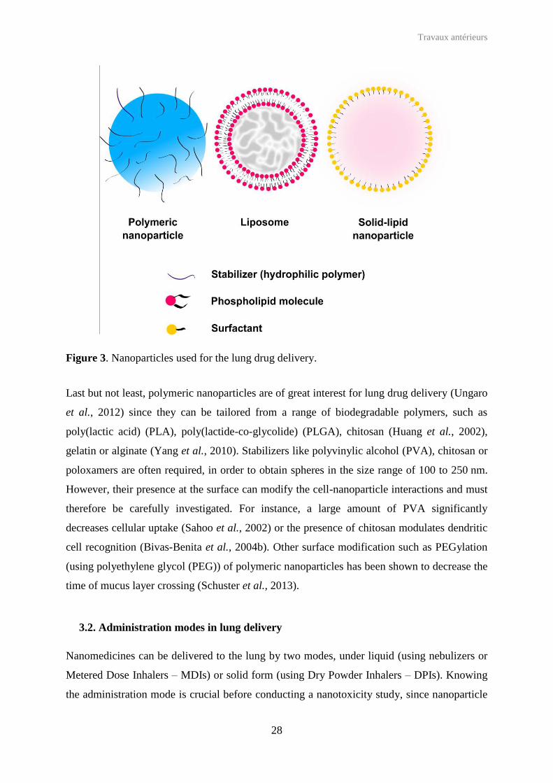
Figure 3. Nanoparticles used for the lung drug delivery.
Last but not least, polymeric nanoparticles are of great interest for lung drug delivery (Ungaro
et al., 2012) since they can be tailored from a range of biodegradable polymers, such as
poly(lactic acid) (PLA), poly(lactide-co-glycolide) (PLGA), chitosan (Huang et al., 2002),
gelatin or alginate (Yang et al., 2010). Stabilizers like polyvinylic alcohol (PVA), chitosan or
poloxamers are often required, in order to obtain spheres in the size range of 100 to 250 nm.
However, their presence at the surface can modify the cell-nanoparticle interactions and must
therefore be carefully investigated. For instance, a large amount of PVA significantly
decreases cellular uptake (Sahoo et al., 2002) or the presence of chitosan modulates dendritic
cell recognition (Bivas-Benita et al., 2004b). Other surface modification such as PEGylation
(using polyethylene glycol (PEG)) of polymeric nanoparticles has been shown to decrease the
time of mucus layer crossing (Schuster et al., 2013).
3.2. Administration modes in lung delivery
Nanomedicines can be delivered to the lung by two modes, under liquid (using nebulizers or
Metered Dose Inhalers – MDIs) or solid form (using Dry Powder Inhalers – DPIs). Knowing
the administration mode is crucial before conducting a nanotoxicity study, since nanoparticle

Travaux antérieurs
29
biodistribution will be different according to their mode of entry, which might impact the type
of cells that will be first in contact with nanoparticles.
3.2.1. Delivery under liquid form
Nebulization is the easiest administration mode to deliver aqueous suspensions of
nanoparticles or liposomes around 200 nm (Zaru et al., 2007), but it is not convenient for the
delivery of large particles (0.3 µm to 3 µm) that can provoke nasal obstructions or be filtered
by the nasal mucosa (Schwab and Zenkel, 1998). To achieve nebulization, two devices are
available: nebulizers (delivery under controlled rate) and MDIs. The MDI is a pressured
device that allows the delivery of uniformed doses of suspension / solution to the patient
(Nowacki et al., 1985). Droplets are formed under pressure and further inhaled by the patient.
MDI were originally developed to deliver anti-asthmatic treatments. Although there is a little
number of applications of MDI for the delivery of nanoparticles, they have demonstrated an
interest for the delivery of chitosan nanoparticles to the deep lung (Sharma et al., 2012). On
the contrary, nebulization is the preferred mode that in most cases is achieved in vivo in small
animals (rats and mice) with a specific device, as example the MicroSprayTM aerosolizer
(Penn-Century for liquid) which was firstly used to deliver an aerosol of fluorescent
nanoparticles to mice (Bivas-Benita et al., 2005). No mortality was induced by the technique,
and one day after administration, mice showed full recovery (Bivas-Benita et al., 2005).
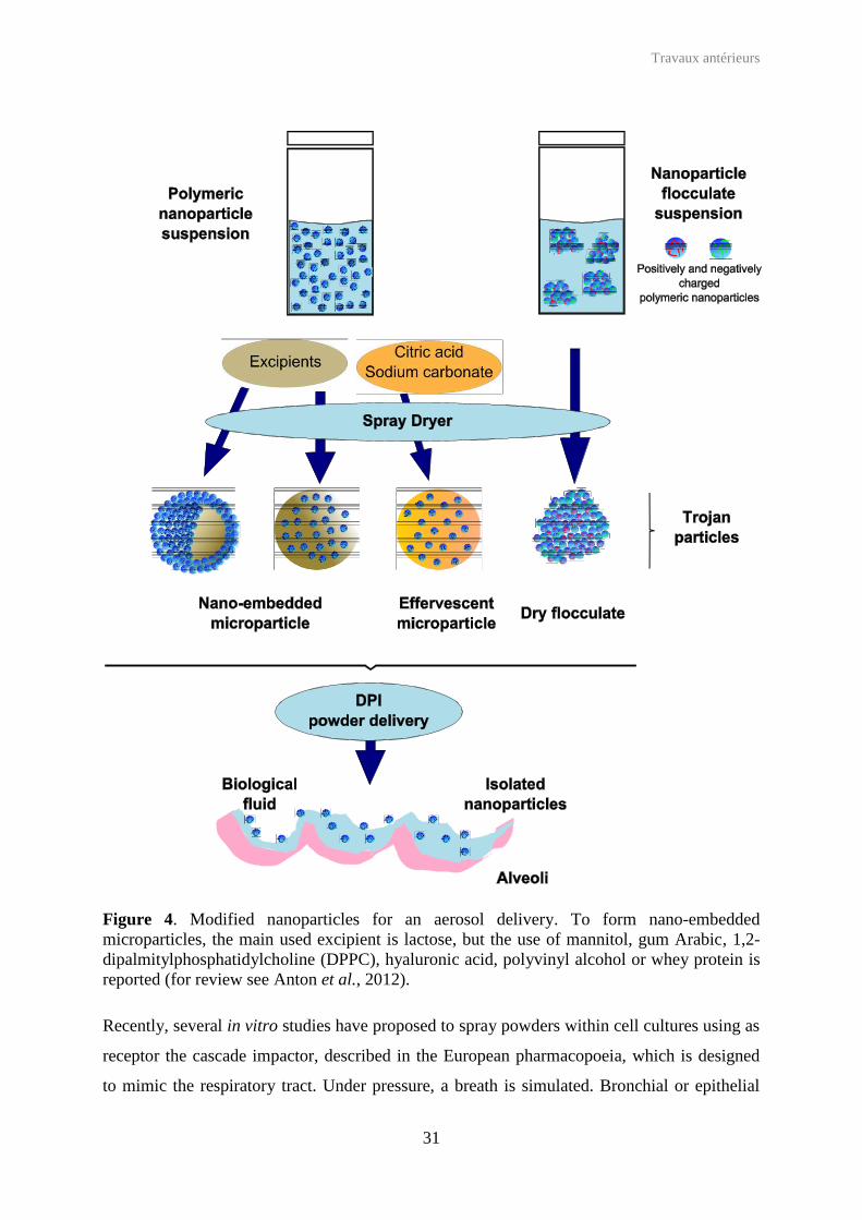
3.2.2 Delivery under solid form: the example of Trojan microparticles
Reproducible doses of solid particles can be delivered using DPI (Newhouse, 1992). A special
veterinary device is available, called Dry Powder InsufflatorTM Pulmonary Aerosol Kit (Penn-
Century for solid) for in vivo studies in small animals (Ohashi et al., 2009). However, solid
nanoparticles are never used as such in DPI because they are too small to be delivered directly
in deep lung regions (i.e alveolar ducts), being eliminated mainly by the mucociliary
clearance. To overpass this constraint, different methods have been employed to deliver
nanoparticle micro-aggregates that turn into the native nanoparticles once reaching the target
(Figure 4). Tsapis et al. have proposed to spray-dry nanoparticles into controlled micro-
aggregates, called Trojan microparticles (Tsapis et al., 2002) that deliver the initial
nanoparticles once in contact with biological fluids. Similarly, insulin was loaded into
chitosan nanoparticles (Grenha et al., 2005) and rifampicin (Tomoda et al., 2008), TAS-103

Travaux antérieurs
30
(anticancer drug) (Tomoda et al., 2009), or dexamethasone (Gómez-Gaete et al., 2008) were
associated to PVA-coated PLGA nanoparticles. The nanoparticles were spray dried in the
presence of trehalose (Tomoda et al., 2008, Tomoda et al., 2009) or 1,2-dipalmitoyl-sn-
glycero-3-phosphocholine (DPPC) and haluronic acid (Gómez-Gaete et al., 2008) forming
microparticles ranging from 2 to 5 µm. For such aerodynamic diameter values, the deposition
in deep lung is favored (Grenha et al., 2005). The efficacy of the decomposition of
microparticles into nanoparticles in contact with water is dependent on the spray-dried
optimal temperature and the ratio of primary nanoparticles (Tomoda et al., 2008). The size of
microparticles is in addition dependent on the diameter of primary nanoparticles. Conversely,
when the spray-drying process is performed at optimal temperature, the fine particle fraction
is 20 to 30 times higher than those of primary nanoparticles, and is almost not affected by the
ratio and the size of primary nanoparticles (Tomoda et al., 2009). However, compared to
primary nanoparticles, the drug was released more rapidly. Indeed, during the spray dried
process, samples can become amorphous that accelerate the moisture absorption and thus the
drug release (Zeng et al., 2007). Shi et al. have formed PLGA nanoparticle flocs (Shi et al.,
2007). In this process, PLGA nanoparticles were pre-formulated by emulsion / solvent
extraction with two oppositely charged polyelectrolytes (PVA vs poly(ethylene-maleic
anhydride)) and then mixed in water under controlled rate. Following electrostatic
interactions, flocs were formed with a large superstructure which were then freeze-dried to
obtain a dried powder formulation. To be delivered in deep lungs, tobramycin was loaded in
pre-formulated PLGA nanoparticles embedded into microparticles after a bath in lactose
aqueous solution (Ungaro et al., 2012). Microparticles were finally freeze dried to be
intratracheally delivered to rats. Last, but not least, the use of large porous effervescent
microparticles to deliver ciprofloxacin (Ely et al., 2007) or doxorubicin (Al -Hallak et al.,
2012, Kim et al., 2012) to the lung was reported. Primary drug-loaded polymeric
nanoparticles (polycyanoacrylate, PLGA) were spray dried with, among other excipients,
lactose (charge), ammonia (to ensure pH to 8) that were mixed to citric acid and sodium
carbonate that cause effervescence in contact with water. Results have shown that the shape,
the size, the density and the fine particle fraction of microparticles are mediated by amounts
of lactose, PEG, leucine (Ely et al., 2007) and bicarbonate (Yang et al., 2009b). Moreover,
thanks to the effervescence of the mixture, the release rate of the ciprofloxacin was
demonstrated higher than with lactose microparticles.
Travaux antérieurs
31
Figure 4. Modified nanoparticles for an aerosol delivery. To form nano-embedded microparticles, the main used excipient is lactose, but the use of mannitol, gum Arabic, 1,2-dipalmitylphosphatidylcholine (DPPC), hyaluronic acid, polyvinyl alcohol or whey protein is reported (for review see Anton et al., 2012).
Recently, several in vitro studies have proposed to spray powders within cell cultures using as
receptor the cascade impactor, described in the European pharmacopoeia, which is designed
to mimic the respiratory tract. Under pressure, a breath is simulated. Bronchial or epithelial

Travaux antérieurs
32
cells have been cultivated on Transwell® at the air-liquid interface and, once the confluence
was reached, they were incorporated to the impactor system and exposed to dry powder. The
complex system allowed studying the effect of active drugs (Hein et al., 2011) or diesel
particles (Cooney and Hickey, 2011) that reached cell cultures by impaction.
Trojan particles have several advantages, such as reduction of the delivered dose, increasing
the drug bioavailability, controlling the release, reducing the toxicity, and as a result
improving the therapeutic index and patient compliance (Anton et al., 2012).
3.3. Fate of nanomedicine after pulmonary delivery
3.3.1. In vivo biodistribution
Biodistribution studies have been performed after nanoparticle delivery in several animal
species (mainly mice and rats), and tissue distribution has been limited to the pulmonary
regions or extended to peripheral organs. Fluorescent or radio-labeled nanoparticles have been
mostly utilized for this purpose.
Ungaro et al. (Ungaro et al., 2012) have administer to rats, through the Dry Insufflator
deviceTM, spray dried / PLGA nano-embedded microparticles (in lactose) containing
rhodamine. After administration, animals were anesthetized, the abdominal cavity incised and
lung infused, firstly with phosphate buffer and then with formaldehyde. Respiratory tissues
were removed, cut into thin slices to be observed and analyzed by fluorescence microscopy
(Ungaro et al., 2010). The results have demonstrated that in vivo nanoparticle distribution was
mediated by surface recovering (Ungaro et al., 2012). Indeed, microparticles were found in
trachea, bronchia and bronchioles when primary nanoparticles were stabilized with PVA,
whereas microparticles were mainly found in alveolar ducts when primary nanoparticles were
stabilized by chitosan (Figure 5).
Other biodistribution studies have shown that after lung delivery into healthy or tumor-
bearing mice, nanoparticle fate differs. Indeed, when fluorescent gelatin nanoparticles were
delivered by nebulization, healthy mice rapidly eliminated nanoparticles towards kidneys,
whereas tumor-bearing mice retained them in lungs, until 24 h (Tseng et al., 2008). Moreover,
30 min after administration, most of the nanoparticles were found in association with
epithelial cells lining the trachea. Gelatin nanoparticles were also indentified in blood vessels,
heart, spleen, brain and liver. After aerosolization of effervescent microparticles (Al-Hallak et

Travaux antérieurs
33
al., 2012, Kim et al., 2012) containing radio-labeled nanoparticles loaded with doxorubicin,
the latest were disseminated in the lung whereas the heart, a tissue sensitive to the toxicity of
doxorubicin, remained free of nanoparticles.
Figure 5. In vivo biodistribution of Rhodamine-PLGA nano-embedded microparticles in rat lungs. Photomicrographs show localization of fluorescent nanoparticles (red) in section of trachea (A), left primary bronchus (B), lobar bronchus (C) and alveolar ducts (D). (Modified from Ungaro et al., 2010).
Finally, free or SLN-loaded radio-labeled amikacin (170 nm, spherical) were delivered to rats
by nebulization or intravenous (iv) administration (Varshosaz et al., 2013). While after lung
delivery of SLN, sustained concentrations were obtained in the lungs, with all other
conditions, the rate of amikacin reaches a maximum 30 min following the delivery, after
which it started to be cleared. Moreover, 6 h after delivery of SLN by pulmonary route, a
significant amount of amikacin was found in the stomach, most likely because the animal
swallowed exhaled SLN. As far as the amikacin rate in blood is concerned, the delivery of
SLN, either by pulmonary route or iv. administration, led to lower drug concentration than
after the administration of free drug.

Travaux antérieurs
34
3.3.2. Translocation
It is suggested that a small fraction of nanoparticles that are not trapped in alveolar
macrophages or in lung epithelial cells have the potential to cross the lung blood-air barrier to
reach extrapulmonary spaces and blood circulation (Muhlfeld et al., 2008). Kunzli and co-
coworkers (Kunzli and Tager, 2005) have reviewed the impact of inhaled air nanoparticles on
the cardio-vascular system. Adverse effects, such as heart rate, blood pressure or
inflammatory state, were attributed to nanoparticle translocation from lung to the blood
circulation. A strong in vivo comparative studies including inorganic (silica, quantum
dots) / organic (polystyrene, human serum albumin) hybrid nanoparticles have demonstrated
that translocation is size and surface-dependent, whereas the composition of the core
(organic / inorganic) does not have an influence (Choi et al., 2010). Regarding the size, it was
shown that below 34 nm, nanoparticles rapidly translocate from the alveolar space to the
lymph nodes, followed by a second translocation towards the bloodstream. Secondly,
zwitterionic, anionic and polar nanoparticles are able to translocate, whereas cationic
nanoparticles are probably trapped by epithelial cells or macrophages which slow down the
translocation. Finally, zwitterionic nanoparticles with an aerodynamic diameter under 6 nm
can be found in lymph nodes within 3 min after exposure. After 30 min they start
accumulating in the kidney before being excreted (Choi et al., 2010).
3.3.3. Mechanisms of cellular uptake in vitro and in vivo
Cellular uptake results from the ability of nanoparticles to cross cell barriers. Nanomedicines
are generally taken up by cells following endocytosis mechanisms, either phagocytosis or
pinocytosis. Phagocytosis is generally performed by phagocytic cells such as macrophages,
whereas pinocytosis is performed by all kind of cells. Pinocytosis mechanisms can be
clathrin-dependent endocytosis or clathrin-independent endocytosis. This last pathway is
classified as caveolae-dependent endocytosis, clathrin and caveolae-independent endocytosis
and macropinocytosis (Sahay et al., 2010, Hillaireau and Couvreur, 2009). To address the
question of uptake by endocytosis, a simple method consists in performing uptake studies
simultaneously at 37 °C and 4 °C (Figure 6). A nanoparticle uptake at 4 °C similar to the one
at 37 °C, characterizes a non-energy dependent mechanism such adsoprtpition whereas
differences of uptake results from endocytosis. Then, to determine the endocytosis pathways,
various chemical (amiloride, chlorpromazine, genestein or filipin) or biological (AP180 or
caveolins) inhibitors can be used to block one or more pathways (Iversen et al., 2011). For

Travaux antérieurs
35
instance, genestein can inhibit several tyrosine kinases that inhibit caveolae pinching, or
caveolin can stabilize receptors in caveolae. Some endocytosis mechanisms are clathrin-
independent and cholesterol-dependent (such as macropynocitosis).
Figure 6. Confocal microscopy imaging (section showing interior of cells) of primary pulmonary epithelium cells after exposure to gelatin nanoparticles at 37°C and 4°C. The cell membrane is bounded with TRITC-concanavalin is coded red and nanoparticle of gelatin bound with FITC is coded green, at 37 °C and 4 °C. (Modified from Brzoska et al., 2004).
In order to visualize nanoparticle trafficking within cells and their exact location, two
microscopic modes may be used, which are confocal laser scanning microscopy (CLSM) as
well as transmission electronic microscopy (TEM). In the first case, fluorescent nanoparticles
are required. For that reason, polymeric nanoparticles can be loaded with fluorescent
compounds, such as 6-coumarin (Panyam et al., 2003) or for ensuring greater stability,
covalently labeled with rhodamine (Mura et al., 2011b) or fluorescein derivates (Huang et al.,
2002), or near infra-red emitting fluorescent probe (Reul et al., 2012). For more accuracy and
complete details, each cell compartment, i.e. endoplasmic reticulum, endosome, golgi
apparatus, lysosome, mitochondria, nucleous or plasma membrane, can be labeled with
specific commercial fluorophores (Cell Staining Simulation Tool Life Technologies). In the
case of transmission electronic microscopy, nanoparticles must be electron dense. Otherwise,
it is not possible to discriminate between nanoparticles and cell vesicles. For this purpose, in
one study, polymeric nanoparticles have been loaded with osmium tetraoxide, to be detectable
(Panyam et al., 2003).

Travaux antérieurs
36
The quantification of the in vitro (Cartiera et al., 2009) cellular uptake is performed by flow
cytometry but some studies use radioactivity sampling or fluorescence intensity
measurements (Yang et al., 2009a, Ohashi et al., 2009).
Nanoparticle cellular fate after pulmonary delivery is a crucial point to be investigated when
carrying out toxicity analyses. Indeed, the same type of nanoparticle can be taken up by all the
previously described ports of entry. Clearance of radioactive-labeled ultrafine nanoparticles
was followed after rat intratracheal intubation (Semmler et al., 2004, Semmler-Behnke et al.,
2007). The nanoparticles were mostly retained in lungs up to 6 months after exposure, and the
clearance was mainly performed by excretion. The small fraction of free nanoparticles found
in bronchoalveolar lavages is mostly associated with alveolar or epithelial cells. Translocation
into the circulation and accumulation in other organs was also reported, but nanoparticle
levels in the extrapulmonary spaces quickly decreased within 7 days (Semmler-Behnke et al.,
2007). According to the size, nanoparticles were either retained by epithelium or sequestered
by alveolar macrophages, beyond the epithelium.
4. Toxicity endpoints
The term toxicity is very large and embraces several parameters (Figure 7). This section
should provide an overview of the different in vitro, in vivo or ex vivo tests reported in the
literature to study nanoparticle-related toxicity.
4.1. Cell integrity
Cell integrity can routinely be restimated by trypan blue, eosin or acridine orange which are
stains that selectively color damaged cells (Tennant, 1964). MTT (3-(4,5-dimethylthiazol-2-
yl)-2,5-diphenyltetrazolium bromide) assay, or equivalents [MTS (3-(4,5-dimethylthiazol-2-
yl)-5-(3-carboxymethoxyphenyl)-2-(4-sulfophenyl)-2H-tetrazolium), XTT (2,3-bis-(2-
methoxy-4-nitro-5-sulfophenyl)-2H-tetrazolium-5-carboxanilide), WST-1 (Water Soluble
Tetrazolium salts)] (Mosmann, 1983) are widely used to assess the in vitro metabolic activity
of cells. In these tests, the tetrazolium dye is reduced to formazan purple crystals in living
cells by Nicotinamide Adenine Dinucleotide Phosphate-Oxidase-(NADPH) dependent oxido-
reductase enzymes. The measurement of the absorbance of the colored solution is directly
related to the cellular metabolic activity.

Travaux antérieurs
37
Figure 7. Nanotoxicology can be expressed by several endpoints: evaluation of mitochondrial activity evolution, membrane integrity, inflammatory response, oxidative stress. Tests are performed in supernatants, or inside lung cells (GSH / GSSG: glutathione sulfhydryl / oxidized disulfide forms, ROS: Reactive oxygen species, RNS: Reactive Nitrogen Species, SOD: Super Oxide Dismutase), apoptosis / necrosis detection, analyses of the complement activation, and DNA damage.)
Membrane integrity can also be evaluated by different methods. One of the most commonly
used tests is the lactate dehydrogenase (LDH) assay, suitable for both in vitro and in vivo
models (Dailey et al., 2006). When cell membrane gets disrupted, the intracellular LDH is
released in extracellular medium. This release, which is quantified by an indirect enzymatic
assay, correlates to an enhancement of membrane permeability. The LDH released reduces
NAD+ into NADH which catalyzes the reduction of a tetrazolium salt to form a formazan
crystal with an absorbance wavelength of 540 nm. By the use of a different principle, the
Live/dead® test – or equivalent – also allows investigating membrane integrity. To achieve
these tests, cells are collected and put in contact with two fluorescent probes able to link

Travaux antérieurs
38
nucleic acids, i) one red-orange, generally propidium iodide (or 7-Aminoactinomycin D (7-
AAD)), an intercalating agent, able to penetrate cells with damaged membrane, and ii) one
green, such as the acetomethoxy derivate of calcein or Syto® 24, able to penetrate all cells.
Cells can then be observed by microscopy, or the intracellular fluorescent level can be
determined by flow cytometry.
4.2. Apoptosis detection
Apoptosis corresponding to programmed cell death is generally opposed to necrosis that is
“accidental” cell death (Kerr et al., 1972). In apoptotic cells, membrane phospholipids
undergo a “flip-flop” event in which phosphatidylserine molecules are translocated to the
outer leaflet, to be recognized by macrophages, without disruption of the membrane in the
early stages (Fadok et al., 1992), whereas the necrotic cell membrane is disrupted.
Quantification of apoptotic cells can be performed by detecting caspase-3, an activated
protease in apoptotic cells (Jänicke et al., 1998), or by flow cytometry after staining of
phosphatidylserines with annexin V, an anticoagulant (Koopman et al., 1994). To
discriminate apoptotic cells from necrotic and intact cells, annexin V, coupled with FITC
(green fluorescence) is combined with propidium iodide (or 7-AAD) (red fluorescence)
(VanEngeland et al., 1996). Annexin V is added in cell culture medium for a short time.
Detached cells are pooled with harvested cells (in case of adherent cells), and propidium
iodide (or 7-AAD) is added. Intact, early apoptotic, necrotic and late apoptotic cells are
respectively untagged, fluorescent in green channel, fluorescent in red channel,
simultaneously fluorescent in both green and red channels. Quantification of each cell
population is performed by flow cytometry. Fluorescent microscopy can give supplementary
morphology-based information concerning the cellular viability of epithelial or bronchial
cells. Indeed, apoptotic cells are characterized by membrane blebbing.
4.3. DNA damage - Genotoxicity
Nanoparticles can penetrate inside cells, causing damage to DNA by single or double strands
breaking. The single cell gel assays (SCG) also called “comet assay” are able to detect DNA
single strand breaks, alkali labile sites or incomplete excision repair sites with a strong
sensitivity to determine the lower DNA damage (Singh et al., 1988). However these tests
require the extraction of single-strand DNA, obtained after different steps including: i) cells
Travaux antérieurs
39
loading into an agarose gel slice, ii) treatment with a lysis solution and iii) DNA unwinding
with an alkaline buffer (Tice et al., 2000). The staining is generally achieved after a
neutralization step with fluorescent compound such as ethidium bromide, propidium iodide or
4',6'-diamidino-2-phénylindole (DAPI), or non fluorescent dye such as the silver nitrate.
Quantitative analysis can be performed by determining the proportion of cells with altered
migration onto the agarose gel, or by classifying them according to the length of migration.
The ability of lung cancer cells to form colonies is another relevant endpoint of cytotoxicity
and genotoxicity (Shoemaker et al., 1985). A very small amount of treated cells were seeded
on plates in order to perform single cell forming colonies. After a long incubation period (few
days), cells were fixed to finally count colonies after trypan blue staining (Jantzen et al.,
2012).
4.4. Oxidative stress assay
Oxygen is required for all aerobic organisms, but it is also a strong oxidant molecule that can
be responsible for undesirable oxidations in cells. This hazardous phenomenon is called
oxidative stress (Sies, 1997) and can be caused by free radicals, reactive oxygen species
(ROS) or reactive nitrogen species (RNS), that are superoxide anion (O2•−), hydroxyl radical
(OH•), nitric oxide (NO•), peroxyl radical (ROO•), hydrogen peroxide (H2O2), ozone (O3),
hypochlorite anion (ClO−), peroxynitrite (NO3−), and also copper and iron ions (Cu2+, Fe3+)
that catalyze Haber-Weiß and Fenton reactions (Sorg, 2004) (Figure 8). In physiological
conditions, reactive species are neutralized by antioxidants, such as super oxide dismutase
(SOD), catalase, glutathione peroxidase (GPX), reductase (GPR) and S-transferase (GST),
ascorbic acid (vitamin C), retinoids (vitamin A), caroteinoids, tocopherols (vitamin E), and
selenium. However, when the organism is exposed to hazardous compounds like pollutants,
ultraviolet, tobacco smoke or nanoparticles, the oxidant / antioxidant balance is destabilized,
and ROS and RNS are formed.
As it was widely shown in the literature, lungs are the site of several oxidative stress reactions
(MacNee, 2001, Li et al., 2008, Repine et al., 1997). For instance, one of the causes of
emphysemia or acute respiratory distress syndrome is related to oxidant release in alveolar
tissues or pulmonary endothelium (Babior, 2000), and SOD plays an antioxidant key role in
asthma and in chronic obstructive pulmonary disease (Tsukagoshi et al., 2000).

Travaux antérieurs
40
4.4.1. Reactive Oxygen Species
To quantify intracellular located ROS, before exposure to nanoparticles, cells are shortly
incubated with 2',7'-dichlorodihydrofluorescein diacetate (H2DCFDA) that penetrates inside
cells (LeBel et al., 1992) and which upon cleavage of the acetate group by intracellular
esterase is converted into β’,7’-dichlorofluorescein (DCF) that expresses a high fluorescent
intensity after ROS oxidation. Quantification is achieved by flow cytometry.
Figure 8. Oxidants (in red) and anti-oxidants (in green) species involved in oxidative stress reactions. ROS = Reactive Oxygen Species, RNS, Reactive Nitrogen Species; SOD, Super Oxide Dismutase; GSH/GSSH, reduced sulfydryl / oxidezed disulfide gluthathione (Modified from Sorg, 2004).
4.4.2. Reactive Nitrogen Species
Contrary to ROS, cells release RNS in the extracellular medium. The most common method
to quantify them is the Griess reagent (Green et al., 1982, Hensley et al., 2003) containing
sulfanilic acid and α-naphtylamine in acetic acid. In presence of RNS, a pink compound that
absorbed at a wavelength of 560 nm will be formed. However, this method has a poor
O2-• SODH2OH2O2ONOO-
OH- + OH• + O2
Fe2+ + H2O2
O2
catalase
GSH GSSG
• O2•
Oxidativeconditions
H2O + • O2•
Metal-ion catalysedHaber-Weiss reaction
Fe(III)/Cu(II)
Fe3+ + OH- + HO.Fenton
NO •
Oxidants (ROS / RNS)Antioxidants

Travaux antérieurs
41
sensitivity, the nitrite concentration being detectable in the range of 1 to 5 µM. The 2,3-
diaminonaphtalene is also able to react with nitrites forming a fluorescent compound
(excitation at 355 nm and emission at 460 nm) allowing the detection of lower nitrite
concentrations (from 0.02 to 10 µM) (Nussler et al., 2006). Moreover, this method can
quantify the nitrates, by pre-converting them into nitrites with NADPH.
4.4.3. Superoxide Dismutase
The SOD catalyzes the dismutation of super oxide radicals (O2•− + O2
•− + 2H+ O2 + H2O2)
(McCord and Fridovich, 1969). Its increase in the extracellular medium characterizes the
presence of superoxide radical that is immediately neutralized by the enzyme. To quantify
SOD, superoxide radical anions are formed in situ. They are either neutralized by the SOD, if
it is present in the extracellular medium, or they react with nitroblue tetrazolium forming
colored nitroblue tetrazolium-formazan (NBT-formazan) (absorbance at a wavelength of
540 nm) (Beauchamp and Fridovich, 1971, Sun et al., 1988). In presence of SOD, production
of NBT-formazan is inhibited and the quantity of SOD is accessible by a colorimetric assay.
4.4.4. Glutathione
Physiologically, glutathione exists within the cell under, both, the reduced sulfhydryl form
(GSH) and the oxidized disulfide form (GSSG). In healthy cells, GSSG is quickly reduced by
glutathione reductase into GSH, but in presence of hifh number of free radicals, GSSG cannot
be reduced. GSH can react with the 5,5’-dithio-bis(2-nitrobenzoic acid) (DTNB) to form the
5-thio-2-nitrobenzoic acid (TNB) (absorbance at a wavelength of 412 nm) and a glutathione
derivate (GS-TNB) (Rahman et al., 2007). The addition of glutathione reductase allows the
reduction of GS-TNB into GSH, and the reduction of GSSG into two molecules of GSH. The
total quantity of glutathione is [GDH]total = [GSH] + 2[GSSG]. To determine the exact amount
of each glutathione form, a second assay can be performed: the excess of 2-vinylpiridine
reacting with GSH is neutralized by triethanolamine. GSSG assay is then performed as
previously described.

Travaux antérieurs
42
4.5. Inflammatory response
4.5.1. Cytokine assay
The immune system is composed of the innate system that is non-specific and the acquired
system that is antibody mediated. After even a slight perturbation in lungs, the immune
response is activated and regulated (amplitude and duration) thanks to low molecular weight
proteins, called cytokines, released in response to cellular signals (Kelley, 1990). Cytokine
denomination includes chemokine, interleukine (IL), growth factor and interferon (IFN).
Every cytokine – that can be secreted in cascade – can cause multiple effects on growth and
differentiation (mitosis, chemotaxis, angiogenesis, cytoskeleton arrangement, immuno-
modulation and extracellular production) in a large variety of cells, even at low concentration
(Kelley, 1990, Galley and Webster, 1996). Pro-inflammatory chemotactic chemokines (IL-8,
Monocyte Chemoattractant Protein (MCP)-1) are able to attract primarily neutrophils and
cells of the innate immune system, regulate cell trafficking (Deshmane et al., 2009), and can
be produced by macrophages or alveolar type-II epithelial-like cells (Sallusto et al., 2000,
Cromwell et al., 1992). By regulating the migration and infiltration of monocytes, MCP-1 is
also involved in various diseases (Polito and Proud, 1998). The tumor necrosis factor-α (TNF-
α), IL-1, IL-6, mostly secreted by alveolar macrophages, are involved in pulmonary diseases;
TNF-α increase transitory, with high levels in a short time, but will cause important injuries in
lung and fever or tachycardia (Kelley, 1990).
The inflammatory response can be quantified by assaying released cytokines in extracellular
medium. Cytokines have been historically quantified by ELISA (Enzyme Linked
Immunosorbent Assay) based on cytokine recognition with specific antibodies linked to an
enzyme detected by a colorimetric reaction. However by ELISA, only one cytokine per
sample can be quantified. More recently new methods have been developed to quantify
several cytokines (until 30) simultaneously (duPont et al., 2005). Among them, BDTM CBA
(Cytometric Beads Array – BD Biosciences) (Tarnok et al., 2003, Morgan et al., 2004),
Luminex®system (Invitrogen), BioPlex cytokine assays (Bio Rad) were successfully used.
Such systems use calibrated beads of different sizes and fluorescence wavelengths that
specifically recognize cytokines. The final addition of antibodies that also recognize each
cytokine allows quantifying the amount of each cytokine. These methods lead to important
improvement and a higher sensitivity compared to ELISA tests (duPont et al., 2005, Djoba
Siawaya et al., 2008).

Travaux antérieurs
43
4.5.2. Complement activation
The complement system is a multiple-component triggered enzyme cascade which attracts
phagocytes to micro-organisms increasing capillarity, permeability and neutrophil chemotaxis
and adhesion (Galley and Webster, 1996). As previously shown, epithelial lining fluid
contains the complement system proteins (Robertson et al., 1976). In one example of
complement activation determination, bovine heat-inactivated serum exposed to nanoparticles
was loaded into the bottom of 96-well neuroprobe chemotaxis chamber, equipped with
polycarbonate filter with 5 µm pores. Murine macrophages were placed at the top of the
insert, in serum free media. The positive control employed was rich in chemotaxis
complement proteins C3a (recognized by phagocytes that will process to engulfment of
nanoparticles) and C5a (chemotaxis for polymorphonuclear). After incubation, the insert was
removed and cells were stained with Romanowsky reagent (mixture of eosin and methylene
blue). Absorbance measurement allows the assessment of the amount of macrophages passed
through the filter, which number was function of complement activation.
4.5.3. Polymorphonuclear counting in bronchoalveolar lavages
Polymorphonuclear (PMN) are locally recruited in lungs and an increased number
characterizes a disorder. By collecting the bronchoalveolar lavages (BAL) on animals, cells
can be characterized and counted on a hematocytometer. After nanoparticle lung delivery,
BAL are withdrawn from anesthetized animals. Bronchoalveolar lavage fluids (BALF) are
then stained with Romanowsky reagent to estimate the number of PMN present.
4.6. Mucus interactions
As the mucus is a strong defense barrier in the pulmonary tract, interactions with inhalable
nanoparticles are important, and different parameters can be investigated to characterize
mucus alterations. To begin with, an overproduction of mucus is synonym of airway
inflammation. Four mucins are generally quantified: MUC2, MUC5AC, MUC5B and
MUC19. The quantification of these proteins or their respective mRNA expression can give
essential information about the mucus state. The quantity of mucin in BALF can be obtained
by ELISA (Lin et al., 1989, Phillips et al., 2006) or electrophoresis (Spurr-Michaud et al.,
2007). Both methods use multiple anti-mucin antibodies, specific of each mucin proteins. To
investigate the passage of insoluble fluorescent particles across the mucus layer, artificial

Travaux antérieurs
44
mucus composed of water, DNA, egg yolk emulsion, mucin, pentetic acid (DTPA), NaCl,
KCl and cell culture medium (RPMI) (Yang et al., 2011) can be prepared and then coated on
a gelatin layer (Ungaro et al., 2012). The measurement of the turbidity of the gelatin layer is
related to the quantity of particles able to cross the mucus layer. On the other hand, the muco-
adhesion of particles can be estimated by mixing them with a known mucin solution (Zaru et
al., 2007). The suspension is then centrifuged, and supernatant isolated to quantify the free
mucin by a Bradford assay. Finally, mucus layer can be labeled with fluorescent wheat germ
agglutinin, to be observed in confocal microscopy, after incubation with fluorescent particles
(Mura et al., 2011a).
Taken this into account, it is obvious that complete studies are required to deeply understand
nanoparticle effects, firstly because all toxicity endpoints are linked, and secondly because
eventual interferences of nanoparticles with the assay can lead to false results (Wörle-Knirsch
et al., 2006). All tests have shown their interest to evaluate the toxicity of non-biodegradable
nanoparticles (Hoet et al., 2004) and are subsequently relevant for investigating the safety of
biodegradable nanoparticles. For example, titanium dioxide nanoparticles were proved to
induce DNA damage (Trouiller et al., 2009), oxidative stress (Bhattacharya et al., 2009) and
inflammatory response (Park et al., 2009), and apoptosis (Shi et al., 2010). However, these
tests have to be taken with caution. For instance artifacts MTT test were reported (Simon-
Deckers et al., 2008).
5. Models for lung nanotoxicology: in vitro, ex vivo, in vivo
The complexity of the respiratory tract explains the high number of in vitro/in vivo/ex vivo
models available to mimic all physiological or histological conditions present in lungs
(Table 1). Several reviews draw a complete list of in vitro (primary cultured cells; human and
animal cell lines isolated from human cancer or transformed from normal cells; cell co-
cultures), ex vivo (isolated perfused lung – IPL) and in vivo (rodents such as rats, guinea pigs
or mice; larger animals such as rabbits, dogs, monkeys and sheep) models that can be used to
assess pulmonary toxicity (Sakagami, 2006, Fisher and Placke, 1987). Most of these models
were used to study the impact of non-biodegradable nanoparticles – also called manufactured
or engineered nanoparticles–, and only some of them were applied to study effect of
nanoparticles used as drug carriers in lung drug delivery.
Travaux antérieurs
45
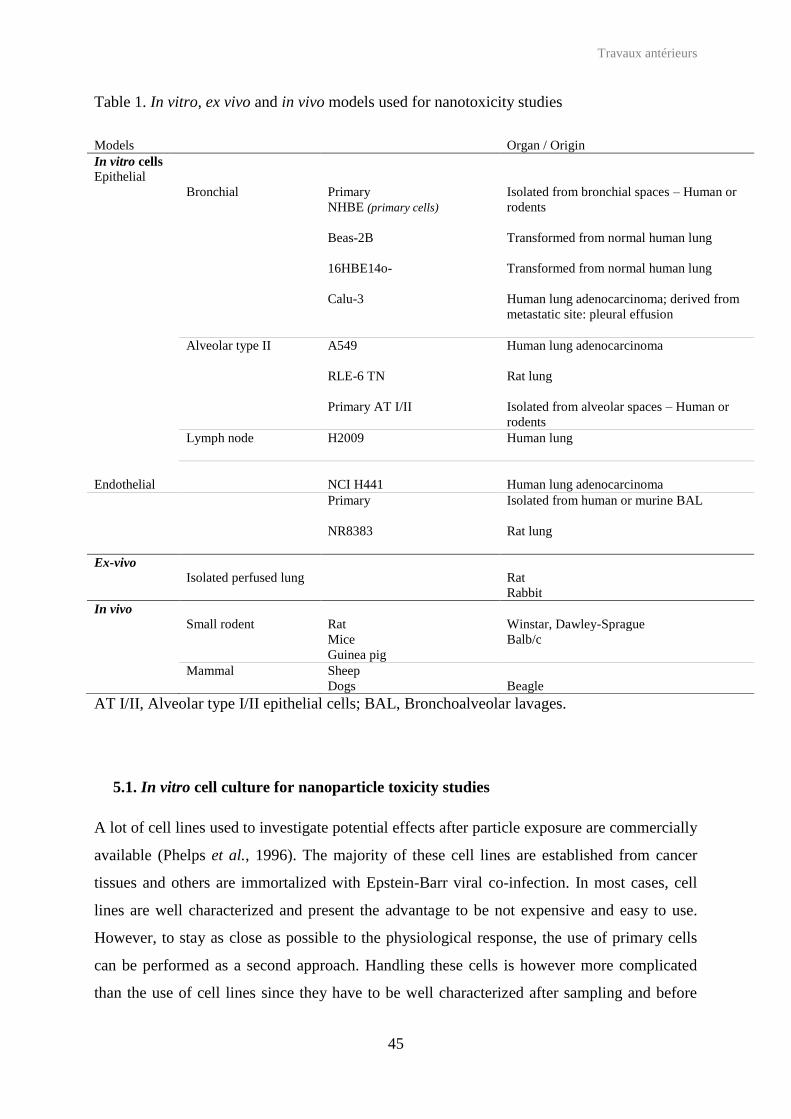
Table 1. In vitro, ex vivo and in vivo models used for nanotoxicity studies
AT I/II, Alveolar type I/II epithelial cells; BAL, Bronchoalveolar lavages.
5.1. In vitro cell culture for nanoparticle toxicity studies
A lot of cell lines used to investigate potential effects after particle exposure are commercially
available (Phelps et al., 1996). The majority of these cell lines are established from cancer
tissues and others are immortalized with Epstein-Barr viral co-infection. In most cases, cell
lines are well characterized and present the advantage to be not expensive and easy to use.
However, to stay as close as possible to the physiological response, the use of primary cells
can be performed as a second approach. Handling these cells is however more complicated
than the use of cell lines since they have to be well characterized after sampling and before
Models Organ / Origin In vitro cells Epithelial Bronchial Primary
NHBE (primary cells) Beas-2B
Isolated from bronchial spaces – Human or rodents Transformed from normal human lung
16HBE14o-
Transformed from normal human lung
Calu-3 Human lung adenocarcinoma; derived from metastatic site: pleural effusion
Alveolar type II A549 Human lung adenocarcinoma
RLE-6 TN Rat lung
Primary AT I/II
Isolated from alveolar spaces – Human or rodents
Lymph node H2009 Human lung Endothelial NCI H441 Human lung adenocarcinoma Primary Isolated from human or murine BAL
NR8383
Rat lung
Ex-vivo Isolated perfused lung Rat
Rabbit In vivo Small rodent Rat Winstar, Dawley-Sprague Mice Balb/c Guinea pig Mammal Sheep Dogs Beagle

Travaux antérieurs
46
their use. Moreover, their stability is generally limited to a small number of passages. To
mimic the lung, human or murine epithelial cells (bronchial, alveolar, lymph nodes),
endothelial cell lines and murine macrophages are available. Human primary macrophages
can be obtained from BAL or by monocyte differentiation.
The delivery to the bronchus can be studied on bronchial epithelial cell lines, such as
16HBE14-o cells, human bronchus cells (Dombu et al., 2012, Brzoska et al., 2004) or Calu-3
cells (Bivas-Benita et al., 2004a), isolated from a human lung carcinoma (Fogh et al., 1977).
These both cell lines can be cultured at the air-liquid interface, to form a confluent monolayer
with good trans-epithelial electrical resistance (TEER) values, due to polarized tight junctions
(Foster et al., 2000) and a dense mucus layer (Fiegel et al., 2003). Using these models, good
correlations with in vivo results were shown (Mathias et al., 2002). Gelatin and human serum
albumin nanoparticles were prepared and stabilized with glutaraldehyde to deliver drug or
gene into primary lung cells (isolated from lobectomies) or 16HBE14o- cells (Brzoska et al.,
2004). Previous confocal microscopy observations have evidenced that nanoparticles were
taken up according to an endocytic mechanism. Then, toxicity studies on both cell types, have
shown that membrane integrity was not affected (LDH), even after the highest tested
concentration (50 µg/mL), and no IL-8 secretions were detected. Another study has
investigated the potential gene delivery polyethylenimine (PEI) coated PLGA nanoparticles
(Bivas-Benita et al., 2004a). Nanoparticle uptake was observed within Calu-3 cells with a
lysosomal co-localization. The uptake level was shown to be PLGA / PEI ratio-dependent, as
demonstrated by the levels of gene expression.
Beas-2 B cells, isolated and modified from normal human bronchial cells (Reddel et al.,
1988), were widely used to study the effect of non-biodegradable nanoparticles such as the
inflammatory response after particulate matter exposure (Steerenberg et al., 1998), the
oxidative stress after exposure to silica nanoparticles (Eom and Choi, 2009) and complete
toxicity studies (oxidative stress, apoptosis, gene expression, DAPI staining) after exposure to
titanium dioxide nanoparticles (Park et al., 2008). In most of these studies the model was
relevant to demonstrate toxicity properties of the tested particles.
Major studies that focused on alveolar conditions used the A549 cell line. These cells are
isolated from a human carcinoma (Lieber et al., 1976) and constitutes a good model of type II
pneumocytes (Rothen-Rutishauser et al., 2012, Foster et al., 1998), giving the possibility to
test, after exposure to biodegradable nanoparticles, several previously detailed toxicity
Travaux antérieurs
47
endpoints. As an example, the uptake of fluorescent chitosan nanoparticles was shown to be
an endocytosis-mechanism initiated by adsorptive steps (Huang et al., 2002), while the SLN
(loaded with paclitaxel) endocytosis was PEG or folate coating-mediated (Yuan et al., 2008).
The low cytotoxicity of SLN based on 6-lauroxyhexyl lysinate and complexed to DNA
allowed to induce a similar rate of gene transfection comparable with those obtained with
Lipofectamine (commercial reagent) (Li et al., 2011). Finally, liposomes loaded with
ciprofloxacin have shown a good antibacterial effect without leading to LDH release (Chono
et al., 2006). Moreover, after exposure of A549 cells to inorganic nanoparticles, apoptosis
detection, cell proliferation assay, ROS quantification and inflammatory response were
observed (Choi et al., 2009).
To expand alveolar condition studies, human primary alveolar type II cells were isolated after
lung resection of carcinoma (Witherden et al., 2004). Such cells express an alveolar cell-type
phenotype and can be immortalized (O'Hare et al., 2001) showing an increased internalization
of latex beads (Kemp et al., 2008).
5.2. Co-culture of lung cells in nanotoxicology
Co-culture systems have provided significant advances in cell biology. Indeed, by mixing
different cell types (2 or more), interactions occur and culture conditions are closer to
physiology than classic in vitro cell line models. Co-culture models can be established with
direct or indirect contact between the cells (Figure 9). Models with indirect contact require the
use of a device such as Transwell® inserts equipped with porous membranes allowing
communication between different cell types placed in wells. Each cell type is then seeded on
each side of the insert. To mimic pulmonary tract, a large variety of cells have been
used: bronchial, alveolar, epithelial, fibroblast, dendritic and macrophages. However, in each
case the relevance of co-cultures needs to be validated by deep comparisons between cells in
mono- and in co-culture.
Bronchial conditions were reproduced by a co-culture of 16HBE14o- cells or human primary
bronchial cells (apical compartment) with lung fibroblast cells Wi-38 (basolateral
compartment) at the air liquid interface (Pohl et al., 2009). The use of primary cells has
shown interest. Indeed, in co-culture with fibroblastes, primary bronchial cells (which are
continuously differentiated) mimic the structure of a native polarized bronchial epithelium

Travaux antérieurs
48
showing mucus-producing, basal and ciliated cells. Such model was used from 3 weeks to
3 months (Pohl et al., 2009).
EpiairwayTM (MatTek Corporation) and MucilAirTM (Epthelix), two commercial differentiated
pulmonary epithelium, are available to perform toxicity tests after exposure to chemical
compounds (Constant et al., 2011a) or manufactured nanoparticles (Constant et al., 2011b,
Hayden et al., 2011). Composed of human primary cells isolated from trachea, bronchus and
nose, MucilAirTM device is cultivated at the air-liquid interface and presents characteristics of
airway tissues (available for healthy or damaged conditions). Indeed, the device contains
basal, ciliated and mucosal cells and displays a regular cilliary beating, tight junctions, ion
transport and metabolic activity. The epithelium remains in homeostasis state for more than
one year, thus, long term studies after single or repeated exposure can be performed. After 28
days repeated exposure to chemical compounds, cytochrome expression was modulated
(Constant et al., 2011a). After delivery of nanoparticles on apical surface, trans-epithelial
resistance, LDH and cytokine released were quantified (Constant et al., 2011b). It was shown
that the epithelium has the potential to recover after low nanoparticle concentration exposure.
EpiairwayTM and MucilAirTM present an interesting potential to drive environmental studies,
as the ones responding to EU regulations in the REACH programme (BéruBé et al., 2010).
Alveolar conditions were simulated by different co-culture models. For example, the indirect
co-culture of A549 cells or NC H441 with primary human pulmonary microvascular
endothelial cells allowed developing a model of an alveolo-capillary barrier (Hermanns et al.,
2004). This model has been used to study environmental cadmium exposure side-effects and
has shown that whereas viability of epithelial cells largely decreases, the viability of
endothelial cells remains unaffected (Papritz et al., 2010). To investigate the predominant role
of resident macrophages following air particulate exposure, lung alveolar epithelial cells,
[A549 (Wottrich et al., 2004, Jantzen et al., 2012), L132 (Abbas et al., 2009), RLE-6N (Wang
et al., 2002)], were co-cultured with macrophages [differentiated from THP-1 monocytes
(St íž et al., 2001, Wottrich et al., 2004), Mono Mac 6 (Wottrich et al., 2004) or human
(Abbas et al., 2009) / murine (Wang et al., 2002) alveolar macrophages isolated from BAL] in
direct or indirect contact. In co-culture with A549, macrophages differentiated from THP-1
monocytes acquire alveolar macrophage phenotypic characteristics (St íž et al., 1993), and
after exposure to ultrafine nanoparticles, co-culture synergistic effects were observed
concerning the release of IL-6 and IL-8 (Wottrich et al., 2004), while no modification of ROS
and DNA damage levels were noticed (Jantzen et al., 2012). The defensive role of alveolar

Travaux antérieurs
49
macrophages against particulate matter was proved in co-culture with L132 cells (Abbas et
al., 2009). In co-culture, the levels of particulate matter metabolizing enzymes produced were
higher than in mono-culture. The significant attenuation of IL-1 and IL-6 cytokines
secretions after exposure to jet fuel on a co-culture of rat alveolar cells and macrophages
compared to mono-culture of each cells confirmed that interactions between cells exist (Wang
et al., 2002).
Figure 9. Representation of co-culture models used to mimic the pulmonary tract using Transwell® device, to established co-culture in indirect contact.
Rothen-Rutishauser et al. (Rothen-Rutishauser et al., 2005) have established a tri-culture of i)
A549 cells seeded on the apical compartment of Transwell®, ii) differentiated dendritic cells
seeded on the basal side of the turned insert and iii) differentiated macrophages added on the

Travaux antérieurs
50
apical surface of the epithelial cell monolayer. This tri-culture was used to investigate effects
of non biodegradable nanoparticles in alveolar conditions (Müller et al., 2010). Following
TEM observations, TiO2 nanoparticles were found in all cell types, whereas carbon nanotubes
were present in epithelial cells. After exposure to carbon nanotubes, ROS production was
observed in majority of cells, with a higher level than in mono-culture of epithelial cells,
whereas IL-8 and TNF-α quantification (ELISA) did not show any synergistic effect.
In order to deeply understand toxicity mechanisms, and more particularly the inflammatory
response after exposure to particulate matter, Alfaro-Moreno and co-workers have deeply
investigated mono and co-cultures of epithelial cells (A549), macrophages (differentiated
THP-1 monocytes), mast cells (HMC-1) and endothelial cells (EAHY926) (Alfaro-Moreno et
al., 2008). Different co-culture combinations were performed: mast cells or macrophages with
epithelial cells in direct contact; mast and epithelial cells and macrophages in direct contact,
with or without endothelial cells in indirect contact. Cytokine release profile was modified
according to the combination and the key role of mast cells and macrophages was
demonstrated (large increase of several cytokines in co-culture), as well as the activation of
endothelial cells by the co-culture. The cytokine release profile of the system composed of the
four cell types was in accordance with in vivo profile and correlated to asthmatic patients who
were exposed to such particulate matter.
5.3. Ex-vivo models
5.3.1. Isolated perfused lungs
The isolated perfused lung (IPL) system represents an interesting alternative when
considering all cell-cell interactions that occurred within the isolated organ (Mehendale et al.,
1981). This technique consists in extracting the respiratory system (lungs and/or trachea) from
rats, guinea pigs or rabbits and keep it as close as possible to the physiological conditions
(Byron et al., 1986). The pulmonary circulation (air flow rate, temperature, blood circulation)
is maintained in a complex track of pumps to provide isolation, perfusion and ventilation of
isolated lungs. IPL are not much used in nanotoxicology even though the radioactive tracking
of iridium particles to an isolated perfused rat lung has shown a similar particle distribution to
the one occurring in vivo, confirming the pertinence of such models (Meiring et al., 2005).
PLGA or branched polyester-based nanoparticles were loaded with a fluorescent compound
(Beck-Broichsitter et al., 2009) or salbutamol (Beck-Broichsitter et al., 2010), for the delivery

Travaux antérieurs
51
to the isolated perfused rat lung. Kinetic spectroscopic studies on the IPL have confirmed the
presence of nanoparticles in deep lungs up to 2 hours after administration, and the release of
active compounds in two phases, the first corresponding to a constant increasing of
compounds in the IPL, and the second being the phase of absorption. Similar studies were
performed with polystyrene nanoparticles, and confocal laser scanning microscopy imaging of
lung slices has shown the presence of such nanoparticles in the deep lung, principally in
alveolar macrophages and in free alveolar spaces (Hamoir et al., 2003) (Figure 9), but no
evidence of translocation from alveolar to the blood circulation was shown (Nemmar et al.,
2005).
IPL studies have certain advantages such as the possibility to achieve complete qualitative and
quantitative studies of drug release, by frequent sample collection, but, unfortunately, in a
short time duration, because structural and functional integrity of organs will quickly decline
(Beck-Broichsitter et al., 2011). Moreover, physiological conditions are not strictly respected,
since lymph flow is altered, bronchial perfusion is suppressed, autonomic innervations are
disconnected and blood cells are not present (Meiring et al., 2005). In addition, the technique
of isolation is hard to control and the perfusion system is expensive.
Figure 10. Particle distribution in lungs after intratracheal administration in isolated perfused organs. (A) Microspheres in red were homogeneously distributed into the lungs. (B) Particles were located in macrophages (M) but free particles (Fp) were also observed. No particles were detected in pulmonary vessels (Ve). Bars indicate 50 μm. (Hamoir et al., 2003).

Travaux antérieurs
52
5.3.2. Ex vitro mucus models
Because of the rigidity of the mucin and the pore size distribution, mucus penetration is
different for each nanoparticles. Indeed, interactions are surface coating-mediates (Kirch et
al., 2012). It was, for instance shown that PEG coating decreases mucus-nanoparticles
interactions (Schuster et al., 2013), while chitosan coating leads to a strong muco-adhesion
(Zaru et al., 2007). Indeed, chitosan-coated nanoparticles displayed more interactions with
mucins (confirmed by an increase of zeta potential values), and a more important passage
through the mucus than equivalent PVA-coated PLGA nanoparticles (Ungaro et al., 2012,
Mura et al., 2011a).
5.4. In vivo models
Compared to other models, in vivo models present the large advantage to be a whole
organism. Moreover, as previously described, in vitro lung delivery can be easily performed
with specific veterinary devices. However experiments are limited by ethical concerns, and
moreover expensive to carry out.
In most cases, small rodents have been employed to follow particle biodistribution and to
performe assays on BAL after lung nanoparticle exposure. For example, insulin loaded into
liposomes based on hydrogenated soybean phosphatidylcholine and cholesterol (Huang and
Wang, 2006) or into SLN (Bi et al., 2009) were administered to diabetic mice or rats by
inhalation. In addition of the expected sustained effect, analyses of BAL have shown no
immunoreaction nor inflammation (no leukocyte increase). Similarly, liposomes loaded with
ciprofloxacin did not induce toxicity after intratracheal delivery to rats (Chono et al., 2006).
SLN stabilized with PVA and loaded with several antibiotics were also demonstrated to be
interesting for the treatment of tuberculosis in guinea pigs (Pandey and Khuller, 2005). In
addition to the sustained effect observed in peripheral organs (until 7 days after delivery), the
absence of alteration in serum bilirubin assays has confirmed the safety of the formulations.
PVA-coated PLGA nanoparticles loaded with elcatonin have shown a low lung retention in
guinea pigs whereas chitosan coated-PLGA nanoparticles were more largely retained. The
presence of chitosan causes of high muco-adhesion properties that may open intracellular
tight junctions (Yamamoto et al., 2005) and increase the cellular uptake (Yang et al., 2009a).
Indeed, positive surface of these nanoparticles induce strong electrostatic interactions with the
exacerbated negative charge of cancer cells.

Travaux antérieurs
53
Very few studies relate experiments with non-rodents for lung drug delivery of nanoparticles.
Beagle dogs were used to follow X-ray contrast agent nanoparticles, stabilized by Pluronic
F68 or F108 (poloxamers) in lung draining lymph nodes, thus detectable with contrast agent
radiographs (McIntire et al., 1998). After administration, dogs were in good clinical
conditions (good appetite, normal breathing and active/alert behavior) despite the presence of
lung lesions detected during the autopsy. These lesions were described as infiltrations of
macrophages signing an inflammatory response, more prevalent with Pluronic F108 than F68.
Sheep have shown a good lung dynamic compliance after intratracheal administration of
liposomes loaded with amikacin (Schreier et al., 1992) and the therapeutic effect of the
liposomal form was higher than the free drug without causing any irritation.
5.5. Compared in vitro / ex vivo / in vivo nanotoxicity studies
Unfortunately, all the models cited previously rapidly reach some limits. Indeed, despite their
human origin, in vitro cell models are more often immortalized by genetic modifications or
originate from tumoral cancer which means they are quite far from the reality. Co-culture
models improve traditional in vitro cell culture by mimicking cell-cell interactions. Ex vivo
and in vivo models represent an interesting alternative, thanks to the presence of cell
interactions or fluid circulation. Yet, the animal physiology is different from the human
physiology. In order to improve our understanding of the behavior and interactions of
nanoparticles towards human organism, compiling together results obtained on different
models may represent a strong interest.
For example, Dailey et al. (Dailey et al., 2006) have firstly proved that polymeric
nanoparticles (PLGA and branched PLGA) do not induce a significant LDH release after 24 h
exposure to A549 cells, whereas after instillation in Balb/c mice, LDH levels in BAL 24 h and
14 days were slightly higher than for non treated animals. In addition, nanoparticles did not
provoke PMN recruitment, neither macrophage inflammatory protein secretion. Nassimi et al.
(Nassimi et al., 2009) have used the same method to investigate the toxicity of SLN: firstly
towards A549 cells, then ex vivo on isolated lung slices and finally on Balb/c mice. In vitro
cytotoxicity results have given a higher IC50 than on ex vivo experiments, that was explained
by the more basic model, and the cell-cell interactions that definitely play a role in ex vivo
models. The three models have shown the absence of LDH release as well as inflammatory
response.
Travaux antérieurs
54
The effect of nanoparticle stabilizers (PVA, chitosan, Pluronic F68) coating PLGA
nanoparticles were investigated towards bronchial and alveolar cells (Mura et al., 2011a,b,
Grabowski et al., 2013). Pluronic F68-covered PLGA nanoparticles penetrate more deeply the
mucus layer on the top of Calu-3 cells than PVA or chitosan-covered nanoparticles (Mura et
al., 2011a), without causing more cytotoxicity or inflammatory response (Mura et al., 2011b).
However, the same particles induce an increasing inflammatory response on A549 cells with a
decrease of the mitochondrial activity, correlated with a higher internalization (Grabowski et
al., 2013). Chitosan-coated PLGA nanoparticles are also more cytotoxic than PVA-coated
nanoparticles on A549 cells, without inducing other side effects (full membrane integrity, no
inflammatory response) (Ungaro et al., 2012). The toxicity of these nanoparticles was
explained by the own cytotoxicity of chitosan in aqueous solution. In addition, chitosan-
coated nanoparticles caused a decrease which was reversible of TEER, without increasing
MUC5AC mRNA expression as well as the other tested nanoparticles (Mura et al., 2011a).
Additionally, an increase in cell marker membrane and in the IFN- production from dendritic
cells after delivery of chitosan nanoparticles to rats was reported (Bivas-Benita et al., 2004b).
In vitro and in vivo comparisons were performed in order to evaluate the radiodiagnostic tool
potential of hematoporphyrin linked to albumin under nanoparticle form (Yang et al., 2010).
Accumulation was shown on A549 cells and correlated to a photodynamic activity after UV
exposure, without any toxicity. The accumulation was confirmed in mice-bearing lung tumors
and nanoparticles have shown a better half-life in rabbits than the free drug.
As there are in first line to face inhaled nanoparticles, alveolar macrophages should be widely
studied, and particularly for phagocytosis (uptake) studies. Rifampicin was encapsulated into
PLGA nanoparticles or microparticles (Ohashi et al., 2009). Nanoparticles, or free drug as
control, were spray-dried with mannitol to form microparticles. High aerosol performances of
rifampicin / PLGA / mannitol microparticles were confirmed with the cascade impactor. In
addition, thanks to the immediate mannitol dissolution, rifampicin / PLGA nanoparticles were
immediately released. Applied on rat alveolar macrophages (NR8383), PLGA nanoparticles
were taken up in significant quantity but less than PLGA microparticles. When rats were
intratracheally administered with the three different formulations, the uptake of rifampicin
associated to PLGA / mannitol microparticles was higher than from the other preparations.
Microscopy observations of rat lungs evidenced that PLGA microparticles were rapidly
excreted by the mucocillary clearance, whereas after mannitol dissolution, PLGA
nanoparticles were retained in lungs. In comparison, rifampicin was loaded in SLN of 850 nm

Travaux antérieurs
55
and evaluated, in vitro on alveolar macrophages, primary epithelial cells (isolated from rats)
and cell lines (NR8383 and A549), and delivered in vivo to rats (Chuan et al., 2013). As
expected, according to the size criteria, alveolar macrophages were specifically targeted
(in vitro and in vivo results), with a sustained effect up to 12 h, and without decreasing the
mitochondrial activity. It is also important to highlight that no differences were observed on
primary cells and cell lines in terms of uptake and cytotoxicty. Moreover, delivery of PVA-
coated PLGA and PLA nanoparticles on rats have shown that the uptake is predominantly
achieved by alveolar macrophages compared to epithelial cells, without increasing the
immune response (Thomas et al., 2010).
Conclusion
Because of their small size, nanoparticles present a specific potential toxicity towards
biological systems. Face to the need to consolidate all toxicity results obtained after
nanoparticle exposure, toxicologists have created a new sub-discipline: the nanotoxicology.
As shown above, nanoparticle toxicity encompasses several endpoints, and in order to suggest
a safe nanomedicine, complete toxicity studies should be performed. For this purpose, to
deeply understand interactions between nanomedicine and living organisms, several toxicity
endpoints and diverse in vitro, in vivo and ex vivo models should be compared.
The use of nanoparticles in biopharmaceutical applications is constantly increasing.
Nanomedicines, obtained through the use of large variety of drugs and nanoparticles, have a
promising future in diagnostic and therapeutic (see Tables 2, 3, 4 for summary). Two major
types of nanoparticles are currently investigated for the lung drug delivery: lipid and
polymeric nanoparticles. Liposomes or SLN have the main advantages to be prepared with
natural compounds, and loaded with insulin they have been shown as an interesting
alternative to iv. administration (Table 2), without causing important toxicity. As far as
polymeric nanoparticles are concerned, the stabilizer has a key role in interactions with
biological systems (Table 3). According to the present report, lipid and polymeric
nanoparticles are good candidates as nanocarriers. Further experimental and clinical studies
are expected to understand human organism/nanoparticle interactions, and thus confirm, or
not, the therapeutic and diagnostic potential of such nanomedicine.

Travaux antérieurs
56
Finally, a little number of other nanoparticles (Table 4) are proposed for lung delivery. For
example, the preparation of contrast agent nanoparticles, stabilized with poloxamers, has
revealed a low toxicity and can shown a strong interest in imaging.

Travaux antérieurs
57
Table 2. In vitro and in vivo pulmonary toxicity endpoints after exposure to lipid-based nanoparticles.
Chem., Chemokine ; AM, Alveolar Macrophages ; AT I, Alveolar Type II Epithelial cells ; Chol, Cholesterol ; DCP, dicetylphosphate ; PC, Phosphatidylcholine ; HSPC, hydrogenated soy phosphatidylcholin ; DPPC, 1,2-dipalmitoyl-sn-glycero-3-phosphocholine ; PEG, poly(ethyleneglycol) ; DOPE, 1,2-Dioleoyl-sn-glycero-3-phosphoethanolamine ; DNA, deoxyribonucleic acid.
Lipid-based nanomedicines Nanoparticle Drug Model
(administration way) Nanoparticle toxicity endpoints References
Liposomes HPC Chol
Insulin Amikacin
Normal / diabetic mice (aerosolization) Sheep (nebulization)
No cell morphology modification, no inflammation No toxicity, no irritation, good dynamic compliance and lung resistance
(Huang and Wang, 2006, Schreier et al., 1992)
HSPC DCP
Ciprofloxacin Rat No toxicity (Chono et al., 2006)
PC DPPC
Rifampicin A549 cells No cytotoxicity (Zaru et al., 2007)
6-laurylhexyl lysiniate
DNA A549 cells Rat
Low toxicity (Li et al., 2011)
PEG Phosphatidyl-ethanolamine Chol DOPE
Manganese DNA
A549 cells LLC1 cells C57B1/6 mice
Cytoplasmic uptake, No cytotoxicity High lung retention after 48 h
(Howell et al., 2013)
Solid-lipid nanoparticles
Lipids Insulin rat (diabetic) No toxicity (Bi et al., 2009)
Triglycerides Phospholipids
A549 Balb/c mice
IC 50 (mg/mL) = 3090 (MTT) – 2090 (neutral red) – 500 (ex vivo) No increasing of chem. KC, LDH, IL-6, IL-8, TNF-α, total proteins, total cells Toxic effects at 500 µg/animal (No effect until 200 µg).
(Nassimi et al., 2010)
Lipids Rifampicin+ Isoniazid + Pyrazinamide
Dunkin Hartley guinea pig No alteration in serum bilirubin Drug release, with sustained effect, after 45 min
(Pandey and Khuller, 2005)
Lipids Paclitaxel A549 PEG and folate enhance cellular uptake without cytotoxicity (Yuan et al., 2008) Soy bean lecithin
Rifampicin N8383 Primary rat AM A549 Primary rat ATII Sprague-Dawleys rats
Low mitochondrial activity decreasing Better pharmacologic effects than the free drug Sustained effect until 12 h in AM/N8383 SLN fluorescence intensities higher in macrophages than in AEC Macrophage targeting
(Chuan et al., 2013)

Travaux antérieurs
58
Table 3. In vitro and in vivo pulmonary toxicity endpoints after exposure to polymer-based nanoparticles.
Polymer-based nanomedicine Nanoparticle
Drug Core Stabilizer
Model (administration mode)
Nanoparticle toxicity endpoints References
PLGA PVA Elcatonin Paclitaxel HBsAg
Guinea pig
Mice Rat Sprague-dawley rat
Low lung retention Taken up by alveolar macrophages Basal immune response
(Yamamoto et al., 2005, Thomas et al., 2010, Yang et al., 2009a)
A549 cells Calu-3 cells HBE cells
Low cytotoxicity and Golgi colocalization (Ungaro et al., 2012, Grabowski et al., 2013, Yang et al., 2009a, Thomas et al., 2010, Mura et al., 2011a,b, Cartiera et al., 2009)
PLGA CS Elcatonin Paclitaxel
Guinea pig
Mice Rat
Long lung retention May opening intracellular tight junctions Strong muco-
(Yamamoto et al., 2005, Yang et al., 2009a)
A549 cells Calu-3 cells
Uptake increase with CS concentration – energy dependent mechanism Medium/high toxicity
(Yang et al., 2009a, Grabowski et al., 2013, Mura et al., 2011a, b)
PLGA PF68 A549 Calu-3
High uptake – energy dependent mechanism Medium/high toxicity, significant inflammatory response, full membrane integrity
(Mura et al., 2011a, b, Grabowski et al., 2013)
PLA PVA HBsAg (Thomas et al., 2010)
Sprague-dawley rat Calu-3
Taken up by alveolar macrophages No immune response
(Thomas et al., 2010)
CS DNA Mice - Dendritic cells Increasing of CD80, CD86, CD83 expression (LPS levels) and INF- production
(Bivas-Benita et al., 2004b)
PLGA, poly(lactide-co-glycolique) acid ; PLA, polylactic acid ; PVA, polyvinylique alcoho ;, CS, Chitosan ; PF68, Pluronic F68 ; DNA, deoxyribonucleic acid.

Travaux antérieurs
59
Table 4. In vitro and in vivo pulmonary toxicity endpoints after exposure to other nanoparticles relevant for biomedical applications.
LPS = Lipopolysaccharides, PF68 / PF 108 = Pluronic F68 / F108, PLGA = poly(lactide-co-glycolide).
Nanomedicine
Nanoparticle
Core Shell Drug Model (administration way) Nanomedicine toxicity endpoints References
Gold
Sodium tribasic dihydrate
Mice
Slow clearance Biodistribution: heart > thymus > spleen
(Hussain et al., 2013) LPS
Fast clearance Biodistribution: spleen > thymus> heart
Human serum albumin Porcine gelatin
Glutaraldehyde 16HBE14o- cells Primary bronchial cells
Nanoparticle internalized inside cells (energy dependent mechanism) No cytotoxicity, Full membrane integrity, No inflammatory response
(Brzoska et al., 2004)
Contrast agent PF68 PF108
Contrast agent
Beagle dog Good clinical conditions Lung lesions, inflammation Macrophage infiltrations prevalent with PF 108P than PF68
(McIntire et al., 1998)
Fe3O4 PLGA Quercitin A549 cells Balb/c mice (nebulization)
No cell morphology modification No cytotoxicity (100 µg/mL) No IL-6 secretion, High GSH levels (transitory)
(Verma et al., 2013)
Antisense oligonucleotides
Mannose receptor Rat alveolar macrophages Full membrane integrity Alveolar macrophages targeting
(Liang et al., 1996)

Travaux antérieurs
60
References
ABBAS, I., SAINT-GEORGES, F., BILLET, S., VERDIN, A., MULLIEZ, P., SHIRALI, P. & GARÇON, G. 2009. Air pollution particulate matter (PM2.5)-induced gene expression of volatile organic compound and/or polycyclic aromatic hydrocarbon-metabolizing enzymes in an in vitro coculture lung model. Toxicology in Vitro, 23, 37-46.
AL-HALLAK, M. H. D. K., SARFRAZ, M. K., AZARMI, S., ROA, W. H., FINLAY, W. H., ROULEAU, C. & LÖBENBERG, R. 2012. Distribution of effervescent inhalable nanoparticles after pulmonary delivery: an in vivo study. Therapeutic Delivery, 3, 725-734.
ALFARO-MORENO, E., NAWROT, T. S., VANAUDENAERDE, B. M., HOYLAERTS, M. F., VANOIRBEEK, J. A., NEMERY, B. & HOET, P. H. M. 2008. Co-cultures of multiple cell types mimic pulmonary cell communication in response to urban PM10. European Respiratory Journal, 32, 1184-1194.
ANDUJAR, P., LANONE, S., BROCHARD, P. & BOCZKOWSKI, J. 2009. Effets respiratoires des nanoparticules manufacturées. Revue des Maladies Respiratoires, 26, 625-637.
ANTON, N., JAKHMOLA, A. & VANDAMME, T. F. 2012. Trojan Microparticles for Drug Delivery. Pharmaceutics, 4, 1-25.
BABIOR, B. M. 2000. Phagocytes and oxidative stress. The American Journal of Medicine, 109, 33-44.
BACHAND, G. D., ALLEN, A., BACHAND, M., ACHYUTHAN, K. E., SEAGRAVE, J. C. & BROZIK, S. M. 2012. Cytotoxicity and inflammation in human alveolar epithelial cells following exposure to occupational levels of gold and silver nanoparticles. Journal of Nanoparticle Research, 14.
BEAUCHAMP, C. & FRIDOVICH, I. 1971. Superoxide dismutase: Improved assays and an assay applicable to acrylamide gels. Analytical Biochemistry, 44, 276-287.
BECK-BROICHSITTER, M., GAUSS, J., GESSLER, T., SEEGER, W., KISSEL, T. & SCHMEHL, T. 2010. Pulmonary targeting with biodegradable salbutamol-loaded nanoparticles. J Aerosol Med Pulm Drug Deliv, 23, 47-57.
BECK-BROICHSITTER, M., GAUSS, J., PACKHAEUSER, C. B., LAHNSTEIN, K., SCHMEHL, T., SEEGER, W., KISSEL, T. & GESSLER, T. 2009. Pulmonary drug delivery with aerosolizable nanoparticles in an ex vivo lung model. International Journal of Pharmaceutics, 367, 169-178.
BECK-BROICHSITTER, M., MERKEL, O. M. & KISSEL, T. 2012. Controlled pulmonary drug and gene delivery using polymeric nano-carriers. Journal of Controlled Release, 161, 214-224.
BECK-BROICHSITTER, M., SCHMEHL, T., SEEGER, W. & GESSLER, T. 2011. Evaluating the Controlled Release Properties of Inhaled Nanoparticles Using Isolated, Perfused, and Ventilated Lung Models. Journal of Nanomaterials, 2011.
BÉRUBÉ, K., PRYTHERCH, Z., JOB, C. & HUGHES, T. 2010. Human primary bronchial lung cell constructs: The new respiratory models. Toxicology, 278, 311-318.
BHATTACHARYA, K., DAVOREN, M., BOERTZ, J., SCHINS, R., HOFFMANN, E. & DOPP, E. 2009. Titanium dioxide nanoparticles induce oxidative stress and DNA-adduct formation but not DNA-breakage in human lung cells. Particle and Fibre Toxicology, 6, 1-11.

Travaux antérieurs
61
BI, R., SHAO, W., WANG, Q. & ZHANG, N. 2009. Solid lipid nanoparticles as insulin inhalation carriers for enhanced pulmonary delivery. J Biomed Nanotechnol, 5, 84-92.
BIVAS-BENITA, M., ROMEIJN, S., JUNGINGER, H. E. & BORCHARD, G. 2004a. PLGA-PEI nanoparticles for gene delivery to pulmonary epithelium. Eur J Pharm Biopharm, 58, 1-6.
BIVAS-BENITA, M., VAN MEIJGAARDEN, K. E., FRANKEN, K. L., JUNGINGER, H. E., BORCHARD, G., OTTENHOFF, T. H. & GELUK, A. 2004b. Pulmonary delivery of chitosan-DNA nanoparticles enhances the immunogenicity of a DNA vaccine encoding HLA-A*0201-restricted T-cell epitopes of Mycobacterium tuberculosis. Vaccine, 22, 1609-15.
BIVAS-BENITA, M., ZWIER, R., JUNGINGER, H. E. & BORCHARD, G. 2005. Non-invasive pulmonary aerosol delivery in mice by the endotracheal route. European Journal of Pharmaceutics and Biopharmaceutics, 61, 214-218.
BRUINENBERG, P., SERISIER, D., CIPOLLA, D. & BLANCHARD, J. 2010. Safety, Tolerability And Pharmacokinetics Of Novel Liposomal Ciprofloxacin Formulations For Inhalation In Healthy Volunteers And Non-Cystic Bronchiectasis Patients. B49. BRONCHIECTASIS: CYSTIC FIBROSIS AND BEYOND, A3192-A3192.
BRZOSKA, M., LANGER, K., COESTER, C., LOITSCH, S., WAGNER, T. O. F. & MALLINCKRODT, C. V. 2004. Incorporation of biodegradable nanoparticles into human airway epithelium cells—in vitro study of the suitability as a vehicle for drug or gene delivery in pulmonary diseases. Biochemical and Biophysical Research Communications, 318, 562-570.
BYRON, P. R., ROBERTS, N. S. & CLARK, A. R. 1986. An isolated perfused rat lung preparation for the study of aerosolized drug deposition and absorption. J Pharm Sci, 75, 168-71.
CARTIERA, M. S., JOHNSON, K. M., RAJENDRAN, V., CAPLAN, M. J. & SALTZMAN, W. M. 2009. The uptake and intracellular fate of PLGA nanoparticles in epithelial cells. Biomaterials, 30, 2790-8.
CELL STAINING SIMULATION TOOL LIFE TECHNOLOGIES. Cell Staining Simulation Tool [Online]. Available: http://www.invitrogen.com/site/us/en/home/support/Research-Tools/Cell-Staining-Tool.html?CID=fl-cellstaintool [Accessed 2013/07/11].
CHOI, H. S., ASHITATE, Y., LEE, J. H., KIM, S. H., MATSUI, A., INSIN, N., BAWENDI, M. G., SEMMLER-BEHNKE, M., FRANGIONI, J. V. & TSUDA, A. 2010. Rapid translocation of nanoparticles from the lung airspaces to the body. Nat Biotechnol, 28, 1300-3.
CHOI, S. J., OH, J. M. & CHOY, J. H. 2009. Toxicological effects of inorganic nanoparticles on human lung cancer A549 cells. J Inorg Biochem, 103, 463-71.
CHONO, S., TANINO, T., SEKI, T. & MORIMOTO, K. 2006. Influence of particle size on drug delivery to rat alveolar macrophages following pulmonary administration of ciprofloxacin incorporated into liposomes. J Drug Target, 14, 557-66.
CHUAN, J., LI, Y., YANG, L., SUN, X., ZHANG, Q., GONG, T. & ZHANG, Z. 2013. Enhanced rifampicin delivery to alveolar macrophages by solid lipid nanoparticles. Journal of Nanoparticle Research, 15, 1-9.
CONSTANT, S., HUANG, S., CAULFUTY, M., BONFANTE, R., MONACHINO, M., FRAUENFELDER, R. & WISZNIEWSKI, L. 2011a. 28 day repeated dose toxicity test on an in vitro cell model. Toxicology Letters, 205, Supplement, S58.

Travaux antérieurs
62
CONSTANT, S., HUANG, S., CAULFUTY, M., BONFANTE, R., MONACHINO, M., FRAUENFELDER, R. & WISZNIEWSKI, L. 2011b. A simple method for testing the toxicity of nanomaterials on 3D air–liquid interface human airway epithelia (MucilAir™). Toxicology Letters, 205, Supplement, S284.
COONEY, D. J. & HICKEY, A. J. 2011. Cellular response to the deposition of diesel exhaust particle aerosols onto human lung cells grown at the air–liquid interface by inertial impaction. Toxicology in Vitro, 25, 1953-1965.
CRAPO, J. D., BARRY, B. E., GEHR, P., BACHOFEN, M. & WEIBEL, E. R. 1982. Cell number and cell characteristics of the normal human lung. The American review of respiratory disease, 126, 332-337.
CROMWELL, O., HAMID, Q., CORRIGAN, C. J., BARKANS, J., MENG, Q., COLLINS, P. D. & KAY, A. B. 1992. Expression and generation of interleukin-8, IL-6 and granulocyte-macrophage colony-stimulating factor by bronchial epithelial cells and enhancement by IL-1 beta and tumour necrosis factor-alpha. Immunology, 77, 330-7.
DAILEY, L. A., JEKEL, N., FINK, L., GESSLER, T., SCHMEHL, T., WITTMAR, M., KISSEL, T. & SEEGER, W. 2006. Investigation of the proinflammatory potential of biodegradable nanoparticle drug delivery systems in the lung. Toxicol Appl Pharmacol, 215, 100-8.
DESHMANE, S. L., KREMLEV, S., AMINI, S. & SAWAYA, B. E. 2009. Monocyte chemoattractant protein-1 (MCP-1): an overview. J Interferon Cytokine Res, 29, 313-26.
DJOBA SIAWAYA, J. F., ROBERTS, T., BABB, C., BLACK, G., GOLAKAI, H. J., STANLEY, K., BAPELA, N. B., HOAL, E., PARIDA, S., VAN HELDEN, P. & WALZL, G. 2008. An Evaluation of Commercial Fluorescent Bead-Based Luminex Cytokine Assays. PLoS ONE, 3, e2535.
DOMBU, C., CARPENTIER, R. & BETBEDER, D. 2012. Influence of surface charge and inner composition of nanoparticles on intracellular delivery of proteins in airway epithelial cells. Biomaterials, 33, 9117-9126.
DONALDSON, K., STONE, V., TRAN, C. L., KREYLING, W. & BORM, P. J. 2004. Nanotoxicology. Occup Environ Med, 61, 727-8.
DUPONT, N. C., WANG, K., WADHWA, P. D., CULHANE, J. F. & NELSON, E. L. 2005. Validation and comparison of luminex multiplex cytokine analysis kits with ELISA: Determinations of a panel of nine cytokines in clinical sample culture supernatants. Journal of Reproductive Immunology, 66, 175-191.
ELY, L., ROA, W., FINLAY, W. H. & LÖBENBERG, R. 2007. Effervescent dry powder for respiratory drug delivery. European Journal of Pharmaceutics and Biopharmaceutics, 65, 346-353.
EOM, H.-J. & CHOI, J. 2009. Oxidative stress of silica nanoparticles in human bronchial epithelial cell, Beas-2B. Toxicology in Vitro, 23, 1326-1332.
EPTHELIX. MucilAirTM [Online]. Available: http://www.epithelix.com/content/view/6/5/lang,en/ [Accessed 2013/09/29].
FADOK, V. A., VOELKER, D. R., CAMPBELL, P. A., COHEN, J. J., BRATTON, D. L. & HENSON, P. M. 1992. Exposure of phosphatidylserine on the surface of apoptotic lymphocytes triggers specific recognition and removal by macrophages. The Journal of Immunology, 148, 2207-16.

Travaux antérieurs
63
FIEGEL, J., EHRHARDT, C., SCHAEFER, U., LEHR, C.-M. & HANES, J. 2003. Large Porous Particle Impingement on Lung Epithelial Cell Monolayers—Toward Improved Particle Characterization in the Lung. Pharmaceutical Research, 20, 788-796.
FISHER, G. L. & PLACKE, M. E. 1987. In vitro models of lung toxicity. Toxicology, 47, 71-93.
FOGH, J., FOGH, J. M. & ORFEO, T. 1977. One hundred and twenty-seven cultured human tumor cell lines producing tumors in nude mice. J Natl Cancer Inst, 59, 221-6.
FOSTER, K. A., AVERY, M. L., YAZDANIAN, M. & AUDUS, K. L. 2000. Characterization of the Calu-3 cell line as a tool to screen pulmonary drug delivery. International Journal of Pharmaceutics, 208, 1-11.
FOSTER, K. A., OSTER, C. G., MAYER, M. M., AVERY, M. L. & AUDUS, K. L. 1998. Characterization of the A549 cell line as a type II pulmonary epithelial cell model for drug metabolism. Exp Cell Res, 243, 359-66.
GALLEY, H. F. & WEBSTER, N. R. 1996. The immuno-inflammatory cascade. British Journal of Anaesthesia, 77, 11-16.
GÓMEZ-GAETE, C., FATTAL, E., SILVA, L., BESNARD, M. & TSAPIS, N. 2008. Dexamethasone acetate encapsulation into Trojan particles. Journal of Controlled Release, 128, 41-49.
GRABOWSKI, N., HILLAIREAU, H., VERGNAUD, J., ARAGAO, L. S., KERDINE-ROMER, S., PALLARDY, M., TSAPIS, N. & FATTAL, E. 2013. Toxicity of surface-modified PLGA nanoparticles toward lung alveolar epithelial cells. International Journal of Pharmaceutics.
GREEN, L. C., WAGNER, D. A., GLOGOWSKI, J., SKIPPER, P. L., WISHNOK, J. S. & TANNENBAUM, S. R. 1982. Analysis of nitrate, nitrite, and [15N]nitrate in biological fluids. Analytical Biochemistry, 126, 131-138.
GRENHA, A., SEIJO, B. & REMUÑÁN-LÓPEZ, C. 2005. Microencapsulated chitosan nanoparticles for lung protein delivery. European Journal of Pharmaceutical Sciences, 25, 427-437.
GRONEBERG, D. A., EYNOTT, P. R., LIM, S., OATES, T., WU, R., CARLSTEDT, I., ROBERTS, P., MCCANN, B., NICHOLSON, A. G., HARRISON, B. D. & CHUNG, K. F. 2002. Expression of respiratory mucins in fatal status asthmaticus and mild asthma. Histopathology, 40, 367-373.
HAMOIR, J., NEMMAR, A., HALLOY, D., WIRTH, D., VINCKE, G., VANDERPLASSCHEN, A., NEMERY, B. & GUSTIN, P. 2003. Effect of polystyrene particles on lung microvascular permeability in isolated perfused rabbit lungs: role of size and surface properties. Toxicology and Applied Pharmacology, 190, 278-285.
HAYDEN, P., KALUZHNY, Y., KUBILUS, J., AYEHUNIE, S., KANDAROVA, H. & KLAUSNER, M. 2011. Use of normal human 3-dimensional (NHu-3D) tissue models (EpiDerm, EpiAirway) for nanotoxicology applications Alternative To Experimental Animals. Montréal, Canada.
HEIN, S., BUR, M., SCHAEFER, U. F. & LEHR, C.-M. 2011. A new Pharmaceutical Aerosol Deposition Device on Cell Cultures (PADDOCC) to evaluate pulmonary drug absorption for metered dose dry powder formulations. European Journal of Pharmaceutics and Biopharmaceutics, 77, 132-138.
HENSLEY, K., MOU, S. & PYE, Q. 2003. Nitrite Determination by Colorimetric and Fluorometric Griess Diazotization Assays. In: HENSLEY, K. & FLOYD, R. (eds.) Methods in Biological Oxidative Stress. Humana Press.

Travaux antérieurs
64
HERMANNS, M. I., UNGER, R. E., KEHE, K., PETERS, K. & KIRKPATRICK, C. J. 2004. Lung epithelial cell lines in coculture with human pulmonary microvascular endothelial cells: development of an alveolo-capillary barrier in vitro. Lab Invest, 84, 736-52.
HILLAIREAU, H. & COUVREUR, P. 2009. Nanocarriers' entry into the cell: relevance to drug delivery. Cell Mol Life Sci, 66, 2873-96.
HOET, P. H., BRUSKE-HOHLFELD, I. & SALATA, O. V. 2004. Nanoparticles - known and unknown health risks. J Nanobiotechnology, 2, 12.
HOWELL, M., MALLELA, J., WANG, C., RAVI, S., DIXIT, S., GARAPATI, U. & MOHAPATRA, S. 2013. Manganese-loaded lipid-micellar theranostics for simultaneous drug and gene delivery to lungs. J Control Release, 167, 210-8.
HUANG, M., MA, Z., KHOR, E. & LIM, L.-Y. 2002. Uptake of FITC-Chitosan Nanoparticles by A549 Cells. Pharmaceutical Research, 19, 1488-1494.
HUANG, Y. Y. & WANG, C. H. 2006. Pulmonary delivery of insulin by liposomal carriers. J Control Release, 113, 9-14.
HUSSAIN, S., VANOIRBEEK, J. A. J., HAENEN, S., HAUFROID, V., BOLAND, S., MARANO, F., NEMERY, B. & HOET, P. H. M. 2013. Prior Lung Inflammation Impacts on Body Distribution of Gold Nanoparticles. BioMed Research International, 2013, 6.
IVERSEN, T.-G., SKOTLAND, T. & SANDVIG, K. 2011. Endocytosis and intracellular transport of nanoparticles: Present knowledge and need for future studies. Nano Today, 6, 176-185.
JÄNICKE, R. U., SPRENGART, M. L., WATI, M. R. & PORTER, A. G. 1998. Caspase-3 Is Required for DNA Fragmentation and Morphological Changes Associated with Apoptosis. Journal of Biological Chemistry, 273, 9357-9360.
JANTZEN, K., ROURSGAARD, M., DESLER, C., LOFT, S., RASMUSSEN, L. J. & MØLLER, P. 2012. Oxidative damage to DNA by diesel exhaust particle exposure in co-cultures of human lung epithelial cells and macrophages. Mutagenesis, 27, 693-701.
KELLEY, J. 1990. Cytokines of the Lung. American Review of Respiratory Disease, 141, 765-788.
KEMP, S. J., THORLEY, A. J., GORELIK, J., SECKL, M. J., O'HARE, M. J., ARCARO, A., KORCHEV, Y., GOLDSTRAW, P. & TETLEY, T. D. 2008. Immortalization of human alveolar epithelial cells to investigate nanoparticle uptake. Am J Respir Cell Mol Biol, 39, 591-7.
KERR, J. F., WYLLIE, A. H. & CURRIE, A. R. 1972. Apoptosis: a basic biological phenomenon with wide-ranging implications in tissue kinetics. Br J Cancer, 26, 239-57.
KIM, I., BYEON, H. J., KIM, T. H., LEE, E. S., OH, K. T., SHIN, B. S., LEE, K. C. & YOUN, Y. S. 2012. Doxorubicin-loaded highly porous large PLGA microparticles as a sustained- release inhalation system for the treatment of metastatic lung cancer. Biomaterials, 33, 5574-5583.
KIRCH, J., SCHNEIDER, A., ABOU, B., HOPF, A., SCHAEFER, U. F., SCHNEIDER, M., SCHALL, C., WAGNER, C. & LEHR, C.-M. 2012. Optical tweezers reveal relationship between microstructure and nanoparticle penetration of pulmonary mucus. Proceedings of the National Academy of Sciences, 109, 18355-18360.
KOOPMAN, G., REUTELINGSPERGER, C. P. M., KUIJTEN, G. A. M., KEEHNEN, R. M. J., PALS, S. T. & VANOERS, M. H. J. 1994. ANNEXIN-V FOR FLOW CYTOMETRIC DETECTION
Travaux antérieurs
65
OF PHOSPHATIDYLSERINE EXPRESSION ON B-CELLS UNDERGOING APOPTOSIS. Blood, 84, 1415-1420.
KUNZLI, N. & TAGER, I. B. 2005. Air pollution: from lung to heart. Swiss Med Wkly, 135, 697-702.
LABIRIS, N.R & DOLOVICH M. B. 2003, Pulmonary drug delivery. Part I: physiological factors affecting therapeutic effectiveness of aerosolized medications, British Journal of Clinical Pharmacology, 56, 588-599.
LEBEL, C. P., ISCHIROPOULOS, H. & BONDY, S. C. 1992. Evaluation of the probe 2',7'-dichlorofluorescin as an indicator of reactive oxygen species formation and oxidative stress. Chemical Research in Toxicology, 5, 227-231.
LI, N., XIA, T. & NEL, A. E. 2008. The role of oxidative stress in ambient particulate matter-induced lung diseases and its implications in the toxicity of engineered nanoparticles. Free Radical Biology and Medicine, 44, 1689-1699.
LI, P., LIU, D. H., SUN, X. L., LIU, C. X., LIU, Y. J. & ZHANG, N. 2011. A novel cationic liposome formulation for efficient gene delivery via a pulmonary route. Nanotechnology, 22.
LIANG, W., SHI, X., DESHPANDE, D., MALANGA, C. J. & ROJANASAKUL, Y. 1996. Oligonucleotide targeting to alveolar macrophages by mannose receptor-mediated endocytosis. Biochimica et Biophysica Acta (BBA) - Biomembranes, 1279, 227-234.
LIEBER, M., SMITH, B., SZAKAL, A., NELSON-REES, W. & TODARO, G. 1976. A continuous tumor-cell line from a human lung carcinoma with properties of type II alveolar epithelial cells. Int J Cancer, 17, 62-70.
LIN, H., CARLSON, D. M., ST. GEORGE, J. A., PLOPPER, C. G. & WU, R. 1989. An ELISA Method for the Quantitation of Tracheal Mucins from Human and Nonhuman Primates. American Journal of Respiratory Cell and Molecular Biology, 1, 41-48.
LIN, W., HUANG, Y. W., ZHOU, X. D. & MA, Y. 2006. In vitro toxicity of silica nanoparticles in human lung cancer cells. Toxicol Appl Pharmacol, 217, 252-9.
LIU, J., GONG, T., FU, H., WANG, C., WANG, X., CHEN, Q., ZHANG, Q., HE, Q. & ZHANG, Z. 2008. Solid lipid nanoparticles for pulmonary delivery of insulin. International Journal of Pharmaceutics, 356, 333-344.
MACNEE, W. 2001. Oxidative stress and lung inflammation in airways disease. European Journal of Pharmacology, 429, 195-207.
MANSOUR, H. M., RHEE, Y. S. & WU, X. 2009. Nanomedicine in pulmonary delivery. Int J Nanomedicine, 4, 299-319.
MATHIAS, N. R., TIMOSZYK, J., STETSKO, P. I., MEGILL, J. R., SMITH, R. L. & WALL, D. A. 2002. Permeability Characteristics of Calu-3 Human Bronchial Epithelial Cells: In Vitro - In Vivo Correlation to Predict Lung Absorption in Rats. Journal of Drug Targeting, 10, 31-40.
MATSUI, H., RANDELL, S. H., PERETTI, S. W., DAVIS, C. W. & BOUCHER, R. C. 1998. Coordinated clearance of periciliary liquid and mucus from airway surfaces. J Clin Invest, 102, 1125-31.
MATTEK_CORPORATION. EpiAirwayTM [Online]. Available: http://www.mattek.com/456-an-in-vitro-model-of-human-airway-epithelium-epiairway-for-in-vitro-metabolism-toxicity-screening-and-drug-delivery-applications [Accessed 2013/09/29].

Travaux antérieurs
66
MCCORD, J. M. & FRIDOVICH, I. 1969. Superoxide Dismutase: AN ENZYMIC FUNCTION FOR ERYTHROCUPREIN (HEMOCUPREIN). Journal of Biological Chemistry, 244, 6049-6055.
MCINTIRE, G. L., BACON, E. R., TONER, J. L., CORNACOFF, J. B., LOSCO, P. E., ILLIG, K. J., NIKULA, K. J., MUGGENBURG, B. A. & KETAI, L. 1998. Pulmonary delivery of nanoparticles of insoluble, iodinated CT x-ray contrast agents to lung draining lymph nodes in dogs. Journal of Pharmaceutical Sciences, 87, 1466-1470.
MEHENDALE, H. M., ANGEVINE, L. S. & OHMIYA, Y. 1981. The isolated perfused lung — A critical evaluation. Toxicology, 21, 1-36.
MEIRING, J. J., BORM, P. J., BAGATE, K., SEMMLER, M., SEITZ, J., TAKENAKA, S. & KREYLING, W. G. 2005. The influence of hydrogen peroxide and histamine on lung permeability and translocation of iridium nanoparticles in the isolated perfused rat lung. Part Fibre Toxicol, 2, 3.
MORGAN, E., VARRO, R., SEPULVEDA, H., EMBER, J. A., APGAR, J., WILSON, J., LOWE, L., CHEN, R., SHIVRAJ, L., AGADIR, A., CAMPOS, R., ERNST, D. & GAUR, A. 2004. Cytometric bead array: a multiplexed assay platform with applications in various areas of biology. Clin Immunol, 110, 252-66.
MOSMANN, T. 1983. Rapid colorimetric assay for cellular growth and survival: application to proliferation and cytotoxicity assays. J Immunol Methods, 65, 55-63.
MU, L. & SPRANDO, R. 2010. Application of Nanotechnology in Cosmetics. Pharmaceutical Research, 27, 1746-1749.
MUHLFELD, C., GEHR, P. & ROTHEN-RUTISHAUSER, B. 2008. Translocation and cellular entering mechanisms of nanoparticles in the respiratory tract. Swiss Med Wkly, 138, 387-91.
MÜLLER, L., RIEDIKER, M., WICK, P., MOHR, M., GEHR, P. & ROTHEN-RUTISHAUSER, B. 2010. Oxidative stress and inflammation response after nanoparticle exposure: differences between human lung cell monocultures and an advanced three-dimensional model of the human epithelial airways. Journal of The Royal Society Interface, 7, S27-S40.
MURA, S., HILLAIREAU, H., NICOLAS, J., KERDINE-ROMER, S., LE DROUMAGUET, B., DELOMENIE, C., NICOLAS, V., PALLARDY, M., TSAPIS, N. & FATTAL, E. 2011a. Biodegradable nanoparticles meet the bronchial airway barrier: how surface properties affect their interaction with mucus and epithelial cells. Biomacromolecules, 12, 4136-43.
MURA, S., HILLAIREAU, H., NICOLAS, J., LE DROUMAGUET, B., GUEUTIN, C., ZANNA, S., TSAPIS, N. & FATTAL, E. 2011b. Influence of surface charge on the potential toxicity of PLGA nanoparticles towards Calu-3 cells. Int J Nanomedicine, 6, 2591-605.
NASSIMI, M., SCHLEH, C., LAUENSTEIN, H.-D., HUSSEIN, R., LÜBBERS, K., POHLMANN, G., SWITALLA, S., SEWALD, K., MÜLLER, M., KRUG, N., MÜLLER-GOYMANN, C. C. & BRAUN, A. 2009. Low cytotoxicity of solid lipid nanoparticles in in vitro and ex vivo lung models. Inhalation Toxicology, 21, 104-109.
NASSIMI, M., SCHLEH, C., LAUENSTEIN, H. D., HUSSEIN, R., HOYMANN, H. G., KOCH, W., POHLMANN, G., KRUG, N., SEWALD, K., RITTINGHAUSEN, S., BRAUN, A. & MULLER-GOYMANN, C. 2010. A toxicological evaluation of inhaled solid lipid nanoparticles used as a potential drug delivery system for the lung. Eur J Pharm Biopharm, 75, 107-16.
NEMMAR, A., HAMOIR, J., NEMERY, B. & GUSTIN, P. 2005. Evaluation of particle translocation across the alveolo-capillary barrier in isolated perfused rabbit lung model. Toxicology, 208, 105-113.

Travaux antérieurs
67
NEWHOUSE, M. T. 1992. Powder Inhaler. USA patent application 484,069.
NOWACKI, C., HEIGHTS, A. & BRISSON, A. G. 1985. Meterd Dose Inhaler. USA patent application 574,340.
NUSSLER, A. K., GLANEMANN, M., SCHIRMEIER, A., LIU, L. & NUSSLER, N. C. 2006. Fluorometric measurement of nitrite/nitrate by 2,3-diaminonaphthalene. Nat. Protocols, 1, 2223-2226.
O'HARE, M. J., BOND, J., CLARKE, C., TAKEUCHI, Y., ATHERTON, A. J., BERRY, C., MOODY, J., SILVER, A. R. J., DAVIES, D. C., ALSOP, A. E., NEVILLE, A. M. & JAT, P. S. 2001. Conditional immortalization of freshly isolated human mammary fibroblasts and endothelial cells. Proceedings of the National Academy of Sciences, 98, 646-651.
OHASHI, K., KABASAWA, T., OZEKI, T. & OKADA, H. 2009. One-step preparation of rifampicin/poly(lactic-co-glycolic acid) nanoparticle-containing mannitol microspheres using a four-fluid nozzle spray drier for inhalation therapy of tuberculosis. Journal of Controlled Release, 135, 19-24.
PANDEY, R. & KHULLER, G. K. 2005. Solid lipid particle-based inhalable sustained drug delivery system against experimental tuberculosis. Tuberculosis (Edinb), 85, 227-34.
PANYAM, J., SAHOO, S. K., PRABHA, S., BARGAR, T. & LABHASETWAR, V. 2003. Fluorescence and electron microscopy probes for cellular and tissue uptake of poly(d,l-lactide-co-glycolide) nanoparticles. International Journal of Pharmaceutics, 262, 1-11.
PAPRITZ, M., POHL, C., WÜBBEKE, C., MOISCH, M., HOFMANN, H., HERMANNS, M. I., THIERMANN, H., KIRKPATRICK, C. J. & KEHE, K. 2010. Side-specific effects by cadmium exposure: Apical and basolateral treatment in a coculture model of the blood–air barrier. Toxicology and Applied Pharmacology, 245, 361-369.
PARK, E.-J., YI, J., CHUNG, K.-H., RYU, D.-Y., CHOI, J. & PARK, K. 2008. Oxidative stress and apoptosis induced by titanium dioxide nanoparticles in cultured BEAS-2B cells. Toxicology Letters, 180, 222-229.
PARK, E.-J., YOON, J., CHOI, K., YI, J. & PARK, K. 2009. Induction of chronic inflammation in mice treated with titanium dioxide nanoparticles by intratracheal instillation. Toxicology, 260, 37-46.
PATTON, J. S. 1996. Mechanisms of macromolecule absorption by the lungs. Advanced Drug Delivery Reviews, 19, 3-36.
PATTON, J. S. & BYRON, P. R. 2007. Inhaling medicines: delivering drugs to the body through the lungs. Nat Rev Drug Discov, 6, 67-74.
PATTON, J. S., FISHBURN, C. S. & WEERS, J. G. 2004. The lungs as a portal of entry for systemic drug delivery. Proc Am Thorac Soc, 1, 338-44.
PHELPS, R. M., JOHNSON, B. E., IHDE, D. C., GAZDAR, A. F., CARBONE, D. P., MCCLINTOCK, P. R., LINNOILA, R. I., MATTHEWS, M. J., BUNN, P. A., CARNEY, D., MINNA, J. D. & MULSHINE, J. L. 1996. NCI-navy medical oncology branch cell line data base. Journal of Cellular Biochemistry, 63, 32-91.
PHILLIPS, J. E., CASE, N. R., CELLY, C., CHAPMAN, R. W., HEY, J. A. & MINNICOZZI, M. 2006. An enzyme-linked immunosorbent assay (ELISA) for the determination of mucin levels in bronchoalveolar lavage fluid. Journal of Pharmacological and Toxicological Methods, 53, 160-167.

Travaux antérieurs
68
POHL, C., HERMANNS, M. I., UBOLDI, C., BOCK, M., FUCHS, S., DEI-ANANG, J., MAYER, E., KEHE, K., KUMMER, W. & KIRKPATRICK, C. J. 2009. Barrier functions and paracellular integrity in human cell culture models of the proximal respiratory unit. European Journal of Pharmaceutics and Biopharmaceutics, 72, 339-349.
POLITO, A. J. & PROUD, D. 1998. Epithelial cells as regulators of airway inflammation. Journal of Allergy and Clinical Immunology, 102, 714-718.
RAHMAN, I., KODE, A. & BISWAS, S. K. 2007. Assay for quantitative determination of glutathione and glutathione disulfide levels using enzymatic recycling method. Nat. Protocols, 1, 3159-3165.
RANDALL, T. D. 2010. Chapter 7 - Bronchus-Associated Lymphoid Tissue (BALT): Structure and Function. In: SIDONIA, F. & ANDREA, C. (eds.) Advances in Immunology. Academic Press.
REDDEL, R. R., KE, Y., GERWIN, B. I., MCMENAMIN, M. G., LECHNER, J. F., SU, R. T., BRASH, D. E., PARK, J. B., RHIM, J. S. & HARRIS, C. C. 1988. Transformation of human bronchial epithelial cells by infection with SV40 or adenovirus-12 SV40 hybrid virus, or transfection via strontium phosphate coprecipitation with a plasmid containing SV40 early region genes. Cancer Res, 48, 1904-9.
REPINE, J. E., BAST, A. & LANKHORST, I. D. A. 1997. Oxidative Stress in Chronic Obstructive Pulmonary Disease. American Journal of Respiratory and Critical Care Medicine, 156, 341-357.
REUL, R., TSAPIS, N., HILLAIREAU, H., SANCEY, L., MURA, S., RECHER, M., NICOLAS, J., COLL, J.-L. & FATTAL, E. 2012. Near infrared labeling of PLGA for in vivo imaging of nanoparticles. Polymer Chemistry, 3, 694-702.
ROBERTSON, J., CALDWELL, J. R., CASTLE, J. R. & WALDMAN, R. H. 1976. Evidence for the Presence of Components of the Alternative (Properdin) Pathway of Complement Activation in Respiratory Secretions. The Journal of Immunology, 117, 900-903.
ROTHEN-RUTISHAUSER, B., CLIFT, M. J. D., JUD, C., FINK, A. & WICK, P. 2012. Human epithelial cells in vitro – Are they an advantageous tool to help understand the nanomaterial-biological barrier interaction? . Euro Nanotox Letters, 4, 1-20.
ROTHEN-RUTISHAUSER, B. M., KIAMA, S. G. & GEHR, P. 2005. A Three-Dimensional Cellular Model of the Human Respiratory Tract to Study the Interaction with Particles. American Journal of Respiratory Cell and Molecular Biology, 32, 281-289.
RUBIN, B. K. 2002. Physiology of airway mucus clearance. Respir Care, 47, 761-8.
RUGE, C. A., KIRCH, J., CAÑADAS, O., SCHNEIDER, M., PEREZ-GIL, J., SCHAEFER, U. F., CASALS, C. & LEHR, C.-M. 2011. Uptake of nanoparticles by alveolar macrophages is triggered by surfactant protein A. Nanomedicine: Nanotechnology, Biology and Medicine, 7, 690-693.
SAHAY, G., ALAKHOVA, D. Y. & KABANOV, A. V. 2010. Endocytosis of nanomedicines. Journal of Controlled Release, 145, 182-195.
SAHOO, S. K., PANYAM, J., PRABHA, S. & LABHASETWAR, V. 2002. Residual polyvinyl alcohol associated with poly (d,l-lactide-co-glycolide) nanoparticles affects their physical properties and cellular uptake. Journal of Controlled Release, 82, 105-114.
SAKAGAMI, M. 2006. In vivo, in vitro and ex vivo models to assess pulmonary absorption and disposition of inhaled therapeutics for systemic delivery. Advanced Drug Delivery Reviews, 58, 1030-1060.

Travaux antérieurs
69
SALLUSTO, F., MACKAY, C. R. & LANZAVECCHIA, A. 2000. The role of chemokine receptors in primary, effector, and memory immune responses. Annu Rev Immunol, 18, 593-620.
SAMET, J. M. & CHENG, P. W. 1994. The role of airway mucus in pulmonary toxicology. Environ Health Perspect, 102 Suppl 2, 89-103.
SCHREIER, H., MCNICOL, K. J., AUSBORN, M., SOUCY, D. M., DERENDORF, H., STECENKO, A. A. & GONZALEZROTHI, R. J. 1992. Pulmonary Delivery of Amikacin Liposomes and Acute Liposome Toxicity in the Sheep. International Journal of Pharmaceutics, 87, 183-193.
SCHUSTER, B. S., SUK, J. S., WOODWORTH, G. F. & HANES, J. 2013. Nanoparticle diffusion in respiratory mucus from humans without lung disease. Biomaterials, 34, 3439-46.
SCHWAB, J. A. & ZENKEL, M. 1998. Filtration of particulates in the human nose. Laryngoscope, 108, 120-4.
SCHWARZ, C., MEHNERT, W., LUCKS, J. S. & MÜLLER, R. H. 1994. Solid lipid nanoparticles (SLN) for controlled drug delivery. I. Production, characterization and sterilization. Journal of Controlled Release, 30, 83-96.
SEMMLER-BEHNKE, M., TAKENAKA, S., FERTSCH, S., WENK, A., SEITZ, J., MAYER, P., OBERDORSTER, G. & KREYLING, W. G. 2007. Efficient elimination of inhaled nanoparticles from the alveolar region: evidence for interstitial uptake and subsequent reentrainment onto airways epithelium. Environ Health Perspect, 115, 728-33.
SEMMLER, M., SEITZ, J., ERBE, F., MAYER, P., HEYDER, J., OBERDORSTER, G. & KREYLING, W. G. 2004. Long-term clearance kinetics of inhaled ultrafine insoluble iridium particles from the rat lung, including transient translocation into secondary organs. Inhal Toxicol, 16, 453-9.
SHARMA, K., SOMAVARAPU, S., COLOMBANI, A., GOVIND, N. & TAYLOR, K. M. 2012. Crosslinked chitosan nanoparticle formulations for delivery from pressurized metered dose inhalers. Eur J Pharm Biopharm, 81, 74-81.
SHELLY, M. P., LLOYD, G. M. & PARK, G. R. 1988. A review of the mechanisms and methods of humidification of inspired gases. Intensive Care Medicine, 14, 1-9.
SHERMAN, M. P. & GANZ, T. 1992. Host Defense in Pulmonary Alveoli. Annual Review of Physiology, 54, 331-350.
SHI, L., PLUMLEY, C. J. & BERKLAND, C. 2007. Biodegradable Nanoparticle Flocculates for Dry Powder Aerosol Formulation. Langmuir, 23, 10897-10901.
SHI, Y., WANG, F., HE, J., YADAV, S. & WANG, H. 2010. Titanium dioxide nanoparticles cause apoptosis in BEAS-2B cells through the caspase 8/t-Bid-independent mitochondrial pathway. Toxicology Letters, 196, 21-27.
SHOEMAKER, R. H., WOLPERT-DEFILIPPES, M. K., KERN, D. H., LIEBER, M. M., MAKUCH, R. W., MELNICK, N. R., MILLER, W. T., SALMON, S. E., SIMON, R. M., VENDITTI, J. M. & VON HOFF, D. D. 1985. Application of a Human Tumor Colony-forming Assay to New Drug Screening. Cancer Research, 45, 2145-2153.
SIES, H. 1997. Oxidative stress: oxidants and antioxidants. Experimental Physiology, 82, 291-295.
SIMON-DECKERS, A., GOUGET, B., MAYNE-L'HERMITE, M., HERLIN-BOIME, N., REYNAUD, C. & CARRIERE, M. 2008. In vitro investigation of oxide nanoparticle and carbon

Travaux antérieurs
70
nanotube toxicity and intracellular accumulation in A549 human pneumocytes. Toxicology, 253, 137-46.
SINGH, N. P., MCCOY, M. T., TICE, R. R. & SCHNEIDER, E. L. 1988. A simple technique for quantitation of low levels of DNA damage in individual cells. Experimental Cell Research, 175, 184-191.
SORG, O. 2004. Oxidative stress: a theoretical model or a biological reality? Comptes Rendus Biologies, 327, 649-662.
SPURR-MICHAUD, S., ARGÜESO, P. & GIPSON, I. 2007. Assay of mucins in human tear fluid. Experimental Eye Research, 84, 939-950.
STEERENBERG, P. A., ZONNENBERG, J. A., DORMANS, J. A., JOON, P. N., WOUTERS, I. M., VAN BREE, L., SCHEEPERS, P. T. & VAN LOVEREN, H. 1998. Diesel exhaust particles induced release of interleukin 6 and 8 by (primed) human bronchial epithelial cells (BEAS 2B) in vitro. Experimental Lung Research, 24, 85-100.
ST ÍŽ, I., SLAVČEV, A., KALANIN, J., JAREŠOVÁ, M. & RENNARD, S. I. β001. Cell–Cell Contacts with Epithelial Cells Modulate the Phenotype of Human Macrophages. Inflammation, 25, 241-246.
STRIZ, I., WANG, Y. M., TESCHLER, H., SORG, C. & COSTABEL, U. 1993. Phenotypic markers of alveolar macrophage maturation in pulmonary sarcoidosis. Lung, 171, 293-303.
SUN, Y., OBERLEY, L. W. & LI, Y. 1988. A simple method for clinical assay of superoxide dismutase. Clin Chem, 34, 497-500.
SUNG, J. C., PULLIAM, B. L. & EDWARDS, D. A. 2007. Nanoparticles for drug delivery to the lungs. Trends Biotechnol, 25, 563-70.
TARNOK, A., HAMBSCH, J., CHEN, R. & VARRO, R. 2003. Cytometric bead array to measure six cytokines in twenty-five microliters of serum. Clin Chem, 49, 1000-2.
TENNANT, J. R. 1964. Evaluation of the Trypan Blue Technique for Determination of Cell Viability. Transplantation, 2, 685-694.
THOMAS, C., RAWAT, A., HOPE-WEEKS, L. & AHSAN, F. 2010. Aerosolized PLA and PLGA Nanoparticles Enhance Humoral, Mucosal and Cytokine Responses to Hepatitis B Vaccine. Molecular Pharmaceutics, 8, 405-415.
TICE, R. R., AGURELL, E., ANDERSON, D., BURLINSON, B., HARTMANN, A., KOBAYASHI, H., MIYAMAE, Y., ROJAS, E., RYU, J. C. & SASAKI, Y. F. 2000. Single cell gel/comet assay: guidelines for in vitro and in vivo genetic toxicology testing. Environ Mol Mutagen, 35, 206-21.
TOMODA, K., OHKOSHI, T., HIROTA, K., SONAVANE, G. S., NAKAJIMA, T., TERADA, H., KOMURO, M., KITAZATO, K. & MAKINO, K. 2009. Preparation and properties of inhalable nanocomposite particles for treatment of lung cancer. Colloids and Surfaces B: Biointerfaces, 71, 177-182.
TOMODA, K., OHKOSHI, T., NAKAJIMA, T. & MAKINO, K. 2008. Preparation and properties of inhalable nanocomposite particles: Effects of the size, weight ratio of the primary nanoparticles in nanocomposite particles and temperature at a spray-dryer inlet upon properties of nanocomposite particles. Colloids and Surfaces B: Biointerfaces, 64, 70-76.

Travaux antérieurs
71
TROUILLER, B., RELIENE, R., WESTBROOK, A., SOLAIMANI, P. & SCHIESTL, R. H. 2009. Titanium Dioxide Nanoparticles Induce DNA Damage and Genetic Instability In vivo in Mice. Cancer Research, 69, 8784-8789.
TSAPIS, N., BENNETT, D., JACKSON, B., WEITZ, D. A. & EDWARDS, D. A. 2002. Trojan particles: large porous carriers of nanoparticles for drug delivery. Proc Natl Acad Sci U S A, 99, 12001-5.
TSENG, C.-L., WU, S. Y.-H., WANG, W.-H., PENG, C.-L., LIN, F.-H., LIN, C.-C., YOUNG, T.-H. & SHIEH, M.-J. 2008. Targeting efficiency and biodistribution of biotinylated-EGF-conjugated gelatin nanoparticles administered via aerosol delivery in nude mice with lung cancer. Biomaterials, 29, 3014-3022.
TSUDA, T., NOGUCHI, H., TAKUMI, Y. & AOCHI, O. 1977. Optimum humidification of air administered to a tracheastomy in dogs: Scanning electron microscopy and surfactant studies. British Journal of Anaesthesia, 49, 965-977.
TSUKAGOSHI, H., SHIMIZU, Y., IWAMAE, S., HISADA, T., ISHIZUKA, T., IIZUKA, K., DOBASHI, K. & MORI, M. 2000. Evidence of oxidative stress in asthma and COPD: potential inhibitory effect of theophylline. Respiratory Medicine, 94, 584-588.
UNGARO, F., D'ANGELO, I., COLETTA, C., D'EMMANUELE DI VILLA BIANCA, R., SORRENTINO, R., PERFETTO, B., TUFANO, M. A., MIRO, A., LA ROTONDA, M. I. & QUAGLIA, F. 2012. Dry powders based on PLGA nanoparticles for pulmonary delivery of antibiotics: modulation of encapsulation efficiency, release rate and lung deposition pattern by hydrophilic polymers. J Control Release, 157, 149-59.
UNGARO, F., GIOVINO, C., COLETTA, C., SORRENTINO, R., MIRO, A. & QUAGLIA, F. 2010. Engineering gas-foamed large porous particles for efficient local delivery of macromolecules to the lung. European Journal of Pharmaceutical Sciences, 41, 60-70.
VANENGELAND, M., RAMAEKERS, F. C. S., SCHUTTE, B. & REUTELINGSPERGER, C. P. M. 1996. A novel assay to measure loss of plasma membrane asymmetry during apoptosis of adherent cells in culture. Cytometry, 24, 131-139.
VARSHOSAZ, J., GHAFFARI, S., MIRSHOJAEI, S. F., JAFARIAN, A., ATYABI, F., KOBARFARD, F. & AZARMI, S. 2013. Biodistribution of Amikacin Solid Lipid Nanoparticles after Pulmonary Delivery. BioMed Research International, 2013, 8.
VERMA, N. K., CROSBIE-STAUNTON, K., SATTI, A., GALLAGHER, S., RYAN, K. B., DOODY, T., MCATAMNEY, C., MACLOUGHLIN, R., GALVIN, P., BURKE, C. S., VOLKOV, Y. & GUN’KO, Y. K. β01γ. Magnetic core-shell nanoparticles for drug delivery by nebulization. Journal of Nanobiotechnology. 2013 Jan 23, 11, 1.
WANG, J. X., CHEN, C. Y., LIU, Y., JIAO, F., LI, W., LAO, F., LI, Y. F., LI, B., GE, C. C., ZHOU, G. Q., GAO, Y. X., ZHAO, Y. L. & CHAI, Z. F. 2008. Potential neurological lesion after nasal instillation of TiO2 nanoparticles in the anatase and rutile crystal phases. Toxicology Letters, 183, 72-80.
WANG, S., YOUNG, R. S., SUN, N. N. & WITTEN, M. L. 2002. In vitro cytokine release from rat type II pneumocytes and alveolar macrophages following exposure to JP-8 jet fuel in co-culture. Toxicology, 173, 211-219.

Travaux antérieurs
72
WEERS, J., METZHEISER, B., TAYLOR, G., WARREN, S., MEERS, P. & PERKINS, W. R. 2009. A gamma scintigraphy study to investigate lung deposition and clearance of inhaled amikacin-loaded liposomes in healthy male volunteers. J Aerosol Med Pulm Drug Deliv, 22, 131-8.
WEIBEL, E. R. & GOMEZ, D. M. 1962. Architecture of the Human Lung: Use of quantitative methods establishes fundamental relations between size and number of lung structures. Science, 137, 577-585.
WITHERDEN, I. R., VANDEN BON, E. J., GOLDSTRAW, P., RATCLIFFE, C., PASTORINO, U. & TETLEY, T. D. 2004. Primary Human Alveolar Type II Epithelial Cell Chemokine Release. American Journal of Respiratory Cell and Molecular Biology, 30, 500-509.
WÖRLE-KNIRSCH, J. M., PULSKAMP, K. & KRUG, H. F. 2006. Oops They Did It Again! Carbon Nanotubes Hoax Scientists in Viability Assays. Nano Letters, 6, 1261-1268.
WOTTRICH, R., DIABATÉ, S. & KRUG, H. F. 2004. Biological effects of ultrafine model particles in human macrophages and epithelial cells in mono- and co-culture. International Journal of Hygiene and Environmental Health, 207, 353-361.
WRIGHT, J. R. 1997. Immunomodulatory functions of surfactant. Physiol Rev, 77, 931-62.
WRIGHT, J. R. 2005. Immunoregulatory functions of surfactant proteins. Nat Rev Immunol, 5, 58-68.
YAMAMOTO, H., KUNO, Y., SUGIMOTO, S., TAKEUCHI, H. & KAWASHIMA, Y. 2005. Surface-modified PLGA nanosphere with chitosan improved pulmonary delivery of calcitonin by mucoadhesion and opening of the intercellular tight junctions. J Control Release, 102, 373-81.
YANG, R., YANG, S.-G., SHIM, W.-S., CUI, F., CHENG, G., KIM, I.-W., KIM, D.-D., CHUNG, S.-J. & SHIM, C.-K. 2009a. Lung-specific delivery of paclitaxel by chitosan-modified PLGA nanoparticles via transient formation of microaggregates. Journal of Pharmaceutical Sciences, 98, 970-984.
YANG, S. G., CHANG, J. E., SHIN, B., PARK, S., NA, K. & SHIM, C. K. 2010. (99)mTc-hematoporphyrin linked albumin nanoparticles for lung cancer targeted photodynamic therapy and imaging. Journal of Materials Chemistry, 20, 9042-9046.
YANG, Y., BAJAJ, N., XU, P., OHN, K., TSIFANSKY, M. D. & YEO, Y. 2009b. Development of highly porous large PLGA microparticles for pulmonary drug delivery. Biomaterials, 30, 1947-1953.
YANG, Y., TSIFANSKY, M. D., SHIN, S., LIN, Q. & YEO, Y. 2011. Mannitol-Guided delivery of ciprofloxacin in artificial cystic fibrosis mucus model. Biotechnology and Bioengineering, 108, 1441-1449.
YUAN, H., MIAO, J., DU, Y. Z., YOU, J., HU, F. Q. & ZENG, S. 2008. Cellular uptake of solid lipid nanoparticles and cytotoxicity of encapsulated paclitaxel in A549 cancer cells. International Journal of Pharmaceutics, 348, 137-45.
ZARU, M., MOURTAS, S., KLEPETSANIS, P., FADDA, A. M. & ANTIMISIARIS, S. G. 2007. Liposomes for drug delivery to the lungs by nebulization. European Journal of Pharmaceutics and Biopharmaceutics, 67, 655-666.
ZENG, X.-M., MACRITCHIE, H. B., MARRIOTT, C. & MARTIN, G. P. 2007. Humidity-induced changes of the aerodynamic properties of dry powder aerosol formulations containing different carriers. International Journal of Pharmaceutics, 333, 45-55.

Travaux expérimentaux
Introduction aux travaux expérimentaux
75
Ce projet de thèse se propose d’évaluer la toxicité in vitro de nanoparticules biodégradables
de PLGA, stabilisées ou non par des polymères de surface, vis-à-vis du système pulmonaire.
Comme l’ont démontré les travaux antérieurs, les études de nanotoxicité doivent aborder
différents paramètres, afin de comprendre les mécanismes d’interaction entre les
nanoparticules et les cellules. Au cours des travaux expérimentaux, les paramètres relatifs à la
nanotoxicologie étudiés seront :
L’activité mitochondriale,
La viabilité par le test au bleu trypan,
L’évaluation de la perméabilité de la membrane cellulaire par le dosage de la LDH
libérée et le marquage par l’iodure de propidium,
Le type de mort cellulaire par le marquage à l’Annexine V et le 7-AAD,
La réponse inflammatoire par le dosage de cytokines sécrétées dans le milieu
extracellulaire,
Le stress oxydant par le dosage des ROS intracellulaires.
Les poumons étant un tissu complexe du point de vue histologique et physiologique, nous
avons décidé d’étudier différents modèles in vitro : les cellules A549 en mono-culture
(Chapitre 1), ou en co-culture avec les macrophages différenciés de monocytes THP-1
(Chapitre 3). Au cours du chapitre 2, le comportement des nanoparticules a été évalué vis-à-
vis de macrophages différenciés de monocytes THP-1, qui présentent les caractéristiques des
macrophages humains.
Enfin, les résultats seront discutés à la lumière de ceux obtenus précédemment au sein de
l’équipe de recherche, d’une part, in vitro sur un modèle d’épithélium bronchique, et d’autre
part, in vivo sur un modèle de souris, afin de dresser un profil de toxicité des nanoparticules
selon leurs propriétés physico-chimiques.


77
Chapitre 1
Toxicité de nanoparticules de PLGA de charge de surface variée vis-à-vis de
cellules épithéliales alvéolaires pulmonaires humaines
Article publié dans l’International Journal of Pharmaceutics
Volume 454, Issue 2, Octobre 2013, Pages 686-694

Chapitre 1
78
Résumé
Au cours de ce premier chapitre, des nanoparticules biodégradables de polymères ainsi que
des nanoparticules non-biodégradables utilisées tout au long des travaux de thèse sont
présentées. Ces nanoparticules ont été incubées en présence de cellules A549, lignée de
cellules d’épithélium alvéolaire pulmonaire dérivée d’un adénocarcinome humain, afin d’en
évaluer l’éventuelle toxicité. Les résultats sont présentés sous forme d’un article scientifique
publié.
Au cours de ce travail, des nanoparticules biodégradables à base de (poly-lactide-co-
glycolide) (PLGA) ont été formulées par émulsion-évaporation de solvant, ou par
nanoprécipation, en présence d’un agent stabilisant capable de s’associer à la surface des
nanoparticules. Les agents qui ont été utilisés sont l’alcool polyvinylique (PVA), le chitosane
(CS) ou le Pluronic F68 (PF68) qui aboutissent respectivement à la formation de
nanoparticules neutres (PLGA/PVA NPs), positives (PLGA/CS NPs) ou négatives
(PLGA/PF6Ř NPs), ayant toutes une taille moyenne de l’ordre de βγ0 nm. Les nanoparticules
préparées sans agent stabilisant possèdent un potentiel zêta négatif et une taille moyenne de
170 nm.
Parallèlement, des nanoparticules non-biodégradables de polystyrène (PS NPs) ainsi que des
nanoparticules de dioxyde de titane (TiO2), de forme cristalline anatase (anatase TiO2 NPs) et
rutile (rutile TiO2 NPs) ont été obtenues commercialement. Elles ont été choisies comme
contrôle positif car il est décrit dans la littérature qu’elles induisent une certaine toxicité, et
particulièrement une réponse inflammatoire (Dailey et al., 2006, Inoue et al., 2008).
Dans un premier temps l’internalisation des nanoparticules par les cellules a été évaluée. Ces
expériences menées après exposition aux différents types de nanoparticules de PLGA
montrent que les nanoparticules chargées négativement, c'est-à-dire les nanoparticules de
PLGA/PF68, sont internalisées plus rapidement et en plus grande quantité que les autres. La
microscopie confocale permet de conclure que les nanoparticules sont bien présentes à
l’intérieur des cellules, de manière homogène pour les nanoparticules de PLGA/PVA, tandis
que les nanoparticules de PLGA/CS et PLGA/PF68 semblent être localisées dans des
vacuoles d’endocytose.
La cytotoxicité des nanoparticules a ensuite été évaluée par la mesure de l’activité
mitochondriale (tests MTT) ainsi que par l’étude de l’intégrité membranaire (mesure de

Chapitre 1
79
l’enzyme lactate deshydrogénase (LDH) libérée dans le milieu extracellulaire, le test au bleu
trypan et le marquage à l’iodure de propidium (PI pour Propidium Iodide)) après exposition
aux nanoparticules. Les cytotoxicités des nanoparticules de PLGA et de polystyrène sont
généralement plus faibles ou équivalentes à celles induites par les nanoparticules de TiO2,
quelque soit la forme cristalline. La toxicité observée après exposition aux nanoparticules de
PLGA/CS peut être expliquée par la cytotoxicité propre du chitosane seul en solution
aqueuse. Quant à la toxicité des nanoparticules de PLGA/PF68 elle peut être expliquée par
l’internalisation en plus grande quantité de ce type de nanoparticules.
La réponse inflammatoire a été quantifiée après exposition aux nanoparticules par le dosage
de plusieurs cytokines, notamment l’IL-6, l’IL-8, le MCP-1 et le TNF-α. Exceptées les
nanoparticules de PLGA/PF68, les nanoparticules testées induisent une réponse inflammatoire
très faible, qui est toujours inférieure à celle induite après exposition au lipopolysaccharides
(LPS). En revanche, après exposition aux nanoparticules de PLGA/PF68, les sécrétions des
cytokines sont plus importantes que celles obtenues après exposition au LPS, en particulier
pour MCP-1. Cette différence dans la réponse inflammatoire peut s’expliquer à nouveau par
une internalisation plus importante des nanoparticules PLGA/PF68.
Le stress oxydant causé par les nanoparticules de PLGA et de polystyrène a été quantifié par
le dosage des espèces réactives de l’oxygène (ROS pour Reactive Oxygen Species)
intracellulaires. Après 1 h, 4 h et 24 h d’exposition, aucune des nanoparticules testées, ni les
polymères de surface associés, n’induisent la production de ROS. La combinaison des ces
résultats permet de conclure que de manière générale les nanoparticules de PLGA entrainent
une faible toxicité vis-à-vis des cellules A549. De plus, nous avons montré que la nature des
nanoparticules, ainsi que leur recouvrement de surface, jouent un rôle des plus importants
dans la réponse cellulaire, en impactant l’internalisation, la cytotoxicité ou la réponse
inflammatoire.

Chapitre 1
80
Toxicity of surface-modified PLGA nanoparticles towards lung alveolar epithelial cells
Nadège Grabowski a,b, Hervé Hillaireau a,b, Juliette Vergnaud a,b, Letícia Aragão Santiago a,b, Saadia Kerdine-Romer c, Marc Pallardy c, Nicolas Tsapis a,b, Elias Fattal a,b
a. Université Paris-Sud, Faculté de pharmacie, Institut Galien Paris-Sud, LabEx LERMIT, 5
rue JB Clément, 92296 Chatenay-Malabry Cedex, France b. CNRS, UMR 8612, 5 rue JB Clément, 92296 Chatenay-Malabry Cedex, France c. INSERM UMR 996 Cytokines, Chimiokines et Immunopathologie, 5 rue JB Clément,
92296 Chatenay-Malabry Cedex, France
Abstract
In vitro cytotoxicity and inflammatory response following exposure to nanoparticles (NPs)
made of poly(lactide-co-glycolide) (PLGA) have been investigated on A549 human lung
epithelial cells. Three different PLGA NPs (230 nm) were obtained using different stabilizers
(polyvinyl alcohol, chitosan, or Pluronic® F68) to form respectively neutral, positively or
negatively charged NPs. Polystyrene NPs were used as polymeric but non-biodegradable NPs,
and titanium dioxide (anatase and rutile) as inorganic NPs, for comparison. Cytotoxicity was
evaluated through mitochondrial activity as well as membrane integrity (lactate
dehydrogenase release, trypan blue exclusion, propidium iodide staining). The cytotoxicity of
PLGA-based and polystyrene NPs was lower or equivalent to the one observed after exposure
to titanium dioxide NPs. The inflammatory response, evaluated through the release of the IL-
6, IL-8, MCP-1, TNF-α cytokines, was low for all NPs. However, some differences were
observed, especially for negative PLGA NPs that led to a higher inflammatory response,
which can be correlated to a higher uptake of these NPs. Taken together, these results show
that both coating of PLGA NPs and the nature of the core play a key role in cell response.
Keywords: Nanoparticles, PLGA, lung cells, cytotoxicity, inflammatory response
Graphical abstract

Chapitre 1
81
1. Introduction
The pulmonary route offers several advantages for drug delivery: it is a non-invasive route
that can be used not only for local but also systemic treatments, due to a high absorption
surface, a high blood flow and a weak enzymatic activity (Patton et al., 2004). Administration
of drug-loaded nanoparticles (NPs) into the lungs represents several advantages. NPs can not
only transport and protect the drug into the targeted lung cells, but also allow a sustained
effect, a decrease of the administering frequency and a diminution of the side effects (Sung et
al., 2007).
However, the safety of these NPs should be proved before being used as drug carriers. The
poly(lactide-co-glycolide) (PLGA), thanks to its biodegradability and biocompatibility, is
already approved for use in medicine and has a promising future in nanomedicine, including
through lung delivery (Cartiera et al., 2009). PLGA NPs are popular drug nanocarriers,
generally designed as a matrix-like core for drug encapsulation and protection, surrounded by
a stabilizer that determines the NP surface charge. Depending on the stabilizer used, uptake
and cytotoxicity profiles can change (Win and Feng, 2005, Menon et al., 2012). Since
polyvinyl alcohol (PVA) is one of the most used stabilizers, several in vitro and in vivo
studies of PLGA/PVA NP safety have been reported (Dailey et al., 2006, Semete et al., 2010).
However, poloxamers, polyacrylic acid or polysaccharides are also good candidates for the
formulation of PLGA NPs and the modulation of their properties (Vandervoort and Ludwig,
2002, Nafee et al., 2009).
Many studies have investigated the toxicity of environmental NPs such as particulate matter
or inorganic NPs like layered metal hydroxide (Choi et al., 2009), silica (Lin et al., 2006),
A549 lung cells
Polystyrene
TiO2(Anatase)
PLGA/Pluronic
PLGA/PVA
PLGA/Chitosan TiO2(Rutile)
Nanoparticles200-400 nm
UptakeCytotoxicity Inflammatory
response
Toxicological evaluation
Biodegradable Non-biodegradable

Chapitre 1
82
titanium dioxide (TiO2) (Johnston et al., 2009), gold or silver (Bachand et al., 2012) NPs. The
pulmonary exposure to these NPs is often unintentional, and could constitute a significant
source of toxicity (Nel et al., 2006), inflammation and oxidative stress (Bhattacharya et al.,
2009, Limbach et al., 2007).
The aim of this study was to investigate the cytotoxicity and inflammatory response following
exposure of A549 alveolar epithelial lung cells to a family of PLGA NPs covered with
different polymeric stabilizers, to correlate these effects to the NP uptake kinetics, and to
understand the potential own effect of the stabilizers. The cytotoxicity and inflammatory
response were compared to those induced by several non biodegradable NPs, either polymeric
(polystyrene) or inorganic (TiO2, in the anatase or rutile form) NPs.
2. Material and methods
2.1. Nanoparticle preparation
PLGA NPs were prepared according to an emulsion/evaporation method. Neutral
(PLGA/PVA) and positive (PLGA/CS) NPs were prepared as described before by our group
(Mura et al., 2011b). Briefly, PLGA (75:25 Resomer® RG756, Ingelheim) was dissolved in a
dichloromethane/acetone (1/1 v/v) mixture and pre-emulsified by vortexing for 1 min, with,
respectively, an aqueous solution containing 2.5 mg/mL PVA (87-89 % hydrolyzed, 30-
70 kDa, Sigma Aldrich), or an aqueous solution containing 0.6 mg/mL of chitosan (CS)
(Protasan® UP CL113, 75-90% deacetylation, 50-150 kDa) and 5 mg/mL PVA (Mowiol®4-
88, Kuraray Specialities Europe GmbH). The pre-emulsion was kept on ice and immediately
sonicated (Branson Digital Sonifier) at 40 % for, respectively, 1 or 2 min. The purification of
NPs (removal of free PVA and CS) was achieved by ultracentrifugation (4 °C, 37 000 x g,
1 h). Pellet resuspension was performed by vortexing.
Negative NPs (PLGA/PF68 NPs) were prepared by dissolving 50 mg PLGA in 5 mL ethyl
acetate. The organic phase was added dropwise to 10 mL of a 10 mg/mL poloxamer 188
(Pluronic® F68 (PF68), BASF) aqueous solution under magnetic stirring. After 1 min
vortexing, the emulsion was kept on ice and homogenized using an Ultra-Turrax® (IKA
works, Inc), equipped with a 18G-stainless steel dispersing tool (10 000 rpm, 2 min). The
organic solvent was then evaporated at room temperature under magnetic stirring for 2 h. The

Chapitre 1
83
purification of NPs (removal of free PF68) was performed by dialysis during 24 h against 1 L
of MilliQ water, using a cellulose ester membrane with a 50 kDa cut off (SpectrumLabs).
All PLGA NPs could be fluorescently labeled, by replacing the total quantity of PLGA by a
90/10 (w/w) mixture of PLGA and Rhodamine B-PLGA conjugate (AV11, Akina, Inc, USA,
MW ~30 kDa). The further steps were performed similarly as for unlabeled NPs, but
protected from light.
TiO2 NP suspensions were obtained by thorough dispersion of anatase (spheres, < 25 nm) or
rutile (rods, 10 x 40 nm) TiO2 nanopowders (Sigma-Aldrich) in water. After powders
incorporation into water at 1 mg/mL and 1 min vortexing, pH was adjusted to 11 with 1 M
sodium hydroxide (previous studies have shown that as soon as sonication of TiO2 NPs is
stopped, re-agglomeration is immediate (Andersson et al., 2011); by adjusting the pH at
eleven at the beginning of the process, re-agglomeration was avoided and suspensions are
homogenous and stable for few days (He et al., 2007)). Suspensions were then sonicated
(40%) on ice during 1.5 h, with 60 s pulsations and 30 s intervals, and subsequently stirred at
24 000 rpm for 5 min on ice using an Ultra-Turrax® equipped with a plastic
(polycarbonate/PEEK) dispersing tool (S25D-14G-KS, IKA). Suspensions could be kept at 4
°C, for few days. NP suspensions were vortexed just before use.
Polystyrene (PS) NP aqueous suspensions (0.20 µm, Polysciences Europe GmbH) were
briefly vortexed before use.
2.2. Nanoparticle characterization
All NPs were characterized by their size and their zeta potential using a Nano ZS (Malvern
instruments, UK). Average hydrodynamic diameters were measured by dynamic light
scattering (25 °C, 173° scattering angle) after appropriate dilutions in water. Zeta potential
measurements were performed after appropriate dilutions in 1 mM sodium chloride, at 25 °C.
NP stability was studied in cell culture medium at the final concentration of 0.1 mg/mL, after
48 h incubation (37 °C). The amount of stabilizers associated to NPs after their purification
was assessed through the formation of colored complexes of PVA and CS, or by 1H NMR in
the case of PF68, as previously described (Joshi et al., 1979, Mura et al., 2011a, Mura et al.,
2011b, Trimaille et al., 2003, Muzzarelli, 1998).

Chapitre 1
84
The fluorescence emission spectra of PLGA-Rhodamine NP suspensions were recorded using
an excitation wavelength of 540 nm. The fluorescence intensities of PLGA/PVA and
PLGA/PF68 NPs were compared to that of PLGA/CS NPs and expressed as the ratio of the
fluorescence intensities collected at the 570 nm maximum emission wavelength (the ratio was
set at 1 for PLGA/CS NPs which were the most fluorescent NPs).
Bacterial contaminations of all NPs suspensions were investigated by overnight incubation of
100 µL of a 0.1 mg/mL suspension at on Luria-Bertani (LB) medium, overnight, at 37 °C.
The NP suspensions do not induced any bacterial development.
2.3. Cell culture
The A549 cell line was obtained from ATCC (catalog number CCL-185) and cultured in
RPMI-1640 growth medium (Lonza), supplemented with 50 U/mL penicillin, 50 U/mL
streptomycin, and 10% fetal bovine serum (Lonza). Cells were maintained at 37 °C in a 5%
CO2 humidified atmosphere. Twice a week, cells were divided at 1/15 ratio, after one rinsing
step with PBS, and 4-5 min of trypsine-EDTA 1 X (Lonza). Cells were used from passage 3
to 12 after thawing.
2.4. Nanoparticle uptake kinetics
2.4.1. Flow cytometry
100 000 cells were seeded on 6-well plates, with 2.5 mL of medium. Two days after plating,
supernatants were discarded and replaced by a fresh suspension of fluorescent PLGA NPs
diluted in medium, at 0.1 mg/mL (final concentration), and incubated at 37 °C or 4 °C. After
2, 6, 24 or 48 h incubation at 37 °C (2 or 6 h at 4 °C), supernatants were discarded, cells
rinsed twice with PBS and harvested by 1X trypsine. Cell suspensions were analyzed by flow
cytometry (Accuri C6, BD Biosciences), and mean fluorescence intensities were collected on
channel FL-2. Results were expressed as the ratio of the mean fluorescence intensity of each
sample to the mean fluorescence intensity of non-treated cells. This value was then corrected
by the fluorescence factor of each NP suspension. All measurements were performed in
triplicate or more.
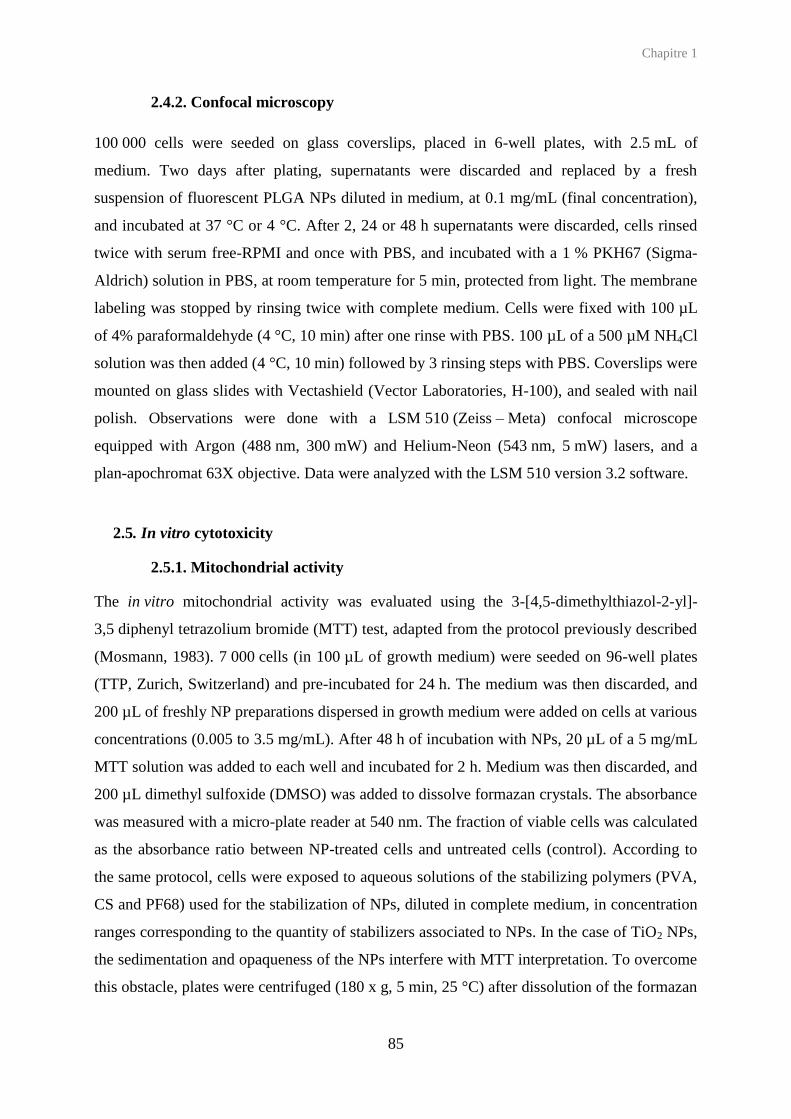
Chapitre 1
85
2.4.2. Confocal microscopy
100 000 cells were seeded on glass coverslips, placed in 6-well plates, with 2.5 mL of
medium. Two days after plating, supernatants were discarded and replaced by a fresh
suspension of fluorescent PLGA NPs diluted in medium, at 0.1 mg/mL (final concentration),
and incubated at 37 °C or 4 °C. After 2, 24 or 48 h supernatants were discarded, cells rinsed
twice with serum free-RPMI and once with PBS, and incubated with a 1 % PKH67 (Sigma-
Aldrich) solution in PBS, at room temperature for 5 min, protected from light. The membrane
labeling was stopped by rinsing twice with complete medium. Cells were fixed with 100 µL
of 4% paraformaldehyde (4 °C, 10 min) after one rinse with PBS. 100 µL of a 500 µM NH4Cl
solution was then added (4 °C, 10 min) followed by 3 rinsing steps with PBS. Coverslips were
mounted on glass slides with Vectashield (Vector Laboratories, H-100), and sealed with nail
polish. Observations were done with a LSM 510 (Zeiss – Meta) confocal microscope
equipped with Argon (488 nm, 300 mW) and Helium-Neon (543 nm, 5 mW) lasers, and a
plan-apochromat 63X objective. Data were analyzed with the LSM 510 version 3.2 software.
2.5. In vitro cytotoxicity
2.5.1. Mitochondrial activity
The in vitro mitochondrial activity was evaluated using the 3-[4,5-dimethylthiazol-2-yl]-
3,5 diphenyl tetrazolium bromide (MTT) test, adapted from the protocol previously described
(Mosmann, 1983). 7 000 cells (in 100 µL of growth medium) were seeded on 96-well plates
(TTP, Zurich, Switzerland) and pre-incubated for 24 h. The medium was then discarded, and
200 µL of freshly NP preparations dispersed in growth medium were added on cells at various
concentrations (0.005 to 3.5 mg/mL). After 48 h of incubation with NPs, 20 µL of a 5 mg/mL
MTT solution was added to each well and incubated for 2 h. Medium was then discarded, and
200 µL dimethyl sulfoxide (DMSO) was added to dissolve formazan crystals. The absorbance
was measured with a micro-plate reader at 540 nm. The fraction of viable cells was calculated
as the absorbance ratio between NP-treated cells and untreated cells (control). According to
the same protocol, cells were exposed to aqueous solutions of the stabilizing polymers (PVA,
CS and PF68) used for the stabilization of NPs, diluted in complete medium, in concentration
ranges corresponding to the quantity of stabilizers associated to NPs. In the case of TiO2 NPs,
the sedimentation and opaqueness of the NPs interfere with MTT interpretation. To overcome
this obstacle, plates were centrifuged (180 x g, 5 min, 25 °C) after dissolution of the formazan

Chapitre 1
86
crystals by DMSO, and 100 µL of supernatants transferred in new plate (Simon-Deckers et
al., 2008). All measurements were performed in triplicate or more.
2.5.2. LDH release test
Lactate dehydrogenase (LDH) released in extracellular medium was quantified using the Cyto
Tox 96, Non-radioactive Cytotoxicity Assay (Promega), according to the manufacturer’s
instructions. 96-well plates seeding and exposure conditions were the same as those described
in section 0. Absorbance was measured at 492 m. The LDH release (%) was calculated as the
ratio between the LDH release after NP exposure minus the LDH spontaneously released by
non-treated cells, and the intracellular LDH, expressed as the difference of the total LDH
measured after cell lysis (Triton X-100, 1/10e diluted in PBS, 1 h, 37° C) minus the LDH
spontaneously released. All measurements were performed in triplicate.
2.5.3. Trypan blue test
Cell viability was also evaluated by trypan blue test. 100 000 cells were seeded in 6-well
plates, in 2.5 mL of medium. After 24 hours, supernatants were discarded and replaced by
fresh NP dilutions in cell culture medium in range concentration of 0.01 to 2 mg/mL. After
48 hours exposure, supernatants were discarded and cells rinsed twice with PBS. Cells
exposed to polymeric NPs (PLGA and polystyrene NPs) were harvested with 1X trypsine, and
cells exposed to TiO2 NPs were scrapped. Cells were mixed with an aqueous solution of
trypan blue 0.4%, in 50/50 (v/v) (Eurobio) according to a method previously described
(Strober, 2001). Cell counting was performed using Kova counting chambers (Kova Glasstic
Slide, Hycor Biomedical) by microscopy. The cell viability was calculated as the ratio
between uncolored cells and total cells. All measurements were performed in triplicate.
2.5.4. Propidum iodide/Syto® 24 test
Viability was also evaluated with two fluorophores, propidium iodide (Sigma-Aldrich) which
binds to nucleic acids of cells with a damaged membrane, and the Syto® 24 (Invitrogen),
which binds to nucleic acids of all cells. 6-well plates seeding, exposure, and harvesting
conditions were the same as those described in part 0. Propidium iodide was first added on
cell suspension at final concentration of 1 µM, and mixing by gentle pipetting
homogenization. After 5 min incubation, Syto® 24 was added to the cell suspension, at 1 µM

Chapitre 1
87
final concentration, and mixing by gentle pipetting homogenization. After 5 min incubation,
cell counting was performed using Kova counting chambers, by fluorescent microscopy,
using the range excitation wavelengths of 515-560 nm (band pass filter) for the propidium
iodide, and 440-480 nm (band pass filter) for the Syto® 24. The cell viability was calculated
as the ratio between green cells and total cells. All measurements were performed in triplicate.
2.6. Cytokine quantification after exposure to nanoparticles
Cytokine secretions after exposure to NPs were investigated in supernatants using the
multiplexing Cytometric Beads Array (CBA) method proposed by BD Biosciences (Morgan
et al., 2004, Tarnok et al., 2003), and performed with the human IL-1, IL-6, IL-8, IL-10, IL-
12p70, MCP-1 and TNF flex sets and the Human Soluble Protein Master Buffer Kit (BD
Biosciences). 40 000 cells were seeded in 12-well plates for 48 hours. Supernatants were then
discarded, and replaced by 2 mL of fresh growth medium (non treated cells), with either
increasing lipopolysaccharide (LPS) concentrations (0.1, 1, 10 µg/mL) or NPs at 0.1 mg/mL.
After 24 h exposure, supernatants were sampled and cells were rinsed once with PBS and
harvested with 1X trypsine, to be counted by flow cytometry. Cytokine results, analyzed with
the FACP ArrayTM Software, were obtained as pg/mL concentrations. All measurements were
performed in triplicate, or more.
3. Results
3.1. Nanoparticle characterization
A family of PLGA NPs displaying various surface properties was prepared using a selection
of hydrophilic polymers as stabilizers (PVA, CS, PF68), as similarly described by our group
(Mura et al., 2011b) with some modifications for PLGA/PF68 NPs, the size of which was
adjusted to around 230 nm. All PLGA NPs have a similar size in the 210-270 nm range, but
differ by their surface charge as shown through zeta potential measurements (around
neutrality for PLGA/PVA NPs, around + 40 mV for PLGA/CS NPs and – 30 mV for
PLGA/PF68 NPs). The amount of stabilizers associated to NPs after preparation and
purification was found in the range of 10 - 45 mg per 100 mg PLGA. Noteworthy, covalently-
labeled, fluorescent counterparts of all these NPs could be obtained without significant
alteration of their size and surface charge. Commercially available polystyrene NPs

Chapitre 1
88
displaying a similar size were characterized similarly and used as non-biodegradable,
polymeric sample NPs (Table 1).
Colloidal suspensions of rutile and anatase TiO2 were obtained through a controlled
dispersion procedure of nanopowders after careful selection of dispersing tools and pH
adjustment, without use of any stabilizing agent. The discrepancy between the size of the
primary particles (< 50 nm) and the particles in suspension can be due to the aggregation of
primary NPs as submicron aggregates (Table 1).
Table. 1. Physicochemical properties of NPs. na = not applicable, nd = not determined
Figure S 1. Fluorescence emission spectra of PLGA-Rhod/PVA (green), PLGA-Rhod/CS (red) and PLGA-Rhod/PF68 (blue) NP suspensions at 540 nm (maximal excitation wavelength).
This NP selection, displaying a variety of core material (polymeric/inorganic, biodegradable
or not) and surface coating was then evaluated on the A549 human lung cells.
NPs Diameter
(nm) (mean ± SD)
Diameter after 48 h in cell culture
medium (nm) (mean ± SD)
Polydispersity index
Zeta Potential
(mV) (mean ± SD)
Core material
Stabilizer (mg/100mg
core)
Crystal form
PLGA/PVA 234 ± 28 212 ± 4 0.099 ± 0.04 - 1.42 ± 1.75 PLGA PVA (11.5)
na
Rhod-PLGA/PVA 247 ± 11 216 ± 11 0.15 ± 0.05 + 3.7 ± 0.2 PLGA nd
PLGA/CS 233 ± 18 269 ± 12 0.210 ± 0.020 + 39.9 ± 7.2 PLGA CS (15.3) / PVA (30.4)
Rhod-PLGA/CS 195 ± 11 200 ± 7 0.16 ± 0.03 + 24.3 ± 1.8 PLGA nd PLGA/PF68 229 ± 19 316 ± 16 0.136 ± 0.036 - 32.2 ± 3.5 PLGA PF 68 (15.5)
Rhod-PLGA/PF68 227 ± 9 238 ± 9 0.12 ± 0.01 - 31.5 ± 2.6 PLGA nd Polystyrene 250 ± 11 259 ± 21 0.02 ± 0.02 - 48.8 ± 1.5 Polystyrene Sulfate ester
Anatase TiO2 421 ± 49 625 ± 46 0.42 ± 0.07 - 35 ± 2 TiO2 none Anatase Rutile TiO2 479 ± 184 795 ± 80 0.77 ± 0.25 - 37.2 ± 6 TiO2 none Rutile

Chapitre 1
89
3.2. Uptake of PLGA nanoparticles
All fluorescent PLGA NPs were taken up according to a similar profile (Figure 1). At 37 °C,
NP uptake increased from to 2 to 24 h, and then plateaued. At 4 °C, the extent of NP uptake
was much lower than at 37 °C, which is characteristic of an energy-dependent uptake
mechanism (Figure 2). Confocal microscopy images confirm the progressive uptake from 2 to
24 h at 37 °C, and the differences between 4 °C and 37 °C (Figure 3). Pictures additionally
show that PLGA NPs are internalized down to the cytoplasm rather than merely associated to
the cell surface. Significant differences are however observed depending on the stabilizer. The
negatively-charged PLGA/PF68 NPs display a lower association to cells at 4 °C compared to
other NPs (Figure 2). In contrast, some colocalization of NPs (red) and cell membrane (green)
is observed in the case of PLGA/CS NPs, showing an adhesion of these positively-charged
NPs onto the cell surface (Figure 3).
Figure 1. Uptake kinetics of PLGA-Rhod/PVA (green), PLGA-Rhod/CS (red) and PLGA-Rhod/PF68 (blue) NPs by A549 cells after 2, 24 and 48 h incubation at 37 °C, compared to non-treated cells, evaluated by flow cytometry. NT = non treated cells, MFI = mean fluorescence intensity (FL-2 channel).

Chapitre 1
90
By measuring the fluorescence intensity of NP suspensions, it appears that the fluorescence
ratios, as defined in the material and methods, of PLGA/PVA NPs and PLGA/PF68 NPs are
respectively 0.77 and 0.37 (Figure S 2). To truly compare the extent of PLGA NP uptake, the
MFI increase (NP-treated cells versus non treated cells) was corrected by the fluorescence
intensity ratios accordingly. Results reveal that at 37 °C, after 48 h exposure, PLGA/PF68
NPs are taken up almost twice as much than the PLGA/CS and PLGA/PVA NPs (Figure 4).
Figure 2. Uptake of PLGA-Rhod/PVA (green), PLGA-Rhod/CS (red) and PLGA-Rhod/PF68 (blue) NPs by A549 cells after 6 h incubation at 4 °C and 37 °C, compared to non-treated (NT) cells, as evaluated by the mean fluorescence intensity (MFI) in flow cytometry.

Chapitre 1
91
Figure 3. Uptake of PLGA-Rhod/PVA, PLGA-Rhod/CS and PLGA-Rhod/PF68 NPs (red) by A549 cells labeled with PKh67 (green) observed in confocal laser scanning microscopy after 2 h (4 h and 37 °C) and 24 h (37 °C) incubation. Scale bars = 20 µm.
The cell granularity was also investigated, using the side scattered (SSC) signal in flow
cytometry. While PLGA/PVA and PLGA/CS NPs did not entail significant modifications
compared to untreated cells, exposure to PLGA/PF68 NPs did increase cell granularity
(Figure 5), which can be correlated to the higher uptake of these NPs.

Chapitre 1
92
2h 6h 2h 6h 24h 48h0
30
60
90
120PLGA/PVA NPs
PLGA/CS NPs
PLGA/PF68 NPs
4°C 37°C
MF
I fo
ld i
ncre
ase
Figure 4. Uptake kinetics of PLGA NPs by A549 cells at 37 °C (full bars) and 4 °C (hatched bars), as evaluated by the mean fluorescence intensity (MFI) in flow cytometry, after correction by relative fluorescence intensity of each NP suspension.
Figure 5. Side-scatter light curves (SSC) of A549 cells after 48 h exposure to PLGA/PVA NPs (green), PLGA/CS NPs (red) and PLGA/PF68 NPs (blue), compared to non treated (NT) cells.
3.3. Cytotoxicity
The cytotoxicity of various NPs was evaluated after exposure of cells to NP concentrations
ranging from 0.01 to 4 mg/mL by addressing several reference toxicity endpoints (Eisenbrand
et al., 2002): reduced mitochondrial function through MTT test, and alteration of membrane
permeability through dye exclusion tests and measurement of released intracellular enzymes.
Detailed results are shown in supplementary data (Figure S 1) and summarized in Table 2.

Chapitre 1
93
0.001 0.01 0.1 1 10
0
60
120
PLGA/PVA NPs
PVA
A
NP (or equivalent) concentrati
Mit
och
on
dri
al
acti
vit
y (
%)
0.001 0.01 0.1 1 10
0
60
120
PLGA/CS NPs
CS
PVA
CS/PVA
B
NP (or equivalent) concentration (mg/mL)
Mit
och
on
dri
al
acti
vit
y (
%)
0.001 0.01 0.1 1 10
0
60
120
PLGA/PF68 NPs
PF68
C
NP (or equivalent) concentrati
Mit
och
on
dri
al
acti
vit
y (
%)
Figure 6. Mitochondrial activity of A549 cells after 48 h exposure to PLGA NPs (plain line) compared to solutions of free stabilizers (dotted line) in equivalent concentrations: PLGA/PVA NPs vs PVA (A), PLGA/CS NPs vs CS/PVA (B) and PLGA/PF68 NPs vs PF68 (C).
The mitochondrial activity measured after 48 h exposure to low concentrations (< 0.1 mg/mL)
of PLGA/PVA NPs is higher than after exposure to PLGA/CS and PLGA/PF6Ř NPs (≥ 90 %
vs. ≥ 70 %), whereas at high concentrations (> 0.1 mg/mL), they are similar for all PLGA NPs
(around 60 %). Polystyrene NPs induce a similar effect to that of PLGA/PVA NPs. The
exposure to TiO2 nanoparticles reduces mitochondrial activity down to 45 % at high NP
concentration (with a slight decrease when concentration increases). At the midpoint
concentration of 0.1 mg/mL, all NPs maintain cellular viability around 60 % or above
(Figures S 1.A, S 1.E). This concentration was chosen for the study of the inflammatory
response.

Chapitre 1
94
0.001 0.01 0.1 1 10
0
60
120
PLGA/PVA NPs
PLGA/CS NPs
PLGA/PF68 NPs
NPs (mg/mL)
Mit
och
on
dri
al
acti
vit
y (
%)
0.001 0.01 0.1 1 10
0
60
120
PLGA/PVA NPs
PLGA/CS NPs
PLGA/PF68 NPs
NPs (mg/mL)
Try
pa
n b
lue n
ega
tive c
ells (
%)
0.001 0.01 0.1 1 10
0
60
120
PLGA/PVA NPs
PLGA/CS NPs
PLGA/PF68 NPs
NPs (mg/mL)
LD
H r
ele
ase
(%
)
0.001 0.01 0.1 1 10
0
60
120
PLGA/PVA NPs
PLGA/CS NPs
PLGA/PF68 NPs
NPs (mg/mL)
Syto
24
-po
sit
ive
ce
lls (
%)
0.0001 0.001 0.01 0.1 1 10
0
60
120
Anatase TiO 2 NPs
Rutile TiO2 NPs
PS NPs
NPs (mg/mL)
Mit
och
on
dri
al
acti
vit
y (
%)
0.001 0.01 0.1 1 10
0
60
120
Anatase TiO 2 NPs
Rutile TiO2 NPs
PS NPs
NP (mg/mL)
Try
pa
n b
lue n
ega
tive c
ells (
%)
0.001 0.01 0.1 1 10
0
60
120
Anatase TiO 2 NPs
Rutile TiO2 NPs
PS NPs
NPs (mg/mL)
LD
H r
ele
ase
(%
)
0.001 0.01 0.1 1 10
0
60
120
Anatase TiO 2 NPs
Rutile TiO2 NPs
PS NPs
NPs (mg/mL)
Syto
24
-po
sit
ive
ce
lls (
%)
A
B
C
D
E
F
G
H
Figure S 2. A549 cell viability estimated with MTT test (A, E), trypan Blue test (B, F), LDH release (C, G) and propidium iodide/Syto® 24 staining (D, H) after 48 h exposure to PLGA NPs (A, B, C and D) or polystyrene and TiO2 NPs (E, F, G and H).

Chapitre 1
95
Cell viability estimated by the trypan blue exclusion test shows homogeneous results for all
PLGA NPs: a low toxicity at low NP concentration, increasing with the concentration.
Cellular viability profile is similar after exposure to TiO2 NPs, although these particles induce
a higher toxicity at high concentrations. Compared to mitochondrial activity, the effect of
PLGA and TiO2 NPs on trypan blue exclusion is more progressive but also more pronounced.
In contrast, the effect of polystyrene NPs seems negligible (Figures S 1.B, S 1.F).
The assessment of membrane integrity through the release of LDH, and the staining of nucleic
acids with propidium iodide and Syto® 24, shows a weak NP effect. High concentrations of
TiO2 NPs are the only conditions leading to a substantial effect, more pronounced for the
anatase than rutile form (Figures S 1.C, S 1.D, S 1.G, S 1.H).
Taken together, these results show that low concentrations of PLGA/PVA and polystyrene
NPs induce no or mild reduction of cell mitochondrial activity, whereas the impact of
PLGA/CS and PLGA/PF68 NPs is more pronounced. The effect on cell membrane integrity is
more pronounced for TiO2 NPs compared to polymeric NPs, especially at high
concentrations.
In order to evaluate the potential contribution to cytotoxicity from the hydrophilic polymers
used as stabilizers to formulate PLGA NPs, cells were exposed to solutions of the
corresponding polymers (PVA, CS and PF68, in the absence of PLGA) in the same range of
concentrations as those corresponding to the PLGA NP suspensions (Figure 6). Cell viability
was evaluated with the MTT test since it was the most discriminant test to compare the
toxicity of PLGA NPs. Results show that solutions of PVA and PF68 do not induce cell
mortality, event at high concentration, and thus may not contribute by themselves to the
toxicity of PLGA/PVA and PLGA/PF68 NPs. In contrast, solutions containing CS induce a
similar toxicity to that of the corresponding PLGA/CS NP suspensions, indicating that the
sole presence of CS can induce cytotoxicity as evaluated by mitochondrial activity.

Chapitre 1
96
Table 2. Cellular toxicity estimated with various tests (MTT, trypan blue staining, LDH release, Propidium iodide/Syto®24 staining) following exposure (48 h) to low (<1 mg/mL) or high (> 1 mg/mL) concentrations of NPs.
Toxicity levels (-, +, ++) were assigned according to the following viability intervals: above 75 % (-); between 75 % and 35 % (+); below 35 % (++). Low concentration corresponds to NP concentration under 0.1 mg/mL; high concentration corresponds to concentration above 0.1 mg/mL. Detailed viability results are shown in figure S 1.
3.4. Inflammatory response following exposure to nanoparticles
Cytokines were quantified in the cell supernatants following exposure to various NPs using
the CBA method that allows the dosage of numerous compounds in one sample. We have
investigated several cytokines known to be involved in the inflammatory response: IL-1, IL-
6, IL-8, IL-10, IL-12p70, TNF-α and MCP-1. LPS, a well known Toll-like agonist, was
chosen as positive control to induce cytokine secretions from A549 cells. Preliminary results
allowed us to select three relevant cytokines: IL6-, IL8 and MCP-1. These cytokines were
found to be secreted according to a dose-response profile and reached a maximum in response
to 1 µg/mL LPS (Figure S 3). Other tested cytokines were under the limit detection, even after
exposure to 1 µg/mL LPS. In accordance with the MTT test performed after exposure to
PLGA NPs, the NP concentration selected was 0.1 mg/mL. IL-6, IL-8 and MCP-1 secretions
following TiO2 NPs exposure are found to be equivalent or above the basal levels (non-treated
cells). PLGA/PVA, PLGA/CS and polystyrene NPs induced cytokine levels between non-
treated and LPS-treated cells, or slightly above. In contrast, PLGA/PF68 NPs induced higher
cytokine secretions than other polymeric NPs, reaching values equivalent or above LPS-
treated cells.
Test MTT Trypan Blue LDH release Propidium Iodide
/ Syto®24 NP concentration Low High Low High Low High Low High
PLGA/PVA - + - + - - - - PLGA/CS + + - + - - - -
PLGA/PF68 + + - + - - - - PS - + - - - - - -
Anatase TiO2 + + - ++ - - - + Rutile TiO2 + + - ++ - - - +

Chapitre 1
97
Figure S 3. Secretions of MCP-1 (A), IL-8 (B) and IL-6 (C) cytokines by A549 cells after 24 h exposure to various concentration of LPS. nd = not determined. n = 3, * p < 0.05 compared to non-treated cells.
0 0.1 1 100
2000
4000
6000
8000
LPS (µg/mL)
MC
P-1
(pg/1
06 c
ells
)
*
A
0 0.1 1 100
500
1000
1500
2000
LPS (µg/mL)
IL-8
(pg/1
06 c
ell) *
B
0 0,1 1 100
50
100
150
200
LPS (µg/mL)
IL-6
(pg/1
06 c
ell)
*
C

Chapitre 1
98
NT
LPS
PLGA/P
VA N
Ps
PVA
PLGA/C
S NPs
CS/P
VA
PLGA/P
F68 N
Ps
PF68
PS N
Ps
NPs
2
Anat
ase
TiO N
Ps
2
Rutil
e TiO
0
500
1000
1500
2000
2500
*
*
§
AM
CP
-1 (
pg
/mL
)
NT
LPS
PLG
A/P
VA N
Ps
PVA
PLG
A/C
S N
Ps
CS/P
VA
PLG
A/P
F68 N
Ps
PF68
PS N
Ps
NPs
2
Anat
ase
TiO N
Ps
2
Rutil
e TiO
0
200
400
600
800
1000
1200
**
*
§
B
IL-8
(p
g/m
L)
NT
LPS
PLG
A/P
VA N
Ps
PVA
PLG
A/C
S N
Ps
CS/P
VA
PLG
A/P
F68 N
Ps
PF68
PS N
Ps
NPs
2
Anat
ase
TiO N
Ps
2
Rutil
e TiO
0
20
40
60 *
*
§
§
C
IL-6
(p
g/m
L)
Figure 7. Secretions of MCP-1 (A), IL-8 (B) and IL-6 (C) cytokines after 24 h exposure to 0.1 mg/mL NPs or 1 µg/mL LPS, quantified with CBA method. NT = non-treated cells, n ≥ 3, p < 0.05 compared to non-treated cells, § p < 0.05 for each stabilizer compared to corresponding NP concentration.
4. Discussion
The goal of the present study was to compare the cytotoxicity and inflammatory response
following exposure to different types of NPs on A549 cells. The self-designed biodegradable
polymeric NPs are comparable in terms of size and size distribution displaying a mean
diameter around 230 nm (regardless of the coating agent) that is compatible with cell
internalization, with a low polydispersity index. In order to compare the role of the core
material and the surface coating of NPs on their toxicity, we have checked in a previous study
that the PLGA NPs are not degraded during the short time cytotoxicity studies (Mura et al.,
2011b). We have also used polystyrene NPs with an equivalent mean size. Only TiO2 NPs

Chapitre 1
99
displayed a larger mean size but still under 500 nm without the use of any additive that would
have changed the behavior of the native NPs.
In this study, the A459 cell line, isolated from a human lung carcinoma (Lieber et al., 1976),
was chosen because these cells constitute an appropriate epithelial alveolar model (Rothen-
Rutishauser et al., 2012). This model allows an evaluation of the impact of NPs able to reach
alveoli, and is complementary to previous studies focused on an in vitro Calu-3 model of the
bronchial region ((Mura et al., 2011a,b). Among the family of PLGA-based NPs tested in this
model, some similarities were observed, such as the uptake mechanism or cytotoxicity at low
concentration, but also multiple divergences. Differences observed in NP uptake between 4
and 37 °C characterize an energy-dependent mechanism of internalization consistent with
previous studies showing that PLGA NPs are taken up by epithelial cells through an
endocytosis pathway in most cases (Cartiera et al., 2009). However, as it was previously
shown on Calu-3 cells (Mura et al., 2011a), PLGA/PF68 NPs are internalized in higher
amount compared to the two other PLGA NPs. This result confirms that on the one hand the
coating plays a role in the uptake (Alexis et al., 2008), and on the other hand explains the
granularity increase. Due to the impossibility to use the same labeling technique, we did not
explore the internalization of the non-biodegradable PS NPs or the manufactured TiO2 NPs.
Cell toxicity encompasses various mechanisms, which have been addressed in this study using
several toxicity endpoints: modification of cell appearance (size, morphology or granular
modifications), reduction of metabolic capacity (mitochondrial activity) and alteration of
membrane integrity. Contrary to Calu-3 cells, for which no significant difference in
cytotoxicity was observed among the three PLGA NPs, some differences were observed on
A549 cells, especially in terms of mitochondrial activity. While at high concentration, all
PLGA NPs have a similar impact on mitochondrial activity, at low concentration PLGA/CS
NPs have a stronger effect over this activity, which can be correlated to the stronger
association to cell membrane as observed by confocal microscopy. Interestingly, the
cytotoxicity of PLGA/CS NPs observed was the same as the one induced by CS only,
indicating an important contribution in this case of the coating agent to the cytotoxicity, which
was not observed for PVA or PF68 (Huang et al., 2004). In the case of PLGA/PF68 NPs, the
higher toxicity compared to PLGA/PVA may result from the significantly higher quantity of
PLGA/PF68 NPs internalized. Besides these differences, all PLGA NPs were found to induce
a low LDH release by the cells, which moslty remained negative to propidium iodide. Taken
together, these results suggest that cell exposure to PLGA NPs results in a significant
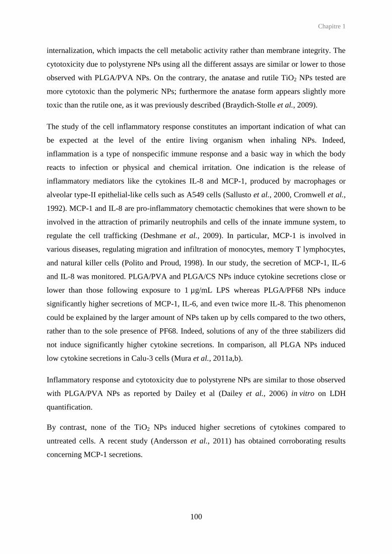
Chapitre 1
100
internalization, which impacts the cell metabolic activity rather than membrane integrity. The
cytotoxicity due to polystyrene NPs using all the different assays are similar or lower to those
observed with PLGA/PVA NPs. On the contrary, the anatase and rutile TiO2 NPs tested are
more cytotoxic than the polymeric NPs; furthermore the anatase form appears slightly more
toxic than the rutile one, as it was previously described (Braydich-Stolle et al., 2009).
The study of the cell inflammatory response constitutes an important indication of what can
be expected at the level of the entire living organism when inhaling NPs. Indeed,
inflammation is a type of nonspecific immune response and a basic way in which the body
reacts to infection or physical and chemical irritation. One indication is the release of
inflammatory mediators like the cytokines IL-8 and MCP-1, produced by macrophages or
alveolar type-II epithelial-like cells such as A549 cells (Sallusto et al., 2000, Cromwell et al.,
1992). MCP-1 and IL-8 are pro-inflammatory chemotactic chemokines that were shown to be
involved in the attraction of primarily neutrophils and cells of the innate immune system, to
regulate the cell trafficking (Deshmane et al., 2009). In particular, MCP-1 is involved in
various diseases, regulating migration and infiltration of monocytes, memory T lymphocytes,
and natural killer cells (Polito and Proud, 1998). In our study, the secretion of MCP-1, IL-6
and IL-8 was monitored. PLGA/PVA and PLGA/CS NPs induce cytokine secretions close or
lower than those following exposure to 1 µg/mL LPS whereas PLGA/PF68 NPs induce
significantly higher secretions of MCP-1, IL-6, and even twice more IL-8. This phenomenon
could be explained by the larger amount of NPs taken up by cells compared to the two others,
rather than to the sole presence of PF68. Indeed, solutions of any of the three stabilizers did
not induce significantly higher cytokine secretions. In comparison, all PLGA NPs induced
low cytokine secretions in Calu-3 cells (Mura et al., 2011a,b).
Inflammatory response and cytotoxicity due to polystyrene NPs are similar to those observed
with PLGA/PVA NPs as reported by Dailey et al (Dailey et al., 2006) in vitro on LDH
quantification.
By contrast, none of the TiO2 NPs induced higher secretions of cytokines compared to
untreated cells. A recent study (Andersson et al., 2011) has obtained corroborating results
concerning MCP-1 secretions.

Chapitre 1
101
Conclusion
In this study we have observed several differences in the cytotoxicity and inflammatory
response between inorganic and polymeric NPs. TiO2 NPs are slightly more cytotoxic than
polymeric NPs but do not induce more inflammation. However, important differences among
PLGA NPs depending on the surface coating were observed particularly in the cell uptake and
induction of inflammatory response. According to those different behaviors, each PLGA NPs
could be used for different applications in drug delivery to the lung. PLGA/PF68 NPs display
an important potential for intracellular delivery, due to their high internalization into cells.
However, a particular attention should be paid to the inflammatory response to these NPs.
PLGA/CS NPs could be used when there is a need of adhesion of the nanocarrier to the cell
membrane. Finally, PLGA/PVA NPs may provide an interesting compromise between
significant cell uptake with limited cytotoxicity and inflammatory response.
These results also show the importance of detailed characterization of the PLGA NPs together
with their stabilizing agents, and their potential involvement in the inflammatory response. To
gain a deeper understanding, complementary studies are worth being investigated, including
complex cell models, and in vivo studies.
Acknowledgments
The authors wish to thank Simona Mura (Institut Galien Paris Sud) for her nanoparticle
expertise, Valérie Nicolas (IFR IPSIT, Université Paris-Sud) for the confocal laser
microscopy observations, Nadia Abed (Institut Galien Paris Sud) for microbiology
experiments, Hélène Chacun and Stéphanie Denis (Institut Galien Paris-Sud) for their
guidance and assistance in cytotoxicity and cell culture experiments. This study was
supported by the ANSES “Emerging risks” program and by ANR (under reference β00ř
CESA 011).
References
ALEXIS, F., PRIDGEN, E., MOLNAR, L. K. & FAROKHZAD, O. C. 2008. Factors affecting the clearance and biodistribution of polymeric nanoparticles. Mol Pharm, 5, 505-15.
ANDERSSON, P. O., LEJON, C., EKSTRAND-HAMMARSTROM, B., AKFUR, C., AHLINDER, L., BUCHT, A. & OSTERLUND, L. 2011. Polymorph- and size-dependent uptake and toxicity of TiO(2) nanoparticles in living lung epithelial cells. Small, 7, 514-23.

Chapitre 1
102
BACHAND, G. D., ALLEN, A., BACHAND, M., ACHYUTHAN, K. E., SEAGRAVE, J. C. & BROZIK, S. M. 2012. Cytotoxicity and inflammation in human alveolar epithelial cells following exposure to occupational levels of gold and silver nanoparticles. Journal of Nanoparticle Research, 14.
BHATTACHARYA, K., DAVOREN, M., BOERTZ, J., SCHINS, R. P., HOFFMANN, E. & DOPP, E. 2009. Titanium dioxide nanoparticles induce oxidative stress and DNA-adduct formation but not DNA-breakage in human lung cells. Part Fibre Toxicol, 6, 17.
BRAYDICH-STOLLE, L., SCHAEUBLIN, N., MURDOCK, R., JIANG, J., BISWAS, P., SCHLAGER, J. & HUSSAIN, S. 2009. Crystal structure mediates mode of cell death in TiO2 nanotoxicity. Journal of Nanoparticle Research, 11, 1361-1374.
CARTIERA, M. S., JOHNSON, K. M., RAJENDRAN, V., CAPLAN, M. J. & SALTZMAN, W. M. 2009. The uptake and intracellular fate of PLGA nanoparticles in epithelial cells. Biomaterials, 30, 2790-8.
CHOI, S. J., OH, J. M. & CHOY, J. H. 2009. Toxicological effects of inorganic nanoparticles on human lung cancer A549 cells. J Inorg Biochem, 103, 463-71.
CROMWELL, O., HAMID, Q., CORRIGAN, C. J., BARKANS, J., MENG, Q., COLLINS, P. D. & KAY, A. B. 1992. Expression and generation of interleukin-8, IL-6 and granulocyte-macrophage colony-stimulating factor by bronchial epithelial cells and enhancement by IL-1 beta and tumour necrosis factor-alpha. Immunology, 77, 330-7.
DAILEY, L. A., JEKEL, N., FINK, L., GESSLER, T., SCHMEHL, T., WITTMAR, M., KISSEL, T. & SEEGER, W. 2006. Investigation of the proinflammatory potential of biodegradable nanoparticle drug delivery systems in the lung. Toxicol Appl Pharmacol, 215, 100-8.
DESHMANE, S. L., KREMLEV, S., AMINI, S. & SAWAYA, B. E. 2009. Monocyte chemoattractant protein-1 (MCP-1): an overview. J Interferon Cytokine Res, 29, 313-26.
EISENBRAND, G., POOL-ZOBEL, B., BAKER, V., BALLS, M., BLAAUBOER, B. J., BOOBIS, A., CARERE, A., KEVEKORDES, S., LHUGUENOT, J. C., PIETERS, R. & KLEINER, J. 2002. Methods of in vitro toxicology. Food Chem Toxicol, 40, 193-236.
HE, Y. R., JIN, Y., CHEN, H. S., DING, Y. L., CANG, D. Q. & LU, H. L. 2007. Heat transfer and flow behaviour of aqueous suspensions of TiO2 nanoparticles (nanofluids) flowing upward through a vertical pipe. International Journal of Heat and Mass Transfer, 50, 2272-2281.
HUANG, M., KHOR, E. & LIM, L. Y. 2004. Uptake and cytotoxicity of chitosan molecules and nanoparticles: effects of molecular weight and degree of deacetylation. Pharm Res, 21, 344-53.
INOUE, K., TAKANO, H., OHNUKI, M., YANAGISAWA, R., SAKURAI, M., SHIMADA, A., MIZUSHIMA, K. & YOSHIKAWA, T. 2008. Size effects of nanomaterials on lung inflammation and coagulatory disturbance. International journal of immunopathology and pharmacology, 21, 197-206.

Chapitre 1
103
JOHNSTON, H. J., HUTCHISON, G. R., CHRISTENSEN, F. M., PETERS, S., HANKIN, S. & STONE, V. 2009. Identification of the mechanisms that drive the toxicity of TiO(2 )particulates: the contribution of physicochemical characteristics. Part Fibre Toxicol, 6, 33.
JOSHI, D. P., LAN-CHUN-FUNG, Y. L. & PRITCHARD, J. G. 1979. Determination of poly(vinyl alcohol) via its complex with boric acid and iodine. Analytica Chimica Acta, 104, 153-160.
LIEBER, M., SMITH, B., SZAKAL, A., NELSON-REES, W. & TODARO, G. 1976. A continuous tumor-cell line from a human lung carcinoma with properties of type II alveolar epithelial cells. Int J Cancer, 17, 62-70.
LIMBACH, L. K., WICK, P., MANSER, P., GRASS, R. N., BRUININK, A. & STARK, W. J. 2007. Exposure of engineered nanoparticles to human lung epithelial cells: influence of chemical composition and catalytic activity on oxidative stress. Environ Sci Technol, 41, 4158-63.
LIN, W., HUANG, Y. W., ZHOU, X. D. & MA, Y. 2006. In vitro toxicity of silica nanoparticles in human lung cancer cells. Toxicol Appl Pharmacol, 217, 252-9.
MENON, J. U., KONA, S., WADAJKAR, A. S., DESAI, F., VADLA, A. & NGUYEN, K. T. 2012. Effects of surfactants on the properties of PLGA nanoparticles. J Biomed Mater Res A, 100, 1998-2005.
MORGAN, E., VARRO, R., SEPULVEDA, H., EMBER, J. A., APGAR, J., WILSON, J., LOWE, L., CHEN, R., SHIVRAJ, L., AGADIR, A., CAMPOS, R., ERNST, D. & GAUR, A. 2004. Cytometric bead array: a multiplexed assay platform with applications in various areas of biology. Clin Immunol, 110, 252-66.
MOSMANN, T. 1983. Rapid colorimetric assay for cellular growth and survival: application to proliferation and cytotoxicity assays. J Immunol Methods, 65, 55-63.
MURA, S., HILLAIREAU, H., NICOLAS, J., KERDINE-ROMER, S., LE DROUMAGUET, B., DELOMENIE, C., NICOLAS, V., PALLARDY, M., TSAPIS, N. & FATTAL, E. 2011a. Biodegradable nanoparticles meet the bronchial airway barrier: how surface properties affect their interaction with mucus and epithelial cells. Biomacromolecules, 12, 4136-43.
MURA, S., HILLAIREAU, H., NICOLAS, J., LE DROUMAGUET, B., GUEUTIN, C., ZANNA, S., TSAPIS, N. & FATTAL, E. 2011b. Influence of surface charge on the potential toxicity of PLGA nanoparticles towards Calu-3 cells. Int J Nanomedicine, 6, 2591-605.
MUZZARELLI, R. A. 1998. Colorimetric determination of chitosan. Anal Biochem, 260, 255-7.
NAFEE, N., SCHNEIDER, M., SCHAEFER, U. F. & LEHR, C. M. 2009. Relevance of the colloidal stability of chitosan/PLGA nanoparticles on their cytotoxicity profile. Int J Pharm, 381, 130-9.
NEL, A., XIA, T., MADLER, L. & LI, N. 2006. Toxic potential of materials at the nanolevel. Science, 311, 622-7.
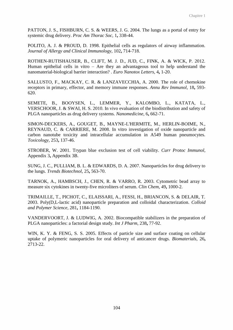
Chapitre 1
104
PATTON, J. S., FISHBURN, C. S. & WEERS, J. G. 2004. The lungs as a portal of entry for systemic drug delivery. Proc Am Thorac Soc, 1, 338-44.
POLITO, A. J. & PROUD, D. 1998. Epithelial cells as regulators of airway inflammation. Journal of Allergy and Clinical Immunology, 102, 714-718.
ROTHEN-RUTISHAUSER, B., CLIFT, M. J. D., JUD, C., FINK, A. & WICK, P. 2012. Human epithelial cells in vitro – Are they an advantageous tool to help understand the nanomaterial-biological barrier interaction? . Euro Nanotox Letters, 4, 1-20.
SALLUSTO, F., MACKAY, C. R. & LANZAVECCHIA, A. 2000. The role of chemokine receptors in primary, effector, and memory immune responses. Annu Rev Immunol, 18, 593-620.
SEMETE, B., BOOYSEN, L., LEMMER, Y., KALOMBO, L., KATATA, L., VERSCHOOR, J. & SWAI, H. S. 2010. In vivo evaluation of the biodistribution and safety of PLGA nanoparticles as drug delivery systems. Nanomedicine, 6, 662-71.
SIMON-DECKERS, A., GOUGET, B., MAYNE-L'HERMITE, M., HERLIN-BOIME, N., REYNAUD, C. & CARRIERE, M. 2008. In vitro investigation of oxide nanoparticle and carbon nanotube toxicity and intracellular accumulation in A549 human pneumocytes. Toxicology, 253, 137-46.
STROBER, W. 2001. Trypan blue exclusion test of cell viability. Curr Protoc Immunol, Appendix 3, Appendix 3B.
SUNG, J. C., PULLIAM, B. L. & EDWARDS, D. A. 2007. Nanoparticles for drug delivery to the lungs. Trends Biotechnol, 25, 563-70.
TARNOK, A., HAMBSCH, J., CHEN, R. & VARRO, R. 2003. Cytometric bead array to measure six cytokines in twenty-five microliters of serum. Clin Chem, 49, 1000-2.
TRIMAILLE, T., PICHOT, C., ELAISSARI, A., FESSI, H., BRIANCON, S. & DELAIR, T. 2003. Poly(D,L-lactic acid) nanoparticle preparation and colloidal characterization. Colloid and Polymer Science, 281, 1184-1190.
VANDERVOORT, J. & LUDWIG, A. 2002. Biocompatible stabilizers in the preparation of PLGA nanoparticles: a factorial design study. Int J Pharm, 238, 77-92.
WIN, K. Y. & FENG, S. S. 2005. Effects of particle size and surface coating on cellular uptake of polymeric nanoparticles for oral delivery of anticancer drugs. Biomaterials, 26, 2713-22.


106
Chapitre 2
Le recouvrement de surface des
nanoparticules de polymère joue un rôle
clef dans la toxicité vis-à-vis de
macrophages dérivés de
monocytes humains

Chapitre 2
107
Dans cette partie expérimentale de la thèse, des macrophages THP-1 ont été exposés aux
différentes nanoparticules de PLGA (PLGA sans stabilisant, PLGA/PVA, PLGA/CS,
PLGA/PF6Ř) précédemment détaillées (chapitre 1), afin d’en étudier la toxicité.
La lignée THP-1, est une lignée de monocytes humains dérivés de cellules leucémiques
(Tsuchiya et al., 1980). Ces monocytes possèdent les caractéristiques des monocytes humains,
de par leur capacité d’internalisation ou encore de par leur capacité à sécréter des composés
biologiques impliqués dans la croissance, la communication ou la mort cellulaire. La
différentiation en macrophages est généralement réalisée grâce à l’action du phorbol-12-
myristate-13-acetate (PMA) (Tsuchiya et al., 1řŘβ). Bien qu’il ne soit pas naturel, le PMA
conduit à une différentiation complète des monocytes en macrophages (Auwerx, 1991), en
augmentant l’expression des marqueurs membranaires CD11b (Mac-1 α, adhérence des
cellules immunitaires) et CD14 (antigène des monocytes) (Schwende et al., 1996). Nos
résultats ont montré que 24 h d’exposition à 10 nM sont suffisantes pour induire la
différentiation (Figure S1).
Lors des études d’internalisation, nous avons montré que les macrophages THP-1 capturaient
les nanoparticules de charge de surface négative (i.e PLGA sans stabilisant et PLGA/PF68)
plus rapidement et en plus grande quantité que les nanoparticules de charge de surface neutre
(PLGA/PVA) ou positive (PLGA/CS) (Figure 1). Afin d’étudier la toxicité induite par ces
nanoparticules, les macrophages THP-1 ont été exposés aux nanoparticules ainsi qu’aux
polymères de surface utilisés en tant que stabilisants. Différents paramètres de toxicité ont été
évalués :
La viabilité cellulaire estimée par l’activité mitochondriale (Figure 2)
Le type de mort cellulaire (apoptose/nécrose) (Figure 3)
La sécrétion des cytokines dans le milieu extracellulaire (Figure 4)
Le stress oxydant par la quantification des ROS et des RNS (espèces réactives de l’oxygène et de l’azote) (Figure 5)
Au cours de cette étude nous avons montré qu’après exposition à de faibles concentrations en
nanoparticules (jusqu’à 0,1 mg/mL), aucun signe de toxicité n’a été détecté. Seules les
nanoparticules de PLGA/CS se montrent légèrement plus toxiques que les autres, et cette
toxicité peut être attribuée à la toxicité propre du chitosane. Les nanoparticules de PLGA ne
provoquent ni la libération des cytokines (IL-6, IL-8, MCP-1 et TNF-α) dans le milieu

Chapitre 2
108
extracellulaire, ni la production de ROS intracellulaires ou la libération de RNS. Enfin, la
mort cellulaire observée est uniquement d’origine nécrotique et non apoptotique.
En revanche, après exposition à des fortes concentrations, les résultats obtenus notamment par
des tests MTT, montrent des profils de toxicité différents pour les nanoparticules stabilisées et
les nanoparticules sans stabilisant. En effet, au dessus de 1 mg/mL, les nanoparticules de
PLGA sans stabilisant n’induisent pas de réduction de l’activité mitochondriale, tandis que les
nanoparticules stabilisées induisent une cytotoxicité légèrement plus élevée et cela en dépit
d’une pénétration cellulaire équivalente. La toxicité des nanoparticules de PLGA/CS peut à
nouveau être attribuée à la toxicité propre du chitosane mais dans le cas du PVA et du PF68,
le polymère contribue à la toxicité lorsqu’il recouvre les nanoparticules et jamais de manière
isolée. Nous avons conclu que les nanoparticules en étant internalisées vont contribuer à
augmenter la toxicité de l’agent de surface qui leur est associé. Il faut cependant noter que
d’une manière générale, aucun effet n’a été observé sur le stress oxydant et qu’à une dose
thérapeutique pertinente de 0,1 mg/mL, les nanoparticules n’induisent qu’une très faible
toxicité vis-à-vis des macrophages.
.

Chapitre 2
109
Surface-coating mediates the toxicity of polymeric nanoparticles towards
human-like macrophages
Nadège Grabowski a,b, Hervé Hillaireau a,b, Juliette Vergnaud a,b, Letícia Aragão Santiago a,b,
Saadia Kerdine-Römer c, Marc Pallardy c, Nicolas Tsapis a,b, Elias Fattal a,b
a. Université Paris-Sud, Faculté de pharmacie, Institut Galien Paris-Sud, LabEx LERMIT, 5 rue JB Clément, 92296 Chatenay-Malabry Cedex, France b. CNRS, UMR 8612, 5 rue JB Clément, 92296 Chatenay-Malabry Cedex, France c. Institut d’Innovation thérapeutique (IFR 141), IPSIT, 5 rue JB Clément, 92296 Chatenay-Malabry Cedex, France d. INSERM UMR 996 Cytokines, Chimiokines et Immunopathologie, 5 rue JB Clément, 92296 Chatenay-Malabry Cedex, France
To be submitted
Abstract
The toxicity of a series of poly(lactide-co-glycolic) (PLGA) nanoparticles was investigated on
human-like macrophages THP-1. Positively-, negatively-charged and neutral nanoparticles
(200 nm) were prepared using chitosan (CS), poloxamer 188 (PF68) and poly(vinyl alcohol)
(PVA) respectively as stabilizers, and compared to nude (stabilizer-free) PLGA nanoparticles
as well as solutions of stabilizers only.
When used at therapeutically relevant concentrations (up to 0.1 mg/mL in vitro), all tested
nanoparticles show no or scarce signs of toxicity, as assessed by cell mitochondrial activity,
induction of apoptosis and necrosis, production of intracellular ROS and secretion of pro-
inflammatory cytokines.
At high concentrations (above 1 mg/mL), toxicity is found to be induced by the presence of
stabilizers, whatever the toxicological pattern of the stabilizer itself. While nude PLGA
nanoparticles exert no toxicity, the slightly cytotoxic CS polymer confers PLGA nanoparticles
significant cytotoxicity when used as nanoparticle stabilizer; more surprisingly, the otherwise
innocuous PVA and PF68 also confer a significant cytotoxicity to PLGA nanoparticles.

Chapitre 2
110
These results unveil the critical toxicological contribution played by stabilizers used for the
formulation of PLGA nanoparticles when used at high concentrations, which may have
implications for local toxicities of PLGA-based nanomedicine, and provide additional insight
in cytotoxic effects of internalized nanoparticles.
Graphical abstract
Keywords: Nanoparticles, PLGA, stabilizer, macrophage, cytotoxicity, inflammatory
response
1. Introduction
Poly(lactide-co-glycolide) (PLGA) is a biodegradable and biocompatible polymer used for
multiple applications in drug delivery, and particularly for the formation of nanocarriers (Jain,
2000). To do so, PLGA nanoparticles are generally prepared using a hydrophilic polymer
such as poly(vinyl alcohol) (PVA), chitosan (CS) or poloxamers (e.g. Pluronic F68 (PF68)) as
stabilizers (Yamamoto et al., 2005), while drug molecules may be incorporated into the
nanoparticle matrix or adsorbed onto their surface.
While PLGA is widely considered as safe and FDA-approved, a detailed understanding of the
potentially deleterious interactions of PLGA nanoparticles with cellular processes, especially
at high doses, is still missing. Macrophages are key candidates for such studies, as they stand
in frontline in the physiological response to therapeutically relevant nanoparticles. After

Chapitre 2
111
intravenous administration, nanoparticles are taken up by macrophages of the mononuclear
phagocyte system in most cases; after drug delivery by inhalation, macrophages located in the
pulmonary alveolar region also play a key role in the response to nanoparticles (Oberdorster
et al., 1992). The potential nanoparticle toxicity can be investigated using a variety of
endpoints, such as cytotoxicity, oxidative stress or inflammatory response, which can be
correlated. For example, production of cytokines (TNF-α and IL-6) is correlated with an acute
toxicity (Arning et al., 1995) while the production of Reactive Oxygen Species (ROS) is
involved in apoptosis and cell proliferation (Forman and Torres, 2002, Ott et al., 2007).
Finally, it was shown that after ATP stimulation, macrophage response is mediated by ROS
production and cytokine secretions (IL-1 and IL-18) (Cruz et al., 2007).
The objective of the present study is to investigate the toxicological pattern of PLGA-based
nanoparticles towards macrophages over a wide range of concentrations, and to “pell” such
nanoparticles in order to understand the role of the nanoparticle PLGA core as well as the
stabilizer shell. To this aim, a series of PLGA-based nanoparticles stabilized with the well-
established hydrophilic polymers PVA, CS and PF68 was prepared and characterized, as well
as stabilizer-free (‘nude’) PLGA nanoparticles. The effect of nanoparticles was compared to
equivalent concentrations of the PVA, CS and PF68 polymers used separately, towards
human THP-1 monocyte-derived macrophages, using various toxicity endpoints: impact on
mitochondrial activity, induction of oxidative stress (production of ROS), induction of
apotosis/necrosis and inflammatory response (secretion of pro-inflammatory cytokines).
2. Materials and methods
2.1. Materials
Unless otherwise noted, all chemicals were purchased from Sigma-Aldrich. All solvents were
of analytical grade. In the following, water refers to purified Milli-Q® (Millipore) water
(resistivity ≥ 18 MΩ∙cm).
2.2. Nanoparticle preparation
Stabilized PLGA nanoparticles were prepared according to an emulsion/evaporation method
as previously described by our group (Mura et al., 2011a, Mura et al., 2011b, Grabowski et
al., 2013).

Chapitre 2
112
PLGA (75:25 Resomer® RG756, Ingelheim) was dissolved in a dichloromethane/acetone
(1/1 v/v) mixture and pre-emulsified by vortexing for 1 min, with either an aqueous solution
containing 2.5 mg/mL PVA (87-89 % hydrolyzed, 30-70 kDa) to prepare PLGA/PVA
nanoparticles, or an aqueous solution containing 0.6 mg/mL CS (Protasan® UP CL113, 75-
90% deacetylation, 50-150 kDa, FMC Biopolymers) and 5 mg/mL PVA (Mowiol®4-88,
Kuraray Specialities Europe GmbH) to prepare PLGA/CS nanoparticles. The pre-emulsion
was kept on ice and immediately sonicated (Branson Digital Sonifier) at 40 % for,
respectively, 1 or 2 min. The purification of nanoparticles (removal of free PVA and CS) was
achieved by ultracentrifugation (4 °C, 37 000 × g, 1 h) and pellet resuspension was performed
by vortexing.
PLGA/PF68 nanoparticles were prepared by dissolving 50 mg PLGA in 5 mL ethyl acetate.
The organic phase was added dropwise to 10 mL of a 10 mg/mL PF68 (BASF) aqueous
solution under magnetic stirring. After 1 min vortexing, the emulsion was kept on ice and
homogenized using an Ultra-Turrax® (IKA works, Inc), equipped with a 18G-stainless steel
dispersing tool (10 000 rpm, 2 min). The organic solvent was then evaporated at room
temperature under magnetic stirring for 2 h. The purification of nanoparticles (removal of free
PF68) was performed by dialysis during 24 h against 1 L of MilliQ water, using a cellulose
ester membrane with a 50 kDa cutoff (SpectrumLabs).
Stabilizer-free PLGA nanoparticles were prepared by nanoprecipitation using a modified
method previously described (Govender et al., 1999). 50 mg PLGA were dissolved in 5 mL
acetonitrile. The organic solution was added dropwise under magnetic stirring to 15 mL
water. After 5 min magnetic stirring, acetonitrile was removed and the suspension
concentrated by evaporation under reduced pressure at 40°C, until a nanoparticle
concentration close to 25 mg/mL.
All PLGA nanoparticles were fluorescently labeled, by replacing the total quantity of PLGA
by a 99/1 (w/w) mixture of PLGA and a custom-made DY700-PLGA conjugate, obtained by
coupling 75:25 PLGA with a NIR (Near Infra Red)-emitting fluorescent dye (DY700,
Dyomics) as previously described (Reul et al., 2012). The further steps were performed
similarly to unlabeled nanoparticles, protected from light.
The absence of bacterial contaminations of all nanoparticle suspensions was investigated after
overnight incubation of 100 µL of a 0.1 mg/mL suspension on a Luria-Bertani gelose at
37 °C. Any bacterial development was observed.

Chapitre 2
113
2.3. Nanoparticle characterization
All PLGA nanoparticles were characterized by their size and their zeta potential using a Nano
ZS (Malvern instruments, UK). Average hydrodynamic diameters were measured by dynamic
light scattering (25 °C, 173° scattering angle) after appropriate dilutions in water. Zeta
potential measurements were performed after appropriate dilutions in 1 mM sodium chloride,
at 25 °C. Nanoparticle stability was studied in cell culture medium at the final concentration
of 0.1 mg/mL, after 48 h incubation (37 °C). The amount of stabilizers associated to
nanoparticles after their purification was assessed through the formation of colored complexes
with PVA and CS, or by 1H NMR in the case of PF68, as previously described (Grabowski et
al., 2013).
The fluorescence emission spectra of fluorescent nanoparticle suspensions were recorded
using an excitation wavelength of 700 nm. The fluorescence intensities of stabilizer free-
PLGA, PLGA/PVA and PLGA/PF68 nanoparticles were compared to that of PLGA/CS
nanoparticles and expressed as the ratio of the fluorescence intensities collected at the 722 nm
emission wavelength (the ratio was set at 1 for PLGA/CS nanoparticles which were the most
fluorescent).
2.4. Cell culture
The THP-1 cell line was obtained from ATCC (catalog number TIB-202) and cultured in
RPMI-1640 growth medium (Lonza), supplemented with 50 U/mL penicillin, 50 U/mL
streptomycin, and 10 % fetal bovine serum (Lonza). Cells were maintained at 37 °C in a
5 % CO2 humidified atmosphere. Twice a week, cells were passaged at a 1/5 ratio. Cells were
used from passage 3 to 12 after thawing.
2.5. Immunocytochemistry
Differentiation of THP-1 monocytes into macrophages was performed using 12-o-
tetradecanoylphorbol-13-acetate (PMA). 107 cells were seeded on petri dishes with 10 nM
PMA for 24, 48 or 72 h. The supernatant was discarded and replaced by fresh growth medium
for 24 h. Cells were then harvested by trypsine, resuspended into PBS and counted on a
Malassez counting chamber. 250 000 cells were seeded into eppendorf, and supernatant was
discarded after centrifugation (3 min, 1000 x g). Cells were resuspended into PBS containing
0.5 % Bovine Serum Albumine (BSA), refered to as ‘PBS/BSA’ in the following, and

Chapitre 2
114
supernatant was discarded after centrifugation (3 min, 1000 x g). Cells were incubated in
PBS/BSA for 20 min at 4 °C with the following monoclonal antibodies: Phycoerythrin (PE)-
conjugated anti-CD14, anti-CD11b, anti-CD54, (Bd Biosciences respectively 555398,
555388, 555511, 555665), AlexaFluor (AF)-647-CD68 (Bd Biosciences) or fluorescein-
conjugated anti-HLADR (BdBiosciences, 555560). After incubation cells were washed once
with PBS/BSA and once with PBS and finally resuspended into PBS to be immediately
analyzed by flow cytometry (Accuri C6, BD Biosciences). For each sample, 10 000 events
were acquired from a gated homogenous cell population selected from a side- versus forward-
scatter plot, fluorescence intensities were collected in the FL-2 channel, and the mean
fluorescence intensity (MFI) determined. The MFI fold increase of each sample was
determined as the ratio of the sample MFI to the MFI of non labeled cells. In addition, to
determine positive cells, histograms with FL-2 parameter against the number of events were
used. The marker was placed to the right of the peak to non-treated cells. All experiments
were performed in triplicate or more.
2.6. Nanoparticle uptake
The phagocytosis capacity of differentiated THP-1 cells was assessed using fluorescent
(fluorescein derivative) latex beads of 200 nm (Invitrogen). 84 000 THP-1 cells were seeded
on glass-bottom 35 mm petri dishes (MatTek Corporation) with 10 nM PMA for 24 h.
Medium was discarded and replaced by fresh growth medium for 24 h. Fresh medium
containing latex beads (0.01 mg/mL) was added. Cells were placed at 37 °C for 2 h and then
observed by microscopy (Videomicroscope Axio-Observer Z1 – Colibri – TIRF 3).
For PLGA nanoparticle uptake studies, 84 000 THP-1cells were seeded on 6-well plates and
exposed to 10 nM PMA for 24 h. After medium renewal and 24 h incubation, supernatant was
discarded and replaced by a fresh suspension of fluorescent nanoparticles diluted in medium
at 0.1 mg/mL (final concentration). After 5 min to 48 h incubation at 37 °C, supernatant was
discarded, cells were rinsed twice with PBS and harvested by trypsine. Cell suspensions were
analyzed by flow cytometry and MFIs were collected using a NIR-filter. Results were
expressed as the ratio of the MFI of each sample to the MFI of non-treated cells. This value
was then corrected by the fluorescence factor of each nanoparticle suspension. All
experiments were performed in triplicate or more.

Chapitre 2
115
2.7. Mitochondrial activity
The mitochondrial activity was evaluated using the 3-[4,5-dimethylthiazol-2-yl]-3,5 diphenyl
tetrazolium bromide (MTT) test, as shown previously (Mosmann, 1983). 50 000 cells were
seeded on 96-well plates (TTP, Zurich, Switzerland) and incubated with 10 nM PMA for
24 h. After medium renewal and 24 h incubation, supernatants were discarded and 200 µL of
freshly-prepared nanoparticle suspensions dispersed in medium was added on cells at various
concentrations (0.005 to 3.5 mg/mL). After 48 h incubation, 20 µL of a 5 mg/mL MTT
solution was added to each well and incubated for 2 h. Supernatants were discarded and
200 µL dimethyl sulfoxide (DMSO) was added to dissolve formazan crystals. The absorbance
at 540 nm was measured with a micro-plate reader. The fraction of viable cells was calculated
as the absorbance ratio between nanoparticle-treated and untreated cells. Using the same
protocol, cells were exposed to aqueous solutions of the stabilizing polymers (PVA, CS and
PF68) used for the stabilization of nanoparticles, diluted in complete medium, in
concentration ranges corresponding to the quantity of stabilizers associated to nanoparticles
(determined in section 2.3.). All experiments were performed in triplicate or more.
2.8. Apoptosis/necrosis detection
Apoptosis/necrosis detection following exposure to nanoparticles were performed using the
annexin-AlexaFluor (AF) 488 (A13201, Life Technologies) / 7-AAD (7-Aminoactinomycin
D) (A9400). 84 000 cells were seeded in 12-well plates for 24 h. Supernatants were then
discarded, and replaced by 2 mL of fresh growth medium (non treated), with either increasing
H2O2 (216763) concentrations (250, 750, 1000 µM), or nanoparticle at 0.1 mg/mL for 24 to
48 h. The cell layer and the supernatants were analyzed separately. In a first step, supernatants
were collected in eppendorf, centrifugated (3 min, 1000 × g) and resuspended into 20 µL
binding buffer that containing 2 µL of annexin-AF 488 (Life Technologies) and 2 µL of 7-
AAD (Sigma-Aldrich) (10 min, room temperature, darkness). Cells were rinsed once with
PBS/BSA, after centrifugation (3 min, 1000 × g) and kept on ice. In a second step, adherent
cell layer were rinsed with cold PBS following by the addition of 200 µL binding buffer
containing 5 µL annexin-AF488 and 5 µL of 7-AAD (10 min, room temperature, darkness).
Cells were rinsed with PBS and harvested by trypsin, to polled them with pellets obtained in
the first step. Cells were immediately analyzed by flow cytometry, and mean fluorescence
intensities were collected on FL-1 (AF488) and FL-3 (7-AAD). To perform fluorescence
compensations each fluorescent probe was used separately on non treated cells. Cells were

Chapitre 2
116
distinguished as: living cells are unlabelled, necrotic cells are 7-AAD positive, early apoptotic
cells are annexin V positive and late apoptotic cells are 7-AAD and annexin V positive. All
experiments were performed in triplicate or more.
2.9. Cytokine secretions
Cytokine secretions following exposure to nanoparticles were monitored in supernatants using
the multiplexed Cytometric Bead Array (CBA) method (BD Biosciences) (Morgan et al.,
2004, Tarnok et al., 2003), and performed with the human IL-1, IL-6, IL-8, IL-10, IL-12p70,
MCP-1 and TNF flex sets. 42 000 cells were seeded in 12-well plates for 48 h. Supernatants
were then discarded, and replaced by 2 mL of fresh growth medium (non treated cells), with
either increasing lipopolysaccharide (LPS) concentrations (0.1, 1, 10 µg/mL) or nanoparticles
at 0.1 mg/mL. After 24 h exposure, supernatants were sampled and cells were rinsed once
with PBS and harvested with trypsin, to be counted by flow cytometry. Cytokines were
simultaneously quantified in supernatants according to the manufacturer protocol. To quantify
MCP-1 and IL-8, 1/50 dilutions were required. Cytokine results, analyzed with the
FACP ArrayTM Software, were obtained as pg/mL concentrations and then expressed as
pg/106 cell. All experiments were performed in triplicate or more.
2.10. Oxidative stress
84 000 THP-1 cells were seeded on 6-well plates and exposed to 10 nM PMA for 24 h. After
medium renewal and 24 h incubation, supernatant were discarded and cells were incubated
with H2DCFDA (Life Technologies) at the final concentration of 5 µM in PBS, during 4 min.
Supernatant were discarded, 2 mL of fresh medium was added, and plates were placed in the
incubator for at least 30 min before use. Fresh PLGA nanoparticle suspensions were then
added at the final concentration of 0.1 mg/mL or 1 mg/mL. After 5 min to 48 h incubation at
37 °C, supernatant were discarded, cells were rinsed twice with PBS and harvested by trypsin.
Treatment with H2O2 (750 µM, 30 min) was used as a positive control at each time point, in
order to confirm that H2DCFDA was still active. Cell suspensions were immediately analyzed
by flow cytometry (Accuri C6, BD Biosciences), and MFIs were collected on the FL-2
channel. Results were expressed as the ratio of the MFI of each sample to the MFIs of non-
treated cells. All experiments were performed in triplicate or more.

Chapitre 2
117
Statistical analyses
Analyses of variance were performed, using the Dunnett test with a significant value of 0.05.
3. Results
3.1. Nanoparticle characterization
PLGA was used to form biodegradable nanoparticles by the addition or not of hydrophilic
polymers as stabilizers. As previously described by our group, PLGA/PVA, PLGA/CS and
PLGA/PF68 nanoparticles in the range of 200-230 nm can be obtained with respectively,
neutral, positive and negative surface charge, with a precise control of the amount of
stabilizers associated to nanoparticles (10-45 mg per 100 mg PLGA) (Mura et al., 2011b,
Grabowski et al., 2013). In addition, in this study, stabilizer-free PLGA nanoparticles were
prepared with a size of 170 nm and a negative zeta potential. All nanoparticle sizes were
stable after 48 h incubation in cell culture medium. The addition of DY700-PLGA to form
fluorescent nanoparticles lead to similar sizes and zeta potentials. The fluorescence ratios of
DY700 PLGA-based nanoparticles were in the range of 0.68-1 (Table 1).
Table 1. Physicochemical properties of PLGA-based nanoparticles.
Nanoparticles Mean diameters (nm)
Polydipersity index
Zeta Potential
(mV)
Stabilizer (mg/100 mg
PLGA)
Fluorescence intensity
ratio Water Cell
culture medium PLGA 171 ± 5 nd 0.044 ± 0.021 −44.8 ± 5.1 None na
DY700-PLGA 172 ± 15 nd 0.052 ± 0.016 −40.0 ± 7.0 None 0.91 PLGA/PVA 233 ± 28 212 ± 6 0.099 ± 0.050 −1.42 ± 2.19 PVA (11.5) na
DY700-PLGA/PVA 221 ± 9 nd 0.084 ± 0.018 −4.10 ± 0.75 nd 0.78
PLGA/CS 233 ± 18 269 ± 12 0.210 ± 0.020 +39.9 ± 7.2 CS (15.3), PVA (30.4)
na
DY700-PLGA/CS 234 ± 1 nd 0.182 ± 0.004 +45.3 ± 0.6 nd 1 PLGA/PF68 228 ± 22 316 ± 16 0.133 ± 0.044 −31.3 ± 4.9 PF68 (15.5) na
DY700-PLGA/PF68
205 ± 27 nd 0.152 ± 0.070 −27.4 ± 7.0 nd 0.68
na: non applicable, nd: not determined
3.2. Cell characterization
Before contact with PMA, THP-1 monocytes are round shaped and in suspension (Tsuchiya et
al., 1980). After 24 h PMA exposure, cells are found flattened and adherent, and maintain this
phenotype after 48-72 h PMA exposure. Immunocytochemical characterization shows an

Chapitre 2
118
increase in the expression of CD11b, CD14 and CD54 from 24 h, which is characteristic of
differentiation into macrophages (Striz et al., 1993, Schwende et al., 1996, Daigneault et al.,
2010). These changes remain stable after 48 and 72 h (Figure S1, Table S1). The 24 h
incubation time was thus chosen for further experiments. CD68 and HLA-DR do not show
significant difference compared to untreated THP-1.
The phagocytosis ability of differentiated THP-1 cells was confirmed by microscopy. After
2 h exposure to 200 nm fluorescent latex beads, and several washing steps with cold PBS in
order to eliminate the potential nanoparticles adsorbed at the cell surface, cells were found
highly loaded with fluorescent nanoparticles (Figure S2).
Figure S1. Immunocytochemical characterization of THP-1 cells before and after 24, 48 and 72 h incubation with 10 nM PMA. Evolution of size/structure ratio, CD14, CD11b and CD54 were followed by flow cytometry. To obtain CD14, CD11b, CD54 histograms, cell population was gated on THP-1 undifferentiated cells or THP-1 differentiated cells, according to the corresponding experiment.

Chapitre 2
119
Table S1. Fraction of positive cells after 10 nM PMA exposure of THP-1 cells and incubation with antibodies against CD14, CD11b and CD54 followed by flow cytometry. The threshold was defined on non-labeled cells.
Figure S2. Uptake of fluorescent latex beads (0.01 mg/mL) by human like-macrophages observed in microscopy, after 24 h PMA exposure. Contrast image before exposure (A), contrast image (B) and fluorescent image (C) after 2 h exposure (after two cold PBS washes (B). Scale bars correspond to 20 µm.
3.3. Uptake of PLGA nanoparticles
All nanoparticles are gradually taken up by THP-1 macrophages until 24 h (Figure 1). Among
stabilizer-associated PLGA, the extent of uptake is ranked as: PLGA/PF68 > PLGA/CS >
PLGA/PVA, which remains after 48 h although with slight differences. Stabilizer-free PLGA
Cell marker Fraction of positive cells (%)
Before treatment PMA (24 h) PMA (48 h) PMA (72 h) CD14 86 ± 2 89 ± 5 63 ± 1 51 ± 4 CD11b 64 ± 1 95 ± 1 96 ± 1 95 ± 1 CD54 17 ± 2 97 ± 1 96 ± 1 92 ± 1

Chapitre 2
120
nanoparticles show a more rapid uptake versus PLGA/PF68 nanoparticles after 24 h. The
subsequent decrease from 24 h to 48 h could be due to nanoparticle degradation, nanoparticle
exocytosis, or on the contrary to nanoparticle accumulation in the cell and fluorescence
quenching. On the whole, after 24 h, all nanoparticles are taken up to a significant extent in
the same order of magnitude by cells.
5 ' 1 0 ' 1 5 ' 3 0 ' 4 5 ' 1 h 2 h 4 h 2 4 h 4 8 h
0
5
1 0
1 5
T Im e
MF
I fo
ld i
nc
re
as
e
P L G A N P s
P L G A /P V A N P s
P L G A /C S N P s
P L G A /P F 6 8 N P s
Figure 1. Uptake of DY700-labeled, PLGA-based nanoparticles (NPs) by THP-1 macrophages at 37 °C, as assessed by cell mean fluorescence intensity increase in flow cytometry (mean ± S.D., n ≥ 3).
3.4. Impact on mitochondrial activity
The impact of exposure to PLGA-based nanoparticles, nude or associated to stabilizers, on
mitochondrial activity was monitored as an evaluation of cell viability (Figure 2 A). For each
stabilized nanoparticle, this effect was also compared to the corresponding stabilizer used
alone as a solution, at a concentration corresponding to the equivalent amount of stabilizer
associated to nanoparticles (Figure 2 B-D).
When exposed to cells at low concentrations (< 0.1 mg/mL), all nanoparticles induce a
moderate decrease of cell mitochondrial activity, the effect of PLGA/CS nanoparticles being
more pronounced (Figure 2 A). At high concentrations (> 1 mg/mL), sharp differences appear
between nude nanoparticles, which do not decrease mitochondrial activity whatever the
concentration, and all stabilized nanoparticles, which decrease it by around 50 % at the
highest concentration.
The comparison between stabilized nanoparticles and the corresponding stabilizers follows
two different trends. In the case of PVA (Figure 2 B) and PF68 (Figure 2 D), the stabilizer
alone has no impact on mitochondrial activity, even at the highest concentration, in contrast to

Chapitre 2
121
the corresponding stabilized nanoparticles. In the case of CS (Figure 2 C), the stabilizer itself
significantly impacts mitochondrial activity (regardless of the small amount of PVA which is
also used in the PLGA/CS nanoparticle formulation). This effect is identical to the one of
PLGA/CS nanoparticles at all concentrations.
Figure 2. Cell mitochondrial activity after 48 h exposure to PLGA nanoparticles (NPs), nude or stabilized (A), and comparison of stabilized PLGA nanoparticles to their corresponding stabilizer in equivalent nanoparticle concentration: PLGA/PVA vs. PVA (B), PLGA/CS vs. CS (±PVA) (C), PLGA/PF68 vs. PF68 (D) (mean ± S.D., n ≥ 3).
3.5. Apoptosis/necrosis
The combined use of 7-AAD and annexin V allows to identify living cells (7-AAD and
annexin V negative), necrotic cells (7-AAD positive cells), early apoptotic cells (annexin V
positive cells) and late apoptotic cells (7-AAD and annexin V positive). Increasing
concentrations of H2O2 were used as positive controls (Figure S3).
0.001 0.01 0.1 1 100
50
100
150
PLGA NPs
PLGA/PVA NPs
PLGA/CS NPs
PLGA/PF68 NPs
A
NPs (mg/mL)
Mitochondrial activity (
%)
0.001 0.01 0.1 1 100
20
40
60
80
100
PLGA/CS NPs
CS (+PVA)
C
NPs (mg/mL)
Mitochondrial activity (
%)
0.001 0.01 0.1 1 100
50
100
150PLGA/PVA NPs
PVA
B
NPs (mg/mL)
Mitochondrial activity (
%)
0.001 0.01 0.1 1 100
50
100
150PLGA/PF68 NPs
PF68
D
NPs (mg/mL)
Mitochondrial activity (
%)

Chapitre 2
122
0 250 750 10000
50
100
Living cells
Necrosis
Early Apoptosis
Late apoptotic
H2O2 (µM)
% p
osit
ive c
ell
s
Figure S3. Identification of cell early/late apoptosis and necrosis after 24 h exposure to increasing H2O2 concentrations.
Results show that compared to non-treated cells, there is no significant increase of apoptosis
after exposure to all nanoparticles, whatever the time and the concentration exposure (Fig. 3).
However, significant differences are observed for necrosis. PLGA/CS nanoparticles induce
significant necrosis, after 48 h exposure at high concentration (1 mg/mL). This effect can be
compared to the CS (+PVA) stabilizer, which also induce significant cell necrosis to a higher
extent, this effect occurring earlier (24 h) or at a lower concentration (0.1 mg/mL nanoparticle
equivalent).
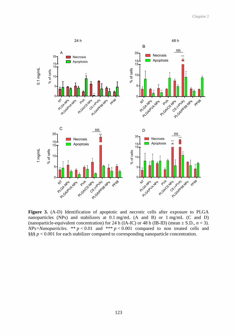
Chapitre 2
123
Figure 3. (A-D) Identification of apoptotic and necrotic cells after exposure to PLGA nanoparticles (NPs) and stabilizers at 0.1 mg/mL (A and B) or 1 mg/mL (C and D) (nanoparticle-equivalent concentration) for 24 h (IA-IC) or 48 h (IB-ID) (mean ± S.D., n = 3). NPs=Nanoparticles. ** p < 0.01 and *** p < 0.001 compared to non treated cells and §§§ p < 0.001 for each stabilizer compared to corresponding nanoparticle concentration.
NT
PLGA N
Ps
PLGA/P
VA NPs
PVA
PLGA/C
S NPs
CS (+
PVA)
PLGA/P
F68 N
Ps
PF68
0
5
10
1515
35 Necrosis
Apoptosis
A
**
**
% o
f ce
lls
NT
PLGA N
Ps
PLGA/P
VA NPs
PVA
PLGA/C
S NPs
CS (+
PVA)
PLGA/P
F68 N
Ps
PF68
0
5
10
1515
35Necrosis
Apoptosis
B
***
**
****
§§§
% o
f cells
NT
PLGA N
Ps
PLGA/P
VA NPs
PVA
PLGA/C
S NPs
CS (+
PVA)
PLGA/P
F68 N
Ps
PF68
0
5
10
1515
35
Necrosis
Apoptosis
C
***
§§§
% o
f cells
NT
PLGA N
Ps
PLGA/P
VA NPs
PVA
PLGA/C
S NPs
CS (+
PVA)
PLGA/P
F68
NPs
PF68
0
5
10
1515
35Necrosis
Apoptosis
D
***
***
§§§%
of
ce
lls
24 h 48 h0.1
mg/m
L1 m
g/m
L
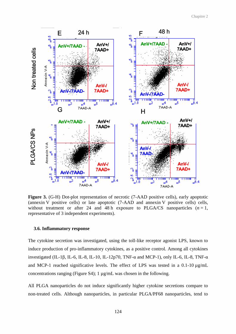
Chapitre 2
124
Figure 3. (G-H) Dot-plot representation of necrotic (7-AAD positive cells), early apoptotic (annexin V positive cells) or late apoptotic (7-AAD and annexin V positive cells) cells, without treatment or after 24 and 48 h exposure to PLGA/CS nanoparticles (n = 1, representative of 3 independent experiments).
3.6. Inflammatory response
The cytokine secretion was investigated, using the toll-like receptor agonist LPS, known to
induce production of pro-inflammatory cytokines, as a positive control. Among all cytokines
investigated (IL-1, IL-6, IL-8, IL-10, IL-12p70, TNF-α and MCP-1), only IL-6, IL-8, TNF-α
and MCP-1 reached significative levels. The effect of LPS was tested in a 0.1-10 µg/mL
concentrations ranging (Figure S4); 1 µg/mL was chosen in the following.
All PLGA nanoparticles do not induce significantly higher cytokine secretions compare to
non-treated cells. Although nanoparticles, in particular PLGA/PF68 nanoparticles, tend to

Chapitre 2
125
induce slightly higher levels than the stabilizers themselves, no difference hit statistical
significance.
0 0.1 1 100
10000
2000020000
300000
150000
LPS (µg/mL)
MC
P-1
(p
g/1
06 c
ell
)
0 0.1 1 100
300
600600
80000
20000
50000
LPS (µg/mL)
IL-6
(p
g/1
06 c
ell
)
0 0.1 1 100
100100
150000
75000
LPS (µg/mL)
TN
F-
(pg
/10
6 c
ell)
0 0.1 1 100
400000
800000
1000000
10000000
5000000
LPS (µg/mL)
IL-8
(p
g/1
06 c
ell)
Figure S4. Cytokines secretions (IL-8, MCP-1, IL-6 and TNF-α) evaluated by CBA method after 24 h exposure to increasing LPS concentrations.

Chapitre 2
126
Figure 4. Secretions of IL-8 (A), MCP-1 (B), IL-6 (C) and TNF-α (D) cytokines after 24 h exposure to PLGA nanoparticles (NPs) or the corresponding stabilizers (0.1 mg/mL nanoparticle-equivalent concentration) and comparison to non treated (NT) cells or LPS-treated cells (positive control), quantified with CBA method (mean ± S.D., n ≥ 3).
3.7. ROS production
The intracellular production of ROS was investigated using the H2DCFDA probe, which
becomes fluorescent after cellular internalization and oxidation. H2O2 was chosen as positive
control in such conditions (750 µM, 30 min preincubation) that cells retain a healthy
phenotype using microscope observations. Preincubation with H2DCFDA allowed
satisfactory ROS detection until 4 h exposure to treatment (data not shown).
NT
LPS 1
PLGA N
Ps
PLGA/P
VA NPs
PVA
PLGA/C
S NPs
CS (+
PVA)
PLGA/P
F68 N
PsPF68
0
500000
1000000
15000001500000
4000000
A
*2500000
IL-8
(pg/1
06 c
ell)
NT
LPS 1
PLGA N
Ps
PLGA/P
VA NPs
PVA
PLGA/C
S NPs
CS (+
PVA)
PLGA/P
F68
NPs
PF68
0
200
400400
80000
C
***
50000
20000
IL-6
(p
g/1
06 c
ell)
NT
LPS 1
PLG
A N
Ps
PLG
A/P
VA N
Ps
PVA
PLG
A/C
S N
Ps
CS (+
PVA)
PLG
A/P
F68 NPs
PF6
8
0
25000
5000050000
400000
B
***
200000
MC
P-1
(pg/1
06 c
ell)
NT
LPS 1
PLGA N
Ps
PLGA/P
VA NPs
PVA
PLGA/C
S NPs
CS (+
PVA)
PLGA/P
F68
NPs
PF68
0
15001500
150000
D
1000
50000
TN
F- a
(pg/1
06 c
ell)

Chapitre 2
127
At 0.1 mg/mL none of the PLGA nanoparticles induce significant ROS production, neither
did their stabilizing polymers in equivalent concentrations. At 1 mg/mL, an increase signal
occurs for nanoparticles, but this effect is transient. After 15 min, ROS production levels turn
back to basal levels.
15' 30' 1h 4h0.0
0.5
1.0
1.5
2.0
PLGA NPs
PLGA/PVA NPs
PLGA/CS NPs
PLGA/PF68 NPs
PVA
CS (+PVA)
PF68
A
NT
Time
MF
I F
old
in
cre
ase
5' 15' 30' 1h 4h
0.0
0.5
1.0
1.5
2.0
2.5
B
***
*
***
Time
MF
I F
old
in
cre
ase
Figure 5. Intracellular ROS production after exposure to PLGA nanoparticles (NPs) or the corresponding stabilizers, at nanoparticle-equivalent concentrations of 0.1 mg/mL (A) or 1 mg/mL (B), quantified with the H2DCFDA probe (mean ± S.D., n ≥ 3). * p < 0.05, *** p < 0.001 compared to non treated cells.

Chapitre 2
128
4. Discussion
It was previously shown that the cytotoxicity of solid lipid nanoparticles of similar sizes,
towards mouse macrophages, can be mediated by surfactant – the shell – (Schöler et al.,
2001) or by the lipid matrix – the core – (Schöler et al., 2002), whereas no influence were
detected concerning the inflammatory response neither the oxidative stress. The objective of
the present study was to investigate the toxicological profile of a family of PLGA-based
nanoparticles towards human-like macrophages, using various cellular toxicity endpoints, in
order to understand the role played by the stabilizers most commonly used in their
preparation. To do so, PLGA nanoparticles were prepared using the hydrophilic polymers
PVA, CS or PF68 as stabilizers, with a homogeneous size (around 200 nm), as previsously
described by our group (Mura et al., 2011a,b, Grabowski et al., 2013). These nanoparticles
are monodisperse (polydispersity index below 0.15) and remain stable after 48 h in cell
culture medium. In addition, these PLGA nanoparticles can undergo covalent fluorescent
labeling without modifying their physicochemical properties (Reul et al., 2012). In the present
study, stabilizer-free (‘nude’) PLGA nanoparticles have been added to this panel. While such
nanoparticles are challenging to prepare (and thus seldom used in the literature), the method
previously described leads to a nanoparticle suspension in the same size range with the same
stability profile, and can be fluorescently labeled similarly.
Human-like macrophages were obtained by differentiation of a THP-1 monocytes cell line,
which has been described as a relevant in vitro macrophage model (Tsuchiya et al., 1980,
Tsuchiya et al., 1982). This model has been used successfully to investigate the toxicity of
nanoparticles, especially in terms of cytotoxicity and cytokine expression (Chellat et al.,
2005, Chen et al., 2006). In order to precisely investigate the toxicological profile of
nanoparticles, we selected key toxicity endpoints: mitochondrial activity, inflammatory
response, oxidative stress, and cell death mechanisms (necrosis, apoptosis).
When exposed to all nanoparticles, human-like macrophages show important differences
depending on the nanoparticle concentrations used. On the one hand, few signs of toxicity
appear for nanoparticle concentrations below 0.1 mg/mL, either in mitochondrial activity,
induction of apoptosis/necrosis, production of intracellular ROS and secretions of pro-
inflammatory cytokines. Such concentrations can be considered in the range (or at the higher
end) of therapeutically relevant doses – in the case of drug delivery, an in vitro concentration
0.1 mg/mL can be correlated to a dose of 370 mg of polymer reaching the total alveoli surface

Chapitre 2
129
(70 m2) of adult human lung. These results confirm the safe toxicological pattern shown with
the same nanoparticles on other cell models such as Calu-3 (Mura et al., 2011a,b) and A549
(Grabowski et al., 2013).
In contrast, the exposure of macrophages to high concentrations of nanoparticles (above
1 mg/mL) leads to different cellular responses among the nanoparticles. Surprisingly,
stabilizer-free PLGA nanoparticles do not decrease cell mitochondrial activity (nor affect
other toxicity endpoints), in sharp contrast to all stabilized PLGA nanoparticles. This
important result highlight the innocuity of PLGA alone in nanoparticle form towards cell
viability, even though high concentrations are used and significant cellular uptake occurs. In
the case of stabilized PLGA nanoparticles, while PLGA/CS are found to induce a similar
toxicity to the CS stabilizer only, the toxicity of PLGA/PVA and PLGA/PF68 nanoparticles
contrasts with the innocuity of the PVA and PF68 stabilizers only.
Taken together, these results suggest that the cytotoxic effects of commonly-used stabilized
PLGA nanoparticles towards macrophages can be attributed to the stabilizers (PVA, CS or
PF68) when they are covering nanoparticles. A slightly toxic polymer like CS would retain its
toxicity when carried with nanoparticles, this toxicity occurring through necrosis with limited
production of ROS and pro-inflammatory cytokines. Moreover, even if a stabilizer has no
effect alone (like PVA, PF68), it may confer toxicity to the nanoparticles. The toxicity
mechanism does not seem to involve apoptosis/necrosis nor important ROS and pro-
inflammatory cytokines. It could be due to the cellular internalization of the stabilizers when
carried by nanoparticles, while these hydrophilic polymers do not interact with cell
membranes when used in the free form.
Conclusion
The comparison of routinely-used stabilized PLGA nanoparticles to stabilizer-free
nanoparticles and to the corresponding stabilizers used separately shows the importance of
precise knowledge of nanoparticule composition, but also that the toxicological profile of
stabilized PLGA nanoparticles is very different from the sum of their components studied
separately. Although these effects are observed at high concentrations exposed to cells, they
can provide an additional understanding of cellular toxicity of such nanoparticles and also
have implications for local/cellular toxic effects of PLGA-based nanomedicine.

Chapitre 2
130
Acknowledgments
The authors thank Simona Mura (Institut Galien Paris–Sud) for the NP characterization
studies, Valérie Nicolas (IFR IPSIT, Université Paris-Sud) for the confocal laser microscopy
observations, Nadia Abed (Institut Galien Paris Sud) for microbiology experiments, Claire
Merlet (Institut Galien Paris Sud) for her help in nanoparticle preparation. Hélène Chacun and
Stéphanie Denis (Institut Galien Paris-Sud) for their guidance and assistance in cell culture
and cytotoxicity experiments. This study was supported by the ANSES “Emerging risks”
program and by the ANR (under reference 2009 CESA 011).
References
ARNING, M., KLICHE, K. O., HEER-SONDERHOFF, A. H. & WEHMEIER, A. 1995. Infusion-related toxicity of three different amphotericin B formulations and its relation to cytokine plasma levels. Mycoses, 38, 459-465.
AUWERX, J. 1991. The human leukemia cell line, THP-1: A multifacetted model for the study of monocyte-macrophage differentiation. Experientia, 47, 22-31.
CHELLAT, F., GRANDJEAN-LAQUERRIERE, A., NAOUR, R. L., FERNANDES, J., YAHIA, L. H., GUENOUNOU, M. & LAURENT-MAQUIN, D. 2005. Metalloproteinase and cytokine production by THP-1 macrophages following exposure to chitosan-DNA nanoparticles. Biomaterials, 26, 961-970.
CHEN, H.-W., SU, S.-F., CHIEN, C.-T., LIN, W.-H., YU, S.-L., CHOU, C.-C., CHEN, J. J. W. & YANG, P.-C. 2006. Titanium dioxide nanoparticles induce emphysema-like lung injury in mice. The FASEB Journal, 20, 2393-2395.
CRUZ, C. M., RINNA, A., FORMAN, H. J., VENTURA, A. L. M., PERSECHINI, P. M. & OJCIUS, D. M. 2007. ATP Activates a Reactive Oxygen Species-dependent Oxidative Stress Response and Secretion of Proinflammatory Cytokines in Macrophages. Journal of Biological Chemistry, 282, 2871-2879.
DAIGNEAULT, M., PRESTON, J. A., MARRIOTT, H. M., WHYTE, M. K. & DOCKRELL, D. H. 2010. The identification of markers of macrophage differentiation in PMA-stimulated THP-1 cells and monocyte-derived macrophages. PLoS One, 5, e8668.
FORMAN, H. J. & TORRES, M. 2002. Reactive Oxygen Species and Cell Signaling. American Journal of Respiratory and Critical Care Medicine, 166, S4-S8.
GOVENDER, T., STOLNIK, S., GARNETT, M. C., ILLUM, L. & DAVIS, S. S. 1999. PLGA nanoparticles prepared by nanoprecipitation: drug loading and release studies of a water soluble drug. Journal of Controlled Release, 57, 171-185.
GRABOWSKI, N., HILLAIREAU, H., VERGNAUD, J., SANTIAGO, L. A., KERDINE-ROMER, S., PALLARDY, M., TSAPIS, N. & FATTAL, E. 2013. Toxicity of surface-

Chapitre 2
131
modified PLGA nanoparticles toward lung alveolar epithelial cells. International Journal of Pharmaceutics.
JAIN, R. A. 2000. The manufacturing techniques of various drug loaded biodegradable poly(lactide-co-glycolide) (PLGA) devices. Biomaterials, 21, 2475-2490.
MORGAN, E., VARRO, R., SEPULVEDA, H., EMBER, J. A., APGAR, J., WILSON, J., LOWE, L., CHEN, R., SHIVRAJ, L., AGADIR, A., CAMPOS, R., ERNST, D. & GAUR, A. 2004. Cytometric bead array: a multiplexed assay platform with applications in various areas of biology. Clin Immunol, 110, 252-66.
MOSMANN, T. 1983. Rapid colorimetric assay for cellular growth and survival: application to proliferation and cytotoxicity assays. J Immunol Methods, 65, 55-63.
MURA, S., HILLAIREAU, H., NICOLAS, J., KERDINE-ROMER, S., LE DROUMAGUET, B., DELOMENIE, C., NICOLAS, V., PALLARDY, M., TSAPIS, N. & FATTAL, E. 2011a. Biodegradable nanoparticles meet the bronchial airway barrier: how surface properties affect their interaction with mucus and epithelial cells. Biomacromolecules, 12, 4136-43.
MURA, S., HILLAIREAU, H., NICOLAS, J., LE DROUMAGUET, B., GUEUTIN, C., ZANNA, S., TSAPIS, N. & FATTAL, E. 2011b. Influence of surface charge on the potential toxicity of PLGA nanoparticles towards Calu-3 cells. Int J Nanomedicine, 6, 2591-605.
OBERDORSTER, G., FERIN, J., GELEIN, R., SODERHOLM, S. C. & FINKELSTEIN, J. 1992. Role of the alveolar macrophage in lung injury: studies with ultrafine particles. Environ Health Perspect, 97, 193-9.
OTT, M., GOGVADZE, V., ORRENIUS, S. & ZHIVOTOVSKY, B. 2007. Mitochondria, oxidative stress and cell death. Apoptosis, 12, 913-22.
REUL, R., TSAPIS, N., HILLAIREAU, H., SANCEY, L., MURA, S., RECHER, M., NICOLAS, J., COLL, J.-L. & FATTAL, E. 2012. Near infrared labeling of PLGA for in vivo imaging of nanoparticles. Polymer Chemistry, 3, 694-702.
SCHÖLER, N., HAHN, H., MÜLLER, R. H. & LIESENFELD, O. 2002. Effect of lipid matrix and size of solid lipid nanoparticles (SLN) on the viability and cytokine production of macrophages. International Journal of Pharmaceutics, 231, 167-176.
SCHÖLER, N., OLBRICH, C., TABATT, K., MÜLLER, R. H., HAHN, H. & LIESENFELD, O. 2001. Surfactant, but not the size of solid lipid nanoparticles (SLN) influences viability and cytokine production of macrophages. International Journal of Pharmaceutics, 221, 57-67.
SCHWENDE, H., FITZKE, E., AMBS, P. & DIETER, P. 1996. Differences in the state of differentiation of THP-1 cells induced by phorbol ester and 1,25-dihydroxyvitamin D3. J Leukoc Biol, 59, 555-61.
STRIZ, I., WANG, Y. M., TESCHLER, H., SORG, C. & COSTABEL, U. 1993. Phenotypic markers of alveolar macrophage maturation in pulmonary sarcoidosis. Lung, 171, 293-303.
TARNOK, A., HAMBSCH, J., CHEN, R. & VARRO, R. 2003. Cytometric bead array to measure six cytokines in twenty-five microliters of serum. Clin Chem, 49, 1000-2.

Chapitre 2
132
TSUCHIYA, S., KOBAYASHI, Y., GOTO, Y., OKUMURA, H., NAKAE, S., KONNO, T. & TADA, K. 1982. Induction of maturation in cultured human monocytic leukemia cells by a phorbol diester. Cancer Res, 42, 1530-6.
TSUCHIYA, S., YAMABE, M., YAMAGUCHI, Y., KOBAYASHI, Y., KONNO, T. & TADA, K. 1980. Establishment and characterization of a human acute monocytic leukemia cell line (THP-1). Int J Cancer, 26, 171-6.
YAMAMOTO, H., KUNO, Y., SUGIMOTO, S., TAKEUCHI, H. & KAWASHIMA, Y. 2005. Surface-modified PLGA nanosphere with chitosan improved pulmonary delivery of calcitonin by mucoadhesion and opening of the intercellular tight junctions. J Control Release, 102, 373-81.


134
Chapitre 3
Co-culture de cellules épithéliales
alvéolaires pulmonaires humaines et de
macrophages : Un outil in vitro
pertinent et efficace en nanotoxicologie

Chapitre 3
135
La co-culture de cellules épithéliales pulmonaires (A549) en présence de macrophages
(différenciés de monocytes THP-1, et notés THP-1-D) a été établie avec un contact direct
entre les deux types cellulaires. Caractérisée par l'identification des récepteurs membranaires
CD11b, CD14 et CD54 ainsi qu'en microscopie confocale, la co-existence de 2 populations
cellulaires a été confirmée, dont 10 % de macrophages. De plus, le marquage avec CD14 a été
utilisé afin d’identifier chaque sous-population cellulaire de la co-culture. Enfin, des
interactions entre les deux populations cellulaires ont été mises en évidence, par l’évolution
d’une partie du phénotype des cellules A54ř. Enfin, il est à noter qu'en présence de
lipopolysaccharide, des effets synergiques ont été observés concernant la sécrétion des
cytokines dans les surnageants de culture, les niveaux de sécrétions des cytokines en co-
culture étant supérieurs aux niveaux attendus, c'est-à-dire ceux issus de l’addition des
sécrétions obtenues pour chaque type cellulaire en mono-culture.
La co-culture a par la suite été utilisée comme un outil in vitro afin d'étudier la toxicité des
nanoparticules présentées au cours des précédents chapitres. La concentration en
nanoparticules choisie pour cette étude a été de 0,1 mg/mL, concentration à laquelle
correspondent 60 à 80 % de viabilité mitochondriale (données MTT) pour chaque type
cellulaire en mono-culture (Chapitres 1 et 2). Les différents paramètres de toxicité étudiés ont
été l’internalisation des nanoparticules, leur cytotoxicité ainsi que la réponse inflammatoire
qu’elles peuvent causer ou augmenter.
L’internalisation des nanoparticules de PLGA a été suivie en cytométrie en flux,
spécifiquement par chaque sous-population cellulaire. Les résultats montrent que les
nanoparticules de PLGA de potentiel zêta négatifs (dépourvues de stabilisant ou stabilisées
par le PF68) sont internalisées en plus grande quantité que les nanoparticules neutres
(PLGA/PVA) et positives (PLGA/CS), et ce, quelque soit la population cellulaire. De plus, les
macrophages THP-1-D captent plus de nanoparticules que les cellules A549 ce qui démontre
leur rôle important dans l'élimination des nanoparticules.
La cytotoxicité des nanoparticules de PLGA a été évaluée par un tri-marquage
annexine V/7AAD/CD14, permettant de différencier les différents types de mort cellulaire
(apoptose précoce ou avancée et nécrose) et ce, au sein de chaque lignée cellulaire. La co-
culture étant un mélange de plusieurs types cellulaires, il ne nous a pas semblé judicieux de
réaliser un test MTT classique, qui n’aurait pas fait la distinction entre les deux types de
cellules. Les résultats montrent qu’à la concentration de 0.1 mg/mL, les nanoparticules de

Chapitre 3
136
PLGA induisent exclusivement la mort par nécrose, et ce, pour chacune des populations
cellulaires.
La réponse inflammatoire a été évaluée par les cytokines MCP-1, IL-6, IL-8 et TNF-α
(Figure 8). Dans une première étape, les nanoparticules, ainsi que les polymères de surface en
solution, ont été appliqués sur la co-culture pendant 24 h, afin de mesurer l'effet
inflammatoire des nanoparticules proprement dites. Les nanoparticules de PLGA, en
particulier celles préparées sans agent stabilisant, induisent de faibles secrétions de cytokines.
De la même façon, les stabilisants en solution aqueuse n’induisent pas de réponse
inflammatoire. Dans une seconde approche, les nanoparticules ont été incubées en présence
de LPS, pour déterminer si les nanoparticules peuvent augmenter la réponse inflammatoire,
ou encore concentrer les effets du LPS qui, en se fixant sur les nanoparticules, pénétrerait
facilement à l’intérieur de la cellule. La réponse inflammatoire après exposition aux
nanoparticules de PLGA est à nouveau faible.
L’évaluation du stress oxydant a été entreprise par le dosage des ROS à l’intérieur de chaque
population cellulaire, par l’ajout de la sonde H2DCFDA et un co-marquage avec le CD14, en
cytométrie en flux. Malheureusement, la sonde H2DCDFA émet trop dans le canal du CD14
(PE), et les compensations n’ont pas permis d’obtenir un résultat convenable. Des anticorps
CD14 couplés à d’autres sondes fluorescentes ont été testés, mais sans donner de résultats
probants.
En conclusion, lors de cette étude, nous avons montré que la co-culture de cellules épithéliales
alvéolaires pulmonaires et de macrophages THP-1-D constitue un modèle in vitro simple et
pertinent pour les études de nanotoxicologie. De plus, une nouvelle fois, nous avons pu
démontrer que les nanoparticules de PLGA représentent un intérêt particulier dans la
formulation de nanovecteurs pour une application pulmonaire, car elles induisent une faible
toxicité ainsi qu'une faible réponse inflammatoire.

Chapitre 3
137
Lung epithelial cells and human-like macrophages co-cultures for the
evaluation of nanoparticles toxicity
Nadège Grabowski a,b, Hervé Hillaireau a,b, Juliette Vergnaud a,b, Valérie Nicolas c,
Saadia Kerdine-Römer d, Elias Fattal a,b
a. Université Paris-Sud, Faculté de pharmacie, Institut Galien Paris-Sud, LabEx LERMIT, 5 rue JB Clément, 92296 Chatenay-Malabry Cedex, France b. CNRS, UMR 8612, 5 rue JB Clément, 92296 Chatenay-Malabry Cedex, France c. Institut d’Innovation thérapeutique (IFR 141), IPSIT, 5 rue JB Clément, 92296 Chatenay-Malabry Cedex, France d. INSERM UMR 996 Cytokines, Chimiokines et Immunopathologie, 5 rue JB Clément, 92296 Chatenay-Malabry Cedex, France
To be submitted
Abstract
A co-culture of lung alveolar epithelial cells (A459) and macrophages (differentiated THP-1
monocytes) was established by putting in direct contact both cell populations. Using
immunocytochemistry and confocal laser scanning microscopy, both cell subpopulations were
evidenced. The synergistic effects observed in the co-culture for cytokine secretions confirm
cell -cell interactions. The co-culture was thus used as a relevant in vitro tool to evaluate the
toxicity of polymeric as well as non-biodegradable nanoparticles. Poly(lactide-co-glycolide)
(PLGA)-based nanoparticles of 200 nm were prepared, with the addition, or not, of
hydrophilic polymers (as stabilizers). Selective uptake kinetics of PLGA nanoparticles by cell
subpopulations, as well as apoptosis/necrosis detection, was achieved by using a selective
label of each cell type, while cytokine secretions were quantified in culture supernatants. Both
cell subpopulations took up PLGA nanoparticles with similar profiles, and induced only
necrotic cells. Mild inflammatory response to stabilized nanoparticles was detected
(compared to well-known inflammatory compounds), slightly higher than the PLGA and
stabilizer components taken individually demonstrating that although biodegradable
nanoparticles are safe they can internalize compounds such as the stabilizing agents used here
and slightly enhance their toxicity.
Keywords Co-culture, A549, THP-1 macrophages, nanoparticles, toxicology

Chapitre 3
138
1. Introduction
In vitro nanotoxicology to the lung must take into account of a large variety of toxicity
endpoints as well as the nature of the nanoparticles tested. Indeed, on the one hand, several
reviews have detailed the toxicity of inhalable manufactured nanoparticles towards the
pulmonary tract, as they could be involved in the induction of inflammation and oxidative
stress (Oberdorster et al., 2005, Horie et al., 2012). On the other hand, the pulmonary route
offers interesting potentialities for drug delivery. Indeed, it is a non invasive route, that can be
used for local or systemic treatments thanks to the high absorption surface, weak enzymatic
activity and a high blood supply of the pulmonary region (Patton et al., 2004). A wide range
of biodegradable nanocarriers, from liposomes to polymeric nanoparticles, can be considered
for lung delivery (Sung et al., 2007, Mansour et al., 2009). Regarding the latter, poly(lactide-
co-glycolide) (PLGA)-based nanoparticles present a high interest for drug delivery since this
copolymer was approved in many countries for controlled drug delivery implants. In addition,
nanoparticles made of such co-polymer can easily be surface-modified for an efficient
targeting and several different drugs have been loaded into these particles from small
molecules to macromolecules (Danhier et al., 2012).
The majority of in vitro studies are driven on mono-culture. Yet, human organism is multi-
cellular, and cannot be reduced to a unique cell line. However, for a first approach, the use of
a single cell line provides several advantages in terms of cost, convenience, and stability upon
splitting. On account of these benefits, the mixture of several cell types (at least two) to create
a co-culture represents an important step in the cell culture area. Hence, co-cultures consider
cell-cell interactions and are thus an in vitro model closer to the physiology than single cell
culture. This is reason why co-cultures have been used to mimic different organs or body
parts, including the lungs.
From the trachea, to the bronchus and the alveoli, the pulmonary epithelium is composed of
multiple cell types (Patton and Byron, 2007, Crapo et al., 1982). The bronchial space was for
instance mimicked by a co-culture of bronchial cells (primary or cell lines) and fibroblasts
(Pohl et al., 2009). Several co-cultures were used as model of aveoli by co-cultivating
alveolar epithelial cells with macrophages (Wottrich et al., 2004, Jantzen et al., 2012, Striz et
al., 2011, Striz et al., 2001, Alfaro-Moreno et al., 2008, Hjort et al., 2003). More complex
models were reported as the co-culture of alveolar epithelial and dendritic cells as well as
macrophages (Rothen-Rutishauser et al., 2005), or the co-culture of macrophages, epithelial,
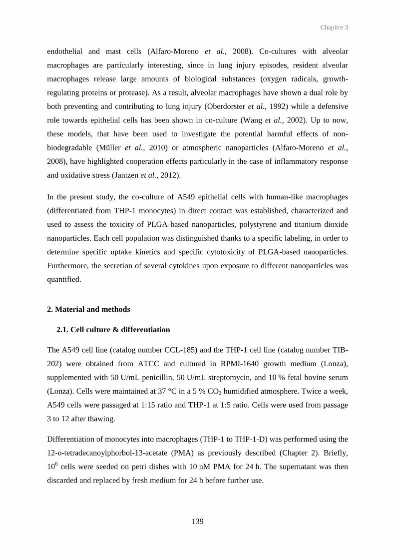
Chapitre 3
139
endothelial and mast cells (Alfaro-Moreno et al., 2008). Co-cultures with alveolar
macrophages are particularly interesting, since in lung injury episodes, resident alveolar
macrophages release large amounts of biological substances (oxygen radicals, growth-
regulating proteins or protease). As a result, alveolar macrophages have shown a dual role by
both preventing and contributing to lung injury (Oberdorster et al., 1992) while a defensive
role towards epithelial cells has been shown in co-culture (Wang et al., 2002). Up to now,
these models, that have been used to investigate the potential harmful effects of non-
biodegradable (Müller et al., 2010) or atmospheric nanoparticles (Alfaro-Moreno et al.,
2008), have highlighted cooperation effects particularly in the case of inflammatory response
and oxidative stress (Jantzen et al., 2012).
In the present study, the co-culture of A549 epithelial cells with human-like macrophages
(differentiated from THP-1 monocytes) in direct contact was established, characterized and
used to assess the toxicity of PLGA-based nanoparticles, polystyrene and titanium dioxide
nanoparticles. Each cell population was distinguished thanks to a specific labeling, in order to
determine specific uptake kinetics and specific cytotoxicity of PLGA-based nanoparticles.
Furthermore, the secretion of several cytokines upon exposure to different nanoparticles was
quantified.
2. Material and methods
2.1. Cell culture & differentiation
The A549 cell line (catalog number CCL-185) and the THP-1 cell line (catalog number TIB-
202) were obtained from ATCC and cultured in RPMI-1640 growth medium (Lonza),
supplemented with 50 U/mL penicillin, 50 U/mL streptomycin, and 10 % fetal bovine serum
(Lonza). Cells were maintained at 37 °C in a 5 % CO2 humidified atmosphere. Twice a week,
A549 cells were passaged at 1:15 ratio and THP-1 at 1:5 ratio. Cells were used from passage
3 to 12 after thawing.
Differentiation of monocytes into macrophages (THP-1 to THP-1-D) was performed using the
12-o-tetradecanoylphorbol-13-acetate (PMA) as previously described (Chapter 2). Briefly,
106 cells were seeded on petri dishes with 10 nM PMA for 24 h. The supernatant was then
discarded and replaced by fresh medium for 24 h before further use.

Chapitre 3
140
2.2. Co-culture
2.2.1. Co-culture establishment
The co-culture was established by direct contact between both cell populations (Figure 1). 106
THP-1 monocytes were firstly differentiated into THP-1 macrophages in petri dishes (t = 0)
as described in section 2.1. The medium was renewed after 24 h. Secondly, 42 000 A549 cells
were plated in 12-well plates (t = 24 h). Thirdly, THP-1-D macrophages were harvested and
centrifuged down (200 × g, 4 min, 4 °C). After pellet resuspension in growth medium, 42 000
THP-1-D macrophages were added in direct contact onto the A549 cells in 12-well plates,
resulting approximately in a 1:2 ratio (THP-1-D : A549) (t = 48 h). The resulting co-culture
was incubated for 24 h to allow cell recovery and potential contact establishment between cell
types. The co-culture was then characterized, or ready to use (12-well plates) for further
studies (t = 72 h).
Figure 1. Experimental setup of the establishment of the co-culture of lung epithelial cells (A549) and human-like macrophages (differentiated from THP-1 monocytes) in direct contact.

Chapitre 3
141
2.2.2. Immunocytochemistry characterization
After 24 h of direct contact between the two cell populations, the cell layer was rinsed with
PBS and harvested with trypsine. 250 000 cells were seeded in 96-well plates with conical
bottom. Supernatants were discarded after centrifugation (4 min, 200 × g, 4 °C), and cells
were resuspended in PBS containing 0.5 % Bovine Serum Albumin (BSA) (refered to as
‘PBS/BSA’ in the following). Supernatants were discarded after centrifugation (4 min,
200 × g, 4 °C) and cells were incubated in PBS/BSA for 20 min at 4 °C (protected from light),
with the following monoclonal antibodies: PE (PhycoErythrin)-conjugated anti-CD14, anti-
CD11b and anti-CD54 (BD Biosciences respectively 555398, 555388, 555511). After
incubation, cells were washed once with PBS/BSA, once with PBS and finally resuspended in
PBS, to be immediately analyzed by flow cytometry (FACScalibur, Beckton Dickinson). For
each sample 10 000 events were acquired from a gated homogeneous cell population from a
side- versus forward-scatter plot. Fluorescence intensities were then collected on the FL-2
channel and mean fluorescence intensities (MFI) determined. The MFI fold increase of each
sample was determined as the ratio of the sample MFI to the MFI of non-labeled cells. To
determine positive cells, histograms with FL-2 parameter against the number of events were
used. The marker was placed to the right of the peak to non-treated cells, to designate positive
cells. All experiments were performed in triplicate or more.
2.2.3. Co-culture characterization by confocal laser scanning microscopy
A549 cells were pre-labeled with 5-(and 6)-carboxyfluorescein diacetate succinimidyl ester
(CFSE) (Biolegend) according to the protocol provided. Briefly, 10 to 100 ×106 cells were
collected, centrifuged (4 min, 200 × g, 37 °C), and incubated with pre-warmed PBS (37 °C)
containing CFSE (5 µM) for 10 min at 37 °C in a 5 % CO2 humidified atmosphere. Ice-cold
culture medium was added, and cells were centrifuged (4 min, 200 × g, 37 °C). Supernatants
were then discarded and 100 000 of such pre-labeled A549 cells were plated in 6-well plates
containing glass coverslips and incubated for 24 h. The day after, pre-differentiated THP-1-D
macrophages were harvested, centrifuged (200 × g, 4 min, 37 °C) and resuspended in medium
containing 0.5 µM Orange Lysotracker (Invitrogen) (5 min, 37 °C). Cells were rinsed once
with culture medium and 100 000 THP-1-D were added in direct contact with pre-labeled
A549 cells. The resulting co-culture was incubated for 24 h. All the protocol was performed
in the absence of light. Observations were done with a LSM confocal microscope (Zeiss-
Meta) equipped with Argon (488 nm, 300 mV) and Helium-Neon (543 nm, 5 mV) lasers, and

Chapitre 3
142
a plan-apochromat 63X objective. During the observation, glass coverslips were kept at
37 °C, using an adapted warming plate, to keep cells alive. Data were analyzed with the LSM
510 software (version 3.2).
2.3. Nanoparticle preparation and characterization
Stabilized PLGA nanoparticles were prepared according to an emulsion/evaporation method
as previously described by our group (chapters 1). PLGA (75:25 Resomer® RG756,
Ingelheim) was dissolved in a dichloromethane/acetone (1/1 v/v) mixture and pre-emulsified
by vortexing for 1 min, with either an aqueous solution containing 2.5 mg/mL PVA (87-89 %
hydrolyzed, 30-70 kDa) to prepare PLGA/PVA nanoparticles, or an aqueous solution
containing 0.6 mg/mL CS (Protasan® UP CL113, 75-90% deacetylation, 50-150 kDa, FMC
Biopolymers) and 5 mg/mL PVA (Mowiol®4-88, Kuraray Specialities Europe GmbH) to
prepare PLGA/CS nanoparticles. The pre-emulsion was kept on ice and immediately
sonicated (Branson Digital Sonifier) at 40 % for, respectively, 1 or 2 min. The purification of
nanoparticles (removal of free PVA and CS) was achieved by ultracentrifugation (4 °C,
37 000 × g, 1 h) and pellet resuspension was performed by vortexing.
PLGA/PF68 nanoparticles were prepared by dissolving 50 mg PLGA in 5 mL ethyl acetate.
The organic phase was added dropwise to 10 mL of a 10 mg/mL PF68 (BASF) aqueous
solution under magnetic stirring. After 1 min vortexing, the emulsion was kept on ice and
homogenized using an Ultra-Turrax® (IKA works, Inc), equipped with a 18G-stainless steel
dispersing tool (10 000 rpm, 2 min). The organic solvent was then evaporated at room
temperature under magnetic stirring for 2 h. The purification of nanoparticles (removal of free
PF68) was performed by dialysis during 24 h against 1 L of MilliQ water, using a cellulose
ester membrane with a 50 kDa cutoff (SpectrumLabs).
Stabilizer-free PLGA nanoparticles were prepared by nanoprecipitation as previously
described (Chapter 2). 50 mg PLGA 75:25 were dissolved in 5 mL acetonitrile. The organic
solution was added dropwise under magnetic stirring to 15 mL water. After 5 min magnetic
stirring, acetonitrile was removed and the suspension concentrated by evaporation under
reduced pressure at 40 °C, until a nanoparticle concentration close to 25 mg/mL.
All PLGA nanoparticles were fluorescently labeled, by replacing the total quantity of PLGA
by a 99/1 (w/w) mixture of PLGA and a custom-made DY700-PLGA conjugate, obtained by
coupling 75:25 PLGA with a Near Infra Red (NIR)-emitting fluorescent dye (DY700,

Chapitre 3
143
Dyomics) as previously described (Reul et al., 2012). The further steps were performed
similarly to unlabeled nanoparticles, protected from light. Particle suspensions were not
contaminated by any bacteria.
As previously described (Chapter 1) TiO2 nanoparticle suspensions were obtained by
thorough dispersion of anatase (spheres < 25 nm) or rutile (rods 10 x 40 nm) TiO2
nanopowders (Sigma-Aldrich) in water. Briefly, powders were incorporated into water at
1 mg/mL and 1 min vortexing, after which pH was adjusted to 11 with 1 M sodium
hydroxide. Suspensions were then sonicated (40 %) on ice during 1.5 h, with 60 s pulsations
and 30 s intervals, and subsequently stirred at 24 000 rpm for 5 min on ice using an Ultra-
Turrax® equipped with a plastic (polycarbonate/PEEK) dispersing tool (S25D-14G-KS,
IKA). Nanoparticle suspensions were vortexed just before use. Polystyrene (PS) nanoparticle
aqueous suspensions (0.20 µm, Polysciences Europe GmbH) were briefly vortexed before
use.
All nanoparticles were characterized by their size and their zeta potential using a Nano ZS
(Malvern instruments, UK). Average hydrodynamic diameters were measured by dynamic
light scattering (25 °C, 173° scattering angle) after appropriate dilutions in water. Zeta
potential measurements were performed after appropriate dilutions in 1 mM sodium chloride,
at 25 °C. Nanoparticle stability was studied in cell culture medium at the final concentration
of 0.1 mg/mL, after 48 h incubation (37°C). The amount of stabilizers associated to
nanoparticles after their purification was assessed through the formation of colored complexes
with PVA and CS, or by 1H NMR in the case of PF68, as previously described (Chapter 1).
The fluorescence emission spectra of fluorescent nanoparticle suspensions were recorded
using an excitation wavelength of 700 nm. The fluorescence intensities of stabilizer free-
PLGA, PLGA/PVA and PLGA/PF68 nanoparticles were compared to that of PLGA/CS
nanoparticles and expressed as the ratio of the fluorescence intensities collected at the 722 nm
emission wavelength (the ratio was set at 1 for PLGA/CS nanoparticles which were the most
fluorescent). Physicochemical characteristics of PLGA-based nanoparticles and of non-
biodegradable nanoparticles are presented on Table 1 and Table 2.

Chapitre 3
144
Table 1. Physicochemical properties of PLGA-based nanoparticles.
Nanoparticles
Average diameter (nm) Polydipersity
index Zeta potential
(mV) Shell
(mg/100 mg PLGA) Fluorescence intensity ratio Water
48 h cell culture medium 37 °C
PLGA 171 ± 5 nd 0.044 ± 0.021 - 44.8 ± 5.1 None na DY700-PLGA 172 ± 15 nd 0.052 ± 0.016 - 40.0 ± 7.0 None 0.91 PLGA/PVA 233 ± 28 212 ± 6 0.099 ± 0.050 - 1.42 ± 2.2 PVA (11.5) na
DY700-PLGA/PVA 221 ± 9 nd 0.084 ± 0.018 - 4.10 ± 0.8 nd 0.78 PLGA/CS 233 ± 18 269 ± 12 0.210 ± 0.020 + 39.9 ± 7.2 CS (15.3)/PVA (30.4) na
DY700-PLGA/CS 234 ± 1 nd 0.182 ± 0.004 + 45.3 ± 0.6 nd 1 PLGA/PF68 228 ± 22 316 ± 16 0.133 ± 0.044 - 31.3 ± 4.9 PF68 (15.5) na
DY700-PLGA/PF68 205 ± 27 nd 0.152 ± 0.070 -27.4 ± 7.0 nd 0.68
na: non applicable, nd: not determined.
Table 2. Physicochemical properties of non-biodegradable nanoparticles.
Nanoparticles Core
Average diameter (nm) Polydipersity
index Zeta
potential (mV) Shell
Cristal form Water 48 h cell culture medium 37 °C
TiO2 Anatase 421 ± 49 625 ± 46 0.42 ± 0.07 - 35 ± 2 None TiO2 Rutile 479 ± 184 795 ± 80 0.77 ± 0.25 - 37.2 ± 6 None
Polystyrene na 250 ± 11 259 ± 21 0.02 ± 0.02 - 48.8 ± 1.5 Sulfate ester
na: non applicable
2.4. Selective uptake of nanoparticles
2.4.1. Flow cytometry
After establishment of the co-culture (Figure 1, t = 72 h), supernatants were discarded and
replaced by a fresh suspension of fluorescent DY700-PLGA nanoparticles diluted in the
medium at 0.1 mg/mL (final concentration). After 5 min to 48 h incubation at 37 °C,
supernatants were discarded, cells were rinsed twice with PBS and harvested by trypsine.
Trypsine was neutralized by PBS/BSA, and cells were labeled with anti-CD14-PE as
described above (section 2.2.2). Cell suspensions were immediately analyzed by flow
cytometry and MFI was collected on FL-2 (corresponding to PE probe) and FL-3 using a NIR
filter (corresponding to DY700 probe). For each sample 10 000 events were acquired from a
gated homogenous cell population selected from a side-versus forward-scatter plot. Cell
populations were distinguished by gating CD-14 positive cell population and CD-14 negative
cell population (Figure 2). For each sample, results were expressed as the ratio of the FL-3
MFI of each gated population to the MFI of corresponding non-treated cells. This value was
then corrected by the fluorescence factor of each nanoparticle suspension. All experiments
were performed in triplicate or more.

Chapitre 3
145
2.4.2. Confocal laser scanning microscopy
After establishment of the co-culture as described in the section 2.2.3 (and Figure 1, t = 72 h),
supernatants were discarded and replaced by 2 mL of DY700-PLGA nanoparticle suspensions
diluted in medium at 0.1 mg/mL (final concentration). After 1 h incubation at 37 °C,
coverslips were rinsed with PBS, and fresh medium was added to perform confocal
microscopy observations in real time on living cells, using similar setup as described in
section 2.2.3, with a supplementary Helium-Neon (633 nm, 5 mV) laser.
2.5. Apotosis/necrosis detection coupled to CD14 identification
After establishment of the co-culture (Figure 1, t = 72 h), supernatants were discarded and
replaced by 2 mL of fresh growth medium containing PLGA nanoparticles at 0.1 mg/mL, and
incubated for 24 to 48 h. The cell layer and the supernatants were analyzed separately. In a
first step, supernatants were collected in eppendorf, centrifugated (3 min, 1000 × g) and
resuspended into 20 µL binding buffer that containing 2 µL of annexin-AlexaFluor (AF) 488
(Life Technologies) and 2 µL of 7-Aminoactinomycin D (7-AAD) (Sigma-Aldrich) (10 min,
room temperature, darkness). Cells were resuspended in with PBS/BSA after centrifugation
(3 min, 1000 × g) and kept on ice. In a second step, the adherent cell layer was rinsed with
ice-cold PBS followed by the addition of 200 µL binding buffer containing 5 µL annexin-
AF488 and 5 µL of 7-AAD (10 min, room temperature, darkness). Cells were rinsed with
PBS and harvested by trypsin, and polled them with pellets obtained in the first step.
250 000 cells were then indentified with anti-CD14-PE as described in the section 2.2.2. Cells
were immediately analyzed by flow cytometry, and mean fluorescence intensities were
collected on FL-1 (AF488 probe), FL-2 (PE probe) and FL-3 (7-AAD) channels (Figure 2).
To perform fluorescence compensations each fluorescent probe was used separately on non
treated cells.
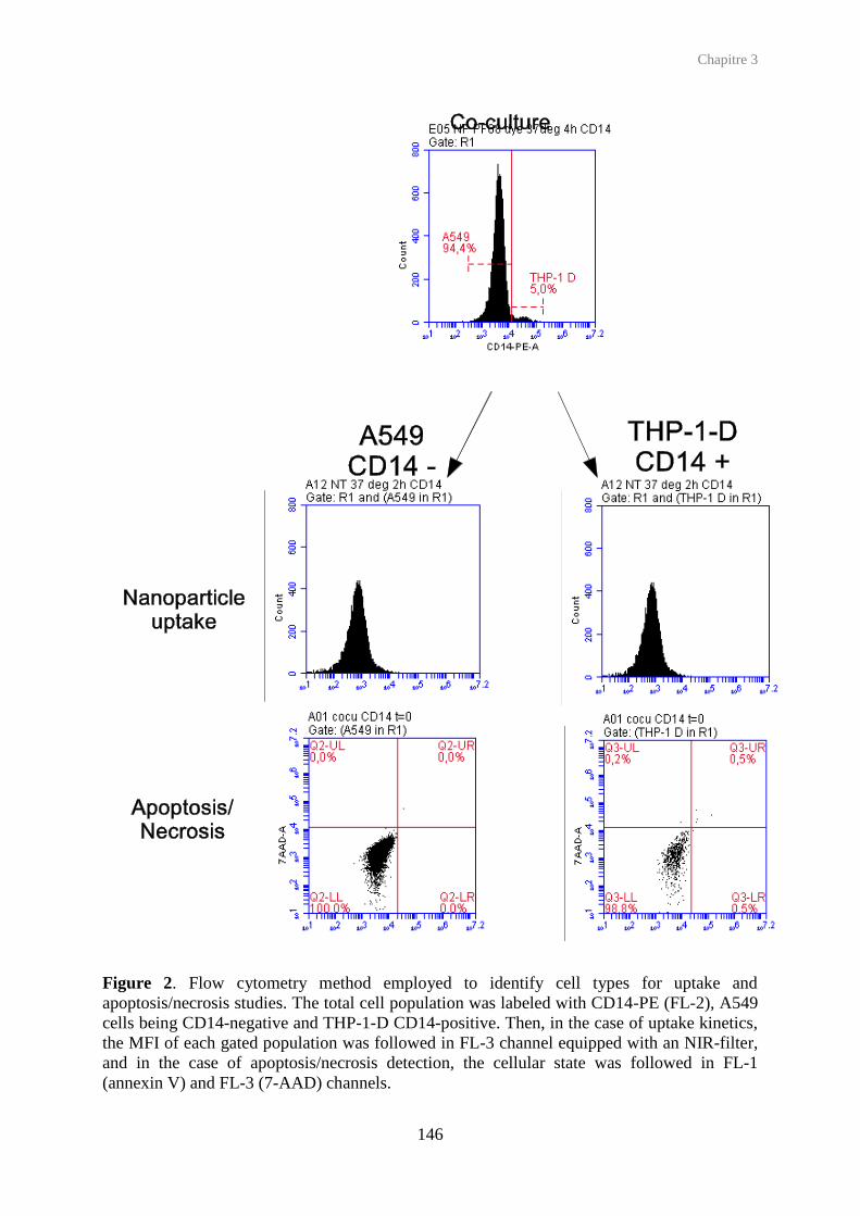
Chapitre 3
146
Figure 2. Flow cytometry method employed to identify cell types for uptake and apoptosis/necrosis studies. The total cell population was labeled with CD14-PE (FL-2), A549 cells being CD14-negative and THP-1-D CD14-positive. Then, in the case of uptake kinetics, the MFI of each gated population was followed in FL-3 channel equipped with an NIR-filter, and in the case of apoptosis/necrosis detection, the cellular state was followed in FL-1 (annexin V) and FL-3 (7-AAD) channels.

Chapitre 3
147
2.6. Quantification of secreted cytokines
Cytokine secretions following nanoparticle exposure were investigated in supernatants using
the multiplexed Cytometric Beads Array (CBA) method (BD Biosciences) (Morgan et al.,
2004, Tarnok et al., 2003), and performed with the human IL-1, IL-6, IL-8, IL-10, IL-12p70,
MCP-1 and TNF flex sets with the Human Soluble Protein Master Buffer Kit (BD
Biosciences). After establishment of the co-culture (Figure 1, t = 72 h), supernatant were
discarded and replaced by 2 mL of fresh growth medium (non-treated cells), with either
increasing lipopolysaccharide (LPS) concentrations (0.1, 1, 10 µg/mL) or nanoparticles at
0.1 mg/mL. After 24 h exposure, supernatants were sampled and cells were rinsed once with
PBS and harvested with trypsine, to be counted by flow cytometry. To asses a large range of
concentrations, each supernatant sample was analyzed without dilution and after a 1:50
dilution in medium. Cytokine results, analyzed with the FACP ArrayTM Software, were
obtained as pg/mL. All experiments were performed in triplicate, or more.
3. Results and discussion
3.1. Co-culture establishment and characterization
In order to investigate in vitro the pulmonary toxicity of a series of nanoparticles, co-culture
models are attractive tools to take into account the diversity of cell populations in the lung
tissue and their interactions. In the alveoli, a region expected to interact significantly with
deposited nanoparticles after inhalation, pneumocytes and macrophages play an important
role, respectively in the maintenance of the epithelial integrity and the inflammatory response.
However, the interaction between these cell types should also be taken into account to stay the
closest to the physiological response, such as in the case of inflammatory response. In this
study, we selected as a model of pneumocytes, A549 cells because they have already been
reported as an appropriate epithelial alveolar model (Rothen-Rutishauser et al., 2012).
Macrophages have been obtained by controlled differentiation of human THP-1 monocytes,
as described previously (chapter 2). They present a phenotype close to alveolar macrophages
(Striz et al., 1993). These two cell types were co-cultured in direct contact in order to mimic
physiological conditions. While such co-culture conditions have already been described
(Wottrich et al., 2004, Jantzen et al., 2012), our goal was (i) to characterize the cell
subpopulations and investigate their common interaction, and (ii) to explore the potential of
this co-culture for nanotoxicology studies, in particular for the investigation of selective

Chapitre 3
148
uptake of nanoparticles, induction of apoptosis and/or necrosis on each subpopulation, and
possible synergistic secretion of cytokines.
3.1.1. Phenotype characterization
The phenotype of cell subpopulations in co-culture was investigated and compared to the
corresponding cell monocultures, by CLSM and immunocytochemistry. CLSM images on
pre-labeled cells show that both cell types exhibit a similar morphology compared to the
corresponding mono-cultures (Figure 3). In particular, macrophages retain their surface
adhesion capacity despite the transfer step when put into contact with A549 cells.
Immunocytochemistry results show that in mono-culture, THP-1-D macrophages are CD11b
and CD14 positive, whereas A549 cells are CD11b and CD14 negative (Figure 4). The
analysis of the co-culture cell layer shows two populations for the CD11b and CD14 markers:
one being CD11b+ and CD14+ (accounting for approximately 90% of total cells), the other
one being CD11b– and CD14– (approximately 10%). This 90/10 ratio is different from the
expected 1:4 ratio after 24 h incubation. However, it is worth noting that this final cell ratio is
consistent with normal human physiological conditions (Crapo et al., 1982). These profiles
also show that anti-CD11b-PE and anti-CD14-PE are good candidates to discriminate both
cell subpopulations for further selective uptake studies and moreover apoptosis/necrosis
detection in subpopulations.
Some indications of A549/THP-1-D interactions are provided by CLSM images, displaying a
close proximity of membranes from the two cell types (Figure 3C). In addition,
immunocytochemistry results show that while A549 and THP-1-D cells are respectively
CD54– and CD54+ in mono-culture, almost all cells are found CD54+ in the co-culture
(Figure 4), suggesting that contact with THP-1-D cells has induced the expression of CD54
by A549 cells. This receptor, also called ICAM-1, is involved in cell adhesion process. This
result is consistent with previous studies showing that A549 cells incubated with conditioned
medium of primary monocytes, can express ICAM-1 (Krakauer, 2002).

Chapitre 3
149
Figure 3. (A) CFSE-labeled A459 cells (green), (B) Orange Lysotracker THP-1-D (red) cells in monoculture, and (C) in a co-culture in direct contact, as observed in confocal laser scanning microscopy (CLSM), in real time with living cells (incubation time shown: 24 h). Scale bar: 20 µm.

Chapitre 3
150
Figure 4. Immunocytochemical characterization of THP-1-D and A549 cells in mono and in co-culture. Cellular morphology and CD-11b, CD-14, CD-54 cell levels were analyzed by flow cytometry. To obtain CD-11b, CD-14, CD-54 histograms, gates were drawn on the major cell population, according to the corresponding experiment. Data shown are representative of 3 independent experiments.
3.1.2. Cytokine secretion
As it was previously shown, interactions between alveolar macrophages and epithelial cells
can promote inflammatory response (Tao and Kobzik, 2002). Synergistic effects in cytokine
production in the co-culture supernatants were thus investigated after exposure to LPS, and
compared to those observed for mono-cultures. Seven cytokines were quantified using the
CBA method: IL-1 , IL-10, IL-6, IL-8, TNF-α, MCP-1 (Figure 5) and IL-12 (data not
shown). Four relevant cytokines were found to be secreted in significant levels in mono and
co-culture: IL-6, IL-8, MCP-1 and TNF-α. Synergistic effects were observed for IL-6, MCP-1
and IL-8. For example, incubation with 0.1 µg/mL LPS led to an increase of IL-6 and IL-8
secretion up to 2.5-fold higher in co-culture than the sum of the secretions obtained in mono-
cultures (Figure 5 B,C). This increase was even 6-fold higher for MCP-1 (Figure 5 E). As a
general trend, cytokine levels peaked upon exposure to 1 µg/mL LPS and then plateaued or
decreased for higher LPS concentrations. Despite their low representation compared to A549
cells, THP-1-D macrophages seem to account for the larger part of IL-6, IL-8 and especially
TNF-α secretion, which is consistent with previous studies showing that in co-culture with

Chapitre 3
151
A549 cells, PBMC are the main source of –between others cytokines- TNF-α and IL-8
cytokines (Krakauer, 2002). These results show the potential of the A549/THP-1-D co-culture
to study the inflammatory response.
0 0.1 1 100
10
20
30
A549 THP-1D Co-culture
A
LPS (µg/mL)
IL-1
(p
g/m
L)
0 0.1 1 100
5
1010
500
A549 THP-1 D co-culture
B
*
250
LPS (µg/mL)
IL-6
(p
g/m
L)
0 0.1 1 100
7575
80000
A549 THP-1 D co-culture
C
**
40000
LPS (µg/mL)
IL-8
(p
g/m
L)
0 0.1 1 100
1
2
3
4
A549 THP-1D Co-culture
D
LPS (µg/mL)
IL-1
0 (
pg
/mL
)
0 0.1 1 100
100100
4000
7900
A549 THP-1 D co-culture
E
***
*
***
LPS (µg/mL)
MC
P-1
(p
g/m
L)
0 0.1 1 100
55
800
A549 THP-1 D co-culture
F
400
LPS (µg/mL)
TN
F-
(pg
/mL
)
Figure 5. Secretions of IL-1 (A), IL-6 (B), IL-8 (C), IL-10 (D), MCP-1 (E) and TNF-α (F) cytokines after 24 h exposure to increasing LPS concentrations ( 0.1, 1, 10 µg/mL) in A549 cells (white bars) and THP-1-D cells (grey bars) mono-cultures, or in co-culture (black bars). n ≥ γ, * p < 0.05, ** p < 0.01, *** p < 0.001 compared to corresponding non treated conditions.

Chapitre 3
152
3.2. Comparative toxicological evaluation of nanoparticles
3.2.1. Nanoparticles
The effect of different nanoparticle types has been investigated on the co-culture. In
particular, PLGA-based nanoparticles have been selected as it is one the major type of
biodegradable polymeric nanoparticles, relevant for the formulation of nanomedicine. A
series of custom made PLGA nanoparticles were prepared as previously described by our
group (Chapters 1 and 2). Nanoparticles in the range size of 170-230 nm were obtained in the
presence of hydrophilic polymers as stabilizers, but also in the absence of any stabilizer.
Uncoated PLGA nanoparticles present a negative surface charge, whereas PVA, CS and PF68
form respectively neutral, positive and negative surface charged nanoparticles. The
replacement of a low percentage of PLGA by a fluorescent PLGA does not modify physico-
chemical properties (Table 1). Moreover, nanoparticle sizes are stable after 48 h in complete
culture medium at 37 °C.
PLGA nanoparticles were compared to non-biodegradable nanoparticles, either polymeric
(polystyrene) or inorganic (TiO2). Sulfate ester-stabilized polystyrene nanoparticles were used
without modifications and were shown to be stable in culture medium. The diameter of
anatase and rutile TiO2 nanoparticles in suspension was reduced using dispersing tools,
without adding any supplementary stabilizer.
3.2.2 Selective uptake of PLGA nanoparticles by cell subpopulations
Uptake kinetic studies (Figure 6) were performed at 37 °C after exposure to fluorescent
DY700-PLGA nanoparticles at 0.1 mg/mL. A549 and THP-1-D subpopulations were
identified using anti-CD14-PE (Figure 2), which did not alter the detection of nanoparticle
fluorescence. All PLGA nanoparticles are taken up with a similar profile by both sub cell
populations. Stabilizer-free PLGA and PLGA/PF68 nanoparticles (displaying both a negative
zeta potential) are taken up earlier and in higher quantity than PLGA/CS (positive zeta
potential) and PLGA/PVA (neutral) nanoparticles, especially by the A549 cells. The same
behavior was observed on the same cells in mono-culture (chapters 1 and 2). Another
observation is the overall higher uptake of nanoparticles by THP-1-D cells compared to A549
cells. After 24 and 48 h, THP-1-D cells take up twice as much PLGA/PVA nanoparticles than
the A549 cells. PLGA nanoparticle uptake by co-culture was also visualized by CLSM in real
time on living cells. Both cell populations were fluorescently labeled as described in section

Chapitre 3
153
2.2.3 (A549 with CFSE, THP-1-D with Orange Lysotracker), and cultivated in co-culture.
The co-culture was finally exposed for 1 h to DY700-PLGA/PF68 nanoparticles and observed
by CLSM (Figure 7). PLGA/PF68 nanoparticles were observed within both sub cell
populations, but mostly in macrophages, confirming the flow cytometry results. These results
come in line with the prominent role of alveolar macrophage in particle clearance from the
alveoli region (Wang et al., 2002), although their represent less than 10 % of the cell
population, similarly to what is observed here.

Chapitre 3
154
15' 30' 1h 2h 4h 24h 48h0
5
10
15
PLGA NPs PLGA/PVA NPs
PLGA/CS NPsPLGA/PF68 NPs
A549 subpopulationA
Time
MF
I fo
ld i
ncre
ase
15' 30' 1h 2h 4h 24h 48h0
5
10
15
PLGA NPs PLGA/PVA NPs
PLGA/CS NPsPLGA/PF68 NPs
THP-1-D subpopulationB
Time
MF
I fo
ld i
ncre
ase
Figure 6. (A) Uptake of PLGA-based nanoparticles by A549 and THP-1-D cells in co-culture at 37 °C, as evaluated by the mean fluorescence intensity (MFI) in flow cytometry after identification of cell subpopulation using anti-CCD14-PE. (B) Co-culture of CFSE-labeled A549 cells (green) and Orange Lysotracker-labeled THP-1-D cells (red) exposed to DY700-PLGA/PF68 nanoparticles (white) for 2 h at 37 °C. Scale bar: 20 µm.

Chapitre 3
155
Figure 7. (A) Uptake kinetics of PLGA-based nanoparticles by A549 and THP-1-D cells in co-culture at 37 °C, as evaluated by the mean fluorescence intensity (MFI) in flow cytometry after correction by relative fluorescence factor. (B) Co-culture of A549 cells (green – CFSE) and THP-1-D cells (red – Orange Lysotracker) exposed to DY700-PLGA/PF68 nanoparticles (white) for 2 h at 37 °C. Scale bar: 20 µm.
3.2.3 Effect on apoptosis/necrosis cell subpopulations
For the detection of apoptosis/necrosis, both cell populations were identified using anti-
CD14-PE labeling (Figure 2). Based on our previous studies (Chapters 1 and 2) the
nanoparticle concentration was set at 0.1 mg/mL corresponding to 60-80 % of viability of
single cell lines as evaluated by mitochondrial activity (MTT tests). After exposure to PLGA
nanoparticles for 24 h (A, C) and 48 h (B, D), the main cellular death mechanism was shown
to be necrosis, and the amount of early/late apoptotic cells was similar in treated and non-
treated cells, whatever the formulations tested (Figure 8). Only slight differences among
PLGA nanoparticles appear in the co-culture. After 24 h, PLGA/PF68 and PLGA/CS
nanoparticles are slightly more toxic on THP-1-D cells than on A549 cells, whereas stabilizer
free-PLGA nanoparticles are more toxic on A549 cells than on THP-1-D cells. After 48 h,
PLGA/PF68 nanoparticles remain more toxic on THP-1-D cells. Interesting differences
appear, though, when comparing the effect of nanoparticles towards the co-culture compared
to the mono-cultures (Table 4). In co-culture conditions, PLGA/PF68 nanoparticles show a
noticeable toxicity compared to other PLGA nanoparticles (10% necrotic cells among the
A549 subpopulation, 21% among THP-1-D), which was not observed in mono-culture
(Chapters 1 and 2).
20 µm

Chapitre 3
156
NT
PLG
A N
Ps
PLG
A/P
VA N
Ps
PLG
A/C
S N
Ps
PLG
A/P
F68 N
Ps
0
5
10
100
Living cells
Necrosis
Early Apoptosis
Late apoptotic
A%
of
cells
NT
PLG
A N
Ps
PLG
A/P
VA N
Ps
PLG
A/C
S N
Ps
PLG
A/P
F68 N
Ps
0
5
10
100
Living cells
Necrosis
Early Apoptosis
Late apoptotic
B
% o
f cells
NT
PLG
A N
Ps
PLG
A/P
VA N
Ps
PLG
A/C
S N
Ps
PLG
A/P
F68 N
Ps
0
5
10
100
Living cells
Necrosis
Early Apoptosis
Late apoptotic
C
% o
f cells
NT
PLG
A N
Ps
PLG
A/P
VA N
Ps
PLG
A/C
S N
Ps
PLG
A/P
F68 N
Ps
0
5
10
100
Living cells
Necrosis
Early Apoptosis
Late apoptotic
D
*
% o
f cells
24 h 48 hA
54
9 s
ub
po
pu
lati
on
TH
P-1
-D s
ub
po
pu
lati
on
Figure 8. Percentage of living cells, early/late apoptotic cells, and necrotic cells, in the A549 subpopulation (A, B) and THP-1-D subpopulation (C, D) of a co-culture after 24 h (A, C) or 48 h (B, D) exposure to PLGA nanoparticles (NPs). Cells are simultaneously labeled with CD14-PE, 7-AAD and annexin V-AlexaFluor 488. THP-1-D (CD14 positive) and A549 (C14 negative) cells are distinguished as well as necrotic cells (7-AAD positive), early apoptotic cells (Annexin V positive) and late apoptotic cells (both 7-AAD and Annexin V positive). * p < 0.05, n = 3.

Chapitre 3
157
Table 3. Comparison of the percentage of necrotic cells after 48 h exposure to PLGA-based nanoparticle among A549 and THP-1-D cells in mono-culture (Chapters 1 and 2) or as subpopulations in co-culture.
NPs, nanoparticles ; NT, Non treated
3.2.4 Effect on global cytokine secretions
One of the major interests of a co-culture using macrophages relates to the inflammatory
response, as macrophages are able to secrete a large variety of multifunctional biological
substances such as cytokines (Nathan, 1987). However, in normal or injured human lungs, all
structural cells can secrete cytokines to ensure communication (Kelley, 1990). Quantification
of cytokines in co-culture supernatants characterizes not only an inflammatory state, but also
cell-cell communications. For example, the quantification of the cytokines of A549 in co-
culture with THP-1 monocytes or macrophages has shown that IL-6 and IL-8 significantly
increase in co-culture conditions compare to mono-cultures in case of hypoxia (Hjort et al.,
2003) or after exposure to ultrafine particles (Wottrich et al., 2004).
The co-culture of A549 and THP-1-D described herein was first used to investigate the own
inflammatory potential of nanoparticles, by exposing the co-culture to nanoparticles at
0.1 mg/mL for 24 h. After exposure to stabilizer-free PLGA nanoparticles no increase in
cytokine secretion was detected compared to non-treated conditions. Similarly, the aqueous
solution of stabilizers did not induce any augmentation in cytokine secretions. On the
contrary, stabilized PLGA nanoparticles induced a significant raise in the secretion of several
cytokines (IL-8 with PLGA/PVA and PLGA/CS nanoparticles, MCP-1 with PLGA/CS and
PLGA/PF68 nanoparticles and IL-6 with PLGA/PF68 nanoparticles) (Figure 9, Table 4).
These results highlight the importance of the PLGA/stabilizer combination in the cellular
response, as the combination entails significant effect whereas each component is innocuous.
Most of the stabilizing agents, being macromolecules, amphiphilic or hydrophilic, does not
diffuse by themselves across cell membranes. However, being adsorbed onto nanoparticle
NPs 0.1 mg/mL Mono-cultures
(Chapters 1 and 2) Co-culture
48 h Necrotic cells (%) Necrotic cells (%) A549 THP-1-D A549 THP-1-D
NT < 1 4 7 3 PLGA NPs 1 3 2 6
PLGA/PVA NPs 1 3 2 8 PLGA/CS NPs < 1 5 7 8
PLGA/PF68 NPs 3 4 10 21

Chapitre 3
158
surface, their internalization is enhanced leading to more toxicity. This inflammatory effect
remains however limited, much below the cytokine secretions caused by an exposure to 0.1
µg/mL LPS. This co-culture model has thus allowed the detection of mild inflammatory
response to stabilized PLGA nanoparticles, slightly higher than the PLGA and stabilizer
components individually, but very limited compared to well-known inflammatory
compounds.
Table 4. Summary of the in vitro inflammatory response after 24 h exposure to polymeric and inorganic nanoparticles on A549 cells and THP-1-D in mono and in co-culture, in presence (co-stimulation) or not of 0.1 µg/mL LPS.
NPs 0.1 mg/mL
Mono-cultures (chapters 1 and 2) Co-culture
24h A549 THP-1-D
without
LPS with LPS
without LPS
with LPS without
LPS with LPS
PLGA - nd - nd - nd
PLGA/PVA + (IL-8) - - - +
(IL-8) +
(IL-6)
PLGA/CS + (IL-8) - - -
+ (MCP-1
IL-8) -
PLGA/PF68 +
(MCP-1, IL-6, IL-8)
- - - +
(MCP-1, IL-6, TNF-α)
+ (IL-6, TNF-α)
PS - - - - - - Anatase TiO2 - - - - - -
Rutile TiO2 - - - - + (IL-8) -
(+) is defined according to observed significant effects (at least p < 0.05) compared to control conditions. nd: not determined.
In addition, co-cultures were exposed to nanoparticles in co-stimulation with 0.1 µg/mL LPS,
in order to check if nanoparticles can increase the inflammatory response when cells are pre-
activated (staggered incubation), or can concentrate LPS that will penetrate the cell easily
(simultaneous incubation). Although few statistically significant differences were observed,
the relative increase is moderate, and no clear tendency can be drawn regarding a potential
effect of nanoparticles on the pro-inflammatory activity of LPS (Figure 9).
These effects were compared to those induced by non-biodegradable nanoparticles, polymeric
or inorganic. Unexpectedly, none of the tested nanoparticles induced higher cytokine
secretions than PLGA nanoparticles. Such a result is not corroborated by several studies

Chapitre 3
159
showing that polystyrene (Dailey et al., 2006) and TiO2 (Inoue et al., 2008) nanoparticles
induce a significant inflammatory response in vivo. However, it was previously shown that
some particles like carbon black, diesel and TiO2 nanoparticles tested in vitro can adsorb
various cytokines, such as IL-6, TNF-α and IL-8, thereby limiting their detection (Val et al.,
2009, Kocbach et al., 2008). We observed that the IL-8 secretion following LPS exposure was
surprisingly reduced in presence of anatase TiO2 nanoparticles (Figures 9, S1, S2), which may
support this hypothesis.
0
2000
40004000
40000
- + - + - + - + - + - + - + - + - + - + - +
CTRL Rutile
TiO2
NPs
Anatase
TIO2
NPs
PS
NPs
PF68PLGA/PF68
NPs
CS
(+PVA)
PLGA/CS
NPs
PVAPLGA/PVA
NPs
PLGA
NPs
LPS
§§§
§
nd nd nd nd
***
*
***
B
IL-8
(p
g/m
L)
0
3500
7000
- + - + - + - + - + - + - + - + - + - + - +
CTRL Rutile
TiO2
NPs
Anatase
TIO2
NPs
PS
NPs
PF68PLGA/PF68
NPs
CS
(+PVA)
PLGA/CS
NPs
PVAPLGA/PVA
NPs
PLGA
NPs
LPS
*** **
§§§1500
5500
nd nd nd nd
A
MC
P-1
(p
g/m
L)

Chapitre 3
160
0
50
100
150150
1000
- + - + - + - + - + - + - + - + - + - + - +
CTRL Rutile
TiO2
NPs
Anatase
TIO2
NPs
PS
NPs
PF68PLGA/PF68
NPs
CS
(+PVA)
PLGA/CS
NPs
PVAPLGA/PVA
NPs
PLGA
NPs
LPS
500
nd nd nd nd
*
*
C
IL-6
(p
g/m
L)
0
5
1010
200
- + - + - + - + - + - + - + - + - + - + - +
CTRL Rutile
TiO2
NPs
Anatase
TIO2
NPs
PS
NPs
PF68PLGA/PF68
NPs
CS
(+PVA)
PLGA/CS
NPs
PVAPLGA/PVA
NPs
PLGA
NPs
LPS
100
nd nd nd nd
*
D
TN
F-
(pg
/mL
)
Figure 9. Secretions in co-culture (A549 cells and THP-1-D) of MCP-1 (A), IL-8 (B), IL-6 (C) and TNF-α (D) after 24 h exposure to nanoparticles (NPs) 0.1 mg/mL, or to the corresponding stabilizers used separately in nanoparticle-equivalent concentration, in absence (-) or in presence (+) of 0.1 µg/mL LPS. CTRL = control, NPs = nanoparticles, n ≥ γ. * p < 0.05, ** p < 0.01, *** p < 0.001, compared to corresponding untreated conditions. § p < 0.005, §§ p < 0.01, §§§ p < 0.001 for each stabilizer compared to corresponding stabilized nanoparticles.

Chapitre 3
161
0
500
1000
1500
LPS - +
CTRL PLGA/PVA
NPs
PLGA
NPs
TiO2
Anatase
NPs
TIO2
Rutile
NPs
PS
NPs
PLGA/PF68
NPs
PLGA/CS
NPs
- + - + - + - + - + - + - +
nd
*
AM
CP
-1 (p
g/m
L)
0
200
400
600
800
LPS - +
CTRL PLGA/PVA
NPs
PLGA
NPs
TiO2
Anatase
NPs
TIO2
Rutile
NPs
PS
NPs
PLGA/PF68
NPs
PLGA/CS
NPs
- + - + - + - + - + - + - +
*
nd
**
***
B
IL-8
(p
g/m
L)
0
20
40
60
80
LPS - +
CTRL PLGA/PVA
NPs
PLGA
NPs
TiO2
Anatase
NPs
TIO2
Rutile
NPs
PS
NPs
PLGA/PF68
NPs
PLGA/CS
NPs
- + - + - + - + - + - + - +
nd
C
IL-6
(p
g/m
L)
Figure S1. Secretions in mono-culture of A549 cells, of MCP-1 (A), IL-8 (B) and IL-6 (C) after 24 h exposure to nanoparticles (NPs) 0.1 mg/mL or to stabilizers in nanoparticle-equivalent concentration, in absence (-) or in presence (+) of LPS 0.1 µg/mL, quantified by CBA methods. CTRL = control, NPs = nanoparticles, n ≥ 3. * p < 0.05, ** p < 0.01, *** p < 0.001, compared to corresponding untreated conditions.

Chapitre 3
162
0
100
200
300
400
LPS - +
CTRL PLGA/PVA
NPs
PLGA
NPs
Anatase
TiO2
NPs
Rutile
TiO2
NPs
PS
NPs
PLGA/PF68
NPs
PLGA/CS
NPs
- + - + - + - + - + - + - +
nd
AM
CP
-1 (
pg
/mL
)
0
6000
12000
LPS - +
CTRL PLGA/PVA
NPs
PLGA
NPs
Anatase
TiO2
NPs
Rutile
TiO2
NPs
PS
NPs
PLGA/PF68
NPs
PLGA/CS
NPs
- + - + - + - + - + - + - +
nd
B
IL-8
(p
g/m
L)
0
55
80
LPS - +
CTRL PLGA/PVA
NPs
PLGA
NPs
PS
NPs
PLGA/PF68
NPs
PLGA/CS
NPs
- + - + - + - + - + - + - +
nd
40
Anatase
TiO2
NPs
Rutile
TiO2
NPs
C
IL-6
(p
g/m
L)
0
5
1010
80
LPS - +
CTRL PLGA/PVA
NPs
PLGA
NPs
PS
NPs
PLGA/PF68
NPs
PLGA/CS
NPs
- + - + - + - + - + - + - +
nd
Anatase
TiO2
NPs
Rutile
TiO2
NPs
40
D
TN
F-
(pg
/mL
)
Figure S2. Secretions in mono-culture of THP-1-D cells, of MCP-1 (A), IL-8 (B) and IL-6 (C) after 24 h exposure to nanoparticles 0.1 mg/mL or to stabilizers in nanoparticle-equivalent concentration, in absence (-) or in presence (+) of LPS 0.1 µg/mL, quantified by CBA methods. CTRL = control, NPs = nanoparticles, n ≥ 3. * p < 0.05, ** p < 0.01, *** p < 0.001, compared to corresponding untreated conditions.
Conclusion
We have shown that a co-culture of A549 epithelial cells and THP-1 macrophages represent
an interesting model for in vitro pulmonary nanotoxicology studies, in particular for the
investigation of selective uptake of nanoparticles, the induction of apoptosis and/or necrosis
in each subpopulation, and possible synergistic effect for the secretion of cytokines, even at
low levels. This co-culture model has enable the detection of mild inflammatory response to
PLGA nanoparticles stabilized by the hydrophilic polymers PVA, CS and PF68, slightly
higher than the stabilizer-free PLGA nanoparticles and than stabilizer components
individually, but very limited compared to well-known inflammatory compounds.

Chapitre 3
163
Acknowledgments
The authors thank Simona Mura (Institut Galien Paris Sud) for her nanoparticle expertise,
Nadia Abed (Institut Galien Paris Sud) for microbiology experiments, and Claire Merlet
(Institut Galien Paris Sud) for her help. Hélène Chacun and Stéphanie Denis (Institut Galien
Paris-Sud) for their guidance and assistance in cell culture and cytotoxicity experiments. This
study was supported by the ANSES “Emerging risks” program and by the ANR (under
reference 2009 CESA 011).
Bibliography
ALFARO-MORENO, E., NAWROT, T. S., VANAUDENAERDE, B. M., HOYLAERTS, M. F., VANOIRBEEK, J. A., NEMERY, B. & HOET, P. H. M. 2008. Co-cultures of multiple cell types mimic pulmonary cell communication in response to urban PM10. European Respiratory Journal, 32, 1184-1194.
CRAPO, J. D., BARRY, B. E., GEHR, P., BACHOFEN, M. & WEIBEL, E. R. 1982. Cell number and cell characteristics of the normal human lung. The American review of respiratory disease, 126, 332-337.
DAILEY, L. A., JEKEL, N., FINK, L., GESSLER, T., SCHMEHL, T., WITTMAR, M., KISSEL, T. & SEEGER, W. 2006. Investigation of the proinflammatory potential of biodegradable nanoparticle drug delivery systems in the lung. Toxicology and Applied Pharmacology, 215, 100-108.
DANHIER, F., ANSORENA, E., SILVA, J. M., COCO, R., LE BRETON, A. & PRÉAT, V. 2012. PLGA-based nanoparticles: An overview of biomedical applications. Journal of Controlled Release, 161, 505-522.
HJORT, M. R., BRENYO, A. J., FINKELSTEIN, J. N., FRAMPTON, M. W., LOMONACO, M. B., STEWART, J. C., JOHNSTON, C. J. & D'ANGIO, C. T. 2003. Alveolar epithelial cell-macrophage interactions affect oxygen-stimulated interleukin-8 release. Inflammation, 27, 137-45.
HORIE, M., KATO, H., FUJITA, K., ENDOH, S. & IWAHASHI, H. 2012. In Vitro Evaluation of Cellular Response Induced by Manufactured Nanoparticles. Chemical Research in Toxicology, 25, 605-619.
INOUE, K., TAKANO, H., OHNUKI, M., YANAGISAWA, R., SAKURAI, M., SHIMADA, A., MIZUSHIMA, K. & YOSHIKAWA, T. 2008. Size effects of nanomaterials on lung inflammation and coagulatory disturbance. International journal of immunopathology and pharmacology, 21, 197-206.

Chapitre 3
164
JANTZEN, K., ROURSGAARD, M., DESLER, C., LOFT, S., RASMUSSEN, L. J. & MØLLER, P. 2012. Oxidative damage to DNA by diesel exhaust particle exposure in co-cultures of human lung epithelial cells and macrophages. Mutagenesis, 27, 693-701.
KELLEY, J. 1990. Cytokines of the Lung. American Review of Respiratory Disease, 141, 765-788.
KOCBACH, A., TOTLANDSDAL, A. I., LÅG, M., REFSNES, M. & SCHWARZE, P. E. 2008. Differential binding of cytokines to environmentally relevant particles: A possible source for misinterpretation of in vitro results? Toxicology Letters, 176, 131-137.
KRAKAUER, T. 2002. Stimulant-dependent modulation of cytokines and chemokines by airway epithelial cells: cross talk between pulmonary epithelial and peripheral blood mononuclear cells. Clin Diagn Lab Immunol, 9, 126-31.
MANSOUR, H. M., RHEE, Y. S. & WU, X. 2009. Nanomedicine in pulmonary delivery. Int J Nanomedicine, 4, 299-319.
MORGAN, E., VARRO, R., SEPULVEDA, H., EMBER, J. A., APGAR, J., WILSON, J., LOWE, L., CHEN, R., SHIVRAJ, L., AGADIR, A., CAMPOS, R., ERNST, D. & GAUR, A. 2004. Cytometric bead array: a multiplexed assay platform with applications in various areas of biology. Clin Immunol, 110, 252-66.
MÜLLER, L., RIEDIKER, M., WICK, P., MOHR, M., GEHR, P. & ROTHEN-RUTISHAUSER, B. 2010. Oxidative stress and inflammation response after nanoparticle exposure: differences between human lung cell monocultures and an advanced three-dimensional model of the human epithelial airways. Journal of The Royal Society Interface, 7, S27-S40.
NATHAN, C. F. 1987. Secretory products of macrophages. J Clin Invest, 79, 319-26.
OBERDORSTER, G., FERIN, J., GELEIN, R., SODERHOLM, S. C. & FINKELSTEIN, J. 1992. Role of the alveolar macrophage in lung injury: studies with ultrafine particles. Environ Health Perspect, 97, 193-9.
OBERDORSTER, G., OBERDORSTER, E. & OBERDORSTER, J. 2005. Nanotoxicology: an emerging discipline evolving from studies of ultrafine particles. Environ Health Perspect, 113, 823-39.
PATTON, J. S. & BYRON, P. R. 2007. Inhaling medicines: delivering drugs to the body through the lungs. Nat Rev Drug Discov, 6, 67-74.
PATTON, J. S., FISHBURN, C. S. & WEERS, J. G. 2004. The lungs as a portal of entry for systemic drug delivery. Proc Am Thorac Soc, 1, 338-44.
POHL, C., HERMANNS, M. I., UBOLDI, C., BOCK, M., FUCHS, S., DEI-ANANG, J., MAYER, E., KEHE, K., KUMMER, W. & KIRKPATRICK, C. J. 2009. Barrier functions and paracellular integrity in human cell culture models of the proximal respiratory unit. European Journal of Pharmaceutics and Biopharmaceutics, 72, 339-349.

Chapitre 3
165
REUL, R., TSAPIS, N., HILLAIREAU, H., SANCEY, L., MURA, S., RECHER, M., NICOLAS, J., COLL, J.-L. & FATTAL, E. 2012. Near infrared labeling of PLGA for in vivo imaging of nanoparticles. Polymer Chemistry, 3, 694-702.
ROTHEN-RUTISHAUSER, B., CLIFT, M. J. D., JUD, C., FINK, A. & WICK, P. 2012. Human epithelial cells in vitro – Are they an advantageous tool to help understand the nanomaterial-biological barrier interaction? . Euro Nanotox Letters, 4, 1-20.
ROTHEN-RUTISHAUSER, B. M., KIAMA, S. G. & GEHR, P. 2005. A Three-Dimensional Cellular Model of the Human Respiratory Tract to Study the Interaction with Particles. American Journal of Respiratory Cell and Molecular Biology, 32, 281-289.
STRIZ, I., BRABCOVA, E., KOLESAR, L., LIU, X. D., BRABCOVA, I., SEKERKOVA, A., POOLE, J. A., JARESOVA, M., SLAVCEV, A. & RENNARD, S. I. 2011. Epithelial cells modulate genes associated with NF kappa B activation in co-cultured human macrophages. Immunobiology, 216, 1110-6.
STRIZ, I., SLAVCEV, A., KALANIN, J., JARESOVA, M. & RENNARD, S. I. 2001. Cell-cell contacts with epithelial cells modulate the phenotype of human macrophages. Inflammation, 25, 241-6.
STRIZ, I., WANG, Y. M., TESCHLER, H., SORG, C. & COSTABEL, U. 1993. Phenotypic Markers of Alveolar Macrophage Maturation in Pulmonary Sarcoidosis. Lung, 171, 293-303.
SUNG, J. C., PULLIAM, B. L. & EDWARDS, D. A. 2007. Nanoparticles for drug delivery to the lungs. Trends in Biotechnology, 25, 563-570.
TAO, F. & KOBZIK, L. 2002. Lung Macrophage–Epithelial Cell Interactions Amplify Particle-Mediated Cytokine Release. American Journal of Respiratory Cell and Molecular Biology, 26, 499-505.
TARNOK, A., HAMBSCH, J., CHEN, R. & VARRO, R. 2003. Cytometric bead array to measure six cytokines in twenty-five microliters of serum. Clin Chem, 49, 1000-2.
VAL, S., HUSSAIN, S., BOLAND, S., HAMEL, R., BAEZA-SQUIBAN, A. & MARANO, F. 2009. Carbon black and titanium dioxide nanoparticles induce pro-inflammatory responses in bronchial epithelial cells: Need for multiparametric evaluation due to adsorption artifacts. Inhalation Toxicology, 21, 115-122.
WANG, S., YOUNG, R. S., SUN, N. N. & WITTEN, M. L. 2002. In vitro cytokine release from rat type II pneumocytes and alveolar macrophages following exposure to JP-8 jet fuel in co-culture. Toxicology, 173, 211-219.
WOTTRICH, R., DIABATÉ, S. & KRUG, H. F. 2004. Biological effects of ultrafine model particles in human macrophages and epithelial cells in mono- and co-culture. International Journal of Hygiene and Environmental Health, 207, 353-361.


Discussion
générale

Discussion générale
168
Au cours de ces travaux de thèse, la toxicité pulmonaire de nanoparticules biodégradables et
non-biodégradables a été étudiée. Ce projet s’inscrit dans un projet global soutenu par
l’ANSES (« Emerging risks »), l’ANR (β00ř CESA) et le Fond pour la Recherche
Respiratoire regroupant des études in vitro et in vivo menées au sein du laboratoire. Deux
précédents articles ont fait état de la toxicité in vitro de nanoparticules biodégradables vis-à-
vis des cellules d’épithélium bronchiques (Calu-3) (Mura et al., 2011a,b), et un article relatant
les effets in vivo est en cours de rédaction (Santiago, submitted). Au cours de la présente
étude, la toxicité in vitro de ces mêmes nanoparticules a été étudiée sur des cellules
alvéolaires. Les objectifs de ce travail de thèse étaient (Figure 1) :
D’enrichir notre bibliothèque de nanoparticules afin d’aboutir à une collection de
particules aux caractéristiques physico-chimiques variées et représentatives des
nanoparticules les plus couramment rencontrées en nanomédecine et possédant une
taille de l’ordre de β00 nm (organiques / inorganiques, biodégradables / non-
biodégradables, charges de surface positives, négatives ou neutres),
D’établir et de caractériser une co-culture mimant les conditions alvéolaires,
D’étudier la toxicité de nanoparticules sur les cellules en mono et en co-culture, à
l'aide de différentes tests,
De tenter de corréler les profils de toxicité des nanoparticules à leurs caractéristiques
physico-chimiques afin de proposer un modèle pertinent de nanovecteurs pour
l’administration pulmonaire.
Les études portant sur la toxicité pulmonaire de nanoparticules non-biodégradables sont
nombreuses. Et pour cause, l’arbre bronchique est exposé à tout type de particules présentes
dans l’atmosphère, incluant les nanoparticules non-biodégradables dans l'environnement,
telles que les nanoparticules de diesel ou en milieu professionnel, les nanoparticules
manufacturées, telles que les nanotubes de carbone. En revanche, assez peu d’études sont
disponibles aujourd’hui sur la toxicité pulmonaire de nanoparticules biodégradables, qui
entrent dans la fabrication de nanomédicaments. Pourtant, la voie pulmonaire est prometteuse
pour l’administration de nanomédicaments, car elle permet d’envisager à la fois des
traitements locaux (asthme, broncho-pneumopathie chronique obstructive ou cancer) et des
traitements systémiques grâce aux échanges constants entre les alvéoles et la circulation
sanguine (diabète, administration de vaccins). Les nanovecteurs qui assurent le transport de
ces principes actifs, doivent évidemment faire preuve d’une totale innocuité vis-à-vis de
l’organisme.

Discussion générale
169
Figure 1. Schéma de l’étude de nanotoxicité menée au sein de l’équipe de recherche. Afin de connaitre les effets de nanoparticules vis-à-vis de la voie pulmonaire, la toxicologie in vitro de nanoparticules est étudiée sur des cellules épithéliales pulmonaires alvéolaires (A549) et des macrophages (différenciés de monocytes THP-1) en mono et en co-culture (projet de thèse), sur des cellules Calu-γ, d’épithélium bronchique, et chez un modèle de souris (Balb/c).
Au sein du projet mené dans l’équipe de recherche (Figure 1), nous étudions la toxicité et le
devenir de nanoparticules de poly(lactide(co-glycolide) (PLGA), afin des les proposer en tant
que candidats nanomédicaments. Dans le cadre de ce projet de thèse, nous nous sommes
particulièrement intéressés à la toxicité de ces nanoparticules vis-à-vis de la région alvéolaire.
1. La bibliothèque de nanoparticules
Des nanoparticules de composition chimique et de charge de surface variées ont été utilisées.
Néanmoins, nous avons œuvré dans la formulation pour que la taille des particules reste
constante, de l’ordre de β00 m. Les nanoparticules de PLGA ont été formulées au sein du
laboratoire et les nanoparticules de TiO2 et de polystyrène ont été obtenues commercialement
(Figure 2).

Discussion générale
170
Figure 2. Nanoparticles de PLGA stabilisées ou non stabilisées par l’alcool polyvinylique (PVA), le chitosane ou le pluronic F68 (PF68) et nanoparticules de dioxyde de titane (TiO2) et de polystyrène (PS) utilisées dans les études de toxicité vis-à-vis de la voie pulmonaire.
Les nanoparticules à base de PLGA ont une taille de l’ordre de β00 nm, et sont formulées par
émulsion-évaporation de solvant ou nanoprécipitation. L’introduction au cours de la
préparation de polymères hydrophiles en tant qu'agents stabilisants s'adsorbant à la surface
des particules, permet de moduler leur charge de surface. L’alcool polyvinylique (PVA)
conduit à la formation de nanoparticules neutres, le Pluronic F6Ř (PF6Ř) permet d’obtenir des
nanoparticules négatives, et le chitosane (CS) des nanoparticules positives (Tableau 1). Au
cours des précédentes études in vitro sur les cellules Calu-3, les nanoparticules stabilisées par
le PF6Ř présentaient une taille de l’ordre de 100 nm, et n’étaient pas purifiées. Suite à une
modification du procédé de formulation, nous avons pu obtenir des nanoparticules de l’ordre
de 200 nm qui ont ensuite été purifiées. Le diamètre des nanoparticules pouvant influer sur la
toxicité de nanoparticules similaires (Schöler et al., 2002), il était important d’avoir des
particules de taille équivalente avant de se focaliser sur la composition de la surface des
nanoparticules, qui constitue un paramètre important du point de vue de la nanotoxicité
(Schöler et al., 2001). Nous avons, en outre, préparé des nanoparticules de PLGA sans ajouter
d’agent stabilisant. Ce type de nanoparticules nous a permis de bien comprendre le rôle des
agents stabilisants dans la toxicité des nanoparticules. Enfin, en remplaçant une petite quantité
de PLGA par un PLGA lié de façon covalente à une sonde fluorescente, la rhodamine (Mura
et al., 2011a) ou le DY700 (Reul et al., 2012), des nanoparticules fluorescentes, dont les
caractéristiques physico-chimiques sont identiques aux nanoparticules non-fluorescentes, ont
été obtenues. La synthèse d’un polymère lié de façon covalente à une sonde fluorescente
permet d’en assurer la stabilité, et d’éviter notamment la libération prématurée de la sonde au

Discussion générale
171
cours du temps. L’intérêt des nanoparticules fluorescentes est de pouvoir étudier leur devenir
in vitro et in vivo en microscopie ou cytométrie en flux (Wu et al., 2008). Des études de
stabilité ont montré que les nanoparticules (fluorescentes et non fluorescentes) sont stables
plusieurs semaines en milieux aqueux, et au moins jusqu’à 4Ř h dans les milieux de culture
(milieu + sérum non décomplémenté + antibiotiques).
Tableau 1. Bilan des caractéristiques physico-chimiques des nanoparticules formulées à base de PLGA, fluorescentes et non-fluorescentes.
NP: nanoparticules, nd: non déterminé, na: non applicable.
Les nanoparticules de TiO2 de formes cristallines anatase (sphérique) et rutile (bâtonnet) sont
obtenues commercialement sous forme de poudres. Lors de la mise en suspension, les
mesures de taille et les analyses microscopiques (données non fournies) montrent la présence
de gros agrégats, de tailles disparates. Une étape de sonication suivie d’une agitation par
Ultra-Turrax a permis d’homogénéiser et de réduire la taille des agrégats, et ce, de manière
reproductible. L’ajustement du pH à 11 en début de procédé permet d’éviter une ré-
agglomération qui est quasi-immédiate à pH alcalin (He et al., 2007). Nous n’avons pas pu
atteindre un diamètre de l’ordre de β00 nm comme nous l’aurions souhaité, mais aucun
adjuvant n’a été ajouté dans la formulation. La nature des nanoparticules n’est donc pas
modifiée. Cependant, une réagrégation est observée lors de la dilution dans le milieu de
culture cellulaire.
Les nanoparticules de polystyrène sont elles aussi obtenues commercialement. Leurs
caractéristiques physico-chimiques sont conformes aux spécifications commerciales, elles ont
une taille de 250 nm et sont stables dans le milieu de culture. La présence d’ester de sulfate à
leur surface leur confère une charge de surface négative.
Nanoparticules
Diamètres moyens Indice de
polydispersité
Potentiel zêta
Ratio d’intensité de fluorescence (1 pour NP PLGA/CS)
Eau 48 h milieu de culture 37 °C
PLGA ~ 170 nm nd ~ 0,05 ~ - 40 mV
na DY700-PLGA 0.91 Rhod-PLGA Impossible à formuler PLGA/PVA
~ 230 nm ~ 200 nm ~ 0,05 ~ 0 mV na
DY700-PLGA/PVA 0.78 Rhod-PLGA/PVA 0.77
PLGA/CS ~ 200 nm ~ 230 nm ~ 0,15 ~ + 40 mV
na DY700-PLGA/CS 1 Rhod-PLGA/CS 1
PLGA/PF68 ~ 200 nm ~ 250 nm ~ 0,15 ~ - 30 mV
na DY700-PLGA/PF68 0.68 Rhod-PLGA/PF68 0.37

Discussion générale
172
2. La co-culture : Un outil in vitro pertinent en nanotoxicologie
Les études réalisées in vitro constituent une étape indispensable aux études de nanotoxicité.
En effet, de nombreux tests permettent de cribler la toxicité de potentiels nanomédicaments,
avant de mener des études in vivo coûteuses et dont l’éthique peut être remise en question.
Néanmoins, l’utilisation de lignées cellulaires classiques ne tient pas compte des interactions
qui existent au sein de l’organisme. Afin de prendre en considération certaines des
interactions cellulaires, nous avons voulu au cours de cette thèse développer un modèle de co-
culture de cellules épithéliales pulmonaires alvéolaires et de macrophages. Le choix des
cellules pour l’établissement de la co-culture a été réalisé selon une démarche scientifique
bien précise. En effet, les cellules épithéliales constituent un des éléments de défense de
l’organisme des plus importants face à tout type de particules présentes dans l’air. De plus,
comme nous l’avons précédemment détaillé (Travaux antérieurs), elles se situent en première
ligne au cours de l'inhalation. Quant à l’emploi des macrophages, il permet d’étudier la
réponse immunitaire mise en place lors de l’exposition aux nanoparticules. En outre, les
macrophages sont des cellules particulièrement intéressantes car ils sécrétent une grande
variété de substances biologiques impliquées dans les désordres pulmonaires (radicaux de
l’oxygène, facteur de croissance, cytokines). Dans la littérature, l’intérêt de la co-culture de
cellules A54ř (lignée isolée d’épithélium alvéolaire pulmonaire humain) (Lieber et al., 1976)
et de macrophages dérivés de monocytes THP-1, notés THP-1-D par la suite, (lignée isolée de
sang leucémique humain) a été démontré pour étudier la réponse inflammatoire induite par
des nanoparticules manufacturées (Müller et al., 2010), atmosphériques (Alfaro-Moreno et
al., 2008) ou ultrafines (Wottrich et al., 2004). De la même manière, au cours de la thèse, la
co-culture a été utilisée afin d’évaluer la réponse inflammatoire causée par les nanoparticules
que nous avons décrites précédemment. Nous avons également suivi le devenir intracellulaire
des nanoparticules ainsi que leur toxicité spécifique grâce à un marquage distinctif de chacune
des sous populations cellulaires. Pour valider les résultats obtenus, et les comprendre
davantage, les études ont été menées en parallèle sur les mêmes cellules en mono-culture.
Les cultures de cellules A549 utilisées sous des conditions contrôlées, présentent les
caractéristiques des cellules épithéliales de type II (Rothen-Rutishauser et al., 2012) et sont
aptes à répondre à un stimuli extérieur, en produisant par exemple des cytokines (IL-8, MCP-
1) (Cromwell et al., 1992). De par leur stade avancé de maturation, les macrophages humains
ne se divisent plus, et n’existent donc pas sous forme de lignée cellulaire. Ils peuvent être
obtenus par lavages broncho-alvéolaires de donneurs mais la méthode est compliquée à mettre

Discussion générale
173
en œuvre pour un rendement trop faible. L’alternative consiste à différencier des monocytes
primaires ou en lignée, ce qui permet d’obtenir des macrophages en quantité suffisante.
L’utilisation des lignées cellulaires présente l’intérêt de réaliser des expériences avec des
cellules ayant des caractéristiques phénotypiques stables et reproductibles. En effet, il a été
démontré que le phénotype des monocytes humains THP-1 était maintenu plusieurs mois en
culture (Tsuchiya et al., 1980), et évoluait lorsqu’ils étaient différenciés en macrophages
(expression de CD11b et CD54) (Tsuchiya et al., 1982, Schwende et al., 1996). Parmi les
composés disponibles pour différencier les monocytes en macrophages (Park et al., 2007),
nous avons choisi le Phorbol-12-myristate-13-acetate (PMA) qui conduit à l’adhésion rapide,
à l’arrêt de prolifération des cellules, ainsi qu’à l’expression des récepteurs membranaires
CD11b et CD54 (Chapitre 2 – Figure S1). Le CD14, récepteur membranaire exprimé par les
cellules immunitaires, est maintenu lors de la différenciation. Le temps d’exposition du PMA
est un facteur important dans la différentiation. Bien qu’il soit décrit comme optimal entre
48 et 72 h (Tsuchiya et al., 1982), nos résultats ont pourtant montré qu’après β4 h
d’exposition, l’expression des récepteurs membranaires CD11b et CD54 était maximale
(Chapitre 2 – Figure S1), et que les macrophages présentaient une bonne capacité de
phagocytose (Chapitre 2 – Figure S2). Les macrophages ainsi différenciés présentent un
phénotype caractéristique des macrophages humains, ils sont donc utilisés dans de
nombreuses études (Auwerx, 1991). Notamment, les macrophages alvéolaires matures
humains, issus de lavages broncho-alvéolaires, expriment CD14 et CD11b (Striz et al., 1993).
L’utilisation des macrophages THP-1 pour l’établissement de la co-culture est ainsi
appropriée. D’autre part, comme toutes les cellules immunitaires, les macrophages THP-1
expriment le récepteur membranaire CD14 (Chapitre 2 – Figure S1), qui sera utilisé dans la
suite des études pour distinguer chaque sous population cellulaire au sein de la co-culture.
La co-culture peut être établie en contact direct ou indirect, par l’utilisation d’un insert
Transwell® (Figure 3), dont la membrane en polycarbonate présente des pores de 0,4 µm.
L’intérêt du modèle en contact indirect est de pouvoir étudier les communications
intercellulaires, établies suite à la sécrétion de facteurs solubles, tels que les cytokines, qui
peuvent traverser les pores de la membrane. Le modèle en contact direct est quant à lui très
proche des conditions physiologiques, mais la distinction entre les différents types cellulaires
peut s’avérer compliquée. Au cours de notre travail nous avons tenté de surmonter cette
complexité.

Discussion générale
174
Figure 3. Etablissement de la co-culture de cellules A549 (épithéliales pulmonaires humaines en violet) et de macrophages différenciés de monocytes (THP-1-D en vert) en contact direct ou indirect. L’établissement en contact indirect requiert l’utilisation d’un insert Transwell® (membrane en polycarbonate avec des pores de 0,4 µm)
Lors de l’établissement de la co-culure, certaines études utilisent les THP-1 sous forme
monocytaire (Hjort et al., 2003), et montrent qu’une différenciation complète en macrophages
se produit, ce que nous n’avons pas observé (données non présentées). Nous avons donc
décidé de pré-différencier les macrophages avant la mise en contact direct. Nous avons établi
les deux types de co-culture et avons étudié l’expression des récepteurs membranaires CD11b,
CD14 et CD54, après 24 h de contact. Comme détaillé plus haut, les THP-1-D en mono-
culture expriment ces trois marqueurs membranaires qui ne sont pas présents sur les cellules
A54ř. L’analyse des récepteurs CD11b et du CD14, nous permet de conclure que nous
sommes bien en présence de deux populations cellulaires dans le modèle de co-culture, selon
un ratio 9 : 1 A549 : THP-1-D, ce qui a été confirmé par l’analyse en microscopie confocale
(Chapitre 3 – Figures 2 et 3). En effet, nous retrouvons une population majoritaire négative
(90 % – cellules A54ř) ainsi qu’une population minoritaire négative (10 % – cellules THP-1-
D). Un tel ratio est cohérent avec la proportion chez l’homme des cellules alvéolaire
épithéliales et des cellules macrophagiques pulmonaires (Crapo et al., 1982). En revanche,
lors de l’analyse du CD54, nous observons une unique population cellulaire, positive. Il a été
précédemment montré qu’en culture avec du milieu conditionné de monocytes primaires
humains, les cellules A549 expriment le CD54, ICAM-1, récepteur de l’adhésion (Krakauer,
2002). Nos données confirment cette observation. De plus, nous observons que le contact
direct est nécessaire à cette modification phénotypique, car la quasi totalité des cellules A549
Discussion générale
175
est positive au CD54, tandis que dans le cas du contact indirect, seul 7 % des cellules A549
sont positives (données non présentées).
Les cellules en mono et co-culture ont par la suite été exposées pendant 24 h au LPS
(Lipopolysaccharide), composé pro-inflammatoire connu. L’intérêt étant de pouvoir étudier la
présence, ou non, d’effets synergiques, en ce qui concerne les sécrétions des cytokines, dosées
dans les surnageants de culture, par la méthode CBA (Cytometric Beads Arrays). On parle
d’effets synergiques lorsque les niveaux de sécrétions des cytokines en co-culture sont plus
élevés que les niveaux théoriques correspondants à la somme de ceux obtenus en mono-
culture pour chacune des sous populations cellulaires. Parmi les cytokines testées, seules l’IL-
6, l’IL-8, MCP-1 et TNF-α ont été sécrétées à des niveaux détectables par les cellules (en
mono et co-culture), et la présence d’effets synergiques a été montrée pour l’IL-6, l’IL-8 et le
MCP-1 (Figure 4). En effet, en co-culture en contact direct, les niveaux de sécrétion sont de
1,5 (IL-6 et IL-8) à 5 (MCP-1) fois plus élevés que les niveaux théoriques, issus de la somme
des sécrétions obtenues pour chaque type cellulaire en mono-culture. Les cytokines MPC-1 et
IL-8, sont des chimiokines chimiotactiques pro-inflammatoires impliquées dans l’attraction
des neutrophiles et la régulation du trafic cellulaire (Deshmane et al., 2009, Polito and Proud,
1998), tandis que l’IL-6, facteur de croissance, et le TNF-α sont impliqués dans la régulation
de la réponse immune (Kishimoto, 1989, Akira et al., 1990). De tels effets synergiques
confirment la communication intercellulaire. Comme précédemment montré par Krakauer
(2002), nos résultats confirment qu’en co-culture, les macrophages constituent la principale
source d’IL-6, l’IL-8 et de TNF-α. De plus, nos résultats montrent que les cellules A549 sont,
dans la co-culture, la principale source de MCP-1. Dans le cas du contact indirect, les dosages
des cytokines ont été réalisés dans les surnageants recueillis dans le compartiment supérieur,
contenant les macrophages. Les fortes concentrations en MCP-1 permettent de conclure que la
cytokine est capable de passer d’un compartiment à l’autre, et que ce modèle est pertinent
dans une approche mécanistique (Figure 4).

Discussion générale
176
0 0.1 1 100
100100
15000
A549
THP-1 D
co-culture direct
co-culture indirect
5000
A
****
***
LPS (µg/mL)
MC
P-1
(p
g/m
L)
0 0.1 1 100
100100
50005000
150000
A549
THP-1 D
co-culture direct
co-culture indirect
B
LPS (µg/mL)
IL-8
(p
g/m
L)
Figure 4. Sécrétions des cytokines MCP-1 (A) et IL-8 (B) par les cellules A549 et THP-1-D en mono et en co-culture (contact direct et indirect), après 24 h d’exposition à des concentrations croissantes (0.1 à 10 µg/mL) de LPS (lypopolysaccharide).
Au cours des études de nanotoxicité qui suivront, seul le modèle en contact direct a été exposé
aux nanoparticules. Pour des études futures, l’utilisation de la co-culture en contact indirect
permettrait de mieux comprendre les mécanismes de communication cellulaires.
3. Devenir in vitro et toxicité des nanoparticules
La toxicité des nanoparticules de PLGA, de TiO2 et de polystyrène, ainsi que la toxicité des
polymères de surface utilisés pour la stabilisation des nanoparticules (en concentrations
équivalentes à celles en interactions avec les nanoparticules (Mura et al., 2011b, Grabowski et
al., 2013)), ont été évaluées sur les cellules A549 et THP-1-D en mono et en co-culture. Afin
d’étudier la pénétration intracellulaire des nanoparticules, les cinétiques d’internalisation ont
été réalisées. Par la suite, nous avons choisi d’évaluer différents paramètres de toxicité :
l’activité mitochondriale, l’état de la membrane cellulaire (dosage de la LDH libérée,
marquage au Bleu Trypan et par l’iodure de propidium/Syto24®), la réponse inflammatoire
(quantification des cytokines dans les surnageants de culture), et le stress oxydant
(quantification des espèces réactives de l’oxygène (ROS)). Les résultats obtenus sont
synthétisés dans les tableaux 2, 3 et 4.

Discussion générale
177
3.1. Internalisation des nanoparticules par les cellules en mono et co-culture
Les cinétiques d’internalisation ont été menées après exposition aux nanoparticules de PLGA
sur les cellules en mono et co-culture, à la concentration de 0,1 mg/mL. Les nanoparticules de
charge de surface négative (PLGA/PF68 et PLGA sans stabilisant) sont internalisées plus
rapidement et en plus grande quantité par les cellules en mono et co-culture – jusqu’à β fois
plus (A549) – que les nanoparticules de charge de surface neutre (PLGA/PVA) et positive
(PLGA/CS) (Chapitre 1 – Figure 4, Chapitre 2 – Figure 1, Chapitre 3 – Figure 5). Les
observations réalisées en microscopie confocale nous ont permis de confirmer que les
nanoparticules sont bien localisées à l’intérieur des cellules A54ř, mais des différences
existent : les nanoparticules de PLGA/PVA sont distribuées dans toute la cellule, tandis que
les nanoparticules de PLGA/CS et PLGA/PF68 forment des agrégats qui semblent localisés
dans des compartiments subcellulaires que nous pourrions identifier grâce à des marquages
spécifiques de chaque organelle (Chapitre 1 – Figure γ). De plus, comme il l’a été
précédemment démontré (Cartiera et al., 2009), les cellules A549 internalisent les
nanoparticules selon un mécanisme énergie-dépendants, de type endocytose (Chapitre 1 –
Figure β). En revanche, nous n’avons pas pu confirmer que le mécanisme est énergie-
dépendant pour les macrophages, car les cellules THP-1-D meurent très rapidement à 4 °C,
sous atmosphère non contrôlée. Pour répondre à cette question il faudrait utiliser des
inhibiteurs ou des marqueurs des différentes voies d’internalisation. Néanmoins, nous
pouvons supposer, que les nanoparticules sont internalisées par phagocytose, principale voie
utilisée par les macrophages pour capter des particules d’environ β00 nm (Sahay et al., 2010,
Hillaireau and Couvreur, 2009). De plus, en co-culture, les macrophages, internalisent
davantage de nanoparticules que les cellules A549 (Chapitre 3 – Figure 5). On remarque
notamment que les nanoparticules de PLGA/PVA sont principalement internalisées par les
macrophages, tandis que leur internalisation avec les cellules A549 ne varie plus après 15
minutes d’exposition. Le rôle protecteur des macrophages, cellules immunitaires de
l’organisme, précédemment avancé (Wang et al., 2002), a ici pu être confirmé. De plus, un tel
résultat confirme le rôle clef des macrophages dans l’élimination de nanoparticules. Lors des
observations en microscopie confocale, nous avons observé que les nanoparticules de
PLGA/PF6Ř sont bien à l’intérieur des deux types cellulaires après 1 h d’exposition (Chapitre
3 – Figure 7).
En conclusion, les études d’internalisation nous permettent de dire que, quelque soit leur
charge de surface, les nanoparticules de PLGA de 200 nm, sont captées en quantité

Discussion générale
178
significative par les cellules épithéliales selon un mécanisme énergie-dépendant. Néanmoins,
les nanoparticules chargées négativement, sont internalisées en plus grande quantité et plus
rapidement que les autres. Enfin, les macrophages internalisent des quantités plus importantes
de nanoparticules que les cellules épithéliales.
L’internalisation des nanoparticules de TiO2 et de polystyrène n’a pas pu être réalisée
quantitativement, car nous ne disposions pas de nanoparticules fluorescentes équivalentes.
Cependant, en ce qui concerne les nanoparticules de TiO2, les analyses en cytométrie en flux,
permettent de mettre en évidence une granularité augmentée des cellules, ce qui a été montré
comme caractéristique d’une internalisation des nanoparticules par les cellules (Suzuki et al.,
2007). L’utilisation d’un cytomètre en flux couplé à la microscopie (Imagestream) nous
permettrait d’approfondir ces résultats.
3.2. Toxicologie pulmonaire des nanoparticules
Afin de mieux appréhender les éléments responsables de la cytotoxicité, nous avons testé
chacun des éléments qui les constituent séparément (Figure 5). De nombreuses études utilisant
les nanoparticules de PLGA stabilisées par des polymères de surface, la valeur ajoutée de ce
projet est d’étudier la toxicité de la matrice de PLGA de taille nanométrique, ainsi que les
effets de la couche stabilisante de polymère en solution aqueuse.
La cytotoxicité des nanoparticules de PLGA stabilisées, i.e PLGA/PVA, PLGA/CS et
PLGA/PF68, est le premier paramètre étudié, car il nous permet de définir notre concentration
de travail. Suite aux résultats obtenus par les tests MTT, nous avons établi cette concentration
à 0.1 mg/mL, pour laquelle l’activité mitochondriale de chaque type cellulaire en mono-
culture est d’au moins 60 à Ř0 % (après 48 h d’exposition). Les nanoparticules de PLGA/PVA
sont généralement moins cytotoxiques que les nanoparticules de PLGA/CS et PLGA/PF68.
L’étude de la mort cellulaire après une exposition de 0.1 mg/mL de nanoparticules sur les
cellules en co-culture, montrent que les nanoparticules de PLGA/PF68 induisent une toxicité
beaucoup plus élevée que les autres nanoparticules stabilisées, mais également une toxicité
plus forte qu’en mono-culture (Tableau 4). La cytotoxicité des nanoparticules PLGA/PF68
peut être corrélée à la forte internalisation de ces nanoparticules. D’autre part, on remarque
que quelque soit le type de cellule étudié, en mono ou en co-culture, les nanoparticules de
PLGA stabilisées déclenchent la mort uniquement par nécrose, et n’induisent pas de
dommage à la membrane. En ce qui concerne la quantification de la réponse inflammatoire,

Discussion générale
179
seules les nanoparticules de PLGA/PF68 provoquent la libération de cytokines dans le milieu
extracellulaire des cellules A549 en mono-culture (Tableau 2). La plus grande internalisation
de ces nanoparticules permet à nouveau d’expliquer ces résultats. En revanche, aucun effet
inflammatoire significatif n’a été relevé ni après exposition sur les THP-1-D en monoculture,
ni sur les cellules en co-culture. De tels résultats laissent penser que la présence des
macrophages dans la co-culture régule la sécrétions de cytokines – notamment IL-6, IL-8 et
MCP-1 – produites par les cellules A54ř lors de l’exposition aux nanoparticules.
Dans le cas des nanoparticules de PLGA sans stabilisant, on remarque qu’à forte
concentration, elles n’ont aucun effet cytotoxique sur les THP-1-D en mono-culture
(Tableau 3), tandis que leur effet sur les cellules A549 et sur la co-culture (Tableaux 2 et 4),
est comparable à celui observé après exposition aux nanoparticules de PLGA stabilisées. De
plus, comme les nanoparticules de PLGA stabilisées, elles induisent la mort uniquement par
nécrose. En ce qui concerne les niveaux de sécrétions des cytokines après exposition aux
nanoparticules de PLGA sans stabilisant, ils sont semblables aux niveaux observés dans les
conditions non traitées. La quantification des ROS intracellulaires au sein des cellules A549 et
THP-1-D, montrent qu’elles induisent un stress oxydant similaire à celui induit par les
nanoparticules de PLGA stabilisées.
Enfin, la toxicité des polymères de surface en solution aqueuse a été étudiée, à des
concentrations équivalentes en nanoparticules. Seules les solutions contenant du chitosane
induisent une cytotoxicité, vis-à-vis des deux types cellulaires en mono-culture, comme cela a
été montré dans la littérature (Huang et al., 2004). Ces résultats permettent d’expliquer la
cytotoxicité des nanoparticules PLGA/CS précédemment observée. Polymère naturel, le
chitosane, est considéré, parfois à tort (Kean and Thanou, 2010), comme un excipient sûr
(Baldrick, 2010). La toxicité qui peut être observée augmente avec le poids moléculaire
(Carreño-Gómez and Duncan, 1997), et la dégradation dépend du degré de déacetylation
(Verheul et al., 2009). Il est tout de même à l’étude pour la formulation de nombreux
médicaments tels que les comprimés, les microcapsules ou les liposomes. D’autre part,
l’administration pulmonaire de nanoparticules de PLGA stabilisées par le chitosan a montré
que les nanoparticules, ont de fortes propriétés muco-adhésives, et ouvrent les jonctions
serrées intracellulaires, et sont ainsi retenues plus longtemps dans les poumons des rats
(Yamamoto et al., 2005).
Discussion générale
180
La combinaison de ces résultats nous permet de conclure que la matrice de PLGA, les
solutions de PVA et de PF6Ř n’induisent que peu, voire pas de toxicité pulmonaire, et
n’expliquent donc pas directement les toxicités des nanoparticules PLGA/PVA et
PLGA/PF68. On peut conclure que les stabilisants PVA et PF68 présentent une toxicité
uniquement lorsqu’ils ont été captés par les cellules. De plus, la toxicité des nanoparticules
PLGA/PF68 peut être attribuée à la plus grande quantité de nanoparticules internalisées par
les cellules. En revanche le chitosane se montrant plus toxique, il permet d’expliquer la
toxicité des nanoparticles PLGA/CS.
Nous avions choisi les nanoparticules de polystyrène (Dailey et al., 2006) et de TiO2 (Inoue et
al., 2008) comme contrôles positifs, car il a été notamment montré que lors de
l’administration in vivo qu’elles sont inductrices de réponse inflammatoire, voire sont
capables de l’augmenter. Ces résultats ont été confirmés lors des tests in vivo réalisés au sein
de notre équipe (Santiago, submitted). Pourtant, bien que nous ayons observé une cytotoxicité
plus importante (tests MTT, bleu de trypan et libération de la LDH) de la part des
nanoparticules de TiO2, aucune réponse inflammatoire n’a été détectée (Tableaux β et γ). Qui
plus est, en co-stimulation avec le LPS, après exposition aux nanoparticules de TiO2 de forme
cristalline anatase, la quantité d’Il-Ř dans le milieu extracellulaire est plus faible qu’après
exposition au LPS seul. Il a été montré que les nanoparticules de diesel, de TiO2 ou les
nanotubes de carbone peuvent adsorber à leur surface certaines cytokines (Val et al., 2009,
Kocbach et al., 2008). Les nanoparticules de TiO2 de forme cristalline anatase semblent
confirmer cette hypothèse, et nous pouvons donc conclure, que la méthode utilisée (dosage
des cytokines dans le milieu extracellulaire) n’est pas forcément adaptée à ce type de
nanoparticules. En revanche, les nanoparticules de PLGA n’adsorbent pas les cytokines à leur
surface, puisque en co-stimulation avec le LPS, les niveaux des cytokines sont identiques à
ceux de la stimulation du LPS seul. La méthode employée est donc valable. De plus, nous
pouvons conclure que les nanoparticules de PLGA n’augmentent pas la réponse
inflammatoire de cellules pré-activées, ni ne permettent d’augmenter les effets du LPS.

Discussion générale
181
Tableau 3. Evaluation de la toxicité des nanoparticules de PLGA, de TiO2 et de polystyrène par différents paramètres après exposition sur les cellules A549 en mono-culture.
Les niveaux d’internalisation (++,+,-) sont établis par rapport aux cellules non traitées. Les niveaux de toxicité (++, + et -) sont définis selon les gammes de viabilité suivantes : supérieure à 75 % (-); entre 75 % et 35 % (+); inférieur à 35 % (++). Les niveaux de la réponse inflammatoire et du stress oxydant sont définis par rapport aux conditions non traitées, et selon leur différence significative (Anova, p ≤ 0.05). nd = non déterminé.
A549 Toxicité
Internalisation MTT Bleu
Trypan Annexine V /
7-AAD LDH release IP / Syto®24
Réponse Inflammatoire
(IL6, IL-6, MCP-1, TNF-α)
Stress Oxydant
(ROS)
Concentration en NP (mg/mL)
0.1 ≤ . > 0.1 ≤ . > 0.1 ≤ . > 0.1 ≤ . > 0.1 ≤ . > 0.1 ≤ . > 0.1 ≤ . > 0.1
PLGA nd - + nd nd - - nd nd nd nd - nd - nd
PLGA/PVA + - + - + - nd - - - - - nd - nd
PLGA/CS + + + - + - nd - - - - - nd + nd
PLGA/PF68 ++ + + - + - nd - - - - + nd - nd
PS nd - + - - nd nd - - - - - nd - nd
Anatase TiO2 nd + + - ++ nd nd - - - + - nd nd nd
Rutile TiO2 nd + + - ++ nd nd - - - + - nd nd nd

Discussion générale
182
Tableau 4. Evaluation de la toxicité des nanoparticules de PLGA, de TiO2 et de polystyrène par différents paramètres après exposition sur les cellules THP-1-D en mono-culture.
Les niveaux d’internalisation (++,+,-) sont établis par rapport aux cellules non traitées. Les niveaux de toxicité (MTT, Annexin V/7-AAD) (++, + et -) sont définis selon les gammes de viabilité suivantes : supérieure à 75 % (-); entre 75 % et 35 % (+); inférieure à 35 % (++). Les niveaux de la réponse inflammatoire et du stress oxydant sont définis par rapport aux conditions non traitées, et selon leur différence significative (Anova, p ≤ 0.05). nd = non déterminé.
THP-1-D Toxicité
Internalisation MTT Annexine V /
7-AAD
Réponse Inflammatoire
(IL6, IL-6, MCP-1, TNF-α)
Stress Oxydant
(ROS)
Concentration en NP (mg/mL)
0.1 ≤ . > 0.1 0.1 1 0.1 1 0.1 1
PLGA ++ - + - - - nd - -
PLGA/PVA + - + - - - nd - -
PLGA/CS + + + - + - nd - -
PLGA/PF68 ++ + + - - - nd - -
PS nd - + nd nd - nd nd nd
Anatase TiO2 nd + + nd nd - nd nd nd
Rutile TiO2 nd + + nd nd - nd nd nd

Discussion générale
183
Table 5. Evaluation de la toxicité des nanoparticules de PLGA, de TiO2 et de polystyrène par différents paramètres après exposition sur les cellules THP-1-D et A549 en co-culture.
Les niveaux d’internalisation (++,+,-) sont établis par rapport aux cellules non traitées. Les niveaux de toxicité (MTT, Annexine V/7-AAD) (++, + et -) sont définis selon les gammes de viabilité suivantes : supérieure à 75 % (-); entre 75 % et 35 % (+); inférieur à 35 % (++). Les niveaux de la réponse inflammatoire sont définis par rapport aux conditions non traitées, et selon leur différence significative (Anova, p ≤ 0.05). nd = non déterminé.
Conclusion : Profil de toxicité et intérêt des nanoparticules de PLGA
Dans l’optique de proposer des nanovecteurs sûrs pour une administration pulmonaire, notre
équipe a développé un axe majeur de recherche afin d’évaluer la toxicité in vitro et in vivo de
nanoparticules biodégradables de PLGA (Tableau 6). Il a été montré sur des projets menés en
parallèle que les nanoparticules de PLGA induisent une toxicité très faible vis-à-vis des
cellules Calu-3 – lignée de cellules épithéliales bronchiques – (Mura et al., 2011a,b), et vis-à-
vis des souris (Santiago, submitted). Au cours de ce projet de thèse nous avons également
montré que vis-à-vis de cellules épithéliales alvéolaires, et de macrophages, la toxicité des
nanoparticules de PLGA est très faible.
La concentration de 0.1 mg/mL choisie lors de ces études, est une dose pertinente en termes
de dose thérapeutique, elle représente environ 400 mg de polymère qui atteindraient la surface
des alvéoles (70 cm2) chez l’homme (Patton and Byron, 2007).
De manière générale, les nanoparticules de PLGA testées induisent une toxicité faible,
toujours inférieure aux composés reconnus toxiques (cytotoxicité plus faible que les
Co-culture Toxicité
Internalisation Annexine V /
7-AAD
Réponse Inflammatoire
(IL6, IL-6, MCP-1, TNF-α)
Nanoparticules à 0.1 mg/mL
A549 THP-1-D A549 THP-1-D Surnageants
PLGA ++ ++ - - -
PLGA/PVA - + - - -
PLGA/CS + + - + -
PLGA/PF68 ++ + - - -
PS nd nd nd nd -
Anatase TiO2 nd nd nd nd -
Rutile TiO2 nd nd nd nd -
Discussion générale
184
nanoparticules de TIO2, réponse inflammatoire plus faible qu’après exposition au LPS).
Cependant, les résultats in vitro obtenus au cours de ces différentes études montrent que le
polymère de surface joue un rôle des plus importants dans le comportement des
nanoparticules vis-à-vis des cellules pulmonaires.
Par exemple, l’emploi du PF6Ř et du chitosane permet d’avoir une très forte internalisation,
certes par les macrophages, mais également par les cellules A549. Les nanoparticules de
PLGA/PF68 ont montré une facilité à traverser la couche de mucus des cellules Calu-3, et
ainsi à être internalisées par ces dernières (Tableau 6). Il est vrai que ces mêmes
nanoparticules provoquent une réponse inflammatoire vis-à-vis des cellules A549 en mono-
culture, mais lors de la co-culture avec les macrophages, cette réponse est régulée. On peut
légitimement penser que cet effet sera observé de manière physiologique chez l’homme,
comme il l’est observé lors des études in vitro (Tableau 6).
En ce qui concerne le chitosane, il n’est pas dénué de toxicité, et confère également une
toxicité aux nanoparticules qui lui sont dérivées. Néanmoins, ses propriétés de muco-adhésion
en font un candidat prometteur (Yamamoto et al., 2005) pour l’administration de
nanomédicaments à libération prolongée ou à effet retard. Nous remarquerons qu’in vivo,
aucune toxicité n’a cependant été observée.
Quant à l’utilisation du PVA, elle est certes, très intéressante, car les nanoparticules
PLGA/PVA sont généralement moins toxiques que les PLGA/CS et PLGA/PF68. En
revanche, bien que les nanoparticules PLGA/PVA soient internalisées en quantité
significative par les cellules A549 en mono-culture, en co-culture, elles sont principalement
capturées par les macrophages et non par les cellules A54ř. Il semble qu’elles aient plus de
mal à échapper à la capture macrophagique que les autres nanoparticules, notamment celles
ayant une charge de surface négative, ce qui pourrait être un frein à leur emploi lorsque les
cellules épithéliales sont ciblées.
Enfin l’emploi des nanoparticules de PLGA sans stabilisant pourrait montrer un très grand
intérêt lorsque les cellules cibles sont des macrophages. En effet, elles sont internalisées en
grande quantité, et de plus, à très forte concentration, elles ne provoquent aucune toxicité.

Discussion générale
185
Tableau 6. Bilan des résultats des études de nanotoxicité in vitro et in vivo obtenus après exposition aux nanoparticules biodégradables de PLGA (poly(lactide-co-glycolide), stabilisées, ou non, par l’alcool polyvinylique (PVA), le chitosane (CS) ou le pluronic F6Ř (PF68).
Nanoparticules de PLGA
In vitro In vivo
Cellules épithéliales alvéolaires A549
Macrophages alvéolaires
THP-1-D
Co-culture contact direct
A549/THP-1-D
Cellules épithéliales bronchiques Calu-3
Souris Balb/c
San
s st
abili
san
t
PLGA Cytotoxicité modérée à forte concentration
Pas de réponse inflammatoire
Pas de cytotoxicité Pas de réponse inflammatoire
Pas de stress oxydant
Pas de cytotoxicité Pas de réponse inflammatoire
Non etudiées Non etudiées
Ave
c ag
ents
sta
bili
san
ts PLGA/PVA
Cytotoxicité modérée à forte concentration
Pas de réponse inflammatoire Cytotoxicité
modérée à forte concentration
Pas de réponse inflammatoire
Pas de stress oxydant
Pas de cytotoxicité Pas de réponse inflammatoire
Très faible cytotoxicité Pas de réponse inflammatoire
Pas de stress oxydant Pas de recrutement
des polymorphonucléaires
Pas de réponse inflammatoire
PLGA/CS Cytotoxicité modérée à forte concentration
Pas de réponse inflammatoire
Pas de cytotoxicité Pas de réponse inflammatoire
Très faible cytotoxicité Pas de réponse inflammatoire
Pas de stress oxydant
PLGA/PF68
Cytotoxicité modérée à forte concentration
Réponse inflammatoire modérée
Cytotoxicité modérée
Pas de réponse inflammatoire
Très faible cytotoxicité Pas de réponse inflammatoire Traverse la couche de mucus
Pénétration intracellulaire plus importante Pas de stress oxydant
PLGA/CS : Toxicité attribuée à la toxicité propre du chitosane
PLGA/PF68 : Toxi ité et répo se i fla atoire attri uée à l’i ter alisatio e plus grande quantité

Discussion générale
186
Références
AKIRA, S., HIRANO, T., TAGA, T. & KISHIMOTO, T. 1990. Biology of multifunctional cytokines: IL 6 and related molecules (IL 1 and TNF). FASEB J, 4, 2860-7.
ALFARO-MORENO, E., NAWROT, T. S., VANAUDENAERDE, B. M., HOYLAERTS, M. F., VANOIRBEEK, J. A., NEMERY, B. & HOET, P. H. M. 2008. Co-cultures of multiple cell types mimic pulmonary cell communication in response to urban PM10. European Respiratory Journal, 32, 1184-1194.
AUWERX, J. 1991. The human leukemia cell line, THP-1: A multifacetted model for the study of monocyte-macrophage differentiation. Experientia, 47, 22-31.
BALDRICK, P. 2010. The safety of chitosan as a pharmaceutical excipient. Regulatory Toxicology and Pharmacology, 56, 290-299.
CARREÑO-GÓMEZ, B. & DUNCAN, R. 1997. Evaluation of the biological properties of soluble chitosan and chitosan microspheres. International Journal of Pharmaceutics, 148, 231-240.
CARTIERA, M. S., JOHNSON, K. M., RAJENDRAN, V., CAPLAN, M. J. & SALTZMAN, W. M. 2009. The uptake and intracellular fate of PLGA nanoparticles in epithelial cells. Biomaterials, 30, 2790-8.
CRAPO, J. D., BARRY, B. E., GEHR, P., BACHOFEN, M. & WEIBEL, E. R. 1982. Cell number and cell characteristics of the normal human lung. The American review of respiratory disease, 126, 332-337.
CROMWELL, O., HAMID, Q., CORRIGAN, C. J., BARKANS, J., MENG, Q., COLLINS, P. D. & KAY, A. B. 1992. Expression and generation of interleukin-8, IL-6 and granulocyte-macrophage colony-stimulating factor by bronchial epithelial cells and enhancement by IL-1 beta and tumour necrosis factor-alpha. Immunology, 77, 330-7.
DAILEY, L. A., JEKEL, N., FINK, L., GESSLER, T., SCHMEHL, T., WITTMAR, M., KISSEL, T. & SEEGER, W. 2006. Investigation of the proinflammatory potential of biodegradable nanoparticle drug delivery systems in the lung. Toxicology and Applied Pharmacology, 215, 100-108.
DESHMANE, S. L., KREMLEV, S., AMINI, S. & SAWAYA, B. E. 2009. Monocyte chemoattractant protein-1 (MCP-1): an overview. J Interferon Cytokine Res, 29, 313-26.
GRABOWSKI, N., HILLAIREAU, H., VERGNAUD, J., SANTIAGO, L. A., KERDINE-ROMER, S., PALLARDY, M., TSAPIS, N. & FATTAL, E. 2013. Toxicity of surface-modified PLGA nanoparticles toward lung alveolar epithelial cells. International Journal of Pharmaceutics.
HE, Y. R., JIN, Y., CHEN, H. S., DING, Y. L., CANG, D. Q. & LU, H. L. 2007. Heat transfer and flow behaviour of aqueous suspensions of TiO2 nanoparticles (nanofluids) flowing upward through a vertical pipe. International Journal of Heat and Mass Transfer, 50, 2272-2281.
HILLAIREAU, H. & COUVREUR, P. 2009. Nanocarriers' entry into the cell: relevance to drug delivery. Cell Mol Life Sci, 66, 2873-96.
HJORT, M. R., BRENYO, A. J., FINKELSTEIN, J. N., FRAMPTON, M. W., LOMONACO, M. B., STEWART, J. C., JOHNSTON, C. J. & D'ANGIO, C. T. 2003. Alveolar epithelial cell-macrophage interactions affect oxygen-stimulated interleukin-8 release. Inflammation, 27, 137-45.

Discussion générale
187
HUANG, M., KHOR, E. & LIM, L. Y. 2004. Uptake and cytotoxicity of chitosan molecules and nanoparticles: effects of molecular weight and degree of deacetylation. Pharm Res, 21, 344-53.
INOUE, K., TAKANO, H., OHNUKI, M., YANAGISAWA, R., SAKURAI, M., SHIMADA, A., MIZUSHIMA, K. & YOSHIKAWA, T. 2008. Size effects of nanomaterials on lung inflammation and coagulatory disturbance. International journal of immunopathology and pharmacology, 21, 197-206.
KEAN, T. & THANOU, M. 2010. Biodegradation, biodistribution and toxicity of chitosan. Advanced Drug Delivery Reviews, 62, 3-11.
KISHIMOTO, T. 1989. The biology of interleukin-6. Blood, 74, 1-10.
KOCBACH, A., TOTLANDSDAL, A. I., LÅG, M., REFSNES, M. & SCHWARZE, P. E. 2008. Differential binding of cytokines to environmentally relevant particles: A possible source for misinterpretation of in vitro results? Toxicology Letters, 176, 131-137.
KRAKAUER, T. 2002. Stimulant-dependent modulation of cytokines and chemokines by airway epithelial cells: cross talk between pulmonary epithelial and peripheral blood mononuclear cells. Clin Diagn Lab Immunol, 9, 126-31.
LIEBER, M., SMITH, B., SZAKAL, A., NELSON-REES, W. & TODARO, G. 1976. A continuous tumor-cell line from a human lung carcinoma with properties of type II alveolar epithelial cells. Int J Cancer, 17, 62-70.
MÜLLER, L., RIEDIKER, M., WICK, P., MOHR, M., GEHR, P. & ROTHEN-RUTISHAUSER, B. 2010. Oxidative stress and inflammation response after nanoparticle exposure: differences between human lung cell monocultures and an advanced three-dimensional model of the human epithelial airways. Journal of The Royal Society Interface, 7, S27-S40.
MURA, S., HILLAIREAU, H., NICOLAS, J., KERDINE-ROMER, S., LE DROUMAGUET, B., DELOMENIE, C., NICOLAS, V., PALLARDY, M., TSAPIS, N. & FATTAL, E. 2011a. Biodegradable nanoparticles meet the bronchial airway barrier: how surface properties affect their interaction with mucus and epithelial cells. Biomacromolecules, 12, 4136-43.
MURA, S., HILLAIREAU, H., NICOLAS, J., LE DROUMAGUET, B., GUEUTIN, C., ZANNA, S., TSAPIS, N. & FATTAL, E. 2011b. Influence of surface charge on the potential toxicity of PLGA nanoparticles towards Calu-3 cells. Int J Nanomedicine, 6, 2591-605.
PARK, E. K., JUNG, H. S., YANG, H. I., YOO, M. C., KIM, C. & KIM, K. S. 2007. Optimized THP-1 differentiation is required for the detection of responses to weak stimuli. Inflamm Res, 56, 45-50.
PATTON, J. S. & BYRON, P. R. 2007. Inhaling medicines: delivering drugs to the body through the lungs. Nat Rev Drug Discov, 6, 67-74.
POLITO, A. J. & PROUD, D. 1998. Epithelial cells as regulators of airway inflammation. Journal of Allergy and Clinical Immunology, 102, 714-718.
REUL, R., TSAPIS, N., HILLAIREAU, H., SANCEY, L., MURA, S., RECHER, M., NICOLAS, J., COLL, J.-L. & FATTAL, E. 2012. Near infrared labeling of PLGA for in vivo imaging of nanoparticles. Polymer Chemistry, 3, 694-702.
ROTHEN-RUTISHAUSER, B., CLIFT, M. J. D., JUD, C., FINK, A. & WICK, P. 2012. Human epithelial cells in vitro – Are they an advantageous tool to help understand the nanomaterial-biological barrier interaction? . Euro Nanotox Letters, 4, 1-20.

Discussion générale
188
SAHAY, G., ALAKHOVA, D. Y. & KABANOV, A. V. 2010. Endocytosis of nanomedicines. Journal of Controlled Release, 145, 182-195.
SANTIAGO, L. A. submitted.
SCHÖLER, N., HAHN, H., MÜLLER, R. H. & LIESENFELD, O. 2002. Effect of lipid matrix and size of solid lipid nanoparticles (SLN) on the viability and cytokine production of macrophages. International Journal of Pharmaceutics, 231, 167-176.
SCHÖLER, N., OLBRICH, C., TABATT, K., MÜLLER, R. H., HAHN, H. & LIESENFELD, O. 2001. Surfactant, but not the size of solid lipid nanoparticles (SLN) influences viability and cytokine production of macrophages. International Journal of Pharmaceutics, 221, 57-67.
SCHWENDE, H., FITZKE, E., AMBS, P. & DIETER, P. 1996. Differences in the state of differentiation of THP-1 cells induced by phorbol ester and 1,25-dihydroxyvitamin D3. J Leukoc Biol, 59, 555-61.
STRIZ, I., WANG, Y. M., TESCHLER, H., SORG, C. & COSTABEL, U. 1993. Phenotypic markers of alveolar macrophage maturation in pulmonary sarcoidosis. Lung, 171, 293-303.
SUZUKI, H., TOYOOKA, T. & IBUKI, Y. 2007. Simple and Easy Method to Evaluate Uptake Potential of Nanoparticles in Mammalian Cells Using a Flow Cytometric Light Scatter Analysis. Environmental Science & Technology, 41, 3018-3024.
TSUCHIYA, S., KOBAYASHI, Y., GOTO, Y., OKUMURA, H., NAKAE, S., KONNO, T. & TADA, K. 1982. Induction of maturation in cultured human monocytic leukemia cells by a phorbol diester. Cancer Res, 42, 1530-6.
TSUCHIYA, S., YAMABE, M., YAMAGUCHI, Y., KOBAYASHI, Y., KONNO, T. & TADA, K. 1980. Establishment and characterization of a human acute monocytic leukemia cell line (THP-1). Int J Cancer, 26, 171-6.
VAL, S., HUSSAIN, S., BOLAND, S., HAMEL, R., BAEZA-SQUIBAN, A. & MARANO, F. 2009. Carbon black and titanium dioxide nanoparticles induce pro-inflammatory responses in bronchial epithelial cells: Need for multiparametric evaluation due to adsorption artifacts. Inhalation Toxicology, 21, 115-122.
VERHEUL, R. J., AMIDI, M., VAN STEENBERGEN, M. J., VAN RIET, E., JISKOOT, W. & HENNINK, W. E. 2009. Influence of the degree of acetylation on the enzymatic degradation and in vitro biological properties of trimethylated chitosans. Biomaterials, 30, 3129-3135.
WANG, S., YOUNG, R. S., SUN, N. N. & WITTEN, M. L. 2002. In vitro cytokine release from rat type II pneumocytes and alveolar macrophages following exposure to JP-8 jet fuel in co-culture. Toxicology, 173, 211-219.
WOTTRICH, R., DIABATÉ, S. & KRUG, H. F. 2004. Biological effects of ultrafine model particles in human macrophages and epithelial cells in mono- and co-culture. International Journal of Hygiene and Environmental Health, 207, 353-361.
WU, C., BULL, B., SZYMANSKI, C., CHRISTENSEN, K. & MCNEILL, J. 2008. Multicolor Conjugated Polymer Dots for Biological Fluorescence Imaging. ACS Nano, 2, 2415-2423.
YAMAMOTO, H., KUNO, Y., SUGIMOTO, S., TAKEUCHI, H. & KAWASHIMA, Y. 2005. Surface-modified PLGA nanosphere with chitosan improved pulmonary delivery of calcitonin by mucoadhesion and opening of the intercellular tight junctions. J Control Release, 102, 373-81.

Conclusions
&
perspectives

Conclusions et perspectives
191
Au cours de ce travail, nous avons mis en évidence la pertinence de la co-culture de cellules
A549 et de macrophages différenciés de monocytes THP-1 pour les études de nanotoxicologie
concernant les nanoparticules biodégradables. De plus, nous avons élargi les tests de toxicité,
grâce à un marquage spécifique des macrophages qui nous a permis de distinguer les deux
populations cellulaires, et ainsi suivre le devenir de nos nanoparticules de PLGA et étudier
leur cytotoxicité au sein de chaque type cellulaire.
D’autre part, au cours des présentes études, nous avons montré que les nanoparticules à base
de PLGA induisent une faible toxicité (in vitro et in vivo) vis-à-vis de cellules pulmonaire.
Néanmoins, bien que le poloxamer (PF68) (Kabanov and Alakhov, 2002), le PVA (DeMerlis
and Schoneker, 2003) et le chitosan (Baldrick, 2010) soient des excipients pharmaceutiques
approuvés, leur ajout en tant que stabilisant de nanoparticules doit faire l’objet d’études
approfondies car ils ne sont pas dénués de toxicité. Nous avons également montré que les
nanoparticules de PLGA n’augmentent pas la réponse inflammatoire de cellules pré-activées,
et pourraient donc être utilisées dans le cas des maladies inflammatoires telles que l’asthme
ou la broncho-pneumopathie chronique obstructive. D’autre part, ces différentes études
soulignent également le fait que des différences existent in vitro et in vivo, et qu’elles
fournissent des résultats complémentaires afin d’obtenir des informations solides concernant
la toxicité de nanoparticules.
Différents types de perspectives peuvent être envisagées à ce travail.
Dans un premier temps, et ce à très court terme, nous avons prévu de compléter la présente
étude en testant une concentration plus élevée en nanoparticules de 1 mg/mL.
D’autre part, plusieurs évolutions du modèle de co-culture peuvent être imaginées :
Pour des études ultérieures, la co-culture en contact indirect permettrait de mieux
comprendre les communications intercellulaires. Les nanoparticules pourraient être
administrées sélectivement soit aux macrophages soit aux cellules épithéliales, et ainsi
nous pourrions étudier les effets d’une internalisation spécifique par une population
cellulaire sur l’autre population.
D’autre part, une co-culture semblable, de cellules épithéliales et de macrophages
pourrait être établie avec des cellules primaires humaines, afin de s’approcher
d’avantage des conditions physiologiques (Krakauer, 2002, Hermanns et al., 2004).

Conclusions et perspectives
192
Les macrophages peuvent être obtenus par lavages-broncho alvéolaires, ou par
différentiation de monocytes isolés du sang.
La co-culture de cellules épithéliales peut être envisagée avec des monocytes (Hjort et
al., 2003, Striz et al., 2011, Striz et al., β001), afin d’observer, ou non, la
différentiation des monocytes en macrophages, lors de l’exposition aux
nanoparticules. Nous avons montré au cours de ces études, que la seule présence des
cellules A549, ou de leur milieu de culture ne suffit pas à obtenir une différentiation
complète en macrophages (pas d’adhésion, et l’expression des marqueurs CD11b et
CD54 est très faible).
La co-culture peut être complexifiée par l’ajout d’autres types cellulaires, tels que les
cellules dendritiques (Müller et al., 2010), des mastocytes (Alfaro-Moreno et al.,
2008), des cellules endothéliales (Hermanns et al., 2004, Papritz et al., 2010, Alfaro-
Moreno et al., 2008), ou des cellules alvéolaires de type I.
Enfin, l’utilisation de cellules épithéliales à l’interface air-liquide permettrait d’étudier
les échanges qui opèrent à l’interface air-sang au sein des alvéoles (Jiang et al., 2001,
Bitterle et al., 2006) lorsque les nanoparticules se déposent à la surface des cellules.
Finalement, nous avons montré que les nanoparticules de PLGA sont un bon vecteur pour une
administration pulmonaire. La prochaine étape est de charger ces nanoparticules d’un principe
actif, pour une application locale (par exemple rifampicine pour traiter la tuberculose ou
tobramycine pour traiter la mucoviscidose) ou pour une application systémique (par exemple
l’insuline pour le traitement du diabète (Klingler et al., 2009)).
Enfin, dans le cadre du projet global mené au sein de l’équipe, l’administration répétée de
nanoparticules de PLGA sur un modèle de souris est envisagée. Cela nous permettrait entre
autres d’étudier les effets des produits de dégradation sur la réponse inflammatoire à plus long
terme.
Références
ALFARO-MORENO, E., NAWROT, T. S., VANAUDENAERDE, B. M., HOYLAERTS, M. F., VANOIRBEEK, J. A., NEMERY, B. & HOET, P. H. M. 2008. Co-cultures of multiple

Conclusions et perspectives
193
cell types mimic pulmonary cell communication in response to urban PM10. European Respiratory Journal, 32, 1184-1194.
ANTON, N., JAKHMOLA, A. & VANDAMME, T. F. 2012. Trojan Microparticles for Drug Delivery. Pharmaceutics, 4, 1-25.
BALDRICK, P. 2010. The safety of chitosan as a pharmaceutical excipient. Regulatory Toxicology and Pharmacology, 56, 290-299.
BITTERLE, E., KARG, E., SCHROEPPEL, A., KREYLING, W. G., TIPPE, A., FERRON, G. A., SCHMID, O., HEYDER, J., MAIER, K. L. & HOFER, T. 2006. Dose-controlled exposure of A549 epithelial cells at the air–liquid interface to airborne ultrafine carbonaceous particles. Chemosphere, 65, 1784-1790.
DEMERLIS, C. C. & SCHONEKER, D. R. 2003. Review of the oral toxicity of polyvinyl alcohol (PVA). Food and Chemical Toxicology, 41, 319-326.
HERMANNS, M. I., UNGER, R. E., KEHE, K., PETERS, K. & KIRKPATRICK, C. J. 2004. Lung epithelial cell lines in coculture with human pulmonary microvascular endothelial cells: development of an alveolo-capillary barrier in vitro. Lab Invest, 84, 736-52.
HJORT, M. R., BRENYO, A. J., FINKELSTEIN, J. N., FRAMPTON, M. W., LOMONACO, M. B., STEWART, J. C., JOHNSTON, C. J. & D'ANGIO, C. T. 2003. Alveolar epithelial cell-macrophage interactions affect oxygen-stimulated interleukin-8 release. Inflammation, 27, 137-45.
JIANG, X., INGBAR, D. H. & O'GRADY, S. M. 2001. Adrenergic Regulation of Ion Transport Across Adult Alveolar Epithelial Cells: Effects on Cl− Channel Activation and Transport Function in Cultures with an Apical Air Interface. The Journal of Membrane Biology, 181, 195-204.
KABANOV, A. V. & ALAKHOV, V. Y. 2002. Pluronic® Block Copolymers in Drug Delivery: from Micellar Nanocontainers to Biological Response Modifiers. 19, 72.
KLINGLER, C., MÜLLER, B. W. & STECKEL, H. 2009. Insulin-micro- and nanoparticles for pulmonary delivery. International Journal of Pharmaceutics, 377, 173-179.
KRAKAUER, T. 2002. Stimulant-dependent modulation of cytokines and chemokines by airway epithelial cells: cross talk between pulmonary epithelial and peripheral blood mononuclear cells. Clin Diagn Lab Immunol, 9, 126-31.
MÜLLER, L., RIEDIKER, M., WICK, P., MOHR, M., GEHR, P. & ROTHEN-RUTISHAUSER, B. 2010. Oxidative stress and inflammation response after nanoparticle exposure: differences between human lung cell monocultures and an advanced three-dimensional model of the human epithelial airways. Journal of The Royal Society Interface, 7, S27-S40.
PAPRITZ, M., POHL, C., WÜBBEKE, C., MOISCH, M., HOFMANN, H., HERMANNS, M. I., THIERMANN, H., KIRKPATRICK, C. J. & KEHE, K. 2010. Side-specific effects by cadmium exposure: Apical and basolateral treatment in a coculture model of the blood–air barrier. Toxicology and Applied Pharmacology, 245, 361-369.
STRIZ, I., BRABCOVA, E., KOLESAR, L., LIU, X. D., BRABCOVA, I., SEKERKOVA, A., POOLE, J. A., JARESOVA, M., SLAVCEV, A. & RENNARD, S. I. 2011. Epithelial
Conclusions et perspectives
194
cells modulate genes associated with NF kappa B activation in co-cultured human macrophages. Immunobiology, 216, 1110-6.
STRIZ, I., SLAVCEV, A., KALANIN, J., JARESOVA, M. & RENNARD, S. I. 2001. Cell-cell contacts with epithelial cells modulate the phenotype of human macrophages. Inflammation, 25, 241-6.
WOTTRICH, R., DIABATÉ, S. & KRUG, H. F. 2004. Biological effects of ultrafine model particles in human macrophages and epithelial cells in mono- and co-culture. International Journal of Hygiene and Environmental Health, 207, 353-361.


Résumé
La toxicologie pulmonaire in vitro de nanoparticules biodégradables, formulées à partir de
poly(lactide-co-glycolide) (PLGA), et des nanoparticules non-biodégradables de polystyrène et de
dioxyde de titane, est évaluée sur des cellules épithéliales alvéolaires et des macrophages, en mono
et co-culture.
Mots-clefs
Nanoparticules, polymères, cellules alvéolaires, macrophages, co-culture
Laboratoire de rattachement
Institut Galien Paris Sud
UMR CNRS 8612
PÔLE : PHARMACOTECHNIE ET PHYSICO-CHIMIE PHARMACEUTIQUE
UNIVERSITÉ PARIS-SUD 11 UFR «FACULTÉ DE PHARMACIE DE CHATENAY-MALABRY » 5, rue Jean Baptiste Clément 92296 CHÂTENAY-MALABRY Cedex