towards the development of a novel vaccine for trichuris
TRANSCRIPT

Towards the development of a
novel vaccine for Trichuris
trichiura
A thesis submitted to The University of Manchester for the degree of
Doctor of Philosophy (PhD) in the Faculty of Biology, Medicine and Health
2019
Ayat Tariq Zawawi
School of Biological Sciences Division of Infection, Immunity and Respiratory
Medicine

2
Table of Contents List of Figures ...................................................................................................................................... 8
List of Tables ..................................................................................................................................... 11
Abstract ............................................................................................................................................. 14
Declaration ........................................................................................................................................ 15
Copyright statement .......................................................................................................................... 15
Acknowledgements ........................................................................................................................... 17
Chapter One ...................................................................................................................................... 18
Introduction........................................................................................................................................ 18
1.1 Human soil-transmitted helminths ..................................................................................... 19
1.2 Trichuris trichiura in humans ............................................................................................. 19
1.3 Trichuris muris in mice as a model of human trichuriasis ................................................. 20
1.4 The lifecycle of Trichuris spp ............................................................................................. 20
1.5 Trichuris genome structure and shared antigens .............................................................. 21
1.6 Immune responses to T. muris infection ........................................................................... 24
1.6.1 Elements contributing to resistance/susceptibility to T. muris infection .................... 24
1.6.2 Acquired immunity to T. muris ................................................................................... 26
1.6.3 Cytokines associated with resistance/susceptibility to T. muris infection ................. 27
1.6.4 Other sources of Th2 cytokines ................................................................................ 29
1.6.5 B-cells and production of IgG antibodies .................................................................. 31
1.7 Mechanisms of T. muris expulsion .................................................................................... 31
1.7.1 Epithelial cell turnover ............................................................................................... 32
1.7.2 Intestinal muscle hyper-contractility .......................................................................... 32
1.7.3 Goblet cells and mucus production ........................................................................... 33
1.8 Experimental Trichuris vaccine candidates ....................................................................... 35
1.9 Epitope-based vaccine against trichuriasis ....................................................................... 37
1.10 Immunoinformatics and epitope prediction ....................................................................... 39
1.10.1 B-cell epitopes prediction .......................................................................................... 39
1.10.2 T-cell epitopes prediction .......................................................................................... 40
1.11 Virus-like particle (VLP) as a promising vaccine delivery system ..................................... 41
1.11.1 Key features of VLP which bestow promise as a vaccine delivery system............... 42
1.11.2 VLPs and the induction of humoral and cellular immune responses ........................ 43
1.12 Hepatitis B core (HBc) particle as a promising VLP platform ........................................... 48

3
1.13 Thesis aim and objectives ................................................................................................. 50
Chapter Two ...................................................................................................................................... 51
2.1 Mice ................................................................................................................................... 52
2.2 Parasite ............................................................................................................................. 52
2.2.1 Maintenance of T. muris Life Cycle ........................................................................... 52
2.2.2 Trichuris muris Passage ............................................................................................ 52
2.2.3 Egg preparation ......................................................................................................... 52
2.2.4 Assessment of egg infectivity .................................................................................... 53
2.2.5 Preparation of Excretory/Secretary (E/S) Antigen .................................................... 53
2.3 In vivo evaluation of mice vaccinated with 25 µg of pre-mixed VLPs (HBc-H112-128, HBc-
CBD36-52, HBc-CBD1243-1259, and HBc-CLSP143-158) ....................................................................... 53
2.4 In vivo evaluation of mice vaccinated with 25 µg of pre-mixed VLPs (HBc-H112-128, HBc-
CBD241-257, HBc-CLSP143-158 and HBc-CLSP398-416) emulsified with an equal volume of
AddaVax™ adjuvant ..................................................................................................................... 53
2.5 In vivo evaluation of mice vaccinated with 50 µg of pre-mixed VLPs (HBc-CBD1243-1259,
HBc-CBD241-257, HBc-CLSP143-158 and HBc-CLSP398-416) ............................................................... 54
2.6 Worm Burden .................................................................................................................... 54
2.7 Serum collection and preparation ..................................................................................... 54
2.8 Mouse tail bleeding ........................................................................................................... 54
2.9 Isolation and stimulation of cells ....................................................................................... 55
2.9.1 Isolation and stimulation of cells from mesenteric lymph nodes (MLN) .................... 55
2.9.2 Isolation and stimulation of mouse bone marrow-derived Macrophages (BMDMs) . 55
2.9.3 Isolation and stimulation of mouse bone marrow-derived Dendritic cells (BMDCs) . 56
2.10 Flow Cytometry ................................................................................................................. 57
2.10.1 BMDC and BMDM staining ....................................................................................... 57
2.11 Cytometric Bead Array (CBA) ........................................................................................... 58
2.12 Mouse Inflammation Cytometric Bead Array (CBA).......................................................... 58
2.13 LEGENDplex ..................................................................................................................... 58
2.14 ELISA measurement of T. muris-specific IgM, IgG1, and IgG2c ...................................... 59
2.15 ELISA measurement of VLP-specific IgM, IgG1, and IgG2c ELISA ................................. 59
2.16 Semi-Dry western blot ....................................................................................................... 60
2.17 Histology ............................................................................................................................ 60
2.17.1 Haematoxylin and Eosin staining of paraffin sections............................................... 60
2.17.2 Goblet cell staining of paraffin sections .................................................................... 61
4
2.17.3 Quantification of histological staining ........................................................................ 61
2.18 HBc-insert plasmid construction........................................................................................ 61
2.19 HBc-HP1-512 and HBc-mosaic plasmid construction .......................................................... 62
2.20 Transformation .................................................................................................................. 69
2.21 Plasmid purification: QIAprep spin miniprep kit ................................................................ 69
2.22 DNA sequencing ............................................................................................................... 69
2.23 Small-scale VLP expression ............................................................................................. 69
2.24 Large-scale VLP expression ............................................................................................. 70
2.25 Preparation of cell lysate ................................................................................................... 70
2.26 Stage 1 purification using StrepTrap™ affinity chromatography ...................................... 70
2.27 Stage 2 purification using size exclusion chromatography (SEC) .................................... 71
2.29 10% Sodium Dodecyl Sulfate Polyacrylamide Gel Electrophoresis (SDS-PAGE) ........... 71
2.30 Designed VLPs construction and solubility ....................................................................... 71
2.31 ELISA-based endotoxin detection assay .......................................................................... 72
2.32 Circular Dichroism spectropolarimeter (CD) ..................................................................... 72
2.33 Transmission Electron microscopy (TEM) ........................................................................ 72
2.34 Labelling VLPs with Fluorescein dye ................................................................................ 73
2.35 Fluorescein-conjugated VLP internalisation and localisation in APCs using an Amnis
ImageStreamX cytometer ............................................................................................................. 73
2.36 Prediction of Physicochemical Properties ......................................................................... 73
2.37 Procedures to reduce Endotoxin (LPS) contamination ..................................................... 73
2.38 Protein storage .................................................................................................................. 74
2.39 Statistical analysis ............................................................................................................. 74
Chapter Three ................................................................................................................................... 75
3.1 Introduction ........................................................................................................................ 76
3.2 Aim and objectives ............................................................................................................ 77
3.3 Methods ............................................................................................................................. 77
3.3.1 Search strategy ......................................................................................................... 77
3.3.2 Construction of an epitope training set ..................................................................... 79
3.3.3 Prediction of MHC-II T-cell epitopes ......................................................................... 80
3.3.4 Evaluation and statistical analysis............................................................................. 80
3.4 Results .............................................................................................................................. 81
3.5 Discussion ......................................................................................................................... 85

5
3.5.1 Evaluation of available MHC-II binding prediction tools ............................................ 85
3.5.2 Updates of the available in silico prediction tools ..................................................... 88
3.5.3 Applications of MHC-II prediction tools ..................................................................... 88
3.6 Summary ........................................................................................................................... 89
Chapter Four ..................................................................................................................................... 91
4.1 Introduction ........................................................................................................................ 92
4.2 Aim and objectives ............................................................................................................ 92
4.2 Methods and results .......................................................................................................... 93
4.2.1 Data acquisition and identification of potential virulent proteins ............................... 93
4.2.2 Identification of non-homologous proteins to human and mouse with high expression
levels 93
4.2.3 Prediction of Trichuris MHC-II T-cell binding epitopes .............................................. 94
4.2.4 Conservation and allergens ...................................................................................... 94
4.3 Discussion ....................................................................................................................... 108
4.3.1 The choice of secreted proteins as vaccine candidates ......................................... 108
4.3.2 Trichuris proteins selected for epitope prediction ................................................... 109
4.4 Summary ......................................................................................................................... 112
Chapter Five .................................................................................................................................... 113
5.1 Introduction ...................................................................................................................... 114
5.2 Aim and objectives .......................................................................................................... 114
5.3 Results ............................................................................................................................ 115
5.3.1 Prediction of the recombinant VLP protein’s physicochemical properties .... 115
5.3.2 Design of the native VLP (HBc-Ag) ..................................................................... 119
5.3.3 Cloning and expression of the native VLP (HBc-Ag) ........................................ 119
5.3.4 Purification of the native VLP (HBc-Ag) ............................................................. 120
5.3.5 Cloning and expression of HBc-Ag containing Trichuris antigens ................. 121
5.3.6 Purification of VLP recombinant proteins .......................................................... 125
5.3.7 Endotoxin levels of the purified VLP recombinant proteins ............................ 127
5.3.8 Circular dichroism (CD) spectroscopy ............................................................... 128
5.3.9 Characterisation of VLP recombinant protein assembly by TEM .................... 130
5.4 Discussion ....................................................................................................................... 133
5.4.1 Selection of E. coli for expressing VLP recombinant proteins. ....................... 133
5.4.2 Insertion capacity and solubility of the native VLP (HBc-Ag) .......................... 134

6
5.4.3 Assessing VLP recombinant proteins’ structure, stability and assembly ...... 134
5.5 Summary ......................................................................................................................... 135
Chapter Six...................................................................................................................................... 138
6.1 Introduction ...................................................................................................................... 139
6.2 Aim and objectives .......................................................................................................... 140
6.3 Results ............................................................................................................................ 141
6.3.1 Activation of BMDCs by VLPs ................................................................................. 141
6.3.2 Inflammatory cytokine production by BMDCs after stimulation with VLPs ............. 144
6.3.3 Inflammatory cytokine production by BMDMs after stimulation with VLPs ............. 146
6.3.4 Uptake of fluorescein-conjugated VLPs by BMDCs ................................................ 149
6.3.5 Direct visualisation of VLP association and co-localization in APCs ...................... 151
6.4 Discussion ....................................................................................................................... 157
6.4.1 Dendritic cell activation by VLPs ............................................................................. 157
6.4.2 Macrophage activation by VLPs .............................................................................. 160
6.5 Summary ......................................................................................................................... 161
Chapter Seven ................................................................................................................................ 162
7.1 Introduction ...................................................................................................................... 163
7.2 Aim and objectives .......................................................................................................... 164
7.3 Results ............................................................................................................................ 165
7.3.1 Experimental protocol for in vivo evaluation of vaccinating mice with 25 µg of pre-
mixed VLPs expressing Trichuris MHC-II T-cell epitopes (HBc-H112-128, HBc-CLSP143-158,
HBc-CBD1243-1259, and HBc-CBD241-257) .................................................................................. 165
7.3.2 Experimental protocol for in vivo evaluation of vaccinating mice with 25 µg of pre-
mixed VLPs + Trichuris MHC-II T-cell epitopes (HBc-H112-128, HBc-CLSP143-158, HBc-CLSP398-
416, and HBc-CBD241-257) formulated with AddaVax™ adjuvant ............................................. 179
7.3.3 Experimental protocol for in vivo evaluation of vaccinating mice with 50 µg of pre-
mixed VLPs + Trichuris MHC-II T-cell epitopes (HBc-CBD1243-1259, HBc-CBD241-257, HBc-
CLSP143-158, and HBc-CLSP398-416).......................................................................................... 184
7.4 Discussion ....................................................................................................................... 206
7.4.1 Protection and immune responses to the VLPs+ T-cell epitopes vaccine candidates
206
7.4.2 VLP-based vaccines against parasite infections ..................................................... 210
7.5 Summary ......................................................................................................................... 211
Chapter Eight .................................................................................................................................. 213

7
2.11 Identification of novel Trichuris MHC-II T-cell epitopes as promising vaccine candidates
215
2.12 VLP (HBc-Ag) as a promising vaccine delivery system .................................................. 217
2.13 Identification of a novel VLP-based vaccine against trichuriasis .................................... 218
2.14 Prospects and challenges for anti-Trichuris vaccine design ........................................... 219
2.15 Conclusions and future perspectives .............................................................................. 221
References ..................................................................................................................................... 222
Appendices Chapter 3: ............................................................................................................... 261
Appendices Chapter 4: ............................................................................................................... 271
Supplementary Table 4.1 Percentage conservation of the T. muris MHC-II T-cell epitopes
(CLSP222-237, CLSP433-450, CLSP143-158, CLSP424-443) to homologous T. trichiura proteins............. 271
Appendices Chapter 7: ............................................................................................................... 274
Appendix 7.1 Serum parasite-specific responses following vaccination of mice with VLPs
expressing Trichuris T-cell epitopes ....................................................................................... 274
Appendix 7.2 Serum VLP+ T-cell epitope-specific responses following vaccination of mice
with VLPs expressing Trichuris T-cell epitopes ...................................................................... 277
Appendix 7.3 The cellular immune responses following vaccination of mice with VLPs
expressing Trichuris T-cell epitopes and infection ................................................................. 278
Appendix 7.4 Crypt hyperplasia following vaccination of mice with VLPs expressing Trichuris
T-cell epitopes ........................................................................................................................ 281
Appendix 7.5 Goblet cells hyperplasia in mice vaccinated with VLPs expressing Trichuris T-
cell epitopes ............................................................................................................................ 283
Word count: 66.878

8
List of Figures
Figure 1.1 Trichuris muris life cycle. ................................................................................................. 21
Figure 1.2 Immune profiles during Trichuris muris infection. ............................................................ 34
Figure 1.3 Reverse Vaccinology (RV) strategy. ................................................................................ 38
Figure 1.4 Key features of virus-like particle (VLP) as a promising vaccine delivery system. .......... 43
Figure 1.5 The different immune responses induced by VLPs ......................................................... 47
Figure 1.6 A schematic representation of the HBc-Ag capsid arrangement as carriers for foreign
epitopes (1QGT, Protein Data Bank). ............................................................................................... 48
Figure 2.1 The complete sequence of the native VLP (HBc-Ag) construct. ..................................... 63
Figure 2.2: DNA construction for HBc-Ag inserted in N -terminally T7-tagged pET-17b vector from
Novagen. ........................................................................................................................................... 63
Figure 3.1 A PRISMA flow diagram of the systematic review screening process for restricted MHC-
II T-cell bioinformatics prediction tools. ........................................................................................... 78
Figure 3.2 NetMHC-II 2.2 and IEDB tools performance. .................................................................. 83
Figure 4.1 The flow diagram of reverse vaccinology approach to identify potential vaccine
candidates (MHC-II T-cell epitopes) from the T. muris genome. ...................................................... 95
Figure 5.1 SDS-PAGE analysis of the native VLP (HBc-Ag) after small-scale expression. ........... 120
Figure 5.2 The native VLP (HBc-Ag) purification profile and SDS-PAGE analysis. ....................... 121
Figure 5.3 Protocol used for VLP recombinant proteins cloning, expression and purification ....... 122
Figure 5.4 SDS-PAGE analysis of VLP recombinant proteins (HBc-CBD36-52, HBc-CBD1243-1259,
HBc-CBD241-257 and HBc-H112-128) after small-scale expression. ..................................................... 123
Figure 5.5 SDS-PAGE analysis of VLP recombinant proteins (HBc-CLSP433-450, HBc-CLSP222-237,
HBc-CLSP143-158, HBc-CLSP424-443 and HBc-CLSP398-416) after small-scale expression.. ............... 123
Figure 5.6 SDS-PAGE analysis of HBc-Pfam256-273 after small-scale expression. ......................... 124
Figure 5.7 SDS-PAGE analysis of HBc-HP1-512 after small-scale expression. ............................... 124
Figure 5.8 SDS-PAGE analysis of HBc-mosaic after small-scale expression. ............................... 125
Figure 5.9 The VLP recombinant proteins (HBc-H112-128, HBc-CBD36-52
, HBc-CBD1243-1259
and HBc-
CBD241-257
, HBc-CLSP433-450, HBc-CLSP143-158, and HBc-CLSP398-416) purification profile and SDS-
PAGE analysis. ............................................................................................................................... 126
Figure 5.10 The recombinant proteins (HBc-CLSP222-237, and HBc-CLSP424-443) purification profile and
SDS-PAGE analysis. ....................................................................................................................... 127
Figure 5.11 The secondary structure composition of the native VLP (HBc-Ag) and VLPs expressing
Trichuris MHC-II T-cell epitopes. .................................................................................................... 129
Figure 5.12 Electron microscopy of negatively stained VLP recombinant proteins. ....................... 132
Figure 6.1 Representative gating strategy of mouse bone marrow-derived DCs (BMDCs) analysed
by flow cytometry.. .......................................................................................................................... 142
Figure 6.2 Phenotypic activation of BMDCs stimulated with VLPs compared to ES and LPS as
positive controls. ............................................................................................................................. 143
Figure 6.3 Inflammatory cytokines production by mouse bone marrow-derived DCs (BMDCs) in
response to VLPs.. .......................................................................................................................... 145

9
Figure 6.4 Representative gating strategy for mouse bone marrow-derived macrophages (BMDMs)
purity.. .............................................................................................................................................. 147
Figure 6.5 Inflammatory cytokines productions by mouse bone marrow-derived macrophages
(BMDMs) in response to VLPs. ....................................................................................................... 148
Figure 6.6 Uptake of VLPs by mouse bone marrow-derived DCs (BMDCs) analysed by flow
cytometry. ........................................................................................................................................ 150
Figure 6.7 Representative images of fluorescein-conjugated VLPs internalisation in the BMDCs.
........................................................................................................................................................ 153
Figure 6.8 Representative images of fluorescein-conjugated VLPs internalisation in the BMDMs.
........................................................................................................................................................ 154
Figure 6.9 Representative images of fluorescein-conjugated VLPs co-localization in the BMDCs.
........................................................................................................................................................ 155
Figure 6.10 Representative images of fluorescein-conjugated VLPs co-localization in the BMDMs..
........................................................................................................................................................ 159
Figure 7.1 Experimental schedule and worm burden of mice vaccinated and challenged with a T.
muris infection.. ............................................................................................................................... 166
Figure 7.2 Parasite-specific IgM, IgG1 and IgG2c serum antibodies levels at day 14.. ................. 168
Figure 7.3 VLPs-specific IgM serum antibody levels at day 14 post-infection. ............................... 170
Figure 7.4 Parasite-specific cytokine production by mesenteric lymph node cells from mice
vaccinated with VLPs+ T-cell epitopes and control mice.. .............................................................. 172
Figure 7.5 The correlation of worm burden with parasite-specific Th2 and anti-inflammatory
cytokine production from mice vaccinated with VLPs+ T-cell epitopes. ......................................... 173
Figure 7.6 Cytokine production by mesenteric lymph node cells from mice vaccinated with VLPs+
T-cell epitopes and control mice. .................................................................................................... 174
Figure 7.7 Changes in intestinal pathology in T. muris infected mice vaccinated with VLPs+ T-cell
epitopes and control mice.. ............................................................................................................. 176
Figure 7.8 Quantification of goblet cell numbers in T. muris infected mice vaccinated with VLPs+ T-
cell epitopes and control mice.. ....................................................................................................... 178
Figure 7.9 Experimental schedule and worm burden of mice vaccinated and challenged with a T.
muris infection. ................................................................................................................................ 180
Figure 7.10 Western blots of serum of vaccinated mice and control groups against T. muris ES..
........................................................................................................................................................ 182
Figure 7.11 Western blots of serum IgG from vaccinated mice and control groups specific for
VLPs+ T-cell epitopes proteins.. ..................................................................................................... 183
Figure 7.12 Experimental schedule and worm burden of mice vaccinated and challenged with a T.
muris infection. ................................................................................................................................ 186
Figure 7.13 Parasite-specific IgM and IgG1 serum antibodies levels at day -1. ............................. 188
Figure 7.14 Parasite-specific IgM, IgG1 and IgG2c serum antibodies levels at day 14 post-infection.
........................................................................................................................................................ 189
Figure 7.15 Pre-mixed VLPs+ T-cell epitopes-specific IgM and IgG1 serum antibodies levels at day
-1.. ................................................................................................................................................... 191
Figure 7.16 Pre-mixed VLP+ T-cell epitope-specific IgM, IgG1 and IgG2c levels at day -1. ... 192

10
Figure 7.17 Native VLP (HBc-Ag) and VLP+ T-cell epitope-specific IgM levels at day 14 post-
infection (p.i.).. ................................................................................................................................. 193
Figure 7.18 Native VLP (HBc-Ag) and VLP+ T-cell epitope-specific IgG1 levels at day 14 post-
infection (p.i.). .................................................................................................................................. 194
Figure 7.19 Native VLP (HBc-Ag) and VLP+ T-cell epitope-specific IgG2c levels at day 14 post-
infection (p.i.). .................................................................................................................................. 195
Figure 7.20 Parasite-specific cytokine production by mesenteric lymph node cells from mice
vaccinated with VLPs+ T-cell epitopes and control mice.. .............................................................. 198
Figure 7.21 Cytokine production by mesenteric lymph node cells from mice vaccinated with VLPs+
T-cell epitopes and control mice. .................................................................................................... 199
Figure 7.22 Cytokine production by mesenteric lymph node cells from mice vaccinated with VLPs+
T-cell epitopes and control mice. .................................................................................................... 200
Figure 7.23 Changes in intestinal pathology in T. muris infected mice vaccinated with VLPs+ T-cell
epitopes and control mice. .............................................................................................................. 202
Figure 7.24 Quantification of goblet cell numbers in T. muris infected mice vaccinated with VLPs+
T-cell epitopes and control mice. .................................................................................................... 204
Figure 7.25 Isolated lymphoid follicle-like structures’ in T. muris infected mice vaccinated with
VLPs+ T-cell epitopes and control mice.......................................................................................... 215
Figure 8.1 Reverse vaccinology strategy used to identify epitope-based vaccine against trichuriasis
infection. .......................................................................................................................................... 217
Supplementary Figure 7.1 Parasite-specific IgG1 serum antibody levels at day -1.. ..................... 275
Supplementary Figure 7.2 Parasite-specific IgM, IgG1 and IgG2c serum antibodies levels at day 14
post-infection.. ................................................................................................................................. 276
Supplementary Figure 7.3 VLPs-specific IgM serum antibody levels at day 14 post-infection. ..... 277
Supplementary Figure 7.4 Parasite-specific cytokine production by mesenteric lymph node cells
from mice vaccinated with VLPs+ T-cell epitopes and control mice.. ............................................. 279
Supplementary Figure 7.5 Cytokine production by mesenteric lymph node cells from mice
vaccinated with VLPs+ T-cell epitopes and control mice. ............................................................... 280
Supplementary Figure 7.6 Changes in intestinal pathology in T. muris infected mice vaccinated with
VLPs+ T-cell epitopes and control mice.......................................................................................... 282
Supplementary Figure 7.7 Quantification of goblet cell numbers in T. muris infected mice
vaccinated with VLPs+ T-cell epitopes and control mice.. .............................................................. 284

11
List of Tables
Table 2.1 Flow cytometry panel. ....................................................................................................... 57
Table 2.2 List of MHC-II T-cell epitope sequences and primers used to amplify the target. ............ 65
Table 2.3 The complete sequences of the selected antigen constructs obtained from GeneArt. .... 66
Table 3.1 List of Keywords used in this review. ................................................................................ 79
Table 3.2 List of MHC-II epitope prediction tools and their number of publication, number of
citations using Google Scholar, the online availability, last update, and community. ....................... 82
Table 3.3 The sensitivity and probability of predicting MHC-II T-cell epitopes using SYFPEITHI,
IEDB, NetMHC-II 2.2, and Rankpep prediction tools. ...................................................................... 84
Table 4.1 The prediction scores of MHC-II T-cell epitope (CBD1243-1259) containing three
overlapping peptides predicted from chitin-binding domain containing protein
(TMUE_s0281000600) using the IEDB prediction tool. .................................................................... 97
Table 4.2 List of 219 identified Trichuris MHC-II T-cell peptides. ..................................................... 98
Table 4.3 List of 10 Trichuris MHC-II T-cell epitopes which have potential as vaccine candidates.
........................................................................................................................................................ 107
Table 5.1 The schematics design and physicochemical properties of the VLP recombinant
constructs. ....................................................................................................................................... 116
Table 5.2 Endotoxin concentration of the purified VLP recombinant proteins determined by
EndoLISA detection assay. ............................................................................................................. 128
Table 5.3 Summary of the VLP recombinant protein properties. .................................................... 132
Table 5.4: Summary of biophysical analysis of VLP constructs reported in the literature. ............. 139
Supplementary Table 3.1 List of websites and sources used to screen for MHC class I and II in
silico prediction tools. ...................................................................................................................... 261
Supplementary Table 3.2 MHC-II T-cell epitopes dataset used to evaluate the MHC-II in silico
prediction tools used in this review. ................................................................................................ 262
Supplementary Table 3.3 List of MHC class I and II and HLA binders peptides in silico prediction
tools. ................................................................................................................................................ 269
Supplementary Table 4.1 Percentage conservation of the T. muris MHC-II T-cell epitopes (CLSP222-
237, CLSP433-450, CLSP143-158, CLSP424-443) to homologous T. trichiura proteins. ............................... 271
Supplementary Table 4.2 Percentage conservation of the T. muris MHC-II T-cell epitopes (CBD36-52
and CBD1243-1259) to homologous T. trichiura proteins. .................................................................... 272
Supplementary Table 4.3 Percentage conservation of the T. muris MHC-II T-cell epitope (CBD241-
257) to homologous T. trichiura proteins. .......................................................................................... 273
Supplementary Table 4.4 Percentage conservation of the T. muris MHC-II T-cell epitope (Pfam256-
273) to homologous T. trichiura proteins........................................................................................... 273
Supplementary Table 4.5 Percentage conservation of the predicted T. muris MHC-II T-cell epitope
(H112-128) to homologous T. trichiura proteins. ................................................................................. 273
12
List of Abbreviations
aa Amino acid
AAM Alternatively activated macrophages
Ag Antigen
ANN Artificial neural network
ANOVA Analysis of variance
APC Antigen-presenting cell
BLAST Basic local alignment search tool
BMDCs Bone marrow-derived dendritic cells
BMDMs Bone marrow-derived macrophages
BSA Bovine serum albumin
CD Cluster of differentiation
CD Circular dichroism
CV Column volume
DCs Dendritic cells
dDCs Dermal dendritic cells
DNA Deoxyribonucleic acid
DNase Deoxyribonuclease
E. coli Escherichia coli
EDTA Ethylene diamine tetra acetic acid
ELISA Enzyme linked immunosorbent assay
EM Electron microscopy
ES Excretory-secretory EV Extracellular vesicle
fDCs Follicular dendritic cells
Fig Figure
FN-y Interferon gamma
GM-CSF Granulocyte-macrophages colony stimulating factor factor (GM-CSF,
H&E Hematoxylin and eosin stain
HBc-Ag Hepatitis B core antigen
HBc-CBD Hepatitis B core-chitin-binding domain
HBc-CLSP Hepatitis B core-chymotrypsin-like serine protease protease
HBc-HP Hepatitis B core-hypothetical protein
HBs-Ag Hepatitis B surface antigen HLA Human leukocyte antigen
HMM Hidden Markov models
hrs Hours
IEDB Immune epitope database and analysis recourse Ig Immunoglobulin (e.g. IgM, IgG)
IL Interleukin (e.g. IL-10)
ILC Innate lymphoid cells
imDCs Immature dendritic cells
IPTG Isopropyl-β-D-1-thiogalactopyranoside
KDa Kilodaltons
KO Knockout mouse
LB Luria Bertani broth
LCs Langerhans cells
LNs Lymph nodes
LPS Lipopolysaccharide
MCP-1 Monocyte chemoattractant protein-1

13
M-CSF Macrophage colony-stimulating factor
MHC Major histocompatibility complex
Min Minute
MIP Macrophage inflammatory proteins
MIR Major immunodominant region
MLAs Machine learning algorithms
MLN Mesenteric lymph node
Mw Molecular weight
NTDs Neglected tropical diseases
ON Over-night
p.i. Post-infection
PAMPs Pathogen-associated molecular pattern motifs
PBS Phosphate buffered saline
PBS-T 0.05% v/v Tween 20 in PBS
PCR Polymerase chain reaction
PRRs Pattern-recognition receptors
QM Quantitative matrices
RELMβ Resistin-like molecule β
RPMI Roswell park memorial institute medium
RT Room temperature
RV Reverse Vaccinology
s.c Subcutaneous
SCID Severe combined immunodeficiency
SDS-PAGE Sodium dodecyl sulphate-polyacrylamide gel electrophoresis
SEC Size exclusion chromatography
SEM Standard error of mean
SLPI Secretory leukocyte protease inhibitor
spp Species
STH Soil-transmitted helminth
SVM Support vector machine
T. muris Trichuris muris
T. spiralis Trichinella spiralis
T. trichiura Trichuris trichiura
TB Tuberculosis
TCR T cell receptor
TEM Transmission electron microscopy
Th T helper cell (e.g. Th1, Th2 and Th17)
TLR Toll-like receptors
TNF Tumour necrosis factor
Treg Regulatory T-cell
TSLP Thymic stromal lymphopoietin
Tween 20 Polyoxyethylene (20) sorbitan monolaurate
U Units
UniProt Universal protein resource database
VLP Virus-like particles
WAP Whey acidic protein
WHO World Health Organization
WT Wild type

14
Abstract
Trichuris trichiura (whipworm) is a soil-transmitted helminth parasite that affects around 500 million
people worldwide, resulting in disability and poor child development, especially in areas of poor
hygiene and sanitation. The ideal vaccine to protect against T. trichiura in humans would include
protein epitopes that elicit a protective T helper cell type 2 immune response. Herein, we used
bioinformatics tools to identify candidate histocompatibility complex class II (MHC-II) molecule T-
cell epitopes from known Trichuris muris proteins selected using inclusion and exclusion criteria.
T. muris is the murine whipworm that is closely related to the human pathogen making it a relevant
model parasite. A number of prediction tools are available for the identification of peptides that bind
to MHC-II molecules. The lack of standardised methodology and the difficulty of MHC-II epitope
prediction make the selection of an appropriate prediction tool difficult. This study reports a
systematic review to choose the most appropriate tools to predict MHC-II epitopes. Subsequently,
up to fifteen epitopes were predicted, from the selected T. muris proteins and expressed on
Hepatitis B core antigen virus-like particles VLP (HBc-Ag). VLPs expressing Trichuris MHC-II T-cell
epitopes were tested in vitro to address whether they could activate and be taken up by antigen-
presenting cells (APCs).
VLPs expressing T-cell epitopes efficiently stimulated both antigen-presenting cells (dendritic cells
and macrophages) to produce a broad range of pro-inflammatory and anti-inflammatory cytokines
and were internalised and well co-localized in the lysosomes of both APCs.
I also immunised mice with VLPs+ T-cell epitopes prior to infection with T. muris to test the
protective immune response in vivo. Notably, upon challenge infection, mice vaccinated with the
VLPs+ T-cell epitopes showed a significantly reduced worm burden in the caecum and colon.
Immunisation of mice with VLPs+ T-cell epitopes followed by infection induced T. muris-specific
IgM and IgG2c antibody responses. High levels of VLPs+ T-cell epitopes-specific IgM and IgG2c,
were also induced after challenge infections. The protection of mice by VLPs+ T-cell epitopes was
also characterised by the production of mesenteric lymph node (MLN)-derived Th2 cytokines. The
predicted epitopes identified using the right combination of immunoinformatics and immunogenicity
screening tools have the potential to bring T. trichiura to a vaccine trial.
15
Declaration
I declare that no portion of the work referred to in the thesis has been submitted in support of an
application for another degree or qualification of this or any other university or other institute of
learning.
Copyright statement
i. The author of this thesis (including any appendices and/or schedules to this thesis) owns
certain copyright or related rights in it (the “Copyright”) and s/he has given The University
of Manchester certain rights to use such Copyright, including for administrative purposes.
ii. Copies of this thesis, either in full or in extracts and whether in hard or electronic copy, may
be made only in accordance with the Copyright, Designs and Patents Act 1988 (as
amended) and regulations issued under it or, where appropriate, in accordance with
licensing agreements which the University has from time to time. This page must form part
of any such copies made.
iii. The ownership of certain Copyright, patents, designs, trademarks and other intellectual
property (the “Intellectual Property”) and any reproductions of copyright works in the thesis,
for example graphs and tables (“Reproductions”), which may be described in this thesis,
may not be owned by the author and may be owned by third parties. Such Intellectual
Property and Reproductions cannot and must not be made available for use without the
prior written permission of the owner(s) of the relevant Intellectual Property and/or
Reproductions.
iv. Further information on the conditions under which disclosure, publication and
commercialisation of this thesis, the Copyright and any Intellectual Property and/or
Reproductions described in it may take place is available in the University IP Policy (see
http://documents.manchester.ac.uk/DocuInfo.aspx?DocID=24420), in any relevant Thesis
restriction declarations deposited in the University Library, The University Library’s
regulations (see http://www.library.manchester.ac.uk/about/regulations/) and in The
University’s policy on Presentation of Theses.

16
I dedicate this PhD thesis to:
my husband, Waleed Gazzaz
and
my children, Khalid and Salam
for their eternal love and support

17
Acknowledgements
First and foremost, I would like to express my sincere gratitude to my great supervisor Professor
Kathryn Else for her continuous support, encouragement, for her patience, motivation, and
immense knowledge. Her guidance helped me in all the time of research and writing of this thesis. I
could not have imagined having a better advisor and mentor for my PhD journey. Besides my
supervisor, I would like to thank my co-supervisors: Professor Jeremy Derrick and Professor Andy
Brass for their insightful comments and encouragement. Without them precious support, it would
not be possible to conduct this research.
Not to forget my colleagues for their wonderful collaboration. You supported me greatly and were
always willing to help me. I would particularly like to thank: Iris Mair, Ruth Forman, Munirah
Albaqshi, Leena Binmahfouz, Murtala Jibril, Faisal Minshawi, Angela Thistlethwaite, and Larisa
Logunova.
A big and warm thank to my precious family, my amazing parents (Tarek and Salam), and my
brothers (Islam and Mustafa). They have been my true inspiration and motivation to pursue my
PhD studies. Finally, I would like to thank King Abdul Aziz University in Jeddah for funding this
research.
Last but not least; I would like to thank my beloved husband Waleed Gazzaz and my beloved
children (Khalid and Salam), for their patience, full support, and unconditional love.

18
Chapter One
Introduction

19
1.1 Human soil-transmitted helminths
Soil-transmitted helminths (STHs) are nematode worms that are transmitted to humans via faecal-
contaminated soil (Jourdan et al., 2018). The four most prevalent STHs worldwide are the
Hookworms Necator americanus and Ancylostoma duodenale, the roundworm Ascaris
lumbricoides, together with the whipworm Trichuris trichiura (Brooker et al., 2006; WHO, 2005).
Since 1980, the global health community have focused their research on ascariasis and neglected
trichuriasis, despite the fact that trichuriasis is the second most common STH infection after
ascariasis (John and Ralph, 1989). Trichuris trichiura is highly distributed throughout moist tropic
and subtropic areas in the developing world, with the highest prevalence in sub-Saharan Africa,
East Asia, India, China and South America (Bethony et al., 2006; Moser et al., 2015). Around 477
million people are estimated to be infected with Trichuris infection, with the highest intensity of
infection seen in school-aged children (Alexander and Blackburn, 2019; Pullan et al., 2014).
1.2 Trichuris trichiura in humans
The whipworm T. trichiura has a simple, direct life cycle. Humans are the primary host, although
some animals, such as lemurs, monkeys, and pigs have also been reported to carry the T. trichiura
worms (Stephenson et al., 2000). Population-based studies demonstrate that the majority of people
harbour asymptomatic light infection with less than 100 worms, while relatively fewer people have
heavy chronic infections with more than 10,000 worms (Nokes et al., 1991; Stephenson et al.,
2000). High-intensity chronic infections are associated with colitis, nutritional disturbances, growth
retardation and Trichuris dysentery syndrome (TDS), which is characterised by rectal prolapse and
chronic iron deficiency anaemia as a result of bleeding lesions (Khuroo et al., 2010).
There are several factors that affect the severity and intensity of infection, including environmental
conditions, such as changes in climate and season (Brooker et al., 2006; Crompton and Savioli,
1993); demographic factors, such as the host’s general health, gender, age and immunogenetics
(Bundy et al., 1987; Williams-Blangero et al., 2002); and socioeconomic factors, such as education,
occupation, sanitation, poverty, behaviour and household clustering (Drake et al., 2000; Montresor
et al., 1998). The highest intensity of infection is seen in school-aged children (5–15 years of age)
infected with Trichuris and Ascaris, while adults often carry the highest hookworm loads (Bundy et
al., 1987a; 1987b; Nokes et al., 1991). These intensity/age profiles confirm that the prevalence and
intensity of T. trichiura infection are age-related (Bundy et al., 1987a; 1988). The decrease in
worms observed with age could be a result of less exposure to the parasites (behavioural changes)
or development of acquired immunity (Anderson and May, 1985; Bundy et al., 1985; Quihui et al.,
2006).
The primary method of preventing STH infections is enhancing individuals’ health and standard of
living. This can be done by improving sanitation and encouraging better health and hygiene
behaviour and attitudes through educational programmes (Pullan et al., 2014). However, the cost
of providing clean food and water and ensuring adequate sanitation makes it difficult for many
developing countries to implement such initiatives (Farrell et al., 2018). Therefore, the World Health
Organisation (WHO), in collaboration with several countries’ ministries of health, are implementing
global mass drug administration (MDA) programmes to reduce the worm burden of all at-risk
20
people, including preschool- and school-aged children and pregnant and breastfeeding women,
living in areas in which STH infection is endemic (Farrell et al., 2018; Freeman et al., 2019; WHO,
2015). The WHO recommends periodic preventive treatment with either mebendazole or
albendazole once annually and, occasionally, twice annually to prevent and reduce morbidity (Anto
and Nugraha, 2019; Becker et al., 2018; Mehta, 2013; WHO, 2013). Although this preventive
chemotherapy strategy is the most common and effective method to control the spread of STHs,
several studies have shown that both drugs have low efficacy against Trichuris infections
compared to Ascaris and hookworm infections (Patel et al., 2019; Speich et al., 2015; Turner et al.,
2016). Also, field studies carried out in Myanmar, Vietnam and Zanzibar have shown that repeated
treatment prevents hosts from developing acquired immunity at an earlier age, and the
development of anthelmintic-resistant parasites (Albonico et al., 2003; Dunn et al., 2019; Flohr et
al., 2007; Mrus et al., 2018). For these reasons and its massive annual cost, MDA alone is unlikely
to provide a long-term solution for STH infection. Thus, there is considerable interest in developing
vaccines against STHs, as these have the potential to be cost-effective, long-term immunological
control strategies for controlling the outcome of drug therapy and reinfection (Becker et al., 2018;
Dixon et al., 2008).
There is still no licensed vaccine against any human STH. However there are two hookworm
vaccine candidates undergoing clinical trials (Diemert et al., 2017; Nagel and Diemert, 2018), and
several pre-clinical vaccine candidates for Schistosoma species (spp) (Hotez et al., 2019) and
Ascaris spp (Gazzinelli-Guimaraes et al., 2018; Tsuji et al., 2004). Comparatively, little progress
has been made towards developing a vaccine for T. trichiura, although several pre-clinical studies
have shown promising results, as discussed in section 1.8 (Briggs et al., 2018; Dixon et al., 2010).
1.3 Trichuris muris in mice as a model of human trichuriasis
Trichuris muris is the most useful experimental animal model used by immunologists to understand
the host-parasite interaction and immune response to Trichuris infection as T. muris and T. trichiura
are genetically, morphologically, antigenically, physiologically and epidemiologically similar (Foth et
al., 2014; Hurst and Else, 2013; Klementowicz et al., 2012; Roach et al., 1988). Thus T. muris in
the mouse is a well-defined model to investigate vaccine design.
1.4 The lifecycle of Trichuris spp
The host becomes infected upon ingesting food or soil contaminated with embryonated eggs (the
infected stage). These eggs hatch in the host’s large intestine and release larvae. The larvae
penetrate the epithelial cells at the base of the crypts, feed and moult to L2, L3, L4 and finally to the
adult stage (Stephenson et al., 2000). As adults, the anterior part of the worm body is embedded
within epithelial cells, leaving the posterior end free into the lumen of the large intestine to facilitate
mating and oviposition (Tilney et al., 2005). In humans, adult parasites usually take 60 to 70 days
after infection to develop in the caecum. After copulation with males, females begin to oviposit
between 3,000 and 20,000 unembryonated eggs per day in the caecum. Later, these eggs will
pass with stool. Under moist, warm soil in the shade, the eggs will develop into the embryonated
infective stage within 15 to 30 days. The lifespan of the T. trichiura adults is around one year. The
21
life cycle of T. muris in mice is similar to that of T. trichiura in humans, with adult worms developing
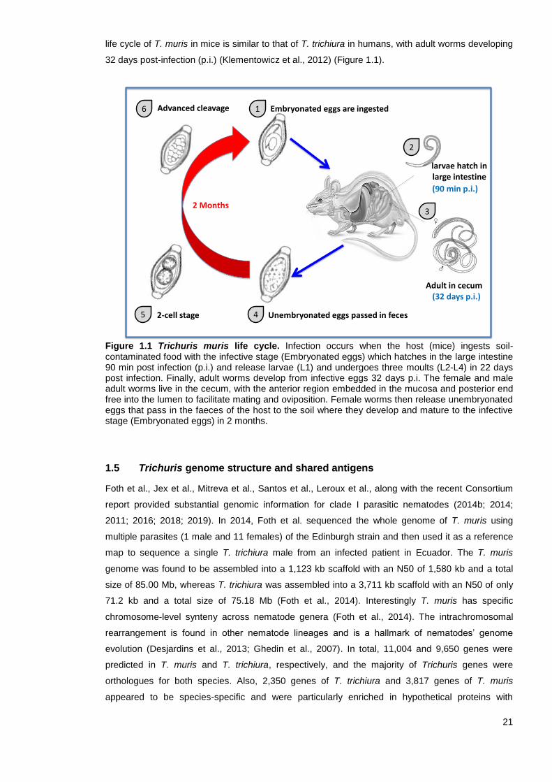
32 days post-infection (p.i.) (Klementowicz et al., 2012) (Figure 1.1).
Figure 1.1 Trichuris muris life cycle. Infection occurs when the host (mice) ingests soil-contaminated food with the infective stage (Embryonated eggs) which hatches in the large intestine 90 min post infection (p.i.) and release larvae (L1) and undergoes three moults (L2-L4) in 22 days post infection. Finally, adult worms develop from infective eggs 32 days p.i. The female and male adult worms live in the cecum, with the anterior region embedded in the mucosa and posterior end free into the lumen to facilitate mating and oviposition. Female worms then release unembryonated eggs that pass in the faeces of the host to the soil where they develop and mature to the infective stage (Embryonated eggs) in 2 months.
1.5 Trichuris genome structure and shared antigens
Foth et al., Jex et al., Mitreva et al., Santos et al., Leroux et al., along with the recent Consortium
report provided substantial genomic information for clade I parasitic nematodes (2014b; 2014;
2011; 2016; 2018; 2019). In 2014, Foth et al. sequenced the whole genome of T. muris using
multiple parasites (1 male and 11 females) of the Edinburgh strain and then used it as a reference
map to sequence a single T. trichiura male from an infected patient in Ecuador. The T. muris
genome was found to be assembled into a 1,123 kb scaffold with an N50 of 1,580 kb and a total
size of 85.00 Mb, whereas T. trichiura was assembled into a 3,711 kb scaffold with an N50 of only
71.2 kb and a total size of 75.18 Mb (Foth et al., 2014). Interestingly T. muris has specific
chromosome-level synteny across nematode genera (Foth et al., 2014). The intrachromosomal
rearrangement is found in other nematode lineages and is a hallmark of nematodes’ genome
evolution (Desjardins et al., 2013; Ghedin et al., 2007). In total, 11,004 and 9,650 genes were
predicted in T. muris and T. trichiura, respectively, and the majority of Trichuris genes were
orthologues for both species. Also, 2,350 genes of T. trichiura and 3,817 genes of T. muris
appeared to be species-specific and were particularly enriched in hypothetical proteins with
Unembryonated eggs passed in feces 2-cell stage
larvae hatch in large intestine
Adult in cecum
Advanced cleavage Embryonated eggs are ingested
45
6 1
2
3
(90 min p.i.)
(32 days p.i.)
2 Months
22
unknown function, extracellular proteins, proteases and protease inhibitors (Foth et al., 2014).
Furthermore, to investigate the immune response and the roles of T-helper (Th1 and Th2), Foth et
al. characterised the gene expression in the caeca and mesenteric lymph nodes (MLNs) of
chronically infected mice using RNA sequencing (RNA-Seq) (2014). Both tissues exhibited an
upregulation of specific genes consistent with a Th1 response (Foth et al., 2014).
A recent comparative study of over 50 parasitic nematode genomes conducted based on 36
published genomes observed gene count expansion of proteases and protease inhibitors in all
parasitic nematodes and platyhelminth families, which are involved in host tissue penetration and
migration, immunomodulation and modification of the host environment (Consortium, 2019). The
trypsin inhibitors have notably expanded as the most abundant protease inhibitor across parasitic
nematodes and platyhelminths (Consortium, 2019; Jex et al., 2018; Zarowiecki and Berriman,
2015).
Transcripts of the anterior region of T. muris were dominated by chymotrypsin A-like serine
proteases and by protease inhibitors, including secretory leukocyte peptidase inhibitors (SLPIs)
(Foth et al., 2014). Chymotrypsin A-like serine proteases are more abundant in both Trichuris
genomes, in terms of both gene number and gene expression, compared to other protease families
and far higher than in other nematodes (Foth et al., 2014). These proteases are known to degrade
the mucus barrier in the host’s intestine by inhibiting intestinal mucins, Muc2 in particular (Drake et
al., 1994; Foth et al., 2014; Hasnain et al., 2012). Chymotrypsin A-like serine proteases are also
thought to play a role as an anticoagulant by regulating blood clotting in the host and in digesting
host tissue, such as fibrinogen (Jex et al., 2014). Jex et al. sequenced the whole genome of a
single adult male and a single adult female pig worm (T. suis) (2014). Interestingly, chymotrypsin
A-like serine proteases were also upregulated in the stichosomes of T. suis species and exhibited
high homology with Schistosoma mansoni serine protease 1 (SP1) and human kallikrein (Golias et
al., 2007), which play a critical role in inhibiting inflammation (Jex et al., 2014).
The T. muris genome contains 44 genes that encode SLPI-like proteins, which are the most
abundant protease inhibitors in the anterior region of T. muris, while T. trichiura contains 20 such
genes and Trichinella spiralis contains 23 such genes (Foth et al., 2014). Host SLPI-like proteins
are predominantly secreted by epithelial cells at mucosal sites, and they exhibit anti-inflammatory
and antimicrobial properties and play a role in modulating inflamed intestinal tissue in the host
(Williams et al., 2006). These proteins are also similar to the mesocentin protein of Caenorhabditis
elegans, which plays a role in the development of the nervous system (Bénard et al., 2006).
Transcripts for DNase II-like proteins, which are known to be involved in mediation of DNA
apoptosis in hosts, were also highly expressed in the anterior region of T. muris, similar to
T. spiralis and C. elegans (Lai et al., 2009; Leroux et al., 2018; Liu et al., 2008). Three male-
specific encoding proteins were also expressed: major sperm protein (MSP), which plays a role in
the amoeboid locomotion of nematode sperm, and proteins with casein-kinase-related and
epidermal-growth-factor-like domains that have roles in male mating functions (Hu et al., 2006;
Leroux et al., 2018; Tarr and Scott, 2005). Chitin-binding domains that are associated with the
23
eggshell formation in C. elegans were also upregulated in female T. muris whipworms (Foth et al.,
2014; Johnston et al., 2010). These proteins are also predominantly expressed in T. trichiura,
T. suis, Nippostrongylus brasiliensis and Heligmosomoides polygyrus (Jex et al., 2014; Santos et
al., 2016; Vannella et al., 2016). The transcriptional landscape of both larval stages (L2 and L3) is
similar to adult worms’ anterior region, except that high levels of ribosomal proteins, collagen and
fibronectin-related proteins are expressed in the larval stages as a result of fast growth and cuticle
synthesis (Foth et al., 2014; Johnston et al., 2010).
Santos et al. (2016) reported the first transcriptomic exploration of the adult stage of T. trichiura
worms obtained from infected Ecuadorian children using next-generation sequencing technology
and a de novo assembly strategy. Among the 40 most highly expressed protein-encoding genes of
T. trichiura, a set of sequences that code vitellogenins, chitin-binding proteins and hypothetical
proteins were identified as potential molecules for the development of a trichuriasis vaccine
(Santos et al., 2016). Also, of the 40 most highly expressed protein-encoding genes of T. trichiura,
26 protein-coding genes were identified to be highly expressed and conserved among the adult
stages of the three Trichuris species (T. suis, T. muris, and T. trichiura) (Caffrey et al., 2018;
Ghedin, 2014; Howe et al., 2015; Santos et al., 2016). The de novo assembled transcriptome of
T. trichiura also predicted 20 transcripts that code for proteins with immunomodulatory properties
(Santos et al., 2016).
Using the nano-LC/mass spectrometry approach, it was also demonstrated that Trichinella spiralis
shared 12 proteins in T. trichiura adult worm fractions (Santos et al., 2013). Among the homology
identified proteins are macrophage migration inhibitory factor homologue (MIFH), fructose-
bisphosphate aldolase (FBPA) and heat shock protein 70 (Santos et al., 2013). These proteins are
known to have immunomodulatory effects that could lead to the development of novel drugs for
allergic and autoimmune diseases (Hauet-Broere et al., 2006; Maizels and Yazdanbakhsh, 2003;
Marques et al., 2008).
The anterior region of the Trichuris worm has a secretory gland and a storage organ called
stichostome, which consists of a row of stichocytes along the oesophagus and serves as a rich
source of excretory/secretory (ES) proteins (Lightowlers and Rickard, 1988). Such ES products are
produced in the stichosome and are thought to be released through the anterior ends of adult
whipworms embedded in the colonic mucosa to facilitate worm feeding (Jenkins and Wakelin,
1977; Lightowlers and Rickard, 1988; Wakelin and Selby, 1973). Due to intimate nature of the host-
parasite interaction, extensive studies across many helminth spp have investigated the ES
components that play a role in immunomodulation and inducing Th2-skewed immune response
(Harnett and Harnett, 2017; Mcsorley et al., 2013). For example, proteomic analysis of ES proteins
isolated from T. suis and Trichinella pseudospiralis at various life stages revealed that the most
abundant classes were proteases, protease inhibitors and uncharacterised proteins (Leroux et al.,
2018; Wang et al., 2017). In a different study, Tritten et al. (2017) sequenced T. muris ES
microRNA (miRNA) isolated from vesicles/particles of the same size as exosomes using polymer
precipitation and analysed the protein profiles by liquid chromatography-tandem mass

24
spectrometry. Most miRNA targets identified in T. muris exosome-like vesicles were conserved
across nematodes and mouse miRNAs, which may reflect host-parasite interactions. Furthermore,
comparison of T. muris-derived proteins and published nematode protein secretomes revealed a
high degree of conservation at the functional level (Tritten et al., 2017). Tritten and co-workers
(2017) also identified trypsin domains in the secretomes of T. muris that could have similar
biological functions to the serine proteases. More recently, Eichenberger et al. (2018a) reported
proteomic and genomic analyses of T. muris ES and extracellular vesicle (EV) fractions purified
using OptiPrep. As expected, proteases (including serine proteases), trypsin domain-containing
proteins and protease inhibitors were among the most abundant proteins found in the ES fractions
of adult T. muris worms (Eichenberger et al., 2018b). Trypsin or trypsin-like domain proteins
involved in proteolysis were also well represented in the T. muris EV fractions (Shears et al.,
2018a). Also, the RNA content of EVs fractions characterised using the Illumina HiSeq platform
demonstrated that hypothetical proteins with “unknown function“ are among the most abundant
domains in the mRNA transcripts mapping of T. muris genes (Eichenberger et al., 2018b).
Collectively, the wealth of information in genomics and proteomics provided an incentive to develop
a vaccine for trichuriasis based on new methods.
1.6 Immune responses to T. muris infection
The type of immune response generated against T. muris is critical in promoting either
susceptibility or resistance to infection. Thus, the vaccine candidate needs to be presented to the
host’s immune system in a manner that stimulates an appropriate protective immune response to
infection. For Trichuris, this response is generally a Th2-dependent immune response, whereas a
Th1 response is associated with chronic infection and increased immunopathology (Hurst and Else,
2013).
1.6.1 Elements contributing to resistance/susceptibility to T. muris infection
Studies conducted with inbred and gene knockout (KO) mice have greatly contributed to our
knowledge of the importance of certain elements in relation to resistance/susceptibility to T. muris
infection, including genetic background, gender, infection dose and parasite strain (Else, 2003;
Hayes et al., 2014; Klementowicz et al., 2012).
First, it has been shown that there is considerable variation in the persistence of T. muris infection
between mouse strains as well as within the same strain (Wakelin, 1967; Worley et al., 1962). The
genetic background of the mouse strain plays a vital role in determining the protective immune
response to infection (Klementowicz et al., 2012). For example, mice with a BALB/c genetic
background are resistant to infection and rapidly expel parasites, while AKR and B10.BR strains
are susceptible to infection (Else et al., 1993a; Else et al., 1992; Else and Wakelin, 1988). Also, the
slower‐responding C57BL/6 mouse strain is normally resistant to infection when infected with a
high-dose of T. muris eggs; mice were able to expel the worms by day 21 p.i., and full protection
was observed by day 28 p.i. (Bancroft et al., 2001; Cliffe et al., 2005; D'elia et al., 2009). Such
variation in immune response between different mouse strains has helped in understanding the

25
different immune responses generated against T. muris and subsequent parasite expulsion (Zhan
et al., 2014).
Second, several studies have clearly demonstrated that the gender of the host affects the immune
response to T. muris infection. For example, Bancroft et al. (2000) showed that male and female
interleukin 4 (IL-4) deficient mice with a BALB/c background responded differently to worm
expulsion; males developed a chronic infection, while females expelled the Trichuris worms
(Bancroft et al., 2000). This difference in expulsion kinetics is thought to be IL-13 dependent, as
treating IL-4 deficient female mice with anti-IL-13 antibodies led to the development of chronic
infection while administration of recombinant IL-13 enabled male IL-4 deficient mice to expel
worms. This study thus highlighted the critical role of the Th2 cytokine (IL-13) in driving worm
expulsion (Bancroft et al., 2000). Further research has revealed that sex hormones may play a role
in the gender differences in worm expulsion. This can be explained as the male-related hormone
dihydrotestosterone seems to decrease the ability of dendritic cells (DCs) to activate T-cells and
promotes T-cell differentiation towards a Th1-type immune response via IL-18-dependent
mechanisms in male IL-4 deficient mice. In contrast, the female-associated hormone 17-β-estradiol
(E2) enhances the generation of a Th2 immune response in vitro (Hepworth et al., 2010).
Third, the infective dose can profoundly influence hosts’ susceptibility or resistance to T. muris
infection (Bancroft et al., 1994). It has been demonstrated that a low-dose of Trichuris infection
(10–40 eggs) favours the development of a long-lasting chronic infection associated with a Th1
immune response, whereas a high-dose (200–400 eggs) favours the development of worm
expulsion associated with a Th2 immune response in most laboratory strains, including C57BL/6
and BALB/c mice (Bancroft et al., 1994; Wakelin, 1973). Bancroft et al. (2001) also demonstrated
that the fate of a secondary infection depends on the level of the primary infection. Interestingly,
BALB/K mice were able to develop a protective immunity when infected with a high-dose of
T. muris followed by a low-dose, whereas mice were susceptible to a high-dose if a low-dose was
administered first (Bancroft et al., 2001). BALB/K and C57BL/6 mice were also able to develop
protective immunity after administration of a repeated ‘trickle’ of low-dose infections (Bancroft et al.,
2001). In contrast, susceptible AKR mice and immuno-compromised strains, such as severe
combined immunodeficient (SCID) mice, were unable to expel high or low doses of Trichuris
infections (Bancroft et al., 2001).
Fourth, T. muris isolates can influence the hosts’ immune response to infection (Klementowicz et
al., 2012). Most inbred mouse strains, including B10.BR, CBA and C57BL/10, are susceptible to a
high-dose of the Sobreda (S) isolate and resistant to the Edinburgh (E) and Japan (J) isolates
(Bellaby et al., 1996; D'elia et al., 2009; Koyama and Ito, 1996). S-isolate-infected mice developed
Th1 immunity characterised by production of high levels of interferon gamma (IFN-γ), secretion of
anti-parasite serum IgG2c and an increased number of regulatory T cells (Tregs) in the gut (Bellaby
et al., 1996; D'elia et al., 2009; Johnston et al., 2005; Koyama and Ito, 1996). On the other hand, E-
and J-isolate-infected mice developed Th2 immunity characterised by the production of high levels
of IL-5 and secretion of anti-parasite serum IgG1 (Bellaby et al., 1996; D'elia et al., 2009). The
T. muris E isolate was used in all the in vivo experiments conducted in the current thesis.

26
Overall, T. muris is a helpful tool for understanding the components involved in the mediation of
susceptibility/resistance to Trichuris infection, and it serves as a model of cytokine-mediated
immunity to all gastrointestinal nematodes (Cliffe and Grencis, 2004).
1.6.2 Acquired immunity to T. muris
In the context of acquired immune responses, Wakelin was the first researcher who demonstrated
that more than 70% of outbred Schofield mice expelled T. muris and developed protective immunity
to primary and secondary infections, with only 30% of the mice were susceptible to infection
(Wakelin, 1967). Immunity was completely suppressed by administration of an immunosuppressive
agent (cortisone acetate) (Selby and Wakelin, 1973). Subsequently, several studies revealed that
immunity can be transferred by antiserum and mesenteric lymph node cells (MLNCs) taken from
infected animals and highlighted the importance of T-cells in mediating T. muris expulsion (Else
and Grencis, 1991; Lee et al., 1983; Selby and Wakelin, 1973). For example, Yoichi showed that
congenitally athymic (nude) mice were susceptible to T. muris infection unless they received
thymus, MLNC or spleen cells, while phenotypically normal mice were resistant to T. muris
infection (1991). This and similar studies confirm that immunity to T. muris is thymus-dependent.
Serial studies subsequently highlighted the role of CD4+ T helper cells and CD8+ cytotoxic T-cells
in mediating cellular protective immunity against T. muris (Humphreys et al., 2004). For instance,
Koyama et al. (1995) depleted CD4+ and CD8+ T-cells in vivo in BALB/c mice. Depletion of CD4+
T-cells, but not CD8+ T-cells, resulted in suppression of worm expulsion, suggesting that CD4+ T-
cells, and not CD8+ T-cells, mediate protective immunity against T. muris infection (Koyama et al.,
1995). Also, Else and Grencis (1996b) demonstrated that adoptive transfer of pure populations of
CD4+ T-cells protected SCID mice from T. muris infection in the complete absence of antibodies
and B-cells. Another study demonstrated that CD4+ T-cells are localised at the site of infection
(large intestinal mucosa) to confer protection against larval stages of T. muris (Betts and Else,
2000; Svensson et al., 2010). Interestingly, inhibition of the molecules necessary for activated
CD4+ T-cell migration to the gut-associated lymphoid tissue, including the gut homing receptors
(β7 and αE integrins) and the gut homing ligand MAdCAM-1, completely abrogated the ability of
transferred CD4+ T-cells to confer resistance in SCID mice (Betts and Else, 2000). Furthermore,
CD4+ T-cells localization to the site of infection is dependent on the Gαi‐coupled receptors;
however, neutralization of the most abundantly expressed chemokine receptors (CCR6 and
CXCR3) by CD4+ T cells in the MLN, which are involved in gut homing by T-cells, did not prevent
expulsion of worms (Svensson et al., 2010). Humphreys and Grencis (2002) also demonstrated
that C57BL/6 mice become more susceptible to infection with age due to a decreased ability of
CD4+ T-cells to respond to stimulation in vitro and were less able to proliferate and polarise into
Th2 cells.
Collectively, the studies described in this section and others confirm the role of CD4+ T
lymphocytes in the development of protective immunity against T. muris.

27
1.6.3 Cytokines associated with resistance/susceptibility to T. muris infection
Based on cytokine production patterns, naïve CD4+ T-cells can be diffrentiated into four subsets: T
helper 1 (Th1), T helper 2 (Th2), T helper 17 cells (Th17) and regulatory T-cells (Treg) (Harrington
et al., 2005; Street and Mosmann, 1991). Early studies comparing susceptible versus resistant
mouse strains to T. muris infection provided initial evidence that both Th1 and Th2 are associated
with susceptibility and resistant immune responses to T. muris infections (Else et al., 1994;
Grencis, 2001; Koyama et al., 1995; Scott and Kaufmann, 1991). It is now well documented that
resistance to T. muris infection is promoted by generation of a Th2 immune response and secretion
of the Th2 cytokines IL-4, IL-5, IL-6, IL-9, IL-10 and IL-13 and tumor necrosis factor α (TNF-α),
whereas susceptibility to infection is promoted by generation of a Th1 immune response and
production of the Th1-associated cytokines IFN-γ and IL-18 (Bancroft et al., 2001; Dixon et al.,
2010; Gause, 2003; Licona-Limon et al., 2017). In humans, host susceptibility and susceptibility to
reinfection have also been linked to whole-blood cytokine responses. Susceptible individuals
infected with T. trichiura are characterised by poor Th2 response and a negative association
between IL-5 and IL-13 responses and infection, suggesting that humans exhibit a protective Th2
response, similar to mice (Faulkner et al., 2002; Jackson et al., 2004a; Turner et al., 2002).
One of the Th2 cytokines produced in response to resistance to infection, IL-5, is involved in the
inducing eosinophilia during T. muris and T. trichiura infection (Else and Grencis, 1991; Jackson et
al., 2004b; Svensson et al., 2011). However, IL‐5 appeared not to be essential for worm expulsion,
as treatment with anti-IL-5 monoclonal antibodies had no effect on worm clearance in resistant
BALB/k mice (Betts and Else, 1999). Furthermore, no difference in worm expulsion was observed
following infection in CCL11‐deficient and CCL11 IL‐5 double-deficient mice (Dixon et al., 2006).
Therefore, despite being a useful Th2 cytokine hallmark of helminth infection, IL-5 is not thought to
play a critical role in mediating worm expulsion.
Several studies have also assessed the importance of IL-4 and IL-13 in promoting T. muris
expulsion. IL-4 and IL-13 share a sequence homology and functions associated with type 2
responses (Rao and Avni, 2000). For example, early studies demonstrated that both IL-4 and IL-13
KO mice are susceptible to T. muris infection (Bancroft et al., 1998). Administration of an IL-4
complex to susceptible AKR mice resulted in the development of a Th2 response and enabled
worm expulsion, and blocking IL-4 function in resistant BALB/k mice prevented the generation of a
protective immune response (Else et al., 1994; Grencis, 1993). Other studies have highlighted the
several effector functions of IL-4 in mast cell (Lorentz et al., 2000), goblet cell responses (Dabbagh
et al., 1999), and stimulation of intestinal muscle hypercontractility (Akiho et al., 2002). Bancroft et
al. (2000) found that IL-4-/- mice with a BALB/c background were able to expel T. muris worms
through IL-13-mediated responses, inferring that IL-13 plays a prominent role in Trichuris infection.
However, an earlier study showed that IL-13 KO mice failed to expel T. muris worms despite
generating a Th2 response involving the production of IL-4, IL-5 and IL-9 and high levels of serum
IgG1 and IgE p.i. (Bancroft et al., 1998). Other studies have provided evidence that IL-13 plays a
role in increasing epithelial cell turnover and producing mucins to promote worm expulsion
(Bancroft et al., 2000; Bancroft et al., 1998; Hasnain et al., 2011a).

28
Another cytokine that plays a major role in resistance to T. muris is IL-9. An early study by Faulkner
et al. (1998) demonstrated that the MLNs of resistant BALB/K mice express high levels of IL-9 as
early as day 4 p.i. compared to susceptible AKR mice (Faulkner et al., 1998). Furthermore, treating
AKR mice with an IL-9 complex resulted in a reduction in worm survival (Faulkner et al., 1998).
Moreover, the importance of IL-9 has been illustrated in the stimulation of intestinal smooth muscle
contractility, which drives T. muris expulsion but not goblet cell hyperplasia and mucosal mast cell
protease-1 production (Khan et al., 2003; Richard et al., 2000). Thus, impaired IL-9 expression
often results in chronic helminth infection (Li et al., 2017). Collectively, these findings clearly
confirm the central role of Th2 response in protective immunity to trichuriasis.
The regulatory molecule IL-10, which is produced by Tregs, B-cells and DCs, has been shown to
play a role in both resistance and survival during T. muris infection (Schopf et al., 2002). Mice
deficient in IL-10 were susceptible to T. muris infection characterised by elevated levels of IFN-γ
and TNF-α, and they displayed fatal intestinal pathology (Schopf et al., 2002). However, both IL-10
and transforming growth factor-β (TGF-β) can be detected in the large intestine during T. muris
infection, and seem to play a role in regulating immunity to T. muris infection (Collison et al., 2010;
Veldhoen et al., 2008). In another study, an increasing number of Foxp3+ regulatory T-cells during
chronic infection was found to be advantageous for the host as this minimised gut damage (D'elia
et al., 2009). However, Treg cells are not considered to be critical for protection (Worthington et al.,
2013).
It has been further demonstrated that the role of TNF-α in susceptibility/resistance to Trichuris
infection is context-dependent, with high levels of TNF-α detected in susceptible hosts during Th1
response (Artis et al., 1999a; Hayes et al., 2007). Hayes et al. (2007) showed that mice deficient in
TNF-α receptor (p55/p75) with a C57BL/6 background are susceptible to T. muris infection, but that
this phenotype can be restored by IL-13 administration. In vivo treatment of resistant C57BL/6 mice
with anti-TNF-α monoclonal antibodies significantly prevented worm expulsion without altering the
magnitude of the type 2 response compared to control mice treated with PBS (Artis et al., 1999a).
Furthermore, female susceptible BALB/c IL-4 KO mice (in which expulsion is mediated by IL-13)
that were infected with T. muris and treated with recombinant TNF-α were able to clear the
infection (Artis et al., 1999a). This suggests that TNF-α plays a role in IL-13-mediated anti-T. muris
effector responses (Artis et al., 1999a). Although TNF-α may enhance either Th1 or Th2 during an
ongoing immune response to T. muris infection, it is not necessary for developing immunity.
Equally importantly, IL-12, IL-18 and IFN-γ are associated with Th1 response and the development
of susceptibility to infection (Bancroft et al., 2004; Belladonna et al., 2002). In one study, in vivo
treatment of resistant mice with recombinant IL-12 promoted the development of chronic intestinal
nematode infection (Bancroft et al., 1997). In contrast, IL-12 KO mice have been reported to be
capable of expelling T. muris parasites, regardless of whether they were administered a high- or
low-dose infection (Bancroft et al., 2001; Helmby et al., 2001). Helmby et al. (2001) also suggested
that IL-18 promotes the development of Trichuris chronic infection by down-regulating IL-13.
Furthermore, Else et al. (1994) provided the first evidence of the critical role of IFN-γ in developing
chronic infection, as depletion of IFN-γ in susceptible AKR mice resulted in the expulsion of the
29
parasite. Collectively, Th1 cytokines (IL-12, IL-18 and IFN-γ) are hallmarks of a type 1 immune
response, whereas Th2 cytokines (IL-4, IL-5, IL-9 and IL-13) are hallmarks of a type 2 immune
response and the generation of worm expulsion in T. muris infection.
1.6.4 Other sources of Th2 cytokines
The key factors that are essential for generation of a protective Th2 immune response have not
been fully characterised, with many different cell types implicated, including mast cells, eosinophils,
natural killer cells (NKs), innate lymphoid cells (ILCs), dendritic cells (DCs), basophils and
macrophages (Grencis et al., 2014).
Increased numbers of mast cells and eosinophils are typical in helminth infection and during
T. muris infection (Mukai et al., 2016), but neither cell type has been shown to play a critical role in
protective immunity against T. muris (Betts and Else, 1999; Knight et al., 2000). For example,
Hepworth et al. (2012) demonstrated that mice deficient in mast cells were characterised by
increased worm burden and decreased levels of epithelial IL-25, IL-33 and thymic stromal
lymphopoietin (TSLP) following T. muris infection. Furthermore, Svensson et al. (2001)
demonstrated that eosinophils accumulate faster and in greater numbers in the MLNs of T. muris-
resistant BALB/c mice compared to susceptible AKR mice. However, mice deficient in eosinophils
(ΔdblGATA-1 mice) were still able to expel T. muris (Svensson et al., 2011). Collectively, these
data suggest that mast cells and eosinophils do not play a critical role in parasite expulsion within
this infection system.
It has also been hypothesised that NK T-cells may play an essential role in driving Th2 responses
that produce protective immunity against T. muris (Hepworth and Grencis, 2009). However,
depletion of NK1.1+ cells in resistant mice (B10.BR) by anti-NK1.1 monoclonal antibody injection
failed to induce the mice to develop susceptibility (Koyama, 2002). This implies that, in the
presence of T. muris, NKs are not involved in the induction of Th2 response and the development
of parasite expulsion.
New insights highlight that the role of the ILCs, which are mainly driven by IL-25 and IL-33
production, is associated with Th2 response and development of resistance to T. muris infection
(Humphreys et al., 2008; Minutti et al., 2017; Saenz et al., 2010; Spencer et al., 2014). Mice
deficient in either cytokine (IL-25 or IL-33) exhibit delayed worm expulsion, while treating
susceptible mice during the early stages of infection with either recombinant IL-25 or IL-33
promoted Th2 immunity and worm expulsion (Fallon et al., 2006; Humphreys et al., 2008; Owyang
et al., 2006). Chronically infected IL-25-deficient mice developed severe infection associated with
heightened expression of IFN-γ and IL-17 and a significant reduction in IL-4 and IL-13 Th2
cytokines (Owyang et al., 2006). However, there is limited evidence regarding the role of Th17 in
protection against T. muris infection (Bouchery et al., 2014).
Intestinal epithelial cells (IECs) are able to sense T. muris infection and initiate Th2 cell-mediated
immunity through the release of TSLP, IL-25 and IL-33, which promotes the development,
proliferation and activation of ILC2s, basophils, mast cells, eosinophils and DCs in the early phases
of infection (Taylor et al., 2009; Zaph et al., 2007).

30
The kinetics of DCs may also be important for regulation of intestinal immune responses by
processing antigens, migrating to MLNs upon activation and priming naïve T-cells that
subsequently drive adaptive immunity (Joeris et al., 2017). Intestinal DCs can be classified into
three distinct subsets based on their expression of CD11b and CD103 and either interferon
regulatory factor 4 or 8 (IRF4 or IRF8) (Joeris et al., 2017). There is some controversy about the
importance of DCs in triggering Th2 responses during T. muris infection. For example, there is
evidence that the IL-27 produced by DCs during T. muris infection directly suppresses CD4+ T-cell
proliferation and Th2 cytokine production and promotes the development of susceptibility and a
Th1 immune response (Artis et al., 2004a; Bancroft et al., 2004). Furthermore, depletion of CD11c+
DC cells during T. muris infection resulted in normal expression of Th2 cytokines (IL-4 and IL-5)
and expulsion of Trichuris worms, suggesting that DCs were not involved in priming CD4+ T-cells
(Perrigoue et al., 2009). However, it was previously reported that DCs mobilise to the site of
infection via epithelial chemokine production more quickly in resistant mice than in susceptible
ones (Bowcutt et al., 2014; Cruickshank et al., 2009). Also, recent studies demonstrated a
dominant role for IRF4-dependent CD11b+ DCs in the induction of Th2 immunity, notably during
infection with T. muris (Demiri et al., 2017; Mayer et al., 2017). Another study found that IRF8-
dependent CD103+ DCs promoted T. muris chronicity by generating type 1 immunity with helper
CD4+ T-cells and cytotoxic CD8+ T-cells (Luda et al., 2016).
It has, however, been suggested that basophils are more important than DCs for priming CD4+ T-
cells. Data indicates that basophils expressing major histocompatibility molecules (MHC) class II in
draining LNs upon activation of the immune system play an important role in promoting Th2-
dependent immunity to T. muris by producing TSLP and IL-4, which facilitate DC-mediated
development (Perrigoue et al., 2009; Sokol et al., 2009; Webb et al., 2019; Wynn, 2009). For
example, Siracusa et al. (2001) demonstrated that TSLPR KO mice exhibited impaired Trichuris
worm expulsion due to a decreased number of basophils and decreased levels of IL-5 and IL-13
during acute T. muris infection. However, adoptive transfer of basophils into TSLPR KO mice led to
a reduction in worm numbers (Siracusa et al., 2011).
Because macrophages exhibit diverse biological functions such as antigen-presenting cells
(APCs), phagocytosis, production of cytokines and in tissue repair, it is not easy to predict their role
in immunity to T. muris infection (Faz-Lopez et al., 2016; Noel et al., 2004). However, alternatively
activated macrophages (AAMs) have been reported to have several roles in type 2 immune
responses induced by IL-4 and IL-13 in intestinal nematode infections (Faz-Lopez et al., 2016;
Kreider et al., 2007). Specifically, AAMs are involved in tissue remodelling, repairing the tissue
affected by large extracellular nematodes, limiting of damage and inflammatory responses, and
smooth muscle hypercontractility to promote nematode expulsion (Kreider et al., 2007; Zhao et al.,
2008). Interestingly, larger numbers of macrophages were found accumulated in the lamina propria
of resistant BALB/c mice to T. muris infection compared to susceptible AKR mice (Little et al.,
2005). However, Bowcutt et al. (2011) found that using a cell-specific targeting strategy, arginase-1
deficiency in macrophages had no effect on resistance to T. muris infection, suggesting that
macrophages have a possible arginase-independent role in the immune response to T. muris.

31
1.6.5 B-cells and production of IgG antibodies
The role of B-cells and B-cell-derived antibodies in mediating immune responses to T. muris has
received a great amount of attention. However, it has been postulated that the role of B-cells in the
immune response to Trichuris infection is context-dependent (Sahputra et al., 2019; Sorobetea et
al., 2018). The number of B-cells is significantly elevated in both T. muris-resistant and susceptible
mice strains (Else and Grencis, 1991), with a marked increase in the production of anti-T. muris
antibodies (IgA and IgG subclasses) in MLNs as early as 14 and 21 days p.i. (Blackwell and Else,
2001; Koyama et al., 1999). It was also found that parasite-specific IgE was negatively correlated
with infection intensity and positively correlated with age, suggesting that IgE is associated with
protection (Faulkner et al., 2002; Turner et al., 2002).
It is widely accepted that IgG1 is correlated with the development of Th2 response in resistance
and IgG2 is correlated with the development of Th1 response in susceptibility to T. muris infection
(Blackwell and Else, 2002). An early study by Lee et al. (1983) showed that transfer of enriched T-
cell populations from MLNCs, but not B-cell enriched populations from T. muris-infected animals, to
naïve recipients transferred immunity to infection. It has also been demonstrated that adaptive
transfer of only CD4+ T-cells purified from BALB/c donor mice to SCID mice (which are deficient in
B- and T-cells) was sufficient to expel T. muris worms, suggesting that antibodies are not essential
for expulsion (Else and Grencis, 1996). However, a different study demonstrated that µMT mice on
a C57BL/6 background which are not able to produce B-cells did not expel T. muris worms
efficiently (Blackwell and Else, 2001). When these animals were reconstituted with B-cells,
however, they were able to expel most of the worms; expulsion was associated with Th2 response
and production of a high level of specific anti-T. muris antibodies (Blackwell and Else, 2001).
Furthermore, the adoptive transfer of serum from resistant mice provided high levels of protection
to susceptible strains of mice (Blackwell and Else, 2001; Selby and Wakelin, 1973). Early studies
have also shown that parasite-specific IgG1 is involved in the memory response of the adaptive
immune system in resistant strains following primary infection (Else et al., 1993a). A recent study
supported the role for B-cells in the generation of protective immunity to T. muris infection and
suggested that the Th2 type immune response is dependent on the host’s genetic background and
is independent of antibodies (Sahputra et al., 2019).
Taken together, the findings of the above-mentioned studies suggest that B-cells might play a role
in stimulating the generation and/or polarisation of T-cell responses by either cytokine secretion or
antigen presentation. This is probably most effective in mice with secondary T. muris infection
(Sorobetea et al., 2018).
1.7 Mechanisms of T. muris expulsion
Once adaptive immunity has been stimulated in the local lymph nodes (LNs), activated effector
cells must home back to the site of the infection ‘large intestine’ where worms expulsion can take
place (Sorobetea et al., 2018). A variety of Th2 regulated immune-mediated mechanisms are
characterised for T. muris expulsion including epithelial cell turn over (Cliffe and Grencis, 2004;
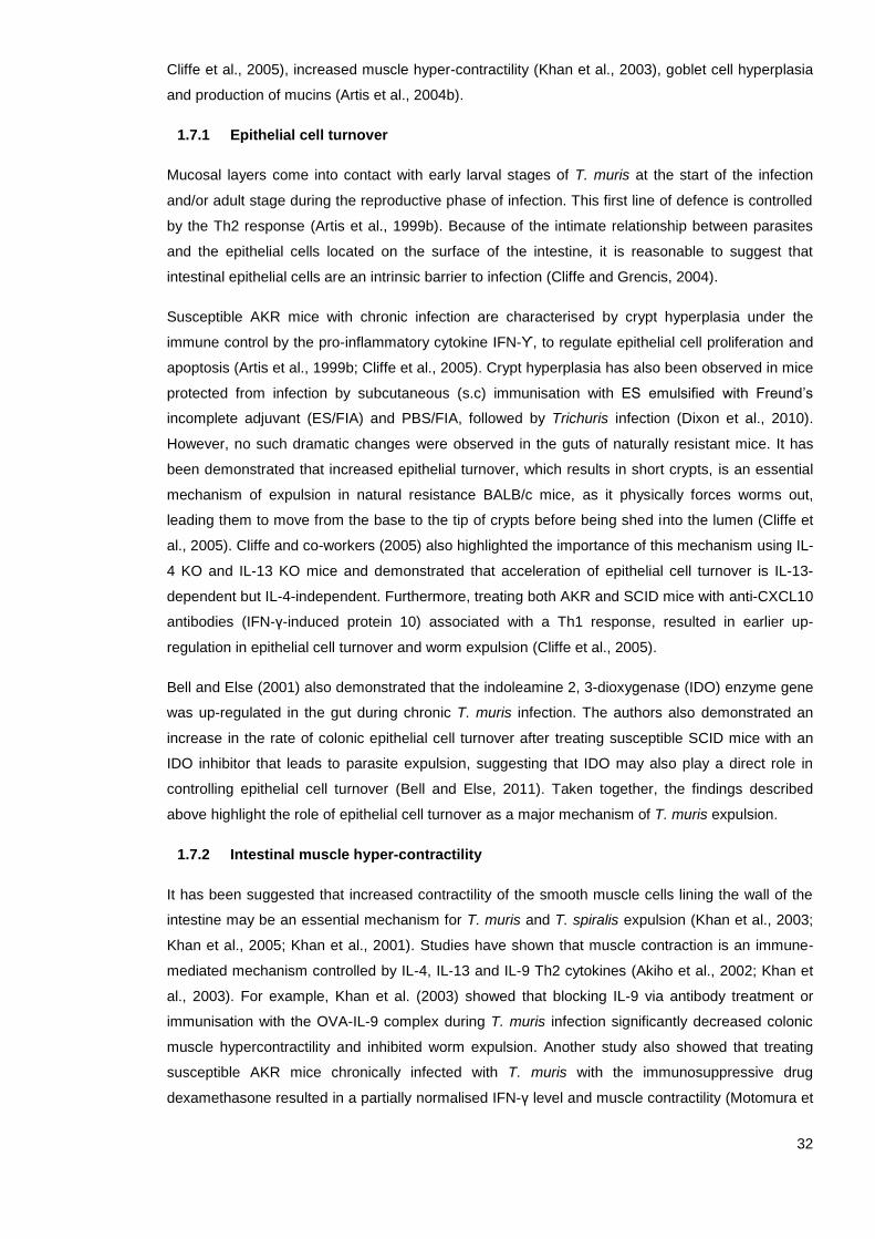
32
Cliffe et al., 2005), increased muscle hyper-contractility (Khan et al., 2003), goblet cell hyperplasia
and production of mucins (Artis et al., 2004b).
1.7.1 Epithelial cell turnover
Mucosal layers come into contact with early larval stages of T. muris at the start of the infection
and/or adult stage during the reproductive phase of infection. This first line of defence is controlled
by the Th2 response (Artis et al., 1999b). Because of the intimate relationship between parasites
and the epithelial cells located on the surface of the intestine, it is reasonable to suggest that
intestinal epithelial cells are an intrinsic barrier to infection (Cliffe and Grencis, 2004).
Susceptible AKR mice with chronic infection are characterised by crypt hyperplasia under the
immune control by the pro-inflammatory cytokine IFN-ϒ, to regulate epithelial cell proliferation and
apoptosis (Artis et al., 1999b; Cliffe et al., 2005). Crypt hyperplasia has also been observed in mice
protected from infection by subcutaneous (s.c) immunisation with ES emulsified with Freund’s
incomplete adjuvant (ES/FIA) and PBS/FIA, followed by Trichuris infection (Dixon et al., 2010).
However, no such dramatic changes were observed in the guts of naturally resistant mice. It has
been demonstrated that increased epithelial turnover, which results in short crypts, is an essential
mechanism of expulsion in natural resistance BALB/c mice, as it physically forces worms out,
leading them to move from the base to the tip of crypts before being shed into the lumen (Cliffe et
al., 2005). Cliffe and co-workers (2005) also highlighted the importance of this mechanism using IL-
4 KO and IL-13 KO mice and demonstrated that acceleration of epithelial cell turnover is IL-13-
dependent but IL-4-independent. Furthermore, treating both AKR and SCID mice with anti-CXCL10
antibodies (IFN-γ-induced protein 10) associated with a Th1 response, resulted in earlier up-
regulation in epithelial cell turnover and worm expulsion (Cliffe et al., 2005).
Bell and Else (2001) also demonstrated that the indoleamine 2, 3-dioxygenase (IDO) enzyme gene
was up-regulated in the gut during chronic T. muris infection. The authors also demonstrated an
increase in the rate of colonic epithelial cell turnover after treating susceptible SCID mice with an
IDO inhibitor that leads to parasite expulsion, suggesting that IDO may also play a direct role in
controlling epithelial cell turnover (Bell and Else, 2011). Taken together, the findings described
above highlight the role of epithelial cell turnover as a major mechanism of T. muris expulsion.
1.7.2 Intestinal muscle hyper-contractility
It has been suggested that increased contractility of the smooth muscle cells lining the wall of the
intestine may be an essential mechanism for T. muris and T. spiralis expulsion (Khan et al., 2003;
Khan et al., 2005; Khan et al., 2001). Studies have shown that muscle contraction is an immune-
mediated mechanism controlled by IL-4, IL-13 and IL-9 Th2 cytokines (Akiho et al., 2002; Khan et
al., 2003). For example, Khan et al. (2003) showed that blocking IL-9 via antibody treatment or
immunisation with the OVA-IL-9 complex during T. muris infection significantly decreased colonic
muscle hypercontractility and inhibited worm expulsion. Another study also showed that treating
susceptible AKR mice chronically infected with T. muris with the immunosuppressive drug
dexamethasone resulted in a partially normalised IFN-γ level and muscle contractility (Motomura et

33
al., 2010). Thus, smooth muscle hypercontractility appears to be an important immune-mediated
mechanism for the expulsion of gastrointestinal parasites.
1.7.3 Goblet cells and mucus production
The extrinsic physical barrier comprised of goblet cells and mucins is known to assist in parasite
expulsion under the control of Th2 cytokines (IL-13 and IL-9 and IL-22) (Artis et al., 2004b; Leung,
2013; Turner et al., 2013). It has been demonstrated that the number of goblet cells increases in
both susceptible and resistant mice during T. muris infection compared to uninfected naïve mice
(Artis et al., 2004b; Cliffe and Grencis, 2004; Datta et al., 2005; Owyang et al., 2006). Goblet cells
produce different defensive compounds, including glycoprotein mucin (the major protein
component of mucus), which acts as a sticky blanket that inhibits the motility and feeding of
T. muris and mediates worm expulsion (Hasnain et al., 2011b; Hasnain et al., 2010; McGuckin et
al., 2011). However, the type of mucin produced by intestinal goblet cells differs between resistant
and susceptible mice (Hasnain et al., 2011a; Hasnain et al., 2010). Muc2 was only up-regulated in
resistant mice (BALB/c, C57BL/6). Moreover, Muc2-deficient mice showed delayed parasite
expulsion compared to resistant wild-type C57BL/6 mice (Hasnain et al., 2010). Hasnain et al.
(2012) also demonstrated that serine protease, the main protein secreted by T. muris, has the
ability to make the mucus barrier more porous by degrading Muc2. Interestingly, Muc5ac, which is
a mucin that is not normally expressed in the intestinal tract, was detected in resistant animals
before T. muris expulsion and was associated with the production of IL-13 (Hasnain et al., 2011b;
Hasnain et al., 2010). Mice deficient in Muc5ac were susceptible to Trichuris infection despite the
robust Th2 effector response and remained highly susceptible to T. muris infection even after
treatment with an IFN-γ neutralising antibody, which further enhanced the Th2 response (Hasnain
et al., 2010). These findings suggest that certain mucins can have a direct effect on worm
expulsion.
Goblet cells produce resistin-like molecule β (RELMβ), which is associated with the production of
Th2 cytokines (IL-13) in animals that are resistant to T. muris infection (Artis et al., 2004b; Nair et
al., 2008). RELMβ are also known to prevent lumen-dwelling worms from feeding and growing and
block their motility and attachment to the host’s epithelium (Artis et al., 2004b; Herbert et al., 2009).
However, Nair et al. (2008) showed that RELMβ KO mice exhibited reduced intestinal inflammation
and were able to expel acute T. muris infection, suggesting that RELMβ does not play an essential
role in the generation of Th2 response in resistant mice (Nair et al., 2008).
In summary, ejection of T. muris worms relies on a combination of physiological mechanisms,
including increased epithelial cell turnover, increased muscle contractility, goblet cell hyperplasia
and production of the mucins summarised in Figure 1.2.
34
Figure 1.2 Immune profiles during Trichuris muris infection. During resistance to T. muris infection, whipworms traverse through the epithelium. Damaged epithelial cells release alarmins such as IL-25 and IL-33 that activate innate lymphoid type 2 cells (ILC2) and antigen-presenting cells such as the dendritic cells (DCs) or macrophages. These alarmins, together with helminth-derived products, promote a Th2 immune response. Activated DCs, migrate to the draining lymph nodes where they induce differentiation and polarisation of naïve CD4+ T-cells to Th2-cells. Th2 cells subsequently secrete Th2 cytokines (IL-4, IL-5, IL-9, and IL-13) which drive the activation of B-cells and induces isotype switching to IgE and IgG1. Furthermore, the activation of Th2 response results in the stimulation of anti-helminth responses, including increase epithelial cell turnover and muscle contractility. In addition to inducing goblet cell hyperplasia and the secretion of Muc5ac and Muc2 mucins that result in thickening of the mucus layer, which have a direct detrimental effect on worm expulsion. In contrast, in susceptibility, T. muris parasites significantly damage the epithelium as they migrate, causing the release of damage signals that stimulate the activation of innate cells present in the tissue. Activated DCs induce differentiation and polarisation of naïve T-cells to Th1-cells, which secrete Th1 cytokines, including IFN-γ. Th: T-helper, IL: interleukin, immunoglobulin: Ig, IFN-γ: interferon gamma.
Susceptible Resistance
Epitheliu
m
Muscle contractility
Epithelial turnover
Muc2
No Muc5a
Mucus Goblet cell
Muscle contractility
Epithelial turnover
Muc2
Muc5ac
Mucins
Epithelium
MLN
Intestinal
mucosa
Naїve
CD4+
T cell
DC
IFNϒ
IL-4
IL-5
IL-9
IL-13IgE
IgG1
IgG2
B-cell B-cell
Th1Th2
IL-12IL-18
IL-2IL-4

35
1.8 Experimental Trichuris vaccine candidates
Vaccines are one of the most cost-effective and well-established interventions to control and
prevent infectious diseases in both the human medicine and veterinary fields (Lombard et al.,
2007). It is now clear that to produce effective immunisations, it requires long-term stimulation of
both the humoral and cellular arms of the immune system to a specific region of the pathogen to
produce effector and memory cells to avoid subsequent re-infection (Clem, 2011). STHs have
complex genomes, proteomes, and correspondingly complex immunomes (Foth et al., 2014). This
complexity represents a great challenge for the development of an effective vaccine (De Sousa and
Doolan, 2016). Thus, the availability of an effective vaccine to most helminth infections does not
exist. However, the development of vaccines for helminth infections has progressed through
generations; the earliest vaccines included attenuated or irradiation-killed parasites, subsequent
subunit vaccines contained excretory/secretory (ES) products, ES fractions and extracellular
vesicles (EVs), and most recently using purified recombinant parasite antigens (Bain, 1999; Briggs
et al., 2018; Dixon et al., 2010; Kifle et al., 2017; Shears et al., 2018a).
Conventional vaccines, such as live attenuated and whole inactivated vaccines, include weakened
or killed microorganisms or their derivatives, such as detoxified toxins (Sinha and Bhattacharya,
2006). Such vaccines have reduced the morbidity of major infectious diseases such as smallpox,
poliovirus and diphtheria, but they have not eliminated existing or newly emerging diseases (Arnon
and Ben-Yedidia, 2003). Irradiated larval vaccines, for example, have been commercially available
since 1959 for Ancylostoma caninum and bovine lungworm, Dictyocaulus viviparus, in animals
(Bain, 1999; Miller, 1978). Despite the fact that they have been used for treating animals, these
vaccines are not stable and cannot be used against human parasites (Schneider et al., 2011).
One of the first attempts at a non-living vaccine for T. trichiura was conducted on the mouse model
T. muris, using adult and larval worm somatic antigens to stimulate protective immunity in infected
animals (Wakelin and Selby, 1973). Vaccination of mice with T. muris somatic antigens that were
isolated from NIH mice and emulsified in Freund’s incomplete adjuvant stimulated protective
immunity and a 92% reduction in worm burden after infection (Wakelin and Selby, 1973). Wakelin
and Selby (1973) also demonstrated that soluble antigens from the anterior region of adult worms
were shown to more effectively stimulate immunity than antigens prepared from the posterior
region. Hence, T. muris adult worm homogenate and ES products from both adult and larval stages
have been utilised as early stages of vaccine development to T. trichiura (Jenkins and Wakelin,
1977; 1983). For example, there was only sufficient protection against T. muris in several
susceptible mouse strains that were administered with homogenised adult antigens combined with
cholera toxin as an oral vaccine (Robinson et al., 1995). However, full protection in all the tested
mice strains was achieved through s.c. vaccination with adult antigens emulsified in Freund’s
adjuvant (Robinson et al., 1995). Jenkins and colleagues (1983) also showed that vaccinating NIH
mice subcutaneously with 100 μg of ES without adjuvant was more effective than intraperitoneal
vaccination; s.c. vaccination resulted in about 70% reduction in worm burden of a high-dose
infection at day 9 post-infection (p.i.) compared to 33% for intraperitoneal vaccination. More
recently, Dixon et al. (2010) showed that s.c. vaccination of naturally susceptible AKR mice with
100 μg of T. muris ES emulsified with Freund’s incomplete adjuvant (IFA) induced expulsion of a

36
high-dose infection. This study also described priming of the immune response to s.c. vaccination
as occurring in peripheral LNs (Dixon et al., 2010).
Interest in identifying host protective material in EV components within helminth secretions has
increased recently (Eichenberger et al., 2018a; Eichenberger et al., 2018b; Hansen et al., 2015;
Tritten and Geary, 2018). For example, Coakley et al. (2017) showed that intraperitoneal
vaccination of mice with H. polygyrus exosome-like vesicles (ELVs) prior to infection resulted in a
significant reduction in H. polygyrus worm burden and egg output. Also, Shears et al. (2018a;
2018b) showed that vaccinating C57BL/6 mice with either ES fractions or EVs isolated from
T. muris without an adjuvant and prior to infection with a low-dose of T. muris eggs significantly
reduced the worm burden compared to the PBS-injected group.
Notwithstanding these achievements, developing a subunit vaccine based on native antigens has
many manufactory complications, such as cost, time consumption, difficulty of purifying large
quantities of worm antigens and control over differences in batches to develop a commercially
stable vaccine (Geldhof et al., 2007; Hewitson and Maizels, 2014). To overcome these and other
crucial limitations and ensure that safer and more reliable vaccines are developed, new
approaches are being considered, especially after the failure of anti-helminthic drugs (Terry et al.,
2014). Such approaches include recombinant, DNA and epitope-based vaccines (Patronov and
Doytchinova, 2013). Recombinant vaccines may contain secreted or extracted fractions from the
pathogen or synthetic peptides or proteins that can lead to a protective immune response produced
by genetic engineering technology, and expressed in a heterologous expression system (Noon and
Aroian, 2017). Of the most successful recombinant vaccines are the two against hookworm
disease caused by Necator americanus, which consist of aspartic protease-1 (Na-APR-1) and
glutathione-S-transferase-1 (Na-GST-1). These vaccines are currently in phase 1 of clinical trials
(Diemert et al., 2018; Diemert et al., 2017; Hotez et al., 2013).
Gomez-Samblas et al. (2017) have also identified a vaccine candidate against some helminth
parasites, including T. muris, based on recombinant serine/threonine phosphatase 2A from the
nematode Angiostrongylus costaricensis (rPP2A), which is formulated as a lipopeptide and
conjugated with a self-adjuvant oleic-vinyl sulfone (OVS). Interestingly, intranasal immunisation of
AKR mice with the vaccine candidate prior to T. muris challenge showed a marked reduction in the
number of nematode eggs and adults. The immunised mice also developed a combined Th17/Th9
response orchestrated by the cytokines IL-25, IL-17 and IL-9 (Gomez-Samblas et al., 2017). The
same vaccine candidate has also been tested in lambs and was found to provide significant
protection against the ovine helminth Haemonchus contortus and Teladorsagia circumcincta
infection (Mohamed Fawzi et al., 2013).
More recently, Briggs et al. (2018) developed two vaccines against trichuriasis based on T. muris
whey acidic protein (rTm-WAP49) and T. muris WAP fragment fusion protein (rTm-WAP-F8+Na-
GST-1) formulated with Montanide ISA 720 adjuvant. Vaccinating AKR mice with the vaccine
candidates three times at two-week intervals prior to T. muris challenge induced a partial reduction
in worm burden (48% and 38%, respectively) (Briggs et al., 2018). The authors also showed that
both humoral and cellular immune responses were induced and characterised by elevated antigen-

37
specific IgG1 and IgG2c antibodies and Th2 (IL-4, IL-9 and IL-13) cytokines in the draining inguinal
LNs, draining mesenteric LNs and spleens of vaccinated mice (Briggs et al., 2018). Despite these
promising results, subunit vaccines require substantial adjuvant and often do not provide sufficient
protective cellular immunity compared with other vaccine approaches (Arnon and Ben-Yedidia,
2003).
Recent advances in helminth genomics and proteomics have made available a wealth of
information for the development of DNA and epitope-based vaccines to induce immune responses
with less risk than conventional vaccines (Gurunathan et al., 2000; Loukas and Giacomin, 2016).
For example, the Ascaris suum vaccine composed of gene-encoding A. suum enolase (As-enol-1)
that was amplified, cloned and expressed in Marc-145 cells resulted in a 61.13% reduction (P <
0.05) in larvae recovery in Kunming mice (Chen et al., 2012). In addition, a recent study tested the
efficacy of a naked DNA vaccine approach without adjuvant that used the serine protease (Ts-
NBLsp) of T. spiralis larvae (Xu et al., 2017). Vaccinating Kunming mice twice with 60 μg of the
vaccine candidate resulted in a 77.93% reduction in muscle larvae following T. spiralis infection
and promoted both type 1 and type 2 immune responses (Xu et al., 2017). More recently, Sun et
al. (2019) demonstrated that intranasal vaccination of BALB/c mice with T. spiralis serine protease
(rTsSP) cDNA cloned and expressed in E. coli and coupled with cholera toxin B subunit (CTB)
resulted in a 71.10% adult and 62.10% larva reduction after challenge.
The emergence of pathogen genomic sequencing and recombinant DNA technology enabled the
development of a methodology called Reverse Vaccinology (RV), which relies on genomic data
and bioinformatics analysis to identify critical antigens derived from the pathogen genome with
minimum risk (De Sousa and Doolan, 2016; Del Tordello et al., 2017). The vaccine against
Neisseria meningitidis serogroup B, which is the primary cause of sepsis and meningitis in children
and young adults, was the first vaccine created with the RV approach to be approved for humans
(Dretler et al., 2018; Pizza et al., 2000). The success of RV for N. meningitidis and the significant
developments in immunoinformatic tools have prompted researchers to apply RV to develop
epitope-based vaccines against a variety of parasites, including Plasmodium falciparum (Pance,
2019), Onchocerca volvulus (Shey et al., 2019), Cystic echinococcosis (Pourseif et al., 2018),
Leishmania infantum (Dias et al., 2018), and T. spiralis (Gu et al., 2016; Wei et al., 2011). For
example, Dias et al. (2018) developed an epitope-based vaccine against leishmaniasis based on
CD4+ and CD8+ T-cell epitopes derived from three different L. infantum proteins predicted using
the NetCTLPan in silico prediction tool. Interestingly, following L. infantum promastigote challenge,
vaccinating mice with the epitope-based vaccine candidate significantly reduced the parasite
burden in several organs compared to control groups (Dias et al., 2018).
1.9 Epitope-based vaccine against trichuriasis
Based on the promising results of the epitope-based vaccines, the present study developed
epitope-based vaccines for Trichuris based on the RV strategy. The RV approach comprises five
major steps, which can be thought of as a funnelling process (Figure 1.3). The first step is the
identification of secreted, upregulated and conserved proteins from the pathogen genome that can
elicit a protective immune response (Khan et al., 2006). Second, after selecting a set of proteins

38
derived from the whole genome, immunoinformatic tools are used to identify potential highly
immunogenic epitopes that have a high probability of stimulating an appropriate immune response
to the pathogen components by mapping the protein sequence (Terry et al., 2014; Vaishnav et al.,
2015). Because the expression frequencies of proteins differ, it is necessary to select multiple
epitopes from different proteins. This allows the pathogen to be identified at different
developmental stages, increasing the chance of antigen coverage and inducing greater immune
response (Thomas and Luxon, 2013). However, not every epitope derived from a protein should be
included; one should select only epitopes in sufficiently high concentrations to stimulate a
protective immune response (De Groot et al., 2002b). In addition, the selected epitopes need to
cover the highly polymorphic nature of the MHC to ensure that the T-cell epitope-based vaccine
has good coverage in the human population (De Groot et al., 2002a; De Groot and Berzofsky,
2004; Soria-Guerra et al., 2015). Further, it is important to consider the human genome and human
microbiome genomes to avoid an allergic response (IgA response) (Hotez et al., 2016; Moise et al.,
2011). Third, these epitopes can then be cloned and expressed on any platform, such as bacteria,
yeast or viruses. Fourth, the vaccine candidates will be tested through in vitro immunogenicity
studies (Moise et al., 2011). Finally, having identified lead antigens to include in the vaccine, the
vaccine needs to be tested in the preclinical stage (animal testing) and then in the clinical stage
(human testing) (Urban et al., 2007).
Figure 1.3 Reverse Vaccinology (RV) strategy. The epitope-based vaccine strategy comprises five steps; First, identifying genes from the pathogen genome that encode proteins with promising vaccine antigen properties. Second, in silico immunoinformatic tools are then used to predict specific epitopes by mapping protein sequence. Third, these epitopes need to be cloned and expressed on any vaccine platform. Fourth, in vitro assays are then used to evaluate the vaccine candidates in inducing a specific immune response. Fifth, the vaccine candidates are then evaluated in vivo for immunogenicity and protection in mice before testing in man. Adapted from
Moise et al. (2011).
Genome analysis to identify lead proteins
Prediction of potential epitopes using in silico prediction tools
In vitro evaluation
In vivo evaluation
Vaccine candidates
Cloning and expressing epitopes on any platform

39
1.10 Immunoinformatics and epitope prediction
Immunoinformatic approaches, such as those involving epitope prediction tools, are powerful
methods for analysing immunological data that combine genetics sequence analysis, proteomics
and mathematical models to better understand how the whole immune system function (Bambini
and Rappuoli, 2009; Lund, 2005). New applications of bioinformatics have increased the number of
diseases that can be prevented by vaccination and dramatically reduced the time and resources
involved in experimental and laboratory-based studies for discovery research and vaccine
development (Comber and Philip, 2014; Lund, 2005; Sidney et al., 2013). Combining systems
biology approaches with bioinformatics tools may ultimately lead to the development of optimised
gene therapy and vaccines against pathogens such as T. trichiura (De Sousa and Doolan, 2016;
Del Tordello et al., 2017).
Epitopes are immunogenic regions of proteins that induce a more selective, specific and potent
immune response than that induced by a whole protein (Huang and Honda, 2006; Kao and
Hodges, 2009; Richard Ag et al., 2002). For instance, Alves-Silva et al. (2017) demonstrated that
vaccination of mice with Leishmania amazonesis NH36 protein reduced footpad lesion sizes by
55%. However, vaccination of mice with the two domains (F1 and F3) containing the most potent
CD4+ and CD8+ T-cell epitopes as a recombinant chimera induced more robust reductions in size
(82%) and the parasite load of skin lesions (Alves-Silva et al., 2017).
Depending on the structure of epitopes and their integration with a paratope, epitopes can be
classified as either linear or nonlinear. Linear (continuous) epitopes are the primary structure of
linear sequences of amino acids that are recognised by T-cell receptors (TCRs), while nonlinear
(discontinuous) epitopes are three-dimensional proteins recognised by B-cells in their native
structure (Huang and Honda, 2006; Richard Ag et al., 2002). Also, based on their respective
receptors, epitopes are categorised as B- or T-cell epitopes (Yang and Yu, 2009).
1.10.1 B-cell epitopes prediction
B-cell epitopes can be conformational and linear peptides, but more than 90% are conformational
epitopes that are recognised by B-cell receptors or antibodies. They can take the form of small,
complete chemical compounds or large compounds like lipids, nucleotides, glycans and proteins
(Horsfall et al., 1991; Richard Ag et al., 2002). Based on the immune response elicited by B-cell
epitopes, they are classified as either class 1, which inhibit antigen function, or class 2, which
destroy the organism (Sollner et al., 2008). Each class should be predicted separately.
The complex conformational (nonlinear) structure of B-cell epitopes, the generality of antigenicity
and the small database for training antigen-antibody complexes make the performance of B-cell
epitope prediction tools very poor compared to that of T-cell epitope prediction tools (Ponomarenko
and Bourne, 2007; Reitmaier, 2007). Again, because it is difficult to predict the 3D structure of
proteins, there are few B-cell epitope databases and software, and the few that do exist exhibit low
accuracy (Yang and Yu, 2009). Therefore, B-cell synthetic vaccines have not achieved much
success, and none have been licensed for human use (Palatnik-De-Sousa et al., 2018).

40
1.10.2 T-cell epitopes prediction
T-cell epitopes are linear peptides that are recognised by T-cells via TCRs and presented on the
surface of APCs, such as DCs, macrophages or B-cells (Nielsen et al., 2010b). Foreign molecules
are taken up by APCs and cleaved into peptides, which bind to MHC Ι or ΙΙ in mice and human
leukocyte antigen (HLA) in humans to induce an immune response in the host (Backert and
Kohlbacher, 2015; Münz, 2012).
Epitope prediction tools can be classified into several categories, including predictors of peptides
that bind transporters associated with antigen processing (TAPs), predictors of the proteasomal
cleavage site, predictors of MHC binding sites and predictors of T- and B-cell epitopes (Patronov
and Doytchinova, 2013). Of these approaches, MHC-peptide binding is the most selective one at
determining T-cell epitopes (Sanchez-Trincado et al., 2017).
MHC-Ι molecules present short peptides (8–11 amino acids) with a unique motif sequence of two
main anchor residues, usually located at the amino and carboxy termini pools of the peptide ligand,
that are recognised by CD8+ T cytotoxic cells (Elliott et al., 1991; Rötzschke et al., 1991). In
contrast, MHC-ΙΙ molecules present longer peptides (12–25 amino acids) with three main anchor
residues located at positions 1, 6 and 9 that are recognised by CD4+ T helper cells (Brown et al.,
1993; Madden, 1995; Wieczorek et al., 2017). Because the MHC-II groove is open at both ends
(unlike the MHC-I binding groove, which is closed at both ends), MHC-II CD4+ T-cell epitope
prediction tools have much lower accuracy compared to MHC-I tools (Godkin et al., 2001; Yang
and Yu, 2009).
Given the fundamental roles of CD4+ T-cells in anti-Trichuris protective immunity (Bouchery et al.,
2014), defining a set of MHC-II T-cell epitopes for protection while excluding extraneous and
dangerous whole proteins using immunoinformatic tools can lead to the development of a novel
epitope-based vaccine to Trichuris infection. However, more than 50 MHC-II epitope prediction
tools are currently available, and not all are equivalent (Brusic et al., 2004; De Groot and Moise,
2007; Yang and Yu, 2009; Zhang et al., 2012). For example, some of the tools are qualitative, such
as RankPep (Reche et al., 2002), while others are quantitative, such as the immune epitope
database and analysis (IEDB) tool (Wang et al., 2008). Furthermore, the sensitivity (inclusion and
identification of a large proportion of ligands efficiency) and specificity (exclusion of irrelevant
sequences efficiency) of each epitope prediction tool varies (Patronov and Doytchinova, 2013). It
has been suggested that the limited performance of some prediction tools is due to the limited
training dataset used to evaluate the tools (Chaves et al., 2012; Hegde et al., 2018; Lin Hh, 2008;
Wang et al., 2008). Thus, choosing an in silico prediction tool is a critical step when designing an
epitope-based vaccine (Yang and Yu, 2009). Therefore, the current study provides a systematic
review of the available bioinformatics tools for prediction of MHC-II T-cell epitopes and compares
the performance of the tools in predicting MHC-II T-cell epitopes. Previous reviews in this area
have evaluated different T-cell epitope prediction tools (Gowthaman and Agrewala, 2007; Lin et al.,
2008), but non to my knowledge have been conducted in a systematic way using the outlined
criteria and the same training sets described in Chapter 3.

41
After predicting and selecting a set of epitopes, it is important to note that they are short peptides,
which usually cannot fold autonomously and will probably lose their original biological activity. As a
result, epitopes may display low solubility, immunogenicity and physiochemical instability compared
to full proteins (Ryadnov, 2012; Usmani et al., 2018). Therefore, the predicted MHC-II T-cell
epitopes need to be expressed in a practical and safe vaccine delivery system, such as virus-like
particles (VLPs), to ensure that an epitope-based vaccine can promote a protective immune
response (Sanchez-Trincado et al., 2017).
1.11 Virus-like particle (VLP) as a promising vaccine delivery system
VLPs are non-infectious, highly organised spheres that self-assemble from virus-derived envelopes
and/or capsid proteins. They possess the immunostimulatory and self-adjuvanting properties of
natural viruses but do not contain genetic material (Choi et al., 2018; Zeltins, 2013). VLPs can be
naturally formed during infection or genetically engineered and produced on a large scale (Mohsen
et al., 2018). Since the beginning of the 1980s, more than 100 types of VLPs have been
successfully used to produce vaccines against foreign epitopes or the viruses from which they are
derived, including hepatitis E and B viruses (Pumpens and Grens, 2001), human adenovirus virus
(Villegas-Mendez et al., 2010), human papillomavirus (Roden et al., 2018), influenza virus (Pushko
et al., 2005), and Enterobacteria phage Qbeta (QP) (Cielens et al., 2000).
VLPs feature remarkably highly immunostimulatory activity and therefore are effective for use in
vaccine delivery (El-Sayed and Kamel, 2018), drug delivery (Ma et al., 2012), allergen
immunotherapy (Anzaghe et al., 2018) and diagnostic activities (Schwarz et al., 2017). Vaccines for
hepatitis B (Recombivax and Engerix) (Haber et al., 2018) and human papillomavirus (Gardasil®
and Cervarix®) (Elliott and Chan, 2018; Monie et al., 2008) are four examples of human licenced
VLP-based vaccines.
Furthermore, VLPs can overcome B-cell tolerance and induction of self-specific antibodies and
therefore have been widely used as self-antigen carriers for the development of therapeutic
vaccines against chronic diseases, such as arthritis (Röhn et al., 2006), hypertension (Ambühl et
al., 2007), asthma (Akache et al., 2016), and cancer (Costa et al., 2018). VLPs have also been
used to develop highly immunogenic and protective vaccines against parasitic infectious diseases.
For example, RTS, S (Mosquirix) was the first licensed VLP-based vaccine generated against
parasitic disease (Hawkes, 2015). It is composed of three tandem repeat (R) and cell (T) epitopes
from the circumsporozoite protein of the P. falciparum malaria parasite, which are displayed on
hepatitis B surface particles (HBs-Ag) (S), co-expressed in Saccharomyces cerevisiae (S) and
reconstituted with an AS01 adjuvant (Pance, 2019). Furthermore, Toxoplasma gondii (Guo et al.,
2019), T. spiralis (Lee et al., 2016), and Clonorchis sinensis vaccine (Lee et al., 2017) are still
restricted to small-scale fundamental research (Schwarz et al., 2017).
Even though there are several VLP-based vaccines on the market, several challenges should be
considered when developing a novel VLP-based vaccine (Mohsen et al., 2018). Some are related
to the VLP platform, and others are related to particular vaccines (Mohsen et al., 2018). Challenges
specific to VLP-based vaccine platforms include choosing the VLP platform and choosing the

42
expression system (Mohsen et al., 2018). Challenges specific to particular vaccines include
selecting the most immunogenic antigen or epitope for an effective vaccine, inducing a sufficient
immune response to produce protection and attracting industrial interest in producing the vaccine
(Kawano et al., 2018; Mohsen et al., 2018).
1.11.1 Key features of VLP which bestow promise as a vaccine delivery system
VLPs can overcome the disadvantages of the soluble antigens that have been widely used in
numerous vaccine approaches, including poor safety, instability and poor immunogenicity
(Chackerian, 2007; Mohsen et al., 2018). Figure 1.4 summarises the key features of VLPs. For
example, the non-replicating nature of non-infectious VLPs represents a safer alternative to
attenuated viruses in vaccine development (D'argenio and Wilson, 2010; Ludwig and Wagner,
2007). Although VLPs lack the genetic material packaged within particles, they usually package
different natural immune modulators (e.g. nucleic acids such as ssRNA and dsRNA) from host cells
in the expression process, which facilitates internalisation by APCs by engaging different pathogen
recognition receptors (Mohsen et al., 2018).
The high stability of VLPs (i.e. their ability to withstand several different conditions during
production and distribution) can improve the utility of vaccines by reducing the costs associated
with longer shelf-life and cold storage (Jeong and Seong, 2017). Another key feature of VLPs is
their ability to display different-size antigens on their surface via chemical crosslinking or genetic
fusion (Fogarty and Swartz, 2018; Patel and Swartz, 2011). However, the choice of method will
impact the overall stability and immunogenicity of VLP-based vaccines (Mohsen et al., 2018).
Furthermore, VLPs can be produced in different expression systems, including bacteria, yeast,
plant, insect and mammalian cells, providing flexibility during manufacturing (Makarkov et al., 2019;
Shirbaghaee and Bolhassani, 2016; Ulmer et al., 2006). However, the host component may have
an unknown effect on the immunogenicity of VLP-based vaccines, and therefore it is essential to
carefully select an expression vector when designing new vaccines (Mohsen et al., 2018). Another
major advantage of using VLPs as vaccine delivery systems is their ability to activate both humoral
and cellular immune responses through various means, as summarised in Figure 1.5 (D'argenio
and Wilson, 2010; Grgacic and Anderson, 2006). In general, the interaction of the VLP with the
immune system depends on the nano-particles size and the surface geometry (Shirbaghaee and
Bolhassani, 2016; Tao et al., 2018).

43
Figure 1.4 Key features of virus-like particle (VLP) as a promising vaccine delivery system. VLPs are self-assembly, non-replicated viruses that lack the genetic material. The well-defined geometry, small size and the repetitive structure on the VLP surface allow efficient taken up by antigen-presenting cells for processing and cross-presentation by MHC-II and MHC-I that can lead to strong cellular and humoral immune responses without the use of an adjuvant. VLPs have the ability to carry a variety of foreign antigens with high stability compared to the soluble antigens; therefore, VLPs have shown dramatic effectiveness as a drug and a vaccine delivery platform. Adapted from Crisci et al. (2012).
1.11.2 VLPs and the induction of humoral and cellular immune responses
Several steps are important for generating potent protective immune responses, including antigen
uptake and processing by APCs, trafficking to LNs and activation of APCs for effective T-cell
priming and activation of B-cells (Jennings and Bachmann, 2008). Uptake of antigens by APCs
depends on several antigen-associated properties, including surface charge, size and shape, that
impact the immune responses that are relevant to the outcome of vaccination (Gause et al., 2017).
For example, VLPs have large surfaces with charged, hydrophobic properties, which facilitates
interaction with APCs (Bachmann and Jennings, 2010).
Studies have also shown that the nano size (20–200 nm in diameter) of VLPs influences
immunogenicity through two mechanisms: first, it allows for free draining from periphery to
TLR Ligands• Activation of innate immunity
Stability
Carrier of foreign epitopes• Genetic fusion or conjugation of epitopes
Safety• Lack the genetic material
Nano particle• Efficient uptake and cross presentation
Highly repetitive surface structure • B cell activation• Self adjuvanting
Particulate Nature
Delivery of antigen and activators of innate immunity• Efficient activation of APCs and subsequent T cell Immune response

44
secondary lymphoid organs, and second, it facilitates interaction with different APCs (Gomes et al.,
2017b). For example, Manolova et al. (2008) showed that bacteriophage Qb-VLPs can be detected
in C57BL/6 mice footpads for at least 40 minutes post-injection and at drainage LNs 2 hours post-
injection without assistance from migratory APCs at the site of injection. After 48 hours post-
injection, VLPs were detected distributed in the sub-capsular space, cortex and paracortex of the
draining LNs, and some were detected inside B-cell follicles, while larger particles were retained at
the injection site and needed to be transported into the LNs by migratory APCs (Manolova et al.,
2008). A more recent study showed that Qb-VLPs can reach popliteal LNs in mouse footpads less
than 10 minutes post-injection (Mohsen et al., 2017a). Other than free lymphatic drainage, VLPs
can be actively transported from the intradermal injection site to the drainage LNs via specialised
cells such as skin-derived macrophages, dermal DCs and Langerhans cells (Bousarghin et al.,
2005; Fausch et al., 2003; Keller et al., 2010; Ruedl et al., 2002).
Furthermore, natural IgM bind strongly to the VLPs repetitive surfaces through multivalent, high-
avidity interactions that facilitate trafficking of bound entities to lymphoid organs, which leads to
recruitment and activation of complement component 1q (C1q) and the classical pathway of the
complement cascade (Bachmann and Jennings, 2010; Link et al., 2012). Link et al. (2012)
demonstrated that Qb-VLP accumulated on murine splenic follicle dendritic cells (FDCs) via B-cells
in vivo in the absence of prior immunity (specific IgM Abs). The authors also reported that natural
IgM and C1q bound directly to the surface of Qb-VLP and led to activation of the classical
complement cascade. However, deficiency in IgM and C1q led to complete failure of Qb-VLP
deposition on FDCs, suggesting that natural IgM Abs and complement components were required
to mediate the capture and transport of VLPs (Link et al., 2012).
The pattern of distribution of VLPs in LNs also facilitates the interaction of VLPs with different APCs
(DCs, macrophages and B-cells) that are essential for activating innate and adaptive immune
responses against those particles (Mohsen et al., 2018; Plummer and Manchester, 2011).
Following uptake by APCs, VLPs will reach the endosomal-lysosomal compartments, where they
are processed and degraded into peptides (Keller et al., 2010; Storni and Bachmann, 2004). These
peptides are presented to the T-cells as exogenous antigens on MHC-II molecule and then activate
CD4+ T-cells. They can also be cross-presented as endogenous antigens on MHC-I molecules and
activate CD8+ T-cells (Keller et al., 2010; Storni and Bachmann, 2004). For instance, immunisation
of mice with T4 phage-VLP that display Yersinia pestis antigens (F1mutV) induced balanced Th1
and Th2 immune responses (Tao et al., 2013). The fact that the VLP size appears to be favourable
for uptake by DCs and subsequent processing and presentation by both MHC I and II and direct
promotion of DC maturation and migration have led some researchers to describe VLPs as ‘self-
adjuvanting’ immunogen delivery systems (Kushnir et al., 2012). However, some VLP-based
vaccines must be formulated with potent adjuvants in order to induce efficient immune responses
(Hill et al., 2018). Targeting DCs is another promising strategy that can further enhance the vaccine
efficacy (Franco et al., 2011).
Following uptake of VLPs by professional APCs in the T-cell zone, VLPs subsequently provide
signals to naïve B-cells to initiate a specific class-switched antibody response (Wykes et al., 1998).

45
VLP-induced monocyte activation is demonstrated by upregulation of cell surface markers (MHC-II,
CD80 and CD86). Furthermore, maturation of naïve B-cells into effector cells (plasma cells) and
memory B-cells and production of antibodies requires ‘help’ from T helper cells in the form of
release of pro-inflammatory cytokines, particularly those associated with a Th2 response (IL-4, IL-
5, IL-6, IL-9, IL-13 and TNF-), and an anti-inflammatory cytokine (IL-10) (Ding et al., 2010; Lenz et
al., 2003; Siegrist and Aspinall, 2009; Speth et al., 2008). For example, Mohsen et al. (2017a)
demonstrated that LN-resident APCs (DCs and macrophages) efficiently took up Qβ-VLPs
characterised by strong expression of MHC-II. Furthermore, two-photon microscopy confirmed the
co-localisation of VLPs in popliteal draining LNs (Mohsen et al., 2017a). Another study analysed
phagocytosis of exogenous HBc-VLPs by bone-marrow-derived DCs (BMDCs) using
immunofluorescence and confocal laser scanning microscopy (CLSM) (Ding et al., 2010).
Phenotypic analysis revealed that the HBc-VLP-pulsed BMDCs significantly increased BMDCs’
surface markers (CD80, CD86 and MHC-II) compared to untreated BMDCs (Ding et al., 2010).
Furthermore, the cytokine expression of HBc-VLP-pulsed BMDCs identified by RT-PCR and
electrophoresis on 1.2% agarose gels confirmed that IL-12 (p40) and IFN-ϒ were secreted, unlike
the control groups (Ding et al., 2010). Other studies have confirmed that macrophages are
activated in response to VLPs characterised by the secretion of the proinflammatory cytokines
TNF-α, IL-6 and IL-1β, unlike unstimulated cells (Lenz et al., 2003; Wahl-Jensen et al., 2005).
A key feature of successful VLP-based vaccines is their ability to induce strong and long-lasting
humoral immune responses, often without adjuvants (Mohsen et al., 2018). Overall, the stimulation
of B-cells by VLPs and the induction of IgM antibodies can be mediated by T-cell-dependent and -
independent immune responses (Fehr et al., 1998; Liu et al., 2012; Milich et al., 1997a; Zeltins,
2013). The immune response induced by VLPs is based on the highly repetitive surface patterns
on the VLPs and the B-cell epitopes at the tip of the spikes of VLPs directly enhance the
immunogenicity of VLPs by cross-linking B-cell receptors (BCRs) and inducing B-cell proliferation,
antibody production and costimulatory molecules (Bessa et al., 2012; Milich et al., 1997a).
Early research on hepatitis B core antigen (HBc-Ag), as an example of a VLP, showed that it binds
specifically to the BCR on naïve murine B-cells, induces B7.1 and B7.2 costimulatory molecules in
vitro and in vivo and primes T cell-independent antibody production (Bachmann et al., 1993; Fehr
et al., 1998; Milich, 1999; Milich et al., 1997a; Milich and Mclachlan, 1986). Although the production
of specific HBc-Ag antibodies occurs through a T-cell-independent pathway, HBc-Ag stimulates an
HBc-Ag specific T-cell immune response (IL-2 production) efficiently in vivo (Lee et al., 2009; Milich
et al., 1987a; Milich et al., 1987b; Schödel et al., 1994). Bessa et al. (2012) demonstrated that
intranasal administration of the Qβ-VLPs in C57BL/6 mice resulted in strong splenic B-cell
responses as the VLPs bind to B-cells via the BCR and then are transported from the lung to
splenic B-cell follicles. More recently, Hong et al. (2018) vaccinated different genetically modified
mice with Qb-VLP as a model antigen. They demonstrated that the B-cells were sufficient to initiate
prompt naïve CD4+ T-cell activation, proliferation and differentiation without DCs in response to
VLP via direct interactions and TLR signalling-mediated cytokine production (Hong et al., 2018).
These results suggest that B-cells might be the dominant APCs for VLP responses and are directly
involved in the stimulation of CD4+ T-cells (Hong et al., 2018). Not only are B-cells the primary
46
APC for HBc-Ag VLP, but also the specific subset of splenic B1a- and B1b-cells are the most
efficient presenters to naïve CD4+ T-cells (Lee et al., 2009).
Another crucial feature of the humoral response elicited by the repetitive structure of VLPs is
pathogen-associated molecular pattern motifs (PAMPs). These motifs directly trigger the immune
system recognised by pattern-recognition receptors (PRRs) such as Toll-like receptors (TLR7 or
TLR9) in host B-cells, which promote isotype switching (Clingan and Matloubian, 2013; Jegerlehner
et al., 2007; Plummer and Manchester, 2011). Finally, the fact that the physical properties of the
VLPs play an important role in activating B-cells and inducing long-lasting antibody responses
suggests that immunisation with low doses of VLPs will be sufficient to elicit a robust immune
response (Crisci et al., 2013). To conclude, the highly immunogenic properties of VLPs based on
the viral fingerprint retained from the parental viruses, including the size and surface geometry and
the ability to present foreign antigens on their surface have tremendous potential for enhancing
vaccine immunity.

47
Figure 1.5 The different immune responses induced by VLPs. The repetitive surface structure of the VLP makes it recognised as pathogen-associated geometric patterns PAMPs by pathogen-recognition receptors (PRR) expressed by DCs that stimulate the innate immune system. Furthermore, the nanoparticle size of the VLP facilitate free intro into the lymph nodes and/or uptake of by the APC near the injection site that leads to migration to the secondary lymphoid organs. Upon reaching the lymph nodes, APCs uptake VLPs were they will be processed and presented by MHC-II and MHC-I pathways, which eventually leads to T-cell activation of both CD8+ and CD4+ T-cells. Following the T-cell activation, proliferation and differentiation, T-cells interact with native B-cells and produce cytokines that stimulate B-cell activation and differentiation into plasma cells which secretes VLPs specific antibodies into the circulation and the differentiation into VLPs specific memory B-cells. Most of these memory cells will migrate to the bone marrow and will only be activated after re-encounter with the same VLP vaccine. The native B-cells can also be activated directly by the VLPs through T-cell independent mechanism. Adapted from (Tao et al., 2018).
Cytokines
TCR
T-cell activation
B-cell activation
T- & B-cell collaboration
APC activation
TLR
CD 28
CD80 CD86
VLP-based vaccine
Secondary lymphoid organ
MHC II
TCR
Antigen-specific antibodies
Plasma Cell
MHC I
Injection site
Mature DC
CD40MHC II
ActivatedB-cell
Antigen-specific memory B-cell
Memory B-cell
Ag uptake and processing
PRR
Vaccine antigen
BCR
NaїveB-cell
Tissue-resident DCs
Blood vessel
Effector and memory CD8+ T cells differentiation
Immature DC
CD40LTCR
NaїveCD4+T-cell
NaїveCD8+T-cell
ActivatedCD4+T-cell

48
1.12 Hepatitis B core (HBc) particle as a promising VLP platform
Although it is now well documented that the VLPs induce strong immune responses, some VLPs
elicit weak T-cell responses. Therefore, choosing the right VLP is critical when designing a VLP-
based vaccine (Fuenmayor et al., 2017). HBc particles were first described as a promising VLP
carrier of foreign immunological epitopes in the mid-1980s (Chanock et al., 1987). Now, HBc
particles are the most flexible and promising VLP for displaying foreign peptide sequences
(Pumpens and Grens, 2001).
HBV is an enveloped virus that contains an icosahedral core (HBc-Ag) and is surrounded by an
HBV surface antigen (HBsAg) embedded in the lipid envelope, which has been used as a vaccine
carrier (Cao et al., 2019; Howard, 1986; Seeger and Mason, 2000). Electron cryomicroscopy,
image reconstruction and X-ray crystallography images have revealed that HBc particles are
organised into two amphipathic α helical hairpins. This results in the formation of a dimer with four
helix-forming spikes on the surface. This can assemble into a small or large capsid composed of a
triangulation number T = 3 subunit or T = 4 packings comprised of 180 or 240 core proteins,
respectively. The icosahedral repeat unit in the T = 4 capsid consists of four subunits (A, B, C and
D) that form two dimers, as shown in Figure 1.6. The T = 3 capsid consists of three subunits (A, B
and C) that form trimers. In general, HBc capsids are characterised by a hydrophobic core that
stabilises folded monomers with a Cys61 residue, which forms an intradimer disulfide bridge
between two monomers but is not essential for capsid formation (Böttcher and Nassal, 2018;
Böttcher et al., 2006; Böttcher et al., 1997; Cao et al., 2019; Crowther et al., 1994).
HBc-core particles have several features that make them attractive carriers for foreign epitopes
(Clarke et al., 1987). For example, they have a high level of expression and efficiency in different
homologous and heterologous expression systems, including various mammalian cell cultures
(Netter et al., 2001), virus expression systems (Villegas-Mendez et al., 2010), insect cell lines such
as Spodoptera cells (Brandenburg et al., 2005) and plants cells like transgenic Nicotiana
benthamiana plants (Makarkov et al., 2019). Also, they have high levels of expression and
efficiency in bacteria, such as Escherichia coli (Wang et al., 2016), Acetobacter (Schröder et al.,
1991) and Salmonella (Schödel et al., 1990), and in yeast like Pichia pastoris (Li et al., 2007).
Another feature is the ability of HBc-Ag polypeptides to induce strong B-cell, T-helper and cytotoxic
T lymphocyte immune responses to foreign epitopes in addition to their dual roles as a T cell-
dependent and independent antigen (Chisari and Ferrari, 1995; Milich and Mclachlan, 1986; Qiao
et al., 2016; Roose et al., 2013). Previously, a variety of studies have observed that immunising
mice with full-length HBc-Ag primed Th1 immune response, as measured by the production of
specific anti-HBc IgG2c and IgG2b and a high level of IFN-ϒ and low level of IL-4. In contrast,
immunising mice with HBc-Ag lacking the C-terminal arginine-rich domain (HBcAg144, and
HBcAg149) primed Th2 immune response, as evidenced by the production of specific anti-HBc IgG1
and low levels of IgG2c and IgG2b as well as the release of a high levels of IL-4 and low levels of
IFN-ϒ (Milich et al., 1997b; Riedl et al., 2002; Sominskaya et al., 2013). The VLP used in this study
is based on HBc-Ag lacking the C-terminal arginine-rich domain.

49
To display the target antigens on the surface of HBc-Ag with high density, the antigens can be
genetically inserted at different sites within the virus structure or fused by chemical techniques,
such as chemical crosslinkers (Akhras et al., 2017; Frietze et al., 2016; Howarth and Brune, 2018).
As a carrier, HBc-Ag has a high capacity for insertion and display of foreign epitopes in three
different regions with high flexibility: the N- and C-terminal regions and the central part, which is
called the major immunodominant region (MIR), at the tip of the spike between amino acids 78 and
79 (Pumpens et al., 1995; Pumpens and Grens, 2002). The MIR has a surprisingly high capacity
for insertion (up to 238 amino acids) and can induce a high level of specific B- and T-cell immune
responses. In contrast, the N- and C-terminus regions failed to accept 120-aa-long fragments
(Koletzki et al., 1999; Kratz et al., 1999). C-terminus insertion, which occurs at the 144, 149 and
156 positions, has a capacity of only 100 amino acids and is able to induce only moderate specific
immunogenicity (Pumpens and Grens, 2001; Yoshikawa et al., 1993). Furthermore, epitope
insertion in the N-terminal region has been shown to provide a high level of specific antibodies to
different inserted epitopes with a vector capacity of around 50 aa in length (Pumpens and Grens,
2001).
Because recombinant HBc-Ag VLPs are generally easy to purify, they have been widely used for
displaying foreign immunodominant peptides and proteins in vaccines developed for both infectious
and non-infectious diseases (Janssens et al., 2010). A number of HBV VLP-based vaccines are
currently available on the market, and others are still in clinical and preclinical trials. For example,
an influenza virus vaccine composed of influenza matrix protein 2 (M2e) fused with HBc-Ag, which
induced protection against a lethal challenge with various influenza A virus subtypes in animal
models, is currently in phase I of its clinical trial (Keshavarz et al., 2019).
Figure 1.6 A schematic representation of the HBc-Ag capsid arrangement as carriers for foreign epitopes (1QGT, Protein Data Bank). (A) Surface representation of the Three-Dimensional full assembly of HBc-Ag. (B) The crystal structure of two HBc-Ag dimers structure made up of 4 subunits. Subunits A, B, C, and D are shown in red, blue, purple and green respectively. (C) The crystal structure of HBc-Ag monomer of subunit B. The Major Immunodominant Region (MIR) is highlighted in pink, the C- terminus region is highlighted in red and the N- terminus region is highlighted in green. Chimera was used for capsid structure assembly https://www.cgl.ucsf.edu/chimera/.

50
1.13 Thesis aim and objectives
Trichuris trichiura is the second most common STH infection and remains a neglected disease.
However, no effective human-licensed vaccine is available (Loukas and Giacomin, 2016). The aim
of this thesis is to develop a novel epitope-based vaccine against trichuriasis based on an RV
approach that overcomes the limitations of conventional approaches to vaccine development.
T. trichiura and T. muris are closely related at the genetic and proteome level (Foth et al., 2014).
Thus, testing of VLPs that express Trichuris MHC-II T-cell epitopes in an animal model of
trichuriasis enables testing of their ability to drive a protective immune response in a relevant
system.
The overall aim of the thesis was split into five objectives, each of which is explored in one of the
subsequent chapters:
Chapter 3: To assess available MHC-II restricted T-cell epitope in silico prediction tools to select
the top MHC class II in silico prediction tools.
Chapter 4: To identify a set of Trichuris MHC-II T-cell epitopes vaccine candidates based on the
reverse vaccinology approach.
Chapter 5: To design, clone, express and purify novel constructs based on VLP (HBc-Ag)
expressing Trichuris MHC-II T-cell epitopes.
Chapter 6: To determine the ability of mouse bone marrow-derived dendritic cells (BMDCs) and
mouse bone marrow-derived macrophages (BMDMs) to function as primary APCs for the native
VLP (HBc-Ag) and for VLPs expressing Trichuris MHC-II T-cell epitopes.
Chapter 7: To evaluate various VLPs expressing different Trichuris MHC-II T-cell epitopes as
potential vaccine candidates in vivo in a murine model of Trichuris infection.

51
Chapter Two
Materials and methods

52
2.1 Mice
Male C57BL/6 male mice were purchased from Envigo (Bicester, UK) at age 6-8 weeks. Severe
combined immunodeficient (SCID) mice used to maintain the life cycle of T. muris were bred at the
Biological Sciences Facilities at the University of Manchester. The mice were fed autoclaved food
and water and were maintained under specific pathogen-free conditions. All animal experiments
were approved by the University of Manchester Animal Welfare and Ethical Review Board and
performed under the regulation of the Home Office Scientific Procedures Act (1986), and the Home
Office approved licence 70/8127.
2.2 Parasite
2.2.1 Maintenance of T. muris Life Cycle
The E isolate of T. muris discovered in Edinburgh zoo in 1954 was used to maintain the life cycle of
T. muris.
2.2.2 Trichuris muris Passage
Around 150-200 infective Trichuris eggs were administered to SCID mice by oral gavage using a
blunt-tip needle. Mice were euthanised by CO2 asphyxiation after 42 days as this is the time when
eggs are fully mature, and egg-laying is maximal. The caecum and the first part of the colon were
removed and opened lengthways and washed three times in pre-warmed at 37°C Phosphate
Buffered Saline (PBS) (1.37 M NaCl, 27 M KCl, 100 mM Na2HPO4, and 18 mM KH2PO4)
supplemented with (100 U/ml) penicillin and (100 µg/ml) streptomycin (Sigma-Aldrich). Live worms
were removed with fine forceps under a microscope. The worms were then placed in 6-well plates
(Costar, Corning Corporation, NY, USA) containing pre-warmed medium (RPMI-1640) obtained
from (Sigma-Aldrich) supplemented with (100 U/ml) penicillin and (100 µg/ml) streptomycin. The
plates were then incubated in a humid environment at 37°C for four hours. Following the 4 hrs
incubation, the supernatant was collected and replaced with a fresh RPMI medium (supplemented
as before) and incubated for overnight. The supernatant containing E/S and eggs from 4 hrs and
overnight culture were harvested for the preparation of Excretory/Secretary (E/S) antigens. Eggs
were harvested from 4 hrs and overnight medium by centrifugation at 720 x g for 5 minutes. Pellets
were then re-suspended in distilled water and filtered through 100 µm nylon strainer (Falcon, BD
Biosciences, Oxford, UK) into 75 cm tissue culture flasks. The tissue culture flasks were then kept
at room temperature (RT) in the dark for 6-8 weeks to allow eggs embryonation and were
monitored regularly under the microscope. Tissue culture flasks containing embryonated eggs were
kept at 4°C for the next infection.
2.2.3 Egg preparation
An aliquot of eggs was taken from a stock tissue culture flask and centrifuged at 448 x g with
deceleration 3 and acceleration 9 for 15 min. The pellet was then re-suspended in Milli-Q water and
eggs mixed continually using a magnetic stirrer. A high dose infection was prepared by counting
150-200 T. muris embryonated eggs in a total volume of 200 µl under 10X objective microscope.
53
2.2.4 Assessment of egg infectivity
To assess the infectivity of each egg batch, 200-250 embryonated eggs were given by oral gavage
to three SCID mice. Day 12 post-infection (p.i.), mice were sacrificed, and the worm burden was
assessed (see below). The infectivity was calculated as; the mean number of larvae established
divided by the number of embryonated eggs administrated.
2.2.5 Preparation of Excretory/Secretary (E/S) Antigen
Excretory/Secretary (E/S) antigen was prepared as described by (Else and Wakelin, 1989). The
supernatant harvested after 4 hrs and overnight culture during the passage were filtered using a
0.22 µm filter (Millex filter, Millipore Corporation) obtained from (Fisher Scientific UK), and then
concentrated to 5 ml using Centriprep YM-10 tubes (Millipore Corporation, MA, USA) at 400 x g for
30 minutes at 4°C. The protein was then dialysed against three changes of 5 Litres PBS for 72
hours at 4°C using molecular weight cut off 12-14000 Dalton tubes. Following dialysis, the four
hour and overnight E/S were stored at -80°C for further analysis. The four hour E/S was used for
stimulating mesenteric lymph nodes, and BMDC and the overnight E/S was used in the ELISA
assay. The protein concentration in the ON and 4 hrs ES was determined using a NanoDrop
spectrophotometer.
2.3 In vivo evaluation of mice vaccinated with 25 µg of pre-mixed VLPs (HBc-H112-128, HBc-CBD36-52, HBc-CBD1243-1259, and HBc-CLSP143-158)
30 male, C57BL/6 (6 weeks old) were divided randomly into three groups: 10 mice in each group
were inoculated subcutaneously (s.c) with 200 µl of sterile PBS as a negative control and with 200
µl of 25 μg native VLP (HBc-Ag). The vaccinated mouse group was administrated with 200 µl of 25
μg pre-mixed VLP (HBc-Ag) proteins (HBc-H112-128, HBc- CBD36-52, HBc-CBD1243-1259, and HBc-
CLSP143-158) at a 1:1:1:1 ratio. All experimental groups were vaccinated on one abdominal site at
(day -20) and were boosted with the same dose at (day -10) in the other abdominal site. At day 10
post vaccination, 5 mice of each group were challenged orally with a high dose of T. muris (150
eggs) per mouse. Mice were sacrificed 14 days after challenge infection by an overdose of CO2.
2.4 In vivo evaluation of mice vaccinated with 25 µg of pre-mixed VLPs (HBc-H112-128, HBc-CBD241-257, HBc-CLSP143-158 and HBc-CLSP398-416) emulsified with an equal volume of AddaVax™ adjuvant
20 male, C57BL/6 (6 weeks old) were divided randomly into 4 groups: 5 mice were inoculated s.c
with overnight ES antigens emulsified with an equal volume of AddaVax™ (InvivoGen) to achieve a
vaccine dose of 100 μg ES in 100 μl AddaVax™ as a positive control. The negative group was
inoculated s.c with 200 µl of sterile PBS/AddaVax. Alternatively, 5 mice were vaccinated with 200
µl of 100 µl 25 μg VLP (HBc-Ag) added to 100 µl AddaVax™. The vaccinated mouse group was
administrated with 200 µl of 25 μg pre-mixed VLP +T-cell epitopes (HBc-H112-128, HBc-CBD241-257,
HBc-CLSP143-158 and HBc-CLSP398-416) at a 1:1:1:1 ratio mixed 1:1 with AddaVax. All experimental
groups were vaccinated in one abdominal site at (day -35) and were boosted with the same dose at
(day -14) in the other abdominal site. At day 9 post the last vaccination, blood was collected from
the tail vein, and serum samples were separated and stored at −20°C. At day 10 post vaccination,
54
mice were challenged orally with a high dose of T. muris (150 eggs) per mouse. Mice were
sacrificed 14 days after challenge infection by an overdose of CO2.
2.5 In vivo evaluation of mice vaccinated with 50 µg of pre-mixed VLPs (HBc-CBD1243-1259, HBc-CBD241-257, HBc-CLSP143-158 and HBc-CLSP398-416)
44 male, C57BL/6 (6 weeks old) were divided randomly into 4 groups: 11 mice were inoculated s.c
with overnight ES antigens emulsified with an equal volume of Alum-an aluminium salt adjuvant
(Hogenesch, 2013) obtained from Thermo Scientific to achieve a vaccine dose of 100 μg ES in 100
μl Alum as a positive control. The negative group was inoculated s.c with 200 µl of sterile PBS.
Alternatively, 11 mice were vaccinated with 200 µl of 50 μg VLP (HBc-Ag). The vaccinated mouse
group was administrated with 200 µl of 50 μg pre-mixed VLPs + T-cell epitopes (HBc-CBD1243-1259,
HBc-CBD241-257, HBc-CLSP143-158 and HBc-CLSP398-416). All experimental groups were vaccinated in
the scruff of the neck at (day -20) and were boosted with the same dose at (day -10) in the same
site. At day 9 post the last vaccination, blood was collected from the tail vein, and sera samples
were separated and stored at −20°C. At day 10 post vaccination, mice were challenged orally with
a high dose of T. muris (150 eggs) per mouse. Mice were sacrificed 14 days after challenge
infection by an overdose of CO2.
2.6 Worm Burden
Worm burdens in the caecum and proximal colon were assessed at day 14 p.i. using the method
described by (Else and Wakelin, 1990b). For L2 and L3 larval counts, at autopsy caecae and
proximal colons were removed and stored at -20°C for a minimum of 24 hrs to aid the epithelial cell
scrape. The frozen gut was cut lengthways in a petri dish filled with distal water and shaken to
remove the gut contents, then placed in a new Petri dish filled with fresh distal water. In order to
remove the epithelial layer together with the larvae, the gut lining was scraped gently using
watchmaker’s forceps. Worms were then counted under a stereo microscope. All worm counts
were performed in a blinded manner.
2.7 Serum collection and preparation
At autopsy, blood samples were collected from mice by cardiac puncture into a 1.5 ml Eppendorf
tube and left at room temperature until clotted. The blood was then centrifuged 2 times each time
for 6 min at 250 x g. The sera was then collected into a new labelled tube and stored at -20°C for
further analysis.
2.8 Mouse tail bleeding
Mice were first placed on a 37°C heating box for up to 5 minutes before restraining using a suitable
mouse restrainer chamber. The tail vein was then punctured using a 20 gauge (G) needle and a 75
µl volume of blood collected using a capillary tube. The puncture site was then compressed with
tissue to stop the bleeding.
55
2.9 Isolation and stimulation of cells
2.9.1 Isolation and stimulation of cells from mesenteric lymph nodes (MLN)
MLN were harvested from mice into 5 ml RPMI-1640 medium obtained from (Sigma-Aldrich, St
Louis, MO) supplemented with 10% foetal calf serum (FCS), 1% L-Glutamine (Invitrogen Ltd., UK),
and 1% penicillin/streptomycin (Invitrogen Ltd., UK). A cell suspension was made by forcing the
MLN through a 70 µm cell strainer filter into a petri dish using a 2 ml syringe plunger under sterile
conditions. Cells were pelleted by centrifuging at 300 x g for 5 min at 4°C. After that, the cell pellet
was resuspended in 1 ml red blood cell lysing buffer containing 8.3 g/L ammonium chloride (Sigma-
Aldrich, St Louis, MO) for 90 seconds to lyse red blood cells. The volume was increased to 2 ml
using complete RPMI© medium, and the cellular suspension washed two times by adding 10 ml
RPMI© medium and centrifuging at 250 x g at 4°C for 5 min. The cells were counted on a Casy
Cell counter (Schärfe System, Reutlingen, Germany) and an appropriate amount of RPMI©
medium added to give 5 x10⁶ cells/ml.
2.9.2 Isolation and stimulation of mouse bone marrow-derived Macrophages
(BMDMs)
BMDMs were generated from the tibia and femurs of 6–8 week old male naïve C57BL/6 mice. First,
tissue was removed from the bones, then the ends of the bones were trimmed. The bone marrow
was flushed out from the bone into a petri dish with complete medium consisting of Dulbecco′s
Modified Eagle′s Medium (DMEM) – high glucose (Sigma-Aldrich) supplemented with 5% heat-
inactivated FCS (Hyclone, Logon, UT), 100 μg/mL penicillin and 100 μg/mL streptomycin (Sigma-
Aldrich), and Eagle′s minimum essential medium (MEM) non-essential amino acid solution (Sigma-
Aldrich). The cell suspension was then washed using a 21 G needle and drawn into a syringe and
filtered through a 70 µm nylon cell strainer (Sigma-Aldrich) to remove clumps of cells and tissue
debris. Following centrifugation at 300 xg for 10 minutes, red blood cells (RBCs) were lysed with 1
ml RBCs lysing buffer Hybri-MAX (Sigma-Aldrich) for 2 minutes at room temperature. Complete
media was then added to stop the reaction. Following centrifugation, the cell pellet was re-
suspended in complete media and counted using a CASY-1 Cell Counter. 25 x106 cells were plated
into 25 ml complete media supplemented with macrophage colony-stimulating factor (M-CSF)
(eBioscience) at 0.1 mg/ml in a T75 cell culture flask and incubated at 37°C with 5% CO2. Fresh
complete media supplemented with M-CSF at 0.1 mg/ml was added after 3 days. BMDMs were
then recovered on day 8 to assess purity by FACS and for stimulation with VLP recombinant
protein by adding 5 ml accutase solution (Sigma-Aldrich) for 5 min at 37°C.
BMDMs purity: Following centrifugation at 300 x g for 10 minutes, 1 x10⁶/ml cells were stained with
F4/80, CD11b, and CD11c antibodies for 30 minutes on ice, before washing, fishing and acquiring
using an LSRII flow cytometer (Becton Dickinson Biosciences, Oxfordshire).
BMDMs stimulation assays: BMDMs on day 8 were harvested, and cell concentration was adjusted
to 1X10⁶/ml. Cells were incubated with 10 µg/ml VLP (HBc-Ag), VLPs + T-cell epitopes (HBc-H112-
128, HBc-CBD1243-1259, HBc-CBD241-257, HBc-CLSP143-158, and HBc-CLSP398-416) overnight at 37°C.
ES at a final concentration 50 µg/ml and lipopolysaccharide (LPS) at final concentration 0.1 µg/ml

56
were used as positive controls, and untreated macrophages were used as a negative control. After
24 hours, the supernatants were harvested separately and stored at -20°C until analysed by
LEGENDplex for cytokine content.
Evaluation of VLP uptake by BMDMs: BMDMs on day 8 were collected at 1 x10⁶/ml and incubated
with fluorescein-conjugated VLP each separately (HBc-Ag, HBc-H112-128, HBc-CBD241-257, and HBc-
CLSP398-416) at a final concentration 10 µg/ml for overnight culture at 37°C. Fluorescein-conjugated
bovine serum albumin (BSA) at a final concentration 10 µg/ml was used as a positive control. Next
day, the non-adherent macrophages were removed, and cells were stained with cell surface
markers and analysed on an LSRII flow cytometer (Becton Dickinson Biosciences, Oxfordshire).
The supernatants were harvested separately and stored at -20°C until analysed by LEGENDplex
for cytokine content.
2.9.3 Isolation and stimulation of mouse bone marrow-derived Dendritic cells
(BMDCs)
BMDCs were generated from the tibia and femurs of 6-8 week old male naïve C57BL/6 mice. First,
tissues were removed from the bones, then the ends of the bones were trimmed. The bone marrow
was flushed out from the bone into a petri dish with complete medium consisting of RPMI 1640©
(Sigma-Aldrich) supplemented with 5% heat-inactivated FCS (Hyclone, Logon, UT), 100 μg/mL
penicillin and 100 μg/mL streptomycin (Sigma-Aldrich), 2 mM L-Glutamine (Sigma-Aldrich), and 50
µM β-mercaptoethanol (Gibco). The cell suspension was then washed using a 21 G needle and
drawn into a syringe and filtered through a 60-100 µm nylon cell strainer (Sigma-Aldrich). Following
centrifugation at 300 x g for 10 minutes, RBCs were lysed with 1 ml RBCs lysing buffer Hybri-MAX
(Sigma-Aldrich) for 2 minutes at room temperature. Complete RPMI 1640 media was then added to
stop the reaction. Following centrifugation, the cell pellet was re-suspended in complete RPMI
1640 media and counted using a CASY-1 Cell Counter. 40 µl (1 x10⁷/ml)
cells were added to the
centre of 2 ml complete RPMI 1640 supplemented with granulocyte-macrophage colony-stimulating
factor (GM-CSF), at 20 ng/ml to give a final cell concentration of 2 x10⁵ in a 6 well plate, and
incubated at 37°C with 5% CO2. Fresh complete RPMI 1640 supplemented with GM-CSF at 20
ng/ml was added after 3 days. 2 ml of the media were then replaced with fresh complete RPMI
1640 supplemented with GM-CSF at 20 ng/ml after 6 days incubation.
BMDCs purity: Following centrifugation at 300 x g for 10 minutes, 1 x10⁶/ml cells were stained with
CD11b, CD11c and F4/80 antibodies for 30 minutes on ice, before washing, fishing and acquiring
using an LSRII flow cytometer (Becton Dickinson Biosciences, Oxfordshire).
BMDCs stimulation assays: BMDCs on day 8 were harvested, and cell concentration adjusted to
1X10⁶/ml. Cells were incubated with 10 µg/ml VLP (HBc-Ag), VLPs + T-cell epitopes (HBc-H112-128,
HBc-CBD1243-1259, HBc-CBD241-257, HBc-CLSP143-158, and HBc-CLSP398-416) for overnight at 37°C. ES
at a final concentration 50 µg/ml and LPS at final concentration 0.1 µg/ml were used as positive
controls, and untreated BMDCs were used as a negative control. After 24 hours, the supernatants
were harvested separately and stored at -20°C until analysed by CBA for cytokine content.

57
2.10 Flow Cytometry
2.10.1 BMDC and BMDM staining
After overnight incubation with VLPs, ES or LPS, 1 x10⁶ BMDCs and BMDMs were washed 3X in
PBS and treated for 20 min on ice with FcR-blocking reagent (Anti-Mouse CD16/CD32 eBiolegend)
in PBS FACS buffer (1X PBS, 2% FCS, 0.05% sodium azite). Live/Dead (Zombie UV™) was then
added for 20 min at 4°C in the dark. Blocked cells were then subsequently stained with cell surface
markers CD11c (BV605), CD11b (FITC), CD40 (PE-Cy7), CD80 (PerCp/cy5.5), CD86 (APC),
F4/80 Alexa (Fluor® 700) and MHC class II I-Ab (BV650) from Biolegend in 100 µl PBS FACS
buffer for 30 min at 4°C followed by washing with 2 ml PBS FACS buffer. After washing, cells were
resuspended in PBS FACS buffer for FACS analysis. At least 30.000 gated events were acquired
per sample. Greater than 95% of live dendritic cells and macrophages measured by Zombie UV™
were positive. Dendritic cells were greater than 95% pure, with less than 1% of cells being positive
for the macrophage marker F4/80. Cells and fluorescence intensity were acquired using an LSRII
flow cytometer (Becton Dickinson Biosciences, Oxfordshire) and analysed using FlowJo X software
(Tree Star, Inc. COMPNAY, ADDRESS). The flow cytometry panel is summarised in Table 2.1.
Table 2.1 Flow cytometry panel.
Antibody Fluorochrome Clone Stock
Concentration mg/ml
Concentration used
Catalogue number
Anti-mouse F4/80
PE BM8 0.2 1:1000 123110
Anti-mouse CD45
Alexa Fluor® 700
30-F11 0.5 1:300 103127
Anti-mouse I-A/I-E
Brilliant Violet 650™
M5/114.15.2
0.2 1:100 107639
Anti-mouse/human CD11b
FITC M1/70 0.5 1:200 101205
Anti-mouse CD11c
Brilliant Violet 605™
N418 0.2 1:200 117334
Anti-mouse CD40
PE/Cy7 3/23 0.2 1:200 124622
Anti-mouse CD45
Alexa Fluor® 700
30-F11 0.5 1:100 103128
Anti-mouse CD80
PE/Cy5 16-10A1 0.2 1:100 104712
Anti-mouse CD86
APC GL-1 0.2 1:100 105012
Live/dead stain Zombie UV™
Fixable NA NA 1:1000
423107
All anti-mouse antibodies were obtained from Biolegend (UK). NA: not available.

58
2.11 Cytometric Bead Array (CBA)
To analyze the cellular immune responses, 5x10⁶/ml MLN cells from mice vaccinated with VLP
(HBc-Ag), VLPs + T-cell epitopes, ES and PBS with and without T. muris infection were stimulated
with ConA at 5 µg/ml and the supernatants were collected after 24 hrs; or with 4 hrs E/S at 50
µg/ml and the supernatant collected after 48 hrs. The concentrations of IFN-γ, TNF, IL-4, IL-5, IL-6,
IL-9, IL-10, IL-13, and IL-17A in the supernatants were measured by cytometric bead array
according to the manufacturer’s instructions from the Mouse/Rat Soluble Protein Flex Set System
kit (BD Biosciences Pharmingen, Oxford, UK). CBA data was analysed on a MACSQuant analyser
with FCAP array software and the CBA analysis software package (BD Biosciences).
2.12 Mouse Inflammation Cytometric Bead Array (CBA)
To analyse the cellular immune responses, 1 x10⁶/ml BMDMs were incubated with 10 µg/ml VLP
(HBc-Ag), VLPs + T-cell epitopes (HBc-H112-128, HBc-CBD1243-1259, HBc-CBD241-257, HBc-CLSP143-
158, and HBc-CLSP398-416), 0.1 µg/ml LPS, and 50 µg/ml ES and the supernatants were collected
after 24 hrs. The concentrations of TNF, IL-6, IL-10, IL-13, IL17, IL-12p70, IL-1β, Monocyte
Chemoattractant Protein-1 (MCP-1), macrophage inflammatory proteins (MIP) and RANTES
(CCL5) in the supernatants were measured by CBA according to the manufacturer’s instructions
from the Mouse/Rat soluble protein flex set system kit (BD Biosciences Pharmingen, Oxford, UK).
In brief, cell supernatants and standards were diluted 1:1 in assay buffer then 12.5 µl was added to
each well. CBA beads were then vortexed for 30 seconds, and 12.5 µl was added to each well.
CBA beads were incubated with cell supernatants for 1 hrs at 40 x g on a plate shaker at RT
followed by washing with washing buffer. Beads were then conjugated with streptavidin-PE for 1
hour and washed prior to sample reading using FACSVerse (FACSuite v1.6) and analysed with
MACSQuant analyser with FCAP array software and the CBA analysis software package (BD
Biosciences).
2.13 LEGENDplex
The supernatants from BMDCs, cultured at 1 x10⁶/ml and incubated with or without VLP (HBc-Ag),
and VLPs + T-cell epitopes (HBc-H112-128, HBc-CBD1243-1259, HBc-CBD241-257, HBc-CLSP143-158, and
HBc-CLSP398-416) at (10 µg/ml), LPS (0.1 µg/ml), or ES (50 µg/ml), were harvested after 24 hours.
Inflammatory cytokines including; IL- 1β, IL-6, IL-10, IL-12p70, IL-17A, IL-23, IL-27, CCL2 (MCP-1),
IL1-β, IFN-γ, TNF-α, and GM-CSF were analysed using a LEGENDplex™ mouse inflammatory
cytokine panel kit from Biolegend according to the manufacturer’s instruction. In brief, cell
supernatants and standards were diluted 1:1 in assay buffer then 10 µl was added to each well.
Legendplex beads were then vortexed for 30 seconds, and 5 µl was added to each well.
Legendplex beads were incubated with cell supernatants for 2 hrs at 40 x g on a plate shaker at RT
followed by washing with washing buffer. Beads were then conjugated with streptavidin-PE for 30
min and washed prior to sample reading using FACSVerse (FACSuite v1.6) and analysed with
BioLegend’s LEGENDplexTM Data Analysis Software.
59
2.14 ELISA measurement of T. muris-specific IgM, IgG1, and IgG2c
To analyse the T. muris specific antibody (IgM, IgG1, and IgG2c) responses of vaccinated mice,
serial dilutions of mice sera were assessed using the ELISA-based assay. 96 well flat-bottomed
ELISA plates (Immulon IV plates-Dynatech- Canada) were coated with 50 μl/well of the overnight
T. muris E/S antigen at 5 µg/ml in 0.5 M carbonate-bicarbonate buffer, pH 9.6 for overnight at 4°C.
The next day, the plates were washed 5 times with PBS Tween-20 (PBS-T20) (0.05% Tween 20,
Sigma-Aldrich) and incubated for 1 hour at 37°C with 100 μl/well of 3% BSA (Sigma-Aldrich) in
PBS-T20 to block non-specific binding. After 1 hour, plates were washed 5 times with PBS-T20.
The sera were diluted from 1:20 to 1:2560 in PBS-T20 and then 50 μl of each dilution was added to
the plates and incubated for 1 hour at 37°C followed by washing 5 times with PBS-T20.
Subsequently, 50 μl of Biotinylated rat anti-mouse IgG1 (Clone: A85-1, BD Biosciences, 1:500) or
IgG2a/c (Clone: R19-15, BD Biosciences, 1:100) or Biotinylated rat anti-mouse IgM (Clone: 11/41,
BD Biosciences, 1:500) was added to the plates and the plates were incubated for 1 hour at room
temperature. The plates were then washed 5 times with PBS-T20. After that, 75 μl/well of SA-POD
(Streptavidin-Peroxidase - Sigma-Aldrich - 1:1000) was added to the plates and incubated for 1
hour at room temperature followed by 5 washes with PBS-T20. 100 μl/well of TMB ELISA substrate
(3, 3’, 5, 5’-tetramethylbenzidine- Thermo) was then added to the plates for colour development.
Finally, 50 µl/well of the stop solution 0.003% (H2SO4) (2N sulphurine acids- R&D) was added to
stop the colour development. Plates were read using a Dynex MRX11 plate reader (DYNEX
Technologies) at 450 nm with a reference of 570 nm. Data were analysed by using the GraphPad
Prism7 software.
2.15 ELISA measurement of VLP-specific IgM, IgG1, and IgG2c ELISA
All sera were individually collected, and levels of IgM, IgG1 and IgG2c specific for VLP (HBc-Ag)
and VLPs expressing MHC-II T-cell epitopes were determined by ELISA. 96 well flat-bottomed
ELISA plates (Immulon IV plates-Dynatech- Canada) were coated with 50 μg/well of the purified
VLP (HBc-Ag) and with VLPs + MHC-II T-cell epitopes at 1 μg/ml in 0.5 M carbonate-bicarbonate
buffer pH 9.6 for overnight at 4°C. The next day, plates were washed 5 times with PBS Tween-20
(PBS-T20) (0.05% Tween 20, Sigma-Aldrich) and incubated for 1 hour at 37°C with 100 μl/well of
3% BSA in PBS to block non-specific binding. The sera were diluted from 1:20 to 1:2560 in PBS-
T20 and then 50 μl of each dilution was added to the plates and incubated for 1 hour at 37°C
followed by washing 5 times with PBS-T20. Subsequently, 50 μl Biotinylated rat anti-mouse IgG1
(Clone: A85-1, BD Biosciences, 1:500) or IgG2c (Clone: R19-15, BD Biosciences, 1:100) or
Biotinylated rat anti-mouse IgM (Clone: 11/41, BD Biosciences, 1:500) was added to the plates and
the plates were incubated for 1 hour at room temperature. The plates were then washed 5 times
with PBS-T20. 75 μl/well of SA-POD (Streptavidin-Peroxidase- Roch- 1:1000) was added to the
plates and incubated for 1 hour at room temperature followed by 5 washes with PBS-T20.
100μl/well of TMB ELISA substrate (3, 3’, 5, 5’-tetramethylbenzidine- Thermo) was then added to
the plates. Finally, 50 µl/well of the stop solution 0.003% (H2SO4) (2N sulphurine acids- R&D) was
added to stop the colour development. Plates were read using a Dynex MRX11 plate reader
(DYNEX Technologies) at 450 nm with a reference of 570 nm. Data were analysed by using the
GraphPad Prism7 software.
60
2.16 Semi-Dry western blot
ON E/S or purified VLP recombinant proteins were first separated by the NuPAGE 10% Bis-Tris
Pre-Cast gels Sodium Dodecyl Sulfate Polyacrylamide Gel Electrophoresis (SDS-PAGE) as
described in section 2.15.11 (Life Technologies). Gels were placed next to a 0.45 µm nitrocellulose
membrane (Bio-Rad), and an electrical current caused the separated proteins to transfer onto the
membrane using a Bio-Rad Transblot SD Semi-Dry transfer cell at 25 V constant voltages for 10
min in Tris-Glycine Transfer Buffer 1XTBS (Bio-Rad). The membranes were then cut into strips,
and each strip was blocked with 3 ml of 5% w/v nonfat dry milk (Cell Signaling) in 1XTBST Tris
Buffered Saline with Tween 20 at room temperature for 1 hour followed by washing three times for
10 min each with 3 ml of 1XTBST. The membranes were then incubated overnight in mouse serum
diluted at (1:80) pooled from individuals within mouse groups. The next day, the membranes were
washed three times with 1XTBST, and peroxidase affiniPure F (ab')₂ Fragment goat anti-mouse
IgM, µ chain specific (Jackson Immuno Research) was added at a 1:10,000 or anti-mouse IgG,
HRP-linked antibody (Cell Signaling) at a 1:3,000 dilution, and incubated at room temperature for 1
hour. After three washes with 1XTBST, membranes were incubated with 10 ml of clarity western
ECL substrate (Bio-Rad) (5 ml Reagent A and 5 ml Reagent B) for 1 min at room temperature to
visualise the target protein. Detection was carried out using the ChemiDoc MP Imager system
(BIO-RAD, UK).
2.17 Histology
A 1 cm segment of the cecum was removed and placed in pre-labelled histology cassettes and
fixed at room temperature in Neutral buffered formalin (NBF) fixative (4% v/v formaldehyde (BDH),
0.154 M NaCl (Sigma-Aldrich), 2% v/v glacial acetic acid (BDH), 1.37 mM hexadecyl trimethyl-
ammonium bromide (Sigma-Aldrich) overnight, prior to processing and embedding in paraffin
(Histocentre2, Shandon). For histochemical analysis, 5 μm thick serial sections were cut using
microtome and collected onto pre-labelled gelatine coated glass slides and allowed to dry for
overnight at 37°C. Slides were then dewaxed in citroclear (TCS Biosciences) for 10 mins, followed
by a five minute rehydration step through a series of washes with decreased serial dilutions of
ethanol (100%, 100%, 90%, 70%, and 50%) (Fisher Scientific), followed by five minutes incubation
in dH2O.
2.17.1 Haematoxylin and Eosin staining of paraffin sections
Hematoxylin and Eosin staining was performed to measure the crypt length. Rehydrated sections
were stained with hematoxylin solution (Sigma-Aldrich) for 4 minutes, followed by washing in tap
water for 5 mins. Slides were differentiated with 1% acid alcohol (1% HCl in 70% alcohol) for 2
seconds to remove excess dye, followed by washing in tap water for 5 mins. Slides were then
treated with eosin (Sigma-Aldrich) for 30 seconds, followed by washing in tap water for 5 minutes.
Sections were then dehydrated through a series of washes with increased serial dilutions of
ethanol (50%, 70%, 90%, and then 100%). Finally, slides were cleared with citroclear for 5 mins
and mounted and coverslipped using DEPEX mounting media (BDH Laboratory Supplies).
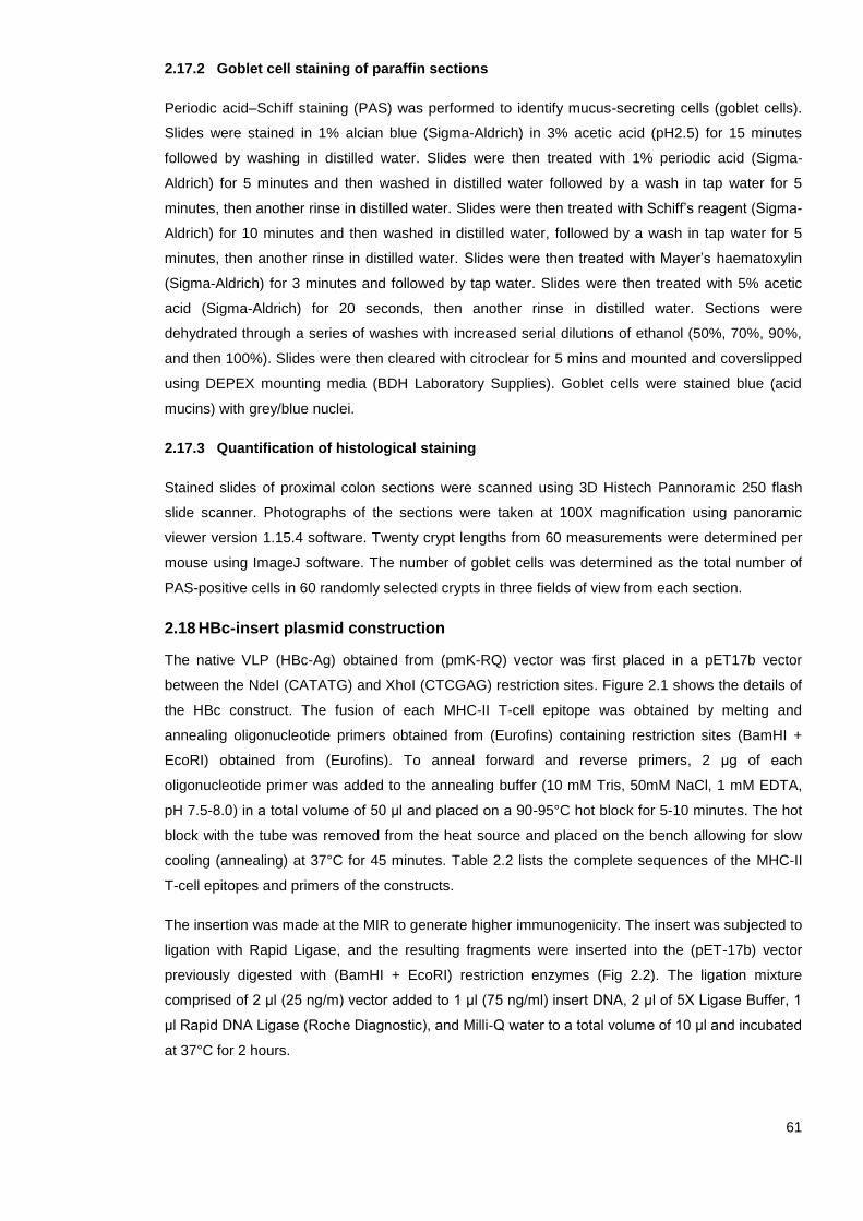
61
2.17.2 Goblet cell staining of paraffin sections
Periodic acid–Schiff staining (PAS) was performed to identify mucus-secreting cells (goblet cells).
Slides were stained in 1% alcian blue (Sigma-Aldrich) in 3% acetic acid (pH2.5) for 15 minutes
followed by washing in distilled water. Slides were then treated with 1% periodic acid (Sigma-
Aldrich) for 5 minutes and then washed in distilled water followed by a wash in tap water for 5
minutes, then another rinse in distilled water. Slides were then treated with Schiff’s reagent (Sigma-
Aldrich) for 10 minutes and then washed in distilled water, followed by a wash in tap water for 5
minutes, then another rinse in distilled water. Slides were then treated with Mayer’s haematoxylin
(Sigma-Aldrich) for 3 minutes and followed by tap water. Slides were then treated with 5% acetic
acid (Sigma-Aldrich) for 20 seconds, then another rinse in distilled water. Sections were
dehydrated through a series of washes with increased serial dilutions of ethanol (50%, 70%, 90%,
and then 100%). Slides were then cleared with citroclear for 5 mins and mounted and coverslipped
using DEPEX mounting media (BDH Laboratory Supplies). Goblet cells were stained blue (acid
mucins) with grey/blue nuclei.
2.17.3 Quantification of histological staining
Stained slides of proximal colon sections were scanned using 3D Histech Pannoramic 250 flash
slide scanner. Photographs of the sections were taken at 100X magnification using panoramic
viewer version 1.15.4 software. Twenty crypt lengths from 60 measurements were determined per
mouse using ImageJ software. The number of goblet cells was determined as the total number of
PAS-positive cells in 60 randomly selected crypts in three fields of view from each section.
2.18 HBc-insert plasmid construction
The native VLP (HBc-Ag) obtained from (pmK-RQ) vector was first placed in a pET17b vector
between the NdeI (CATATG) and XhoI (CTCGAG) restriction sites. Figure 2.1 shows the details of
the HBc construct. The fusion of each MHC-II T-cell epitope was obtained by melting and
annealing oligonucleotide primers obtained from (Eurofins) containing restriction sites (BamHI +
EcoRI) obtained from (Eurofins). To anneal forward and reverse primers, 2 μg of each
oligonucleotide primer was added to the annealing buffer (10 mM Tris, 50mM NaCl, 1 mM EDTA,
pH 7.5-8.0) in a total volume of 50 μl and placed on a 90-95°C hot block for 5-10 minutes. The hot
block with the tube was removed from the heat source and placed on the bench allowing for slow
cooling (annealing) at 37°C for 45 minutes. Table 2.2 lists the complete sequences of the MHC-II
T-cell epitopes and primers of the constructs.
The insertion was made at the MIR to generate higher immunogenicity. The insert was subjected to
ligation with Rapid Ligase, and the resulting fragments were inserted into the (pET-17b) vector
previously digested with (BamHI + EcoRI) restriction enzymes (Fig 2.2). The ligation mixture
comprised of 2 μl (25 ng/m) vector added to 1 μl (75 ng/ml) insert DNA, 2 μl of 5X Ligase Buffer, 1
μl Rapid DNA Ligase (Roche Diagnostic), and Milli-Q water to a total volume of 10 μl and incubated
at 37°C for 2 hours.

62
2.19 HBc-HP1-512 and HBc-mosaic plasmid construction
The HBc-HP1-512 and HBc-mosaic DNA sequence were codon optimised in E. coli and synthesised
by GeneArt. The synthesised DNA obtained from the (pmK-RQ) vector was placed in a pET17b
vector between the NdeI (CATATG) and XhoI (CTCGAG) restriction sites. The synthesised DNA
obtained in the (pmK-RQ) vector was first purified using the QIAprep Spin Miniprep extraction kit
following the original protocol provided by (Qiagen - USA). After the purification, the vector was
then linearized by restriction digests (PstI and BgIII). The digested DNA was confirmed by mixing
the DNA with a loading buffer (Bioline- Germany) and then submitted to 0.5% (w/v) agarose gel
electrophoresis (Bioline- Germany) with Tris- Acetate- EDTA (TAE) with ethidium bromide (Sigma-
Aldrich). The DNA was then extracted from the gel and purified using the QIAquick PCR
Purification Kit following the original protocol provided by (Qiagen-USA). The concentration of the
DNA was measured using a Nano-Drop spectrophotometer. The DNA was then subjected to
ligation with Rapid Ligase, and the resulting fragments were inserted into the (pET-17b) vector
previously digested with (BamHI + EcoRI) restriction enzymes as mentioned before. Table 2.3 lists
the complete sequences of the HBc-HP1-512 and HBc-mosaic constructs.
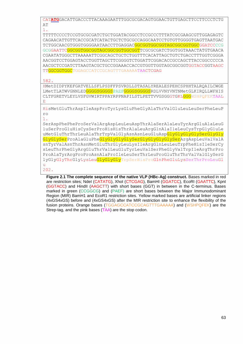
63
CATATGGACATTGACCCTTACAAAGAATTTGGCGCGACAGTGGAACTGTTGAGCTTCCTTCCCTCTG
AT
1.
TTTTTCCCCTCCGTGCGCGATCTGCTGGATACGGCCTCCGCCCTTTATCGCGAAGCGTTGGAGAGTC
CAGAACATTGTTCACCGCATCATACTGCTCTGCGCCAGGCAATCCTGTGTTGGGGTGAGTTAATGAC
TCTGGCAACGTGGGTGGGGAATAACCTTGAGGACGGCGGTGGCGGTAGCGGCGGTGGCGGATCCCCG
GCGGAATTCGGCGGTGGCGGTAGCGGCGGTGGCGGTTCGCGCGATCTGGTGGTAAACTATGTGAACA
CGAATATGGGCTTAAAAATTCGGCAGCTGCTCTGGTTTCACATTAGCTGTCTGACCTTTGGTCGGGA
AACGGTCCTGGAGTACCTGGTTAGCTTCGGGGTCTGGATTCGGACACCGCCAGCTTACCGGCCCCCA
AACGCTCCGATCTTAAGTACGCTGCCGGAAACCACCGTGGTTGGTAGCGGCGGTGGTACCGGTAAGC
TTGGCGGTGGCTGGAGCCATCCGCAGTTTGAAAAATAACTCGAG
582.
HMetDIDPYKEFGATVELLSFLPSDFFPSVRDLLDTASALYREALESPEHCSPHHTALRQAILCWGE
LMetTLATWVGNNLEDGGGGSGGGGSPAEFGGGGSGGGGSRDLVVNYVNTNMetGLKIRQLLWFHIS
CLTFGRETVLEYLVSFGVWIRTPPAYRPPNAPILSTLPETTVVGSGGGTGKLGGGWSHPQFEKTAAL
E
HisMetGluThrAspIleAspProTyrLysGluPheGlyAlaThrValGluLeuLeuSerPheLeuP
ro
1.
SerAspPhePheProSerValArgAspLeuLeuAspThrAlaSerAlaLeuTyrArgGluAlaLeuG
luSerProGluHisCysSerProHisHisThrAlaLeuArgGlnAlaIleLeuCysTrpGlyGluLe
uMetGluThrThrLeuAlaThrTrpValGlyAsnAsnLeuGluAspGlyGlyGlyGlySerGlyGly
GlyGlySerProAlaGluPheGlyGlyGlyGlySerGlyGlyGlyGlySerArgAspLeuValValA
snTyrValAsnThrAsnMetGluThrGlyLeuLysIleArgGlnLeuLeuTrpPheHisIleSerCy
sLeuThrPheGlyArgGluThrValLeuGluTyrLeuValSerPheGlyValTrpIleArgThrPro
ProAlaTyrArgProProAsnAlaProIleLeuSerThrLeuProGluThrThrValValGlySerG
lyGlyGlyThrGlyLysLeuGlyGlyGlyTrpSerHisProGlnPheGluLysSerThrProLeuGl
u
202.
Figure 2.1 The complete sequence of the native VLP (HBc-Ag) construct. Bases marked in red are restriction sites; NdeI (CATATG), XhoI (CTCGAG), BamHI (GGATCC), EcoRI (GAATTC), KpnI (GGTACC) and HindII (AAGCTT) with short bases (GGT) in between in the C-terminus. Bases marked in green (CCGGCG) and (PAEF) are short bases between the Major Immunodominant Region (MIR) BamH1 and EcoR1 restriction sites. Yellow marked bases are artificial linker regions (4xGS4xGS) before and (4xGS4xGS) after the MIR restriction site to enhance the flexibility of the fusion proteins. Orange bases (TGGAGCCATCCGCAGTTTGAAAAA) and (WSHPQFEK) are the Strep-tag, and the pink bases (TAA) are the stop codon.

64
Figure 2.2: DNA construction for HBc-Ag inserted in N -terminally T7-tagged pET-17b vector from Novagen. HBc-Ag indicates the full sequence of HBc-Ag. Twenty-two amino acids as an MHC T-cell epitope for Trichuris were inserted in the MIR of the HBc-Ag. The cloning site for the MHC-II T-cell epitopes is BamH1 and EcoR1. Ampicillin: Ampicillin resistance gene. Ori: vector origin of replication.

65
Table 2.2 List of MHC-II T-cell epitope sequences and primers used to amplify the target.
Protein (Uniprot ID
number)
Epitope name amino acid sequence
Primers sequence
Chitin-binding domain
containing protein (A0A0N5E6C6)
CBD36-52
RPRLLKIKWSPTAASTA
Forward BamHI primer: GATCCtttccgcgcccgcgcctgctgaaaattaaatggagcccgaccgcggcgagcaccgcgattcatG
Reverse EcoRI primer: AATTCatgaatcgcggtgctcgccgcggtcgggctccatttaattttcagcaggcgcgggcgcggaaaG
Chitin-binding domain
containing protein (A0A0N5E6C6)
CBD1243-1259
PAGVVYQCTMPRYTLCV
Forward BamHI primer: GATCCaacctgccggcgggcgtggtgtatcagtgcaccatgccgcgctataccctgtgcG
Reverse EcoRI primer: AATTCgcacagggtatagcgcggcatggtgcactgatacaccacgcccgccggcaggttG
Chitin-binding domain
containing protein (A0A0N5DK22)
CBD241-257
GRTTPVTSAPTTVTTER
Forward BamHI primer: GATCCtgcaccggccgcaccaccccggtgaccagcgcgccgaccaccgtgaccaccgaacgcagcccgG
Reverse EcoRI primer: AATTCcgggctgcgttcggtggtcacggtggtcggcgcgctggtcaccggggtggtgcggccggtgcaG
Pfam-B 9093 domain
containing protein (A0A0N5E6W6)
Pfam 256-273
NFGEYWFTKPVGSVSYKV
Forward BamHI primer: GATCCgaatttaactttggcgaatattggtttaccaaaccggtgggcagcgtgagctataaagtggcgt
gcG
Reverse EcoRI primer: AATTCgcacgccactttatagctcacgctgcccaccggtttggtaaaccaatattcgccaaagttaaat
tcG
Chymotrypsin-like serine proteases (A0A0N5DEK1)
CLSP433-450
SKLRVYAGSPRFSRIR
Forward BamHI primer: GATCCgcgaaaagcaaactgcgcgtgtatgcgggcagcccgcgctttagccgcattcgccgccgcG
Reverse EcoRI primer: AATTCgcggcggcgaatgcggctaaagcgcgggctgcccgcatacacgcgcagtttgcttttcgcG
Chymotrypsin-like serine proteases (A0A0N5E3X0)
CLSP222-237
MEKKRKYLGSPLVCF
Forward BamHI primer: GATCCaaaaaaatggaaaaaaaacgcaaatatctgggcagcccgctggtgtgctttgtgcgcG
Reverse EcoRI primer: AATTCgcgcacaaagcacaccagcgggctgcccagatatttgcgttttttttccatttttttG
Chymotrypsin-like serine proteases (A0A0N5DUC1)
CLSP143-158
AILQLAKPVPFSNTVR
Forward BamHI primer: GATCCggcgtggcgattctgcagctggcgaaaccggtgccgtttagcaacaccgtgcgcccgattG
Reverse EcoRI primer: AATTCaatcgggcgcacggtgttgctaaacggcaccggtttcgccagctgcagaatcgccacgccG
66
Chymotrypsin-like serine proteases (A0A0N5DUC1)
CLSP424-443
CTGSLYASARANFTRTVLTS
BamHI Fw: GATCCgtgatttgcaccggcagcctgtatgcgagcgcgcgcgcgaactttacccgcaccgtgctgacca
gcgcgagcG
Reverse EcoRI primer: AATTCgctcgcgctggtcagcacggtgcgggtaaagttcgcgcgcgcgctcgcatacaggctgccggtg
caaatcacG
Chymotrypsin-like serine proteases (A0A0N5DUC1)
CLSP398-416
SDHQEGYPVSPSVHIVSLA
Forward BamHI primer: GATCCcatgtgagcgatcatcaggaaggctatccggtgagcccgagcgtgcatattgtgagcctggcgg
aaggcG
Reverse EcoRI primer: AATTCgccttccgccaggctcacaatatgcacgctcgggctcaccggatagccttcctgatgatcgctc
acatgG
Hypothetical protein
(A0A0N5DRU5) H112-128
GARKFLPIYQKAVAEEK
Forward BamHI primer: GATCCaaaggcggcgcgcgcaaatttctgccgatttatcagaaagcggtggcggaagaaaaagaagtgG
Reverse EcoRI primer: AATTCcacttctttttcttccgccaccgctttctgataaatcggcagaaatttgcgcgcgccgcctttG
The restriction sites and the two extra amino acids bases are coloured in green and red, respectively.

67
Table 2.3 The complete sequences of the selected antigen constructs obtained from GeneArt.
Synthetic Protein
Protein amino acid Sequence
HBc-Hypothetical
protein
(HBc-HP1-512)
(A0A0N5DRU5)
Molecular weight:
55.29kDa
HMDIDPYKEFGATVELLSFLPSDFFPSVRDLLDTASALYREALESPEHCSPHHTALRQAILCWGELMTLATWVGNNLEDGGGGSGGGGSGGGGGGELGLVAAGHKDECDCI
KGVGPGYEKSHLDYAGKFKGGARKFLPIYEKAVAEEKEVLIKGGPGACDYEPCECRHKRGVEDCGCLKGVGPGYEKSYLDYAGKFKGGARKFLPIYQKAVAEEKEVLIKGG
PGACDYEPCECRHKRGLKDCGCCKGVGPGYEKSYLDYDGKFKGGARKFLPIYEKAVAEEKEVLVKGGPGACDWEPCECRHKRGLKDCGCLKGVGPGYEKSYLDYAGKFKGG
ARKFLPIYEKAVAEEKEVLVKGGPGACDWEPCECRHKRGLKDCGCLKGVGPGYEKSYLDYAGKFKGGARKFLPIYEKAVAEEKEVLIKGGPGARDCGCKHMGLKIRQLLWF
HISCLTFGRETVLEYLVSFGVWIRTPPAYRPPNAPILSTLPETTVVGSGGGTGKLGGGWSHPQFEKLE
HisMetAspIleAspProTyrLysGluPheGlyAlaThrValGluLeuLeuSerPheLeuProSerAspPhePheProSerValArgAspLeuLeuAspThrAlaSerAla
1.
LeuTyrArgGluAlaLeuGluSerProGluHisCysSerProHisHisThrAlaLeuArgGlnAlaIleLeuCysTrpGlyGluLeuMetThrLeuAlaThrTrpValGly
AsnAsnLeuGluAspGlyGlyGlyGlySerGlyGlyGlyGlySerGlyGlyGlyGlyGlyGlyGluLeuGlyLeuValAlaAlaGlyHisLysAspGluCysAspCysIle
LysGlyValGlyProGlyTyrGluLysSerHisLeuAspTyrAlaGlyLysPheLysGlyGlyAlaArgLysPheLeuProIleTyrGluLysAlaValAlaGluGluLys
GluValLeuIleLysGlyGlyProGlyAlaCysAspTyrGluProCysGluCysArgHisLysArgGlyValGluAspCysGlyCysLeuLysGlyValGlyProGlyTyr
GluLysSerTyrLeuAspTyrAlaGlyLysPheLysGlyGlyAlaArgLysPheLeuProIleTyrGlnLysAlaValAlaGluGluLysGluValLeuIleLysGlyGly
ProGlyAlaCysAspTyrGluProCysGluCysArgHisLysArgGlyLeuLysAspCysGlyCysCysLysGlyValGlyProGlyTyrGluLysSerTyrLeuAspTyr
AspGlyLysPheLysGlyGlyAlaArgLysPheLeuProIleTyrGluLysAlaValAlaGluGluLysGluValLeuValLysGlyGlyProGlyAlaCysAspTrpGlu
ProCysGluCysArgHisLysArgGlyLeuLysAspCysGlyCysLeuLysGlyValGlyProGlyTyrGluLysSerTyrLeuAspTyrAlaGlyLysPheLysGlyGly
AlaArgLysPheLeuProIleTyrGluLysAlaValAlaGluGluLysGluValLeuValLysGlyGlyProGlyAlaCysAspTrpGluProCysGluCysArgHisLys
ArgGlyLeuLysAspCysGlyCysLeuLysGlyValGlyProGlyTyrGluLysSerTyrLeuAspTyrAlaGlyLysPheLysGlyGlyAlaArgLysPheLeuProIle
TyrGluLysAlaValAlaGluGluLysGluValLeuIleLysGlyGlyProGlyAlaArgAspCysGlyCysLysHisMetGlyLeuLysIleArgGlnLeuLeuTrpPhe
HisIleSerCysLeuThrPheGlyArgGluThrValLeuGluTyrLeuValSerPheGlyValTrpIleArgThrProProAlaTyrArgProProAsnAlaProIleLeu
SerThrLeuProGluThrThrValValGlySerGlyGlyGlyThrGlyLysLeuGlyGlyGlyTrpSerHisProGlnPheGluLysLeuGlu
512.
HBc-Hypothetical
protein (A0A0N5DRU5) + CLSP398-416 +
HMDIDPYKEFGATVELLSFLPSDFFPSVRDLLDTASALYREALESPEHCSPHHTALRQAILCWGELMTLATWVGNNLEDGGGGSGGGGSGGGGGGELGLVAAGHKDECDCI
KGVGPGYEKSHLDYAGKFKGGARKFLPIYEKAVAEEKEVLIKGGPGACDYEPCECRHKRGVEDCGCLKGVGPGYEKSYLDYAGKFKGGARKFLPIYQKAVAEEKEVLIKGG
PGACDYEPCECRHKRGLKDCGCCKGVGPGYEKSYLDYDGKFKGGARKFLPIYEKAVAEEKEVLVKGGPGACDWEPCECRHKRGLKDCGCLKGVGPGYEKSYLDYAGKFKGG
ARKFLPIYEKAVAEEKEVLVKGGPGACDWEPCECRHKRGLKDCGCLKGVGPGYEKSYLDYAGKFKGGARKFLPIYEKAVAEEKEVLIKGGPGARDCGCKHGGGGSGGGGSG
GGGGGELAKSKLRVYAGSPRFSRIRGGGGSGGGGSGGGGGGELIVRKTFRVGPFPRPRLLKIKWSPTAASTAIHPRWTMGRENAKVAILLVLIALAAFGEAGYSCEGKSNG
DYGDPAEPCSSRYFSCVNGKAIGRQCPRGLYFHPGLDKCDWPHGITACSNVQEKETFRCPAENGDYPDPEHQCSAKFWRCTNGKAEPYAMGLKIRQLLWFHISCLTFGRET
VLEYLVSFGVWIRTPPAYRPPNAPILSTLPETTVVGSGGGTGKLGGGWSHPQFEKLE

68
Chitin binding domain
A0A0N5DK22
(HBc-mosaic)
Molecular weight: 77.70
kDa
HisMetAspIleAspProTyrLysGluPheGlyAlaThrValGluLeuLeuSerPheLeuProSerAspPhePheProSerValArg
1.
AspLeuLeuAspThrAlaSerAlaLeuTyrArgGluAlaLeuGluSerProGluHisCysSerProHisHisThrAlaLeuArgGlnAlaIleLeuCysTrpGlyGluLeu
MetThrLeuAlaThrTrpValGlyAsnAsnLeuGluAspGlyGlyGlyGlySerGlyGlyGlyGlySerGlyGlyGlyGlyGlyGlyGluLeuGlyLeuValAlaAlaGly
HisLysAspGluCysAspCysIleLysGlyValGlyProGlyTyrGluLysSerHisLeuAspTyrAlaGlyLysPheLysGlyGlyAlaArgLysPheLeuProIleTyr
GluLysAlaValAlaGluGluLysGluValLeuIleLysGlyGlyProGlyAlaCysAspTyrGluProCysGluCysArgHisLysArgGlyValGluAspCysGlyCys
LeuLysGlyValGlyProGlyTyrGluLysSerTyrLeuAspTyrAlaGlyLysPheLysGlyGlyAlaArgLysPheLeuProIleTyrGlnLysAlaValAlaGluGlu
LysGluValLeuIleLysGlyGlyProGlyAlaCysAspTyrGluProCysGluCysArgHisLysArgGlyLeuLysAspCysGlyCysCysLysGlyValGlyProGly
TyrGluLysSerTyrLeuAspTyrAspGlyLysPheLysGlyGlyAlaArgLysPheLeuProIleTyrGluLysAlaValAlaGluGluLysGluValLeuValLysGly
GlyProGlyAlaCysAspTrpGluProCysGluCysArgHisLysArgGlyLeuLysAspCysGlyCysLeuLysGlyValGlyProGlyTyrGluLysSerTyrLeuAsp
TyrAlaGlyLysPheLysGlyGlyAlaArgLysPheLeuProIleTyrGluLysAlaValAlaGluGluLysGluValLeuValLysGlyGlyProGlyAlaCysAspTrp
GluProCysGluCysArgHisLysArgGlyLeuLysAspCysGlyCysLeuLysGlyValGlyProGlyTyrGluLysSerTyrLeuAspTyrAlaGlyLysPheLysGly
GlyAlaArgLysPheLeuProIleTyrGluLysAlaValAlaGluGluLysGluValLeuIleLysGlyGlyProGlyAlaArgAspCysGlyCysLysHisGlyGlyGly
GlySerGlyGlyGlyGlySerGlyGlyGlyGlyGlyGlyGluLeuAlaLysSerLysLeuArgValTyrAlaGlySerProArgPheSerArgIleArgGlyGlyGlyGly
SerGlyGlyGlyGlySerGlyGlyGlyGlyGlyGlyGluLeuIleValArgLysThrPheArgValGlyProPheProArgProArgLeuLeuLysIleLysTrpSerPro
ThrAlaAlaSerThrAlaIleHisProArgTrpThrMetGlyArgGluAsnAlaLysValAlaIleLeuLeuValLeuIleAlaLeuAlaAlaPheGlyGluAlaGlyTyr
SerCysGluGlyLysSerAsnGlyAspTyrGlyAspProAlaGluProCysSerSerArgTyrPheSerCysValAsnGlyLysAlaIleGlyArgGlnCysProArgGly
LeuTyrPheHisProGlyLeuAspLysCysAspTrpProHisGlyIleThrAlaCysSerAsnValGlnGluLysGluThrPheArgCysProAlaGluAsnGlyAspTyr
ProAspProGluHisGlnCysSerAlaLysPheTrpArgCysThrAsnGlyLysAlaGluProTyrAlaMetGlyLeuLysIleArgGlnLeuLeuTrpPheHisIleSer
CysLeuThrPheGlyArgGluThrValLeuGluTyrLeuValSerPheGlyValTrpIleArgThrProProAlaTyrArgProProAsnAlaProIleLeuSerThrLeu
ProGluThrThrValValGlySerGlyGlyGlyThrGlyLysLeuGlyGlyGlyTrpSerHisProGlnPheGluLysLeuGlu
723.
Bases marked in Blue are hypothetical protein, purple residues are the CLSP398-416 epitope, and the green residues are chitin-binding domain-containing
protein (A0A0N5DK22). Yellow marked residues are artificial linker regions (4GS4GS5G) to enhance the flexibility of the fusion proteins. The orange bases
are the StrepTag (WSHP).

69
2.20 Transformation
The recombinant plasmid containing the epitope insert was first transformed into XL-I Blue
supercompetent cells and Lemo21 (DE3) competent cells for small-scale expression obtained from
(New England Biolabs) by heat-shock transformation. The recombinant plasmid containing the
synthesised protein insert was transformed into Ultracompetent cells (XLI0-Gold) obtained from
(Agilent Technologies) by heat-shock transformation. For this, 1 µl of the DNA mixture was added
into 50 µl of the heat shock competent cells in a falcon tube and placed on ice for 30 mins. The
tube was then transferred into a 42°C water bath for 45 secs and then transferred to the ice for 2
mins. 250 μl of the Luria-Bertani (LB) Broth were then added to the transformation mixture and
incubated at 37° C for 1 hour. 250 μl of the transformation mixture was then added to the LB plate
containing the appropriate antibiotic (Ampicillin or Kanamycin) and incubated at 37°C for overnight.
2.21 Plasmid purification: QIAprep spin miniprep kit
The recombinant plasmid DNA was purified using the original protocol that is provided by (Qiagen-
USA). Briefly, the bacterial cell pellet was resuspended in 250 μl Buffer P1 and transferred to
microcentrifuge tubes. After that, 250 μl Buffer P2 was added to the tubes and mixed thoroughly by
inverting the tube 4–6 times, followed by adding 350 μl Buffer N3 and mixing immediately. The
tubes were then centrifuged in a table-top microcentrifuge for 10 min at 8,000 x g. To wash the
QIAprep 2.0 spin column, 0.75 ml Buffer PE was added, followed by centrifugation. Finally, to elute
the DNA, the QIAprep 2.0 column was placed in a clean 1.5 ml microcentrifuge tube, and 55 μl of
Milli-Q water was added to the centre of each column, let stand for 3 min and centrifuged for 1 min.
The concentration of the DNA was measured using a NanoDrop spectrophotometer, and the
nucleotide sequence was verified by DNA sequencing (GATC- Biotech, Germany).
2.22 DNA sequencing
The correct insert nucleotide sequence was verified by DNA sequencing (GATC-Biotech,
Germany) using the Sanger Supremerun service. DNA was eluted in Milli-Q water and chipped in a
hydrated form. T7 Promoter primer was provided by the (GATC-Biotech, Germany). The DNA
sequence results were provided in FASTA format and were analysed using SnapGene.
DNA concentrations were assessed using a NanoDrop spectrophotometer. Briefly, 1 µl of Milli-Q
water was first used to calibrate the spectrophotometer followed by sample buffer to clean it before
loading 1 µl of the sample. To calculate the DNA concentration, 1 µl Milli-Q water was used as a
blank, and absorption reading was conducted at 260 nm and 280 nm and the ratio between two.
2.23 Small-scale VLP expression
For small expression of the confirmed inserts, a single colony was picked from the overnight LB
agar plate and inoculated into 50 mL LB broth medium supplemented with 50 µL of 100 μg/mL of
the appropriate antibiotic (Ampicillin or Kanamycin) and incubated at 37°C for overnight in a shaker
incubator to obtain a saturated culture. Next day, 400 µL of the incubated cells were added to 4 ml
LB broth containing 4 µL of 100 μg/mL of the appropriate antibiotic (Ampicillin or Kanamycin) and
incubated for 3-4 hours until the optical density OD600 reached 0.6-0.8. The optical density was
measured using spectrophotometer at 600nm wavelength. Once the OD600 reached 0.6-0.8,

70
flasks were chilled at 4°C for 30 mins then induced by Isopropyl-β-D-1-thiogalactopyranoside
(IPTG) at a final concentration of 0.4 mM and incubated for 3-4 hours to induce recombinant
protein expression. Tubes were then centrifuged at 16,000 x g for 10 minutes at 4°C. Cells were
disrupted by ultra-sonication on ice using Banddelin (Sonoplus) 35% amplitude (Am) for 5 seconds
on and 10 seconds off for 5-7 mins, and the supernatant containing soluble recombinant protein
was harvested by centrifugation at 16,000 x g for 10 minutes at 4°C.
2.24 Large-scale VLP expression
For the overexpression of soluble proteins, the native VLP (HBc-Ag) and the VLPs + T-cell
epitopes (HBc-CBD36-52, HBc-CBD1243-1259, HBc-CBD241-257, HBc-CLSP433-450, HBc-CLSP222-237,
HBc-CLSP143-158, HBc-CLSP424-443, HBc-CLSP398-416 and HBc-H112-128) were transformed into
ClearColi-BL21 (DE3) electrocompetent cells obtained from (Lucigen) and incubated overnight at
37°C. Next day, 40 ml of the transformed cells at a starting optical density (0.1 OD600) were
inoculated into 2 L (2x LB) liquid media supplemented with 100 μg/mL ampicillin for 3-4 hours.
Once the OD600 reached 0.6-0.8, flasks were chilled at 4°C for 30 mins then induced by IPTG at a
final concentration of 0.4 mM. The culture was then grown by continuous shaking for 12-16 hours
at 16°C. The cells were then harvested by centrifugation at 6,000 x g for 20 mins at 4°C using
Sorvall RC-5C plus with the Sorvall SLC -300 rotor. Cell pellets were then stored rapidly at -80°C
after flash freezing in liquid nitrogen until needed.
2.25 Preparation of cell lysate
The cells from the large-scale expression were first thawed and then resuspended at a ratio (4
ml/gram cells) in Strep-Tag washing buffer (100 mM Tris-HCl, 150 mM NaCl, 1 mM EDTA, pH 8). 1
tablet of cOmplete TM EDTA- Free Protease Inhibitor Cocktail (Sigma-Aldrich) was added per 50
mls of the resuspended cells along with 5 µg/ml DNase I (Sigma-Aldrich). The cell suspension was
then disrupted by ultra-sonication on ice using Banddelinuw 3200 (Sonoplus) amplitude (Am) in
35% for 5 sec on and 10 sec off for 5-8 minutes. The supernatant containing soluble recombinant
protein was harvested following centrifugation at 18,900 x g for 40 mins at 4°C in 50 ml Nalgene
PC tubes using Sorvall RC Plus with the Fiber-Lite F21 8x50y rotor. Finally, the soluble
supernatant was filtered through 0.22 µm pore size filters.
2.26 Stage 1 purification using StrepTrap™ affinity chromatography
The soluble native VLP (HBc-Ag) and the soluble VLP expressing Trichuris MHC-II T-cell epitopes
were purified by 5 ml affinity column chromatography using StrepTrap™ column prepacked with
StrepTactin Sepharose for purifying Strep(II)-tag proteins by following the manufacturer’s protocol
provided by GE Healthcare. The binding/wash buffer used was the (100 mM Tris-HCl, 150 mM
NaCl, 1 mM EDTA, pH 8) and the elution buffer was the lysis buffer supplemented with 2.5 mM
desthiobiotin (GE Healthcare). Briefly, the column was first equilibrated with 5 CV (column volume)
washing buffer using a hydrostatic pump at a rate of 1ml/min. To regenerate the column, 10 CV 0.5
mM NaOH was used at 2 ml/min followed by 10 CV H2O, 5 CV binding buffer, loading the samples
to the column at a rate of 1 ml/min followed by a 10 CV binding buffer. 5 CV elution buffer was then

71
applied at 1 ml/min. The purification fractions were collected and concentrated to 0.5 ml using
centrifugal Vivaspin concentrator (20 ml MWCO 10,000) at 6,000 x g at 4°C.
2.27 Stage 2 purification using size exclusion chromatography (SEC)
The native VLP (HBc-Ag) and the VLP recombinant proteins were further purified by SEC
separation (Superose 6, 10/300 GL) using the AKTA FPLC device (GE Healthcare). The column
was first equilibrated with 1.5 CV PBS buffer at pH 7.4 with a flow rate (0.5 ml/min) by following the
original protocol provided by (GE, Healthcare, US). Purified VLPs were first concentrated to 0.5 ml
using Vivaspin concentrator (MWCO 10.000 kDa), then loaded onto a 5 ml loop. Purification was
performed at a flow rate of 0.2 ml/min, and 0.5 ml fractions were collected into a 96 well block. The
purified proteins were visualised using SDS-PAGE analysis. The data for chromatograms in VLPs
purification were obtained from UNICORN software associated with ÄKTA purifier, and the graphs
were plotted using Excel.
The protein concentrations were assessed using a NanoDrop Spectrophotometer. Briefly, 1 µl of
Milli-Q water was first used to calibrate the spectrophotometer followed by sample buffer to clean it
before loading 1 µl of the sample and the protein absorbance measured at 280 nm. The molecular
mass, the number of amino acids, and the extinction coefficient estimated from the protein
sequence using the ExPASy ProtParam tool were used to measure the concentration of the
purified VLP recombinant proteins (Gasteiger et al., 2005).
2.29 10% Sodium Dodecyl Sulfate Polyacrylamide Gel Electrophoresis (SDS-PAGE)
The SDS-PAGE analysis of the total protein fraction before purification and the pellet after first and
the second purification was carried out to confirm the presence of the recombinant protein. The
samples were mixed with 4X Laemmli Loading Buffer (Bioline) containing 5% of 2-mercaptoethanol
(Sigma-Aldrich) in a total volume of 20 µl. Before loading, the samples were boiled at 90°C on a hot
plate for 3 mins, and 15 µl of the sample was loaded on to NuPAGE 10% Bis-Tris Pre-Cast gels
(Life Technologies) and electrophoresed for 60 min at 160 V. 8 µl of the Bio-Rad using 1X MES
running buffer containing 50 mM MES, 50 mM Tris-base, 0.1% SDS, I mM EDTA, pH 7.3 and
obtained from NuPAGE gels, Invitrogen. The precision plus protein unstained protein standard was
used as a reference ladder (Bio-Rad). The precision plus protein stained protein standard (Bio-
Rad) was used as a reference ladder if the gel was used for western blot. The gel surface was
washed with Milli-Q water after separation was complete and then stained with instant blue protein
stain (Expedeon) if not used for Western Blot.
2.30 Designed VLPs construction and solubility
The translation of the DNA vaccine (nucleotide) sequence into a protein sequence (amino acids)
was conducted by http://web.expasy.org/translate/. The Vendruscolo Lab, CamSol method
Software, Centre for Misfolding Diseases (University of Cambridge) was then used to predict the
solubility of the constructs based on the protein sequence (One score per residue) using
http://www-mvsoftware.ch.cam.ac.uk/index.php/camsolintrinsic. Proteins with scores ≥1 are
considered highly soluble, whereas scores ≤-1 poorly soluble ones (Sormanni et al., 2017;

72
Sormanni et al., 2015). Finally, the synthesised DNA was ordered from
https://www.thermofisher.com/uk/en/home.html.
2.31 ELISA-based endotoxin detection assay
The level of endotoxin in all the purified VLPs was measured with an ELISA-based endotoxin
detection assay (Hyglos) following the manufacturer protocol provided by from Biotech. Briefly,
purified VLPs were diluted in 10 fold steps and endotoxin standard (E. coli 055: B5) was diluted to
(500 EU/ml, 50 EU/ml, 5 EU/ml, 0.5 EU/ml, and 0.0 5EU/ml) using endotoxin-free water provided
with the kit. 100 μl of the VLPs, standard dilutions and endotoxin-free water as negative control
were added in duplicate into the microtiter wells pre-coated with LPS-binding phage recombinant
protein. 20 μl of binding buffer was then added to the wells and incubated for 90 minutes at 37°C in
the dark followed by washing 3 times with washing buffer. Finally, 150 μl/well of assay reagent
substrate was added to the plate. Plate fluorescence signals were read at time 0 mins and after
incubation for 90 mins using microplate fluorescence reader (FLx800™) supplied by BioTek
Instruments. To calculate the endotoxin levels in the VLPs, the values at time 0 min were
subtracted from 90 mins values, and the endotoxin standards were used to draw a 4 parameter
logistic, nonlinear curve to back-calculate the endotoxin levels.
2.32 Circular Dichroism spectropolarimeter (CD)
The secondary structure of VLP recombinant proteins was examined using Circular Dichroism
Spectropolarimeter (JASCO, J-810, Tokyo, Japan) using Spectra Manager software (Greenfield,
2006). The sample was first equilibrated with nitrogen gas for 10 minutes, and the Peltier
temperature controller was brought to 20°C. After that, 180 µl of 0.5 mg/ml protein was loaded into
a 0.5 mm UV- quartz cell cuvette then placed into the spectropolarimeter holder. All circular
dichroism (CD) experiments were performed in a 0.2 mm path-length cuvette in the 260nm-190nm
wavelength range, 0.5-sec response, 1 nm bandwidth, 10 accumulations (the number of scans)
and 0.5 cm cell length. To wash cuvettes between samples, 5% Decon was used first, followed by
1 M Nitric acid and finally with Milli-Q H2O. PBS buffer was used as blank, and the results were
extracted from the protein containing spectra. DichroWeb was then used to interpret the results
(Whitmore and Wallace, 2009).
2.33 Transmission Electron microscopy (TEM)
The presence and assembly of the purified VLP proteins were verified by transmission electron
microscopy with negative staining. First, the proteins were dialysed from PBS to 1X TRIS buffer
using Slide –A-Lyser MINI Dialysis Devices, 10K MWCO, 0.5 ml, 25/PK (Thermo Scientific). After
that, 10 μl of the VLPs at 0.5 mg/ml was loaded onto a carbon-coated grid (JEOL, Tokyo, Japan),
and left to stand for 5 min. Grids were then stained with for 3% uranyl acetate (UA) for 1 min, and
then the excess drained. The grid was then examined directly under FEI Tecnai 12 Biotwin
Transmission Electron Microscope operating at 120 kV at magnifications ranging from 6000× to
100,000×.

73
2.34 Labelling VLPs with Fluorescein dye
To assess virus-like particles uptake by BMDCs and BMDMs, VLPs were labelled with Fluorescein
dye following the manufacturer's protocol of Lightning-Link® Fluorescein Conjugation Kit obtained
from (Expedeon- Innova Biosciences). Briefly, 10 µl of VLPs (HBc-Ag, HBc-H112-128, HBc-CBD241-
257, and HBc-CLSP398-416) at 1 mg/ml concentration were added to 1 µl LL-modifier reagent and
mixed gently to give a final concentration of 0.9 mg/ml. The mixture was then added to a glass vial
of Lightning-Link® mix and incubated for 3 hours @ RT (20-25⁰C) in the dark. 1 µl LL-quencher FD
reagent was then added to quench the reaction. Free fluorescein dye was separated from the VLPs
proteins by dialysis into PBS. Fluorescence intensity was examined using LSRII flow cytometer
(Becton Dickinson Biosciences, Oxfordshire) and was analysed using FlowJo X software (Tree
Star, Inc. COMPNAY, ADDRESS). The fluorescein-conjugated VLPs were then stored at -20 °C
freezer to be used for the in vitro assays.
2.35 Fluorescein-conjugated VLP internalisation and localisation in APCs using an Amnis ImageStreamX cytometer
BMDCs and BMMs on day 8 were collected at 1X10⁶/ml and incubated with 10 µg/ml with
fluorescein-conjugated VLP separately (HBc-Ag, HBc-H112-128, HBc-CBD241-257, and HBc-CLSP398-
416) for overnight at 37°C in 5% CO2. Fluorescein-conjugated BSA and fluorescein-conjugated
dextran at a final concentration 10 µg/ml were used as positive controls, and unstimulated BMDCs
were used as a negative control. Next day, cells were incubated with 50 mM LysoTracker dye
obtained from (Invitrogen) for 45 min prior to harvesting to visualise the lysosomal localisation of
fluorescein-conjugated VLPs by ImageStreamX cytometry. A total of 5000 events were collected
per sample. A Brightfield-1 filter was employed to image dendritic cells and macrophages, a
fluorescein isothiocyanate (FITC-488 nm) filter to image fluorescein-conjugated VLPs, and an
(APC-592 nm) filter to image lysosomes. The data were analysed using IDEAS software version
6.2.187.0. To correct spectral overlaps, a compensation matrix was applied to all the data after the
acquisition. The degree of co-localization between green fluorescent conjugated VLPs (FITC) and
LysoTracker was quantitatively measured by the Bright Detail Similarity (BDS-R3) on a cell-by-cell
basis. A Bright Detail Similarity value of 1.0 indicates a high degree of similarity between two
images in the same spatial location (correlated) and a value around 0 has no significant similarity
(uncorrelated).
2.36 Prediction of Physicochemical Properties
The physico-chemical properties of the purified proteins were characterised using Expasy
ProtParam server (http://expasy.org/cgi-bin/protpraram) deduced from the protein sequence. This
server was used to determine the molecular weight, the number of amino acids and the extinction
coefficient and the GRAVY (a hydropathic index) (Gasteiger et al., 2005).
2.37 Procedures to reduce Endotoxin (LPS) contamination
To prevent endotoxin contamination, all the containers used for purifying the proteins were cleaned
with virkon then distilled water then filled with 1 M NaOH, and allowed to soak for 24 hours before

74
use. All the buffers and water were UV sterilised and filtered with a 0.22 µm strainer filter and
autoclaved t before use.
2.38 Protein storage
The purified protein samples were aliquoted into 450 µl, cryofreezed with liquid nitrogen and stored
at -80°C freezer to be used for in vivo and in vitro assays.
2.39 Statistical analysis
Statistical analyses were performed using Graph Pad Prism, version 7.00 software. In all tests, *
P≤0.05, ** P≤0.01 *** P≤0.001, **** P≤0.0001 were considered statistically significant and were
determined using the Kruskal–Wallis non-parametric ANOVA for comparing multiple groups.

75
Chapter Three
Systematic review and evaluation
of MHC-II restricted T-cell epitope
prediction tools

76
3.1 Introduction
Immunoinformatics approaches, including epitope prediction tools, are powerful bioinformatics tools
that analyse immunological data by combining genetics sequence analysis, proteomics and
mathematical models. In addition, they dramatically reduce the time, cost and effort involved in
experimental and laboratory-based studies (Comber and Philip, 2014; Khalili et al., 2014; Lund,
2005; Sidney et al., 2013). Combinations of systems biology approaches and bioinformatics tools is
very promising and may ultimately lead to the development of an epitope-based vaccine against
microbial pathogens based on the reverse vaccinology (RV) approach (Brusic and Petrovsky,
2005; Yan, 2010). For example, the P. falciparum (RTS,S) vaccine, which contains three tandem
repeat and T-cell epitopes identified from the circumsporozoite protein using in silico prediction
tools, displayed on hepatitis B surface participles (HBs-Ag) that were co-expressed in
Saccharomyces cerevisiae and reconstituted with an AS01 adjuvant, is currently undergoing a
clinical trial (Pance, 2019).
It is well documented that CD4+ T-cells play essential roles in the development of protective
immunity against Trichuris spp, which is driven by type 2 cytokines (Bouchery et al., 2014).
Initiation of an antigen-specific immune response requires antigenic peptides to be presented to
CD4+ T-cells in the context of MHC class II molecules in mice and HLA molecules in humans
(Dhanda et al., 2017). MHC class ΙΙ peptides are 12–25 amino acids in length, and they have three
main anchor residues located at positions 1, 6 and 9 that bind to CD4+ T-cells (Patronov and
Doytchinova, 2013). Unlike MHC-I molecules’ peptide-binding groove, which is closed at both
ends, the groove in MHC-II molecules is open at both ends. For this reason, MHC-II CD4+ T-cell
epitope prediction tools have much lower accuracy compared to MHC-I tools (Godkin et al., 2001;
Yang and Yu, 2009). Therefore, selection of an appropriate MHC class II prediction tool is a critical
first step in the development of an epitope-based vaccine against T. trichiura (Usmani et al., 2018).
MHC prediction models can be broadly divided into two groups: sequence-based approaches,
which analyse amino acid sequences, and structure-based approaches, which analyse the three-
dimensional structures of proteins (Dhanda et al., 2017). Sequence-based approaches include
binding motifs, artificial neural networks (ANNs), support vector machines (SVMs), and Hidden
Markov Models (HMMs), while structure-based approaches include threading algorithms, homology
modelling, peptide docking, simulations, binding energy, and molecular dynamics (Patronov and
Doytchinova, 2013). The sensitivity (i.e. the inclusion and efficiency of identifying a large proportion
of ligands) and specificity (i.e. the efficiency of exclusion of irrelevant sequences) of each epitope
prediction tool vary. For instance, motif-based approaches define peptide binding motifs in each
MHC class I and II binding peptide that contain anchor residues, which are frequently occurring
amino acids that interact with specific anchor positions in the polymorphic pockets of specific MHC
alleles (Vivona et al., 2008). However, the absence of motifs in some MHC binders and the
potential for secondary anchor residues to be located at nonconserved positions can limit the
specificity and sensitivity of motif-based approaches compared to other tools (Brusic et al., 2004).
This shortcoming has led to the development of quantitative matrices (QMs) and machine learning
algorithms (MLAs), such as ANNs and SVMs (Martin et al., 2003; Meister et al., 1995; Wang et al.,
2008). Although QMs assume that each amino acid in a peptide independently contributes to each

77
position at which an MHC binds, instead of anchor positions and residues, they cannot handle
nonlinear data (Lafuente and Reche, 2009). MLA approaches can more accurately predict MHC
binders as they can handle both linear and non-linear data (Dönnes and Elofsson, 2002; Singh and
Mishra, 2008).
Comparison of different in silico prediction tools remains a complex task for many reasons,
including inadequate documentation of the datasets used for model building, lack of availability of
the training dataset used to evaluate the tools, and lack of a unified output format, which makes
combining the results of several servers complicated (Soria-Guerra et al., 2015). Furthermore,
there is no standardised methodology to compare the utility of prediction tools. To overcome these
challenges and enable selection of an appropriate prediction tool for predicting Trichuris MHC-II T-
cell epitopes, this chapter provides a systematic review of the available bioinformatics tools to
evaluate the performance of the tools. Previous reviews have evaluated different T-cell epitope
prediction tools (Gowthaman and Agrewala, 2007; Lin et al., 2008), but none to my knowledge
have been conducted systematically using the criteria outlined below.
3.2 Aim and objectives
The overall aim of this chapter was to select the top MHC class II in silico prediction tools. This aim
was achieved via two objectives:
To identify the available MHC class II in silico prediction tools by conducting a systematic
review based on defined inclusion/exclusion criteria.
To evaluate the sensitivity and probability of predicting MHC-II T-cell epitopes using a
unique protein training set to select the top MHC class II in silico prediction tools.
3.3 Methods
3.3.1 Search strategy
A protocol was designed to identify the bioinformatics tools that can predict MHC-II T-cell epitopes
in accordance with the well-defined Preferred Reporting Items for Systematic Reviews and Meta-
Analyses (PRISMA) (Moher et al., 2010). The search was limited to the English language and used
the search terms shown in Table 3.1. A list of the search terms was first used in December 2015 on
Google and other websites (Supplementary Table 3.1) to screen for MHC class I and II in silico
prediction tools. The number of citations (>200), number of publications (>200), online availability,
last update and community were considered to determine whether the tools would be selected for
further analysis. The number of publications and citations for each tool were obtained from Google
Scholar. In the case of duplicate citations, the highest number of citations was used. A flow
diagram of the systematic review screening process for MHC-II T-cell bioinformatics prediction
tools is shown in Figure 3.1.

78
Figure 3.1 A PRISMA flow diagram of the systematic review screening process for restricted MHC-II T-cell bioinformatics prediction tools. A list of search terms was used in Google website and other website sources to screen for MHC class I and II in silico prediction tools. Aspects relating to the number of citations (>200), number of publications, online availability, last update and community were used as inclusion/exclusion criteria to select the top MHC-II in silico prediction tools. n= number of key words, websites sources and in silico prediction tools.
Key words database searching (n = 37)
Scre
en
ing
Incl
ud
ed
Elig
ibili
tyId
en
tifi
cati
on
Websites sources (n = 22)
MHC class I epitopes prediction tools (n = 37)
MHC class II epitopes prediction tools (n = 21)
MHC-II Prediction tools (n = 48)
High Citations, availability, update,
community tools (n= 5)
Predict human and mouse alleles (n= 4)
PRISMA Flow Diagram
MHC class I and II epitopes prediction
tools (n = 30)
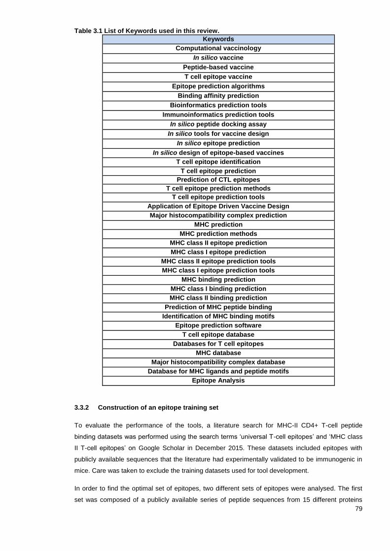
79
Table 3.1 List of Keywords used in this review.
Keywords
Computational vaccinology
In silico vaccine
Peptide-based vaccine
T cell epitope vaccine
Epitope prediction algorithms
Binding affinity prediction
Bioinformatics prediction tools
Immunoinformatics prediction tools
In silico peptide docking assay
In silico tools for vaccine design
In silico epitope prediction
In silico design of epitope-based vaccines
T cell epitope identification
T cell epitope prediction
Prediction of CTL epitopes
T cell epitope prediction methods
T cell epitope prediction tools
Application of Epitope Driven Vaccine Design
Major histocompatibility complex prediction
MHC prediction
MHC prediction methods
MHC class II epitope prediction
MHC class I epitope prediction
MHC class II epitope prediction tools
MHC class I epitope prediction tools
MHC binding prediction
MHC class I binding prediction
MHC class II binding prediction
Prediction of MHC peptide binding
Identification of MHC binding motifs
Epitope prediction software
T cell epitope database
Databases for T cell epitopes
MHC database
Major histocompatibility complex database
Database for MHC ligands and peptide motifs
Epitope Analysis
3.3.2 Construction of an epitope training set
To evaluate the performance of the tools, a literature search for MHC-II CD4+ T-cell peptide
binding datasets was performed using the search terms ‘universal T-cell epitopes’ and ‘MHC class
II T-cell epitopes’ on Google Scholar in December 2015. These datasets included epitopes with
publicly available sequences that the literature had experimentally validated to be immunogenic in
mice. Care was taken to exclude the training datasets used for tool development.
In order to find the optimal set of epitopes, two different sets of epitopes were analysed. The first
set was composed of a publicly available series of peptide sequences from 15 different proteins
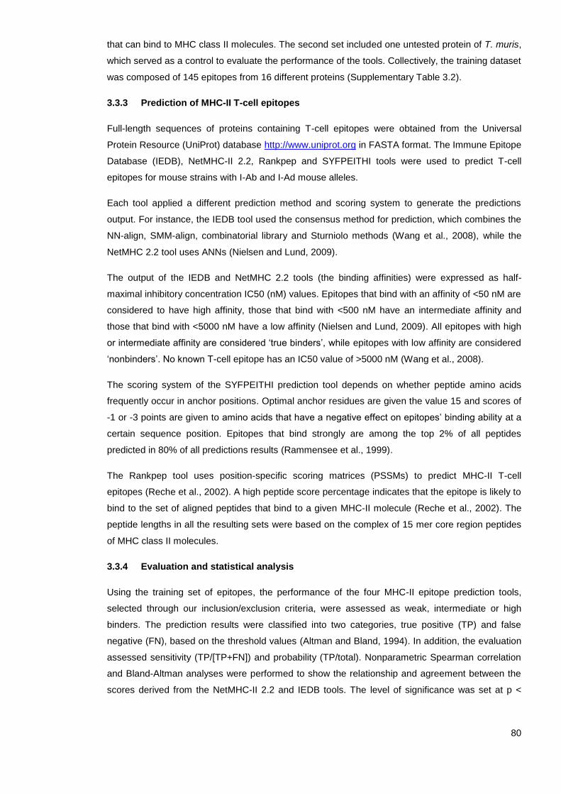
80
that can bind to MHC class II molecules. The second set included one untested protein of T. muris,
which served as a control to evaluate the performance of the tools. Collectively, the training dataset
was composed of 145 epitopes from 16 different proteins (Supplementary Table 3.2).
3.3.3 Prediction of MHC-II T-cell epitopes
Full-length sequences of proteins containing T-cell epitopes were obtained from the Universal
Protein Resource (UniProt) database http://www.uniprot.org in FASTA format. The Immune Epitope
Database (IEDB), NetMHC-II 2.2, Rankpep and SYFPEITHI tools were used to predict T-cell
epitopes for mouse strains with I-Ab and I-Ad mouse alleles.
Each tool applied a different prediction method and scoring system to generate the predictions
output. For instance, the IEDB tool used the consensus method for prediction, which combines the
NN-align, SMM-align, combinatorial library and Sturniolo methods (Wang et al., 2008), while the
NetMHC 2.2 tool uses ANNs (Nielsen and Lund, 2009).
The output of the IEDB and NetMHC 2.2 tools (the binding affinities) were expressed as half-
maximal inhibitory concentration IC50 (nM) values. Epitopes that bind with an affinity of <50 nM are
considered to have high affinity, those that bind with <500 nM have an intermediate affinity and
those that bind with <5000 nM have a low affinity (Nielsen and Lund, 2009). All epitopes with high
or intermediate affinity are considered ‘true binders’, while epitopes with low affinity are considered
‘nonbinders’. No known T-cell epitope has an IC50 value of >5000 nM (Wang et al., 2008).
The scoring system of the SYFPEITHI prediction tool depends on whether peptide amino acids
frequently occur in anchor positions. Optimal anchor residues are given the value 15 and scores of
-1 or -3 points are given to amino acids that have a negative effect on epitopes’ binding ability at a
certain sequence position. Epitopes that bind strongly are among the top 2% of all peptides
predicted in 80% of all predictions results (Rammensee et al., 1999).
The Rankpep tool uses position-specific scoring matrices (PSSMs) to predict MHC-II T-cell
epitopes (Reche et al., 2002). A high peptide score percentage indicates that the epitope is likely to
bind to the set of aligned peptides that bind to a given MHC-II molecule (Reche et al., 2002). The
peptide lengths in all the resulting sets were based on the complex of 15 mer core region peptides
of MHC class II molecules.
3.3.4 Evaluation and statistical analysis
Using the training set of epitopes, the performance of the four MHC-II epitope prediction tools,
selected through our inclusion/exclusion criteria, were assessed as weak, intermediate or high
binders. The prediction results were classified into two categories, true positive (TP) and false
negative (FN), based on the threshold values (Altman and Bland, 1994). In addition, the evaluation
assessed sensitivity (TP/[TP+FN]) and probability (TP/total). Nonparametric Spearman correlation
and Bland-Altman analyses were performed to show the relationship and agreement between the
scores derived from the NetMHC-II 2.2 and IEDB tools. The level of significance was set at p <

81
0.05 for the correlation test. All statistical analyses were performed using the software package
GraphPad Prism version 6 for Windows.
3.4 Results
Based on a list of search terms used on Google and other websites, 88 servers that predict T-cell
epitopes based on MHC class I and II binding were identified (Supplementary Table 3.3). Off the 88
tools, only 48 tools that can predict MHC-II epitopes were identified (Table 3.2). Since our primary
focus was MHC-II T-cell epitope prediction tools, only those tools were further evaluated based on
the inclusion criteria. The top five tools that met the inclusion criteria were IEDB, SYPEITHI,
NetMHC-II 2.2, Rankpep and ProPred. They were then scanned for the ability to predict MHC-II T-
cell epitopes in two mouse alleles, I-Ab and I-Ad. The ProPred tool was excluded from further
analysis because it only predicts HLA-DR binding sites. The four epitope prediction tools that met
all the selection criteria were subsequently evaluated using the epitope training set to calculate the
sensitivity and probability with which they could predict MHC II T-cell epitopes.
Using the epitope training set (Supplementary Table 3.2), it was observed that IEDB and NetMHC-
II 2.2 tools have high levels of sensitivity (~78.00%) and probability (~17.00%) for predicting MHC-
II T-cell binding epitopes with high affinities, while Rankpep and SYFPETHI exhibited low sensitivity
(10.61% and 8.33%, respectively). Collectively, the data indicate that the best prediction tools
across all MHC-II T-cell prediction servers considered in this study are IEDB and NetMHC-II 2.2
(Table 3.3).
Statistically significant differences were found when correlating the scores returned by the
NetMHC-II 2.2 and IEDB tools for MHC I-Ab (p < 0.0001; r = 0.78, 95% CI = 0.66–0.86) and MHC
I-Ad (p = 0.0071; r = 0.46, 95% CI = 0.13–0.70), as shown in Figure 3.2 (A and B). A Bland-Altman
plot compares the average affinity of peptide binding predicted by NetMHC-II 2.2 and IEDB plotted
against the difference in peptide-binding affinity scores given by the two tools for the prediction of
MHC-II T-cell epitopes from I-Ab and I-Ad alleles (Figure 3.2 C, and D). The two tools were more
likely to agree for peptides with high-affinity scores (<50 nM) than those with intermediate and low-
affinity scores (<500 and <5000 nM, respectively). In addition, the majority of scores are distributed
symmetrically around the mean, indicating no systematic bias. In general, the statistical evidence
suggests that the NetMHC-II 2.2 and IEDB tools are able to predict peptides with high-affinity
binding scores equally well. Also, the predictions with high-affinity binding scores are mainly
estimated for MHC (I-Ab) molecules than MHC (I-Ad), as shown in Figure 3.2 (C and D).
Collectively, the work in this chapter has systematically reviewed and evaluated available MHC-II
cell epitope prediction tools and included two such tools to use in the project going forward.

82
Table 3.2 List of MHC-II epitope prediction tools and their number of publication, number of citations using Google Scholar, the online availability, last update, and community.
Prediction Tool No of
publication No of
Citation
Availability
Last Update in
2015 Community
IEDB 440 1550 Yes 12/2015 National Institute of Allergy and Infectious Diseases
NetMHCII 2.2 363 202 Yes 03/02/2012 Center for Biological Sequence analysis CBS
NNAlign (NetMHCII-2.0)
163 13 Yes 17/03/2015 CBS
NetMHCIIpan 3.1 83 156 Yes 17/11/2015 CBS, IMMI, and LIAI.
PickPocket 1.1 26 26 Yes 01/05/2015 CBS, IMMI, and LIAI.
ProPred 715 544 Yes 07/06/2002 Raghava’s Group
FDR4 12 1 Yes 07/12/2014 Raghava’s Group
HLAPRED 63 91 Yes NA Raghava’s Group
HLA-DR4 Pred 563 124 Yes 27/09/2014 Raghava’s Group
MHC2Pred 114 55 Yes 07/12/2014 Raghava’s Group
IMHC 1 5 No NA Raghava’s Group
MHCBench 53 7 Yes 11/03/2014 Raghava’s Group
IL4pred 3 5 Yes NA Raghava’s Group
IFNepitope 17 4 Yes 07/12/2014 Raghava’s Group
IL-10Pred 1 16 Yes NA Raghava’s Group
TmhcPred 4 NA Yes NA Raghava’s Group
MULTIPRED2 40 33 Yes 22/07/2014
Bioinformatics Core at Cancer Vaccine Center, Dana-Farber Cancer Institute.
PREDBALB/c 47 50 Yes NA
Bioinformatics Core at Cancer Vaccine Center, Dana-Farber Cancer Institute.
RANKPEP 709 272 Yes 16/12/2015
Bioinformatics Core at Cancer Vaccine Center, Dana-Farber Cancer Institute.
MHCMIR 1.0 41 13 Yes 29/04/2015 Laboratory of data mining and intelligent information processing.
MHC2SKpan-1.0. 6 4 Yes NA Laboratory of data mining and intelligent information processing.
TEPITOPEpan 35 27 Yes 11/12/2014 Laboratory of data mining and intelligent information processing.
MetaMHC 21 13 NA NA Laboratory of data mining and intelligent information processing.
MetaMHCIIPan 4 1 NA NA Laboratory of data mining and intelligent information processing.
MHC2MIL 7 4 Yes NA Laboratory of data mining and intelligent information processing.
MultiRTA 38 26 NA NA NA
EpiTOP ? 13 Yes NA Faculty of Pharmacy Medical University of Sofia
EpiDOCK 29 17 Yes NA Faculty of Pharmacy Medical University of Sofia
VaxiJen 185 141 Yes NA Faculty of Pharmacy Medical University of Sofia
Vaxign 3 132 Yes NA University of Michigan Medical School
MHCpred 387 58 Yes 11/05/2011 Faculty of Pharmacy Medical University of Sofia

83
PREDIVAC 26 13 Yes 27/112014 University of Queensland
MHC-BPS 40 32 NA 11/12/ 2014
Bioinformatics and Drug Design Group, Department of Pharmacy, National University of Singapore
SYFPEITHI 3,280 2013 Yes 27 /08/2012 Applied Bioinformatics Group at University of Tuebingen
SVMHC 385 260 Yes NA Applied Bioinformatics Group at University of Tuebingen
EpiToolKit 2.0 43 20 Yes NA Applied Bioinformatics Group at University of Tuebingen
OptiTope 33 34 Yes NA Applied Bioinformatics Group at the University of Tuebingen
SNEPv2 83 31 Yes NA Applied Bioinformatics Group at University of Tuebingen
FRED 1 16 Yes NA Applied Bioinformatics Group at University of Tuebingen
POPI2.0 221 82 Yes NA Institute of Bioinformatics, National Chaio Tung University
EpiMatrix (iVAX) 1,010 115 NA 2014 EpiVax
Epitopemap 34 1 Yes NA Damien Farrell (web2py platform)
Hotspot Hunter 36 19 No NA NA
TEpredict 290 14 Yes 26/08/2012 Russian Foundation for Basic Research grant
EpDis 1 0 Yes 24/06/ 2016 Bioinformatics Research Group
SVRMHC 125 83 Yes NA Biolead
POPI2.0 134 85 Yes 11/12/2009 Institute of Bioinformatics, National Chaio Tung University
PepCrawler 30 32 Yes 01/02/2016 BioInfo3D Group
NA: Not available.
Table 3.3 The sensitivity and probability of predicting MHC-II T-cell epitopes using SYFPEITHI, IEDB, NetMHC-II 2.2, and Rankpep prediction tools.
Server Sensitivity Probability
SYFPEITHI 8.33% 7.63%
IEDB 77.87% 15.68%
NETMHCII 77.87% 19.42%
RANKPEP 10.61% 6.25%
Using a handcrafted training set of proteins, the sensitivity as TP/(TP+FN) and the probability as TP/total of MHC-II T-cell epitopes prediction were calculated.
84
Figure 3.2 NetMHC-II 2.2 and IEDB tools performance. (A) The correlation score of the NetMHC-II 2.2 and IEDB tools for predicting MHC-II (I-Ab) epitopes. (B) The correlation score of the NetMHC-II 2.2 and IEDB tools for predicting MHC-II (I-Ad) epitopes. (C) The Bland-Altman plot of the difference and average of NetMHC-II 2.2 and IEDB prediction of MHC-II (I-Ab) epitopes and (D) in predicting MHC-II (I-Ad) epitopes. The red line means 50 nM and the green line means 500 nM.

85
3.5 Discussion
Prediction of which protein epitopes are potential vaccine candidates using immunoinformatics
tools is a powerful strategy for development of epitope-based vaccines (Rasmussen et al., 2016).
There are more than 80 computer-based prediction tools for identifying peptides that bind to MHC
class I and II molecules, but not all are equivalent; some epitope prediction tools may fail to predict
all significant epitopes (Yang and Yu, 2009). The performance of in silico prediction tools is
affected by several factors. For example, MHC-II CD4+ T-cell epitope prediction tools have much
lower accuracy than MHC-I tools because the MHC-I binding groove is closed, while the MHC-II
groove is open at both ends (Godkin et al., 2001; Yang and Yu, 2009). Also, the use of a limited
training dataset to evaluate the tools may affect performance (Hegde et al., 2018; Lin Hh, 2008;
Wang et al., 2008). It was suggested that the low performance of MHC class II prediction tools is
not due only to poor algorithm performance; the genetic diversity of human populations present
further significant challenge (Paul et al., 2015). Thus, choosing the best bioinformatics tool to
predict MHC class II T-cell epitopes is critical when designing epitope-based vaccines (Pappalardo
et al., 2015; Schumacher et al., 2017). This chapter presents a systematic review of existing MHC-
II restricted T-cell epitope prediction tools and evaluated four tools intended to establish the most
appropriate bioinformatics tools currently available for predicting Trichuris MHC-II T-cell epitopes.
Four tools met the inclusion/exclusion criteria and were further analysed to determine the specificity
and probability with which they can predict MHC class II T-cell epitopes. The IEDB and NetMHC-II
2.2 tools achieved similarly high levels of sensitivity and probability of predicting binding epitopes
with high affinities, while Rankpep and SYFPETHI exhibited low sensitivity. Each tool applies
different methods of prediction; the IEDB tool uses a quantitative consensus method that combines
the strengths of various methods (Wang et al., 2008); the NetMHC-II 2.2, another quantitative tool,
uses an NN-align algorithm and weight matrix (Nielsen and Lund, 2009); and both SYFPEITHI
(Rammensee et al., 1999) and Rankpep (Reche et al., 2002) are qualitative tools that use motif
PSSMs. All the tools were ‘user-friendly’, but the IEDB tool has features that the other three do not,
including a browser to input protein sequence formats in a national centre for biotechnology
information (NCBI) database, seven different prediction methods and an easy method of
downloading the prediction output into an Excel spreadsheet. In contrast, SYFPETHI is limited to
specific alleles compared to the three other tools.
3.5.1 Evaluation of available MHC-II binding prediction tools
Several studies have compared the performance of different MHC class II peptide binding
prediction tools (Borrás-Cuesta et al., 2000; Gowthaman and Agrewala, 2007; Lin et al., 2008), but
the comparison presented in this chapter is different for two main reasons. First, the MHC-II
prediction tools were selected in a systematic way using inclusion/exclusion criteria. Second, a
unique dataset was used for comparison.
Because training sets of peptides were used for the development of epitope prediction tools, it is
important to evaluate their performance using unique sets that do not overlap with the original
training set (Murugan and Dai, 2005; Nielsen et al., 2004; Yasser et al., 2008). However, some
86
published studies which have aspired to evaluate the performance of T-cell epitope prediction tools
have used training datasets that are similar to the original training sets (Yasser et al., 2008).
Therefore, to more rigorously assess the relative performance of the available prediction tools, this
study excluded training datasets and introduced new peptides to the tools.
Gowthaman and Agrewala (2007) evaluated the performance of six class II binding peptide
prediction tools (ProPred, MHC2Pred, Rankpep, SVMHC, MHCPred and MHC-BPS) and
concluded that none can be efficiently used to predict which epitopes are crucial for vaccination.
However, the limited number of peptides, HLA class II alleles and prediction methods (matrix-
based methods and SVM) used to evaluate the tools limited the significance of their results
(Gowthaman and Agrewala, 2007).
Other published comparisons claim that the IEDB tool, using the consensus method, is the gold
standard tool since it appears to be the strongest predictor of MHC binding epitopes amongst the
other online available tools (Wang et al., 2008). For example, Wang et al. (2008) evaluated the
performance of nine MHC class II peptide binding tools (consensus, SYFPEITHI, Rankpep,
average relative binding [ARB], ProPred, SMM-align, SVRMHC, MHC2Pred and MHCPred) for
different human and animal alleles using a comprehensive three datasets. The first is an
unpublished experimental dataset of 10,017 peptide-binding affinities for 16 MHC class II
molecules, and it was used to evaluate the binding affinities of the prediction tools. The second
consists of 29 structures associated with 14 different MHC class II molecules extracted from the
Protein Data Bank (PDB), and it was used to evaluate the ability of prediction tools to locate the 9-
mer core of epitopes. The third dataset consists of 664 peptides experimentally tested in a
C57BL/6 (H-2b) strain of laboratory mouse for CD4+ T-cell activation (Wang et al., 2008). The
IEDB tool using consensus method outperformed all other MHC class II prediction methods in
terms of sensitivity in predicting binding affinity and T-cell activation, followed by Rankpep, SMM-
align and ProPred. It was suggested that the increased epitope prediction accuracy of the
consensus method is attributed to the combination of the top performance methods and access to
the most comprehensive online database (Salimi et al., 2010; Wang et al., 2008). It was also
suggested that the great performance of the SMM-align tool can be attributed to its utilisation of the
IEDB dataset (Wang et al., 2008). This would confirm that the size of the training set is an
important factor contributing to better performance (Wang et al., 2008). Furthermore, comparison of
the predicted 9-mer core with the true cores extracted from crystal structures revealed that the
ProPred tool was able to most accurately predict cores, followed by SYFPEITHI and SMM-align
(Wang et al., 2008). The fact that the ProPred tool is based on the TEPITOPE database of
experimental assays further confirms that the performance of the tool is directly affected by the
type of training database applied (Wang et al., 2008). However, this study did not assess the
performance of the consensus approach for predicting 9-mer cores that interact with the binding
groove of the MHC class II molecule.
Ogishi and Yotsuyanagi demonstrated that integration of MHC class II in vitro binding and naturally
eluted ligands derived from mass spectrometry (MS) data can improve the predictive performance
of the NetMHCII and NetMHCIIpan tools regarding identification of T-cell epitopes (Ogishi and
87
Yotsuyanagi, 2019). Another study also suggested that the poor performance of the SYFPEITHI
matrix-based MHC-binding prediction tool is due to the fact that it helps locate epitopes in known
target proteins but does not predict epitopes in unknown proteins (Pelte et al., 2004). The results
of their study agree with those of the present study, which found that the SYFPEITHI tool has low
accuracy for predicting MHC-II T-cell epitopes. Another study also reported that the SYFPEITHI
server is a poor predictor of MHC-I peptide binding affinity compared to SMM-QMs and ANN-based
methods (Peters et al., 2006).
De Groot and Martin (2009) compared the EpiMatrix tool to other MHC class II prediction tools,
including IEDB and SYFPEITHI, using MHC class II-peptide complex crystal structures extracted
from the Protein Data Bank (PDB). EpiMatrix was 5% better than the IEDB tool using the ARB
method of prediction and 9% better than SYFPEITHI (De Groot and Martin, 2009). However,
EpiMatrix was not evaluated in this study or similar studies comparing epitope prediction algorithms
because it is a commercial tool that is limited to commercial users and selected academic
collaborators.
A different study compared the performance of 21 HLA class II binding affinity tools using a dataset
of 103 peptides from four independent studies (Lin et al., 2008). However, this study did not
compare the performance of the tools for predicting MHC-II binding affinity peptides. Lin et al.
(2008) suggested that the NetMHCIIpan server, which is based on a combination of binding ligand
and structure-based methods, ProPred, consensus and MULTIPRED (SVM), were the best
predictors Another study conducted by Ivan et al. (2011) has also identified NetMHCII and
NetMHCIIpan as the best publicly available MHC class II epitope predictors compared to ProPred,
Rankpep and EpiTOP.
A more recent study by Zhao and Sher (2018) evaluated the MHC-II prediction tools hosted on the
IEDB analysis resource server using newly available, untested data of both synthetic and naturally
processed epitopes. Among the 18 predictors that were benchmarked, an ANN-based approach,
NN-align (NetMHCII2), outperformed all other tools, including NetMHCIIpan and the consensus
method for both MHC class I and class II predictions (Zhao and Sher, 2018). Furthermore,
Andreatta et al. (2018) created an automated platform to benchmark six commonly used MHC
class II peptide binding prediction tools using 59 datasets that were newly entered into the IEDB
database and had not yet been made public to prevent biased assessment of the available
prediction tools. Their evaluation suggested that NetMHCIIpan is currently the most accurate tool,
followed by NN-align and the IEDB (consensus) tool (Andreatta et al., 2018). Despite differences in
the datasets used for comparison, these studies agree with the comparison study in this chapter,
which found that the IEDB (consensus) and NETMHC-II 2.2 (ANN) tools are among the best MHC-
II prediction tools. NetMHCIIpan was not included in this study because it did not meet the inclusion
criteria. Collectively, I recommend the use of IEDB and NETMHC-II 2.2 prediction tools in any MHC
class II epitope prediction study to reduce the experimental cost of identifying epitopes. However,
the output of these tools needs to be carefully evaluated in vitro and in vivo before they are used to
bring an epitope-based vaccine to trial.

88
3.5.2 Updates of the available in silico prediction tools
During the time period covered by this systematic review, the available in silico prediction tools
have been improved and updated. For example, the performance of MHC-II peptide binding
prediction tools (NetMHCII and NetMHCIIpan) was recently improved using a dataset of
quantitative MHC peptides extracted from the IEDB (Jensen et al., 2018; Jurtz et al., 2017).
Moreover, the IEDB Analysis Resource developed an online tool called TepiTool, which provides a
set of step-by-step instructions for how to predict MHC class I and class II epitopes, including which
alleles to include, which lengths to consider and which cut-offs should be considered relevant (Paul
et al., 2016). TepiTool uses the consensus, NN-align, SMM-align (also known as NetMHCII-1.1,
version 1.1), Sturniolo and combinatorial library prediction algorithms to examine hundreds of
alleles from different species, including humans, chimpanzees, bovines, gorillas, macaques, mice
and pigs (Paul et al., 2016). More recently, an allele-integrated deep learning framework named
Allele-Integrated MHC (AI-MHC) was developed to improve class I and class II HLA binding
predictions by allowing the tool to be trained on effectively larger datasets (Sidhom et al., 2018).
Also, in 2017, the VacSol pipeline was introduced for rapidly and efficiently identifying vaccine
candidates from the whole proteomes of bacterial pathogens (Rizwan et al., 2017). The pipeline
screens for proteome analysis using several well-known tools (BLAST, Pftools2.3, PSORTb3.0,
HMMTOP 2.0, DEG 10.0, VFDB, ABCPred, ProPred-I, ProPred and UniProt-SwissProt) for
prediction of B- and T-cell epitopes, subcellular protein localisation and transmembrane topology
(Rizwan et al., 2017).
All the above-mentioned MHC-II binding prediction tools apply indirect methods to predict T-helper
epitopes. However, there are a few direct methods for predicting cytokine-specific peptides that can
induce IL-4 (Dhanda et al., 2013a), IL-10 (Nagpal et al., 2017) and IFN-gamma production (Dhanda
et al., 2013b). Immune recognition of peptides by T-cells depends on the formation of a ternary
complex ‘sandwich’ consisting of protein peptides between MHC and TCR. Most of the current
research focuses on evaluating the MHC-facing agretopes. However, the JanusMatrix and
Repitope tools evaluate both TCR-facing and MHC-facing peptides in a single analysis to improve
the selection of vaccine antigens (He et al., 2014; Moise et al., 2013; Ogishi and Yotsuyanagi,
2019). Also, ITCell was introduced in 2018 to predict T-cell epitopes based on information about
the three stages of the immune response pathway: antigen cleavage, MHC-II presentation and
TCR recognition (Schneidman-Duhovny et al., 2018). Using benchmarks, it was found that the
ITCell method had better accuracy than the NetMHCIIpan single-stage epitope prediction algorithm
(Schneidman-Duhovny et al., 2018).
3.5.3 Applications of MHC-II prediction tools
The IEDB and NetMHC-II prediction tools have become widely used for developing epitope-based
vaccines against many parasites, including Plasmodium falciparum (Singh et al., 2010),
Trypanosoma brucei (Teh-Poot et al., 2014), Echinococcus granulosus (Ma et al., 2013),
Leishmania infantum (Agallou et al., 2014; Dias et al., 2018), and Onchocerca volvulus (Shey et al.,
2019). Also, in an effort to identify helminth immunomodulatory proteins, Homan and Bremel (2018)
89
used IEDB approaches to predict MHC-I and II binding epitopes from secreted proteins from 17
helminth spp and common co-infections (P. falciparum, Mycobacterium tuberculosis and H. pylori).
Not only were MHC binding epitopes predicted but also amino acid motifs (T-cell-exposed motifs
[TCEMs]) with a high likelihood of being recognised by T-cell receptors and eliciting Treg
responses were identified (Homan and Bremel, 2018). TCEMs were found to be shared to a high
degree across helminth species and with P. falciparum and M. tuberculosis when these contribute
to co-infection (Homan and Bremel, 2018).
Despite its inferior performance, the SYFPEITHI tool has been used to predict T-cell epitopes from
the nematode parasite Trichinella spiralis. For example, Gu et al. (2016) used this tool to predict a
set of 12 CD4+ T-cell epitopes from a T. spiralis paramyosin protein (Ts-Pmy). Five of the epitopes
significantly induced splenocyte proliferation and secretion of the Th2 cytokines IL-4 and IL-5 in
splenocytes from mice vaccinated with recombinant Ts-Pmy protein (rTs-Pmy) (Gu et al., 2016). In
addition, splenocytes from mice vaccinated with individual synthesised peptides emulsified with an
ISA50 V2 adjuvant stimulated with the corresponding peptides induced strong T-cell proliferation
and secretion of a mix of Th1 and Th2 cytokines (Gu et al., 2016). However, this study was limited
as it did not assess protective immunity to the parasite.
Given that all MHC class II T-cell epitope prediction tools provide false positives and false
negatives to varying degrees, it is recommended that multiple bioinformatics tools be combined to
predict MHC class II T-cell epitopes. For example, Agallou et al. (2017) developed a multi-peptide-
based nanovaccine against leishmaniasis based on Leishmania cysteine protease A (CPA160-189)
co-encapsulated with monophosphoryl lipid A (MPLA) in Poly(D,L-lactic-co-glycolide) (PLGA)
nanoparticles. The potential MHC class I and II binding epitopes derived from the L. infantum CPA
protein were predicted by three in silico analysis tools: SYFPEITHI, BIMAS and NetMHCII (Agallou
et al., 2014). Subcutaneous vaccination of BALB/c mice with the vaccine candidate induced
specific anti-CPA160-189 cellular and humoral immune responses characterised by the production
of high levels of IL-2, IFN-γ and TNFα cytokines and IgG1/IgG2c antibodies (Agallou et al., 2017).
Furthermore, the vaccinated mice displayed significantly reduced hepatic (48%) and splenic (90%)
parasite load prior to L. infantum promastigote challenge (Agallou et al., 2017).
In summary, almost every aspect of the field of immunoinformatics is rapidly expanding, including
the design of epitope-based vaccines. Accurate prediction of MHC II T-cell epitopes using in silico
prediction tools is a critical first step in the development of an epitope-based vaccine. Therefore, a
systematic review and evaluation of the available MHC class II in silico prediction tools was
conducted in this chapter to determine which ones have the best predictive ability.
3.6 Summary
Based on a list of search terms used on Google and other websites, 44 servers that can
predict MHC-II peptides were identified.
Aspects relating to the number of citations (>200), number of publications (>200), online
availability, last update and community were used as inclusion/exclusion criteria to select

90
the top MHC-II in silico prediction tools. The five tools that met the inclusion criteria were
IEDB, SYPEITHI, NetMHC-II 2.2, Rankpep and ProPred.
A unique training data set was used to evaluate the performance of the top tools. The IEDB
and NetMHC-II 2.2 tools exhibited similarly high levels of sensitivity and probability for
predicting epitopes with strong affinities, while the Rankpep and SYFPETHI prediction
tools exhibited low sensitivity.

91
Chapter Four
Trichuris MHC-II T-cell epitope
prediction
92
4.1 Introduction
Designing a vaccine for a neglected tropical parasite, such as T. trichiura, is one of the most
challenging tasks for medical scientists. Although anthelmintic treatments for Trichuris infections
exist, the available drugs have low efficacy and are threatened by drug resistance (Mrus et al.,
2018; Patel et al., 2019). Thus, as with all neglected tropical infectious diseases (NTDs), control
and prevention of trichuriasis are superior to a cure (Briggs et al., 2018). Therefore, developing a
vaccine would be the most effective method to control Trichuris infections (Dixon et al., 2010).
However, while many research scientists have put substantial effort into developing such a
vaccine, there is currently no licensed vaccine against Trichuris, as much work must be done to
improve the specificity, sensitivity and safety of candidate vaccines (Blair and Diemert, 2015).
The advent of the genome era has provided new effective strategies for vaccine development (Del
Tordello et al., 2017). The reverse vaccinology (RV) approach, in particular, which relies on both
genome information and the tremendous advances in immunological and bioinformatics tools, has
been used to develop epitope-based vaccines (Bambini and Rappuoli, 2009). One of the
advantages of the RV approach is that every antigen encoded in the Trichuris genome can be
screened using immunoinformatics-based software to determine its ability to induce an immune
response (De Sousa and Doolan, 2016; Del Tordello et al., 2017). Thus, it can overcome some of
the limitations of conventional methods of screening vaccine candidates (Arnon and Ben-Yedidia,
2003; Capecchi et al., 2004; Moise et al., 2011).
There are several challenges that can arise when screening for potential vaccine candidates, such
as identifying immunogenic candidates in a large-scale genome and choosing a suitable animal
model for testing immunogenicity and protective immunity (Thomas and Luxon, 2013). To address
these challenges, a set of Trichuris proteins can be selected from the genomes of T. trichiura and
its murine equivalent, T. muris, based on selected parameters associated with antigenic peptides
that would provide protection against trichuriasis in C57BL/6 mice.
Designing vaccines based on multi-stage antigens has been an effective strategy for complex
pathogens, such as M. tuberculosis and Plasmodium (Cabrera-Mora et al., 2016; Mosavat et al.,
2016). Therefore, the ultimate goal of this study is to design a novel multi MHC class II T-cell
epitope-based vaccine predicted from multi-stage Trichuris proteins, which will likely induce
effective protective immunity, using the RV approach. The T-cell epitopes will then be incorporated
into a VLP to generate a candidate vaccine against human trichuriasis.
4.2 Aim and objectives
The overall aim of this chapter was to identify novel MHC-II T-cell epitopes from the Trichuris
genome as promising vaccine candidates using an RV approach. This chapter addressed the
following two objectives as part of the overall aim:
To identify a set of proteins from the Trichuris genome based on inclusion/exclusion
criteria to select proteins with the high potential to be lead antigens in the development
of an anti-Trichuris vaccine.

93
To predict the most conserved and antigenic Trichuris MHC-II T-cell epitopes from the
selected proteins using the IEDB prediction tool identified in Chapter 3.
4.2 Methods and results
4.2.1 Data acquisition and identification of potential virulent proteins
The stichosome in the whipworm anterior region secrete ES products which have been suggested
to trigger the host immune response and induce host immunity after infection (Dixon et al., 2010;
Shears et al., 2018b). Furthermore, previous research conducted by Dixon et al., (2008) and Else
et al., (1990), showed that antibody recognition of high molecular weight proteins in both T. muris
adult and larval ES correlated with resistance to T. muris infection. To increase the chance of
antigen coverage to induce a strong immune response, secreted and surface-exposed proteins in
L2, L3, male and female adult worms’ anterior region were selected for this study as potential
vaccine candidates based on published information (Foth et al., 2014).
The 85-Mb genome (∼11,004 protein-encoding genes) of T. muris and the 73-Mb genome (∼9650
protein-coding genes) of T. trichiura sequenced by Foth et al. (2014) served as rich sources of
potential vaccine candidates and therefore were used as starting points (Foth et al., 2014). From
the whole genome of the Trichuris, 637 proteins expressed in L2, L3, male and female adult
worms’ anterior region were selected. Of the total of 637 proteins, only proteins that possess signal
peptides and do not exhibit transmembrane domains were included, resulting in a sample of 156
proteins.
Full-length sequences of the Trichuris proteins were obtained from the Universal Protein Resource
(UniProt) database in FASTA format http://www.uniprot.org/.
4.2.2 Identification of non-homologous proteins to human and mouse with high
expression levels
To eliminate autoimmune reactions when tested in mice and humans, the proteins identified in the
previous step were checked to determine their homology with human and mouse proteins using the
basic local alignment search tool (BLAST), which is available online in the UniProt database
https://www.uniprot.org/blast/. All proteins with any degree of homology with humans or mice were
excluded, leaving 60 candidate proteins.
Of these, only upregulated proteins in L2, L3 and adult worms’ anterior region were selected based
on their transcript expression level. Based on the high-throughput transcriptome data generated
from the RNA of T. muris and Gene Ontology (GO) term enrichment analyses, a transcriptional
upregulation in a particular protein refers to (≥10 Log2 (normalised read count) transcript
expression level (Foth et al., 2014). Of the 60 selected proteins, only 27 proteins were selected to
predict Trichuris MHC-II T-cell epitopes.

94
4.2.3 Prediction of Trichuris MHC-II T-cell binding epitopes
Since antibodies are not essential for protection against Trichuris infection and protective immunity
is mediated by CD4+ T-cells, not CD8+ T-cells, the selected proteins were screened to predict
MHC-II T-cell epitopes rather than B-cell epitopes (Else and Grencis, 1996; Koyama et al., 1995).
All 27 selected proteins were screened to predict T-cell MHC class II epitopes using the IEDB
(consensus method) prediction tool (Wang et al., 2008).
There are many different human MHC (human leukocyte antigen [HLA]) alleles, and the
distributions of particular types are associated with different populations. The analysis was carried
out to predict the binding affinity to the MHC class II allele I-Ab mouse strain and the 11 most
prevalent human class II HLA allele supertypes (Nielsen et al., 2010a; Nielsen and Lund, 2009;
Wang et al., 2008; Wang et al., 2010).
The predicted peptide output (i.e. binding affinity) of the NN-align and SMM-align methods was
expressed as IC50 (nM) values. IC50 values of <50 nM indicate high affinity, values of <500 nM
indicate intermediate affinity and values of <5000 nM indicate low affinity. The median percentile
rank (%) of three prediction methods (combinatorial library, SMM-align and Sturniolo) was also
used to generate the rank for the consensus method. A small numbered percentile rank (%)
indicates that a peptide has a high binding affinity to MHC class II alleles (Wang et al., 2008). The
consensus method was used to select only those peptides with a low median percentile rank
according to three different prediction methods to reduce the chance of failure during prediction.
Table 4.1 shows examples of the prediction scores of three peptides predicted from the T. muris
chitin-binding domain-containing protein (TMUE_s0281000600). The (PAGVVYQCTMPRYTL)
peptide, for example, has high and intermediate binding to H2-Iab and to the HLA-DRB1*01:01,
HLA-DRB1*03:01, HLA-DRB1*04:01, HLA-DRB1*07:01, HLA-DRB1*08:02, HLA-DRB1*11:01,
HLA-DRB1*13:02, HLA-DRB1*15:01, HLA-DRB3*01:01 and HLA-DRB3*02:02 alleles. The two
other peptides have high and intermediate binding affinity to H2-Iab and only seven human alleles.
Due to the large volume of data, the prediction scores for the rest of the predicted Trichuris MHC-II
T-cell peptides are presented in the enclosed CD. To cover common global alleles, peptides were
selected based on their ability to bind to at least three different human alleles. Table 4.2 lists the
MHC-II T-cell peptides with high or intermediate binding affinity to the mouse I-Ab allele and at
least three different human alleles that were selected for further analysis.
4.2.4 Conservation and allergens
One of the critical steps in the development of an effective vaccine is the identification of a
conserved antigen from the genome to overcome antigenic variation and reduce the chance of
immune escape (Khan et al., 2006). To assess how well the predicted MHC-II T-cell peptides were
conserved within the T. trichiura genome, the IEDB conservancy analysis tool was used. This tool
calculates the degree of conservancy (i.e. similarity) of a peptide within a specified protein
sequence (Bui et al., 2007). Only peptides that are >70% conserved with at least one homologous
T. trichiura protein were selected for further analysis. Of the 219 MHC-II T-cell peptides, only 33

95
(shown in red in Table 4.2) met these criteria. The conservancy analysis results of the 33 MHC-II
peptides are shown in Supplementary Tables 4.1–4.6. For example, the three overlapping peptides
predicted from the T. muris hypothetical protein (A0A0N5DRU5) that cover the H112-128 epitope and
used in the first in vivo study in Chapter 7, have more than 86% conservation across eight different
T. trichiura homologous proteins (Supplementary Table 4.6). The conservation of all the predicted
MHC-II peptides is presented in the enclosed CD.
The 33 MHC-II peptides were then assessed for the prediction of IgE epitopes and allergenic
potential using the AllerTOP v.2.0 server http://www.ddg-pharmfac.net/AllerTOP/ (Dimitrov et al.,
2014). All the 33 MHC-II T-cell peptides were predicted to have no allergenic potential.
Table 4.3 lists the final set of 10 potential Trichuris MHC-II T-cell epitopes containing 33
overlapping peptides that emerged as promising vaccine candidates after application of the
selection criteria detailed in Figure 4.1. These epitopes, which were predicted from a Pfam domain-
containing protein (B_9093-A0A0N5E6W6), hypothetical protein (A0A0N5DRU5), chymotrypsin-
like serine proteases (A0A0N5DEK1, A0A0N5E3X0 and A0A0N5DUC1) and chitin-binding domain-
containing proteins (A0A0N5DK22 and A0A0N5E6C6), will be fused into VLPs as potential vaccine
candidates in Chapter 5.
A flow diagram of the RV approach used in this study to identify potential vaccine candidates from
Trichuris genome is shown in Figure 4.1.
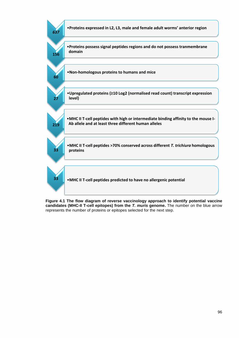
96
Figure 4.1 The flow diagram of reverse vaccinology approach to identify potential vaccine candidates (MHC-II T-cell epitopes) from the T. muris genome. The number on the blue arrow
represents the number of proteins or epitopes selected for the next step.
637 •Proteins expressed in L2, L3, male and female adult worms’ anterior region
156
•Proteins possess signal peptides regions and do not possess tranmembrane domain
60 •Non-homologous proteins to humans and mice
27 •Upregulated proteins (≥10 Log2 (normalised read count) transcript expression level)
219
•MHC II T-cell peptides with high or intermediate binding affinity to the mouse I-Ab allele and at least three different human alleles
33 •MHC II T-cell peptides >70% conserved across different T. trichiura homologous proteins
33 •MHC II T-cell peptides predicted to have no allergenic potential

97
Table 4.1 The prediction scores of MHC-II T-cell epitope (CBD1243-1259) containing three overlapping peptides predicted from chitin-binding domain containing protein (TMUE_s0281000600) using the IEDB prediction tool (Wang et al., 2008).
Allele Prediction method
Peptide sequence
PAGVVYQCTMPRYTL AGVVYQCTMPRYTLC GVVYQCTMPRYTLCV
H2-Iab
% rank 14 14 18
smm X X X
nn 389 390 400
HLA-DRB1 01:01
% rank 26 25 26
smm 169 174 174
nn 32 29 20
HLA-DRB1 03:01
% rank 1 1 5
smm X X X
nn 306 X X
HLA-DRB1 04:01
% rank 5 5 10
smm 436 436 X
nn 94 114 160
HLA-DRB1 07:01
% rank 6 6 6
smm 248 246 238
nn 32 34 30
HLA-DRB1 08:02
% rank 7 8 10
smm X X X
nn 308 252 428
HLA-DRB1 11:01
% rank 1 1 1
smm 193 198 193
nn 44 35 23
HLA-DRB1 13:02
% rank X X X
smm X X X
nn X X X
HLA-DRB1 15:01
% rank 8 8 8
smm X X X
nn 82 81 71
HLA-DRB3 01:01
% rank 9 X X
smm X X X
nn 324 X X
HLA-DRB3 02:02
% rank 4 X X
smm 150 X X
nn 52 X X
HLA-DRB4 01:01
% rank X X X
smm X X X
nn X X X
X: Non-binding peptide.
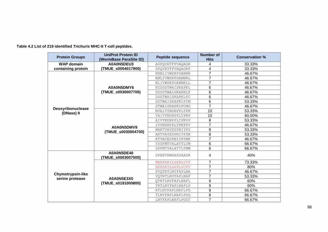
98
Table 4.2 List of 219 identified Trichuris MHC-II T-cell peptides.
Protein Groups UniProt Protein ID
(WormBase ParaSite ID) Peptide sequence
Number of Hits
Conservation %
WAP domain containing protein
A0A0N5DEU3 (TMUE_s0004017900)
ASPQYETFPVAQAGK 4 33.33% SPQYETFPVAQAGKP 4 33.33%
Deoxyribonuclease (DNase) II
A0A0N5DMY6 (TMUE_s0030007700)
KKNLIVWSKPDANNR 7 46.67% KNLIVWSKPDANNRL 7 46.67% NLIVWSKPDANNRLL 7 46.67% KDDSSTWAISKAPKL 6 46.67% DDSSTWAISKAPKLF 6 46.67% DSSTWAISKAPKLFC 6 46.67% SSTWAISKAPKLFCM 6 53.33% STWAISKAPKLFCMG 7 46.67%
A0A0N5DMV9 (TMUE_s0030004700)
NVAIVYNDHPPLTPK 10 53.33% VAIVYNDHPPLTPKP 10 60.00% AIVYNDHPPLTPKPP 8 53.33% IVYNDHPPLTPKPPV 7 46.67% WAKTVAYESPNIYFS 8 53.33% AKTVAYESPNIYFSN 8 53.33% KTVAYESPNIYFSNK 7 46.67% YSSFMTVALATTLTM 6 66.67% SSFMTVALATTLTMW 6 66.67%
Chymotrypsin-like serine protease
A0A0N5DE40 (TMUE_s0003007500)
DPERTRWGHSSAASK 4 40%
A0A0N5E3X0 (TMUE_s0191000800)
MEKKRKYLGSPLVCF 7 73.33% EKKRKYLGSPLVCFV 7 80% SVQTRTLNYFAPLNA 7 46.67% VQTRTLNYFAPLNAF 7 53.33% QTRTLNYFAPLNAFL 9 60% TRTLNYFAPLNAFLP 9 60% RTLNYFAPLNAFLPG 9 66.67% TLNYFAPLNAFLPGG 8 66.67% LNYFAPLNAFLPGGT 7 66.67%
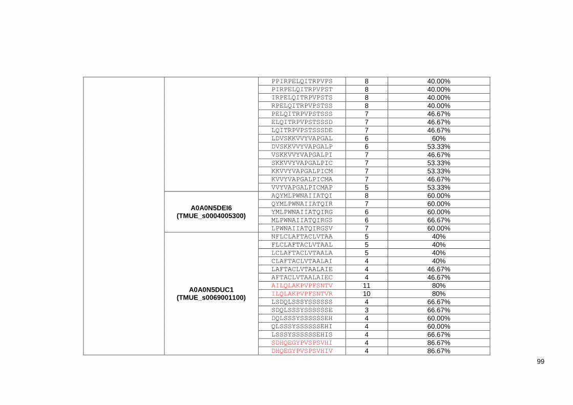
99
PPIRPELQITRPVPS 8 40.00% PIRPELQITRPVPST 8 40.00% IRPELQITRPVPSTS 8 40.00% RPELQITRPVPSTSS 8 40.00% PELQITRPVPSTSSS 7 46.67% ELQITRPVPSTSSSD 7 46.67% LQITRPVPSTSSSDE 7 46.67% LDVSKKVVYVAPGAL 6 60% DVSKKVVYVAPGALP 6 53.33% VSKKVVYVAPGALPI 7 46.67% SKKVVYVAPGALPIC 7 53.33% KKVVYVAPGALPICM 7 53.33% KVVYVAPGALPICMA 7 46.67% VVYVAPGALPICMAP 5 53.33%
A0A0N5DEI6 (TMUE_s0004005300)
AQYMLPWNAIIATQI 8 60.00% QYMLPWNAIIATQIR 7 60.00% YMLPWNAIIATQIRG 6 60.00% MLPWNAIIATQIRGS 6 66.67% LPWNAIIATQIRGSV 7 60.00%
A0A0N5DUC1 (TMUE_s0069001100)
NFLCLAFTACLVTAA 5 40% FLCLAFTACLVTAAL 5 40% LCLAFTACLVTAALA 5 40% CLAFTACLVTAALAI 4 40% LAFTACLVTAALAIE 4 46.67% AFTACLVTAALAIEC 4 46.67% AILQLAKPVPFSNTV 11 80% ILQLAKPVPFSNTVR 10 80% LSDQLSSSYSSSSSS 4 66.67% SDQLSSSYSSSSSSE 3 66.67% DQLSSSYSSSSSSEH 4 60.00% QLSSSYSSSSSSEHI 4 60.00% LSSSYSSSSSSEHIS 4 66.67% SDHQEGYPVSPSVHI 4 86.67% DHQEGYPVSPSVHIV 4 86.67%

100
HQEGYPVSPSVHIVS 5 86.67% QEGYPVSPSVHIVSL 5 86.67% EGYPVSPSVHIVSLA 5 93.33% EVICTGSLYASARAN 4 53.33% VICTGSLYASARANF 7 60.00% ICTGSLYASARANFT 8 66.67% CTGSLYASARANFTR 9 73.33% TGSLYASARANFTRT 9 73.33% GSLYASARANFTRTV 10 73.33% SLYASARANFTRTVL 9 66.67% LYASARANFTRTVLT 6 66.67% YASARANFTRTVLTS 5 73.33% SNFMVYMGSVRSFAA 8 46.67% NFMVYMGSVRSFAAG 8 53.33% FMVYMGSVRSFAAGG 7 46.67% KWLPIYRISTKPIMA 12 40% WLPIYRISTKPIMAH 12 40% LPIYRISTKPIMAHH 12 40% PIYRISTKPIMAHHS 12 40% IYRISTKPIMAHHSA 11 40% YRISTKPIMAHHSAL 8 40% GVCEHGLSAAAPVRI 6 40% VCEHGLSAAAPVRIL 6 40% CEHGLSAAAPVRILS 6 40% EHGLSAAAPVRILSP 6 40% HGLSAAAPVRILSPQ 7 40% GLSAAAPVRILSPQE 5 46.67% QYGIYDFTPEKAHLN 4 46.67% YGIYDFTPEKAHLND 4 53.33% GIYDFTPEKAHLNDI 4 53.33% IYDFTPEKAHLNDIN 4 40%
A0A0N5E1M5 (TMUE_s0144000100)
WNNRYGINIASPSDI 9 40% NNRYGINIASPSDIS 10 40% NRYGINIASPSDISV 9 46.67%
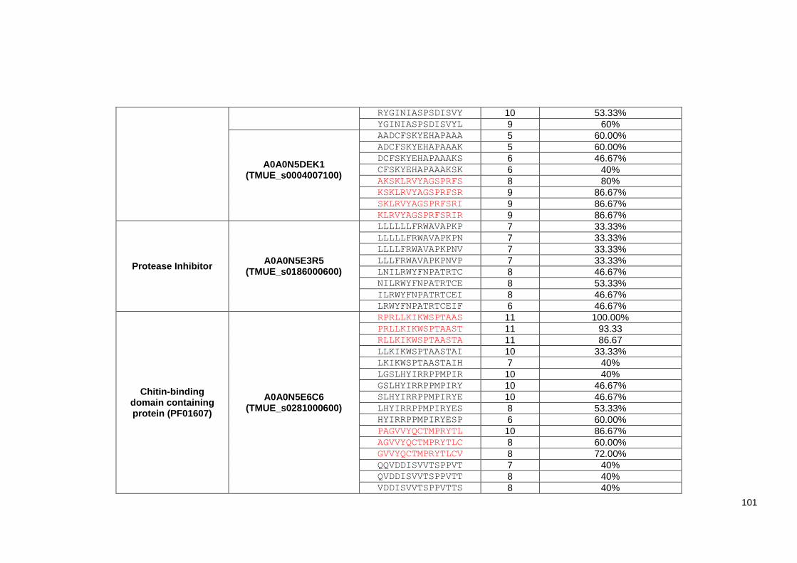
101
RYGINIASPSDISVY 10 53.33% YGINIASPSDISVYL 9 60%
A0A0N5DEK1 (TMUE_s0004007100)
AADCFSKYEHAPAAA 5 60.00% ADCFSKYEHAPAAAK 5 60.00% DCFSKYEHAPAAAKS 6 46.67% CFSKYEHAPAAAKSK 6 40% AKSKLRVYAGSPRFS 8 80% KSKLRVYAGSPRFSR 9 86.67% SKLRVYAGSPRFSRI 9 86.67% KLRVYAGSPRFSRIR 9 86.67%
Protease Inhibitor A0A0N5E3R5
(TMUE_s0186000600)
LLLLLLFRWAVAPKP 7 33.33% LLLLLFRWAVAPKPN 7 33.33% LLLLFRWAVAPKPNV 7 33.33% LLLFRWAVAPKPNVP 7 33.33% LNILRWYFNPATRTC 8 46.67% NILRWYFNPATRTCE 8 53.33% ILRWYFNPATRTCEI 8 46.67% LRWYFNPATRTCEIF 6 46.67%
Chitin-binding domain containing protein (PF01607)
A0A0N5E6C6 (TMUE_s0281000600)
RPRLLKIKWSPTAAS 11 100.00% PRLLKIKWSPTAAST 11 93.33 RLLKIKWSPTAASTA 11 86.67 LLKIKWSPTAASTAI 10 33.33% LKIKWSPTAASTAIH 7 40% LGSLHYIRRPPMPIR 10 40% GSLHYIRRPPMPIRY 10 46.67% SLHYIRRPPMPIRYE 10 46.67% LHYIRRPPMPIRYES 8 53.33% HYIRRPPMPIRYESP 6 60.00% PAGVVYQCTMPRYTL 10 86.67% AGVVYQCTMPRYTLC 8 60.00% GVVYQCTMPRYTLCV 8 72.00% QQVDDISVVTSPPVT 7 40% QVDDISVVTSPPVTT 8 40% VDDISVVTSPPVTTS 8 40%

102
DDISVVTSPPVTTSD 8 40% DISVVTSPPVTTSDS 8 60.00% ISVVTSPPVTTSDSV 8 66.67%
A0A0N5DK22 (TMUE_s0018008000)
GRTTPVTSAPTTVTT 5 100.00% RTTPVTSAPTTVTTE 5 93.33% TTPVTSAPTTVTTER 5 86.67%
Cystatin domain containing protein
A0A0N5E055 (TMUE_s0122001300)
AFALFGFASGAPAAV 7 33.33% FALFGFASGAPAAVD 7 33.33% ALFGFASGAPAAVDT 7 33.33% LFGFASGAPAAVDTA 6 33.33% FGFASGAPAAVDTAS 6 33.33%
Pfam-B 9093 domain containing protein
A0A0N5E6W6 (TMUE_s0327000100)
NFGEYWFTKPVGSVS 5 80% FGEYWFTKPVGSVSY 5 80% GEYWFTKPVGSVSYK 5 73.33% EYWFTKPVGSVSYKV 5 73.33%
A0A0N5E4A5 (TMUE_s0201000900)
LLVTLGYFQFSTALG 6 53.33% LVTLGYFQFSTALGD 6 46.67% VTLGYFQFSTALGDI 6 40% TLGYFQFSTALGDIC 6 46.67% LGYFQFSTALGDICS 6 46.67%
Thioredoxin 8 domain containing protein
A0A0N5DPB5 (TMUE_s0037005600)
LPAVVVVNPSGIPIA 5 26.67% PAVVVVNPSGIPIAK 5 33.33% AVVVVNPSGIPIAKY 5 26.67% VVVVNPSGIPIAKYT 4 26.67%
Myoglobin A0A0N5DMA8
(TMUE_s0027006900)
ALLALSFAAVGTYAS 6 20% LLALSFAAVGTYASS 6 26.67% LALSFAAVGTYASSA 7 26.67% ALSFAAVGTYASSAS 5 20% LSFAAVGTYASSASI 6 20% SFAAVGTYASSASIV 6 26.67% FAAVGTYASSASIVK 7 26.67% AAVGTYASSASIVKD 7 26.67% AVGTYASSASIVKDQ 6 26.67% HVGLTELKGAKPILL 6 40%

103
VGLTELKGAKPILLK 6 40% GLTELKGAKPILLKF 7 40% LTELKGAKPILLKFM 8 33.33%
Poly cysteine and histidine tailed protein
isoform
A0A0N5DW69 (TMUE_s0083002300)
DGYEPHSVAPANVHI 4 53.33%
GYEPHSVAPANVHIP 4 53.33%
Hypothetical protein
A0A0N5DVH8 (TMUE_s0077002200)
MIAALPLSLAALLLA 7 53.33% SLAALLLASVSAVTN 8 46.67% LAALLLASVSAVTNE 8 46.67%
A0A0N5DPK5 (TMUE_s0038007500)
TVEQYMRIYKSSSAT 6 26.67% VEQYMRIYKSSSATK 8 40% EQYMRIYKSSSATKT 8 40% QYMRIYKSSSATKTT 8 40% YMRIYKSSSATKTTH 8 40% MRIYKSSSATKTTHL 8 46.67%
A0A0N5DN19 (TMUE_s0031002800)
RSKILAAKPDESFAA 4 33.33% SKILAAKPDESFAAR 4 40%
A0A0N5DI21 (TMUE_s0012009900)
MRTLYILAASALALV 9 40% RTLYILAASALALVW 9 40% TLYILAASALALVWT 9 40% LYILAASALALVWTV 9 40% YILAASALALVWTVY 9 40% ALALVWTVYAAPIPD 6 46.67% LALVWTVYAAPIPDQ 6 46.67% ALVWTVYAAPIPDQV 6 46.67% LVWTVYAAPIPDQVG 6 46.67% VWTVYAAPIPDQVGA 6 40%
A0A0N5DRU4 (TMUE_s0052001000)
MQFYYLVVFAGLAAA 7 46.67% QFYYLVVFAGLAAAG 7 46.67% FYYLVVFAGLAAAGH 7 46.67% YYLVVFAGLAAAGHW 7 46.67% YLVVFAGLAAAGHWK 6 40% LVVFAGLAAAGHWKC 6 46.67%
A0A0N5DDM9 MHVYLVILLAGAAAA 9 53.33%
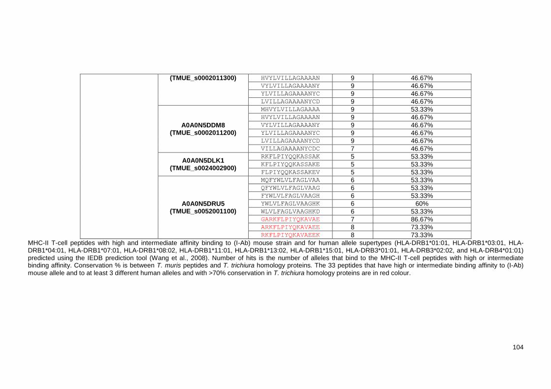
104
(TMUE_s0002011300) HVYLVILLAGAAAAN 9 46.67% VYLVILLAGAAAANY 9 46.67% YLVILLAGAAAANYC 9 46.67% LVILLAGAAAANYCD 9 46.67%
A0A0N5DDM8 (TMUE_s0002011200)
MHVYLVILLAGAAAA 9 53.33% HVYLVILLAGAAAAN 9 46.67% VYLVILLAGAAAANY 9 46.67% YLVILLAGAAAANYC 9 46.67% LVILLAGAAAANYCD 9 46.67% VILLAGAAAANYCDC 7 46.67%
A0A0N5DLK1 (TMUE_s0024002900)
RKFLPIYQQKASSAK 5 53.33% KFLPIYQQKASSAKE 5 53.33% FLPIYQQKASSAKEV 5 53.33%
A0A0N5DRU5 (TMUE_s0052001100)
MQFYWLVLFAGLVAA 6 53.33% QFYWLVLFAGLVAAG 6 53.33% FYWLVLFAGLVAAGH 6 53.33% YWLVLFAGLVAAGHK 6 60% WLVLFAGLVAAGHKD 6 53.33% GARKFLPIYQKAVAE 7 86.67% ARKFLPIYQKAVAEE 8 73.33% RKFLPIYQKAVAEEK 8 73.33%
MHC-II T-cell peptides with high and intermediate affinity binding to (I-Ab) mouse strain and for human allele supertypes (HLA-DRB1*01:01, HLA-DRB1*03:01, HLA-DRB1*04:01, HLA-DRB1*07:01, HLA-DRB1*08:02, HLA-DRB1*11:01, HLA-DRB1*13:02, HLA-DRB1*15:01, HLA-DRB3*01:01, HLA-DRB3*02:02, and HLA-DRB4*01:01) predicted using the IEDB prediction tool (Wang et al., 2008). Number of hits is the number of alleles that bind to the MHC-II T-cell peptides with high or intermediate binding affinity. Conservation % is between T. muris peptides and T. trichiura homology proteins. The 33 peptides that have high or intermediate binding affinity to (I-Ab) mouse allele and to at least 3 different human alleles and with >70% conservation in T. trichiura homology proteins are in red colour.
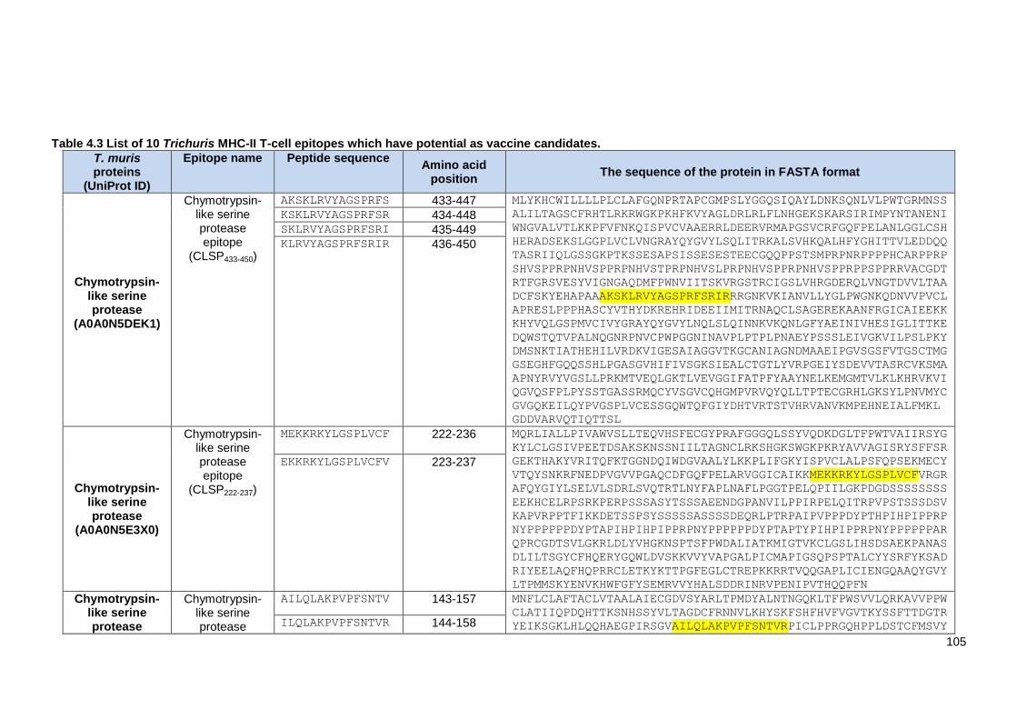
105
Table 4.3 List of 10 Trichuris MHC-II T-cell epitopes which have potential as vaccine candidates.
T. muris proteins
(UniProt ID)
Epitope name Peptide sequence Amino acid
position The sequence of the protein in FASTA format
Chymotrypsin-like serine protease
(A0A0N5DEK1)
Chymotrypsin-like serine protease epitope
(CLSP433-450)
AKSKLRVYAGSPRFS 433-447 MLYKHCWILLLLPLCLAFGQNPRTAPCGMPSLYGGQSIQAYLDNKSQNLVLPWTGRMNSS
ALILTAGSCFRHTLRKRWGKPKHFKVYAGLDRLRLFLNHGEKSKARSIRIMPYNTANENI
WNGVALVTLKKPFVFNKQISPVCVAAERRLDEERVRMAPGSVCRFGQFPELANLGGLCSH
HERADSEKSLGGPLVCLVNGRAYQYGVYLSQLITRKALSVHKQALHFYGHITTVLEDDQQ
TASRIIQLGSSGKPTKSSESAPSISSESESTEECGQQPPSTSMPRPNRPPPPHCARPPRP
SHVSPPRPNHVSPPRPNHVSTPRPNHVSLPRPNHVSPPRPNHVSPPRPPSPPRRVACGDT
RTFGRSVESYVIGNGAQDMFPWNVIITSKVRGSTRCIGSLVHRGDERQLVNGTDVVLTAA
DCFSKYEHAPAAAKSKLRVYAGSPRFSRIRRRGNKVKIANVLLYGLPWGNKQDNVVPVCL
APRESLPPPHASCYVTHYDKREHRIDEEIIMITRNAQCLSAGEREKAANFRGICAIEEKK
KHYVQLGSPMVCIVYGRAYQYGVYLNQLSLQINNKVKQNLGFYAEINIVHESIGLITTKE
DQWSTQTVPALNQGNRPNVCPWPGGNINAVPLPTPLPNAEYPSSSLEIVGKVILPSLPKY
DMSNKTIATHEHILVRDKVIGESAIAGGVTKGCANIAGNDMAAEIPGVSGSFVTGSCTMG
GSEGHFGQQSSHLPGASGVHIFIVSGKSIEALCTGTLYVRPGEIYSDEVVTASRCVKSMA
APNYRVYVGSLLPRKMTVEQLGKTLVEVGGIFATPFYAAYNELKEMGMTVLKLKHRVKVI
QGVQSFPLPYSSTGASSRMQCYVSGVCQHGMPVRVQYQLLTPTECGRHLGKSYLPNVMYC
GVGQKEILQYPVGSPLVCESSGQWTQFGIYDHTVRTSTVHRVANVKMPEHNEIALFMKL
GDDVARVQTIQTTSL
KSKLRVYAGSPRFSR 434-448 SKLRVYAGSPRFSRI 435-449 KLRVYAGSPRFSRIR
436-450
Chymotrypsin-like serine protease
(A0A0N5E3X0)
Chymotrypsin-like serine protease epitope
(CLSP222-237)
MEKKRKYLGSPLVCF
222-236 MQRLIALLPIVAWVSLLTEQVHSFECGYPRAFGGGQLSSYVQDKDGLTFPWTVAIIRSYG
KYLCLGSIVPEETDSAKSKNSSNIILTAGNCLRKSHGKSWGKPKRYAVVAGISRYSFFSR
GEKTHAKYVRITQFKTGGNDQIWDGVAALYLKKPLIFGKYISPVCLALPSFQPSEKMECY
VTQYSNKRFNEDPVGVVPGAQCDFGQFPELARVGGICAIKKMEKKRKYLGSPLVCFVRGR
AFQYGIYLSELVLSDRLSVQTRTLNYFAPLNAFLPGGTPELQPIILGKPDGDSSSSSSSS
EEKHCELRPSRKPERPSSSASYTSSSAEENDGPANVILPPIRPELQITRPVPSTSSSDSV
KAPVRPPTFIKKDETSSPSYSSSSSASSSSDEQRLPTRPAIPVPPPDYPTHPIHPIPPRP
NYPPPPPPDYPTAPIHPIHPIPPRPNYPPPPPPDYPTAPTYPIHPIPPRPNYPPPPPPAR
QPRCGDTSVLGKRLDLYVHGKNSPTSFPWDALIATKMIGTVKCLGSLIHSDSAEKPANAS
DLILTSGYCFHQERYGQWLDVSKKVVYVAPGALPICMAPIGSQPSPTALCYYSRFYKSAD
RIYEELAQFHQPRRCLETKYKTTPGFEGLCTREPKKRRTVQQGAPLICIENGQAAQYGVY
LTPMMSKYENVKHWFGFYSEMRVVYHALSDDRINRVPENIPVTHQQPFN
EKKRKYLGSPLVCFV 223-237
Chymotrypsin-like serine protease
Chymotrypsin-like serine protease
AILQLAKPVPFSNTV 143-157 MNFLCLAFTACLVTAALAIECGDVSYARLTPMDYALNTNGQKLTFPWSVVLQRKAVVPPW
CLATIIQPDQHTTKSNHSSYVLTAGDCFRNNVLKHYSKFSHFHVFVGVTKYSSFTTDGTR
YEIKSGKLHLQQHAEGPIRSGVAILQLAKPVPFSNTVRPICLPPRGQHPPLDSTCFMSVYILQLAKPVPFSNTVR 144-158

106
(A0A0N5DUC1) epitope (CLSP143-158)
HKQQNIIDEVDAPLIFGANCGEFNAVLHKAKGYCSYYPPRQHVTTLGSPLMCIVGVKVFQ
YGVYTTEFQNYIQGKSQYEKLGFFNSVHDAARETIAELPEDRPAATEMPPLSDQLSSSYS
SSSSSEHISSSFSDSQSKEQCPPHCGQSDGSTEEIPTHPELPSSPEKPEPAPLPAPAPLP
KPIPQPIPQPEQPEGNVPSIDDLYPSMPESSDAHAHVSDHQEGYPVSPSVHIVSLAEGKP
EVICTGSLYASARANFTRTVLTSASCVWSRVVSNFMVYMGSVRSFAAGGHGKWLPIYRIS
TKPIMAHHSALKMMGVGVVKLKKPVSAGKGVGFQMPDMFDYATENSQCFVAGVCEHGLSA
AAPVRILSPQECRSRLRGKFYPNIEYCALMRKGAMMKVVGAALVCRTDLEWQWIQYGIYD
FTPEKAHLNDINSKNIKTQEIGIFMKVQGGIKFVKRKEADELLGLYGK
Chymotrypsin-like serine protease epitope
(CLSP424-443)
CTGSLYASARANFTR 424-438 TGSLYASARANFTRT 425-439 GSLYASARANFTRTV 426-440 YASARANFTRTVLTS 429-443
Chymotrypsin-like serine protease epitope
(CLSP398-416)
SDHQEGYPVSPSVHI 398-412 DHQEGYPVSPSVHIV 399-413 HQEGYPVSPSVHIVS 400-414 QEGYPVSPSVHIVSL 401-415 EGYPVSPSVHIVSLA 402-416
Pfam domain-containing
protein (B_9093)
(A0A0N5E6W6)
Pfam domain-containing
protein epitope (Pfam256-273)
NFGEYWFTKPVGSVS 256-270 MQWTSILFVAFGCLQFSAVFSDRCNAESSTSYYYDYKKVQAERVYWKYVKRERTVAAGCV
LDSGEKLDFNQTYRTDFFLLRCDRINETAVVLTPVKCIMHGQELDRGSSVQQGSFIYTCL
EEGGRIGLVITGCVGDKSVVVKFGETFMRKSFLFMCVGEGTRVIHKAVGCVIGGQVVNIH
KTINIGKYWYKCSRYGNGGVQVEVMGCVDSTGKQIDAGDKYRDGGFLFHCKSVGSAVNIV
FAGCIAREFGVLKEFNFGEYWFTKPVGSVSYKVACAGDEKSVVPQVVQCIANMDQGRMVL
KVGECAKYGTDRMFTCMKRKDGTVVARLVTIEEHALSFQTYTAIEGDQCPLPPK
FGEYWFTKPVGSVSY 257-271
GEYWFTKPVGSVSYK 258-272 EYWFTKPVGSVSYKV 259-273
Hypothetical protein
(A0A0N5DRU5)
Hypothetical epitope (H112-128)
GARKFLPIYQKAVAE 112-126 MQFYWLVLFAGLVAAGHKDECDCIKGVGPGYEKSHLDYAGKFKGGARKFLPIYEKAVAEE
KEVLIKGGPGACDYEPCECRHKRGVEDCGCLKGVGPGYEKSYLDYAGKFKGGARKFLPIY
QKAVAEEKEVLIKGGPGACDYEPCECRHKRGLKDCGCCKGVGPGYEKSYLDYDGKFKGGA
RKFLPIYEKAVAEEKEVLVKGGPGACDWEPCECRHKRGLKDCGCLKGVGPGYEKSYLDYA
GKFKGGARKFLPIYEKAVAEEKEVLVKGGPGACDWEPCECRHKRGLKDCGCLKGVGPGYE
KSYLDYAGKFKGGARKFLPIYEKAVAEEKEVLIKGGPGARDCGCKH
ARKFLPIYQKAVAEE 113-127
RKFLPIYQKAVAEEK 114-128
Chitin-binding domain
containing protein
(PF01607) (A0A0N5E6C6)
Chitin-binding
domain containing
protein epitope (CBD36-52)
PRLLKIKWSPTAAST 36-50 MRFVVHPKLATQVASFLGGSSCSIVRKTFRVGPFPRPRLLKIKWSPTAASTAIHPRWTMG
RENAKVAILLVLIALAAFGEAGYSCEGKSNGDYGDPAEPCSSRYFSCVNGKAIGRQCPRG
LYFHPGLDKCDWPHGITACSNVQEKETFRCPAENGDYPDPEHQCSAKFWRCTNGKAEPYA
CMNGQVFDERLARCSPASGKCAFCLHQPKGQYRWTEEEVCSPEYLECVENYGRTTVKRCP
NDLVFNDETKLCQPKSEVAECNPQLLHNEHEEDGGHHLAAGIGVPKARPPLAPAVVPSPH
PAARSVHSCTSPRATGRCTGEYEGCDHGKPVRRLCAVGLVYFEEYGGCVEPTHSPECPKT
VRQASKFHCTDRPGAKLARGRCKPEYVHCMGAIAVRQWCAPDEVFDQQSESCVHVALTDD
CTPAAEPHSHSQTHDTHSIANSICSKEDRQLRRRRALEQCSKYYVQCDDHSGDQVRSCSN
GIYDEMYDRCLPPDLSSNCHKVDGIPFYPLNRRRKRIERSLRHGRQNSMFNPTLFKGDGL
PQIHSAQIGIPPQLELRDEELRANENLPRRETHFGLNKHVKMEMEVGGETIPLPGGEVAQ
VPSIAAASQGRTAVIANLEGLNFDEGPRTAATAVPLIEQNLMPQTGDGIVVPSLDVAQQV
RPRLLKIKWSPTAAS 37-51 RLLKIKWSPTAASTA 38-52
Chitin-binding PAGVVYQCTMPRYTL 1243- 1257
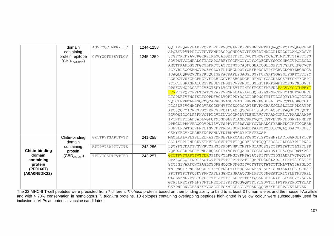
107
domain containing
protein epitope (CBD1243-1259)
AGVVYQCTMPRYTLC 1244-1258
QQIAVPQANVHAPPVQEDLPEPPVGYGAVPPPPPVDNVVETPAQWQQPPQADVQFGRPLP
APQEYVPVTPPFGVTPVFANPPAGPQGMPQAIVPRKVSDTNALGPIKPGSFCANQKDGVY
SYGPCRRDYAVCRGKREIRLSCAIGEIFDPYLFVCTSNYGYQCALTTMTTTTTIAPTTPS
SDVPSTVCLNKADGFYAIAPCSRFYYGCFNGLYQLYQCQFGEVYSQIQHRCIVPGLSCLG
AMQTPKAPLGTFPGTSLPRFCSASFEIWSDCASPCGEATCGLLNPPTTCGRPCKPGCVCR
PGYVRLSQQSRMCVPQEVCLQYTLTNRGLDQTYCRFRPDGLYPYPGRVCDQRYLRCRGGA
IDAQLCQRGEVFDPTKSQCISERACRAPEPSAGGLDSYFCRGKPSGAYNLPGKTCFTIYY
LCSDGTVSFGKCPNGYVFDLHLGCVPPSHCDDGPLDPNDLYCAGKRDGSYTFGNYKCFPI
YYTCISGRANFAICRSVDEDLVFNGHYCVPRNSCLGSLHYIRRPPMPIRYESPFNLSGSF
DPSFCVNQPDGAYPIVECTSPYLVCINSVTTIHYCFPGEIFARVNLPAGVVYQCTMPRYT
LCVPTYPQPYPPFTTATTTVAPTVNNNLCAAPAVDGQLHPLSNNPCERHYINCTGGSFFL
LTCPDRTVFNSTSLTCQPNFACLVQPPPDYRQLCLDRPNGFYTFTLCSQYYLYCQDGIGW
VQTCLNPHWAFNGQTMQCAPRHSVAGCRPAGLHHMPNRPGDLSALDMRCQTLGDRGYEIT
PCQSDFIVCNMGFGDVRSCGSNMVFYGEQQRCARTSEVPACRAKGGDSLCLGKPDGAYPF
APCSQQFYICWRSFSYVERCGPRQIFSAQLQSCVDITSIASCLAQSDPPAQSDPSPQCTT
PGVQSIGQCLPSFHVCTPLGVLILVQCGRGDVFDEHLRVCVPAAACGRSQVPAANAAAPV
IVTNPPPTLADSADLVGFCTNLNDGLYPIANDCRRFVQCSHGTVHVRNCPDGRVFNMDPR
DPHCDLPRNVPACQQVDDISVVTSPPVTTSDSVSNYCVDKADGFYRHMTDCTRFIQCFRR
RSFVLRCATGLVFNPLINVCDFPRRVPDCNIRMEDTAADTPMDSICSQAQDGAFVRDPST
CSAYYRCVHGKAHRFNCPANLVFNTRNNVCDYPTRVPECSP
GVVYQCTMPRYTLCV
1245-1259
Chitin-binding domain
containing protein
(PF01607) (A0A0N5DK22)
Chitin-binding domain
containing protein
(CBD241-257)
GRTTPVTSAPTTVTT
241-255
MAQLLALVFLSSLLELHASVQHSEFLKRCAHIPDGNYPISPCSHNYLACTGARGLIRTCP
SGLIYDPLRNRCEVKTNVPSCCVPTTTTTPQSDVPSTTEQQTFDCSGLLPGDYPLRPNSC
LQQYFTCDADSVGVVRVCPNSLYFDPVNRVCNFFNNIASCSGSTTPPTTATTTLGPTLPP
VQFDCSSRPDGFYPNPARQCSGIYYACTGGQARRLFCGSGLAYDVITRACQSPDNTYACT
GRTTPVTSAPTTVTTERSPIDCVTLPNGIYPNPASACSRIFFVCSDGIAERFVCPGQLYF
DPARQSCQRFNDIFACTGVTTTTTTTPPTTTATTPQMPFDCSSLADGLYPNPTSICSTFY
YICSGSVARRQNCPAGLYYDPEMQQCNSFGNIFVCTGTRQTATTTTTMLVTATSAPGLDC
TNLPNGIYPNPRSQCSPIYFYCTNGFTYEHRCLDDLFFNPELKICDRYSNIFQCTGTRST
PPTTPTPTTPQSDVPFNCAFLPNGNYPNPAAQCSNIFFTCSNGKATIRICPLETFFDPEL
QLCLAFNDVPVCTGTPRTTTTATTTTPLSDVTTPFFQCSNRPNGNYPLGPCEQVYVSCVD
GTPSLRECPPNLFYDFTINECDYIYRIPSCGGQRTTTPISDVTITIPTPPPEFDCTRLAD
GKYPNPRNVCHSNFYYVCAGGRTSKMLCPAGLVYDANLQQCVFYRRPPPCVKTLPVSN
RTTPVTSAPTTVTTE
242-256
TTPVTSAPTTVTTER 243-257
The 33 MHC-II T-cell peptides were predicted from 7 different Trichuris proteins based on their binding ability to bind to at least 3 human alleles and the mouse I-Ab allele and with > 70% conservation in homologous T. trichiura proteins. 10 epitopes containing overlapping peptides highlighted in yellow colour were subsequently used for inclusion in VLPs as potential vaccine candidates.

108
4.3 Discussion
As it infects more than 540 million people worldwide, trichuriasis is one of the world’s most
important neglected infectious diseases (Pullan et al., 2014). Existing anthelmintic treatments are
trusted, but the increasing rates of drug resistance and re-infection pose significant threats to public
health (Zhan et al., 2014). Developing a vaccine is the most desirable and cost-effective approach
to control Trichuris infection, and thus the search for an efficient vaccine against Trichuris infection
has been ongoing for many years (Dixon et al., 2010). To date, researchers have focused on
designing vaccines against Trichuris infection based on ES products or EVs, ES or EV fractions
and, most recently, the T. muris whey acidic protein (WAP) (Briggs et al., 2018; Dixon et al., 2008;
Shears et al., 2018a; Zhan et al., 2014). However, this is the first study to identify a set of novel
MHC-II T-cell epitopes from the Trichuris genome that may induce cross-protection act not only
against T. muris but also against T. trichiura using the RV approach.
Since the advent of immunoinformatics tools for prediction of antigenic epitopes and protein
analysis, promising epitope-based vaccines have been developed against several parasites. For
example, Gu et al. (2017) developed a multi-epitope vaccine against T. spiralis infection based on
CD4+ T-cell epitopes and B-cell epitopes predicted from T. spiralis paramyosin protein (Ts-Pmy)
that were expressed as a soluble recombinant protein (rMEP) in Escherichia coli. Mice immunised
with the rMEP exhibited 55.4% muscle larval reduction, while those immunised with paramyosin
recombinant protein (rTs-Pmy) prior to T. spiralis infection exhibited only 34.4% reduction.
Protection was associated with the production of high levels of anti-rMEP-specific IgG1/IgG2c
antibodies and the secretion of IFN-γ, IL-4 and IL-5 cytokines (Gu et al., 2017).
Based on the promising results of Gu et al.’s study and others, the present study designed a multi
epitope-based vaccine against Trichuris infection. However, since CD4+ T-cells, not B-cells,
protect against Trichuris infection (Else and Grencis, 1996; Koyama et al., 1995), only MHC-II T-
cell epitopes were predicted from different Trichuris proteins. The selected epitopes will be fused
into VLPs, which will serve as a vaccine delivery system.
4.3.1 The choice of secreted proteins as vaccine candidates
Proteins are the first choice for vaccine candidates for many researchers because proteins can be
easily identified and produced in large quantities for inclusion in commercial vaccines (Hein and
Harrison, 2005). In addition, many secreted proteins found in parasite ES products have different
antigenic properties, suggesting that they are great vaccine targets (Dalton et al., 2003; Zarowiecki
and Berriman, 2015). For example, proteins secreted from Leishmania spp have been found to
play an important role in the host-parasite interaction, generating a strong immune response and
protection against infection in mice (John et al., 2012; Santarém et al., 2007). Another study
indicated that boosting bacillus Calmette-Guerin (BCG)-immunised guinea pigs with a purified
recombinant Mycobacterium tuberculosis major secreted protein (30-kDa protein) significantly
boosted the animals’ cell-mediated and humoral immune responses and enhanced protection
against tuberculosis compared to non-boosted BCG-immunised animals (Horwitz et al., 2005).

109
Based on the fact that ES proteins secreted from both adult and larval T. muris elicit strong
immunity and protection against infection in mice (Dixon et al., 2010; Shears et al., 2018b), only
secreted, surface exposed and highly expressed proteins were selected for this study as they
should have a great immunogenic potential during infection and consequently induce protection as
vaccine candidates.
4.3.2 Trichuris proteins selected for epitope prediction
From the whole parasite genome, 10 epitopes containing 33 overlapping peptides were identified.
CBD36-52, CBD1243-1259, and CBD241-257 epitopes were predicted from two different chitin-binding
domain-containing proteins, and the H112-128 epitope was predicted from a hypothetical protein. In
addition, the CLSP433-450, CLSP222-237, CLSP143-158, CLSP424-443 and CLSP398-416 epitopes were
predicted from three different chymotrypsin-like serine proteases, and the Pfam256-273 epitope was
predicted from a Pfam B domain-containing protein.
Although several studies have provided rich information about the host-parasite interaction, the
precise biological function of each Trichuris protein is unclear (Zarowiecki and Berriman, 2015).
Several studies have demonstrated that chitin-binding domain genes are highly expressed in
Caenorhabditis elegans, N. brasiliensis, H. polygyrus, Ascaris lumbricoides, Ancylostoma
ceylanicum, and Anopheles gambiae insects (Foor, 1967; Shen and Jacobs-Lorena, 1998; Tjoelker
et al., 2000; Vannella et al., 2016; Wei et al., 2016). In the nematode C. elegans, chitin-binding
domain-containing proteins are highly expressed in adults, larvae and pharynx cells, suggesting
that they have different functional roles (Veronico et al., 2001). Specifically, they are thought to be
associated with eggshell formation and early single-cell developmental stages, such as secretion of
the chitinous eggshell following fertilisation, maintenance of the osmotic/permeability barrier in the
middle layer of the eggshell, polar-body extrusion and initiation of single-cell embryo (zygote)
polarisation (Johnston et al., 2006, 2010; Nance, 2005; Zhang et al., 2005). Several studies have
also suggested that the acidic mammalian chitinase (AMCase) that constitutively degrades chitin
substrates in the mouse gastrointestinal environment are critical initiators of protective type 2
responses to intestinal nematodes (Ohno et al., 2016; Zhu et al., 2004). For example, Vannella et
al. (2016), demonstrated that, following infection with N. brasiliensis and H. polygyrus, AMCase-
deficient mice developed impaired type 2 immunity as well as reduced mucus production and
decreased intestinal expression of the signature type 2 response genes which regulate T-cell
homing. Taken together, these results suggest that AMCase may play a role in priming protection
against gastrointestinal nematode infections.
There is little published research on Pfam B domain-containing proteins. Nevertheless, based on
the BLAST analyses conducted in this study, they appear to have homologues in other parasites,
including T. trichiura, T. suis, Trichinella spp, Ancylostoma ceylanicum, Diploscapter pachys,
Caenorhabditis spp, Strongyloides spp, Enterobius vermicularis, Ascaris spp, Brugia malayi and
others.
Hypothetical proteins, also known as uncharacterised proteins, are noticeably transcriptionally
upregulated in various stages and sexes in Trichuris spp and other nematode genomes (Foth et al.,
110
2014; Hunt et al., 2016; Jex et al., 2014; Leroux et al., 2018). Also, the RNA content of T. muris EV
fractions, which are characterised using the Illumina HiSeq platform, showed that hypothetical
proteins with unknown function are among the most abundant domains in T. muris (Eichenberger
et al., 2018b). Shears et al. (2018a) also suggested that hypothetical proteins isolated from
T. muris ES and EV proteins are promising vaccine candidates. Thus, vaccinating C57BL/6 mice
with EVs isolated from T. muris ES without an adjuvant prior to infection with a low-dose of T. muris
eggs induced significant protection characterised with low worm burden and a high level of IgG1
anti-parasite antibodies against soluble ES proteins (Shears et al., 2018a).
Chymotrypsin-like serine proteases could also be promising vaccine candidates, given that the
most abundant proteases in the anterior region of T. muris are chymotrypsin A-like serine
proteases (Foth et al., 2014). These proteases have also been found as conserved in the
secretomes of more than 40 helminth spp (Consortium, 2019; Cuesta-Astroz et al., 2017). For
example, Strongyloides stercoralis (Marcilla et al., 2012), Necator americanus (Cantacessi et al.,
2010), A. caninum (Zhan et al., 2003), Trichinella pseudospiralis (Xu et al., 2017), T. spiralis (Wei
et al., 2011), and T. suis (Cantacessi et al., 2011) express high levels of chymotrypsin-like serine
proteases, which have thus been proposed to play an essential role in inducing the host immune
response. Interestingly, the chymotrypsin-like serine protease (TMUE_s0003007500) and
hypothetical proteins (TMUE_s0038007500 and TMUE_s0031002800) identified in this study were
among the secreted proteins with signal peptides in T. muris ES products (Eichenberger et al.,
2018b), confirming that these proteins play a role in the secretory pathway.
Trichuris spp, like other helminth parasites, secrete different proteinases, such as serine, cysteine
and metalloproteases, to overcome the host tissue barrier or obtain essential nutrients for survival
(Hewitson and Maizels, 2014; Izvekova and Frolova, 2017; Viney, 2017). Although the biological
functions of the genes encoding proteases or peptidases remain unclear, helminth serine
proteases seem to play a central role in either the invasion process or modulation of the host
immune response to enhance the survival in the host (Balasubramanian et al., 2010; Santos et al.,
2013; Gomez-Fuentes et al., 2019; Toubarro et al., 2010; Yang et al., 2015). For example,
Strongyloides spp, like many other helminth parasites, produce proteases to break down the host
tissue to facilitate migration to the gut and to digest the host tissue to acquire food (Viney, 2017).
Also, T. muris secretes serine proteases, which are involved in degrading host intestinal mucins 2
(Muc2) polymers within the mucus barrier facilitating mating and egg laying, increasing the host’s
susceptibility to T. muris infection (Drake et al., 1994; Hasnain et al., 2012). Chymotrypsin-like (S1)
serine proteases are also upregulated in the stichosome of T. suis (Jex et al., 2014). It has also
been suggested that some T. suis chymotrypsin-like serine proteases assist in digesting
components of the host, such as blood, serum and tissue (Jex et al., 2014).
Interestingly, immunising BALB/c mice with a recombinant vaccine based on the serine protease
gene (TspSP-1.2) encoding a 35.5 kDa protein from T. spiralis muscle larvae prior to challenging
the mice with T. spiralis infective larvae induced a partial reduction in intestinal adult worms and
muscle larvae (Wang et al., 2013). Furthermore, when the serum from mice vaccinated with
TspSP-1.2 was added to a human colon carcinoma cell line (HCT-8 cells) monolayer overlaid with

111
infective larvae for 1 hour at 37ºC, the invasion rate of the infective larvae into the cell monolayer
was partially prevented (Wang et al., 2013). Another study showed that vaccinating BALB/c mice
with a recombinant serine protease of adult Trichinella spiralis (Ts-Adsp) formulated with an alum
adjuvant prior to T. spiralis challenge induced a reduction in worm burden (Feng et al., 2013). The
results indicated that both humoral and cellular immune responses were induced to serine
proteases, as demonstrated by the elevated levels of specific anti–rTs-Adsp IgG and IgE
antibodies and mixed Th1 (IFN-γ and IL-2) and Th2 (IL-4, IL-10 and IL-13) cytokines (Feng et al.,
2013). Moreover, vaccinating Kunming mice twice with serine proteases from new-born T. spiralis
(Ts-NBLsp) larvae as a naked DNA vaccine prior to challenging the mice with T. spiralis muscle
larvae (ML) led to a 77.93% reduction in ML and boosted anti-Ts-NBLsp-specific IgG responses
(Xu et al., 2017). In another study, Ren et al. (2018) demonstrated that vaccinating BALB/c mice
with recombinant T. spiralis 31 kDa protein (rTs31) containing a domain of a trypsin-like serine
protease prior to challenging the mice with T. spiralis larvae elicited a significant humoral response
and protection, as demonstrated by the 56.93% reduction in adult worms at day 6 p.i. and 53.50%
reduction in muscle larvae at day 42 p.i.. More recently, Sun et al. (2019) demonstrated that
intranasal vaccination of BALB/c mice with T. spiralis serine protease (rTsSP) coupled with cholera
toxin B subunit (CTB) resulted in a 71.10% adult and 62.10% larva reduction after challenge.
Collectively, these studies suggest that Trichuris chymotrypsin-like serine protease could be a
promising protein candidate for inclusion in a vaccine for trichuriasis.
In 2018, Shears et al. fractionated T. muris ES using a combination of size exclusion
chromatography (Superose 12) and gel filtration chromatography (Superdex 75), to identify a set of
potential immunogenic candidates using mass spectrometry. Interestingly, C57BL/6 mice
vaccinated with the ES fraction induced high antibody responses. In addition, stimulating infection-
primed MLN lymphocytes from C57BL/6 mice with ES fractions for 42 hours induced a high level of
Th2 cytokines (IL-9 and IL-13) in the supernatant (Shears et al., 2018b). Interestingly, some of the
proteins identified in this chapter were identified in Shears et al.’s report, including a WAP domain
containing protein (TMUE_s0004017900), Deoxyribonuclease (DNase) II (TMUE_s0030007700),
chymotrypsin-like serine proteases (TMUE_s0191000800, TMUE_s0003007500,
TMUE_s0004005300, TMUE_s0069001100, and TMUE_s0004007100), chitin-binding domain
containing protein (TMUE_s0281000600 and s0018008000), cystatin-domain containing protein
(TMUE_s0122001300), myoglobin (TMUE_s0027006900), a poly cysteine and histidine tailed
protein isoform (TMUE_s0083002300), and hypothetical proteins (TMUE_s0077002200,
TMUE_s0038007500, TMUE_s0031002800, TMUE_s0012009900, TMUE_s0002011300).
Notably, the chymotrypsin-like serine proteases TMUE_s0191000800 and TMUE_s0003007500;
the chitin-binding domain containing protein TMUE_s0281000600; and the hypothetical proteins
TMUE_s0038007500, TMUE_s0031002800, and TMUE_s0012009900, which were selected to
predict the MHC-II T-cell epitopes, were identified in the most immunogenic ES fractions (Shears et
al., 2018b). Most importantly, the final set of selected epitopes identified in this chapter included
three MHC-II T-cell epitopes predicted from these proteins: CLSP222-237, which was predicted from
the chymotrypsin-like serine protease TMUE_s0191000800, and CBD36-52 and CBD1243-1259, which
were predicted from the chitin-binding domain-containing protein TMUE_s0281000600.

112
A better understanding of the biological function of Trichuris proteins might shed new light on host-
parasite interactions, which will help identify potential vaccine candidates. Certainly, the RV
approach facilitated screening for Trichuris immunogenic candidates by reducing the required time
and effort. Otherwise, screening would be difficult to carry out experimentally due to the
complicated genome and variations in immune responses. The provided framework for identifying
potential epitope vaccine candidates within the T. muris and T. trichiura genomes could be used in
future studies to identify epitope-based vaccine candidates against other parasites.
4.4 Summary
Based on the RV approach, a set of proteins were identified from the Trichuris genome
using inclusion and exclusion criteria. A T. muris genome was used as a reference genome
and short-listed proteins according to their secretion pathway and expression level. The
selected proteins were then checked to determine their homology with T. trichiura, human
and mouse proteins using the BLAST tool. After short-listing, 27 proteins were selected as
suitable vaccine targets.
The selected vaccine target proteins were then used to predict MHC-II T-cell epitopes as
promising vaccine antigen candidates using the IEDB in silico prediction tools and
subsequently the predicted peptides were screened for allergenicity and conservancy,
resulting in a final set of 10 Trichuris MHC-II T-cell epitopes containing 33 overlapping
peptides.
113
Chapter Five
Design of HBc-Ag VLP expressing
Trichuris MHC-II T-cell epitopes

114
5.1 Introduction
Virus-like particles (VLPs) are viral structural proteins that have an intrinsic ability to self-assemble
into viral capsids, but they possess the immunostimulatory and self-adjuvanting properties of
natural viruses (Grgacic and Anderson, 2006; Whitacre et al., 2009). Over the years, many VLP-
based vaccines have been engineered to display foreign pathogenic antigens and have proven to
be more immunogenic than recombinant protein-based vaccines (Jennings and Bachmann, 2008;
Roldao et al., 2010). Some of these VLP-based vaccines are used worldwide, such as the hepatitis
B virus (HBV) vaccine (GlaxoSmithKline’s Engerix®) (Keatinggm, 2003) and the human
papillomavirus vaccine (Cervarix®) (Mckeage and Romanowski, 2011). While the anti-malaria
vaccine (RTS, S/AS01) is in clinical development (Dobano et al., 2019), and many others are
undergoing pre-clinical evaluation, such as the human immunodeficiency virus (HIV) and influenza
vaccines (Franco et al., 2011; Song et al., 2011). Also, several VLP-based vaccine candidates are
under investigation, including those against Trichinella spiralis and Clonorchis sinensis (Lee et al.,
2017; Lee et al., 2016).
Several types of VLPs have been used for vaccine development, one of the most promising of
which is the HBV core particle (HBc-Ag) (Chisari and Ferrari, 1995; Qiao et al., 2016; Roose et al.,
2013). This is a self-assembling viral capsid that can induce potent B-cell and T-cell dependent and
independent immune responses without the need for an adjuvant (Milich et al., 1988; Milich and
Mclachlan, 1986). HBc-Ag VLP displaying foreign epitopes has been used as a promising vaccine
carrier against several diseases, including Crohn’s disease (Guan et al., 2009), Anthrax (Yin et al.,
2008), hand-foot-and-mouth disease (Huo et al., 2017), hepatitis B and C (Sominskaya et al.,
2010), malaria caused by Plasmodium falciparum (Nardin et al., 2004), and toxoplasmosis caused
by T. gondii (Guo et al., 2019). Furthermore, HBc-Ag VLP displaying large foreign antigens has
been used as a promising vaccine carrier for diseases such as Mycobacterium tuberculosis (Yin et
al., 2011) and Lyme disease (Nassal et al., 2008). These studies confirm that HBc-Ag VLP is a
safe, efficient and economical vaccine delivery platform that can display antigens of different sizes
and be inexpensively produced in different expression systems.
5.2 Aim and objectives
The overall aim of this chapter was to design and purify HBc-Ag VLPs expressing novel Trichuris
MHC-II T-cell epitopes. To achieve this aim, the current chapter addressed the following objectives:
To design, clone and express a native VLP (HBc-Ag) construct that can be used as a base
construct to insert Trichuris MHC-II T-cell epitopes.
To design, clone and express novel constructs based on VLP (HBc-Ag) expressing the
following Trichuris MHC-II T-cell epitopes: CLSP433-450, CLSP222-237, CLSP143-158, CLSP424-
443, CLSP398-416, Pfam 256-273, H112-128, CBD36-52, CBD1243-1259, and CBD241-257.
To design, clone and express novel constructs based on VLP (HBc-Ag) expressing larger
amino acid sequences in order to enhance the immunogenicity of the construct. The first
construct design, named HBc-mosaic, provides a single expression system that can
display multiple Trichuris epitopes (hypothetical protein A0A0N5DRU5 + CLSP398-416
epitope + chitin-binding domain-containing protein A0A0N5DK22). The second construct,

115
named HBc-HP1-512, is based on VLP (HBc-Ag) expressing the Trichuris hypothetical
protein (A0A0N5DRU5).
To purify VLP recombinant proteins by Strep-Tag affinity chromatography and size
exclusion chromatography (SEC).
To assess the endotoxin contamination of VLP recombinant proteins using the EndoLISA
endotoxin detection assay.
To assess VLP recombinant proteins’ structure and assembly by circular dichroism (CD)
spectroscopy and transmission electron microscopy (TEM).
5.3 Results
5.3.1 Prediction of the recombinant VLP protein’s physicochemical properties
The physicochemical properties derived from the primary structure of each VLP–antigen fusion
(e.g. the molecular mass and isoelectric point) were identified using the Expasy ProtParam server
(Gasteiger et al., 2005) to assist in the identification of stable vaccine candidates (Cox and
Onraedt, 2012; Jain et al., 2015). Table 5.1 details the design and derived physicochemical
properties of the VLP recombinant constructs. The in vivo half-life of all the proteins expressed and
purified in E. coli was estimated to be >10 hours by the Expasy ProtParam server (Varshavsky,
1997). A hydropathic index, the GRAVY, was also calculated. Positive GRAVY values indicated
hydrophobicity, and negative GRAVY values indicated hydrophilicity (Kyte and Doolittle, 1982). All
the constructs have negative values, suggesting that they are soluble.
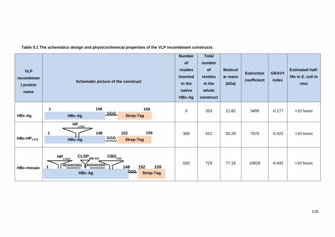
116
Table 5.1 The schematics design and physicochemical properties of the VLP recombinant constructs.
VLP
recombinan
t protein
name
Schematic picture of the construct
Number
of
resides
inserted
in the
native
HBc-Ag
Total
number
of
resides
in the
whole
construct
Molecul
ar mass
(kDa)
Extinction
coefficient
GRAVY
index
Estimated half-
life in E. coli in
vivo
HBc-Ag 0 203 21.82 3495 -0.177 >10 hours
HBc-HP1-512 309 512 55.29 7575 -0.422 >10 hours
HBc-mosaic
520 723 77.16 10818 -0.442 >10 hours
HBc-Ag
1 148
Strep-Tag
159 GGG
HBc-Ag
1 148
Strep-Tag
159 152
GGG
HP1-512
HBc-Ag Strep-Tag GGG 159 152 4GS4GS5G 4GS4GS5G
1 148
HP1-512
CLSP398-416
CBDK22

117
HBc-
Pfam256-273
9 212 22.84 4355 -0.165 >10 hours
HBc-HP112-
128
8 211 22.66 3967 -0.069 >10 hours
HBc-
CLSP433-450
11 214 23.13 3656 -0.242 >10 hours
HBc-
CLSP222-237
12 215 23.27 3669 -0.220 >10 hours
HBc-
CLSP143-158 11 214 22.85 3507 -0.078 >10 hours
HBc-Ag
1 148
Strep-Tag
159 152
GGG
Pfam 256-273
HBc-Ag
1 148
Strep-Tag
159 152 GGG
CLSP433-
450
HBc-Ag
1 148
Strep-Tag
159 152 GGG
CLSP143-158
HBc-Ag
1 148
Strep-Tag
159 152
GGG
CLSP222-237
HBc-Ag
1 148
Strep-Tag
159 152 GGG
H112-128
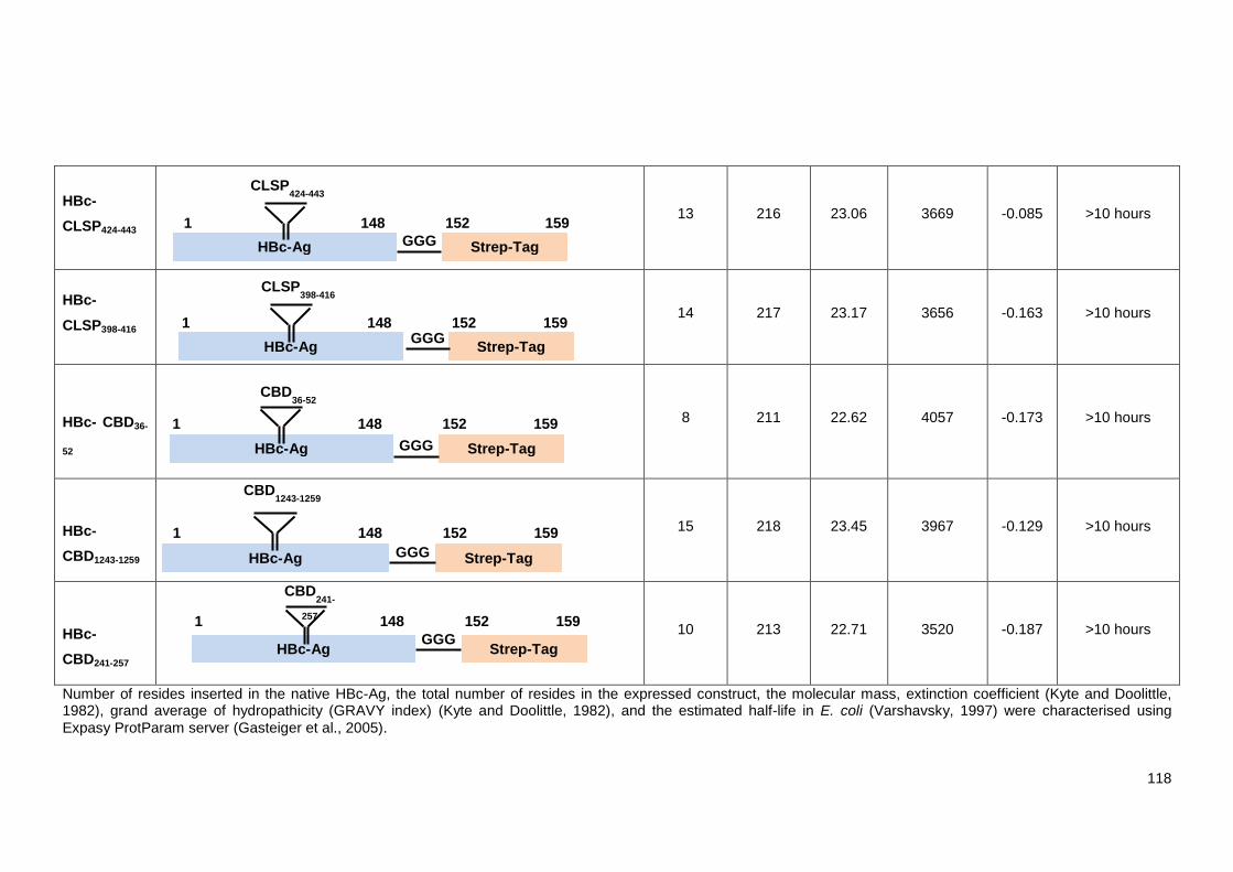
118
HBc-
CLSP424-443
13 216 23.06 3669 -0.085 >10 hours
HBc-
CLSP398-416
14 217 23.17 3656 -0.163 >10 hours
HBc- CBD36-
52
8 211 22.62 4057 -0.173 >10 hours
HBc-
CBD1243-1259
15 218 23.45 3967 -0.129 >10 hours
HBc-
CBD241-257
10 213 22.71 3520 -0.187 >10 hours
Number of resides inserted in the native HBc-Ag, the total number of resides in the expressed construct, the molecular mass, extinction coefficient (Kyte and Doolittle, 1982), grand average of hydropathicity (GRAVY index) (Kyte and Doolittle, 1982), and the estimated half-life in E. coli (Varshavsky, 1997) were characterised using
Expasy ProtParam server (Gasteiger et al., 2005).
HBc-Ag
1 148
Strep-Tag
159 152
CBD36-52
GGG
HBc-Ag
1 148
Strep-Tag
159 152
GGG
CBD241-
257
HBc-Ag
1 148
Strep-Tag
159 152 GGG
CLSP398-416
HBc-Ag
1 148
Strep-Tag
159 152
CBD1243-1259
GGG
HBc-Ag
1 148
Strep-Tag
159 152
GGG
CLSP424-443

119
5.3.2 Design of the native VLP (HBc-Ag)
The design of VLPs expressing Trichuris MHC-II T-cell epitopes was based on the original HBc-Ag,
which consists of a 183-residue sequence (GenBank AAK62975.1). Three important modifications
were made to the native VLP (HBc-Ag) used in this study. First, arginine-rich residues in the C-
terminal domain (150–183 aa) act as RNA-binding sites, which are required for capsid stability and
RNA encapsidation, but not for structural assembly (Birnbaum and Nassal, 1990; Gallina et al.,
1989; Nassal, 1992). Therefore, these sequences were truncated, leaving the N-terminal domain
(148 aa) without disturbing the assembly of the structure. Second, to enhance the flexibility of
insertion of foreign epitopes and improve folding, short linkers (4xGS3xGS) were inserted before
and after the major immunodominant region (MIR) region (Chen et al., 2013; Janssens et al.,
2010). Finally, to facilitate purification of the recombinant constructs using Strep-Tag affinity
chromatography, the Strep-Tag sequence (WSHPQFEK) and glycine-rich (GGG) flexible linkers
were inserted at the C-terminus as described in Chapter 2 (Figure 2.1).
5.3.3 Cloning and expression of the native VLP (HBc-Ag)
The first objective was to verify that the native VLP (HBc-Ag) construct could be cloned, expressed,
purified and used as a base construct to insert Trichuris MHC-II T-cell epitopes. Thus, first, the
pET17b plasmid vector system was digested using Ndel and Xhol restriction enzymes to ligate the
native VLP (HBc-Ag) to construct a recombinant plasmid. The VLP (HBc-Ag) construct was
synthesised (GeneArt) and cloned into the pET17b expression vector for recombinant expression
in Escherichia coli. The construct was verified by DNA sequencing and subsequently transformed
into E. coli Lemo21 (DE3) competent cells for small-scale expression by isopropyl β-d-1-
thiogalactopyranoside (IPTG) induction. Lemo21 (DE3) chemically competent E. coli cells are a
BL21(DE3)-derived strain that is suitable for tuneable T7-driven expression of routine and
challenging proteins under the control of IPTG (Kuipers et al., 2017). After 14–16 hours, cells were
harvested and disrupted by sonication, and the cell lysate was collected in the supernatant after
centrifugation. Monitoring by SDS-PAGE revealed that the native VLP (HBc-Ag) is soluble; a band
at the expected molecular mass of approximately 22 kDa was observed in both the cell lysate (total
protein [TP]) and the soluble protein (SP) (Figure 5.1).
For large-scale expression, the native VLP (HBc-Ag) construct was transformed into ClearColi-
BL21 (DE3) electrocompetent cells for overnight growth and IPTG induction at 16°C.

120
Figure 5.1 SDS-PAGE analysis of the native VLP (HBc-Ag) after small-scale expression. SDS-PAGE (10% gels) stained with Coomassie blue. M: protein marker (Precision Plus protein standard, unstained, Bio-Rad). Lanes ’TP’ and ‘SP’ represent cell lysate (total protein) after sonication and soluble protein supernatant after centrifugation, respectively. A band corresponding to the estimated molecular mass from the HBc-Ag protein sequence (~22 kDa) is present in both the TP and SP lanes is indicated by the arrow.
5.3.4 Purification of the native VLP (HBc-Ag)
The presence of a C-terminal affinity Strep-Tag at the end of the pET17b vector facilitates
purification of the native VLP (HBc-Ag). The HBc-Ag was purified by Strep-Tag affinity
chromatography using StrepTrap HP, as shown in Figure 5.2 (B, Lane 1). The eluted fractions were
then concentrated to 0.5 ml using a Vivaspin concentrator (MWCO 10.000 kDa). Next, the proteins
were subjected to a second round of purification performed by SEC. The SEC chromatogram of the
purified native VLP (HBc-Ag) revealed the purity of the protein; a clear single peak was eluted in
fractions of 7–10 ml, as shown in Figure 5.2 (A). The 120 dimers (T=4) fused via dimer-dimer
interactions to assemble into the HBc-Ag (2x120X22=5.2 MDa). SDS-PAGE analyses also indicate
the presence of a single band of the VLP (HBc-Ag) monomer, with an expected molecular mass of
~22 kDa, as shown in Figure 5.2 (B, Lane 2). Purifying the VLP recombinant protein using two
different methods was essential to remove contaminants and transfer the protein to a more
physiologically appropriate vaccine buffer (1X PBS PH7.4).
KDa25015010050
75
37252015
10
VLP (HBc-Ag)
M TP SP

121
Figure 5.2 The native VLP (HBc-Ag) purification profile and SDS-PAGE analysis. (A) Chromatogram of the native VLP (HBc-Ag) purified by SEC. Separation was carried out on a Superose 6, 10/300 GL (GE Healthcare), at a flow rate of 0.2 ml/min. HBc-Ag was eluted in 0.5 ml fraction and a sample of the SEC elution peak was analysed on SDS-PAGE. Vertical red lines indicate the elution volume of mass standards, from left to right: 2MDa, 670kDa. HBc-Ag was eluted in a single peak at elution voulme= 7 ml. (B) Purification monitored by SDS-PAGE (10% gel) stained with Coomassie blue. M: protein marker (Precision Plus protein standard, unstained, Bio-Rad); Lane 1: HBc-Ag after Strep-Tag affinity purification using the StrepTrap HP, and Lane 2: HBc-Ag after SEC purification. The arrow represents HBc-Ag band that corresponds to the estimated molecular mass of ~22 kDa.
5.3.5 Cloning and expression of HBc-Ag containing Trichuris antigens
Foreign epitope sequences can be inserted and displayed in three different regions on HBc
particles: the N- and C-terminal regions and the MIR at the tips of the spike (position 79/80)
(Pumpens and Grens, 1999, 2001). The MIR has been identified as a superior site as it allows for
insertion of up to 238 amino acid residues and thus can induce a stronger immune response in
mice compared to the N- and C- termini (Koletzki et al., 1999; Kratz et al., 1999; Schödel et al.,
1992). Therefore, all the target antigens were inserted in the MIR of the native VLP (HBc-Ag).
The processes of cloning, expressing and purifying VLP recombinant protein is divided into six
steps (Figure 5.3). The process of expression of VLPs with Trichuris MHC-II T-cell epitopes was
similar to that for expressing native VLP (HBc-Ag), except the MHC-II T-cell epitope primers were
annealed and the resulting ligation was inserted into linearised VLP (HBc-Ag) in the MIR before
expression and transformation into Lemo21 (DE3) competent cells for small-scale expression in
order to monitor the proteins’ solubility. The MHC-II T-cell epitope sequences and primers used to
amplify the target can be found in Chapter 2 (Table 2.2).
Visualisation of protein solubility by SDS PAGE revealed that the VLP recombinant proteins (HBc-
CBD36-52, HBc-CBD1243-1259, HBc-CBD241-257, HBc-CLSP433-450, HBc-CLSP222-237, HBc-CLSP143-158,
HBc-CLSP424-443, HBc-CLSP398-416, and HBc-H112-128) were found to be expressed as soluble
KDa25015010075
5037
2520
15
10
M 1 2
VLP (HBc- Ag) 0
200
400
600
800
1000
1200
1400
0 5 10 15 20
UV
Ab
sorp
tio
n (
mA
U)
Elute Volume, Ve (ml)
5MDa 670kDa
A B

122
products. A band with the expected molecular mass (approximately 22 kDa) was present in both
the crude lysates and soluble proteins after centrifugation (Figures 5.4 and 5.5). Also, the total cell
lysate showed a wide range of proteins prior to sonication and centrifugation (Figures 5.4 and 5.5,
TP lanes). However, HBc-Pfam 256-273 did not give a band in the soluble protein (Fig 5.6),
suggesting that it was insoluble when overexpressed in E. coli.
Larger amino acid sequences (HBc-HP1-512, and HBc-mosaic) were also inserted in the MIR of the
native VLP (HBc-Ag). The full genetic sequences of the constructs can be found in Chapter 2
(Table 2.3). The CamSol method (Sormanni et al., 2015) was used to predict whether large foreign
antigens were compatible with the VLP assembly (Frietze et al., 2016; Joshi et al., 2013). Although
the software predicted that HBc-HP1-512 and HBc-mosaic could be soluble, the two constructs were
insoluble when expressed in E. coli (Figures 5.7 and 5.8). Therefore, they were not purified further.
The constructs identified as soluble were transformed into ClearColi-BL21 (DE3) electrocompetent
cells for overnight growth and IPTG induction for large-scale expression.
Figure 5.3 Protocol used for VLP recombinant proteins cloning, expression and purification. First, the Trichuris antigen was cloned into the MIR of the VLP (HBc-Ag). The correct construct confirmed by the DNA sequencing were expressed after IPTG induction for small-scale expression to assess the solubility. Only soluble proteins were then expressed for large-scale expression. The VLP recombinant proteins were then purified twice using Strep-Tag affinity and SEC. Finally, the VLP recombinant protein structure and assembly were assessed using CD and TEM analysis tools.
Cloning Trichuris antigens into the MIR of the native VLP (HBc-Ag)
Small Scale Expression
Soluble VLP recombinant protein
Large -scale expression
Strep-Tag affinity chromotogrophy
Size exclusion chromotogrogy
CD and TEM
Insoluble VLP recombinant protein
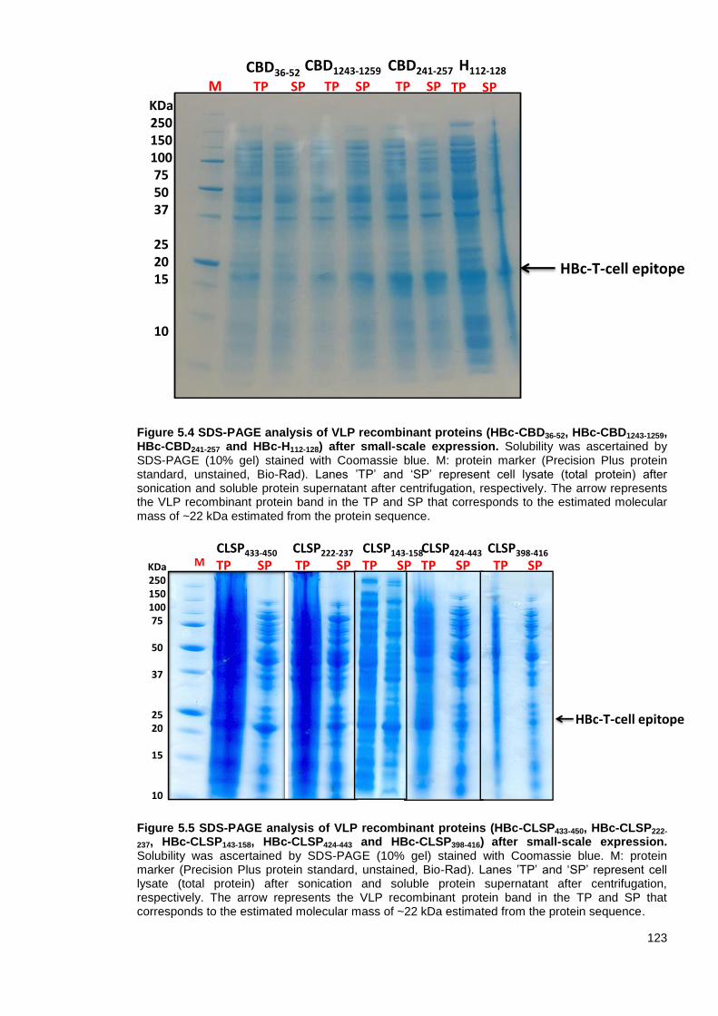
123
Figure 5.4 SDS-PAGE analysis of VLP recombinant proteins (HBc-CBD36-52, HBc-CBD1243-1259, HBc-CBD241-257 and HBc-H112-128) after small-scale expression. Solubility was ascertained by SDS-PAGE (10% gel) stained with Coomassie blue. M: protein marker (Precision Plus protein standard, unstained, Bio-Rad). Lanes ’TP’ and ‘SP’ represent cell lysate (total protein) after sonication and soluble protein supernatant after centrifugation, respectively. The arrow represents the VLP recombinant protein band in the TP and SP that corresponds to the estimated molecular mass of ~22 kDa estimated from the protein sequence.
Figure 5.5 SDS-PAGE analysis of VLP recombinant proteins (HBc-CLSP433-450, HBc-CLSP222-
237, HBc-CLSP143-158, HBc-CLSP424-443 and HBc-CLSP398-416) after small-scale expression. Solubility was ascertained by SDS-PAGE (10% gel) stained with Coomassie blue. M: protein marker (Precision Plus protein standard, unstained, Bio-Rad). Lanes ’TP’ and ‘SP’ represent cell lysate (total protein) after sonication and soluble protein supernatant after centrifugation, respectively. The arrow represents the VLP recombinant protein band in the TP and SP that corresponds to the estimated molecular mass of ~22 kDa estimated from the protein sequence.
HBc-T-cell epitope
MKDa250150100755037
252015
10
TP SP TP SP TP SP
CBD36-52CBD1243-1259 CBD241-257 H112-128
TP SP
HBc-T-cell epitope
MKDa25015010075
50
37
2520
15
10
CLSP433-450
TP SPCLSP222-237
TP SPCLSP143-158
TP SPCLSP424-443
TP SPCLSP398-416
TP SP

124
Figure 5.6 SDS-PAGE analysis of HBc-Pfam256-273 after small-scale expression. Solubility was ascertained by SDS-PAGE (10% gel) stained with Coomassie blue. M: protein marker (Precision Plus protein standard, unstained, Bio-Rad Lanes ’TP’ and ‘SP’ represent cell lysate (total protein) after sonication and soluble protein after centrifugation, respectively. The arrow represents the HBc-Pfam256-273 band in the TP that corresponds to the estimated molecular mass of ~22 kDa estimated from the protein sequence.
Figure 5.7 SDS-PAGE analysis of HBc-HP1-512 after small-scale expression. Solubility was ascertained by SDS-PAGE (10% gel) stained with Coomassie blue. M: protein marker (Precision Plus protein standard, unstained, Bio-Rad). Lanes ’TP’ and ‘SP’ represent cell lysate (total protein) after sonication and soluble protein after centrifugation, respectively. The arrow represents the HBc-HP1-512 band in the TP that corresponds to the estimated molecular mass of ~55.29 kDa
estimated from the protein sequence.
KDa250150100755037
252015
10
HBc-Pfam256-273
M TP SP
KDa25015010075
50
37
252015
10
HBc-HP1-512
M TP SP

125
Figure 5.8 SDS-PAGE analysis of HBc-mosaic after small-scale expression. Solubility was ascertained by SDS-PAGE (10% gel) stained with Coomassie blue. M: protein marker (Precision Plus protein standard, unstained, Bio-Rad); TP: total protein (cell lysate) after sonication; SP: soluble protein after centrifugation. The arrow represents the HBc-mosaic band in the TP that corresponds to the estimated molecular mass of ~77.70 kDa estimated from the protein sequence.
5.3.6 Purification of VLP recombinant proteins
The VLP recombinant proteins HBc-H112-128 (Fig 5.9 B, Lane 1), HBc-CBD36-52
(D), HBc-CBD1243-1259
(F), and HBc-CBD241-257 (H), HBc-CLSP433-450 (J), HBc-CLSP143-158 (L), and HBc-CLSP398-416 (N)
were successfully purified by Strep-Tag affinity chromatography using the StrepTrap HP. The
eluted fractions were then concentrated using a Vivaspin concentrator (MWCO 10.000 kDa). A
second round of purification with SEC was performed to produce a homologous population of
folded VLPs. Monitoring the SEC chromatogram of the purified VLP recombinant proteins revealed
the purity of the proteins, which featured a clear single peak in fractions 7–10 ml (Fig 5.9 A, C, E,
G, I, K and M). The SDS-PAGE analyses also revealed a single band, confirming the purity of the
protein monomer with an expected molecular mass of ~22 kDa (Fig 5.9 B, D, F, H, J, L and N,
Lane 2). However, two defined peaks were observed in the SEC profiles of HBc-CLSP222-237 and
HBc-CLSP424-443 (Fig 5.10 A and C), suggesting that these two VLP recombinant proteins were not
assembled properly. The apparent small difference in the molecular mass of the native HBc-Ag and
HBc expressing Trichuris MHC-II T-cell epitopes is due to the small number of residues inserted in
the HBc-Ag. It was also observed that HBc-Ag, HBc-H112-128, HBc-CBD241-257, HBc-CLSP143-158, and
HBc-CLSP398-416 exhibited adequate expression, with an average yield of 8 mg per 2L culture.
However, HBc-CBD36-52, HBc-CBD1243-1259, HBc-CLSP433-450 and HBc-CLSP143-158 had an
expression with an average yield of 4 mg per 2L culture, while HBc-CLSP222-237 and HBc-CLSP424-
443 had an expression with an average yield <4 mg per 2L culture.
KDa25015010075
50
37
252015
10
HBc-mosaic
TP SP M

126
Figure 5.9 The VLP recombinant proteins (HBc-H112-128, HBc-CBD36-52
, HBc-CBD1243-1259
and
HBc-CBD241-257
, HBc-CLSP433-450, HBc-CLSP143-158, and HBc-CLSP398-416) purification profile
and SDS-PAGE analysis. Left: Chromatogram of the VLP recombinant proteins purified using SEC. Separation was carried out on a Superose 6, 10/300 GL (GE Healthcare), at a flow rate of 0.2 ml/min. VLP recombinant proteins were eluted in 0.5 ml fraction and a sample of SEC the elution peak was visualised by SDS-PAGE. Vertical red lines indicate the elution volume of mass standards, from left to right: 2MDa, 670kDa. Right: Purification visualised by SDS-PAGE (10% gel) analysis stained with Coomassie blue. M: protein marker (Precision Plus protein standard, unstained, Bio-Rad); Lane 1: VLP recombinant protein after Strep-Tag affinity purification using the StrepTrap HP, and Lane 2: VLP recombinant protein after SEC purification. The arrow represents VLP recombinant protein monomer with an expected molecular mass of ~22 kDa.
0
100
200
300
400
0 5 10 15 20UV
Ab
sorp
tio
n (
mA
U)
Elute volume, Ve (ml)
KDa
25
20
15
10
HBc-H112-128
5MDa 670kDa1 2
0
50
100
150
0 5 10 15 20UV
Ab
sorp
tio
n (
mA
U)
Elute Volume, Ve (ml)
0
100
200
300
0 5 10 15 20UV
Ab
sorp
tio
n (
mA
U)
Elute Volume, Ve (ml)
0
100
200
300
400
0 5 10 15 20
Elute Volume, Ve (ml)UV
Ab
sorp
tio
n (
mA
U)
KDa
25
20
15
10
KDa
25
20
15
10
B
KDa
25
20
15
10
C
HBc-CBD36-52
HBc-CBD1243-1259
HBc-CBD241-257
5MDa 670kDa
HBc-CLSP433-450
KDa
25
20
15
100
50
100
150
200
0 5 10 15 20Elute volume, Ve (ml)
UV
Ab
sorp
tio
n (
mA
U)
5MDa 670kDa
HBc-CLSP143-158
KDa
25
20
15
10
0
200
400
600
800
1000
0 5 10 15 20Elute Volume, Ve (ml)
UV
Ab
sorp
tio
n (
mA
U)
5MDa 670kDa
HBc-CLSP398-416
KDa
25
20
15
10
0200400600800
100012001400
0 5 10 15 20
UV
Ab
sorp
tio
n
Elute Volume, Ve (ml)
5MDa 670kDa
5MDa 670kDa
D
E F
5MDa 670kDaG H
I J
K L
NM
1 2
1 2
1 2
1 2
1 2
1 2

127
Figure 5.10 The recombinant proteins (HBc-CLSP222-237, and HBc-CLSP424-443) purification profile and SDS-PAGE analysis. Left: Chromatogram of the VLP recombinant proteins purified using SEC. Separation was carried out on a Superose 6, 10/300 GL (GE Healthcare), at a flow rate of 0.2 ml/min. VLP recombinant proteins were eluted in 0.5 ml fraction and a sample of the SEC elution peak 1 was visualised by SDS-PAGE. Vertical red lines indicate the elution volume of mass standards, from left to right: 2MDa, 670kDa. Right: Purification visualised by SDS-PAGE (10% gel) stained with Coomassie blue. M: protein marker (Precision Plus protein standard, unstained, Bio-Rad); Lane 1: VLP recombinant protein after Strep-Tag affinity purification using the StrepTrap HP, and Lane 2: VLP recombinant protein after SEC purification. The arrow represents VLP recombinant protein monomer with an expected molecular mass of ~22 kDa.
5.3.7 Endotoxin levels of the purified VLP recombinant proteins
Gram-negative E. coli bacteria are a widely preferred prokaryotic expression system for producing
vaccines and therapeutic products because of their rapid growth, high efficacy and ease of culture
(Kyriakopoulos and Kontoravdi, 2013). However, overexpression of VLPs in E. coli introduces an
increased risk of lipopolysaccharide (LPS) contamination. LPS, also known as an endotoxin, is the
primary element in the outer membranes of gram-negative bacteria. It consists of a hydrophobic
domain (lipid A), an oligosaccharide (core) and a polysaccharide (O-antigen) (Liu et al., 1997).
When LPS is recognised by TLR4, a strong inflammatory response is induced, which can lead to
septic shock (Raetz and Whitfield, 2002; Shimazu et al., 1999). It is therefore, essential to prevent
endotoxin contamination when developing a vaccine or drug intended for human use.
VLPs have a large surface area, which increases their susceptibility to LPS contamination and can
trap endotoxin within the VLP structure. Therefore, all the procedures and methods used to purify
the proteins, such as cleaning the containers and chromatography columns with 1 M NaOH prior to
purification and use of two different methods to purify the recombinant proteins, were specifically
designed to eliminate endotoxin contamination (Petsch and Anspach, 2000; Reichelt et al., 2006).
Furthermore, to purify VLP recombinant proteins with minimal endotoxic activity, E. coli (ClearColi
KDa
25
20
15
10
1 2
HBc- CLSP222-237
KDa
25
20
15
10
1 2
HBc- CLSP424-443
A
020406080
100120
0 10 20 30 40UV
Ab
so
rpti
on
(m
AU
)
Elute Volume, Ve (ml)
Peak 2
Peak 1
5MDa 670kDa
0
50
100
150
200
0 10 20 30 40
Elute Volume, Ve (ml)
UV
Ab
sorp
tio
n (
mA
U)
Peak 2Peak 1
B
5MDa 670kDa

128
BL21) electrocompetent cells were used in this study (Huang et al., 2017). The ClearColi strain
contains seven genetic deletions within the lipid A synthesis pathway and thus lacks all the
carbohydrate decorations that are usually attached to LPS to produce a genetically modified LPS
(Lipid IVA) (Mamat et al., 2013). The Lipid IVA is sufficient for cell viability but is incapable of
interacting with TLR4 receptors and inducing an endotoxic response in humans (Miroux and
Walker, 1996; Rosano and Ceccarelli, 2014).
The manufacturer of Lucigen ClearColi cells indicated in the user manual that the standard
methods used to measure endotoxin levels, such as the Limulus amebocyte lysate (LAL) assay,
were unable to discriminate between the modified (Lipid IVA) and unmodified LPS (Lipid A).
https://www.lucigen.com/faq-clearcoli.htm. The LAL assay relies on blood cells (amoebocytes)
extracted from the horseshoe crab, Limulus Polyphemus, to detect endotoxins (Novitsky, 1998).
Therefore, LPS contamination was tested using the EndoLISA endotoxin detection assay, which
relies on a recombinant bacteriophage protein (Factor C) that is activated upon contact with
endotoxins, facilitating reliable quantification of the degree of enzyme activity as a surrogate
measurement of LPS activation (Grallert et al., 2011). However, as with the LAL assay, there were
no assurances that the EndoLISA would be capable to differentiate between the modified and
unmodified LPS (Lipid IVA and Lipid A).
Endotoxin limits are expressed as international endotoxin units (IUs or EUs). According to the
United States Pharmacopeia (USP) and the United States Food and Drug Administration (FDA),
the recommended endotoxin level in recombinant subunits and polysaccharide vaccines is < 20
EU/ml (~3 ng/ml) and < 10 EU/ml (~1.5 ng/ml) for gene vector vaccines (Brito and Singh, 2011).
The level of endotoxins in purified VLP recombinant proteins was unacceptably high (~100 ng/ml),
as shown in Table 5.2. The fact that specific measures were taken into consideration when
purifying the proteins to minimise LPS contamination suggests that the EndoLISA detection assay,
like the LAL assay, cannot discriminate between modified and unmodified LPS.
Table 5.2 Endotoxin concentration of the purified VLP recombinant proteins determined by EndoLISA detection assay.
VLP Recombinant Protein ng/ml
HBc-CBD36-52 47.07
HBc-CBD1243-1259 56.13
HBc-H112-128 108.9
HBc-CLSP433-450 154.2
5.3.8 Circular dichroism (CD) spectroscopy
Circular dichroism (CD) spectroscopy was used to examine the composition of the secondary
structure to verify that the folding of the native VLP (HBc-Ag) and VLPs expressing Trichuris MHC-
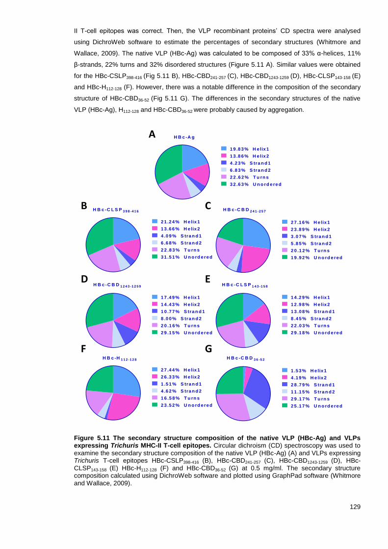
129
II T-cell epitopes was correct. Then, the VLP recombinant proteins’ CD spectra were analysed
using DichroWeb software to estimate the percentages of secondary structures (Whitmore and
Wallace, 2009). The native VLP (HBc-Ag) was calculated to be composed of 33% α-helices, 11%
β-strands, 22% turns and 32% disordered structures (Figure 5.11 A). Similar values were obtained
for the HBc-CSLP398-416 (Fig 5.11 B), HBc-CBD241-257 (C), HBc-CBD1243-1259 (D), HBc-CLSP143-158 (E)
and HBc-H112-128 (F). However, there was a notable difference in the composition of the secondary
structure of HBc-CBD36-52 (Fig 5.11 G). The differences in the secondary structures of the native
VLP (HBc-Ag), H112-128 and HBc-CBD36-52 were probably caused by aggregation.
Figure 5.11 The secondary structure composition of the native VLP (HBc-Ag) and VLPs expressing Trichuris MHC-II T-cell epitopes. Circular dichroism (CD) spectroscopy was used to examine the secondary structure composition of the native VLP (HBc-Ag) (A) and VLPs expressing Trichuris T-cell epitopes HBc-CSLP398-416 (B), HBc-CBD241-257 (C), HBc-CBD1243-1259 (D), HBc-CLSP143-158 (E) HBc-H112-128 (F) and HBc-CBD36-52 (G) at 0.5 mg/ml. The secondary structure composition calculated using DichroWeb software and plotted using GraphPad software (Whitmore and Wallace, 2009).
A
B C
D E
F G
1 9 .8 3 % H e lix 1
1 3 .8 6 % H e lix 2
4 .2 3 % S tra n d 1
6 .8 3 % S tra n d 2
H B c -A g
2 2 .6 2 % T u rn s
3 2 .6 3 % U n o rd e re d
2 7 .4 4 % H e lix 1
2 6 .3 3 % H e lix 2
1 .5 1 % S tra n d 1
4 .6 2 % S tra n d 2
H B c -H 1 1 2 -1 2 8
1 6 .5 8 % T u rn s
2 3 .5 2 % U n o rd e re d
1 .5 3 % H e lix 1
4 .1 9 % H e lix 2
2 8 .7 9 % S tra n d 1
1 1 .1 5 % S tra n d 2
H B c -C B D 3 6 -5 2
2 9 .1 7 % T u rn s
2 5 .1 7 % U n o rd e re d
1 7 .4 9 % H e lix 1
1 4 .4 3 % H e lix 2
1 0 .7 7 % S tra n d 1
8 .0 0 % S tra n d 2
H B c -C B D 1 2 4 3 -1 2 5 9
2 0 .1 6 % T u rn s
2 9 .1 5 % U n o rd e re d
2 7 .1 6 % H e lix 1
2 3 .8 9 % H e lix 2
3 .0 7 % S tra n d 1
5 .8 5 % S tra n d 2
H B c -C B D 2 4 1 -2 5 7
2 0 .1 2 % T u rn s
1 9 .9 2 % U n o rd e re d
1 4 .2 9 % H e lix 1
1 2 .9 8 % H e lix 2
1 3 .0 8 % S tra n d 1
8 .4 5 % S tra n d 2
H B c -C L S P 1 4 3 -1 5 8
2 2 .0 3 % T u rn s
2 9 .1 8 % U n o rd e re d
2 1 .2 4 % H e lix 1
1 3 .6 6 % H e lix 2
4 .0 9 % S tra n d 1
6 .6 8 % S tra n d 2
H B c -C L S P 3 9 8 -4 1 6
2 2 .8 3 % T u rn s
3 1 .5 1 % U n o rd e re d

130
5.3.9 Characterisation of VLP recombinant protein assembly by TEM
The self-assembly of the purified VLP recombinant proteins was verified by TEM with negative
staining. Within the resolution limits of TEM, native VLP (HBc-Ag) particles appeared to be uniform
and spherical at diameters around 30 nm (Figure 5.12 A). In contrast to the native VLP, HBc-Ag
expressing Trichuris MHC-II T-cell epitopes appeared to be heterogeneous (Figure 5.12 C–F). In
addition, HBc-H112-128 was aggregated (Figure 5.12 B). Table 5.3 summarises the VLP recombinant
proteins expression trials.
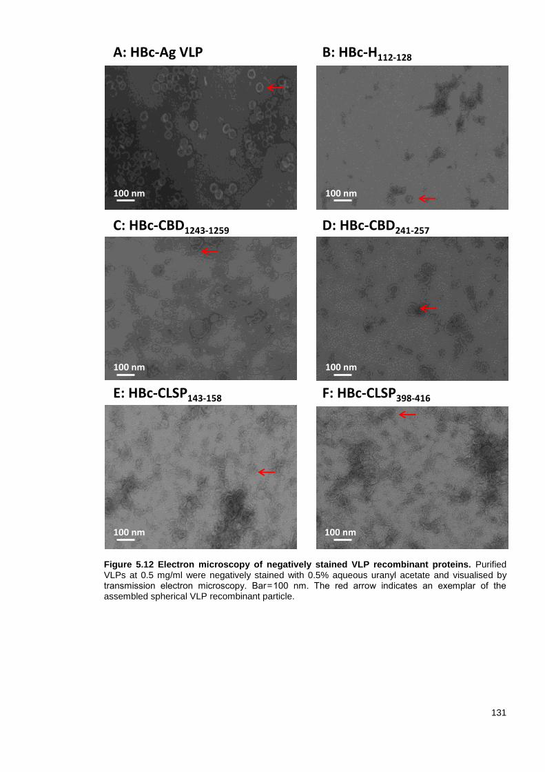
131
Figure 5.12 Electron microscopy of negatively stained VLP recombinant proteins. Purified VLPs at 0.5 mg/ml were negatively stained with 0.5% aqueous uranyl acetate and visualised by transmission electron microscopy. Bar = 100 nm. The red arrow indicates an exemplar of the assembled spherical VLP recombinant particle.
F: HBc-CLSP398-416
A: HBc-Ag VLP B: HBc-H112-128
C: HBc-CBD1243-1259
E: HBc-CLSP143-158
D: HBc-CBD241-257
100 nm
100 nm 100 nm
100 nm
100 nm 100 nm

132
Table 5.3 Summary of the VLP recombinant protein properties.
VLP recombinant
protein name
Confirmed
expression
in E. coli
Average
yield (mg) in
2L culture
EM analysis
Similar secondary
structure
composition to the
native HBc-Ag
VLP (HBc-Ag) Soluble 6
Uniform spherical
particles
Folded
HBc-HP1-512 insoluble ND ND ND
HBc-mosaic insoluble ND ND ND
HBc-Pfam 256-273 insoluble ND ND ND
HBc-H112-128 Soluble 6 Aggregates Yes
HBc-CLSP433-450 Soluble 4 ND ND
HBc-CLSP222-237 Soluble ≤4 ND ND
HBc-CLSP143-158 Soluble 4 Heterogeneous
particles Yes
HBc-CLSP424-443 Soluble ≤4 ND ND
HBc-CLSP398-416 Soluble 6 Heterogeneous
particles Yes
HBc-CBD36-52 Soluble 4 ND
Notable difference
in the composition
of the secondary
structure
HBc-CBD1243-1259 Soluble 4 Heterogeneous
particles Yes
HBc-CBD241-257 Soluble 6 Heterogeneous
particles Yes
ND: Not done.

133
5.4 Discussion
HBc-Ag was the first identified VLP carrier, and it remains one of the most flexible and promising
VLP-based vaccine delivery systems for use in humans (Pumpens and Grens, 2001). HBc-Ag has
several key features that make it suitable for vaccine delivery, such as its non-replicating, non-
infective and self-assembled nature; the high efficiency with which foreign epitopes can be
inserted; and its ability to induce a strong immune response without the need for an adjuvant
(Pumpens and Grens, 1999; Qiao et al., 2016; Roose et al., 2013).
5.4.1 Selection of E. coli for expressing VLP recombinant proteins.
Major factors affecting VLP formulation are the safety and cost of the expression system to be
scaled up for vaccine production (Kushnir et al., 2012). HBc-Ag VLP, as a vaccine carrier, has high
levels of expression in several prokaryotic and eukaryotic expression systems, including bacteria
(Bian et al., 2006; Wang et al., 2016; Zhang et al., 2007), yeast (Li et al., 2007; Rolland et al.,
2001), insect (Brandenburg et al., 2005), and plant expression systems (Makarkov et al., 2019).
All the VLP recombinant proteins were expressed in E. coli because it is the most common
expression system for industrial production due to its ease of use, cost-effectiveness and high level
of expression of recombinant proteins compared to yeast and insect cells (Choi et al., 2006; Huang
et al., 2017; Middelberg et al., 2011). However, there is a high risk of LPS or endotoxin
contamination in the purified proteins (Grgacic and Anderson, 2006; Kushnir et al., 2012).
Nevertheless, several recombinant proteins obtained from E. coli have been approved by the FDA
and are widely used in therapy and vaccines. One example is the first recombinant human insulin
(Humulin-US/Humuline-EU) to be used for treating diabetes (Altman, 1982) and the first VLP-based
vaccine against hepatitis E virus (HEV) to be licensed in China (Wedemeyer and Pischke, 2011). In
addition, two VLP-based vaccines against human papillomavirus (HPV), the causative agent of
cervical cancer, are undergoing clinical trials (Wu et al., 2015). The level of endotoxin in the purified
VLPs was unacceptably high; however, the method used to measure the LPS failed to differentiate
between the modified and unmodified LPS. Future work may include quantifying LPS in the purified
proteins using a more reliable assay such as the HEK-Blue™ hTLR4 cells detection assay (Govers
et al., 2016).
Despite the success of the bacterial expression system in VLP production for many vaccines, it
should be noted that yeast expression systems are effective alternatives that overcome the
problem of endotoxin contamination during vaccine preparation. For example, the current HBV
vaccine used in the US is composed of a hepatitis B surface antigen (HBsAg) VLP expressed on
Saccharomyces cerevisiae yeast (Mcaleer et al., 1984). However, the expression of VLPs in yeast
has several limitations compared to expression in bacteria (Freivalds et al., 2006; Zeltins, 2013).
For instance, the VLP yield in yeast expression system is less than that in E. coli, and the yeast
system is more suitable for production of non-enveloped VLPs than enveloped VLPs (Zeltins,
2013). A mammalian expression system has also been effectively utilised for VLP vaccine
production, but it is not commonly used (Noad and Roy, 2003).
134
5.4.2 Insertion capacity and solubility of the native VLP (HBc-Ag)
A VLP’s solubility and ability to assemble is highly dependent on its type, the site of insertion and
the characteristics of the inserted peptides, such as their size, isoelectric point and hydrophobicity
(Hill et al., 2018). Insertion of short peptides by genetic fusion into a VLP is often easier than
insertion of larger antigens, and it usually does not interfere with VLP assembly. However, insertion
of large proteins or mosaics (i.e. multiple epitopes) into one VLP carrier is more challenging
(Pumpens and Grens, 2001). To overcome this problem, chemical conjugation, whereby VLPs and
target antigens are expressed separately and brought together by means of chemical cross-linking
to allow for more precise surface decoration of VLPs with large foreign antigens (Fogarty and
Swartz, 2018; Patel and Swartz, 2011; Vogel and Bachmann, 2019). Chemical conjugation is
primarily based on the modification of free amino group residues, such as lysine (Lys), carboxylic
groups, sulfhydryl groups or hydroxyl groups, on the surface of VLPs using commercially available
bifunctional cross-linker reagents (Mohsen et al., 2017b).
Previous studies have demonstrated the capacity of native VLP (HBc-Ag) to accommodate a whole
protein by chemical coupling or genetic engineering (Akhras et al., 2017; Hill et al., 2018; Howarth
and Brune, 2018; Peacey et al., 2007; Qi et al., 2015). In addition to the ability to insert a large
target antigen at one site, a study of the Malariavax vaccine demonstrated that, as carriers, HBc-Ag
VLPs have the capacity to accommodate different foreign epitopes at different sites and elicit
specific immunological responses to them (Gregson et al., 2008). HBc-HP1-512 and the HBc-mosaic
construct were insoluble when expressed in E. coli, and therefore, they were not further purified by
SEC. Future work may include chemical coupling to solve this problem or use a different
expression system to attempt expression of large proteins.
5.4.3 Assessing VLP recombinant proteins’ structure, stability and assembly
It is well-established that VLP-based vaccines are often safer and more effective than conventional
vaccines (Roldao et al., 2010). However, the VLPs must be correctly folded to induce a strong and
long-lasting immune response (Frietze et al., 2016). Also, the native conformation of the fused
foreign antigen within the native VLP should be maintained during vaccine formation, storage and
administration to ensure a successful vaccine product (Josefsberg and Buckland, 2012). Therefore,
identification of the physicochemical properties of the VLP in the early stages of vaccine
formulation is essential to hasten identification of stable VLP antigen candidates (Cox and Onraedt,
2012; Jain et al., 2015; Volkin and Middaugh, 2010).
It is also important to assessing long-term stability while designing vaccines to overcome chemical
and physical stresses that occur during and after development, such as changes in temperature
and storage conditions (Brandau et al., 2003; Schumacher et al., 2018; Volkin and Middaugh,
2010). However, the thermal stability of the purified constructs was not addressed in this study
because it is well-known that the complete icosahedral structure of the native VLP (HBc-Ag) is
surprisingly stable and can withstand temperatures up to 80°C (Lee and Tan, 2008; Lu et al., 2015;
Nath et al., 1992; Schumacher et al., 2018). For example, Li et al. (2018) investigated the use of
the thermal stability of HBc particles to remove host cell proteins by heat treatment. They found
135
that the HBc particles were stable at 80°C for 30 min based on CD spectra, fluorescence spectra,
EM, multi-angle laser light scattering and dynamic light scattering (Li et al., 2018). Although the
thermal stability of the purified constructs was not considered, similar constructs that were
designed and purified in our group were assessed using the ThermoFluor assay and proved to be
highly stable (data not published) (Aston-Deaville, 2017).
Assessing the correct assembly of the VLP is critical to prevent the formation of protein aggregates
that may affect the vaccine’s stability, safety and efficacy (Mohr et al., 2013; Moussa et al., 2016).
The secondary structure content of purified VLP recombinant proteins was measured by CD
spectroscopy, and it was confirmed that inserting Trichuris epitopes into the MIR of the HBc-Ag
VLP did not change the tertiary structure compared to native HBc-Ag VLP in which no epitopes
were inserted.
TEM provided the final visual evidence that the native VLPs (HBc-Ag) perfectly self-assembled into
uniform, spherical particles. However, the VLPs (HBc-Ag) expressing Trichuris epitopes appeared
to have more heterogeneous particle composition than native VLP (HBc-Ag). Future work may
include bioprocessing steps such as post-purification disassembly and reassembly treatment to
further improve VLPs’ homogeneity and stability in order to fulfil manufacturing requirements (Zhao
et al., 2012a; Zhao et al., 2013; Zhao et al., 2012b).
Although characterising the structure and assembly of the VLPs is essential for development of
vaccine candidates, few prior studies have monitored VLP assembly or reported whether fusing a
target antigen to the VLP affects the assembly structure of the native VLP. For example, Milich et
al. (2001) developed a V12.PF3.1 malaria vaccine based on HBc-Ag expressing T- and B-cell
epitopes in the MIR and C-terminus. The vaccine elicited long-lasting sporozoite-specific antibodies
and primed CSP-specific CD4+ T-cells (Milich et al., 2001). However, they did not report the
physicochemical properties of VLPs or examine the structural assembly (Milich et al., 2001). Other
studies assessed VLPs’ assembly using TEM but did not compare the assembly of VLPs
expressing foreign antigens to that of native VLPs or assess the physicochemical properties or
structural composition of the VLPs (Lee et al., 2017; Lee et al., 2018; Lee et al., 2016). Table 5.4
summarises some examples in which VLP was used as a vaccine delivery system in the literature.
VLPs represent a step forward in vaccine development. However, like any new approach, there are
still limitations to overcome related to the production of VLPs expressing foreign antigens and
manufacturing processes. However, recent research on these topics is very promising, highlighting
the flexibility of VLP-based technology for vaccine and immunotherapy development.
In conclusion, the current chapter focused on the initial development of VLPs (HBc-Ag) expressing
Trichuris MHC-II T-cell epitopes. These E. coli-derived VLPs are expected to be efficacious and
economical vaccine candidates to combat Trichuris infection.
5.5 Summary
All the predicted Trichuris MHC-II T-cell epitopes identified in Chapter 4 were cloned and
expressed in native VLPs (HBc-Ag) using the pET17b vector.

136
Only soluble VLP recombinant proteins confirmed by SDS-PAGE analysis were purified
with Strep-Tag affinity chromatography and SEC.
Examination of VLPs by CD and TEM confirmed their structural assembly.
Only soluble and highly purified HBc-Ag VLPs expressing Trichuris MHC-II T-cell epitopes
(HBc-H112-128, HBc-CBD36-52, HBc-CBD1243-1259, HBc-CBD241-257, HBc-CLSP143-158 and HBc-
CLSP398-416) will be used for the in vivo and in vitro assays to confirm antigenicity in the
next chapters.

137
Table 5.4: Summary of biophysical analysis of VLP constructs reported in the literature.
VLP platform Vaccine antigen Measure
endotoxin level
secondary structure analysis
VLP Assembly
Physicochemical
properties
Evaluated in vitro or in
vivo Reference
HBc-Ag Non NR
NR NR (Suffian et al., 2017)
HBc-Ag Ovalbumin
NR
(Akhras et al., 2017)
HBc-Ag Porcine Reproductive and
Respiratory Syndrome Virus NR
NR NR (Murthy et al., 2015)
HBc-Ag Influenza M2 protein (M2e) NR NR NR NR
(Sun et al., 2015)
HBc-Ag HBV and HCV Epitopes NR NR
(Sominskaya et al., 2010)
HBc-Ag Heat shock protein 65 (Hsp65) NR NR NR NR
(Yang et al., 2007)
Influenza matrix
protein (M1)
C. sinensis tegumental protein
(22.3 kDa) NR NR
NR
(Lee et al., 2017)
Influenza matrix
protein (M1)
Coronavirus (MERSCoV) spike (S)
protein NR NR
NR
(Lan et al., 2018)
Dengue virus Non NR NR
NR
(Boigard et al., 2018)
Bovine parvovirus Foot-and-mouth virus conserved
neutralizing epitope 8E8 NR
NR
(Chang et al., 2018)
Reported. NR: Not reported.
138
Chapter Six
In vitro evaluation of VLPs
activating Antigen-Presenting Cells

139
6.1 Introduction
An ideal vaccine has to be highly immunogenic to promote the activation and orchestration of a
specific immune response to produce protection against the target pathogen (Singha et al., 2018).
However, one of the most challenging aspects of vaccine development is assessing the
immunogenicity of new vaccine candidates.
The use of hepatitis B virus core antigen HBc-Ag (VLP) as a carrier of foreign epitopes has been
well established as one of the most flexible and promising platforms in the construction of novel
vaccines over the past several decades (Pumpens and Grens, 2001). As an antigen carrier, HBc-
Ag derived VLP, either as a native VLP without an insert or with the fusion antigen, has unique
immunologic features that distinguish it from other vaccine delivery systems. These features
include the ability to (a) promote the uptake of antigens by different antigen-presenting cells (APCs)
to activate T-helper and cytotoxic T-lymphocyte responses to foreign epitopes without the need for
an adjuvant, (b) stimulate the innate immune response through toll-like receptors (TLRs) and other
pattern-recognition receptors (PRRs), and (c) drive the production of potent, long-lasting antibodies
via its dual role as a T-cell-dependent and independent antigen (Roose et al., 2013; Whitacre et al.,
2009).
The self-adjuvanting effects of the highly immunogenic capsid shell of HBc-Ag (VLP) are inherent
in their small size and surface geometry (Gamvrellis et al., 2004; Manolova et al., 2008; Vogel and
Bachmann, 2019). These characteristics facilitate free entry into lymphatic vessels and direct
drainage into local lymph nodes. Once in the lymph node, adaptive immune responses are
induced. VLPs are preferentially taken up by lymph node-resident dendritic cells and macrophages,
the two main APCs (Cubas et al., 2009; Gomes et al., 2017b; Manolova et al., 2008; Shirbaghaee
and Bolhassani, 2016). Other studies have also suggested that the strong antibody immune
response to HBc-Ag (VLP) is due to B-cells preferentially functioning as the primary APC rather
than non-B cell professional APCs (dendritic cells or macrophages). Uptake is followed by antigen
processing, MHC-I and MHC-II cross-presentation, and APC activation and maturation through the
upregulation of costimulatory molecules and cytokine production, as well as the stimulation of a T-
cell immune response (Cubas et al., 2009; Gomes et al., 2017b; Manolova et al., 2008;
Shirbaghaee and Bolhassani, 2016).
In particular, the ability of HBc-Ag VLPs to target APCs is a critical advantage of VLP-based
vaccines. Targeting APCs is now understood to represent potentially the most effective strategy of
inducing strong humoral and cellular immune responses and immunological memory in vaccine
development (Hill et al., 2018; Itano and Jenkins, 2003; Itano et al., 2003; Mccullough and Sharma,
2017).

140
6.2 Aim and objectives
In an attempt to clarify our understanding of the interactions between VLPs and APCs, the overall
aim of this chapter was to determine the ability of mouse bone marrow-derived dendritic cells
(BMDCs) and mouse bone marrow-derived macrophages (BMDMs) to function as primary APCs
for the native VLP (HBc-Ag) and for VLP expressing Trichuris MHC-II T-cell epitopes. This aim was
addressed via four objectives:
To address whether the native VLP (HBc-Ag) and VLP expressing Trichuris MHC-II T-cell
epitope can activate APCs, BMDCs were stimulated with native VLP (HBc-Ag), VLP + T-
cell epitopes (HBc-H112-128, HBc-CBD1243-1259, HBc-CBD241-257, HBc-CLSP143-158, and HBc-
CLSP398-416), ES or LPS for 24 hours. The cell surface markers, CD80, CD86, CD40, and
MHC-II (I-a/I-b) were analysed using flow cytometry.
To address the quality of immune responses to VLPs, BMDCs and BMDMs were
stimulated with native VLP (HBc-Ag), VLP + T-cell epitopes (HBc-H112-128, HBc-CBD1243-
1259, HBc-CBD241-257, HBc-CLSP143-158, and HBc-CLSP398-416), ES or LPS for 24 hours.
Supernatants were collected from stimulated BMDCs and BMDMs to measure the
secretion of inflammatory cytokines using cytometric bead array (CBA).
To address whether a native VLP (HBc-Ag) and VLP expressing Trichuris MHC-II T-cell
epitope (HBc-H112-128, HBc-CBD241-257, and HBc-CLSP398-416) are taken up by BMDCs,
VLPs were labelled with fluorescein dye, and the fluorescent intensity was measured and
compared to that of fluorescein-labelled bovine serum albumin (BSA) stimulated cells using
flow cytometry.
To confirm the internalisation and co-localization of fluorescein-conjugated VLPs (HBc-Ag,
HBc-H112-128, HBc-CBD241-257, and HBc-CLSP398-416) inside the APCs, BMDCs, and BMDMs
were stained with LysoTracker dye, and intracellular lysosomal co-localization was
assessed using Amnis ImageStreamX.
141
6.3 Results
6.3.1 Activation of BMDCs by VLPs
It is now well-known that cellular mediated immunity plays a central role in promoting potent
immunity and protection. However, a fundamental step to mount an effective immune response to
VLPs from naïve T-cells and later, the generation of memory cells is the activation of APCs
(Gomes et al., 2017a). The state of activation of dendritic cells, in particular, is critical for priming
potent T-cell responses. Activated dendritic cells (DCs) are generally characterised by up-
regulation of cell surface costimulatory molecules (such as CD80 and CD86, MHC class I and II),
and cytokine production (Banchereau et al., 2000). To assess whether the VLPs can activate the
DCs APCs in vitro, were generated from murine bone marrow cells by incubation with 20 ng/ml of
GM-CSF for 8 days. BMDCs on day 8 were stimulated with 10 µg/ml native VLP (HBc-Ag), VLP
expressing Trichuris MHC-II T-cell epitope (HBc-H112-128, HBc-CBD1243-1259, HBc-CBD241-257, HBc-
CLSP143-158, and HBc-CLSP398-416) for 24 hours. It is known that T. muris ES and LPS activate
BMDCs (D’elia and Else, 2009; Pooley et al., 2001); therefore, they were used as positive controls.
As a negative control, BMDCs were left unstimulated.
BMDCs were then subsequently stained for DC surface markers (CD11b+, CD11c
+, and F4/80
-)
prior to FACS analysis. Figure 6.1 represents the gating strategy used to identify the DC
population. The DC population was negative for F4/80, excluding the presence of macrophages in
the cell population. Single cell suspensions of DCs were gated for the high co-expression (>90%)
of CD11b+CD11c
+.
Stimulation of BMDCs with different VLPs, LPS and ES for 24 hours resulted in the activation of the
DCs, as demonstrated by the upregulation in expression of co-stimulatory molecules including
CD80 (Fig 6.2 A), CD86 (B), CD40 (C), and MHC class II (D) compared to the unstimulated DCs.
All the VLPs had similar effectiveness in activating BMDCs in vitro and to the same extent as the
LPS and ES positive controls. However, unstimulated DCs expressed variable levels of MHC-II
across the three experiments (Fig 6.2 D). The high level of variation in expression of MHC-II may
be due to the activation during harvesting. Overall, these results demonstrate that VLPs activate
BMDCs.

142
Figure 6.1 Representative gating strategy of mouse bone marrow-derived DCs (BMDCs)
analysed by flow cytometry. BMDCs were gated first by size and granularity, doublets excluded,
followed by gating on live cells. BMDCs were gated as CD11b+CD11c+ and F4/80-. Gates were
set using respective Fluorescence Minus One (FMOs).
11.00.029
3.97 85.0
All Cells 56.7 Single Cells 83.9 Single Cells2 96.8
FSC-A
FSC
-H
SSC
-H
SSC-A
SSC
-A
CD11b
CD
11
c
CD11b+CD11c+ 93.6 Live Cells 99.8
FSC-A
DA
PI
CD11b
F4/8
0

143
Figure 6.2 Phenotypic activation of BMDCs stimulated with VLPs compared to ES and LPS as positive controls. Day 8 BMDCs were incubated with either 0.1 µg/ml LPS, 50 µg/ml 4hr ES, or with 10 µg/ml VLPs (HBc-Ag, HBc-H112-128, HBc-CBD1243-1259, HBc-CBD241-257, HBc-CLSP143-158, and HBc-CLSP398-416). After 24hrs of culture, mean fluorescent intensity (MFI) of the cell surface expression of (A) CD80, (B) CD86, (C) CD40, and (D) MHC-II (I-a/I-b) was determined by flow cytometry. Background unstimulated BMDCs are shown as a pink line. VLPs, LPS and ES stimulated the expression of high levels of all markers compared to unstimulated BMDCs. The bars represent mean ± SEM. Statistical analyses were carried out using the Kruskal-Wallis test against BMDCs unstimulated negative control. Significant differences are represented by (*P≤0.05) with a line. Chart bars represent the mean fluorescence intensity of BMDC grown from three individual mice from one representative experiment of two separate experiments. Histograms are the results of one representative mouse of two separate experiments.
CD40
Florescence intensity
CD86
Florescence intensity
CD80
Florescence intensity
MHC II
Florescence intensity
Un
sti
mu
late
dE
S
LP
S
HB
c-A
g
HB
c-H
112-1
28
HB
c-C
BD 1
243-1
259
HB
c-C
BD 2
41-2
57
HB
c-C
LS
P 143-1
58
HB
c-C
LS
P 398-4
16
0
1 0 0 0
2 0 0 0
3 0 0 0
Me
an
Flu
ore
sc
en
ce
In
ten
sit
y
**
Un
sti
mu
late
dE
S
LP
S
HB
c-A
g
HB
c-H
112-1
28
HB
c-C
BD 1
243-1
259
HB
c-C
BD 2
41-2
57
HB
c-C
LS
P 143-1
58
HB
c-C
LS
P 398-4
16
0
2 0 0 0
4 0 0 0
6 0 0 0
8 0 0 0
Me
an
Flu
ore
sc
en
ce
In
ten
sit
y
*
Un
sti
mu
late
dE
S
LP
S
HB
c-A
g
HB
c-H
112-1
28
HB
c-C
BD 1
243-1
259
HB
c-C
BD 2
41-2
57
HB
c-C
LS
P 143-1
58
HB
c-C
LS
P 398-4
16
0
5 0 0 0
1 0 0 0 0
1 5 0 0 0
2 0 0 0 0
2 5 0 0 0
Me
an
Flu
ore
sc
en
ce
In
ten
sit
y
* *
Un
sti
mu
late
dE
S
LP
S
HB
c-A
g
HB
c-H
112-1
28
HB
c-C
BD 1
243-1
259
HB
c-C
BD 2
41-2
57
HB
c-C
LS
P 143-1
58
HB
c-C
LS
P 398-4
16
0
5 0 0 0
1 0 0 0 0
1 5 0 0 0
2 0 0 0 0
Me
an
Flu
ore
sc
en
ce
In
ten
sit
y
B
C
D
No
rmali
sed
to
mo
de
No
rmali
sed
to
mo
de
No
rmali
sed
to
mo
de
No
rmali
se
d t
o m
od
e
A

144
6.3.2 Inflammatory cytokine production by BMDCs after stimulation with VLPs
As a next step, we focused on the immune responses of BMDCs to VLPs. BMDCs harvested on
day 8 of culture with GM-CSF were stimulated at 1X10⁶/ml with 10 µg/ml VLPs (HBc-Ag, HBc-H112-
128, HBc-CBD1243-1259, HBc-CBD241-257, HBc-CLSP143-158 and HBc-CLSP398-416), and with 50 µg/ml ES
and 0.1 µg/ml LPS as positive controls overnight at 37°C. Cells not exposed to any stimulant were
used as a negative control. The concentration of the secreted cytokines (TNF-α, CCL2, IL-27, IL-
1β, IL-10, IL-12p70, IL-6, and IL-17) in the culture supernatant of stimulated BMDCs was quantified
by CBA.
The results showed that BMDCs stimulated with different VLPs, irrespective of whether they
include CD4 T-cell epitopes produced high levels of IL-6 (Fig 6.3 A), TNF-α (B), IL-27 (E), and
CCL2 (F). Levels were as high in general as the LPS and ES positive controls stimulants. However,
the levels of IL-12p70 (Fig 6.3 C) were not significantly increased in BMDCs in response to VLPs,
or LPS stimulation compared to the unstimulated group.
Interestingly, different VLPs induced the release of different levels of IL-10 and IL-27. For instance,
BMDCs stimulated with the native VLP (HBc-Ag), HBc-H112-128, HBc-CBD1243-1259, and HBc-CLSP143-
158 produced high levels of IL-10 compared to the ES and LPS positive controls. In contrast,
BMDCs stimulated with HBc-CBD241-257 produced the lowest levels of IL-10 compared to the other
VLP stimuli, as shown in Figure 6.3 (D). Moreover, BMDCs stimulated with HBc-CLSP143-158
induced the release of significantly high levels of IL-27, equivalent to seen after exposure to ES,
compared to the other VLP stimuli, as shown in Figure 6.3 (E).
Collectively, these results demonstrate that VLPs nanoparticles efficiently stimulated BMDCs to
produce a broad range of pro-inflammatory and anti-inflammatory cytokines with the potential to
polarise T-cells.

145
Figure 6.3 Inflammatory cytokines production by mouse bone marrow-derived DCs (BMDCs) in response to VLPs. BMDCs at 1X10
6/ml were stimulated in vitro with 10 µg/ml VLPs (HBc-Ag,
HBc-H112-128, HBc-CBD1243-1259, HBc-CBD241-257, HBc-CLSP143-158 and HBc-CLSP398-416) and with 50 µg/ml ES and 0.1 µg/ml LPS as positive controls. Unstimulated BMDCs served as negative controls. Supernatants were harvested after 24 hours for multiple cytokine analyses (A) IL-6, (B) TNF α, (C) IL-17p70, (D) IL-10, (E) IL-27, and (F) CCL2 measured by CBA. The bars represent mean ± SEM. Statistical analyses were carried out using the Kruskal-Wallis test (multiple comparisons). Significant differences between groups are represented by (*P≤0.05) with a line. Chart bars represent BMDM grown from three individual mice from one representative experiment of two separate experiments.
Un
sti
mu
late
d
LP
SE
S
VL
P (
HB
c-A
g)
HB
c-H
112-1
28
HB
c-C
BD 1
243-1
259
HB
c-C
BD 2
41-2
57
HB
c-C
LS
P 143-1
58
HB
c-C
LS
P 398-4
16
0
1 0 0 0 0
2 0 0 0 0
3 0 0 0 0
4 0 0 0 0
5 0 0 0 0
TN
Fa
C
on
ce
ntr
ati
on
p
g/m
L
Un
st i
mu
late
d
LP
SE
S
VL
P (
HB
c-A
g)
HB
c-H
112-1
28
HB
c-C
BD 1
243-1
259
HB
c-C
BD 2
41-2
57
HB
c-C
LS
P 143-1
58
HB
c-C
LS
P 398-4
16
0
5 0 0
1 0 0 0
1 5 0 01 5 0 0
2 0 0 0
2 5 0 0
3 0 0 0
CC
L2
Co
nc
en
tra
tio
n
pg
/mL
Un
st i
mu
late
d
LP
SE
S
VL
P (
HB
c-A
g)
HB
c-H
112-1
28
HB
c-C
BD 1
243-1
259
HB
c-C
BD 2
41-2
57
HB
c-C
LS
P 143-1
58
HB
c-C
LS
P 398-4
16
0
5 0
1 0 0
1 5 0
2 0 0
IL-1
2P
70
Co
nc
en
tra
tio
n
pg
/mL
Un
st i
mu
late
d
LP
SE
S
VL
P (
HB
c-A
g)
HB
c-H
112-1
28
HB
c-C
BD 1
243-1
259
HB
c-C
BD 2
41-2
57
HB
c-C
LS
P 143-1
58
HB
c-C
LS
P 398-4
16
0
1 0 0
2 0 0
3 0 0
4 0 0
IL-2
7 C
on
ce
ntr
ati
on
p
g/m
L
*
Un
sti
mu
late
d
LP
SE
S
VL
P (
HB
c-A
g)
HB
c-H
112-1
28
HB
c-C
BD 1
243-1
259
HB
c-C
BD 2
41-2
57
HB
c-C
LS
P 143-1
58
HB
c-C
LS
P 398-4
16
0
5 0 0 0 0
1 0 0 0 0 0
1 5 0 0 0 0
IL-6
Co
nc
en
tra
tio
n
pg
/mL
Un
st i
mu
late
d
LP
SE
S
VL
P (
HB
c-A
g)
HB
c-H
112-1
28
HB
c-C
BD 1
243-1
259
HB
c-C
BD 2
41-2
57
HB
c-C
LS
P 143-1
58
HB
c-C
LS
P 398-4
16
0
1 0 0 0
2 0 0 0
3 0 0 0
4 0 0 0
5 0 0 0
IL-1
0 C
on
ce
ntr
ati
on
p
g/m
L
*
*C D
E F
A B
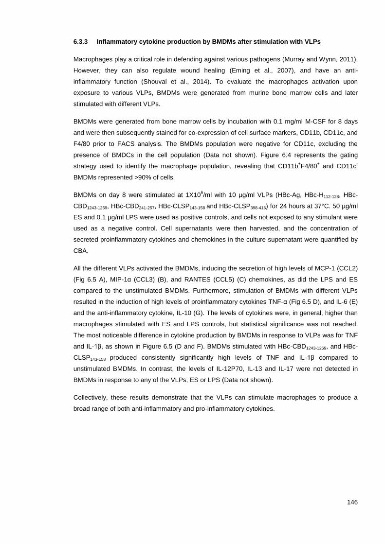
146
6.3.3 Inflammatory cytokine production by BMDMs after stimulation with VLPs
Macrophages play a critical role in defending against various pathogens (Murray and Wynn, 2011).
However, they can also regulate wound healing (Eming et al., 2007), and have an anti-
inflammatory function (Shouval et al., 2014). To evaluate the macrophages activation upon
exposure to various VLPs, BMDMs were generated from murine bone marrow cells and later
stimulated with different VLPs.
BMDMs were generated from bone marrow cells by incubation with 0.1 mg/ml M-CSF for 8 days
and were then subsequently stained for co-expression of cell surface markers, CD11b, CD11c, and
F4/80 prior to FACS analysis. The BMDMs population were negative for CD11c, excluding the
presence of BMDCs in the cell population (Data not shown). Figure 6.4 represents the gating
strategy used to identify the macrophage population, revealing that CD11b+F4/80
+ and CD11c
-
BMDMs represented >90% of cells.
BMDMs on day 8 were stimulated at 1X10⁶/ml with 10 µg/ml VLPs (HBc-Ag, HBc-H112-128, HBc-
CBD1243-1259, HBc-CBD241-257, HBc-CLSP143-158 and HBc-CLSP398-416) for 24 hours at 37°C. 50 µg/ml
ES and 0.1 µg/ml LPS were used as positive controls, and cells not exposed to any stimulant were
used as a negative control. Cell supernatants were then harvested, and the concentration of
secreted proinflammatory cytokines and chemokines in the culture supernatant were quantified by
CBA.
All the different VLPs activated the BMDMs, inducing the secretion of high levels of MCP-1 (CCL2)
(Fig 6.5 A), MIP-1α (CCL3) (B), and RANTES (CCL5) (C) chemokines, as did the LPS and ES
compared to the unstimulated BMDMs. Furthermore, stimulation of BMDMs with different VLPs
resulted in the induction of high levels of proinflammatory cytokines TNF-α (Fig 6.5 D), and IL-6 (E)
and the anti-inflammatory cytokine, IL-10 (G). The levels of cytokines were, in general, higher than
macrophages stimulated with ES and LPS controls, but statistical significance was not reached.
The most noticeable difference in cytokine production by BMDMs in response to VLPs was for TNF
and IL-1β, as shown in Figure 6.5 (D and F). BMDMs stimulated with HBc-CBD1243-1259, and HBc-
CLSP143-158 produced consistently significantly high levels of TNF and IL-1β compared to
unstimulated BMDMs. In contrast, the levels of IL-12P70, IL-13 and IL-17 were not detected in
BMDMs in response to any of the VLPs, ES or LPS (Data not shown).
Collectively, these results demonstrate that the VLPs can stimulate macrophages to produce a
broad range of both anti-inflammatory and pro-inflammatory cytokines.

147
Figure 6.4 Representative gating strategy for mouse bone marrow-derived macrophages (BMDMs) purity. BMDMs analysed by flow cytometry. BMDMs cells were gated first by size to identify single cells followed by gating on live cells. BMDMs gated as F4/80
+ CD11b
+. Gates were
set using respective Fluorescence minus one (FMOs).
FSC-A
SSC
-A
SSC
-A
SSC
-H
SSC-A
FSC-AF4/80
DA
PI
CD
11
b
All cells 48.9 Single Cells 77.2 Single Cells2 94.5
Live96.3
F4/80 CD11b 94.1 Live cells 96.3
148
Figure 6.5 Inflammatory cytokines productions by mouse bone marrow-derived macrophages (BMDMs) in response to VLPs. BMDMs at 1X10
6/ml were stimulated in vitro with
10 µg/ml VLPs (HBc-Ag, HBc-H112-128, HBc-CBD1243-1259, HBc-CBD241-257, HBc-CLSP143-158 and HBc-CLSP398-416) and with 50 µg/ml ES and 0.1 µg/ml LPS as positive controls. As a negative control, unstimulated BMDMs were examined. Supernatants were harvested after 24 hours for multiple chemokines and cytokine analyses (MCP-1 (CCL2), MIP (CCL3), RANTES (CCL5), TNF-α, IL-6, IL1β, and IL-10) measured by LEGENDplex™. The bars represent mean ± SEM. Statistical analyses were carried out using the Kruskal-Wallis test (multiple comparisons). Significant differences between groups are represented by (*P≤0.05) with a line. Chart bars represent BMDMs grown from three individual mice from one representative experiment of two separate experiments.
C
A B
Un
sti
mu
late
d
LP
SE
S
VL
P (
HB
c-A
g)
HB
c-H
112-1
28
HB
c-C
BD 2
41-2
57
HB
c-C
BD 1
243-1
259
HB
c-C
LS
P 143-1
58
HB
c-C
LS
P 398-4
16
0
5
1 0
1 5IL
-1B
Co
nc
en
tra
tio
n
pg
/mL
*
Un
sti
mu
late
d
LP
SE
S
VL
P (
HB
c-A
g)
HB
c-H
112-1
28
HB
c-C
BD 2
41-2
57
HB
c-C
BD 1
243-1
259
HB
c-C
LS
P 143-1
58
HB
c-C
LS
P 398-4
16
0
2 0 0 0
4 0 0 0
6 0 0 0
8 0 0 0
1 0 0 0 0
IL-6
Co
nc
en
tra
tio
n
pg
/mL
Un
st i
mu
late
d
LP
SE
S
VL
P (
HB
c-A
g)
HB
c-H
112-1
28
HB
c-C
BD 2
41-2
57
HB
c-C
BD 1
243-1
259
HB
c-C
LS
P 143-1
58
HB
c-C
LS
P 398-4
16
0
5 0
1 0 0
1 5 0
2 0 0
IL-1
0 C
on
ce
ntr
ati
on
p
g/m
L
Un
st i
mu
late
d
LP
SE
S
VL
P (
HB
c-A
g)
HB
c-H
112-1
28
HB
c-C
BD 2
41-2
57
HB
c-C
BD 1
243-1
259
HB
c-C
LS
P 143-1
58
HB
c-C
LS
P 398-4
16
0
1 0 0 0
2 0 0 0
3 0 0 0
4 0 0 0
TN
F C
on
ce
ntr
ati
on
p
g/m
L
*
*
Un
sti
mu
late
d
LP
SE
S
VL
P (
HB
c-A
g)
HB
c-H
112-1
28
HB
c-C
BD 2
41-2
57
HB
c-C
BD 1
243-1
259
HB
c-C
LS
P 143-1
58
HB
c-C
LS
P 398-4
16
0
5 0 0
1 0 0 0
1 5 0 0
2 0 0 0
2 5 0 0
RA
NT
ES
Co
nc
en
tra
tio
n
pg
/mL
Un
st i
mu
late
d
LP
SE
S
VL
P (
HB
c-A
g)
HB
c-H
112-1
28
HB
c-C
BD 2
41-2
57
HB
c-C
BD 1
243-1
259
HB
c-C
LS
P 143-1
58
HB
c-C
LS
P 398-4
16
0
5 0 0 0
1 0 0 0 0
1 5 0 0 0
MIP
1-a
Co
nc
en
tra
tio
n
pg
/mL
Un
st i
mu
late
d
LP
SE
S
VL
P (
HB
c-A
g)
HB
c-H
112-1
28
HB
c-C
BD 2
41-2
57
HB
c-C
BD 1
243-1
259
HB
c-C
LS
P 143-1
58
HB
c-C
LS
P 398-4
16
0
1 0 0
2 0 0
3 0 0
4 0 0
5 0 0
MC
P C
on
ce
ntr
ati
on
p
g/m
L
D
E
G
F
149
6.3.4 Uptake of fluorescein-conjugated VLPs by BMDCs
To further investigate whether the VLP can serve as a delivery tool to enhance antigen uptake by
APCs, BMDCs were harvested on day 8, and cultured overnight at 37°C with 10 µg/ml fluorescein-
conjugated VLPs (HBc-Ag, HBc-H112-128, HBc-CLSP398-416 and HBc-CBD241-257) and with fluorescein-
conjugated BSA as a control antigen. Antigen uptake by DCs was then examined by flow cytometry
by gating for CD11b+CD11c
+ cells also positive for FITC (Fig 6.6 A).
Figure 6.6 (B) reveals that all the fluorescent-labelled VLPs and control fluorescent-labelled BSA
were successfully taken up by a proportion of DCs. However, the percentage of uptake varied
between different VLPs, although no differences reached significance. Uptake of native VLP (HBc-
Ag) and HBc-CLSP398-416 was lower than uptake of BSA, while HBc-H112-128 and HBc-CBD241-257
were taken up by a higher % of DCs than BSA.
In order to assess whether the activation status of DCs that had taken up VLPs differed from the
activation status of the overall DC population, CD86+ expression on FITC
+ DCs was compared to
the whole DC population. While around 80% of BMDCs were CD86+
(Fig 6.6 C), not all FITC+
BMDCs also expressed CD86+. Thus, of the BMDCs that took up the FITC labelled VLPs and FITC
labelled BSA, only 50%-60% were activated DCs expressing CD86+, as shown in Figure 6.6 (D).

150
Figure 6.6 Uptake of VLPs by mouse bone marrow-derived DCs (BMDCs) analysed by flow cytometry. BMDCs on day 8 were stimulated with 10 µg/ml (HBc-Ag, HBc-H112-128, HBc-CLSP398-
416 and HBc-CBD241-257) and with fluorescein-conjugated BSA as a positive control for 24 hours. Cells were then subsequently stained for flow analysis (A) Representative gating strategy of VLP uptake by BMDCs. DCs were gated first by size and granularity, doublets excluded, followed by gating on live cells. DCs were gated as CD11b
+CD11c
+ and F4/80
- and VLP uptake by BMDCs
was gated as FITC+. Gates were set using respective Fluorescence minus one (FMOs). (B) The
percentage of uptake of different fluorescein-conjugated VLPs by BMDCs. (C) The percentage BMDCs expressing CD86
+ stimulated with different VLPs and BSA. (D) The percentage of CD86
+
BMDCs that took up fluorescein-conjugated VLPs. The bars represent as mean ± SEM. Statistical analyses were carried out using the Kruskal-Wallis test (multiple comparisons). Significant differences are represented by (*P≤0.05) with a line. Chart bars represent BMDC grown from three individual mice from one experiment.
BS
A
HB
c-A
g
HB
c-H
112-1
28
HB
c-C
LS
P 398-4
16
HB
c-C
BD 2
41-2
57
0
2 0
4 0
6 0
8 0
1 0 0
CD
86
(%
of
FIT
C+
BM
DC
s)
*
BSA
HBc-
Ag
HBc-
H 11
2-12
8
HBc-
CLS
P 398-
416
HBc-
CBD 24
1-25
7
0
20
40
60
80
FIT
C (
% B
MD
Cs)
BS
A
HB
c-A
g
HB
c-A
g
HB
c-H
112-1
28
HB
c-C
LS
P 398-4
16
HB
c-C
BD 2
41-2
57
0
2 0
4 0
6 0
8 0
1 0 0
CD
86
+ (
%B
MD
Cs
)
Fluorescence + - + + + +
B
C D
Cells Single Cells Single Cells 2 Live Cells DCs
FSC-A
CD
11
b
SSC
-A
FSC
-H
SSC-A
SSC
-H
FSC-A
DA
PI
CD 11c
FSC
-A
FITC
A Uptake + 48.4

151
6.3.5 Direct visualisation of VLP association and co-localization in APCs
From the flow-based analysis, it was uncertain whether fluorescein-conjugated VLPs remained
exposed on the surface of the BMDCs or were subsequently internalised. Therefore we used
Amnis ImageStreamX cytometer to confirm VLPs association and co-localization inside both
BMDCs and BMDMs.
To visualise VLP association in DCs, BMDCs at 1X10⁶/ml were stimulated with 10 µg/ml
fluorescein-conjugated VLPs (HBc-Ag, HBc-H112-128, HBc-CBD241-257, and HBc-CLSP398-416) for 24
hours. Fluorescein-conjugated BSA and fluorescein-conjugated dextran at 10 µg/ml were used as
positive controls, and cells not exposed to any stimulant were used as a negative control.
Similarly, to visualise VLP association in macrophages, BMDMs at 1X10⁶/ml were stained with
Lysotracker to visualise the cellular lysosome compartment and were then subsequently stimulated
with 10 µg/ml fluorescein-conjugated VLPs (HBc-Ag, HBc-H112-128, and HBc-CLSP398-416) for 24
hours. Fluorescein-conjugated dextran at 10 µg/ml was used as a positive control, and cells not
exposed to any stimulant were used as a negative control.
The ImageStreamX captured digital images of each cell in 2 channels for the ‘internalisation’
analysis, which include brightfield (BF), and FITC (green) channels. It is necessary here to clarify
that the ImageStreamX analysis tool uses the term ‘internalisation’. However, it is not clear at this
stage if VLPs were truly internalised or were just exposed on the surface of the APCs. Images of
individual cells taken by a merge of the two channels demonstrated that the fluorescein-conjugated
VLPS were ‘internalised’ in both BMDCs and BMDMs, as shown in Figure 6.7 (A-D) and Figure 6.8
(A-C) compared to the unstimulated BMDCs (Fig 6.7 G) and unstimulated BMDMs (Fig 6.8 E).
The IDEAS software was then used to quantify the ‘internalisation’ by calculating the internalisation
mean absolute deviation (MAD). The ‘Internalisation’ feature calculates the ratio of the intensity
inside the cell to the intensity of the entire cell. Cells with a high level of ‘internalisation’ will have
positive values while cells with no ‘internalisation’ will have negative values. The values are
presented above each reprehensive cell, as shown in Figure 6.7 (A-G) and Figure 6.8 (A-E).
Interestingly, BMDCs and BMDMs stimulated with different fluorescein-conjugated VLPs showed
similar ‘internalisation’ MAD value around 1.3, which was higher than the fluorescein-conjugated
BSA and fluorescein-conjugated dextran positive controls. In contrast, the unstimulated BMDCs
and unstimulated BMDMs yielded a value of 0 indicating that the image pair had no association
had taken place, as shown in Figure 6.7 (G) and Figure 6.8 (E).
To further confirm that the VLPs were co-localized within the lysosome of the APCs and not just
associated on the cell surface, BMDCs and BMDMs at 1X10⁶/ml were stained with LysoTracker
dye to stain the lysosomes within the cells. Cells were then subsequently stimulated with 10 µg/ml
fluorescein-conjugated VLPs (HBc-Ag, HBc-CBD241-257, HBc-CLSP398-416 and HBc-H112-128) for 24
hours. Fluorescein-conjugated BSA and fluorescein-conjugated dextran at 10 µg/ml were used as
positive controls, and cells not exposed to any stimulant were used as a negative control.

152
The ImageStreamX captured digital images of each cell in 3 channels for the co-localization
analysis, which include channels for brightfield (BF), FITC (green), and Lysotracker (red). Images
of individual cells taken by a merge of the three channels revealed that the fluorescein-conjugated
VLPs, fluorescein-conjugated BSA and fluorescein-conjugated dextran could be found
accumulated in the lysosome compartment of the BMDCs (Fig 6.9 A-F) and BMDMs (Fig 6.10 A-D)
and not just localised on the cell surface. In contrast, BMDCs (Fig 6.9 G) and BMDMs (Fig 6.10 E)
not exposed to any stimulus showed no localisation.
Finally, the similarity feature on the IDEAS software was used to quantify the co-localization of the
VLPs inside the APCs. The similarity feature utilises an algorithm developed to quantify the degree
of co-localization to two images that are linearly correlated within a masked region. Poorly co-
localized events will have similarity bright detail score (SBDS) of 0, and well co-localized events will
have SBDS values around 1 (Ploppa et al., 2011). BMDCs and BMDMs stimulated with different
fluorescein-conjugated VLPs yielded an SBDS value around 1, indicating that the image pair had a
high degree of similarity which was equivalent to the fluorescein-conjugated BSA and fluorescein-
conjugated dextran positive controls. In contrast, the SBDS values of the unstimulated BMDCs and
the unstimulated BMDMs were 0, indicating that the image pair had no degree of similarity.
Collectively, these results indicate that fluorescein-conjugated VLPs were internalised and well co-
localized in the lysosomes of both BMDCs and BMDMs.

153
Figure 6.7 Representative images of fluorescein-conjugated VLPs internalisation in the BMDCs. BMDCs at 1X10
6/ml were incubated with 10 μg/ml fluorescein-conjugated VLPs; (A) HBc-
Ag, (B) HBc-H112-128, (C) HBc-CBD241-257, and (D) HBc-CLSP398-416 for 24 hours. BMDMs were stimulated with 10 µg/ml fluorescein-conjugated BSA (E) and with 10 µg/ml fluorescein-conjugated dextran (F) as positive controls. As a negative control, unstimulated BMDMs were examined (G). Cell internalisation was determined by Amnis ImageStreamX cytometer compared to unstimulated BMDCs. Images shown, from left to right, show individual Brightfield images (BF) in the white channel, fluorescent-labelled stimulus (FITC) in the green channel and the combination of both BF/FITC merged channels. The internalisation mean absolute deviation (MAD) is included above its images. The positive MAD value represents internalisation, and negative values represent poor internalisation. Scale bars represent 10 μm.
(E) BSA
(F) Dextran
(A) HBc-Ag
(C) HBc-CBD241-257
(D) HBc-CLSP398-416
(B) HBc-H112-128
(G) Unstimulated BMDCs
MAD=0
MAD=0.8275
MAD=1.574
MAD=1.514
MAD=1.656
MAD=0.9667
MAD=1.586
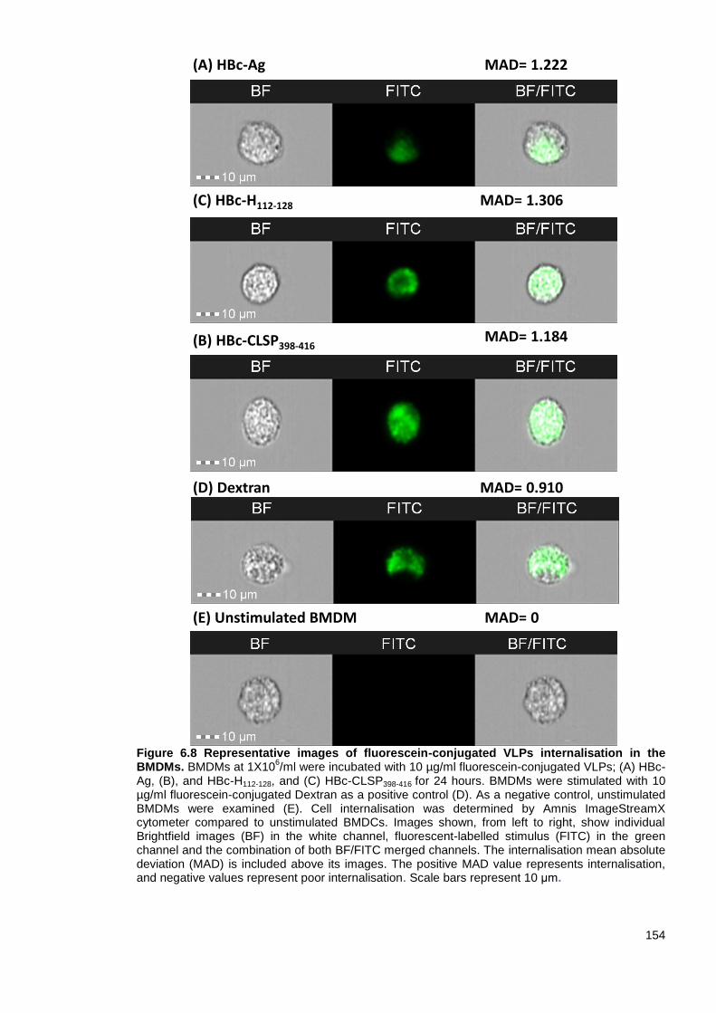
154
Figure 6.8 Representative images of fluorescein-conjugated VLPs internalisation in the BMDMs. BMDMs at 1X10
6/ml were incubated with 10 µg/ml fluorescein-conjugated VLPs; (A) HBc-
Ag, (B), and HBc-H112-128, and (C) HBc-CLSP398-416 for 24 hours. BMDMs were stimulated with 10 µg/ml fluorescein-conjugated Dextran as a positive control (D). As a negative control, unstimulated BMDMs were examined (E). Cell internalisation was determined by Amnis ImageStreamX cytometer compared to unstimulated BMDCs. Images shown, from left to right, show individual Brightfield images (BF) in the white channel, fluorescent-labelled stimulus (FITC) in the green channel and the combination of both BF/FITC merged channels. The internalisation mean absolute deviation (MAD) is included above its images. The positive MAD value represents internalisation, and negative values represent poor internalisation. Scale bars represent 10 μm.
(A) HBc-Ag
(B) HBc-CLSP398-416
(C) HBc-H112-128
(E) Unstimulated BMDM MAD= 0
MAD= 1.184
MAD= 1.306
MAD= 1.222
(D) Dextran MAD= 0.910

155
Figure 6.9 Representative images of fluorescein-conjugated VLPs co-localization in the BMDCs. BMDCs at 1X10
6/ml were stained with Lysotracker to visualise the cellular lysosome
compartment and subsequently stimulated with 10 μg/ml fluorescein-conjugated VLPs; (A) HBc-Ag, (B) HBc-H112-128, (C) HBc-CBD241-257, and (D) HBc-CLSP398-416 for 24 hours. BMDMs were stimulated with 10 µg/ml fluorescein-conjugated BSA (E) and with 10 µg/ml fluorescein-conjugated dextran (F) as positive controls. As a negative control, unstimulated BMDMs were examined (G). Intracellular co-localization was determined by Amnis ImageStreamX cytometer. Images shown, from left to right, show individual Brightfield images (BF) in the white channel, fluorescent-labelled stimulus (FITC) in the green channel, stained lysosome (Lysotracker) in the red channel and the combination of both FITC/ Lysotracker merged channels. The Similarity bright detail score (SBDS) score from the IDEAS quantitative co-localization analysis is included above its image. SBDS values around 1 represent co-localization, and 0 values represent poor co-localization. Scale bars represent 10 μm.
(E) BSA
(F) Dextran
(A) HBc-Ag
(G) Unstimulated BMDCs
(C) HBc-CBD241-257
(D) HBc-CLSP398-416
(B) HBc-H112-128
SBDS= 0
SBDS= 0.8604
SBDS= 1.106
SBDS= 1.155
SBDS= 1.036
SBDS= 0.984
SBDS= 1.018

156
Figure 6.10 Representative images of fluorescein-conjugated VLPs co-localization in the BMDMs. BMDMs at 1X10
6/ml were stained with Lysotracker to visualise the cellular lysosome
compartment and subsequently stimulated with 10 μg/ml fluorescein-conjugated VLPs; (A) HBc-Ag, (B) HBc-H112-128 and (C) HBc-CLSP398-416 for 24 hours. BMDMs were stimulated with 10 µg/ml fluorescein-conjugated Dextran (D) as a positive control. As a negative control, unstimulated BMDMs were examined (E). Intracellular co-localization was determined by Amnis ImageStreamX cytometer. Images shown, from left to right, show individual Brightfield images (BF) in the white channel, fluorescent-labelled stimulus (FITC) in the green channel, stained lysosome (Lysotracker) in the red channel and the combination of both FITC/Lysotracker merged channels. The Similarity bright detail score (SBDS) score from the IDEAS quantitative co-localization analysis is included above its image. SBDS values around 1 represent co-localization, and 0 values represent poor co-localization. Scale bars represent 10 μm.
(A) HBc-Ag
(E) Unstimulated BMDM
(C) HBc-CLSP398-416
(B) HBc-H112-128
SBDS= 0
SBDS= 0.9492
SBDS= 1.129
SBDS= 1.061
(D) Dextran SBDS= 1.036

157
6.4 Discussion
The role of VLP as a vaccine delivery system has been a point of speculation since its discovery.
VLPs have been proven to induce stronger protective immune responses against foreign carrier
antigens when compared to other delivery systems (Vogel and Bachmann, 2019). However, the
precise mechanism of how VLP activates the immune system and leads to an immune response
against the antigen it carries is only partially understood. In particular, the uptake of HBc-Ag (VLP)
by antigen-presenting cells has not been extensively studied compared to other VLPs obtained
from other viruses such as the human papilloma virus-like particle (HPV-VLP).
The VLP as a vaccine platform can target both MHC-I and MHC-II processing and presentation
pathways and generate cytokine production, which is essential to initiating a specific immune
response (Cubas et al., 2009; Gomes et al., 2017b; Manolova et al., 2008; Shirbaghaee and
Bolhassani, 2016). Most importantly, VLPs are advantageous vaccine delivery systems, as they
can directly target the vaccine to different APCs. Among the APCs, DCs are the most potent APC
that play crucial roles in delivering antigens from the site of exposure to the secondary lymphoid
tissues for the initiation of specific T-cell (cytotoxic and helper) mediated immunity (Beyer et al.,
2001; Steinman, 2001). In this study, we aimed to evaluate the innate stimulatory potential of the
native VLP (HBc-Ag) that lacks C-terminal arginine and the VLPs expressing Trichuris MHC-II T-
cell epitopes, by using two types of APCs, BMDCs and BMDMs. We also aimed to examine the
ability of APCs to take up and internalise VLPs.
6.4.1 Dendritic cell activation by VLPs
It is now well established from a variety of studies, that VLPs derived from different viruses, such
as human papillomavirus (HPV-16), Newcastle disease virus, murine polyomavirus, and SARS-
Like coronavirus, can avidly bind and stimulate human and murine DCs (Bai et al., 2008; Bickert et
al., 2007; Lenz et al., 2001; Lenz et al., 2003; Mccullough and Sharma, 2017; Rudolf et al., 2001;
Xu et al., 2019; Yang et al., 2004). The present chapter demonstrated that the VLPs used in this
study provided two types of immunoregulator signals to BMDCs: (a) the upregulation of DC surface
markers (CD40, CD80 and CD86) essential for the optimal activation of Ag-specific T-cells and (b)
the mediation of inflammatory responses characterized by the secretion of high levels of cytokines,
such as IL-6, IL-10 and TNF-α, which are involved in the immune regulation of B- and T-
lymphocytes. Furthermore, VLPs were proven to be taken up by BMDCs efficiently and were not
merely attached to the surface of APCs, suggesting that APCs will process and present the VLPs
in the context of MHC molecules.
Interestingly, not all the BMDCs that took up the fluorescent-labelled VLPs expressed CD86+. For
example, of the BMDCs that took up the fluorescent-labelled native VLP (HBc-Ag), only 50% were
activated DCs expressing CD86+. This effect could be due to the enhanced ability of non-activated
DCs to take up antigens compared to activated DCs (Sousa, 2006).
Collectively, these results suggest that VLPs are well placed to act as delivery systems to drive
immune responses via DCs (Barth et al., 2005; Bosio et al., 2004; Da Silva et al., 2001; Lin et al.,
2014; Rudolf et al., 2001; Warfield et al., 2003). In line with these observations, Ding et al. (2010)

158
demonstrated the phagocytosis of exogenous HBc-VLPs by BMDCs analysed by
immunofluorescence and confocal laser scanning microscopy (CLSM). The phenotypic analysis
also revealed that the HBc-VLP-pulsed BMDCs significantly increased the BMDC surface markers
(CD80, CD86 and MHC-II) compared to the untreated BMDCs. Furthermore, the cytokine
expression of HBc-VLP pulsed BMDCs identified by RT-PCR and electrophoresis on 1.2% agarose
gel confirmed the secretion of IL-12 (p40) and IFN-ϒ compared to the control groups (Ding et al.,
2010). The data are largely consistent with the results presented here although IL-12 (p40) and
IFN-ϒ were not assessed and a broader profile of cytokines was measured, including
IL-12p70.
More recently, Serradell et al. (2019) developed a (VLP-HA/VSP-G) vaccine composed of an
influenza virus expressing influenza virus hemagglutinin (HA) antigen, coexpressed with the
extracellular region of Giardia lamblia variant-specific surface proteins (VSPs) as an adjuvant. The
recombinant protein (VLP-HA/VSP-G) significantly increased the expression of BMDC
costimulatory molecules (CD40 and CD86) and induced the secretion of high levels of TNF-α, IL-
10, and IL-6 cytokines in vitro. They also showed that oral vaccination with the recombinant protein
protected mice from influenza infection and generated protective humoral and cellular immunity
(Serradell et al., 2019). These data correlate well with the results presented here as the VLPs
stimulated BMDCs to secrete high levels of IL-6 and TNF cytokines in vitro and increased the
expression of BMDC costimulatory molecule (CD40).
Of the cytokines produced by DCs in response to VLP stimulation, IL-6, identified initially as a B-
cell stimulatory factor 2, enhances antibody synthesis by activated B-cells (Kishimoto and Tanaka,
2015). IL-12, also produced by BMDCs activated by VLPs, plays an essential role as a bridge
between the innate and adaptive immune systems. The secretion of IL-12 in response to infection
polarises the immune system toward a primary Th-1 response by stimulating T-cells to produce
IFN-ϒ (Fayette et al., 1998; Trinchieri, 1993). In the present study, the level of IL-12p70 produced
by BMDCs in response to the native VLP (HBc-Ag) and VLPs expressing Trichuris epitopes
stimulation was not significantly high. The native VLP (HBc-Ag) utilised in the current study is
equivalent to the HBc-Ag lacking the C-terminal arginine (HBcAg-149). This finding is consistent
with data on the Th1/ Th2 polarisation in immune responses to HBc-Ag (Chen et al., 2009; Milich et
al., 1995). Thus, the full-length HBc-Ag, but neither of the HBc-Ag lacking the C-terminal arginine
(HBcAg-144 and HBcAg-149), preferentially primed a Th1 immune response as measured by the
production of IL-12p70 by dendritic cells from mice immunised with HBc-Ag and the subsequent
release of IFN-ϒ by splenocytes (Riedl et al., 2002). Another study also showered that the full-
length HBc-Ag induced the transcription and secretion of IL-18, a potent inductor of IFN-ϒ in
peripheral blood mononuclear cells (PBMCs) in healthy controls and patients with chronic or acute
hepatitis B infection, whereas the HBc-Ag lacking the C-terminal arginine-rich domain did not
induce IL-18 in all groups (Manigold et al., 2003).
In all studies involving VLPs, it is essential to confirm that DC activation and the release of
cytokines are in response to the interaction of DCs with the VLP protein structure and not due to
endotoxin contamination whilst the VLPs were being made. For example, there were no alterations
159
in cytokine production when the DCs stimulated with VLPs were pretreated with polymyxin B, a
compound that binds and inactivates LPS. Moreover, destroying the structure of the VLPs by
boiling inhibited the cytokine production, while boiling LPS led to no alterations in the cytokines
produced by VLP-stimulated DCs (Bosio et al., 2004; Lenz et al., 2001). Furthermore, when Ebola
virus VLPs were depleted with specific antibodies and then incubated with DCs, cells did not
upregulate cell-surface markers and failed to secrete cytokines (Bosio et al., 2004; Lenz et al.,
2001). Although these types of studies were not undertaken in the current research, future analysis
should include checks for endotoxin contamination.
Several studies have also addressed whether different APCs can take up VLPs. For example,
electron microscopy and confocal imaging of mature DCs obtained from peripheral blood
mononuclear cells (PBMCs) exposed to human immunodeficiency virus type 1 (HIV-VLPs) for 4
hours showed that VLPs were co-localized in the intracellular vesicular compartment. Further
intracellular accumulation increased over time and in a dose-dependent manner (Izquierdo-Useros
et al., 2009). Zhang et al. (2007) also showed that HBc-Ag expressing tumor epitopes bind and
internalise to BMDCs efficiently in vitro when detected using immunofluorescence confocal
microscopy. Furthermore, Link et al. (2012) suggested that a VLP’s size and repetitive structure
play an essential role in Ag presentation to B-cells. To analyse Ag trafficking, naïve wild-type mice
were immunised with VLPs derived from the bacteriophage QB (QB-VLP), and Ag deposition on
murine splenic follicular dendritic cells (FDCs) was determined 24 h after the challenge. The
fluorescence images revealed the deposition of the VLP inside B-cell follicles (Link et al., 2012). In
a second experiment, mice were passively immunised with anti-Qb IgM serum and then challenged
with QB-VLPs to allow the formation of immune complexes (ICs) in vivo. Mice were then
challenged with QB-VLPs, and Ag deposition was assessed 24 h after the challenge. The
fluorescence images also revealed the accumulation of QB-VLPs inside the B-cell follicles as
immune complexes via a process involving IgM antibodies and complement (Link et al., 2012).
Given that VLPs activate DCs and have the ability to interact with B-cells to facilitate Ab production
after VLP vaccination without the need for an adjuvant, modifying a VLP using immunostimulatory
adhesion molecules and cytokines co-displayed on the VLP to target APCs can enhance the
activation of antigen-specific T lymphocytes responses (Akhras et al., 2017; Caminschi and
Shortman, 2012; Hill et al., 2018; Gogesch et al., 2018; Gomes et al., 2017a; Mohsen et al., 2017b;
Rohovie et al., 2017). For example, membrane-bound CD40 ligand (CD40L) expressed on the
surface of simian immunodeficiency virus (SIV) VLPs and HIV-1 VLPs can be used to target CD40
receptor on DCs to enhance both CD4+ and CD8+ T-cell responses to HIV-1
glycosylphosphatidylinositol (GPI) protein (Franco et al., 2011; Skountzou et al., 2007; Zhang et al.,
2010). The activity of CD40L-displaying HIV-1 VLPs significantly enhanced both humoral and
cellular immune responses to HIV-1 Gag protein in immunised mice compared to the effect in
immunised CD40-/- mice (Franco et al., 2011). In another study, the immunization of mice with a
membrane-anchored granulocyte-macrophage colony stimulating factor (GM-CSF) expressed on
SIV VLPs containing Gag, to target GM-CSF-receptor positive APCs significantly enhanced the
production of SIV VLP-specific antibodies and activated both CD4+ and CD8+ T-cell responses

160
compared to the control immunized groups (SIV VLPs, SIV VLPs containing CD40L, or SIV VLPs
mixed with soluble GM-CSF) (Skountzou et al., 2007).
6.4.2 Macrophage activation by VLPs
Macrophages, as APCs, play a role in promoting Th1 and Th2 immunity and controlling infection by
producing proinflammatory and regulatory cytokines. However, the origin, size, and shape of
exogenous stimuli influence the efficiency of phagocytosis, macrophage phenotype, and activation
threshold (Daigneault et al., 2010; Dalgėdienė et al., 2018; Murray and Wynn, 2011).
The present chapter carried out in vitro studies to determine the BMDMs activation upon exposure
to the native VLP (HBc-Ag) and VLPs expressing different Trichuris T-cells epitopes. High levels of
IL-10, TNF-α, IL-6, and IL1β cytokines and MCP-1 (CCL2), MIP (CCL3), and RANTES (CCL5)
chemokines were detected by BMDMs after treatment with different VLPs. The data suggest that
the VLP is a potent inducer of proinflammatory cytokines. I further demonstrated that not only DCs,
but also macrophages were able to take up the VLPs which co-localized in lysosomes.
These results are consistent with those of Lenz et al. (2003) and Wahl-Jensen et al. (2005) who
examined the activation of macrophages in response to VLPs. For example, Lenz et al. (2003)
found that bovine papillomavirus BPV VLPs labelled with green fluorescence protein (GFP) bind to
the cell surface of human macrophages when analysed by flow cytometry. Furthermore, they found
that human macrophages secreted the proinflammatory cytokines TNF-α, IL-6 and IL- 1β in
response to HPV16 VLPs, unlike the unstimulated DCs, as assessed by cytokine ELISA and
intracellular cytokine staining. However, they did not asses the immune response to the native HPV
VLP (Lenz et al., 2003). These results are consistent with Wahl-Jensen et al.’s work (2005), which
found that VLPs with and without Ebola virus glycoproteins (GP) produced higher levels of
proinflammatory cytokines (TNF-a, IL-6 and IL- 1β) and IL-8 compared to the unstimulated cells
when measured by real-time PCR. Also, VLP-GP induced higher levels of macrophage activation
compared to the native VLP (Wahl-Jensen et al., 2005).
Furthermore, Cooper et al. (2005) examined the capacity of the HBc-Ag VLP (HBc-144) which lacks
the genetic material compared to the full-length HBc-Ag, to induce proinflammatory and regulatory
cytokines in human macrophages. The full-length HBc capsid, but not the HBc-144 capsid, efficiently
bound differentiated THP-1 macrophages. However, both induced the production of
proinflammatory and regulatory cytokines (TNF-α, IL-6, and IL-12p40) in a dose- and time-
dependent manner (Cooper et al., 2005). The cytokine levels were lower in response to HBc-144
compared to the full-length HBc-Ag. The recombinant full-length HBc-Ag and HBc-144 were
expressed and purified using E. coli. In order to rule out that the cytokines were induced in
response to residual LPS contamination, the HBc-Ag, HBc-144 and LPS control were preboiled
before adding them to the THP-1 macrophages to measure cytokine induction using ELISA and
RT-PCR. Both preboiled recombinant capsids but not the LPS control failed to induce cytokine
production (Cooper et al., 2005).
It is also important to highlight the fact that there were differences in the responses of APCs after
exposure to VLPs expressing different Trichuris T-cell epitopes. However, these changes could be

161
due to the nature of the inserted epitope or the structural assembly of the VLP. To further confirm
and understand the differences in activation responses seen in vitro, future studies will need to
focus on evaluating the VLPs in vivo. It will also be interesting to vaccinate mice with VLPs and
then evaluate the activation responses to different DC subsets and to other APCs, including B-
cells, in vivo.
Collectively, the findings reported in this chapter provide evidence that both APCs (dendritic cells
and macrophages) effectively recognise and respond to VLPs expressing Trichuris MHC-II T-cell
epitopes, supporting an orchestrated priming of both humoral and cellular immune responses in the
development of VLP-mediated immunity even at low doses and in the absence of an adjuvant.
Given the experimental explanation in vitro of the remarkable immunogenicity of HBc-Ag VLPs as a
promising vaccine delivery system, I propose that HBc-VLPs expressing Trichuris MHC-II T-cell
epitopes may drive strong Trichuris specific T-cell responses in vivo and, thus, could be used as a
novel, effective vaccine against trichuriasis.
6.5 Summary
The native VLP (HBc-Ag) and the VLP expressing Trichuris MHC-II T-cell epitopes (HBc-
Ag, HBc-H112-128, HBc-CBD241-257, and HBc-CLSP398-416) activated BMDCs as demonstrated
by upregulation in the expression of costimulatory molecules CD80, CD86, CD40, and
MHC-II.
The native VLP (HBc-Ag) and the VLP expressing Trichuris MHC-II T-cell epitopes (HBc-
Ag, HBc-H112-128, HBc-CBD1243-1259, HBc-CBD241-257, HBc-CLSP143-158, and HBc-CLSP398-416)
efficiently stimulated BMDCs and BMDMs to produce a broad range of pro-inflammatory
and anti-inflammatory cytokines.
The fluorescein-conjugated VLPs (HBc-Ag, HBc-H112-128, HBc-CBD241-257, and HBc-
CLSP398-416) were taken up by BMDCs. However, the percentage of uptake varied between
different VLPs.
The fluorescein-conjugated VLPs (HBc-Ag, HBc-H112-128, HBc-CBD241-257, and HBc-
CLSP398-416) were internalised and co-localized in the lysosomes of both BMDCs and
BMDMs.

162
Chapter Seven
VLPs expressing Trichuris T-cell
epitopes promote protective
immune responses in vivo
163
7.1 Introduction
Trichuriasis, caused by the whipworm Trichuris trichiura, is one of the most widespread soil-
transmitted helminths in the world (Alexander and Blackburn, 2019). Global mass drug
administration (MDA) programmes are being implemented, but repeated treatments are costly and
may prevent the development of acquired immunity, and the existence of drug-resistant parasites is
well established (Farrell et al., 2018; Speich et al., 2015; WHO, 2015). Thus, there is considerable
interest in developing vaccines against these parasites (Becker et al., 2018; Dixon et al., 2008). A
vaccine that protects against Trichuris infection was first demonstrated in mice immunised
subcutaneously (s.c) with Trichuris muris excretory/secretory (ES) products (Jenkins and Wakelin,
1977), followed by fractions of ES (Shears et al., 2018b), and extracellular vesicles (EVs) (Shears
et al., 2018a), and, more recently with T. muris whey acidic protein (Briggs et al., 2018).
It has been clearly demonstrated that susceptibility to infection and the development of chronic
infection and colitis is driven by CD4+ Th1 immune responses, serum IgG2c, and the production of
Th1 cytokines IL-12 and IFN-γ (Bancroft et al., 2001; Else et al., 1993b). In contrast, resistance to
infection is driven by Th2 immune responses, serum IgG1, and the production of Th2 cytokines (IL-
4, IL-9, and IL-13 (Dixon et al., 2010; Klementowicz et al., 2012). Th2 cytokines are known to
stimulate different effector mechanisms critical to driving worm expulsion for the development of a
protective immune response to T. muris. These effector mechanisms include epithelial cell turnover
in the caecum and proximal colon (Cliffe et al., 2005), intestinal hyper-contractility (Khan et al.,
2003), and the increased production of mucins by goblet cells (Artis et al., 2004b).
Despite the success of the previously mentioned vaccines, developing a vaccine for STHs based
on native antigens has encountered many manufacturing complexities, such as cost, time
consumption, as well as difficulty in purifying large quantities of the worm antigens, and controlling
batch differences to develop a commercially stable vaccine (Geldhof et al., 2007; Hewitson and
Maizels, 2014). An alternative is to develop a vaccine based on virus-like particles (VLPs)
expressing Trichuris MHC-II T-cell epitopes to simplify the production challenges inherent in
developing a safe, inexpensive vaccine that can induce an effective immune response (El-Sayed
and Kamel, 2018; Geldhof et al., 2007; Jain et al., 2015).
The fast-growing knowledge of VLP-based vaccines against several parasites, including
Plasmodium spp (Nardin et al., 2001; Salman et al., 2017), Leishmania infantum (Moura et al.,
2017), Toxoplasma gondii (Guo et al., 2019), Trichinella spiralis (Lee et al., 2016), and Clonorchis
sinensis (Lee et al., 2017) has escalated interest in developing a VLP-based vaccine against
trichuriasis. To the best of our knowledge, this is the first study that has developed a vaccine
against trichuriasis based on VLPs expressing novel Trichuris MHC-II T-cell epitopes.
164
7.2 Aim and objectives
The overall aim of this Chapter was to evaluate various VLPs expressing different Trichuris MHC-II
T-cell epitopes as potential vaccine candidates in vivo in the murine model of Trichuris infection.
This aim was addressed via three objectives:
To test the immunogenic and protective immune response of a low-dose of a vaccine
candidate, C57BL/6 mice were vaccinated with 25 µg of pre-mixed VLPs expressing
Trichuris T-cell epitopes (HBc-H112-128, HBc-CLSP143-158, HBc-CBD1243-1259, and HBc-
CBD241-257) at day -20 and boosted at day -10. As controls for susceptibility to infection,
mice were injected with PBS and 25 µg native VLP (HBc-Ag). Mice were then challenged
with a high-dose of Trichuris infection at day 0, and infection levels were assessed two
weeks post infection (p.i.) for evidence of accelerated expulsion in vaccinated mice.
To assess the potential of AddaVax adjuvant in enhancing the vaccine efficacy, C57BL/6
mice were vaccinated with 25 µg of pre-mixed VLPs expressing Trichuris T-cell epitopes
(HBc-H112-128, HBc-CLSP143-158, HBc-CLSP398-416, and HBc-CBD241-257) formulated with
AddaVax at day -35 and boosted at day -14. As a control for protective immunity, mice
were vaccinated with 50 µg ES/AddaVax, and as controls for susceptibility to infection,
mice were injected with PBS/AddaVax and 50 µg native VLP (HBc-Ag)/AddaVax. Mice
were then challenged with a high-dose of T. muris infection at day 0, and infection levels
were assessed at day 14 p.i.
To test the immunogenic and protective immune response of a high-dose of a vaccine
candidate, C57BL/6 mice were vaccinated with 50 µg of pre-mixed VLPs expressing
Trichuris T-cell epitopes (HBc-CBD1243-1259, HBc-CBD241-257, HBc-CLSP143-158, and HBc-
CLSP398-416) at day -20 and boosted at day -10. Control groups were injected with 50 µg
ES/Alum, PBS or 50 µg native VLP (HBc-Ag) at day -20 and boosted at day -10. Mice were
then challenged with a high-dose of T. muris infection at day 0, and infection levels were
assessed at day 14 p.i.

165
7.3 Results
7.3.1 Experimental protocol for in vivo evaluation of vaccinating mice with 25 µg of pre-
mixed VLPs expressing Trichuris MHC-II T-cell epitopes (HBc-H112-128, HBc-CLSP143-
158, HBc-CBD1243-1259, and HBc-CBD241-257)
In order to investigate whether VLPs expressing Trichuris T-cell epitopes are capable of stimulating
protective immunity, male C57BL/6 mice were subcutaneously vaccinated with 25 μg of pre-mixed
VLPs+ T-cell epitopes (HBc-H112-128, HBc-CLSP143-158, HBc-CBD1243-1259, and HBc-CBD241-257) on
day -20 and boosted on day -10. As controls for susceptibility to infection, mice were injected with
PBS and 25 μg of native VLP (HBc-Ag). 5 mice from each group were immunised but were not
infected with T. muris, and 5 mice from each group were immunised and then infected by oral
gavage with approximately 150 infective T. muris eggs on day 0 and sacrificed at day 14 p.i.
(Figure 7.1 A).
The protective efficacy of the vaccine was assessed two weeks p.i. by counting the worms from the
proximal colon and caecum of each mouse. MLNs at day 14 p.i. were collected and re-stimulated in
vitro with Trichuris ES for cytokine production, while blood was collected to measure serum
parasite and VLP+ T-cell epitope-specific IgM, IgG1 and IgG2c antibodies levels. Proximal colon
sections were collected to assess the pathology and count goblet cells.
7.3.1.1 25 µg of pre-mixed VLPs expressing Trichuris T-cell epitopes immunisation did not
reduce the T. muris worm count following challenge infection
Previous studies have shown that the C57BL/6 mouse strain is normally resistant to infection when
infected with a high-dose of T. muris eggs. Mice were able to expel the worms by day 21 p.i. and
full protection was seen by day 28 p.i. (Bancroft et al., 2001; Cliffe et al., 2005; D'elia et al., 2009).
However, no expulsion is normally seen two weeks p.i. unless the host has been previously
infected or vaccinated. Based on these studies, all the vaccination studies conducted in this
chapter used C57BL/6 mouse strain. Furthermore, only male C57BL/6 mice were subcutaneously
vaccinated to ensure that expulsion and cytokine production was not subjective by gender
differences (Else et al., 1994). The decision to administer the vaccine candidates subcutaneously
was based on a previous study that showed that subcutaneous vaccination of NIH mice with ES
not formulated with an adjuvant induced better protection (70%) than intraperitoneal vaccination
(33%) at day 9 p.i. (Jenkins and Wakelin, 1983).
Caecum and colon worm loads following T. muris infection are the most prominent indicator to
assess protective efficacy. In order to investigate whether the selected VLPs expressing Trichuris
T-cell epitopes (HBc-H112-128, HBc-CLSP143-158, HBc-CBD1243-1259, and HBc-CBD241-257) are
protective in vivo, worm burdens were assessed at day 14 p.i. and compared to those of mice
injected with PBS and native VLP (HBc-Ag) control groups. This time point was chosen as
resistance post vaccination will present as accelerated expulsion (Bancroft et al., 2001; Cliffe et al.,
2005; D'elia et al., 2009).

166
As shown in Figure 7.1 (B), no reduction in worm burden was demonstrated in the VLPs+ T-cell
epitopes vaccinated mouse group compared to the PBS and native VLP (HBc-Ag) control injected
mice groups. However, one mouse expelled all the worms in the group immunised with VLPs+ T-
cell epitopes.
Figure 7.1 Experimental schedule and worm burden of mice vaccinated and challenged with a T. muris infection. (A) 6-week old male C57BL/6 mice were vaccinated subcutaneously with 25 μg of pre-mixed of VLPs + T-cell epitopes (HBc-H112-128, HBc-CLSP143-158, HBc-CBD1243-1259, and HBc-CBD241-257), 25 μg of native VLP (HBc-Ag), or PBS on day -20 and boosted on day -10. 5 mice from each group were infected by oral gavage with approximately 150 infective T. muris eggs on day 0 and sacrificed at day 14 post-infection. (B) Comparison of worm burden at day 14 p.i.. Statistical analyses were carried out using the Kruskal-Wallis test (multiple comparisons). Results are shown as mean ± SEM. n= 5 mice per group. The results presented are from one experiment.
PB
S
VL
P (
HB
c-A
g)
VL
Ps+ T
-Cell e
pit
op
es
0
2 0
4 0
6 0
8 0
1 0 0
1 2 0
Wo
rm
Bu
rd
en
B
• PBS Infection
• 25 μg of VLP (HBc-Ag) Infection
• 25 μg of pre mixed VLPs+ T-Cell epitopes Infection
A
Prime Boost Challenge
d-20 d-10 d0 d14
Autopsy

167
7.3.1.2 Serum parasite-specific responses following vaccination of mice with VLPs
expressing Trichuris T-cell epitopes
Parasite-specific IgG1 and IgG2c serum antibodies are often used as markers of
resistance/susceptibility to Trichuris infection. The production of parasite-specific IgG1 is
associated with Th2 protective immune response in mice resistant to infection. Whereas the
production of parasite-specific IgG2c is associated with Th1 immune response in susceptible mice
to infection (Else et al., 1993b). To analyse parasite-specific antibody responses of vaccinated
mice, serial dilutions of sera were assessed using an ELISA-based assay with Trichuris
excretory/secretory (ES) as a target antigen.
In general, IgM, IgG1 and IgG2ca isotype responses were similar in mice given a T. muris infection
compared to those without challenge infection (Figure 7.2 A-C). There was a significant increase in
parasite-specific IgM detected in the serum of mice vaccinated with VLPs+ T-cell epitopes
compared to the native VLP (HBc-Ag) and PBS-injected mice with and without T. muris challenge
(Fig 7.2 A). Importantly, significant levels of IgG1 (Fig 7.2 B) and IgG2c (Fig 7.2 C) were detected
in the serum of mice vaccinated with VLPs+ T-cell epitopes compared to the native VLP (HBc-Ag),
and PBS-injected mice.

168
Figure 7.2 Parasite-specific IgM, IgG1 and IgG2c serum antibodies levels at day 14. 6 week old male C57BL/6 mice were vaccinated subcutaneously with 25 µg of pre-mixed VLPs+ T-cell epitopes (HBc-H112-128, HBc-CLSP143-158, HBc-CBD1243-1259, and HBc-CBD241-257), VLP (HBc-Ag), or PBS on day -20 and day -10 (10 mice per group). 5 mice from each of the three groups were infected by oral gavage with approximately 150 infective T. muris eggs and sacrificed at d14. 5 mice from each of the three groups were left uninfected. Day 14 sera were titrated against T. muris ES antigens to assess T. muris specific IgM levels by ELISA (A), IgG1 (B), and IgG2c (C) in the infected and uninfected mice groups with Trichuris eggs. Serum antibodies levels were measured for each mouse by ELISA (reading at 405 nm). Statistical analyses were carried out at 1:40 (IgM and IgG1) and 1:80 (IgG2c) serum dilutions using Kruskal-Wallis test (multiple comparisons). Significant differences between groups are represented by (*P≤0.05, **P≤0.01) with a line. Results are shown as mean ± SEM. n= 5 mice per group. The results presented are from one experiment.
PB
S
VL
P (
HB
c-A
g)
VL
Ps+ T
-cell e
pit
op
es
0 .0
0 .5
1 .0
1 .5
OD
1/4
0
*
*
PB
S
VL
P (
HB
c-A
g)
VL
Ps+ T
-cell e
pit
op
es
0 .0
0 .5
1 .0
1 .5
OD
1/4
0
* *
PB
S
VL
P (
HB
c-A
g)
VL
Ps+ T
-cell e
pit
op
es
0 .0
0 .1
0 .2
0 .3
0 .4
OD
1/4
0
PB
S
VL
P (
HB
c-A
g)
VL
Ps+ T
-cell e
pit
op
es
0 .0
0 .1
0 .2
0 .3
0 .4O
D1
/40 * *
PB
S
VL
P (
HB
c-A
g)
VL
Ps+ T
-cell e
pit
op
es
0 .0
0 .1
0 .2
0 .3
0 .4
OD
1/8
0 * *
PB
S
VL
P (
HB
c-A
g)
VL
Ps+ T
-cell e
pit
op
es
0 .0
0 .1
0 .2
0 .3
0 .4
OD
1/8
0
* *
0
0 .0
0 .5
1 .0
1 .5
D ilu t io n F a c to r s
V L P s + T -c e ll e p ito p e s + In fe c tio n
V L P s + T -c e ll e p ito p e s
V L P (H B c -A g ) + In fe c tio n
V L P (H B c -A g )
P B S + In fe c t io n
P B S
2 0 4 0 8 0 1 6 0 3 2 0 6 4 0 1 2 8 0 2 5 6 0
IgM
Op
t ic
al
De
ns
ity
(O
D)
0
0 .0
0 .1
0 .2
0 .3
D ilu t io n F a c to r s
IgG
1 O
pti
ca
l D
en
sit
y (
OD
)
V L P s + T -c e ll e p ito p e s + In fe c tio n
V L P s + T -c e ll e p ito p e s
V L P (H B c -A g ) + In fe c tio n
V L P (H B c -A g )
P B S + In fe c t io n
P B S
20 40 80 1 6 0 3 2 0 6 4 0 1 2 8 0 2 5 6 0
0
0 .0 0
0 .0 5
0 .1 0
0 .1 5
0 .2 0
V L P s + T -c e ll e p ito p e s + In fe c tio n
V L P s + T -c e ll e p ito p e s
V L P (H B c -A g ) + In fe c tio n
V L P (H B c -A g )
P B S + In fe c t io n
P B S
2 0 4 0 8 0 1 6 0 3 2 0 6 4 0 1 2 8 0 2 5 6 0
IgG
2c
Op
tic
al
De
ns
ity
(O
D)
D i lu t io n F a c to r s
A) Parasite-specific IgM: Immunised mice, non infected (d14) Immunised mice, (d14) p.i.
B) Parasite-specific IgG1: Immunised mice, non infected (d14) Immunised mice, (d14) p.i.
C) Parasite-specific IgG2c: Immunised mice, non infected (d14) Immunised mice, (d14) p.i.

169
7.3.1.3 Serum VLP+ T-cell epitope-specific responses following vaccination of mice with
VLPs expressing Trichuris T-cell epitopes
The strategy used in all the in vivo studies conducted in this chapter to identify potential vaccine
candidate against trichuriasis was based on using a mixture of four VLPs expressing four different
Trichuris MHC-II T-cell epitopes. Therefore, assessing VLPs+ T-cell epitope-specific antibody
responses may provide valuable information about the most immunogenic ones amongst these
epitopes.
To analyse VLP+ T-cell epitope specific-IgM antibody responses, serial dilutions of immunised
mouse serum were assessed using an ELISA-based assay against individual VLP+ T-cell epitope
recombinant protein (HBc-H112-128, HBc-CLSP143-158, HBc-CBD1243-1259, and HBc-CBD241-257), and
native VLP (HBc-Ag) as target antigens.
The VLPs+ T-cell epitopes mouse sera reacted with their corresponding VLP+ T-cell epitope
recombinant protein but not to the native VLP (HBc-Ag), as shown in Figure 7.3 (A) and (B).
Interestingly, significantly higher levels of HBc-CLSP143-158, HBc-CBD1243-1259, and HBc-CBD241-257
specific-IgM were detected in the serum of mice vaccinated with VLPs+ T-cell epitopes without (Fig
7.3 A) and after Trichuris infection (Fig 7.3 B) compared to the native VLP (HBc-Ag), and PBS-
injected mice. However, only negligible levels of HBc-H112-128 specific-IgM were detected in the
serum of mice vaccinated with VLPs+ T-cell epitopes and with no significant production after
fourteen days post T. muris infection challenge.
Collectively, these results demonstrate that vaccinating mice with a VLPs+ T-cell epitope vaccine
induced strong VLPs+ T-cell epitopes-specific humoral immune responses to three VLPs+ T-cell
epitopes recombinant proteins in the pre-mixed vaccine candidate.

170
Figure 7.3 VLPs-specific IgM serum antibody levels at day 14 post-infection. 6 week old male C57BL/6 mice were vaccinated subcutaneously with 25 µg of pre-mixed VLPs+ T-cell epitopes (HBc-H112-128, HBc-CLSP143-158, HBc-CBD1243-1259, and HBc-CBD241-257), VLP (HBc-Ag), or PBS on day -20 and day -10. 5 mice from each of the three groups were infected by oral gavage with approximately 150 infective T. muris eggs and sacrificed at d14. 5 mice from each of the three groups were left uninfected. Day 14 sera were titrated against VLPs+ T-cell epitopes proteins (HBc-Ag, HBc-H112-128, HBc-CLSP143-158, HBc-CBD1243-1259, and HBc-CBD241-257) to assess VLPs specific IgM levels in the uninfected (A) and infected (B) mice groups with Trichuris eggs. Serum antibody levels were measured by ELISA (reading at 405 nm). Statistical analyses were carried out at 1:80 serum dilutions out using the Two-way ANOVA (multiple comparisons). Significant differences between groups are represented by (*P≤0.05, **P≤0.01, ***P≤0.001, ****P≤0.0001) with a line. ND: Not done. Results are shown as mean ± SEM. n= 5 mice per group. The results presented are from one experiment.
HB
c-A
g
HB
c-H
112-1
28
HB
c-C
LS
P 143-1
58
HB
c-C
BD 1
243-1
259
HB
c-C
BD 2
41-2
57
0 .0
0 .5
1 .0
1 .5
OD
1/8
0
P B S
V L P (H B c -A g )
V L P + T -c e ll e p ito p e
****
**
**
**** ****
****
****
****
****
****
*
HB
c-A
g
HB
c-H
112-1
28
HB
c-C
LS
P 143-1
58
HB
c-C
BD 1
243-1
259
HB
c-C
BD 2
41-2
57
0 .0
0 .2
0 .4
0 .6
0 .8
OD
1/8
0
P B S + In fe c t io n
V L P (H B c -A g ) + In fe c tio n
V L P + T -c e ll e p ito p e + In fe c tio n
****
***
****
***
N D
*
****
**
A) VLPs-specific IgM levels in vaccinated, not infected mice with Trichuris infection
B) VLPs-specific IgM levels in vaccinated and infected mice with Trichuris infection

171
7.3.1.4 The cellular immune responses following vaccination of mice with VLPs expressing
Trichuris T-cell epitopes and infection
It is already known that Th2 cytokines including IL-4, IL-5, IL-9 and IL-13 are produced during a
protective immune response to Trichuris, while Th1 cytokines including IFN-ϒ are produced during
an inappropriate immune response (Bancroft et al., 2001; Klementowicz et al., 2012). To analyse
the cellular immune responses at the primary site of adaptive immune cell activation following
T. muris infection (King et al., 2017), MLN cells of mice vaccinated with VLPs+ T-cell epitopes were
re-stimulated in vitro at 5x10⁶/ml with 50 µg/ml 4 hours ES for 48 hours. MLN cells were also re-
stimulated in vitro at 5x10⁶/ml with 5 µg/ml ConA for 24 hours. As a readout of cell activation,
supernatants were analysed for the concentrations of a broad range of cytokines production by
cytometric bead array (CBA) and compared to the PBS, and native VLP (HBc-Ag) injected mice
and uninfected mice.
Re-stimulation of MLN cells taken from the VLPs+ T-cell epitopes vaccinated mice p.i. with
Trichuris ES resulted in the induction of Th2 cytokines IL-4 (Fig 7.4 A), IL-5 (B), IL-9 (C) and IL-13
(D), while MLN cells taken from PBS and VLP (HBc-Ag) injected mice did not produce these
cytokines at greater than uninfected levels.
Trichuris infection-primed MLN cells from mice immunised with VLPs+ T-cell epitopes, native VLP
(HBc-Ag) and PBS groups produce significant high levels of Th1 cytokine IFN-ϒ (Fig 7.4 E), pro-
inflammatory cytokines TNF (F), IL-6 (G), IL-17 (H), and anti-inflammatory cytokine IL-10 (I) in
response to re-stimulation with worm ES antigens.
There were significant associations (*p<0.05) between increased cytokine production and worm
burden. In particular, the mouse that expelled all the worms showed high levels of Th2 cytokines
IL-4 (Figure 7.5 A), IL-5 (B), IL-9 (C), IL-13 (D) and the anti-inflammatory cytokine IL-10 (I) and low
levels of Th1 cytokine IFN-g (E). Furthermore, ConA re-stimulated MLN cells showed a slight but
not significant (P>0.05) increase in production of Th2 cytokines IL-4 (Fig 7.6 A), IL-9 (C) and IL-13
(D) in all groups following T. muris infection. In contrast, ConA re-stimulated MLN cells showed
significant production of Th1 cytokine IFN-ϒ (Fig 7.6 E), pro-inflammatory cytokine IL-6 (G) and
anti-inflammatory cytokine IL-10 (I) in all groups following T. muris infection. However, ConA re-
stimulated MLN cells showed no difference in the production of IL-5 (B) and pro-inflammatory
cytokines TNF (G) and IL-17 (I) in all mice groups following Trichuris challenge.

172
Figure 7.4 Parasite-specific cytokine production by mesenteric lymph node cells from mice vaccinated with VLPs+ T-cell epitopes and control mice. MLNs from mice vaccinated with 25 µg of pre-mixed VLPs+ T-cell epitopes (HBc-H112-128, HBc-CLSP143-158, HBc-CBD1243-1259, and HBc-CBD241-257), PBS and 25 µg VLP (HBc-Ag) groups, with and without T. muris infection were stimulated at 5x10
6/ml with 50 µg/ml T. muris ES. After 48 hours of stimulation, cell culture
supernatants were harvested and assayed by cytometric bead array for IL-4 (A), IL-5 (B), IL-9 (C), IL-13 (D), IFN-ϒ (E), TNF (F), IL-6 (G), IL-17 (H) and IL-10 (I) production. Statistical analyses were carried out using the Two-way ANOVA (multiple comparisons). Significant differences between groups are represented by (*P≤0.05, **P≤0.01, ***P≤0.001, ****P≤0.0001) with a line. Results are shown as mean ± SEM. n= 5 mice per group. The results presented are from one experiment.
PB
S
VL
P (
HB
c-A
g)
VL
Ps+ T
-cell e
pit
op
es
0
2 0
4 0
6 0
8 0
1 0 0
IL-4
Co
nc
en
tra
tio
n p
g/m
l
PB
S
VL
P (
HB
c-A
g)
VL
Ps+ T
-cell e
pit
op
es
0
2 0 0
4 0 0
6 0 0
8 0 0
IL-5
Co
nc
en
tra
tio
n p
g/m
l
PB
S
VL
P (
HB
c-A
g)
VL
Ps+ T
-cell e
pit
op
es
0
1 0 0
2 0 0
3 0 0
4 0 0
5 0 0
IL-9
Co
nc
en
tra
tio
n p
g/m
l
In fe c te d
U n in fe c te d
PB
S
VL
P (
HB
c-A
g)
VL
Ps+ T
-cell e
pit
op
es
0
2 0 0 0
4 0 0 0
6 0 0 0
8 0 0 0
IFN
g C
on
ce
ntr
ati
on
pg
/ml
* * * * * * * * * * * *
PB
S
VL
P (
HB
c-A
g)
VL
Ps+ T
-cell e
pit
op
es
0
5 0 0
1 0 0 0
1 5 0 0
2 0 0 0
IL-6
Co
nc
en
tra
tio
n p
g/m
l
* * * * *
PB
S
VL
P (
HB
c-A
g)
VL
Ps+ T
-cell e
pit
op
es
0
1 0 0
2 0 0
3 0 0
4 0 0
IL-1
7 C
on
ce
ntr
ati
on
pg
/ml
*
PB
S
VL
P (
HB
c-A
g)
VL
Ps+ T
-cell e
pit
op
es
0
1 0 0
2 0 0
3 0 0
4 0 0
5 0 0
TN
F C
on
ce
ntr
ati
on
pg
/ml
*
PB
S
VL
P (
HB
c-A
g)
VL
Ps+ T
-cell e
pit
op
es
0
5 0 0
1 0 0 0
1 5 0 0
2 0 0 0
IL-1
0 C
on
ce
ntr
ati
on
pg
/ml
*
PB
S
VL
P (
HB
c-A
g)
VL
Ps+ T
-cell e
pit
op
es
0
5 0 0
1 0 0 0
1 5 0 0
IL-1
3 C
on
ce
ntr
ati
on
pg
/ml
A CB
D
HG
E F
I

173
Figure 7.5 The correlation of worm burden with parasite-specific Th2 and anti-inflammatory cytokine production from mice vaccinated with VLPs+ T-cell epitopes. 6 week old male C57BL/6 mice were vaccinated subcutaneously with 25 µg of pre-mixed VLPs+ T-cell epitopes (HBc-H112-128, HBc-CLSP143-158, HBc-CBD1243-1259, and HBc-CBD241-257) on day -20 and day -10. Mice were then infected by oral gavage with approximately 150 infective T. muris eggs and sacrificed at d14. Worm burden were asses at day 14 p.i. by counting the worms from the cecum and colon. MLNs were re-stimulated in vitro at 5x10
6/ml with 50 µg/ml T. muris ES. After 48 hours
of stimulation, cell culture supernatants were harvested and assayed by cytometric bead array for IL-4 (A), IL-5 (B), IL-9 (C), IL-13 (D), and IFN-ϒ (E), and IL-10 (F) production. n= 5 mice per group. Significant correlations are represented by (*P≤0.05). The results presented are from one experiment.
5 0 1 0 0 1 5 0
-5 0
0
5 0
1 0 0
W o rm C o u n t
IL-4
Co
nc
en
tra
tio
n p
g/m
l
P V a lu e = 0 .0 1 2 3 *
R s q u a re = 0 .9 0 7 7
5 0 1 0 0 1 5 0
-2 0 0
0
2 0 0
4 0 0
6 0 0
8 0 0
W o rm C o u n tIL-5
Co
nc
en
tra
tio
n p
g/m
l
P V a lu e = 0 .0 1 0 1 *
R s q u a re = 0 .9 1 8 5
0 5 0 1 0 0 1 5 0
0
5 0 0
1 0 0 0
1 5 0 0
2 0 0 0
W o rm C o u n t
IL1
0 C
on
ce
ntr
ati
on
p
g/m
L
P V a lu e = 0 .0 1 6 3 *
R s q u a re = 0 .8 8 8 7
5 0 1 0 0 1 5 0
-5 0 0
0
5 0 0
1 0 0 0
1 5 0 0
W o rm C o u n t
IL-1
3 C
on
ce
ntr
ati
on
p
g/m
L
P V a lu e = 0 .0 0 7 6 **
R s q u a re = 0 .9 3 2 4
0 5 0 1 0 0 1 5 0
4 0 0 0
4 5 0 0
5 0 0 0
5 5 0 0
6 0 0 0
6 5 0 0
W o rm C o u n t
IFN
-g
Co
nc
en
tra
tio
n
pg
/mL
P V a lu e = 0 .0 1 2 3 *
R s q u a re = 0 .9 0 7 6
5 0 1 0 0 1 5 0
-2 0 0
0
2 0 0
4 0 0
6 0 0
W o rm C o u n t
IL-9
Co
nc
en
tra
tio
n
pg
/mL
P V a lu e = 0 .0 0 7 3 **
R s q u a re = 0 .9 3 4 2
A B
C D
E F

174
Figure 7.6 Cytokine production by mesenteric lymph node cells from mice vaccinated with VLPs+ T-cell epitopes and control mice. MLNs from mice vaccinated with 25 µg of pre-mixed VLPs+ T-cell epitopes (HBc-H112-128, HBc-CLSP143-158, HBc-CBD1243-1259, and HBc-CBD241-257), PBS and 25 µg VLP (HBc-Ag) groups, with and without T. muris infection. MLNs were stimulated at 5x10
6/ml with 5 µg/ml ConA and after 24 hours of stimulation, cell culture supernatants were
harvested and assayed by cytometric bead array for IL-4 (A), IL-5 (B), IL-9 (C), IL-13 (D), IFN-ϒ (E), TNF (F), IL-6 (G), IL-17 (H) and IL-10 (I) production. Statistical analyses were carried out using Two-way ANOVA (multiple comparisons). Significant differences between groups are represented by (*P≤0.05, **P≤0.01, ***P≤0.001, ****P≤0.0001) with a line. n= 5 mice per group. Results are shown as mean ± SEM. The results presented are from one experiment.
PB
S
VL
P (
HB
c-A
g)
VL
Ps+ T
-cell e
pit
op
es
0
5 0 0
1 0 0 0
1 5 0 0
2 0 0 0
2 5 0 0
TN
F C
on
ce
ntr
ati
on
pg
/ml
PB
S
VL
P (
HB
c-A
g)
VL
Ps+ T
-cell e
pit
op
es
0
1 0 0
2 0 0
3 0 0
4 0 0
IL-1
3 C
on
ce
ntr
ati
on
pg
/ml
PB
S
VL
P (
HB
c-A
g)
VL
Ps+ T
-cell e
pit
op
es
0
1 0 0
2 0 0
3 0 0
4 0 0IL
-10
Co
nc
en
tra
tio
n p
g/m
l*
PB
S
VL
P (
HB
c-A
g)
VL
Ps+ T
-cell e
pit
op
es
0
2 0
4 0
6 0
IL-4
Co
nc
en
tra
tio
n p
g/m
l
PB
S
VL
P (
HB
c-A
g)
VL
Ps+ T
-cell e
pit
op
es
0
2 0 0 0
4 0 0 0
6 0 0 0
8 0 0 0
IFN
g C
on
ce
ntr
ati
on
pg
/ml
* * * * * * *
PB
S
VL
P (
HB
c-A
g)
VL
Ps+ T
-cell e
pit
op
es
0
1 0 0 0
2 0 0 0
3 0 0 0
IL-6
Co
nc
en
tra
tio
n p
g/m
l
*
PB
S
VL
P (
HB
c-A
g)
VL
Ps+ T
-cell e
pit
op
es
0
5 0 0
1 0 0 0
1 5 0 0
IL-1
7 C
on
ce
ntr
ati
on
pg
/ml
PB
S
VL
P (
HB
c-A
g)
VL
Ps+ T
-cell e
pit
op
es
0
5 0
1 0 0
1 5 0
IL-5
Co
nc
en
tra
tio
n p
g/m
l
PB
S
VL
P (
HB
c-A
g)
VL
Ps+ T
-cell e
pit
op
es
0
2 0 0
4 0 0
6 0 0
IL-9
Co
nc
en
tra
tio
n p
g/m
l
*U n in fe c te d
In fe c te d
A CB
D
HG
E F
I
175
7.3.1.5 Crypt hyperplasia following vaccination of mice with VLPs expressing Trichuris T-
cell epitopes
A variety of Th2-regulated effector mechanisms are involved in parasite clearance including
epithelial cell turn over (Cliffe and Grencis, 2004; Cliffe et al., 2005), increased muscle hyper-
contractility (Khan et al., 2003), goblet cell hyperplasia and production of mucins (Artis et al.,
2004b). It has been demonstrated that increased epithelial turnover resulting in short crypts is an
important mechanism of expulsion in natural resistance BALB/c mice (Cliffe et al., 2005). In
contrast, susceptible AKR mice with chronic infection are characterised by crypt hyperplasia under
immune control by IFN-ϒ to regulate epithelial cell proliferation (Artis et al., 1999b; Cliffe et al.,
2005). However, crypt hyperplasia was also seen in mice protected from infection by s.c
immunisation with ES emulsified with Freund’s incomplete adjuvant ES/FIA and PBS/FIA, followed
by Trichuris infection (Dixon et al., 2010).
The length of colonic crypts was measured in VLPs+ T-cell epitopes, VLP (HBc-Ag) and PBS
immunised mice with and without Trichuris infection as an index of crypt hyperplasia, as shown in
Figure 7.7 (A). Prior to Trichuris infection, there was no difference in the average length of colonic
crypts between VLPs+ T-cell epitopes, VLP (HBc-Ag), and PBS immunised mice. However,
following a challenge infection, all mice groups exhibited crypt hyperplasia, with significant crypt
hyperplasia (P<0.05) seen in mice vaccinated with VLPs+ T-cell epitopes compared to the PBS,
and native VLP (HBc-Ag) injected mice, as shown in Figure 7.7 (B).
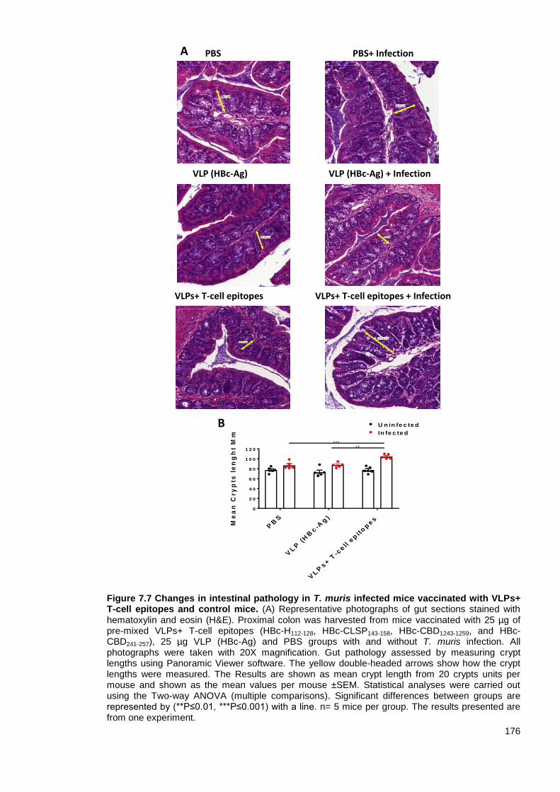
176
Figure 7.7 Changes in intestinal pathology in T. muris infected mice vaccinated with VLPs+ T-cell epitopes and control mice. (A) Representative photographs of gut sections stained with hematoxylin and eosin (H&E). Proximal colon was harvested from mice vaccinated with 25 µg of pre-mixed VLPs+ T-cell epitopes (HBc-H112-128, HBc-CLSP143-158, HBc-CBD1243-1259, and HBc-CBD241-257), 25 µg VLP (HBc-Ag) and PBS groups with and without T. muris infection. All photographs were taken with 20X magnification. Gut pathology assessed by measuring crypt lengths using Panoramic Viewer software. The yellow double-headed arrows show how the crypt lengths were measured. The Results are shown as mean crypt length from 20 crypts units per mouse and shown as the mean values per mouse ±SEM. Statistical analyses were carried out using the Two-way ANOVA (multiple comparisons). Significant differences between groups are represented by (**P≤0.01, ***P≤0.001) with a line. n= 5 mice per group. The results presented are from one experiment.
VLPs+ T-cell epitopes + Infection
VLP (HBc-Ag) + Infection
PBS+ Infection
VLPs+ T-cell epitopes
VLP (HBc-Ag)
PBS
PB
S
VL
P (
HB
c-A
g)
VL
Ps+
T-c
ell e
pit
op
es
0
2 0
4 0
6 0
8 0
1 0 0
1 2 0
Me
an
Cry
pts
le
ng
ht
Mm
*****
U n in fe c te d
In fe c te d
A
B
177
7.3.1.6 Goblet cells hyperplasia in mice vaccinated with VLPs expressing Trichuris T-cell
epitopes
As mentioned before, goblet cells and the production of mucins are important in assisting parasite
expulsion under the control of Th2 cytokines (IL-13 and IL-9) (Artis et al., 2004b). It has been
demonstrated that a marked goblet cell hyperplasia is seen in both susceptible and resistant mice
during T. muris infection compared to the uninfected naïve mice (Artis et al., 2004b; Cliffe and
Grencis, 2004; Datta et al., 2005; Owyang et al., 2006). However, there is a difference in the type
of mucin (the major protein component of mucus) produced by intestinal goblet cells between
resistant and susceptible mice (Hasnain et al., 2011a; Hasnain et al., 2010).
PAS-stained goblet cells were measured in the mice vaccinated with VLPs+ T-cell epitopes
compared to the native VLP (HBc-Ag), and PBS-injected mice with and without T. muris infection
as an index of goblet cells hyperplasia, as shown in Figure 7.8 (A).
There was no significant increase (P>0.05) in the numbers of PAS-stained goblet cells in all the
mouse groups prior to T. muris infection. However, following Trichuris challenge, significant
(P<0.05) goblet cell hyperplasia was seen in the group vaccinated with VLPs+ T-cell epitopes
compared to the native VLP (HBc-Ag) and PBS-injected mice (Figure 7.8 B).

178
Figure 7.8 Quantification of goblet cell numbers in T. muris infected mice vaccinated with VLPs+ T-cell epitopes and control mice. (A) Representative photographs of gut sections stained with Periodic acid–Schiff staining (PAS). Proximal colon was harvested from mice vaccinated with 25 µg of pre-mixed VLPs+ T-cell epitopes (HBc-H112-128, HBc-CLSP143-158, HBc-CBD1243-1259, and HBc-CBD241-257), 25 µg VLP (HBc-Ag), and PBS groups with and without T. muris infection. All photographs were taken with 20X magnification. The slides were scanned, and the goblet cells were quantified using Panoramic Viewer software. All photographs were taken with 20X magnification. (B) Goblet cells were quantified by counting the number of alcian blue-stained cells per crypt unit in three fields of view from each section and are shown as mean cell numbers per 20 crypt units (cu) ±SEM. Statistical analyses were carried out using Two-way ANOVA (multiple comparisons). Significant differences between groups are represented by (*P≤0.05, **P≤0.01, ****P≤0.0001) with a line. n= 5 mice per group. The results presented are from one experiment.
VLPs+ T-cell epitopes + Infection
VLP (HBc-Ag) + Infection
PBS+ Infection
VLPs+ T-cell epitopes
VLP (HBc-Ag)
PBSA
B
PB
S
VL
P (
HB
c-A
g)
VL
Ps+ T
-cell e
pit
op
es
0
1 0 0
2 0 0
3 0 0
4 0 0
Go
ble
t c
ell
s p
er 2
0 c
ry
pt
un
its
* * * *
U n in fe c te d
In fe c te d
179
7.3.2 Experimental protocol for in vivo evaluation of vaccinating mice with 25 µg of pre-
mixed VLPs + Trichuris MHC-II T-cell epitopes (HBc-H112-128, HBc-CLSP143-158, HBc-
CLSP398-416, and HBc-CBD241-257) formulated with AddaVax™ adjuvant
To determine if the addition of an AddaVax™ adjuvant can enhance the immune response to VLPs
expressing Trichuris T-cell epitopes, 6-week old male C57BL/6 mice were vaccinated
subcutaneously with 25 µg of pre-mixed VLPs+ T-cell epitopes (HBc-H112-128, HBc-CLSP143-158,
HBc-CLSP398-416, and HBc-CBD241-257) formulated with an equal volume of AddaVax adjuvant on
day -35 and boosted on day -14. As controls for susceptibility to infection, mice were injected with
25 μg of native VLP (HBc-Ag)/AddaVax, and PBS/AddaVax and as a control for protective
immunity, mice were vaccinated with 50 µg ES/AddaVax. Two weeks after the last vaccination,
mice were infected by oral gavage with approximately 150 infective T. muris eggs on day 0 and
sacrificed at day 14 post-infection (Figure 7.9 A).
The vaccine dosing regime was changed to allow efficient time for antibody production and
formation of memory immune response in response to the vaccine.
On day 14 p.i., the proximal colon and caecum were removed for worm burden estimation; colonic
sections were also collected to assess the pathology and count goblet cells. MLNs were collected
and were re-stimulated in vitro with Trichuris ES for cytokine analysis, while blood was collected to
measure serum parasite and VLP+ T-cell epitope-specific IgM, IgG1 and IgG2c antibody levels.
7.3.2.1 25 µg of pre-mixed VLPs expressing Trichuris T-cell epitopes formulated with
AddaVax adjuvant immunisation did not reduce the T. muris worm count following
challenge infection
Activation of the innate immune system is essential for the successful induction of protective T-cell
responses to control infection. Adjuvants have been traditionally used to stimulate the innate
immune system efficiently to increase the magnitude of an adaptive response to a vaccine
(Coffman et al., 2010). The AddaVax adjuvant, in particular, is a squalene-based oil-in-water
emulsion with a formulation analogous to MF59 has been licensed in most of Europe for pandemic
influenza vaccine (Ott et al., 2000; Podda, 2001). The AddaVax adjuvant has been demonstrated
to stimulates potent cellular and humoral immune responses and generates marked memory
responses (Calabro et al., 2013; Ott et al., 1995), with mixed Th1-Th2 immune responses
(Knudsen et al., 2016; O’hagan et al., 2013). The success of AddaVax adjuvant is dependent on
inducing the expression of genes involved in leukocyte migration, and antigen-presentation that are
both larger and distinct from that induced by other adjuvants including Alum (Mosca et al., 2008).
A vaccine of VLPs expressing Trichuris T-cell epitopes (HBc-H112-128, HBc-CLSP143-158, HBc-
CLSP398-416, and HBc-CBD241-257) formulated with AddaVax was evaluated against the protective
vaccine ES/AddaVax and controls of native VLP (HBc-Ag)/AddaVax, and PBS/AddaVax. Choosing
to vaccinate mice with 50 µg ES/AddaVax was based on a previous study that showed that
vaccinating AKR susceptible mice with 50 µg ES/FIA significantly reduced worm burden at day 21
p.i. (Dixon, 2007). No significant differences were observed in the worm count at day 14 p.i.
between VLPs+ T-cell epitopes/AddaVax and control groups. However, vaccinating mice with
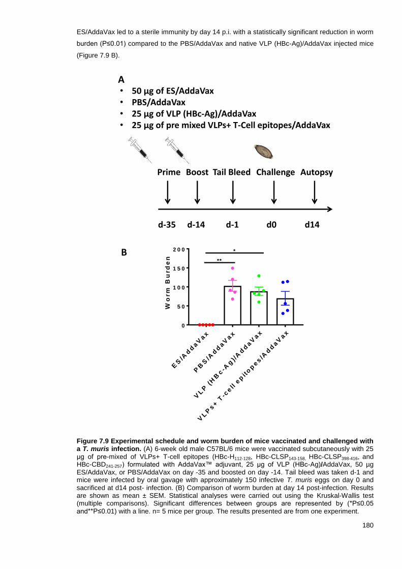
180
ES/AddaVax led to a sterile immunity by day 14 p.i. with a statistically significant reduction in worm
burden (P≤0.01) compared to the PBS/AddaVax and native VLP (HBc-Ag)/AddaVax injected mice
(Figure 7.9 B).
Figure 7.9 Experimental schedule and worm burden of mice vaccinated and challenged with a T. muris infection. (A) 6-week old male C57BL/6 mice were vaccinated subcutaneously with 25 μg of pre-mixed of VLPs+ T-cell epitopes (HBc-H112-128, HBc-CLSP143-158, HBc-CLSP398-416, and HBc-CBD241-257) formulated with AddaVax™ adjuvant, 25 μg of VLP (HBc-Ag)/AddaVax, 50 µg ES/AddaVax, or PBS/AddaVax on day -35 and boosted on day -14. Tail bleed was taken d-1 and mice were infected by oral gavage with approximately 150 infective T. muris eggs on day 0 and sacrificed at d14 post- infection. (B) Comparison of worm burden at day 14 post-infection. Results are shown as mean ± SEM. Statistical analyses were carried out using the Kruskal-Wallis test (multiple comparisons). Significant differences between groups are represented by (*P≤0.05 and**P≤0.01) with a line. n= 5 mice per group. The results presented are from one experiment.
A
B
• 50 μg of ES/AddaVax• PBS/AddaVax• 25 μg of VLP (HBc-Ag)/AddaVax• 25 μg of pre mixed VLPs+ T-Cell epitopes/AddaVax
Prime Boost Challenge
d-35 d-14 d0 d14
Autopsy
d-1
Tail Bleed
ES
/Ad
daV
ax
PB
S/A
dd
aV
ax
VL
P (
HB
c-A
g) /
Ad
daV
ax
VL
Ps+ T
-cell e
pit
op
es/A
dd
aV
ax
0
5 0
1 0 0
1 5 0
2 0 0
Wo
rm
Bu
rd
en **
*

181
7.3.2.2 Antigen profile in the sera of vaccinated mice
From the ELISA analysis (see Supplementary Figure 7.2), the serum of mice vaccinated with
VLPs+ T-cell epitopes contains IgM antibody recognising T. muris ES. Therefore, serum samples
from mice vaccinated with ES/AddaVax, and VLPs+ T-cell epitopes (HBc-H112-128, HBc-CLSP143-158,
HBc-CLSP398-416, and HBc-CBD241-257)/AddaVax and mice injected with PBS/AddaVax, native VLP
(HBc-Ag)/AddaVax were tested for their antibody recognition of T. muris ES antigens.
T. muris ES components were resolved by SDS-PAGE, transferred to nitrocellulose membrane and
subsequently incubated with the serum of each vaccinated mouse separately. Previous work by
Dixon et al. (2010) and Else et al. (1990) showed that resistance to T. muris infection correlated
with serum antibody recognition of high molecular weight proteins in the T. muris adult and larval
ES. Similarly, the immune serum of mice vaccinated with ES/AddaVax recognised a range of high
molecular weight proteins from 37-250 kDa range. The banding pattern recognised by anti-IgM and
anti-IgG in the immune serum appeared to reveal no qualitative difference in protein bands, as
shown in Figure 7.10 (Lane 1 and 2). In contrast, the immune sera of mice injected with
PBS/AddaVax and native VLP (HBc-Ag)/AddaVax did not recognise any protein in the T. muris ES.
Importantly, serum from mice vaccinated with VLPs+ T-cell epitopes recognised high molecular
weight proteins in the T. muris ES, suggesting that the H112-128, CLSP143-158, CBD1243-1259, and
CBD241-257 are highly-conserved immunodominant epitopes expressed in several secretory proteins
in the ES products of the T. muris worm (Fig 7.10 Lane 5).
Furthermore, the ELISA assay revealed that the serum from mice vaccinated with ES/AddaVax
recognised the VLPs+ T-cell epitopes recombinant proteins (HBc-H112-128, HBc-CLSP143-158, and
HBc-CBD1243-1259) (see Supplementary Figure 7.3). Therefore, the presence of an antibody directed
against the anti-VLPs+ T-cell epitopes in the serum of mice vaccinated with ES/AddaVax was
assessed using western blot.
Moreover, in order to assess whether the antibody in the serum of the mice vaccinated with VLPs+
T-cell epitopes/AddaVax and infected with T. muris is specific to the vaccine candidate, the antigen
profile in the serum of vaccinated mice was assessed using western blot. A mix of four VLPs+ T-
cell epitope proteins was resolved by SDS-PAGE and then subsequently plotted against the serum
of each vaccinated mouse separately. Anti-IgG in the serum of mice vaccinated with VLPs+ T-cell
epitopes/AddaVax recognised a band around ~22 kDa corresponding to the size of the VLPs+ T-
cell epitopes. Remarkably, the IgG in the serum of mice vaccinated with ES/AddaVax recognised
the same 22 kDa band. In contrast, the immune sera of PBS/AddaVax-injected mice did not
recognise any band, as shown in Figure 7.11.

182
Figure 7.10 Western blots of serum of vaccinated mice and control groups against T. muris ES. (M) Precision plus protein all standard blue marker (BIO-RAD) was used as a reference. Mice were vaccinated subcutaneously with 50 µg ES/AddaVax, PBS, 25 μg native VLP (HBc-Ag), and 25 μg VLPs +T-cell epitopes (HBc-H112-128, HBc-CLSP143-158, HBc-CBD1243-1259, and HBc-CBD241-
257), infected by oral gavage with approximately 150 infective T. muris eggs and sacrificed at d14. Day 14 sera were blotted against T. muris ES. Anti-mouse IgM and anti-IgG were used as secondary antibodies. WB was performed using (1:160) serum dilutions. One mouse per group was chosen to represent the result of its respective group. The results presented are from one experiment.
kDa25015010075503725201510
M 1 2 3 4 5
IgMIgG

183
Figure 7.11 Western blots of serum IgG from vaccinated mice and control groups specific for VLPs+ T-cell epitopes proteins. (M) Precision plus protein all standard blue marker (BIO-RAD) was used as a reference. Mice were vaccinated subcutaneously with 50 µg ES/AddaVax, PBS/AddaVax and 25 μg VLPs+ T-cell epitopes (HBc-H112-128, HBc-CLSP143-158, HBc-CLSP398-416, and HBc-CBD241-257), on day -35 and boosted on day -14. Mice were infected by oral gavage with approximately 150 infective T. muris eggs and sacrificed at d14. Day 14 sera were blotted against pre-mixed VLPs+ T-cell epitopes proteins. Anti-mouse IgG was used as secondary antibodies. WB was performed using (1:160) serum dilutions. One mouse per group was chosen to represent the result of its respective group. The results presented are from one experiment.
kDa25015010075503725201510
M 1 2 3
184
7.3.3 Experimental protocol for in vivo evaluation of vaccinating mice with 50 µg of pre-
mixed VLPs + Trichuris MHC-II T-cell epitopes (HBc-CBD1243-1259, HBc-CBD241-257,
HBc-CLSP143-158, and HBc-CLSP398-416)
Based on the first in vivo study, immunisation of mice with 25 μg VLPs+ T-cell epitopes did not
protect mice from Trichuris infection; however, one mouse expelled all the worms. Therefore, a
decision was made to increase the dose of the VLPs+ T-cell epitopes vaccine candidate from 25
μg to 50 μg. Furthermore, based on the previous in vivo studies, no HBc-H112-128-specific IgM
antibodies were detected in the serum of mice vaccinated with pre-mixed VLPs+ T-cell epitopes.
Therefore, a decision was made to replace this VLPs+ T-cell epitope recombinant protein with
HBc-CLSP398-416 in the pre-mixed VLPs+ T-cell epitopes vaccine candidate. As including an
adjuvant and altering the vaccination regime did not result in protective immunity, the vaccination
strategy used in the first experiment was followed.
To determine the protective efficacy of a high-dose of VLPs expressing Trichuris T-cell epitopes,
C57BL/6 mice were vaccinated subcutaneously with 50 μg of pre-mixed VLPs+ T-cell epitopes
(HBc-CBD1243-1259, HBc-CBD241-257, HBc-CLSP143-158, and HBc-CLSP398-416) on day -20 and boosted
on day -10. As controls for susceptibility to infection, mice were injected with PBS and 50 μg of
native VLP (HBc-Ag). As a control for protective immunity, mice were vaccinated with 50 µg ES
(Dixon et al., 2010) formulated with an equal volume of Alum-aluminium salt adjuvant (Hogenesch,
2013). Ten days after the last vaccination, mice were infected by oral gavage with approximately
150 infective T. muris eggs on day 0 and sacrificed at day 14 p.i. (Figure 7.12 A).
The protective efficacy of the vaccine candidate was assessed two weeks post-infection by
counting the worms from the proximal colon and caecum of each mouse. MLNs at day 14 p.i. were
collected and re-stimulated in vitro with Trichuris ES for cytokine production, while blood was
collected to measure serum parasite and VLP+ T-cell epitope-specific IgM, IgG1 and IgG2c
antibody levels.
185
7.3.3.1 50 µg of pre-mixed VLPs expressing Trichuris T-cell epitopes immunisation reduced
the T. muris worm count following challenge infection
Worm burden in mice immunised with pre-mixed VLPs+ T-cell epitopes (HBc-CBD1243-1259, HBc-
CBD241-257, HBc-CLSP143-158, and HBc-CLSP398-416), ES/Alum, native VLP (HBc-Ag), and PBS at
day 14 p.i. is shown in Figure 7.12 (B).
Worm count in susceptible control native VLP (HBc-Ag) group was then used to estimate the mean
% reduction in infection as described previously by Chung et al. (2004) using the formula:
% of reduction = ((C-V)/C) × 100
Where C is the worm count in mice immunised with native VLP (HBc-Ag), and V is the worm count
in mice vaccinated with VLPs+ T-cell epitopes following T. muris infection.
Remarkably, vaccination with VLPs+ T-cell epitopes showed a statistically significant (P<0.05)
reduction in worm burden (51.2%) by day 14 p.i.

186
Figure 7.12 Experimental schedule and worm burden of mice vaccinated and challenged with a T. muris infection. 6-week old male C57BL/6 mice were vaccinated subcutaneously with 50 μg of pre-mixed of VLPs+ T-cell epitopes (HBc-CBD1243-1259, HBc-CBD241-257, HBc-CLSP143-158, and HBc-CLSP398-416), 50 μg of VLP (HBc-Ag), or PBS on day -20 and boosted on day -10. Tail bleed was taken d-1 and mice were infected by oral gavage with approximately 150 infective T. muris eggs on day 0 and sacrificed at d14 post-infection. (B) Comparison of worm burden at day 14 post-infection. Statistical analyses were carried out using the Kruskal-Wallis test (multiple comparisons). Significant differences between groups are represented by (****P≤0.0001) with a line. Results are shown as mean ± SEM. n= 11 mice per group. The results presented are from two separated experiments pooled together.
PB
S
VL
P (
HB
c-A
g)
VL
Ps+ T
-Cell e
pit
op
es
0
5 0
1 0 0
1 5 0
2 0 0
2 5 0
Wo
rm
Bu
rd
en
* **
A
B
Prime Boost Challenge
d-20 d-10 d0 d14
Autopsy
d-1
Tail Bleed
• 50 μg ES/Alum
• PBS
• 50 μg VLP (HBc-Ag)
• 50 μg of pre mixed VLPs+ T-cell epitopes

187
7.3.3.2 Serum parasite-specific responses following vaccination of mice with VLPs
expressing Trichuris T-cell epitopes
To evaluate T. muris-specific serum antibody responses induced by vaccination with VLPs+ T-cell
epitopes, parasite-specific IgM, IgG1, and IgG2c serum antibody levels were determined at d-1 and
d14 p.i.. Notably, mice vaccinated with VLPs+ T-cell epitopes and ES/Alum produced similarly
statistically significant higher levels of parasite-specific IgM serum antibody compared to the PBS,
and native VLP (HBc-Ag) injected control groups prior to infection (Figure 7.13 A). In contrast, mice
vaccinated with ES/Alum produced significantly higher levels of parasite-specific IgG1 prior to
infection compared to the PBS, and native VLP (HBc-Ag) injected control groups. However, mice
vaccinated with VLP+T cell epitopes produced only negligible levels of parasite-specific IgG1 prior
to infection, as shown in Figure 7.13 (B).
Following vaccination and infection, VLPs+ T-cell epitopes and ES/Alum vaccinated mice had
statistically significant higher levels parasite-specific IgM (Figure 7.14 A) and IgG2c (C) compared
to the control PBS and native VLP (HBc-Ag) injected mice. However, high levels of parasite-
specific IgG1 were only detected in the serum of mice vaccinated with ES/Alum following Trichuris
infection (Figure 7.14 B). There was no or very low levels of parasite-specific IgM, IgG1, and IgG2c
detected in the serum of native VLP (HBc-Ag) and PBS/FIA injected mice at day 14 p.i., as shown
in Figure 7.14 (A-C).
Collectively, these results demonstrate that the VLPs+ T-cell epitopes vaccine candidate boosted
parasite-specific humoral immune responses.
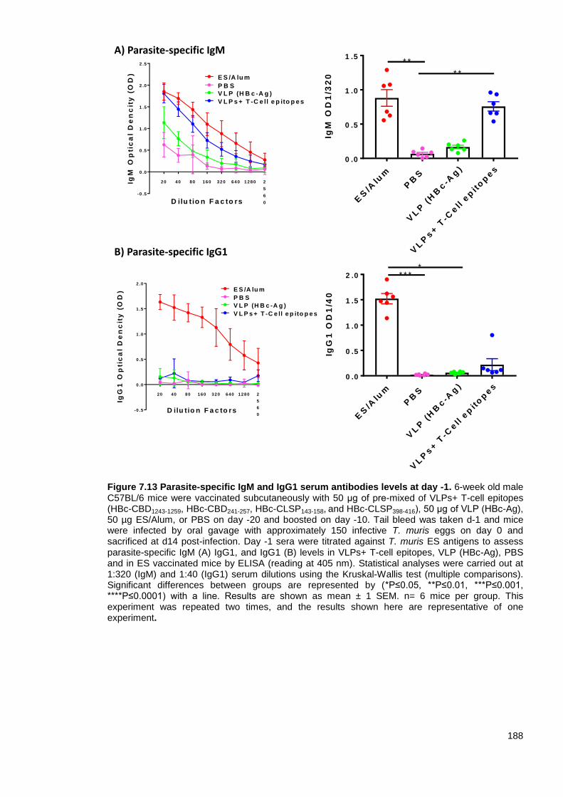
188
Figure 7.13 Parasite-specific IgM and IgG1 serum antibodies levels at day -1. 6-week old male C57BL/6 mice were vaccinated subcutaneously with 50 μg of pre-mixed of VLPs+ T-cell epitopes (HBc-CBD1243-1259, HBc-CBD241-257, HBc-CLSP143-158, and HBc-CLSP398-416), 50 μg of VLP (HBc-Ag), 50 µg ES/Alum, or PBS on day -20 and boosted on day -10. Tail bleed was taken d-1 and mice were infected by oral gavage with approximately 150 infective T. muris eggs on day 0 and sacrificed at d14 post-infection. Day -1 sera were titrated against T. muris ES antigens to assess parasite-specific IgM (A) IgG1, and IgG1 (B) levels in VLPs+ T-cell epitopes, VLP (HBc-Ag), PBS and in ES vaccinated mice by ELISA (reading at 405 nm). Statistical analyses were carried out at 1:320 (IgM) and 1:40 (IgG1) serum dilutions using the Kruskal-Wallis test (multiple comparisons). Significant differences between groups are represented by (*P≤0.05, **P≤0.01, ***P≤0.001, ****P≤0.0001) with a line. Results are shown as mean ± 1 SEM. n= 6 mice per group. This experiment was repeated two times, and the results shown here are representative of one experiment.
ES
/Alu
m
PB
S
VL
P (
HB
c-A
g)
VL
Ps+ T
-Cell e
pit
op
es
0 .0
0 .5
1 .0
1 .5
2 .0
IgG
1 O
D1
/40
* * * *
-0 .5
0 .0
0 .5
1 .0
1 .5
2 .0
D ilu tio n F a c to rs
IgG
1 O
pti
ca
l D
en
cit
y (
OD
)
20 40 80 160 320 640 1 280 2
5
6
0
P B S
E S /A lu m
V L P (H B c -A g )
V L P s + T -C e ll e p ito p e s
-0 .5
0 .0
0 .5
1 .0
1 .5
2 .0
2 .5
D ilu tio n F a c to rs
IgM
Op
tic
al
De
nc
ity
(O
D)
20 40 80 160 320 640 1 280 2
5
6
0
P B S
E S /A lu m
V L P (H B c -A g )
V L P s + T -C e ll e p ito p e s
ES
/Alu
m
PB
S
VL
P (
HB
c-A
g)
VL
Ps+ T
-Cell e
pit
op
es
0 .0
0 .5
1 .0
1 .5
IgM
OD
1/3
20 * *
* *
A) Parasite-specific IgM
B) Parasite-specific IgG1

189
Figure 7.14 Parasite-specific IgM, IgG1 and IgG2c serum antibodies levels at day 14 post-infection. 6-week old male C57BL/6 mice were vaccinated subcutaneously with 50 μg of pre-mixed of VLPs+ T-cell epitopes (HBc-CBD1243-1259, HBc-CBD241-257, HBc-CLSP143-158, and HBc-CLSP398-416), 50 μg of VLP (HBc-Ag), 50 µg ES/Alum, or PBS on day -20 and boosted on day -10. Tail bleed was taken d-1 and mice were infected by oral gavage with approximately 150 infective T. muris eggs on day 0 and sacrificed at d14 post-infection. Day 14 p.i. sera were titrated against T. muris ES antigens to assess parasite-specific IgM (A) IgG1 (B), and IgG2c (C) levels in VLPs+ T- cell epitopes, VLP (HBc-Ag), PBS and in ES vaccinated mice by ELISA (reading at 405 nm). Statistical analyses were carried out at 1:320 (IgM and IgG1) and 1:40 (IgG2c) serum dilutions using Kruskal-Wallis test (multiple comparisons). Significant differences between groups are represented by (*P≤0.05, **P≤0.01, ***P≤0.001, ****P≤0.0001) with a line. Results are shown as mean ± SEM. n= 6 mice per group. This experiment was repeated two times, and the results
shown here are representative of the two experiments.
ES
/Alu
m
PB
S
VL
P (
HB
c-A
g)
VL
Ps+ T
-Cell e
pit
op
es
0 .0
0 .5
1 .0
1 .5
IgG
2c
OD
1/4
0
*
*
*
*
-0 .2
0 .0
0 .2
0 .4
0 .6
0 .8
1 .0
D ilu t io n F a c to rs
IgG
2c
Op
tic
al
De
nc
ity
(O
D)
20 40 80 1 6 0 3 2 0 6 4 0 1 2 8 0 2 5 6 0
P B S
E S /A lu m
V L P (H B c -A g )
V L P s + T -C e ll e p ito p e s
ES
/Alu
m
PB
S
VL
P (
HB
c-A
g)
VL
Ps+ T
-Cell e
pit
op
es
0 .0
0 .5
1 .0
1 .5
2 .0
IgM
OD
1/3
20
*
* * * *
-0 .5
0 .0
0 .5
1 .0
1 .5
2 .0
2 .5
D ilu t io n F a c to rs
IgM
Op
tic
al
De
nc
ity
(O
D)
20 40 80 1 6 0 3 2 0 6 4 0 1 2 8 0 2 5 6 0
P B S
E S /A lu m
V L P
V L P s + T -C e ll e p ito p e s
ES
/Alu
m
PB
S
VL
P (
HB
c-A
g)
VL
Ps+ T
-Cell e
pit
op
es
0
1
2
3
IgG
1 O
D 1
/32
0
* * *
*
-1
0
1
2
3
D ilu t io n F a c to rs
IgG
1 O
pti
ca
l D
en
cit
y (
OD
)
20 40 80 1 6 0 3 2 0 6 4 0 1 2 8 0 2 5 6 0
P B S
E S /A lu m
V L P (H B c -A g )
V L P s + T -C e ll e p ito p e s
B) Parasite-specific IgG1
C) Parasite-specific IgG2c
A) Parasite-specific IgM

190
7.3.3.3 Serum VLP+T cell epitope-specific responses following vaccination of mice with
VLPs expressing Trichuris T-cell epitopes
The isotype profiles of antibodies elicited by VLPs+ T-cell epitopes were analysed using an ELISA-
based assay at d-1 and d 14 p.i.. ELISA plates were coated with pre-mixed VLPs+ T-cell epitopes
(HBc-CBD1243-1259, HBc-CBD241-257, HBc-CLSP143-158, and HBc-CLSP398-416) and with individual
VLP+T cell epitope recombinant protein as detection antigens. All serum from mice vaccinated with
VLPs+ T-cell epitopes displayed strong reactivity to the pre-mixed VLPs+ T-cell epitopes proteins.
Statistically significant higher levels of VLPs+ T-cell epitopes-specific IgM (Figure 7. 15 A) and
IgG1 (B) were measured at d-1 compared to the ES/Alum, PBS and VLP (HBc-Ag) immunised
groups. Similarly, statistically significant higher levels of VLPs+ T-cell epitopes-specific IgM (Figure
7.16 A), IgG1 (B) and IgG2c (C) were produced following Trichuris infection. Notably, mice
vaccinated with ES/Alum also produced high levels of VLPs+ T-cell epitopes-specific IgM following
Trichuris infection, as shown in Figure 7.16 (A).
Furthermore, serum from mice vaccinated with the VLPs+ T-cell epitopes reacted with their
corresponding VLP+T cell epitope recombinant protein but not the native VLP (HBc-Ag) protein.
Significantly higher levels of VLP+ T-cell epitope specific-IgM (Figure 7.17 A-D), IgG1 (Figure 7.18
A-D), and IgG2c (Figure 7.19 A-D) were measured in the serum of mice vaccinated with VLPs+ T-
cell epitopes compared to the ES/Alum, and PBS immunised mice groups. Furthermore, lower or
background levels of native VLP (HBc-Ag) specific-IgM (Figure 7.17 A), and IgG2c (Figure 7.19 A)
were detected in the sera of mice vaccinated with VLPs+ T-cell epitopes. However, low levels of
native VLP (HBc-Ag) specific-IgG1 were detected in the sera of mice vaccinated with VLPs+ T-cell
epitopes and native VLP (HBc-Ag) immunised group (Figure 7.18 A).
Collectively, these results demonstrate that vaccinating mice with VLPs+ T-cell epitopes induced
strong humoral immune responses specific to the four VLPs+ T-cell epitopes recombinant proteins.

191
Figure 7.15 Pre-mixed VLPs+ T-cell epitopes-specific IgM and IgG1 serum antibodies levels at day -1. 6-week old male C57BL/6 mice were vaccinated subcutaneously with 50 μg of pre-mixed of VLPs+ T-cell epitopes (HBc-CBD1243-1259, HBc-CBD241-257, HBc-CLSP143-158, and HBc-CLSP398-
416), 50 μg of VLP (HBc-Ag), 50 µg ES/Alum, or PBS on day -20 and boosted on day -10. Tail bleed was taken d-1 and mice were infected by oral gavage with approximately 150 infective T. muris eggs on day 0 and sacrificed at d14 post-infection. Day -1 sera were titrated against a cocktail of VLPs+ T-cell epitopes proteins (HBc-CBD1243-1259, HBc-CBD241-257, HBc-CLSP143-158 and HBc-CLSP398-416) to assess VLPs-specific IgM (A) and IgG1 (B) levels in VLPs+ T-cell epitopes, VLP (HBc-Ag), PBS and in ES vaccinated mice by ELISA (reading at 405 nm). Statistical analyses were carried out at 1:320 (IgM) and 1:640 (IgG1) serum dilutions using the Kruskal-Wallis test (multiple comparisons). Significant differences between groups are represented by (*P≤0.05, **P≤0.01, ***P≤0.001, ****P≤0.0001) with a line. Results are shown as mean ± SEM. n= 6 mice per group. This experiment was repeated two times, and the results shown here are representative of the two experiments.
ES
/Alu
mP
BS
VL
P (
HB
c-A
g)
VL
Ps+ T
-Cell e
pit
op
es
0 .0
0 .5
1 .0
1 .5
IgM
OD
1/3
20
* * *
*
*
-1 .0
-0 .5
0 .0
0 .5
1 .0
1 .5
2 .0
D ilu t io n F a c to rsIgM
Op
tic
al
De
nc
ity
(O
D)
20 40 80 160 320 640 1 280 2
5
6
0
P B S
E S /A lu m
V L P (H B c -A g )
V L P s + T -C e ll e p ito p e s
ES
/Alu
mP
BS
VL
P (
HB
c-A
g)
VL
Ps+ T
-Cell e
pit
op
es
0 .0
0 .5
1 .0
1 .5
IgG
1 O
D 1
/64
0 * *
* *
-0 .5
0 .0
0 .5
1 .0
1 .5
2 .0
D ilu t io n F a c to rsIgG
1 O
pti
ca
l D
en
cit
y (
OD
)
20 40 80 160 320 640 1 280 2
5
6
0
P B S
E S /A lu m
V L P (H B c -A g )
V L P s + T -C e ll e p ito p e s
A) Pre-mixed VLPs+ T-cell epitopes-specific IgM
B) Pre-mixed VLPS+ T-cell epitopes-specific IgG1

192
Figure 7.16 Pre-mixed VLP+ T-cell epitope-specific IgM, IgG1 and IgG2c levels at day 14 post-infection (p.i.). 6-week old male C57BL/6 mice were vaccinated subcutaneously with 50 μg of pre-mixed of VLPs+ T-cell epitopes (HBc-CBD1243-1259, HBc-CBD241-257, HBc-CLSP143-158, and HBc-CLSP398-416), 50 μg of VLP (HBc-Ag), 50 µg ES/Alum, or PBS on day -20 and boosted on day -10. Tail bleed was taken d-1 and mice were infected by oral gavage with approximately 150 infective T. muris eggs on day 0 and sacrificed at d14 post-infection. Day 14 p.i. sera were titrated against pre-mixed VLPs (HBc-CBD1243-1259, HBc-CBD241-257, HBc-CLSP143-158 and HBc-CLSP398-416) to assess VLPs-specific IgM (A), IgG1 (B), and IgG2c (C) levels in VLPs+ T-cell epitopes, VLP (HBc-Ag), PBS and in ES vaccinated mice by ELISA (reading at 405 nm). Statistical analyses were carried out at 1:320 (IgM) and 1:1280 (IgG1), 1:640 (IgG2c) serum dilutions using the Kruskal-Wallis test (multiple comparisons). Significant differences between groups are represented by (*P≤0.05, **P≤0.01, ***P≤0.001, ****P≤0.0001) with a line. Negative values were adjusted to 0. Results are shown as mean ± SEM. n= 6 mice per group. This experiment was repeated two times, and the results shown here are representative of the two experiments.
ES
/Alu
mP
BS
VL
P (
HB
c-A
g)
VL
Ps+ T
-Cell e
pit
op
es
0 .0
0 .2
0 .4
0 .6
0 .8
IgG
2a
OD
1/6
40
* * *
* * *
-2
-1
0
1
D ilu t io n F a c to rs
IgG
2a
Op
tic
al
De
nc
ity
(O
D)
20 40 80 1 6 0 3 2 0 6 4 0 1 2 8 0 2 5 6 0
P B S
E S /A lu m
V L P (H B c -A g )
V L P s + T -C e ll e p ito p e s
-1 .0
-0 .5
0 .0
0 .5
1 .0
1 .5
D ilu tio n F a c to rsIgG
1 O
pti
ca
l D
en
cit
y (
OD
)
20 40 80 1 6 0 3 2 0 6 4 0 1 2 8 0 2 5 6 0
P B S
E S /A lu m
V L P (H B c -A g )
V L P s + T -C e ll e p ito p e s
ES
/Alu
mP
BS
VL
P (
HB
c-A
g)
VL
Ps+ T
-Cell e
pit
op
es
0 .0
0 .5
1 .0
1 .5
IgG
1 O
D 1
/12
80
* * *
* * *
ES
/Alu
mP
BS
VL
P (
HB
c-A
g)
VL
Ps+T
Cell e
pit
op
es
0 .0
0 .5
1 .0
1 .5
IgM
OD
1/3
20
* *
* * *
-0 .5
0 .0
0 .5
1 .0
1 .5
D ilu t io n F a c to r s
IgM
Op
tic
al
De
nc
ity
(O
D)
20 40 80 1 6 0 3 2 0 6 4 0 1 2 8 0 2 5 6 0
P B S
E S /A lu m
V L P (H B c -A g )
V L P s + T -C e ll e p ito p e s
B) Pre-mixed VLPs+ T-cell epitopes-specific IgG1
C) Pre-mixed VLPs+ T-cell epitopes-specific IgG2c
A) Pre-mixed VLPs+ T-cell epitopes-specific IgM
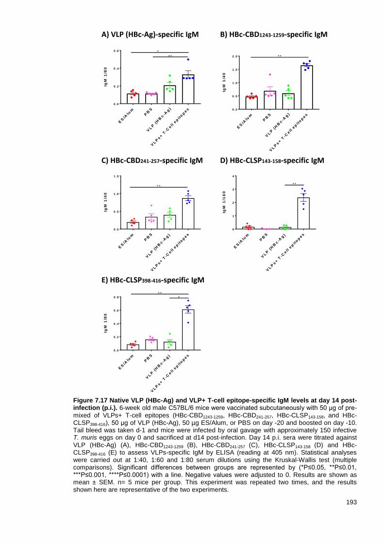
193
Figure 7.17 Native VLP (HBc-Ag) and VLP+ T-cell epitope-specific IgM levels at day 14 post-infection (p.i.). 6-week old male C57BL/6 mice were vaccinated subcutaneously with 50 μg of pre-mixed of VLPs+ T-cell epitopes (HBc-CBD1243-1259, HBc-CBD241-257, HBc-CLSP143-158, and HBc-CLSP398-416), 50 μg of VLP (HBc-Ag), 50 µg ES/Alum, or PBS on day -20 and boosted on day -10. Tail bleed was taken d-1 and mice were infected by oral gavage with approximately 150 infective T. muris eggs on day 0 and sacrificed at d14 post-infection. Day 14 p.i. sera were titrated against VLP (HBc-Ag) (A), HBc-CBD1243-1259 (B), HBc-CBD241-257 (C), HBc-CLSP143-158 (D) and HBc-CLSP398-416 (E) to assess VLPs-specific IgM by ELISA (reading at 405 nm). Statistical analyses were carried out at 1:40, 1:60 and 1:80 serum dilutions using the Kruskal-Wallis test (multiple comparisons). Significant differences between groups are represented by (*P≤0.05, **P≤0.01, ***P≤0.001, ****P≤0.0001) with a line. Negative values were adjusted to 0. Results are shown as mean ± SEM. n= 5 mice per group. This experiment was repeated two times, and the results shown here are representative of the two experiments.
ES
/Alu
mP
BS
VL
P (
HB
c-A
g)
VL
Ps+ T
-Cell e
pit
op
es
0 .0
0 .5
1 .0
1 .5
2 .0
IgM
1/4
0
* *
ES
/Alu
mP
BS
VL
P (
HB
c-A
g)
VL
Ps+ T
-Cell e
pit
op
es
0
1
2
3
4
IgM
1/1
60
* *
ES
/Alu
mP
BS
VL
P (
HB
c-A
g)
VL
Ps+ T
-Cell e
pit
op
es
0 .0
0 .2
0 .4
0 .6
0 .8
IgM
1/8
0
** *
ES
/Alu
mP
BS
VL
P (
HB
c-A
g)
VL
Ps+ T
-Cell e
pit
op
es
0 .0
0 .5
1 .0
1 .5
IgM
1/4
0
* *
A) VLP (HBc-Ag)-specific IgM
ES
/Alu
mP
BS
VL
P (
HB
c-A
g)
VL
Ps+ T
-Cell e
pit
op
es
0 .0
0 .2
0 .4
0 .6
IgM
1/8
0
* *
*
B) HBc-CBD1243-1259-specific IgM
C) HBc-CBD241-257-specific IgM D) HBc-CLSP143-158-specific IgM
E) HBc-CLSP398-416-specific IgM
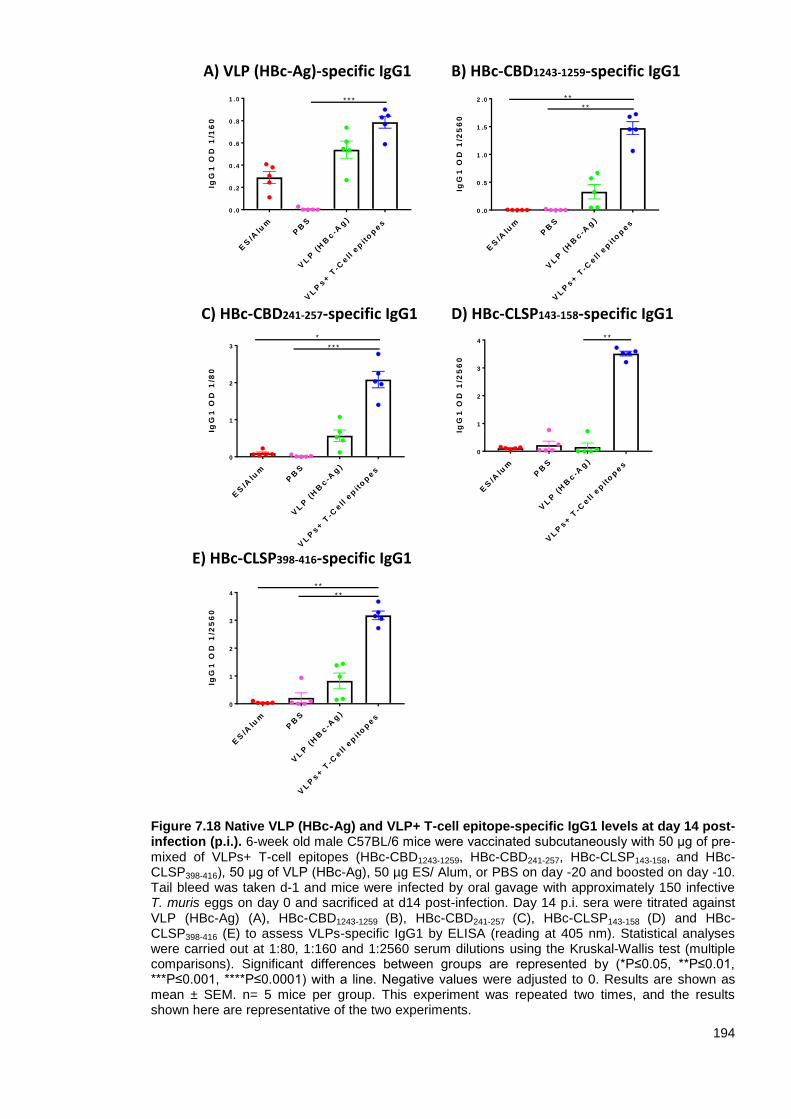
194
Figure 7.18 Native VLP (HBc-Ag) and VLP+ T-cell epitope-specific IgG1 levels at day 14 post-infection (p.i.). 6-week old male C57BL/6 mice were vaccinated subcutaneously with 50 μg of pre-mixed of VLPs+ T-cell epitopes (HBc-CBD1243-1259, HBc-CBD241-257, HBc-CLSP143-158, and HBc-CLSP398-416), 50 μg of VLP (HBc-Ag), 50 µg ES/ Alum, or PBS on day -20 and boosted on day -10. Tail bleed was taken d-1 and mice were infected by oral gavage with approximately 150 infective T. muris eggs on day 0 and sacrificed at d14 post-infection. Day 14 p.i. sera were titrated against VLP (HBc-Ag) (A), HBc-CBD1243-1259 (B), HBc-CBD241-257 (C), HBc-CLSP143-158 (D) and HBc-CLSP398-416 (E) to assess VLPs-specific IgG1 by ELISA (reading at 405 nm). Statistical analyses were carried out at 1:80, 1:160 and 1:2560 serum dilutions using the Kruskal-Wallis test (multiple comparisons). Significant differences between groups are represented by (*P≤0.05, **P≤0.01, ***P≤0.001, ****P≤0.0001) with a line. Negative values were adjusted to 0. Results are shown as mean ± SEM. n= 5 mice per group. This experiment was repeated two times, and the results shown here are representative of the two experiments.
ES
/Alu
mP
BS
VL
P (
HB
c-A
g)
VL
Ps+ T
-Cell e
pit
op
es
0 .0
0 .5
1 .0
1 .5
2 .0
IgG
1 O
D 1
/25
60
* *
* *
ES
/Alu
mP
BS
VL
P (
HB
c-A
g)
VL
Ps+ T
-Cell e
pit
op
es
0
1
2
3
4
IgG
1 O
D 1
/25
60
* ** *
ES
/Alu
mP
BS
VL
P (
HB
c-A
g)
VL
Ps+ T
-Cell e
pit
op
es
0
1
2
3
4
IgG
1 O
D 1
/25
60
* *
ES
/Alu
mP
BS
VL
P (
HB
c-A
g)
VL
Ps+ T
-Cell e
pit
op
es
0
1
2
3
IgG
1 O
D 1
/80
*
* * *
ES
/Alu
mP
BS
VL
P (
HB
c-A
g)
VL
Ps+ T
-Cell e
pit
op
es
0 .0
0 .2
0 .4
0 .6
0 .8
1 .0
IgG
1 O
D 1
/16
0
* * *
A) VLP (HBc-Ag)-specific IgG1 B) HBc-CBD1243-1259-specific IgG1
C) HBc-CBD241-257-specific IgG1 D) HBc-CLSP143-158-specific IgG1
E) HBc-CLSP398-416-specific IgG1

195
Figure 7.19 Native VLP (HBc-Ag) and VLP+ T-cell epitope-specific IgG2c levels at day 14 post-infection (p.i.). 6-week old male C57BL/6 mice were vaccinated subcutaneously with 50 μg of pre-mixed of VLPs+ T-cell epitopes (HBc-CBD1243-1259, HBc-CBD241-257, HBc-CLSP143-158, and HBc-CLSP398-416), 50 μg of VLP (HBc-Ag), 50 µg ES/Alum, or PBS on day -20 and boosted on day -10. Tail bleed was taken d-1 and mice were infected by oral gavage with approximately 150 infective T. muris eggs on day 0 and sacrificed at d14 post-infection. Day 14 p.i. sera were titrated against VLP (HBc-Ag) (A), HBc-CBD1243-1259 (B), HBc-CBD241-257 (C), HBc-CLSP143-158 (D) and HBc-CLSP398-416 (E) to assess VLPs-specific IgG2c by ELISA (reading at 405 nm). Statistical analyses were carried out at 1:40, 1:80, 1:1280 and 1:2560 serum dilutions using the Kruskal-Wallis test (multiple comparisons). Significant differences between groups are represented by (*P≤0.05, **P≤0.01, ***P≤0.001, ****P≤0.0001) with a line. Negative values were adjusted to 0. Results are shown as mean ± SEM. n= 5 mice per group. This experiment was repeated two times, and the results shown here are representative of the two experiments.
ES
/Alu
mP
BS
VL
P (
HB
c-A
g)
VL
Ps+ T
-Cell e
pit
op
es
0
1
2
3
IgG
2a
OD
1/8
0
*
* *
ES
/Alu
mP
BS
VL
P (
HB
c-A
g)
VL
Ps+ T
-Cell e
pit
op
es
0
1
2
3
4
5
IgG
2c
OD
1/1
28
0
* *
*
ES
/Alu
mP
BS
VL
P (
HB
c-A
g)
VL
Ps+ T
-Cell e
pit
op
es
0 .0
0 .5
1 .0
1 .5
2 .0
2 .5
IgG
2a
OD
1/2
56
0
* ** *
ES
/Alu
mP
BS
VL
P (
HB
c-A
g)
VL
Ps+ T
-Cell e
pit
op
es
0
1
2
3
4
IgG
2a
OD
1/4
0
* ** *
ES
PB
S
ES
/Alu
m
VL
Ps+ T
-Cell e
pit
op
es
0 .0
0 .5
1 .0
1 .5
IgG
2c
OD
1
/16
0
*
*
A) VLP (HBc-Ag)-specific IgG2c B) HBc-CBD-1243-1259-specific IgG2c
C) HBc-CBD241-257-specific IgG2c D) HBc-CLSP143-158-specific IgG2c
E) HBc-CLSP398-416-specific IgG2c

196
7.3.3.4 The cellular immune responses following vaccination of mice with VLPs expressing
Trichuris T-cell epitopes
To analyze the cellular immune responses of mice vaccinated with VLPs+ T-cell epitopes
compared to control ES/Alum, native VLP (HBc-Ag) and PBS immunised mice groups after
T. muris infection, MLN cells were re-stimulated in vitro with Trichuris ES and with pre-mixed
VLPs+ T-cell epitopes proteins (HBc-CBD1243-1259, HBc-CBD241-257, HBc-CLSP143-158, and HBc-
CLSP398-416) for 48 hours and with ConA for 24 hours. Supernatants were assayed for Th2
cytokines (IL-4, IL-5, IL-9 and IL-13), Th1/Th17 cytokines (IL-2, IFN-γ and IL-17), proinflammatory
cytokines (IL-6 and TNF-α), and the anti-inflammatory cytokine (IL-10) production by CBA. MLN
cells from mice vaccinated with VLPs+ T-cell epitope produced higher levels of Th2 cytokines IL-4
(A), IL-5 (B), IL-9 (C), and IL-13 (D) compared to all immunised mice including ES/Alum group but
statistical significance was not reached. Significantly higher levels of the proinflammatory cytokine
IL-6 (Figure 7.20 H) were also detected in response to re-stimulation in vitro with ES compared to
ES/Alum, and PBS immunised mice. Notably, re-stimulated MLN cells from mice vaccinated with
VLPs+ T-cell epitope and ES/Alum produced similarly high levels of Th1 cytokine (IFN-γ) (Figure
7.20 F). In contrast, MLN cells from all vaccinated mice groups produced similar levels of IL-2
(Figure 7.20 E), TNF (G), IL-17 (I) cytokines and anti-inflammatory cytokine IL-10 (J) upon their re-
stimulation in vitro with worm ES antigens.
The most noticeable difference in cytokine production in response to in vitro re-stimulation with
VLPs+ T-cell epitopes was for IL-5, IL-6 and IL-10. Interestingly, re-stimulated MLN cells from mice
vaccinated with VLPs+ T-cell epitopes and ES/Alum showed similarly higher levels of IL-5 (Figure
7.21 B), IL-6 (H) and IL-10 (J) compared to the PBS, and VLP (HBc-Ag) injected mice groups.
Furthermore, MLN cells from mice vaccinated with ES/Alum produced significantly higher levels of
IL-13 upon re-stimulation in vitro with VLPs+ T-cell epitopes proteins compared to all immunised
groups (Figure 7.21D).
Furthermore, ConA re-stimulated MLN cells from mice vaccinated with VLPs+ T-cell epitopes and
infected with T. muris showed a slight increase in the production of IL-5 (Figure 7.22 B), IL-9 (C) IL-
13 (D), IL-2 (E), TNF (G) and IL-6 (H) compared to the three other immunised mouse groups.
Collectively, these results demonstrate that the VLPs+ T-cell epitopes vaccine candidate efficiently
boosted the cellular immune response.

197
Figure 7.20 Parasite-specific cytokine productions by mesenteric lymph node cells from mice vaccinated with VLPs+ T-cell epitopes and control mice. MLNs from mice vaccinated with 50 μg of pre-mixed of VLPs+ T-cell epitopes epitopes (HBc-CBD1243-1259, HBc-CBD241-257, HBc-CLSP143-158 and HBc-CLSP398-416), 50 μg of VLP (HBc-Ag), 50 µg ES/Alum, or PBS on day -20 and boosted on day -10 were stimulated at 5x10
6/ml with 50 µg/ml T. muris ES. After 48 hours of
stimulation, cell culture supernatants were harvested and assayed by cytometric bead array for IL-4 (A), IL-5 (B), IL-9 (C), IL-13 (D), IL-2 (E), IFN-g (F), TNF (G), IL-6 (H), IL-17 (I) and IL-10 (J) production. Statistical analyses were carried out using the Kruskal-Wallis test (multiple comparisons). Significant differences between groups are represented by (*P≤0.05) with a line. Results are shown as mean ± SEM. n= 5 mice per group. This experiment was repeated two times, and the results shown here are representative of the two experiments.
ES
PB
S
VL
P (
HB
c-A
g)
VL
P+T
cell e
pit
op
es
0
1 0
2 0
3 0
IL2
Co
nc
en
tra
tio
n
pg
/mL
ES
PB
S
VL
P (
HB
c-A
g)
VL
P+T
cell e
pit
op
es
0
2 0 0
4 0 0
6 0 0
8 0 0
1 0 0 0
IL5
Co
nc
en
tra
tio
n
pg
/mL
ES
PB
S
VL
P (
HB
c-A
g)
VL
P+T
cell e
pit
op
es
0
5 0
1 0 0
1 5 0
2 0 0
IL4
Co
nc
en
tra
tio
n
pg
/mL
ES
PB
S
VL
P (
HB
c-A
g)
VL
P+T
cell e
pit
op
es
0
1 0 0
2 0 0
3 0 0
4 0 0
IL1
7 C
on
ce
ntr
ati
on
p
g/m
L
ES
PB
S
VL
P (
HB
c-A
g)
VL
P+T
cell e
pit
op
es
0
2 0 0 0
4 0 0 0
6 0 0 0
IL1
3 C
on
ce
ntr
ati
on
p
g/m
L
ES
PB
S
VL
P (
HB
c-A
g)
VL
P+T
cell e
pit
op
es
0
2 0 0
4 0 0
6 0 0
TN
F C
on
ce
ntr
ati
on
p
g/m
L
ES
PB
S
VL
P (
HB
c-A
g)
VL
P+T
cell e
pit
op
es
0
5 0 0
1 0 0 0
1 5 0 0
IL9
Co
nc
en
tra
tio
n
pg
/mL
ES
PB
S
VL
P (
HB
c-A
g)
VL
P+T
cell e
pit
op
es
0
5 0 0 0
1 0 0 0 0
1 5 0 0 0
2 0 0 0 0
IFN
-g C
on
ce
ntr
ati
on
p
g/m
L
A B C
D E F
G H I
J
ES
PB
S
VL
P (
HB
c-A
g)
VL
P+T
cell e
pit
op
es
0
1 0 0 0
2 0 0 0
3 0 0 0
IL1
0 C
on
ce
ntr
ati
on
p
g/m
L
ES
PB
S
VL
P (
HB
c-A
g)
VL
P+T
cell e
pit
op
es
0
2 0 0
4 0 0
6 0 0
8 0 0
1 0 0 0
IL6
Co
nc
en
tra
tio
n
pg
/mL

198
Figure 7.20 Parasite-specific cytokine production by mesenteric lymph node cells from mice vaccinated with VLPs+ T-cell epitopes and control mice. MLNs from mice vaccinated with 50 μg of pre-mixed of VLPs+ T-cell epitopes (HBc-CBD1243-1259, HBc-CBD241-257, HBc-CLSP143-158 and HBc-CLSP398-416), 50 μg of VLP (HBc-Ag), 50 µg ES/Alum, or PBS on day -20 and boosted on day -10 were stimulated at 5x10
6/ml with 50 µg/ml T. muris ES. After 48 hours of stimulation, cell
culture supernatants were harvested and assayed by cytometric bead array for IL-4 (A), IL-5 (B), IL-9 (C), IL-13 (D), IL-2 (E), IFN-g (F), TNF (G), IL-6 (H), IL-17 (I) and IL-10 (J) production. Statistical analyses were carried out using the Kruskal-Wallis test (multiple comparisons). Significant differences between groups are represented by (*P≤0.05) with a line. Results are shown as mean ± SEM. n= 5 mice per group. This experiment was repeated two times, and the results shown here are representative of the two experiments.
A B
C D
E F
G H
I J
ES
/Alu
m
PB
S
VL
P
VL
Ps+ T
-cell e
pit
op
es
0
2 0 0
4 0 0
6 0 0
8 0 0
TN
F C
on
ce
ntr
ati
on
p
g/m
L
ES
/Alu
m
PB
S
VL
P
VL
Ps+ T
-cell e
pit
op
es
0
1 0 0
2 0 0
3 0 0
4 0 0
IL-1
7 C
on
ce
ntr
ati
on
p
g/m
L
ES
/Alu
m
PB
S
VL
P
VL
Ps+ T
-cell e
pit
op
es
0
2 0 0 0
4 0 0 0
6 0 0 0
IL-1
3 C
on
ce
ntr
ati
on
p
g/m
L
ES
/Alu
m
PB
S
VL
P
VL
Ps+ T
-cell e
pit
op
es
0
1 0 0 0
2 0 0 0
3 0 0 0
IL-1
0 C
on
ce
ntr
ati
on
p
g/m
L
ES
/Alu
m
PB
S
VL
P
VL
Ps+ T
-cell e
pit
op
es
0
5 0 0
1 0 0 0
1 5 0 0
IL-9
Co
nc
en
tra
tio
n
pg
/mL
ES
/Alu
m
PB
S
VL
P
VL
Ps+ T
-cell e
pit
op
es
0
2 0 0
4 0 0
6 0 0
8 0 0
1 0 0 0
IL-6
Co
nc
en
tra
tio
n
pg
/mL
* *
ES
/Alu
m
PB
S
VL
P
VL
Ps+ T
-cell e
pit
op
es
0
2 0 0
4 0 0
6 0 0
8 0 0
1 0 0 0
IL-5
Co
nc
en
tra
tio
n
pg
/mL
ES
/Alu
m
PB
S
VL
P
VL
Ps+ T
-cell e
pit
op
es
0
5 0
1 0 0
1 5 0
2 0 0
IL-4
Co
nc
en
tra
tio
n
pg
/mL
ES
/Alu
m
PB
S
VL
P
VL
Ps+ T
-cell e
pit
op
es
0
5
1 0
1 5
2 0
2 5
IL-2
Co
nc
en
tra
tio
n
pg
/mL
ES
/Alu
m
PB
S
VL
P
VL
Ps+ T
-cell e
pit
op
es
0
5 0 0
1 0 0 0
1 5 0 0
2 0 0 0
2 5 0 0
IFN
-g C
on
ce
ntr
ati
on
p
g/m
L

199
Figure 7.21 Cytokine production by mesenteric lymph node cells from mice vaccinated with VLPs+ T-cell epitopes and control mice. MLNs from mice vaccinated with 50 μg of pre-mixed of VLPs+ T-cell epitopes (HBc-CBD1243-1259, HBc-CBD241-257, HBc-CLSP143-158 and HBc-CLSP398-416), 50 μg of VLP (HBc-Ag), 50 µg ES/Alum, or PBS on day -20 and boosted on day -10. MLNs were stimulated at 5x10
6/ml with 10 µg/ml pre-mixed of VLPs+ T-cell epitopes. After 48 hours of
stimulation, cell culture supernatants were harvested and assayed by cytometric bead array for IL-4 (A), IL-5 (B), IL-9 (C), IL-13 (D), IL-2 (E), IFN-g (F), TNF (G), IL-6 (H), IL-17 (I) and IL-10 (J) production. Statistical analyses were carried out using the Kruskal-Wallis test (multiple comparisons). Results are shown as mean ± SEM. n= 6 mice per group. This experiment was repeated two times, and the results shown here are representative of the two experiments.
A B
C D
E F
G H
I J
ES
/Alu
m
PB
S
VL
P (
HB
c-A
g)
VL
Ps+ T
-cell e
pit
op
es
0
1 0
2 0
3 0
4 0
5 0
IL-1
3 C
on
ce
ntr
ati
on
p
g/m
L
*
ES
/Alu
m
PB
S
VL
P (
HB
c-A
g)
VL
Ps+ T
-cell e
pit
op
es
0
2
4
6
8
1 0
IL-2
Co
nc
en
tra
tio
n
pg
/mL
ES
/Alu
m
PB
S
VL
P (
HB
c-A
g)
VL
Ps+ T
-cell e
pit
op
es
0
1
2
3
4
IL-4
Co
nc
en
tra
tio
n
pg
/mL
ES
/Alu
m
PB
S
VL
P (
HB
c-A
g)
VL
Ps+ T
-cell e
pit
op
es
0
1 0
2 0
3 0
4 0
IL-5
Co
nc
en
tra
tio
n
pg
/mL
ES
/Alu
m
PB
S
VL
P (
HB
c-A
g)
VL
Ps+ T
-cell e
pit
op
es
0
1 0 0 0
2 0 0 0
3 0 0 0
IL-6
Co
nc
en
tra
tio
n
pg
/mL
ES
/Alu
m
PB
S
VL
P (
HB
c-A
g)
VL
Ps+ T
-cell e
pit
op
es
0
1 0
2 0
3 0
IL-9
Co
nc
en
tra
tio
n
pg
/mL
ES
/Alu
m
PB
S
VL
P (
HB
c-A
g)
VL
Ps+ T
-cell e
pit
op
es
0
1 0 0
2 0 0
3 0 0
IL-1
0 C
on
ce
ntr
ati
on
p
g/m
L
ES
/Alu
m
PB
S
VL
P (
HB
c-A
g)
VL
Ps+ T
-cell e
pit
op
es
0
2 0
4 0
6 0
8 0
IL-1
7 C
on
ce
ntr
ati
on
p
g/m
L
ES
/Alu
m
PB
S
VL
P (
HB
c-A
g)
VL
Ps+ T
-cell e
pit
op
es
0
1 0 0
2 0 0
3 0 0
4 0 0
5 0 0
TN
F C
on
ce
ntr
ati
on
p
g/m
L
ES
/Alu
m
PB
S
VL
P (
HB
c-A
g)
VL
Ps+ T
-cell e
pit
op
es
0
5 0 0
1 0 0 0
1 5 0 0
2 0 0 0
2 5 0 0
IFN
-g C
on
ce
ntr
ati
on
p
g/m
L

200
Figure 7.22 Cytokine production by mesenteric lymph node cells from mice vaccinated with VLPs+ T-cell epitopes and control mice. MLNs from mice vaccinated with 50 μg of pre-mixed of VLPs+ T-cell epitopes (HBc-CBD1243-1259, HBc-CBD241-257, HBc-CLSP143-158 and HBc-CLSP398-416), 50 μg of VLP (HBc-Ag), 50 µg ES/Alum, or PBS on day -20 and boosted on day -10. MLNs were stimulated at 5x10
6/ml with 5 µg/ml ConA. After 24 of stimulation, cell culture supernatants were
harvested and assayed by cytometric bead array for IL-4 (A), IL-5 (B), IL-9 (C), IL-13 (D), IL-2 (E), IFN-g (F), TNF (G), IL-6 (H), IL-17 (I) and IL-10 (J) production. Statistical analyses were carried out using the Kruskal-Wallis test (multiple comparisons). Results are shown as mean ± SEM. n= 5 mice per group. This experiment was repeated two times, and the results shown here are representative of the two experiments.
A B
C D
E F
G H
I J
ES
/Alu
m
PB
S
VL
P (
HB
c-A
g)
VL
Ps+ T
-cell e
pit
op
es
0
2 0 0
4 0 0
6 0 0
8 0 0
IL-1
7 C
on
ce
ntr
ati
on
p
g/m
L
ES
/Alu
m
PB
S
VL
P (
HB
c-A
g)
VL
Ps+ T
-cell e
pit
op
es
0
2 0 0 0
4 0 0 0
6 0 0 0
8 0 0 0
1 0 0 0 0
IFN
-g C
on
ce
ntr
ati
on
p
g/m
L
ES
/Alu
m
PB
S
VL
P (
HB
c-A
g)
VL
Ps+ T
-cell e
pit
op
es
0
5 0 0
1 0 0 0
1 5 0 0
TN
F C
on
ce
ntr
ati
on
p
g/m
L
ES
/Alu
m
PB
S
VL
P (
HB
c-A
g)
VL
Ps+ T
-cell e
pit
op
es
0
5 0 0
1 0 0 0
1 5 0 0
2 0 0 0
IL-1
3 C
on
ce
ntr
ati
on
p
g/m
L
ES
/Alu
m
PB
S
VL
P (
HB
c-A
g)
VL
Ps+ T
-cell e
pit
op
es
0
5 0
1 0 0
1 5 0
2 0 0
2 5 0
IL-5
Co
nc
en
tra
tio
n
pg
/mL
ES
/Alu
m
PB
S
VL
P (
HB
c-A
g)
VL
Ps+ T
-cell e
pit
op
es
0
2 0 0
4 0 0
6 0 0
8 0 0
IL-6
Co
nc
en
tra
tio
n
pg
/mL
ES
/Alu
m
PB
S
VL
P (
HB
c-A
g)
VL
Ps+ T
-cell e
pit
op
es
0
5 0 0
1 0 0 0
1 5 0 0
IL-2
Co
nc
en
tra
tio
n
pg
/mL
ES
/Alu
m
PB
S
VL
P (
HB
c-A
g)
VL
Ps+ T
-cell e
pit
op
es
0
2 0
4 0
6 0
8 0
1 0 0
IL-4
Co
nc
en
tra
tio
n
pg
/mL
ES
/Alu
m
PB
S
VL
P (
HB
c-A
g)
VL
Ps+ T
-cell e
pit
op
es
0
5 0 0
1 0 0 0
1 5 0 0
2 0 0 0
2 5 0 0
IL-9
Co
nc
en
tra
tio
n
pg
/mL
ES
/Alu
m
PB
S
VL
P (
HB
c-A
g)
VL
Ps+ T
-cell e
pit
op
es
0
2 0 0
4 0 0
6 0 0
IL-1
0 C
on
ce
ntr
ati
on
p
g/m
L

201
7.3.3.5 Crypt hyperplasia following vaccination of mice with VLPs expressing Trichuris T-
cell epitopes
Figure 7.23 (A) shows colonic crypt lengths at day 14 p.i. in ES/Alum, PBS, native VLP (HBc-Ag),
and VLPs+ T-cell epitopes vaccinated mice. VLPs+T-cell epitopes and ES/Alum vaccinated mice
exhibited crypt hyperplasia with a significant difference compared to the PBS, and VLP (HBc-Ag)
injected mice groups (P<0.05), as shown in Figure 7.23 (B).

202
Figure 7.23 Changes in intestinal pathology in T. muris infected mice vaccinated with VLPs+ T-cell epitopes and control mice. (A) Representative photographs of gut sections stained with hematoxylin and eosin (H&E). Proximal colon was harvested from mice vaccinated with 50 μg of pre-mixed of VLPs+ T-cell epitopes (HBc-CBD1243-1259, HBc-CBD241-257, HBc-CLSP143-158 and HBc-CLSP398-416), 50 μg of VLP (HBc-Ag), 50 µg ES/Alum, or PBS on day -20 and boosted on day -10. All photographs were taken with 15X magnification. Gut pathology assessed by measuring crypt lengths using Panoramic Viewer software. The yellow double-headed arrows show how the crypt lengths were measured. Results are shown as mean crypt length from 20 crypts units per mouse and shown as the mean values per mouse ±SEM. Statistical analyses were carried out using the Kruskal-Wallis test (multiple comparisons). Significant differences between groups are represented by *P≤0.05, **P≤0.01, ***P≤0.001, ****P≤0.0001) with a line. n= 11 mice per group. The results presented are from two separated experiment pooled together.
ES
/Alu
mP
BS
VL
P (
HB
c-A
g)
VL
Ps+ T
-cell e
pit
op
es
0
5 0
1 0 0
1 5 0
Me
an
Cry
pts
le
ng
ht
µm
* * *
* * * *
* *
*
ES/Alum
VLPs+ T-cell epitopes
PBS
VLP (HBc-Ag)
A
B

203
7.3.3.6 Goblet cells hyperplasia in mice vaccinated with VLPs expressing Trichuris T-cell
epitopes
PAS-stained goblet cells were measured in mice vaccinated with ES/Alum, PBS, VLP (HBc-Ag)
and VLPs+ T-cell epitopes day 14 post T. muris infection as an index of goblet cell hyperplasia
(Figure 7.24 A).
Interestingly, VLPs+T-cell epitopes and ES/Alum vaccinated mice exhibited goblet cells
hyperplasia with a significant difference (P<0.05) compared to the PBS and native VLP (HBc-Ag)
injected mice, as shown in Figure 7.24 (B). Isolated lymphoid follicle-like structures were found in
the gut sections of mice immunised with VLPs+ T-cell epitopes and native VLP (HBc-Ag) following
Trichuris challenge, as shown in Figure 7.25 (A and B).
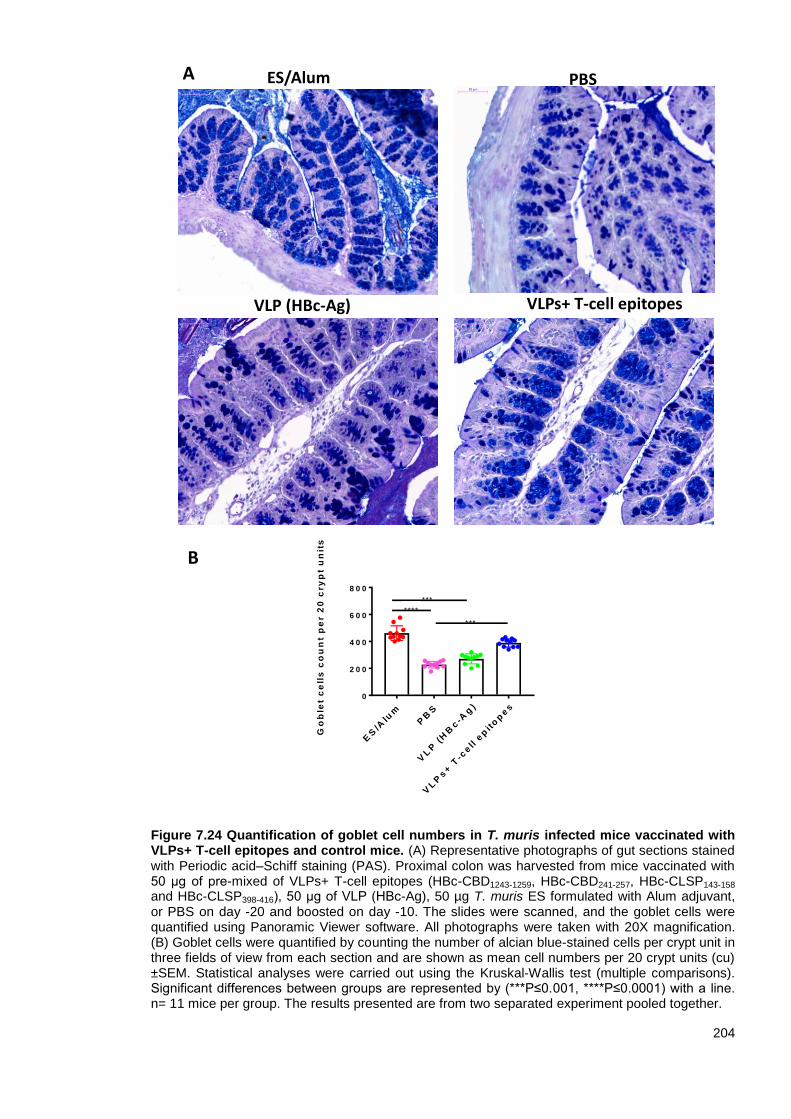
204
Figure 7.24 Quantification of goblet cell numbers in T. muris infected mice vaccinated with VLPs+ T-cell epitopes and control mice. (A) Representative photographs of gut sections stained with Periodic acid–Schiff staining (PAS). Proximal colon was harvested from mice vaccinated with 50 μg of pre-mixed of VLPs+ T-cell epitopes (HBc-CBD1243-1259, HBc-CBD241-257, HBc-CLSP143-158
and HBc-CLSP398-416), 50 μg of VLP (HBc-Ag), 50 µg T. muris ES formulated with Alum adjuvant, or PBS on day -20 and boosted on day -10. The slides were scanned, and the goblet cells were quantified using Panoramic Viewer software. All photographs were taken with 20X magnification. (B) Goblet cells were quantified by counting the number of alcian blue-stained cells per crypt unit in three fields of view from each section and are shown as mean cell numbers per 20 crypt units (cu) ±SEM. Statistical analyses were carried out using the Kruskal-Wallis test (multiple comparisons). Significant differences between groups are represented by (***P≤0.001, ****P≤0.0001) with a line. n= 11 mice per group. The results presented are from two separated experiment pooled together.
ES/Alum
VLPs+ T-cell epitopes
PBS
VLP (HBc-Ag)
ES
/Alu
mP
BS
VL
P (
HB
c-A
g)
VL
Ps+ T
-cell e
pit
op
es
0
2 0 0
4 0 0
6 0 0
8 0 0
G
ob
let
ce
lls
co
un
t p
er 2
0 c
ry
pt
un
its
***
***
****
A
B

205
Figure 7.25 Isolated lymphoid follicle-like structures’ in T. muris infected mice vaccinated with VLPs+ T-cell epitopes and control mice. Representative photographs of gut sections stained with hematoxylin and eosin (H&E). Proximal colon was harvested from mice immunised with 50 μg of pre-mixed of VLPs+ T-cell epitopes (A) or 50 μg of VLP (HBc-Ag) (B) on day -20 and boosted on day -10. The slides were scanned, and the pictures were taken using Panoramic Viewer software. All photographs were taken with 20X magnification. The results presented are from one of two separate experiments.
A
B

206
7.4 Discussion
Trichuris trichiura is one of the most common human soil-transmitted helminth (STH) parasites and
remains a major health concern for humans worldwide (Alexander and Blackburn, 2019). Up to
date, various pre-clinical vaccines against trichuriasis have been reported, containing ES/EVs
products or fractions (Dixon et al., 2008; Shears et al., 2018a; Shears et al., 2018b). However,
developing a vaccine for Trichuris based on native antigens has several limitations (Hewitson and
Maizels, 2014). The current study investigated for the first time the protective efficacy of VLPs
expressing novel Trichuris MHC-II T-cell epitopes that precisely target CD 4+ T-cells via antigen-
presenting cells to elicit a Trichuris-specific immune response with minimal risks rather than using a
whole Trichuris antigen. Based on the Foth et al.'s (2014) work, Trichuris MHC-II T-cell epitopes
were predicted from different Trichuris proteins (chitin-binding domain-containing protein,
chymotrypsin-like serine protease, and hypothetical protein) based on inclusion and exclusion
criteria as discussed in Chapter 4.
7.4.1 Protection and immune responses to the VLPs+ T-cell epitopes vaccine candidates
Three in vivo experiments were conducted in this chapter to test the protective immune response of
VLP-based vaccine candidates expressing different Trichuris T-cell epitopes prior to infection with
T. muris. The first in vivo study in this chapter assessed the immunisation of mice with 25 μg of pre-
mixed VLPs+ T-cell epitopes (HBc-H112-128, HBc-CLSP143-158, HBc-CBD1243-1259, and HBc-CBD241-
257), followed by challenged infection. The vaccine candidate significantly boosted the immune
response but was not sufficient to drive protection form Trichuris infection. High levels of parasite-
specific IgM were detected in the serum of mice vaccinated with VLPs+ T-cell epitopes upon
T. muris challenge infection, and to those without a challenge infection. These results suggest that
these VLPs+ T-cell epitopes are antigenic and can boost strong antibody response enough to
recognise specific small peptides in the T. muris ES. However, little or no parasite-specific IgG1 or
IgG2c antibodies were measured in the serum of mice vaccinated with VLPs+ T-cell epitopes.
These findings may suggest that the ability of the VLPs+ T-cell epitopes vaccine to drive a class-
switched humoral immune response is different from that of the ES/Alum vaccine. Therefore, the
antibody immune response was measured against the corresponding antigen rather than using ES
as a detection antigen. High levels of VLPs+ T-cell epitopes-specific IgM, IgG1, and IgG2c were
detected in the sera of mice vaccinated with VLPs+ T-cell epitopes. However, only negligible levels
of HBc-H112-128-specific-IgM were detected. These results suggest the H112-128 epitope is not highly
immunogenic compared to the other Trichuris epitopes in the vaccine cocktail, and therefore this
VLP+T cell epitope recombinant protein was excluded from the vaccine cocktail in the third in vivo
experiment.
Although VLPs are strong immunogens, several studies included an adjuvant in the VLP-based
vaccine to induce stronger immune responses (Cimica and Galarza, 2017; Gomes et al., 2017a).
Cimica et al. (2016) for example, developed a VLP-based vaccine for the human respiratory
syncytial virus (RSV) based on a human metapneumovirus (hMPV) matrix protein (M) fused to an
RSV glycoprotein formulated with AddaVax adjuvant. The VLP-based vaccine showed full
protection against RSV infection and induced a mix of Th1/Th2 and Th17-mediated types of
207
immune responses (Cimica et al., 2016). However, this study did not assess the efficacy of the
VLP-based vaccine without including the adjuvant.
Based on this study and others, a vaccination experiment was carried out using AddaVax adjuvant
to improve the immunogenicity of the vaccine candidate. Surprisingly, immunisation of mice with
VLPs+ T-cell epitopes (HBc-H112-128, HBc-CLSP143-158, HBc-CLSP398-416, and HBc-CBD241-257)
formulated with AddaVax adjuvant followed by infection showed no enhanced potency of the
VLPs+ T-cell epitopes vaccine compared to the control injected mice. However, antibody
recognition in the serum of mice vaccinated with VLPs+ T-cell epitopes/AddaVax confirms that the
antibodies in the serum were specific to the vaccine candidate. Importantly, antibodies in the serum
of mice vaccinated with ES/AddaVax recognised a 22 kDa band corresponding to the size of the
VLPs+ T-cell epitopes recombinant proteins, suggesting that antibodies induced by nature
protective antigens recognised the Trichuris epitopes expressed in the VLPs. These results also
explain the high levels of VLPs+ T-cell epitopes-specific IgM detected in the sera of mice
vaccinated with ES.
Previous research conducted by Dixon et al., (2008) and Else et al., (1990) showed that antibody
recognition of high molecular weight proteins in both adult and larval T. muris ES correlated with
resistance to T. muris infection. Given that the serum of mice vaccinated with VLPs+ T-cell
epitopes recognised a range of high molecular weight proteins in T. muris ES, suggests that H112-
128, CLSP143-158, CBD1243-1259, and CBD241-257 are highly-conserved immunodominant epitopes and
may be expressed in several Trichuris secretory proteins of high molecular weights, making them
potential vaccine candidates.
After ruling out AddaVax adjuvant in the vaccine formula, it was logical to assess whether the
vaccine dose may improve the efficacy of the vaccine. Remarkably, upon challenge infection, mice
vaccinated with a high-dose of 50 μg of pre-mixed VLPs+ T-cell epitopes (HBc-CBD1243-1259, HBc-
CBD241-257, HBc-CLSP143-158, and HBc-CLSP398-416) showed a significantly lower worm burden
reduction of 51.2% in the caecum and colon. Furthermore, ES/Alum conferred as full protection as
seen with ES/AddaVax. Replacing HBc-H112-128 with HBc-CLSP398-416 in the pre-mixed VLPs+ T-cell
epitopes, with an optimal dose of 50 μg, may explain the improved vaccine efficacy.
It has been postulated that the role of B-cells in the immune response to Trichuris infection is
context dependent (Sahputra et al., 2019; Sorobetea et al., 2018). For instance, B-cell-deficient
μMT mice on a C57BL/6 background appeared to be susceptible to infection (Blackwell and Else,
2001). Earlier studies have also demonstrated that the adaptive transfer of CD4+ T-cells alone,
purified from BALB/c donor mice into SCID mice, was sufficient to expel T. muris worms (Else and
Grencis, 1996). However, the adoptive transfer of serum from resistant mice provided high levels of
protection to susceptible strains of mice (Blackwell and Else, 2001; Selby and Wakelin, 1973).
Early studies also showed that parasite-specific IgG1 is involved in the memory response of the
adaptive immune system in resistant strains following primary infection (Else et al., 1993a).
In the third in vivo study presented here, parasite-specific IgM and IgG2c were detected in the sera
of mice vaccinated with VLPs+ T-cell epitopes upon T. muris infection. Importantly, analysis of the

208
serum from immunised mice showed that the VLPs+ T-cell epitopes elicited the highest level of
neutralising antibody to their corresponding VLP+T cell epitope recombinant protein (HBc-CBD1243-
1259, HBc-CBD241-257, HBc-CLSP143-158, and HBc-CLSP398-416) but not the native VLP (HBc-Ag)
protein. These results suggest that all four VLPs+ T-cell epitopes recombinant proteins had a
similar effect on class switching and enhancing both Th1/Th2 mediated immune responses. These
findings also suggest that the antibody-dependent mechanism may be crucial to the expulsion of
T. muris in vaccinated mice. Thus, it would be instructive to vaccinate transgenic B-cell-deficient
mice with a susceptible background to determine whether susceptible mice can be protected in the
absence of antibodies.
Mice vaccinated with VLPs+ T-cell epitopes were also characterised by the production of MLN-
derived Th2 cytokines. Remarkably, MLN cells from mice vaccinated with different VLP-based
vaccine candidates produced high levels of Th2 (IL-4, IL-5, IL-9 and IL-13) cytokines and
proinflammatory cytokines (IFN-ϒ and IL-6) in response to re-stimulation in vitro with ES. In
contrast, mice immunised with native (HBc-Ag) and PBS showed similar MLN cytokine profiles to
that documented for the susceptible mice strains during primary infection (Taylor et al., 2000).
These findings broadly support other studies linking Th2 cytokines and worm expulsion
(Klementowicz et al., 2012). Of the Th2 cytokines produced in response to resistant to infection,
IL-5, is involved in inducing eosinophilia during T. muris infection; however, IL-5 and eosinophils
appeared not to be essential for worm expulsion in resistant BALB/k mice (Betts and Else, 1999;
Svensson et al., 2011). In contrast, IL-9 is required to stimulate the intestinal muscle
hypercontractility that drives T. muris expulsion (Khan et al., 2003; Richard et al., 2000). It has also
been known that IL-13 plays essential roles in increasing epithelial cell turnover and increasing
production of mucins in promoting worms expulsion (Bancroft et al., 2000; Bancroft et al., 1998).
However, to test whether these Th2 cytokines induce similar responses in the VLPs+ T-cell
epitopes vaccinated mice, future work may include using cytokine transgenic and knockout mice.
Given the critical role played by Th2 cytokines in worm expulsion, our results demonstrating the
protective immunity elicited by VLPs+ T-cell epitopes, strongly support their potential as vaccine
candidates.
The results presented here are in agreement with those obtained by Dixon et al. (2010) who
showed that vaccinating susceptible AKR mice with 100 µg ES in 100 µl FIA prior to infection with a
high-dose of T. muris eggs induced sterile immunity when compared to the PBS/FIA injected mice.
Protection was mediated in the presence of high levels of Th2 (IL-5, IL-9, and IL-13) cytokines and
high levels of parasite-specific IgG1 and IgG2c. However, both immunised groups produced similar
high levels of IFN-ϒ (Dixon et al., 2010). Other studies also showed that vaccinating C57BL/6 mice
with either ES fractions or extracellular vesicles (EVs) isolated from T. muris without an adjuvant
and prior to infection with a low-dose of T. muris eggs significantly induced a reduction in the worm
burden compared to the PBS-injected group (Shears et al., 2018a; Shears et al., 2018b). High
levels of both IgG1 and IgG2c anti-parasite antibodies against soluble ES antigens were detected
in sera of mice vaccinated with T. muris EVs (Shears et al., 2018a). Notably, there was a significant
increase in IL-13 and IL-9 production by infection-primed lymphocytes in response to the ES
209
fractions that induced a reduction in the worm burden (Shears et al., 2018b). The data are largely
consistent with the results presented, here although the present study assessed the protective
immune response of the vaccine candidates in an accelerated expulsion animal model and a
broader profile of cytokines was measured. Although the ES/Alum vaccine led to full protection, the
levels of Th2 cytokines in the re-stimulated MLNs were not dramatically high; however, this finding
might be related to the accelerated expulsion and autopsy date (day 14 p.i.) being too late to detect
peak cytokine production.
Furthermore, vaccinating AKR mice with either a T. muris whey acidic protein (rTm-WAP49) or a
T. muris WAP fragment fusion protein (rTm-WAP-F8+Na-GST-1) formulated with Montanide ISA
720 adjuvant three times at two-week intervals prior to T. muris challenge induced a partial
reduction in the worm burden by 48% and 38%, respectively (Briggs et al., 2018). The results
showed that both humoral and cellular immune responses were induced, characterised by elevated
antigen-specific IgG1 and IgG2c antibodies and Th2 (IL-4, IL-9, and IL-13) cytokines detected in
the draining inguinal lymph nodes, draining mesenteric lymph nodes, and spleen of vaccinated
mice (Briggs et al., 2018). These data correlate well with the results presented here; however,
vaccinating mice with VLPs expressing Trichuris epitopes induced a reduction in the worm burden
by 51.2%. Similarly, mice vaccinated with VLPs+ T-cell epitopes were characterised by the
production of MLN-derived Th2 cytokines (IL-4, IL-9, and IL-13) in response to re-stimulation in
vitro with Trichuris ES. High levels of VLP+T cell epitopes-specific IL-5, IL-6, and IL-10 cytokines
were also detected in the re-stimulated MLNs of vaccinated mice.
Crypt hyperplasia is typically associated with chronic infection accompanied by both increased
epithelial cell proliferation and apoptosis under the control of the IFN-ϒ pro-inflammatory cytokine
(Artis et al., 1999b). In contrast, no such dramatic changes were observed in the gut of ‘naturally
resistant’ mice. It has been demonstrated that resistant animals have accelerated epithelial cell
turnover, resulting in short crypts, a mechanism that is functionally associated with Trichuris
expulsion (Cliffe et al., 2005). However, the results described here are in good agreement with
Dixon’s (2010) findings, which showed that crypt hyperplasia was seen in both ES/FIA- and
PBS/FIA-immunised mice groups following Trichuris infection. These results suggest that the
increased rate of epithelial turnover, which appears to be an important mechanism to expel the
worms in 'natural resistant' mice, is unlikely to be responsible for expulsion in 'vaccinated resistant'
mice. The high levels of MLNs IFN-ϒ in VLPs+ T-cell epitopes and ES/Alum vaccinated mice may
explain the crypt hyperplasia in both groups. These findings also suggest that epithelial cell
proliferation is likely to occur at a similar rate in these mice compared to the PBS and VLP (HBc-
Ag) injected mice.
The VLPs+ T-cell epitopes and ES/Alum vaccinated mice also showed marked goblet cell
hyperplasia, which is a known IL-13-dependent mechanism of Trichuris expulsion in mice resistant
to Trichuris infection (Datta et al., 2005). Goblet cells have been implicated in mucus production
(Muc2 and Muc5ac) during Th2-mediated defence in resistant animals (Hasnain et al., 2010) by
protecting the barrier from degradation by the worm-secreted serine proteases (Hasnain et al.,
2012). Goblet cells also secrete several molecules other than mucins that contribute to T. muris

210
expulsion, including resistin-like molecule β (RELMβ) (Artis et al., 2004b). This putative mechanism
of expulsion may also explain the vaccination-induced protection to challenge infection.
Interestingly, we also identified isolated lymphoid follicle-like structures in the large intestine of mice
immunised with VLPs+ T-cell epitopes and native VLP (HBc-Ag). These follicular structures are
filled mainly with B-cells interspersed by small clusters of CD4+/CD8+ T-cells and have been
previously identified in the large intestine of both AKR and BALB/c mice infected with T. muris
(Little et al., 2005). Similar structures have also been identified in the mouse small intestine in
response to normal gut flora (Lorenz et al., 2003). The presence of these follicular structures may
explain the strong antibody response in the mice vaccinated with VLPs+ T-cell epitopes, especially,
that previous study has shown that IgG1 must be present in the mouse large intestine for effective
larval clearance (Dixon et al., 2010). However, we are unsure, at this stage, whether these
structures are isolated lymphoid follicles. Therefore, future work may include the
immunohistochemical staining of B220+ cells to confirm lymphoid structures, and analyse the local
tissue levels of antibodies post-infection.
7.4.2 VLP-based vaccines against parasite infections
The results presented here represent the first example of successful VLP-based vaccination
against the Trichuris parasite using Trichuris MHC-II T-cell epitopes. However, there is a clear
trend toward identifying and evaluating VLP-based vaccines expressing a whole protein or short
peptides for the development of many anti-parasitic vaccines.
For example, Lee and co-workers (2016) developed a vaccine for human trichinellosis based on
influenza matrix protein 1 (M1) VLP containing the whole 53 kDa ES protein of T. spiralis (T653K)
with and without a cholera toxin (CT). Notably, BALB/c mice vaccinated twice with 25 μg of T653K
VLP induced parasite-specific IgG, IgG1, and IgG2c antibody responses before and after an oral
challenge with 100 T. spiralis larvae/mouse and showed a significant reduction in the worm burden
upon infection. However, protective immune responses were significantly improved by vaccinating
mice with T653K VLP/CT (Lee et al., 2016). Lee et al. (2017) also developed a vaccine for human
clonorchiasis, based on the influenza matrix protein 1 (M1) VLP containing whole C. sinensis
protein 22.3 kDa (CsTP 22.3). Rats vaccinated twice with 200 μg of CsTP VLP and challenged with
C. sinensis metacercariae induced parasite-specific cellular and humoral immune responses. This
result was confirmed with significant production of IgG, IgG1, and IgG2c in the serum and IgA in
the faeces and intestines of the vaccinated group before and after challenge when compared to the
naïve control. Moreover, vaccinated rats induced CD4+/CD8+ T-cell responses in the spleen
compared to the native and the non-immunized control rats. Most importantly, a significant
reduction in the worm burden was seen upon challenge infection without the need for an adjuvant
in the vaccine formulation (Lee et al., 2017). The same group developed a VLP-based vaccine
against Toxoplasma gondii based on the same VLP (influenza M1) containing T. gondii rhoptry
protein 18 (ROP18) or microneme protein 8 (MIC8) (Lee et al., 2018). Mice vaccinated with a
combination of ROP18 VLP or MIC8 VLP and challenged with T. gondii tachyzoites, induced a
stronger immune response compared to the vaccinated groups with each VLP individually (Lee et
al., 2018).

211
Hepatitis B VLP, in particular, has proved to be one of the most promising parasite delivery
systems (Chisari and Ferrari, 1995; Qiao et al., 2016; Roose et al., 2013). The malaria (Malarivax
ICC-1132) vaccine, for example, is composed of HBc-Ag expressing P. falciparum CS B-cell
epitopes inserted into the MIR and T-cell epitopes in the C terminal expressed in E. coli and
purified by gel filtration column chromatography. Mice and monkeys vaccinated with the Malarivax
vaccine formulated in Freund’s adjuvant developed long-lasting immunity and elected an anti-CSP
specific antibody and CD4+ immune response (Birkett et al., 2002; Birkett et al., 2001; Milich et al.,
2001). Based on the strong experimental evidence in vivo, the vaccine was tested in human clinical
trials. Healthy volunteers were vaccinated with the HBc-Ag containing P. falciparum CS T- and B-
cell epitopes expressed in E. coli and purified by ammonium sulfate precipitation, size exclusion,
hydrophobic interaction, and hydroxyapatite chromatography. The vaccine, formulated with alum or
Montanide ISA 720 adjuvant, was found to be highly immunogenic and produced a specific
antibody response to the CS and secretion of IFN-γ and IL-2 cytokines. Other than proving the
immunogenicity of the vaccine, all the healthy volunteers showed no serious side effects from the
vaccine (Gregson et al., 2008; Nardin et al., 2004; Oliveira et al., 2005).
The results of these studies support some critical insights into VLP-based vaccines, including
confirming that an HBc virus-like particle, in particular, is an excellent delivery system for
developing potential vaccine candidates for parasites. Besides, in order to enhance the immunity of
the potential vaccine, it is recommended to include several antigens in the vaccine formula. Despite
the promising results discussed in this chapter, it may be necessary to optimise vaccination
schedules to prolong the exposure of antigens to the immune system or increase the dose to
achieve an appropriate level of protection (Bachmann and Jennings, 2010; Johansen et al., 2008).
Future work should also include investigating the efficacy of the vaccine candidate in a low-dose
infection (25 eggs), which would ordinarily lead to chronic infection in C57BL/6 mice (Bancroft et
al., 1994). To examine whether antibody production is required for vaccine-induced immunity,
additional work could also investigate whether transferring serum from mice vaccinated with VLPs+
T-cell epitopes can protect against infection in naïve mice.
In summary, the current chapter identified a novel vaccine candidate against trichuriasis based on
VLPs expressing different Trichuris MHC-II T-cell epitopes predicted from chitin-binding domain
containing proteins and chymotrypsin-like serine proteases. Collectively, the data presented in this
chapter further justify focussing on Trichuris epitopes predicted from the Trichuris genome that
stimulate Th2 immune response rather than using whole or fractions of Trichuris ES antigens to
identify potential vaccine candidates, as has been used for malaria vaccines (Gregson et al., 2008;
Nardin et al., 2004; Oliveira et al., 2005).
7.5 Summary
Vaccination of mice with 25 µg of pre-mixed VLPs expressing Trichuris MHC-II T-cell
epitopes (HBc-H112-128, HBc-CLSP143-158, HBc-CBD1243-1259, and HBc-CBD241-257) was able
to boost the immune response but was not sufficient to drive protection form a high-dose
Trichuris infection.

212
An oral challenge with T. muris infective eggs following vaccination with 25 µg of pre-mixed
VLPs+ T-cell epitopes (HBc-H112-128, HBc-CLSP143-158, HBc-CLSP398-416, and HBc-CBD241-
257) formulated with AddaVax adjuvant did not improve the efficacy of the vaccine
candidate.
Oral challenge with T. muris infective eggs following vaccination with 50 µg of pre-mixed
VLPs+ T-cell epitopes (HBc-CBD1243-1259, HBc-CBD241-257, HBc-CLSP143-158, and HBc-
CLSP398-416) led to a significant reduction in the worm burden (51.2%). Protective immunity
was characterised by the production of MLN-derived Th1 and Th2 cytokines, high levels of
serum antigen-specific IgM, IgG1, and IgG2c antibodies along with goblet cell hyperplasia.

213
Chapter Eight
Summary discussion

214
Trichuris trichiura infection remains a global public health issue, especially in the tropical and
subtropical areas with poor hygiene and sanitation (Alexander and Blackburn, 2019). The ability to
control such infection depends almost exclusively on administration of anthelmintic drugs to at-risk
populations (Farrell et al., 2018; Freeman et al., 2019; WHO, 2015). However, the appearance of
anthelmintic-resistant parasites has increased interest in developing a vaccine to provide a cost-
effective, long-term immunological method to control helminth infection (Becker et al., 2018; Dixon
et al., 2008; Doyle and Cotton, 2019). Despite research progress and significant early successes,
there are no licensed vaccines for human STHs (Sacks et al., 2016). However, tremendous efforts
toward state-of-the-art vaccine development have improved the understanding of the host-parasite
interaction, parasite component isolation and characterisation and gene sequence analysis (Smith
and Zarlenga, 2006).
The overall aim of this thesis was to develop an anti-Trichuris vaccine based on VLPs expressing
Trichuris T-cell epitopes. This was done using an RV approach to overcome some of the
challenges associated with developing helminth vaccines (Figure 8.1). To achieve this aim, first, a
systematic review was performed to select the top MHC class II in silico prediction tools (Chapter
3). Second, the potential Trichuris MHC-II T-cell epitope vaccine candidates obtained from the
Trichuris genome were identified using the selected in silico prediction tool (Chapter 4). Third,
these epitopes were produced in a commercially viable manner by fusing epitopes in the MIR of the
VLP (HBc-Ag) vaccine delivery system (Chapter 5). These VLPs + T-cell epitope vaccine
candidates were then tested in vitro for their ability to activate APCs (Chapter 6). Finally, three in
vivo experiments were conducted to test the protective immune response of VLPs expressing
different Trichuris T-cell epitopes prior to infection with T. muris (Chapter 7). Collectively, the
results of this research represent the first significant progress towards identifying a novel epitope-
based vaccine for trichuriasis.

215
Figure 8.1 Reverse vaccinology strategy used to identify epitope-based vaccine against trichuriasis infection. Challenges begin with choosing the top MHC-II restricted T-cell epitope prediction tools (Chapter 3) followed by genome analysis to identify lead proteins with promising vaccine antigen properties from the whole genome of T. trichiura and T. muris (Chapter 4). In silico immunoinformatics tools were then used to predict Trichuris MHC-II T-cell epitopes by mapping protein sequence (Chapter 4). The selected Trichuris MHC-II T-cell epitopes were then cloned, expressed and purified in the VLP (HBc-Ag) delivery system (Chapter 5). In vitro assays were then conducted to evaluate the efficacy of the vaccine candidates to activate and stimulate APCs (Chapter 6). The vaccine candidates were then evaluated in vivo for immunogenicity and protection
in mice (Chapter 7).
2.11 Identification of novel Trichuris MHC-II T-cell epitopes as promising vaccine candidates
There have been many attempts to develop vaccines for helminth parasites, but these parasites
are relatively large multicellular organisms with a large genome (Noon and Aroian, 2017), which
makes vaccine development challenging.
Over the past decade, considerable progress has been made in identification of candidate vaccine
antigens for several important helminth parasites, such as the recombinant vaccine for the
hookworm N. americanus based on Na-GST-1 and Na-APR-1 antigens (Diemert et al., 2018; Hotez
et al., 2013). These parasite antigens are known to be involved in the breakdown of host
haemoglobin and detoxification of haem, which is essential for parasite survival (Hotez et al.,
In vivo evaluation of VLPs expressing Trichuris T-cell epitopes
In vitro evaluation of VLPs expressing Trichuris T-cell epitopes
Prediction of Trichuris T cell epitopes using In silico prediction tools
Designing, cloning and purifying VLPs expressing Trichuris T-cell epitopes
Vaccine candidate
Genome analysis to identify lead proteinsChapter 4:
Chapter 4:
Chapter 5:
Chapter 6:
Chapter 7:
Chapter 3: Assessment of MHC–II restricted T-cell epitope prediction tools

216
2013). The vaccine has been proven to be safe and highly immunogenic and is currently
undergoing clinical trials in the United States, Brazil and Gabon (Diemert et al., 2017; Hotez et al.,
2013).
T. muris excretory/secretory (ES) products (Jenkins and Wakelin, 1977), ES fractions (Shears et
al., 2018b), extracellular vesicles (EVs) (Shears et al., 2018a), and, more recently, T. muris whey
acidic protein (Briggs et al., 2018) have shown considerable potential in a number of pre-clinical
protection trials. Despite these successes, developing a vaccine based on native antigens is
associated with many manufacturing challenges, such as cost, time consumption, difficulties in
purifying large quantities of worm antigens and control over differences between batches (Geldhof
et al., 2007; Hewitson and Maizels, 2014). The current era of genome sequencing and the advent
of immunoinformatics tools enable identification of potential vaccine parasite antigens in silico as
an alternative to composing native antigens (Sousa and Doolan, 2016; Tordello et al., 2017). Such
approaches may enable development of a safer and more reliable vaccine based on VLPs
expressing Trichuris MHC-II T-cell epitopes.
Initiation of an antigen-specific immune response requires presentation of antigenic peptides to
CD4+ T-cells in the context of MHC Class II molecules (Dhanda et al., 2017). Selection of
appropriate MHC Class II binding peptides is therefore a critical first step in developing an epitope-
based vaccine. Systematic review and evaluation of existing MHC-II restricted T-cell epitope
prediction tools were carried out in Chapter 3 in order to select the top tools. The work described in
Chapter 3 recommends the use of IEDB (Zhang et al., 2008) and/or NETMHC-II 2.2 (Buus et al.,
2015) prediction tools in any MHC class II epitope prediction study. However, the output results for
these in silico prediction tools need to be carefully evaluated in vitro and in vivo.
To our knowledge, the data presented in Chapter 4 is the first to identify novel Trichuris MHC-II T-
cell epitopes as potential vaccine antigen candidates. When designing an anti-Trichuris vaccine,
one must remember that the end goal is development of a vaccine for humans, not mice. Thus,
based on the work of Foth et al. (2014), a set of candidate proteins were selected from the T. muris
and T. trichiura genomes using inclusion and exclusion criteria. Then, MHC-II T-cell epitopes were
predicted using the IEDB in silico prediction tool and were screened for allergenicity and
conservancy. The framework used here to identify potential epitope vaccine candidates within the
T. muris and T. trichiura genome could be used in the future to identify potential vaccine candidates
within other parasite species.
The final set of Trichuris MHC-II T-cell epitopes vaccine candidates were identified from chitin-
binding domain-containing proteins, chymotrypsin-like serine proteases, hypothetical protein, and
from Pfam B domain-containing protein. Chitin-binding domains genes are highly expressed in
different life stages in many parasites including T. trichiura, Caenorhabditis elegans, Ascaris
lumbricoides and Ancylostoma ceylanicum (Foor, 1967; Santos et al., 2016; Veronico et al., 2001;
Wei et al., 2016). In particular, these proteins are thought to be associated with eggshell formation
and early development in the single-cell stage (Johnston et al., 2006, 2010). Furthermore, given
that T. muris and more than 40 helminths species express a high level of chymotrypsin-like serine
proteases, these proteases may be promising vaccine candidates (Cuesta-Astroz et al., 2017; Foth

217
et al., 2014). They seem to play a central role in either the invasion process or modulation of the
host immune response to enhance the survival in the host (Balasubramanian et al., 2010; Santos
et al., 2013; Toubarro et al., 2010; Yang et al., 2015).
Numerous publications have noted that the success of the most consistent vaccines against
various helminth parasites has been achieved using helminth protease molecules (Dalton et al.,
2003). For example, vaccinating BALB/c mice with the whole recombinant serine protease of
T. spiralis prior to challenging them with T. spiralis infection led to a reduction in the worm burden
and induced a mix of Th1 and Th2 immune responses (Feng et al., 2013; Wang et al., 2013; Xu et
al., 2017). Furthermore, Shears et al. (2018b) showed that vaccinating mice with the ES fraction
containing serine proteases induced high antibody responses. Remarkably, all the proteins
selected in this study, except Pfam B domain-containing proteins, have been identified in most
immunogenic fractions of T. muris ES following vaccination of mice (Shears et al., 2018b).
Moreover, previous work by Dixon et al. (2008) and Else et al. (1990) showed that antibody
recognition of high-molecular-weight proteins in both adult and larval T. muris ES was correlated
with resistance to T. muris infection. Given that the serum of mice vaccinated with VLPs + T-cell
epitopes recognised a range of high-molecular-weight proteins in T. muris ES, this suggests that
the Trichuris epitopes predicted from chymotrypsin-like serine proteases and chitin-binding
domains are highly conserved immunodominant epitopes that may be expressed in several
Trichuris secretory proteins with high molecular weight. Collectively, the results of this study
suggest that the selected Trichuris MHC-II T-cell epitopes are potential vaccine candidates and
may explain the protective immune response developed in mice following vaccination (Chapter 7).
2.12 VLP (HBc-Ag) as a promising vaccine delivery system
Purified antigens can sometimes have poor immunogenicity. Therefore, choosing an economically
viable antigen delivery system is critical, as this will have a tremendous impact on the immune
pathways following vaccination (Cunningham et al., 2016; Wang, 2018). Numerous VLP-based
vaccines have been engineered to exhibit foreign parasite antigens and have been proved to be
highly immunogenic. Some of these VLP-based vaccines are in clinical development, such as the
anti-malaria vaccine (RTS) (Dobano et al., 2019), and others are still under major research
investigation, such as the vaccines against Trichinella spiralis (Lee et al., 2017), Clonorchis
sinensis (Lee et al., 2017), and Toxoplasma gondii for example (Guo et al., 2019).
Chapter 5 highlighted the potential of native VLP (HBc-Ag) as a vaccine delivery system as it
expresses Trichuris T-cell epitopes and simplifies the inherent production challenges associated
with development of a safe and economical vaccine. All the predicted Trichuris MHC-II T-cell
epitopes identified in Chapter 4 were successfully cloned in the MIR of the native VLP (HBc-Ag)
and expressed using an E. coli expression system. Only the VLPs+ T-cell epitope recombinant
proteins confirmed to be soluble by SDS-PAGE analysis were purified using Strep-Tag affinity
chromatography and size-exclusion chromatography. Then, they were examined by CD and TEM
to confirm their structural integrity and assembly.
218
The insertion of whole hypothetical protein (HBc-HP1-512) and a mosaic construct (HBc-mosaic)
providing a single expression system displaying multiple Trichuris epitopes (HBc-hypothetical
protein (A0A0N5DRU5) + CLSP398-416 epitope + chitin binding domain (A0A0N5DK22)) into HBc-Ag
was successful. However, these two constructs were insoluble when expressed in E. coli and were
not further purified by affinity chromatography. In future work aiming to develop these VLP
recombinant proteins, chemical coupling may be used to solve this problem as this allows for more
precise surface decoration of VLPs with large foreign antigens (Fogarty and Swartz, 2018; Patel
and Swartz, 2011). Only soluble and highly purified VLPs expressing Trichuris MHC-II T-cell
epitopes (HBc-H112-128, HBc-CBD36-52, HBc-CBD1243-1259, HBc-CBD241-257, HBc-CLSP143-158 and HBc-
CLSP398-416) were used in the in vitro and in vivo assays performed to confirm antigenicity in
Chapters 6 and 7.
The data presented in Chapter 6 demonstrated that the native VLP (HBc-Ag) and HBc-Ag
expressing Trichuris MHC-II T-cell epitopes provided two types of immunoregulator signals to both
APCs (DCs and macrophages). This can enable orchestrated priming of both humoral and cellular
immune responses during the development of VLP-mediated immunity. First, the up-regulation of
cell surface markers essential for optimal activation of Ag-specific T-cells. Second, stimulation of
inflammatory responses characterised by the secretion of high levels of pro- and anti-inflammatory
cytokines, which are involved in the immune regulation of B- and T-lymphocytes. Third, VLPs were
proven to be taken up efficiently by both types APCs and were not merely attached to the surface
of the APCs. These results suggest that the VLPs are well placed to act as delivery systems to
drive immune responses via both APCs. Given that VLPs activate both APCs, future work may
include evaluating the VLPs’ activation responses to different DC subsets and other APCs
including B-cells, in vivo. Additional work is also needed to modify the VLP using
immunostimulatory adhesion molecules and cytokines co-displayed on the VLP to target APCs to
enhance the activation of antigen-specific T-and B-cell effector cells.
2.13 Identification of a novel VLP-based vaccine against trichuriasis
One of the most important lessons learned from previous anti-parasite vaccines is that the use of
multiple antigens induces more reliably high levels of protection that offered by a single antigen
(Birkett et al., 2002; Cabrera-Mora et al., 2016). The work described in Chapter 7 evaluated the
immunogenic and protective immune responses of a mixture of four VLPs expressing four different
Trichuris MHC-II T-cell epitopes as a potential vaccine candidate in vivo in the murine model of
Trichuris infection.
Mice vaccinated with 25 µg of pre-mixed VLPs expressing Trichuris MHC-II T-cell epitopes with
and without an adjuvant boosted immune responses, but the responses were not sufficient to drive
protection against a high-dose Trichuris infection. However, oral challenge with T. muris infective
eggs following vaccination with a dose of 50 μg of pre-mixed VLPs+ T-cell epitopes (HBc-CBD1243-
1259, HBc-CBD241-257, HBc-CLSP143-158 and HBc-CLSP398-416) led to a significant reduction in worm
burden (51.2%). Also, mice vaccinated with VLPs+ T-cell epitopes produced MLN-derived Th1
(IFN-γ) and Th2 cytokines (IL-4, IL-5, IL-9, and IL-13), crypt hyperplasia and goblet cells
hyperplasia. While it is known that goblet cell hyperplasia is an IL-13-dependent mechanism of

219
Trichuris expulsion in mice that are resistant to Trichuris infection (Datta et al., 2005), further work
is required to elucidate whether goblet cell hyperplasia is a critical protective effector mechanism
following vaccination of Muc2 KO mice with VLPs+ T-cell epitopes.
VLPs+ T-cell epitopes were found to elicit strong neutralising antibody responses involving isotype
class-switching to IgG1 and IgG2c specific to their corresponding VLP+ T-cell epitope recombinant
protein (HBc-CBD1243-1259, HBc-CBD241-257, HBc-CLSP143-158 and HBc-CLSP398-416). These results
suggest that all four VLPs+ T-cell epitopes recombinant proteins had a similar effect on class
switching and enhanced both Th1- and Th2-mediated immune responses. Although the
significance of the follicular structures identified in the guts of some vaccinated mice is not yet
clear, they may be related to the strong antibody response in mice vaccinated with VLPs+ T-cell
epitopes. Collectively, these findings suggest that antibody-dependent mechanism may be crucial
for expulsion of T. muris in vaccinated mice; yet, it is not an indicator of the degree of protection.
Further studies placing greater focus on the role of B-cells as the primary APCs should be
undertaken using B-cell KO mice.
This study supports the work of Dixon et al. (2010), who found that Th1 (IFN-γ) and Th2 (IL-5, IL-9,
IL-13) cytokines, IgG1 and IgG2c antibodies, goblet cell hyperplasia and crypt hyperplasia are
important elements of vaccine-induced resistance to T. muris following ES vaccination. Taken
together, the results of this study suggest that there may be differences in the mechanisms of
expulsion between those with natural resistance and those with vaccinated resistance. Thus, the
correlation between immune response and protection offered by the VLPs+ T-cell epitope vaccine
is a research priority. Also, despite the promising results of this study, additional technical issues
need to be better explored. For example, the vaccine schedule and dose need to be optimised to
achieve an appropriate level of protection.
2.14 Prospects and challenges for anti-Trichuris vaccine design
Development of new vaccines is a complex and multidisciplinary task that requires an
understanding of host-pathogen interactions, epidemiology and manufacturing parameters
(Cunningham et al., 2016). Most importantly, vaccine researchers must have an understanding of
the immune mechanisms involved in diseases and protection in order to select appropriate
antigenic targets and delivery systems to shape the immune response induced by the vaccine
(Howarth and Brune, 2018; Knox et al., 2001). These factors determine the biomechanical
requirements of production and the design of the laboratory and clinical trials (Howarth and Brune,
2018; Knox et al., 2001).
Recognition and understanding of a number of biological, socioeconomic and ethical factors are
fundamental for developing long-lived protective vaccines (Ota et al., 2013). Clear knowledge of
the risk groups and populations, age-specific infection rates and infection demographics is
important as these factors are known to influence immune responses to vaccines (Cunningham et
al., 2016). Additionally, political, ethical and socioeconomic issues such as education, behavioural
and cultural factors affect not only vaccine selection but also evaluation of vaccine immune
responses (Coughlin, 2006; Hotez, 2017).
220
Previous studies have demonstrated that most infants in endemic areas are infected with
T. trichiura and school-aged children are at particularly high risk of T. trichiura infection (Awasthi et
al., 2003; Bethony et al., 2006). Immunisation during the neonatal period may be a promising
strategy as neonates are often capable of producing a Th2 response and antibody isotype
switching to vaccine antigens (Adkins et al., 2001; Barrios et al., 1996). Thus, the ideal vaccine
would be given to infants prior to risk as well as to older children and adults with ongoing infection.
However, this strategy is problematic as an early study showed that vaccination could not protect
susceptible mice with a pre-existing infection and subsequent challenge infection (Else and
Wakelin, 1990a).
Considering the target populations that are most afflicted by trichuriasis infection, the administration
route of the vaccine is another important factor that can influence the design of a vaccine and its
accessibility to patients (Jain et al., 2015; Noon and Aroian, 2017). Adaptive immune responses
are mainly induced in secondary lymphoid organs. Thus, it is critical for APCs to transport vaccine
antigens through the lymphatic system from peripheral tissues to lymphoid organs (Bachmann and
Jennings, 2010). Most currently approved vaccines are administered by intramuscular or
subcutaneous injection. However, it has been suggested by many researchers that vaccine-
induced protection against mucosal disease requires oral or nasal mucosal administration
(Bachmann and Jennings, 2010). The particulate nature of VLPs enables them to be transported to
lymph organs. In addition, they are very stable and can withstand adverse environments, such as
those with acidic pH, making VLPs an attractive carrier for mucosally administered vaccines.
Future work developing these vaccines should focus on assessing thermal stability and ensuring
that vaccines can be stored without refrigeration to reduce delivery restrictions (Fogarty and
Swartz, 2018).
It is also important to consider the epidemiology of infection during vaccine development. The
viability of the vaccine candidates described in this thesis has been tested against one high-dose
infection. A more realistic mouse model of human infection would involve chronic infection, as this
would produce greater confidence that an anti-Trichuris vaccine could have a protective effect in
the field. Future studies could vaccinate male C57BL/6 mice with the vaccine candidate, followed
by a low-dose of T. muris infection to develop chronicity. The worm burdens should be assessed a
number of weeks post-vaccination to determine how long vaccine-induced immunity to Trichuris
lasts.
A key observation made during phase II of the clinical trial of the N. americanus vaccine was that
pre-vaccination volunteers living in endemic areas experienced allergic reactions triggered by high
IgE responses to infective larvae antigens (Diemert et al., 2012). Thus, it is critical to prevent
allergic responses when developing a vaccine. Although the Trichuris T-cell epitopes selected for
this study were screened for allergenicity in silico, future work must examine IgE responses in the
sera from populations in countries with endemic STH infections.
Furthermore, all the pre-clinical vaccine evaluations of human STH infections used various
laboratory infection models. Although there is no way of conducting these studies directly on
humans, these models likely have limitations that could cause variation in the results, such as use

221
of different laboratory host species or strains; unnatural infection; and different reagents, equipment
or researchers (Noon and Aroian, 2017; Zeng et al., 2016).
Coinfections with two or multiple soil-transmitted nematode infections including, A. lumbricoides
and hookworms, mostly Necator americanus, are extremely common in young people (Alexander
and Blackburn, 2019; Salim et al., 2015). Thus, researchers have proposed a single pan-
anthelmintic vaccine against the three major human STHs to generate strong, lasting immunity with
minimal side effects (Zhan et al., 2014). Although each of these STHs occupies a different location
within the gastrointestinal tract and/or exhibits different feeding habits, which may present
enormous obstacles for a single vaccine, these three helminths spp share highly conserved
antigens that are likely to have very similar biological functions (Noon and Aroian, 2017). Thus, a
Pan-anthelmintic vaccine based on protective epitopes from Trichuris and other cross-protective
antigens may be achievable using the RV strategy.
2.15 Conclusions and future perspectives
Vaccines can serve as one of the most effective means to prevent trichuriasis. Approaches to anti-
Trichuris vaccine development have focused mainly on Trichuris native antigens, limiting these
vaccines’ suitability for production and distribution in the developing world. Today, however, it may
be prudent to consider alternatives to conventional thinking and appraise the available genetic
information to develop an epitope-based vaccine to overcome these limitations.
The work presented in this thesis represents significant progress towards the development of a
novel epitope-based vaccine against trichuriasis. We assert that VLPs (HBc-Ag) expressing
Trichuris T-cell epitopes are potential vaccine candidates, as they meet the major vaccine design
criteria: (1) self-assembly and display of the target antigens; (2) inexpensive production in E. coli
expression systems; (3) stimulation of APCs for prompt activation of both humoral and cellular
immune responses; (4) safety in a pre-clinical model; and (5) demonstration of efficacy in
protecting against challenge infections without the need for an adjuvant.
There are multiple future works priorities in which to move forward. First, it is necessary to modify
the VLP-based vaccine to target APCs in order to induce stronger humoral and cellular immune
responses and immunological memory. Second, the protective efficacy and prolonged responses
of these vaccine candidates must be tested in different animal models. Third, to determine the
immune response that must be elicited by the VLP-based vaccine to develop protection. Finally,
promising epitopes for Trichuris and other STH parasites must be identified to develop a pan-
anthelmintic vaccine.
In conclusion, given the right combination of immunoinformatics and immunogenicity screening
tools, epitope-based vaccines will undoubtedly limit the cost and effort associated with bringing a
Trichuris vaccine to trial.

222
References
Adkins, B., Bu, Y. & Guevara, P. (2001). 'The generation of Th memory in neonates versus adults: prolonged primary Th2 effector function and impaired development of Th1 memory effector function in murine neonates', The Journal of Immunology, 166(2), pp. 918-925.
Agallou, M., Athanasiout, E., Koutsonit, O., Dotsika, E. & Karagouni, E. (2014). 'Experimental validation of multi-epitope peptides including promising MHC class I- and II-restricted epitopes of four known Leishmania infantum proteins', Frontiers in Immunology, 5.
Agallou, M., Margaroni, M., Athanasiou, E., Toubanaki, D. K., Kontonikola, K., Karidi, K., Kammona, O., Kiparissides, C. & Karagouni, E. (2017). 'Identification of BALB/c Immune Markers Correlated with a Partial Protection to Leishmania infantum after Vaccination with a Rationally Designed Multi-epitope Cysteine Protease A Peptide-Based Nanovaccine', PLoS Negl Trop Dis, 11(1), p. e0005311.
Ahlborg, N., Ling, I. T., Holder, A. A. & Riley, E. M. (2000). 'Linkage of exogenous T-cell epitopes to the 19-kilodalton region of Plasmodium yoelii merozoite surface protein 1 (MSP119) can enhance protective immunity against malaria and modulate the immunoglobulin subclass response to MSP119', Infect Immun, 68(4), pp. 2102-2109.
Ahlborg, N., Sterky, F., Haddad, D., Perlmann, P., Nygren, P.-a., Andersson, R. & Berzins, K. (1997). 'Predominance of H-2 d-and H-2 k-restricted T-cell epitopes in the highly repetitive Plasmodium falciparum antigen Pf332', Molecular immunology, 34(5), pp. 379-389.
Akache, B., Weeratna, R. D., Deora, A., Thorn, J. M., Champion, B., Merson, J. R., Davis, H. L. & McCluskie, M. J. (2016). 'Anti-IgE Qb-VLP conjugate vaccine self-adjuvants through activation of TLR7', Vaccines, 4(1), p. 3.
Akhras, S., Toda, M., Boller, K., Himmelsbach, K., Elgner, F., Biehl, M., Scheurer, S., Gratz, M., Vieths, S. & Hildt, E. (2017). 'Cell-permeable capsids as universal antigen carrier for the induction of an antigen-specific CD8+ T-cell response', Scientific reports, 7(1), p. 9630.
Akiho, H., Blennerhassett, P., Deng, Y. & Collins, S. M. (2002). 'Role of IL-4, IL-13, and STAT6 in inflammation-induced hypercontractility of murine smooth muscle cells', 282(2), pp. G226-G232.
Albonico, M., Bickle, Q., Ramsan, M., Montresor, A., Savioli, L. & Taylor, M. (2003). 'Efficacy of mebendazole and levamisole alone or in combination against intestinal nematode infections after repeated targeted mebendazole treatment in Zanzibar', Bulletin of the World Health Organization, 81, pp. 343-352.
Alexander, T. Y. & Blackburn, B. G. (2019). 'Soil-Transmitted Helminths: Ascaris, Trichuris, and Hookworm Infections', Water and Sanitation-Related Diseases and the Environment: In the Age of Climate Change, p. 95.
Altman, D. G. & Bland, J. M. (1994). 'Diagnostic tests. 1: Sensitivity and specificity', BMJ: British Medical Journal, 308(6943), p. 1552.
Altman, L. K. (1982). 'A new insulin given approval for use in US', New York Times, 30. Alves-Silva, M. V., Nico, D., Morrot, A., Palatnik, M. & Palatnik-de-Sousa, C. B. (2017). 'A Chimera
Containing CD4+ and CD8+ T-Cell Epitopes of the Leishmania donovani Nucleoside Hydrolase (NH36) Optimizes Cross-Protection against Leishmania amazonesis Infection', Front Immunol, 8, p. 100.
Ambühl, P. M., Tissot, A. C., Fulurija, A., Maurer, P., Nussberger, J., Sabat, R., Nief, V., Schellekens, C., Sladko, K. & Roubicek, K. (2007). 'A vaccine for hypertension based on virus-like particles: preclinical efficacy and phase I safety and immunogenicity', Journal of hypertension, 25(1), pp. 63-72.
Andersen, P., Andersen, A. B., Sørensen, A. & Nagai, S. (1995). 'Recall of long-lived immunity to Mycobacterium tuberculosis infection in mice', The Journal of Immunology, 154(7), pp. 3359-3372.
Anderson, R. & May, R. (1985). 'Herd immunity to helminth infection and implications for parasite control'.

223
Andreatta, M., Trolle, T., Yan, Z., Greenbaum, J. A., Peters, B. & Nielsen, M. (2018). 'An automated benchmarking platform for MHC class II binding prediction methods', Bioinformatics, 34(9), pp. 1522-1528.
Anto, E. J. & Nugraha, S. E. J. O. A. M. J. o. M. S. (2019). 'Efficacy of Albendazole and Mebendazole With or Without Levamisole for Ascariasis and Trichuriasis', 7.
Anzaghe, M., Schülke, S. & Scheurer, S. (2018). 'Virus-Like Particles as Carrier Systems to Enhance Immunomodulation in Allergen Immunotherapy', Current allergy and asthma reports, 18(12), p. 71.
Arnon, R. & Ben-Yedidia, T. (2003). 'Old and new vaccine approaches', International immunopharmacology, 3(8), pp. 1195-1204.
Artis, D., Humphreys, N. E., Bancroft, A. J., Rothwell, N. J., Potten, C. S. & Grencis, R. K. (1999a). 'Tumor Necrosis Factor α Is a Critical Component of Interleukin 13–Mediated Protective T Helper Cell Type 2 Responses during Helminth Infection', 190(7), pp. 953-962.
Artis, D., Potten, C. S., Else, K. J., Finkelman, F. D. & Grencis, R. K. (1999b). 'Trichuris muris: host intestinal epithelial cell hyperproliferation during chronic infection is regulated by interferon-γ', Experimental parasitology, 92(2), pp. 144-153.
Artis, D., Villarino, A., Silverman, M., He, W., Thornton, E. M., Mu, S., Summer, S., Covey, T. M., Huang, E., Yoshida, H., Koretzky, G., Goldschmidt, M., Wu, G. D., de Sauvage, F., Miller, H. R. P., Saris, C. J. M., Scott, P. & Hunter, C. A. (2004a). 'The IL-27 Receptor (WSX-1) Is an Inhibitor of Innate and Adaptive Elements of Type 2 Immunity', The Journal of Immunology, 173(9), pp. 5626-5634.
Artis, D., Wang, M. L., Keilbaugh, S. A., He, W., Brenes, M., Swain, G. P., Knight, P. A., Donaldson, D. D., Lazar, M. A. & Miller, H. R. (2004b). 'RELMβ/FIZZ2 is a goblet cell-specific immune-effector molecule in the gastrointestinal tract', Proceedings of the National Academy of Sciences, 101(37), pp. 13596-13600.
Aston-Deaville, S. J. (2017). Development Of Improved Bioprocessing Methods For The Manufacture Of A Vaccine Against Meningococcal Meningitis. PhD, The ersity of Manchester, Manchester
Astori, M. & Kraehenbuhl, J.-P. (1996). 'Recombinant fusion peptides containing single or multiple repeats of a ubiquitous T-helper epitope are highly immunogenic', Molecular immunology, 33(13), pp. 1017-1024.
Awasthi, S., Bundy, D. A. & Savioli, L. (2003). 'Helminthic infections', Bmj, 327(7412), pp. 431-433. Bachmann, M. F. & Jennings, G. T. (2010). 'Vaccine delivery: a matter of size, geometry, kinetics
and molecular patterns', Nature Reviews Immunology, 10(11), p. 787. Bachmann, M. F., Rohrer, U. H., Kundig, T. M., Burki, K., Hengartner, H. & Zinkernagel, R. M.
(1993). 'The influence of antigen organization on B cell responsiveness', Science, 262(5138), pp. 1448-1451.
Backert, L. & Kohlbacher, O. (2015). 'Immunoinformatics and epitope prediction in the age of genomic medicine', Genome Med, 7, p. 119.
Bai, B., Hu, Q., Hu, H., Zhou, P., Shi, Z., Meng, J., Lu, B., Huang, Y., Mao, P. & Wang, H. (2008). 'Virus-like particles of SARS-like coronavirus formed by membrane proteins from different origins demonstrate stimulating activity in human dendritic cells', PloS one, 3(7), p. e2685.
Bain, R. (1999). 'Irradiated vaccines for helminth control in livestock', International journal for parasitology, 29(1), pp. 185-191.
Balasubramanian, N., Toubarro, D. & Simoes, N. (2010). 'Biochemical study and in vitro insect immune suppression by a trypsin‐like secreted protease from the nematode Steinernema carpocapsae', Parasite immunology, 32(3), pp. 165-175.
Bambini, S. & Rappuoli, R. J. D. d. t. (2009). 'The use of genomics in microbial vaccine development', 14(5-6), pp. 252-260.
Banchereau, J., Briere, F., Caux, C., Davoust, J., Lebecque, S., Liu, Y.-J., Pulendran, B. & Palucka, K. (2000). 'Immunobiology of dendritic cells', Annual review of immunology, 18(1), pp. 767-811.
224
Bancroft, A. J., Artis, D., Donaldson, D. D., Sypek, J. P. & Grencis, R. K. (2000). 'Gastrointestinal nematode expulsion in IL‐4 knockout mice is IL‐13 dependent', European journal of immunology, 30(7), pp. 2083-2091.
Bancroft, A. J., Else, K. J. & Grencis, R. K. (1994). 'Low-level infection with Trichuris muris significantly affects the polarization of the CD4 response', European journal of immunology, 24(12), pp. 3113-3118.
Bancroft, A. J., Else, K. J., Humphreys, N. E. & Grencis, R. K. (2001). 'The effect of challenge and trickle Trichuris muris infections on the polarisation of the immune response', International journal for parasitology, 31(14), pp. 1627-1637.
Bancroft, A. J., Else, K. J., Sypek, J. P. & Grencis, R. K. (1997). 'Interleukin-12 promotes a chronic intestinal nematode infection', Eur J Immunol, 27(4), pp. 866-70.
Bancroft, A. J., Humphreys, N. E., Worthington, J. J., Yoshida, H. & Grencis, R. K. (2004). 'WSX-1: a key role in induction of chronic intestinal nematode infection', The Journal of Immunology, 172(12), pp. 7635-7641.
Bancroft, A. J., McKenzie, A. N. & Grencis, R. K. (1998). 'A critical role for IL-13 in resistance to intestinal nematode infection', The Journal of Immunology, 160(7), pp. 3453-3461.
Barrios, C., Brawand, P., Berney, M., Brandt, C., Lambert, P. H. & Siegrist, C. A. (1996). 'Neonatal and early life immune responses to various forms of vaccine antigens qualitatively differ from adult responses: predominance of a Th2‐biased pattern which persists after adult boosting', European journal of immunology, 26(7), pp. 1489-1496.
Barth, H., Ulsenheimer, A., Pape, G. R., Diepolder, H. M., Hoffmann, M., Neumann-Haefelin, C., Thimme, R., Henneke, P., Klein, R. & Paranhos-Baccalà, G. (2005). 'Uptake and presentation of hepatitis C virus–like particles by human dendritic cells', Blood, 105(9), pp. 3605-3614.
Becker, S. L., Liwanag, H. J., Snyder, J. S., Akogun, O., Belizario Jr, V., Freeman, M. C., Gyorkos, T. W., Imtiaz, R., Keiser, J. & Krolewiecki, A. (2018). 'Toward the 2020 goal of soil-transmitted helminthiasis control and elimination', PLoS neglected tropical diseases, 12(8), p. e0006606.
Bell, L. V. & Else, K. J. (2011). 'Regulation of colonic epithelial cell turnover by IDO contributes to the innate susceptibility of SCID mice to Trichuris muris infection', 33(4), pp. 244-249.
Bellaby, T., Robinson, K. & Wakelin, D. (1996). 'Induction of differential T-helper-cell responses in mice infected with variants of the parasitic nematode Trichuris muris', Infection and immunity, 64(3), pp. 791-795.
Belladonna, M. L., Renauld, J.-C., Bianchi, R., Vacca, C., Fallarino, F., Orabona, C., Fioretti, M. C., Grohmann, U. & Puccetti, P. (2002). 'IL-23 and IL-12 have overlapping, but distinct, effects on murine dendritic cells', The Journal of Immunology, 168(11), pp. 5448-5454.
Bénard, C. Y., Boyanov, A., Hall, D. H. & Hobert, O. (2006). 'DIG-1, a novel giant protein, non-autonomously mediates maintenance of nervous system architecture', Development, 133(17), pp. 3329-3340.
Bergman, M. A., Cummings, L. A., Alaniz, R. C., Mayeda, L., Fellnerova, I. & Cookson, B. T. (2005). 'CD4+-T-cell responses generated during murine Salmonella enterica serovar Typhimurium infection are directed towards multiple epitopes within the natural antigen FliC', Infect Immun, 73(11), pp. 7226-7235.
Bessa, J., Zabel, F., Link, A., Jegerlehner, A., Hinton, H. J., Schmitz, N., Bauer, M., Kündig, T. M., Saudan, P. & Bachmann, M. F. (2012). 'Low-affinity B cells transport viral particles from the lung to the spleen to initiate antibody responses', Proceedings of the National Academy of Sciences, 109(50), pp. 20566-20571.
Bethony, J., Brooker, S., Albonico, M., Geiger, S., Loukas, A., Diemert, D. & Hotez, P. (2006). 'Soil-transmitted helminth infections: ascariasis, trichuriasis, and hookworm', Lancet, 6(367), pp. 1521-32.
Betts, C. & Else, K. (2000). 'Trichuris muris: CD4+ T cell-mediated protection in reconstituted SCID mice', Parasitology, 121(6), pp. 631-637.
Betts, C. J. & Else, K. J. (1999). 'Mast cells, eosinophils and antibody-mediated cellular cytotoxicity are not critical in resistance to Trichuris muris', Parasite immunology, 21(1), pp. 45-52.

225
Beyer, T., Herrmann, M., Reiser, C., Bertling, W. & Hess, J. (2001). 'Bacterial carriers and virus-like-particles as antigen delivery devices: role of dendritic cells in antigen presentation', Current Drug Targets-Infectious Disorders, 1(3), pp. 287-302.
Bian, Z., Hua, Z., Yan, W., Liu, M. & Zheng, Z. (2006). 'Expression in Escherichia coli of hepatitis B virus genes from minority nationality patients in Yunnan province with chronic hepatitis B and their antigenicity', Zhonghua yi xue za zhi, 86(14), pp. 983-986.
Bickert, T., Wohlleben, G., Brinkman, M., Trujillo-Vargas, C. M., Ruehland, C., Reiser, C. O., Hess, J. & Erb, K. J. (2007). 'Murine polyomavirus-like particles induce maturation of bone marrow-derived dendritic cells and proliferation of T cells', Medical microbiology and immunology, 196(1), pp. 31-39.
Birkett, A., Lyons, K., Schmidt, A., Boyd, D., Oliveira, G., Siddique, A., Nussenzweig, R., Calvo-Calle, J. & Nardin, E. (2002). 'A modified hepatitis B virus core particle containing multiple epitopes of the Plasmodium falciparum circumsporozoite protein provides a highly immunogenic malaria vaccine in preclinical analyses in rodent and primate hosts', Infection and immunity, 70(12), pp. 6860-6870.
Birkett, A., Thornton, B., Milich, D., Oliveira, G., Siddique, A., Nussenzweig, R., Calvo-Calle, J. & Nardin, E. (2001). 'Hepatitis B virus core antigen particles containing minimal T and B cell epitopes of Plasmodium falciparum CS protein elicit high levels of malaria specific immune responses in mice and non-human primates', Am. J. Trop. Med. Hyg, 65, p. 258.
Birnbaum, F. & Nassal, M. (1990). 'Hepatitis B virus nucleocapsid assembly: primary structure requirements in the core protein', Journal of virology, 64(7), pp. 3319-3330.
Blackwell, N. M. & Else, K. J. (2001). 'B cells and antibodies are required for resistance to the parasitic gastrointestinal nematode Trichuris muris', Infection and immunity, 69(6), pp. 3860-3868.
Blackwell, N. M. & Else, K. J. (2002). 'A comparison of local and peripheral parasite-specific antibody production in different strains of mice infected with Trichuris muris', Parasite immunology, 24(4), pp. 203-211.
Blair, P. & Diemert, D. (2015). 'Update on prevention and treatment of intestinal helminth infections', Current infectious disease reports, 17(3), p. 12.
Boigard, H., Cimica, V. & Galarza, J. M. (2018). 'Dengue-2 virus-like particle (VLP) based vaccine elicits the highest titers of neutralizing antibodies when produced at reduced temperature', Vaccine, 36(50), pp. 7728-7736.
Borrás-Cuesta, F., Golvano, J.-J., Garcıa-Granero, M., Sarobe, P., Riezu-Boj, J.-I., Huarte, E. & Lasarte, J.-J. (2000). 'Specific and general HLA-DR binding motifs: comparison of algorithms', Human immunology, 61(3), pp. 266-278.
Bosio, C. M., Moore, B. D., Warfield, K. L., Ruthel, G., Mohamadzadeh, M., Aman, M. J. & Bavari, S. (2004). 'Ebola and Marburg virus-like particles activate human myeloid dendritic cells', Virology, 326(2), pp. 280-287.
Böttcher, B. & Nassal, M. (2018). 'Structure of Mutant Hepatitis B Core Protein Capsids with Premature Secretion Phenotype', Journal of molecular biology.
Böttcher, B., Vogel, M., Ploss, M. & Nassal, M. (2006). 'High plasticity of the hepatitis B virus capsid revealed by conformational stress', Journal of molecular biology, 356(3), pp. 812-822.
Böttcher, B., Wynne, S. & Crowther, R. (1997). 'Determination of the fold of the core protein of hepatitis B virus by electron cryomicroscopy', Nature, 386(6620), p. 88.
Bouchery, T., Kyle, R., Ronchese, F. & Le Gros, G. (2014). 'The Differentiation of CD4(+) T-Helper Cell Subsets in the Context of Helminth Parasite Infection', Front Immunol, 5, p. 487.
Bousarghin, L., Hubert, P., Franzen, E., Jacobs, N., Boniver, J. & Delvenne, P. (2005). 'Human papillomavirus 16 virus-like particles use heparan sulfates to bind dendritic cells and colocalize with langerin in Langerhans cells', Journal of General Virology, 86(5), pp. 1297-1305.
Bowcutt, R., Bell, L., Little, M., Wilson, J., Booth, C., Murray, P., Else, K. & Cruickshank, S. J. P. i. (2011). 'Arginase‐1‐expressing macrophages are dispensable for resistance to infection with the gastrointestinal helminth Trichuris muris', 33(7), pp. 411-420.
226
Bowcutt, R., Bramhall, M., Logunova, L., Wilson, J., Booth, C., Carding, S. R., Grencis, R. & Cruickshank, S. (2014). 'A role for the pattern recognition receptor Nod2 in promoting recruitment of CD103+ dendritic cells to the colon in response to Trichuris muris infection', Mucosal Immunol, 7(5), pp. 1094-105.
Brandau, D. T., Jones, L. S., Wiethoff, C. M., Rexroad, J. & Middaugh, C. R. (2003). 'Thermal stability of vaccines', Journal of pharmaceutical sciences, 92(2), pp. 218-231.
Brandenburg, B., Stockl, L., Gutzeit, C., Roos, M., Lupberger, J., Schwartlander, R., Gelderblom, H., Sauer, I. M., Hofschneider, P. H. & Hildt, E. (2005). 'A novel system for efficient gene transfer into primary human hepatocytes via cell‐permeable hepatitis B virus–like particle', Hepatology, 42(6), pp. 1300-1309.
Brandt, L., Oettinger, T., Holm, A., Andersen, A. B. & Andersen, P. (1996). 'Key epitopes on the ESAT-6 antigen recognized in mice during the recall of protective immunity to Mycobacterium tuberculosis', The Journal of Immunology, 157(8), pp. 3527-3533.
Briggs, N., Wei, J., Versteeg, L., Zhan, B., Keegan, B., Damania, A., Pollet, J., Hayes, K. S., Beaumier, C. & Seid, C. A. (2018). 'Trichuris muris whey acidic protein induces type 2 protective immunity against whipworm', PLoS pathogens, 14(8), p. e1007273.
Brito, L. A. & Singh, M. (2011). 'Acceptable levels of endotoxin in vaccine formulations during preclinical research', J Pharm Sci, 100(1), pp. 34-7.
Brooker, S., Clements, A. & Bundy, D. (2006). 'Global epidemiology, ecology and control of soil-transmitted helminth infections ', Advances in parasitology, 62, pp. 221-261.
Brown, J. H., Jardetzky, T. S., Gorga, J. C., Stern, L. J., Urban, R. G., Strominger, J. L. & Wiley, D. C. (1993). 'Three-dimensional structure of the human class II histocompatibility antigen HLA-DR1', NATURE-LONDON-, 364, pp. 33-33.
Brusic, V., Bajic, V. B. & Petrovsky, N. (2004). 'Computational methods for prediction of T-cell epitopes—a framework for modelling, testing, and applications', Methods, 34(4), pp. 436-443.
Brusic, V. & Petrovsky, N. (2005). 'Immunoinformatics and its relevance to understanding human immune disease'.
Bui, H. H., Sidney, J., Li, W., Fusseder, N. & Sette, A. (2007). 'Development of an epitope conservancy analysis tool to facilitate the design of epitope-based diagnostics and vaccines', BMC Bioinformatics, 8, p. 361.
Bundy, D. A., Cooper, E. S., Thompson, D. E., Anderson, R. M. & Didier, J. M. (1987). 'Age-related prevalence and intensity of Trichuris trichiura infection in a St. Lucian community', Transactions of the Royal Society of Tropical Medicine and Hygiene, 81(1), pp. 85-94.
Bundy, D. A., Thompson, D. E., Golden, M. H., Cooper, E. S., Anderson, R. M. & Harland, P. S. (1985). 'Population distribution of Trichuris trichiura in a community of Jamaican children', Transactions of the Royal Society of Tropical Medicine and Hygiene, 79(2), pp. 232-237.
Buus, S., Andreatta, M., Stryhn, A., Rasmussen, M., Karosiene, E. & Nielsen, M. (2015). Prediction of peptide-MHC class II binding affinity with improved binding core identification; implications for the interpretation of T cell cross-reactivity. In: 6th Argentinian Conference on Bioinformatics and Computational Biology, 2015. A2B2C.
Cabrera-Mora, M., Fonseca, J. A., Singh, B., Zhao, C., Makarova, N., Dmitriev, I., Curiel, D. T., Blackwell, J. & Moreno, A. (2016). 'A recombinant chimeric Ad5/3 vector expressing a multistage plasmodium antigen induces protective immunity in mice using heterologous prime-boost immunization regimens', The Journal of Immunology, p. 1501926.
Caffrey, C. R., Goupil, L., Rebello, K. M., Dalton, J. P. & Smith, D. (2018). 'Cysteine proteases as digestive enzymes in parasitic helminths', PLoS neglected tropical diseases, 12(8), p. e0005840.
Calabro, S., Tritto, E., Pezzotti, A., Taccone, M., Muzzi, A., Bertholet, S., De Gregorio, E., O’Hagan, D. T., Baudner, B. & Seubert, A. (2013). 'The adjuvant effect of MF59 is due to the oil-in-water emulsion formulation, none of the individual components induce a comparable adjuvant effect', Vaccine, 31(33), pp. 3363-3369.
Calvo-Calle, J., Hammer, J., Sinigaglia, F., Clavijo, P., Moya-Castro, Z. R. & Nardin, E. (1997). 'Binding of malaria T cell epitopes to DR and DQ molecules in vitro correlates with

227
immunogenicity in vivo: identification of a universal T cell epitope in the Plasmodium falciparum circumsporozoite protein', The Journal of Immunology, 159(3), pp. 1362-1373.
Caminschi, I. & Shortman, K. (2012). 'Boosting antibody responses by targeting antigens to dendritic cells', Trends in immunology, 33(2), pp. 71-77.
Cantacessi, C., Mitreva, M., Jex, A. R., Young, N. D., Campbell, B. E., Hall, R. S., Doyle, M. A., Ralph, S. A., Rabelo, E. M., Ranganathan, S., Sternberg, P. W., Loukas, A. & Gasser, R. B. (2010). 'Massively parallel sequencing and analysis of the Necator americanus transcriptome', PLoS Negl Trop Dis, 4(5), p. e684.
Cantacessi, C., Young, N. D., Nejsum, P., Jex, A. R., Campbell, B. E., Hall, R. S., Thamsborg, S. M., Scheerlinck, J.-P. & Gasser, R. B. (2011). 'The transcriptome of Trichuris suis–first molecular insights into a parasite with curative properties for key immune diseases of humans', PloS one, 6(8), p. e23590.
Cao, J., Zhang, J., Lu, Y., Luo, S., Zhang, J. & Zhu, P. (2019). 'Cryo-EM structure of native spherical subviral particles isolated from HBV carriers', Virus research, 259, pp. 90-96.
Capecchi, B., Serruto, D., Adu-Bobie, J., Rappuoli, R. & Pizza, M. (2004). 'The genome revolution in vaccine research', Current issues in molecular biology, 6, pp. 17-28.
Chackerian, B. (2007). 'Virus-like particles: flexible platforms for vaccine development', Expert review of vaccines, 6(3), pp. 381-390.
Chang, J., Zhang, Y., Yang, D., Jiang, Z., Wang, F. & Yu, L. (2018). 'Potent neutralization activity against type O foot-and-mouth disease virus elicited by a conserved type O neutralizing epitope displayed on bovine parvovirus virus-like particles', Journal of General Virology.
Chanock, R. M., Lerner, R., Brown, F. & Ginsberg, H. (1987). 'Vaccines 87, modern approaches to new vaccines: Prevention of AIDS and other viral, bacterial and parasitic diseases'.
Chaves, F. A., Lee, A. H., Nayak, J. L., Richards, K. A. & Sant, A. J. (2012). 'The utility and limitations of current Web-available algorithms to predict peptides recognized by CD4 T cells in response to pathogen infection', J Immunol, 188(9), pp. 4235-48.
Cheever, A. W., Duvall, R. H., Hallack Jr, T. A., Minker, R. G., Malley, J. D. & Malley, K. G. (1987). 'Variation of hepatic fibrosis and granuloma size among mouse strains infected with Schistosoma mansoni', The American journal of tropical medicine and hygiene, 37(1), pp. 85-97.
Chen, N., Yuan, Z. G., Xu, M. J., Zhou, D. H., Zhang, X. X., Zhang, Y. Z., Wang, X. W., Yan, C., Lin, R. Q. & Zhu, X. Q. (2012). 'Ascaris suum enolase is a potential vaccine candidate against ascariasis', Vaccine, 30(23), pp. 3478-3482.
Chen, W., Shi, M., Shi, F., Mao, Y., Tang, Z., Zhang, B., Zhang, H., Chen, L., Chen, L. & Xin, S. (2009). 'HBcAg‐pulsed dendritic cell vaccine induces Th1 polarization and production of hepatitis B virus‐specific cytotoxic T lymphocytes', Hepatology Research, 39(4), pp. 355-365.
Chen, X., Zaro, J. L. & Shen, W.-C. (2013). 'Fusion protein linkers: property, design and functionality', Advanced drug delivery reviews, 65(10), pp. 1357-1369.
Chisari, F. V. & Ferrari, C. (1995). 'Hepatitis B virus immunopathogenesis', Annu Rev Immunol, 13(1), pp. 29-60.
Choi, B., Kim, H., Choi, H. & Kang, S. (2018). 'Protein Cage Nanoparticles as Delivery Nanoplatforms', Biomimetic Medical Materials: Springerpp. 27-43.
Choi, J. H., Keum, K. C. & Lee, S. Y. (2006). 'Production of recombinant proteins by high cell density culture of Escherichia coli', Chemical Engineering Science, 61(3), pp. 876-885.
Chung, B.-S., Zhang, H., Choi, M.-H., Jeon, D., Li, S., Lee, M. & Hong, S.-T. (2004). 'Development of resistance to reinfection by Clonorchis sinensis in rats', The Korean journal of parasitology, 42(1), p. 19.
Cielens, I., Ose, V., Petrovskis, I., Strelnikova, A., Renhofa, R., Kozlovska, T. & Pumpens, P. (2000). 'Mutilation of RNA phage Qbeta virus-like particles: from icosahedrons to rods', FEBS Lett, 482(3), pp. 261-4.
Cimica, V., Boigard, H., Bhatia, B., Fallon, J. T., Alimova, A., Gottlieb, P. & Galarza, J. M. (2016). 'A novel respiratory syncytial virus-like particle (VLP) vaccine composed of the postfusion and prefusion conformations of the F glycoprotein', Clinical and Vaccine Immunology, pp. CVI. 00720-15.

228
Cimica, V. & Galarza, J. M. (2017). 'Adjuvant formulations for virus-like particle (VLP) based vaccines', Clinical immunology, 183, pp. 99-108.
Clarke, B., Newton, S., Carroll, A., Francis, M., Appleyard, G., Syred, A., Highfield, P., Rowlands, D. & Brown, F. (1987). 'Improved immunogenicity of a peptide epitope after fusion to hepatitis B core protein', Nature, 330(6146), p. 381.
Clem, A. S. (2011). 'Fundamentals of vaccine immunology', Journal of global infectious diseases, 3(1), p. 73.
Cliffe, L. J. & Grencis, R. K. (2004). 'The Trichuris muris system: a paradigm of resistance and susceptibility to intestinal nematode infection', Advances in parasitology, 57, pp. 255-307.
Cliffe, L. J., Humphreys, N. E., Lane, T. E., Potten, C. S., Booth, C. & Grencis, R. K. (2005). 'Accelerated intestinal epithelial cell turnover: a new mechanism of parasite expulsion', Science, 308(5727), pp. 1463-1465.
Clingan, J. M. & Matloubian, M. (2013). 'B Cell–Intrinsic TLR7 Signaling Is Required for Optimal B Cell Responses during Chronic Viral Infection', The Journal of Immunology, p. 1300244.
Coakley, G., McCaskill, J. L., Borger, J. G., Simbari, F., Robertson, E., Millar, M., Harcus, Y., McSorley, H. J., Maizels, R. M. & Buck, A. H. (2017). 'Extracellular Vesicles from a Helminth Parasite Suppress Macrophage Activation and Constitute an Effective Vaccine for Protective Immunity', Cell Reports, 19(8), pp. 1545-1557.
Coffman, R. L., Sher, A. & Seder, R. A. (2010). 'Vaccine adjuvants: putting innate immunity to work', Immunity, 33(4), pp. 492-503.
Coler, R. N. & Reed, S. G. (2005). 'Second-generation vaccines against leishmaniasis', Trends in parasitology, 21(5), pp. 244-249.
Collison, L. W., Chaturvedi, V., Henderson, A. L., Giacomin, P. R., Guy, C., Bankoti, J., Finkelstein, D., Forbes, K., Workman, C. J., Brown, S. A., Rehg, J. E., Jones, M. L., Ni, H. T., Artis, D., Turk, M. J. & Vignali, D. A. (2010). 'IL-35-mediated induction of a potent regulatory T cell population', Nat Immunol, 11(12), pp. 1093-101.
Comber, J. D. & Philip, R. (2014). 'MHC class I antigen presentation and implications for developing a new generation of therapeutic vaccines', Therapeutic advances in vaccines, 2(3), pp. 77-89.
Consortium, I. H. G. (2019). 'Comparative genomics of the major parasitic worms', Nature genetics, 51(1), p. 163.
Conti-Fine, B. M. (1998). Diphtheria toxin epitopes. Google Patents. Cooper, A., Tal, G., Lider, O. & Shaul, Y. (2005). 'Cytokine induction by the hepatitis B virus capsid
in macrophages is facilitated by membrane heparan sulfate and involves TLR2', The Journal of Immunology, 175(5), pp. 3165-3176.
Costa, A. P. F., Gonçalves, A. K., Machado, P. R. L., de Souza, L. B., Sarmento, A., Cobucci, R. N. O., Giraldo, P. C. & Witkin, S. S. (2018). 'Immune response to human papillomavirus one year after prophylactic vaccination with AS04-adjuvanted HPV-16/18 vaccine: HPV-specific IgG and IgA antibodies in the circulation and the cervix', Asian Pacific journal of cancer prevention: APJCP, 19(8), p. 2313.
Coughlin, S. S. (2006). 'Ethical issues in epidemiologic research and public health practice', Emerging themes in epidemiology, 3(1), p. 16.
Cox, M. M. & Onraedt, A. (2012). 'Innovations in vaccine development: can regulatory authorities keep up?', Expert review of vaccines, 11(10), pp. 1171-1173.
Crisci, E., Barcena, J. & Montoya, M. (2012). 'Virus-like particles: the new frontier of vaccines for animal viral infections', Vet Immunol Immunopathol, 148(3-4), pp. 211-25.
Crisci, E., Bárcena, J. & Montoya, M. (2013). 'Virus-like particle-based vaccines for animal viral infections', Inmunologia, 32(3), pp. 102-116.
Crompton, D. & Savioli, L. (1993). 'Intestinal parasitic infections and urbanization', Bulletin of the World Health Organization, 71(1), p. 1.
Crowther, R., Kiselev, N., Böttcher, B., Berriman, J., Borisova, G., Ose, V. & Pumpens, P. (1994). 'Three-dimensional structure of hepatitis B virus core particles determined by electron cryomicroscopy', Cell, 77(6), pp. 943-950.

229
Cruickshank, S. M., Deschoolmeester, M. L., Svensson, M., Howell, G., Bazakou, A., Logunova, L., Little, M. C., English, N., Mack, M. & Grencis, R. K. (2009). 'Rapid dendritic cell mobilization to the large intestinal epithelium is associated with resistance to Trichuris muris infection', The Journal of Immunology, 182(5), pp. 3055-3062.
Cubas, R., Zhang, S., Kwon, S., Sevick-Muraca, E. M., Li, M., Chen, C. & Yao, Q. (2009). 'Virus-like particle (VLP) lymphatic trafficking and immune response generation after immunization by different routes', Journal of immunotherapy (Hagerstown, Md.: 1997), 32(2), p. 118.
Cuesta-Astroz, Y., de Oliveira, F. S., Nahum, L. A. & Oliveira, G. (2017). 'Helminth secretomes reflect different lifestyles and parasitized hosts', International journal for parasitology, 47(9), pp. 529-544.
Cunningham, A. L., Garçon, N., Leo, O., Friedland, L. R., Strugnell, R., Laupèze, B., Doherty, M. & Stern, P. (2016). 'Vaccine development: From concept to early clinical testing', Vaccine, 34(52), pp. 6655-6664.
Curry, A. J., Jardim, A., Olobo, J. & Olafson, R. W. (1994). 'Cell-mediated responses of immunized vervet monkeys to defined Leishmania T-cell epitopes', Infect Immun, 62(5), pp. 1733-1741.
D'argenio, D. A. & Wilson, C. B. (2010). 'A decade of vaccines: Integrating immunology and vaccinology for rational vaccine design', Immunity, 33(4), pp. 437-440.
D'Elia, R., Behnke, J. M., Bradley, J. E. & Else, K. J. (2009). 'Regulatory T cells: a role in the control of helminth-driven intestinal pathology and worm survival', The Journal of Immunology, 182(4), pp. 2340-2348.
D Hill, B., Zak, A., Khera, E. & Wen, F. (2018). 'Engineering virus-like particles for antigen and drug delivery', Current Protein and Peptide Science, 19(1), pp. 112-127.
D’Elia, R. & Else, K. (2009). 'In vitro antigen presenting cell-derived IL-10 and IL-6 correlate with Trichuris muris isolate-specific survival', Parasite immunology, 31(3), pp. 123-131.
Da Silva, D. M., Velders, M. P., Nieland, J. D., Schiller, J. T., Nickoloff, B. J. & Kast, W. M. (2001). 'Physical interaction of human papillomavirus virus-like particles with immune cells', International immunology, 13(5), pp. 633-641.
Dabbagh, K., Takeyama, K., Lee, H. M., Ueki, I. F., Lausier, J. A. & Nadel, J. A. (1999). 'IL-4 induces mucin gene expression and goblet cell metaplasia in vitro and in vivo', J Immunol, 162(10), pp. 6233-7.
Daigneault, M., Preston, J. A., Marriott, H. M., Whyte, M. K. & Dockrell, D. H. (2010). 'The identification of markers of macrophage differentiation in PMA-stimulated THP-1 cells and monocyte-derived macrophages', PloS one, 5(1), p. e8668.
Dalgėdienė, I., Lučiūnaitė, A. & Žvirblienė, A. (2018). 'Activation of Macrophages by Oligomeric Proteins of Different Size and Origin', Mediators of inflammation, 2018.
Dalton, J. P., Brindley, P. J., Knox, D. P., Brady, C. P., Hotez, P. J., Donnelly, S., O'Neill, S. M., Mulcahy, G. & Loukas, A. (2003). 'Helminth vaccines: from mining genomic information for vaccine targets to systems used for protein expression', International journal for parasitology, 33(5-6), pp. 621-640.
Datta, R., Hedeler, C., Paton, N., Brass, A. & Else, K. (2005). 'Identification of novel genes in intestinal tissue that are regulated after infection with an intestinal nematode parasite', Infection and immunity, 73(7), pp. 4025-4033.
De Groot, A., Rayner, J. & Martin, W. (2002a). 'Modelling the immunogenicity of therapeutic proteins using T cell epitope mapping', Developments in biologicals, 112, pp. 71-80.
De Groot, A. S. & Berzofsky, J. A. (2004). 'From genome to vaccine—new immunoinformatics tools for vaccine design', Methods, 34(4), pp. 425-428.
De Groot, A. S. & Martin, W. (2009). 'Reducing risk, improving outcomes: bioengineering less immunogenic protein therapeutics', Clinical Immunology, 131(2), pp. 189-201.
De Groot, A. S. & Moise, L. (2007). 'Prediction of immunogenicity for therapeutic proteins: state of the art', Current Opinion in Drug Discovery and Development, 10(3), p. 332.
De Groot, A. S., Sbai, H., Saint Aubin, C., McMurry, J. & Martin, W. (2002b). 'Immuno-informatics: mining genomes for vaccine components', Immunology and Cell Biology, 80(3), pp. 255-269.
230
De Sousa, K. P. & Doolan, D. L. (2016). 'Immunomics: a 21st century approach to vaccine development for complex pathogens', Parasitology, 143(2), pp. 236-44.
Del Tordello, E., Rappuoli, R. & Delany, I. (2017). 'Reverse Vaccinology: Exploiting Genomes for Vaccine Design', Human Vaccines: Emerging Technologies in Design and Development, pp. 65-86.
Demiri, M., Muller-Luda, K., Agace, W. W. & Svensson-Frej, M. (2017). 'Distinct DC subsets regulate adaptive Th1 and 2 responses during Trichuris muris infection', Parasite Immunol, 39(10).
Demotz, S., Lanzavecchia, A., Eisel, U., Niemann, H., Widmann, C. & Corradin, G. (1989). 'Delineation of several DR-restricted tetanus toxin T cell epitopes', The Journal of Immunology, 142(2), pp. 394-402.
Desjardins, C. A., Cerqueira, G. C., Goldberg, J. M., Hotopp, J. C. D., Haas, B. J., Zucker, J., Ribeiro, J. M., Saif, S., Levin, J. Z. & Fan, L. (2013). 'Genomics of Loa loa, a Wolbachia-free filarial parasite of humans', Nature genetics, 45(5), pp. 495-500.
Dhanda, S. K., Gupta, S., Vir, P. & Raghava, G. P. (2013a). 'Prediction of IL4 inducing peptides', Clin Dev Immunol, 2013, p. 263952.
Dhanda, S. K., Usmani, S. S., Agrawal, P., Nagpal, G., Gautam, A. & Raghava, G. P. S. (2017). 'Novel in silico tools for designing peptide-based subunit vaccines and immunotherapeutics', Brief Bioinform, 18(3), pp. 467-478.
Dhanda, S. K., Vir, P. & Raghava, G. P. (2013b). 'Designing of interferon-gamma inducing MHC class-II binders', Biol Direct, 8, p. 30.
Dias, D. S., Ribeiro, P. A., Martins, V. T., Lage, D. P., Costa, L. E., Chávez-Fumagalli, M. A., Ramos, F. F., Santos, T. T., Ludolf, F. & Oliveira, J. S. J. T. R. (2018). 'Vaccination with a CD4+ and CD8+ T-cell epitopes-based recombinant chimeric protein derived from Leishmania infantum proteins confers protective immunity against visceral leishmaniasis', 200, pp. 18-34.
Diemert, D., Campbell, D., Brelsford, J., Leasure, C., Li, G., Peng, J., Zumer, M., Younes, N., Bottazzi, M. E. & Mejia, R. (2018). Controlled Human Hookworm Infection: Accelerating Human Hookworm Vaccine Development. In: Open Forum Infectious Diseases, 2018. Oxford University Press US. p. ofy083.
Diemert, D. J., Freire, J., Valente, V., Fraga, C. G., Talles, F., Grahek, S., Campbell, D., Jariwala, A., Periago, M. V. & Enk, M. (2017). 'Safety and immunogenicity of the Na-GST-1 hookworm vaccine in Brazilian and American adults', PLoS neglected tropical diseases, 11(5), p. e0005574.
Diemert, D. J., Pinto, A. G., Freire, J., Jariwala, A., Santiago, H., Hamilton, R. G., Periago, M. V., Loukas, A., Tribolet, L. & Mulvenna, J. (2012). 'Generalized urticaria induced by the Na-ASP-2 hookworm vaccine: implications for the development of vaccines against helminths', Journal of Allergy and Clinical Immunology, 130(1), pp. 169-176. e6.
Diethelm-Okita, B. M., Okita, D. K., Banaszak, L. & Conti-Fine, B. M. (2000). 'Universal epitopes for human CD4+ cells on tetanus and diphtheria toxins', Journal of Infectious Diseases, 181(3), pp. 1001-1009.
Diethelm-Okita, B. M., Raju, R., Okita, D. K. & Contl-Fine, B. M. (1997). 'Epitope repertoire of human CD4+ T cells on tetanus toxin: identification of immunodominant sequence segments', Journal of Infectious Diseases, 175(2), pp. 382-391.
Dimitrov, I., Bangov, I., Flower, D. R. & Doytchinova, I. (2014). 'AllerTOP v. 2—a server for in silico prediction of allergens', Journal of molecular modeling, 20(6), pp. 1-6.
Ding, F.-X., Xian, X., Guo, Y.-J., Liu, Y., Wang, Y., Yang, F., Wang, Y.-Z., Song, S.-X., Wang, F. & Sun, S.-H. (2010). 'A preliminary study on the activation and antigen presentation of hepatitis B virus core protein virus-like particle-pulsed bone marrow-derived dendritic cells', Molecular bioSystems, 6(11), pp. 2192-2199.
Dixon, H. (2007). Vaccination and leukocyte homing in gastrointestinal nematode infection models. University of Manchester.
Dixon, H., Blanchard, C., deschoolmeester, M. L., Yuill, N. C., Christie, J. W., Rothenberg, M. E. & Else, K. J. J. E. j. o. i. (2006). 'The role of Th2 cytokines, chemokines and parasite products

231
in eosinophil recruitment to the gastrointestinal mucosa during helminth infection', 36(7), pp. 1753-1763.
Dixon, H., Johnston, C. & Else, K. (2008). 'Antigen selection for future anti‐Trichuris vaccines: a comparison of cytokine and antibody responses to larval and adult antigen in a primary infection', Parasite immunology, 30(9), pp. 454-461.
Dixon, H., Little, M. C. & Else, K. J. (2010). 'Characterisation of the protective immune response following subcutaneous vaccination of susceptible mice against Trichuris muris', International journal for parasitology, 40(6), pp. 683-693.
Dobano, C., Sanz, H., Sorgho, H., Dosoo, D., Mpina, M., Ubillos, I., Aguilar, R., Ford, T., Diez-Padrisa, N., Williams, N. A., Ayestaran, A., Traore, O., Nhabomba, A. J., Jairoce, C., Waitumbi, J., Agnandji, S. T., Kariuki, S., Abdulla, S., Aponte, J. J., Mordmuller, B., Asante, K. P., Owusu-Agyei, S., Tinto, H., Campo, J. J., Moncunill, G., Gyan, B., Valim, C. & Daubenberger, C. (2019). 'Concentration and avidity of antibodies to different circumsporozoite epitopes correlate with RTS,S/AS01E malaria vaccine efficacy', Nat Commun, 10(1), p. 2174.
Dönnes, P. & Elofsson, A. (2002). 'Prediction of MHC class I binding peptides, using SVMHC', BMC bioinformatics, 3(1), p. 1.
Doyle, S. R. & Cotton, J. A. (2019). 'Genome-wide Approaches to Investigate Anthelmintic Resistance', Trends in parasitology.
Drake, L., Bianco, A., Bundy, D. & Ashall, F. (1994). 'Characterization of peptidases of adult Trichuris muris', Parasitology, 109(5), pp. 623-630.
Drake, L., Jukes, M., Sternberg, R. & Bundy, D. (2000). Geohelminth infections (ascariasis, trichuriasis, and hookworm): cognitive and developmental impacts. In: Seminars in Pediatric Infectious Diseases, 2000. Elsevier. pp. 245-251.
Dretler, A. W., Rouphael, N. G. & Stephens, D. S. (2018). 'Progress toward the global control of Neisseria meningitidis: 21st century vaccines, current guidelines, and challenges for future vaccine development', Hum Vaccin Immunother, 14(5), pp. 1146-1160.
Dunn, J. C., Bettis, A. A., Wyine, N. Y., Lwin, A. M. M., Tun, A., Maung, N. S. & Anderson, R. M. (2019). 'Soil-transmitted helminth reinfection four and six months after mass drug administration: results from the delta region of Myanmar', PLoS neglected tropical diseases, 13(2), p. e0006591.
e Sousa, C. R. (2006). 'Dendritic cells in a mature age', Nature Reviews Immunology, 6(6), p. 476. Eichenberger, R. M., Ryan, S., Jones, L., Buitrago, G., Polster, R., Montes de Oca, M., Zuvelek, J.,
Giacomin, P. R., Dent, L. A., Engwerda, C. R., Field, M. A., Sotillo, J. & Loukas, A. (2018a). 'Hookworm Secreted Extracellular Vesicles Interact With Host Cells and Prevent Inducible Colitis in Mice', Front Immunol, 9, p. 850.
Eichenberger, R. M., Talukder, M. H., Field, M. A., Wangchuk, P., Giacomin, P., Loukas, A. & Sotillo, J. (2018b). 'Characterization of Trichuris muris secreted proteins and extracellular vesicles provides new insights into host–parasite communication', Journal of extracellular vesicles, 7(1), p. 1428004.
El-Sayed, A. & Kamel, M. (2018). 'Advanced applications of nanotechnology in veterinary medicine', Environmental Science and Pollution Research, pp. 1-14.
Elliott, T., Cerundolo, V., Elvin, J. & Townsend, A. (1991). 'Peptide-induced conformational change of the class I heavy chain'.
Elliott, W. & Chan, J. J. I. M. A. (2018). 'Human Papillomavirus Vaccine (Gardasil 9)', 40(21). Else, K., Entwistle, G. & Grencis, R. (1993a). 'Correlations between worm burden and markers of
Th1 and Th2 cell subset induction in an inbred strain of mouse infected with Trichuris muris', Parasite immunology, 15(10), pp. 595-600.
Else, K., Finkelman, F., Maliszewski, C. & Grencis, R. (1994). 'Cytokine-mediated regulation of chronic intestinal helminth infection', Journal of Experimental Medicine, 179(1), pp. 347-351.
Else, K. & Grencis, R. (1991). 'Cellular immune responses to the murine nematode parasite Trichuris muris. I. Differential cytokine production during acute or chronic infection', Immunology, 72(4), p. 508.

232
Else, K., Hültner, L. & Grencis, R. (1992). 'Cellular immune responses to the murine nematode parasite Trichuris muris. II. Differential induction of TH-cell subsets in resistant versus susceptible mice', Immunology, 75(2), p. 232.
Else, K. & Wakelin, D. (1988). 'The effects of H-2 and non-H-2 genes on the expulsion of the nematode Trichuris muris from inbred and congenic mice', Parasitology, 96(3), pp. 543-550.
Else, K. & Wakelin, D. (1989). 'Genetic variation in the humoral immune responses of mice to the nematode Trichuris muris', Parasite Immunology, 11(1), pp. 77-90.
Else, K. & Wakelin, D. (1990a). 'Genetically-determined influences on the ability of poor responder mice to respond to immunization against Trichuris muris', Parasitology, 100(3), pp. 479-489.
Else, K., Wakelin, D., Wassom, D. & Hauda, K. (1990). 'MHC-restricted antibody responses to Trichuris muris excretory/secretory (E/S) antigen', Parasite immunology, 12(4‐5), pp. 509-527.
Else, K. J., Entwistle, G. M. & Grencis, R. K. (1993b). 'Correlations between worm burden and markers of Th1 and Th2 cell subset induction in an inbred strain of mouse infected with Trichuris muris', Parasite Immunology, 15(10), pp. 595-600.
Else, K. J. & Grencis, R. K. (1996). 'Antibody-independent effector mechanisms in resistance to the intestinal nematode parasite Trichuris muris', Infection and immunity, 64(8), pp. 2950-2954.
Else, K. J. & Wakelin, D. (1990b). 'Genetically-determined influences on the ability of poor responder mice to respond to immunization against Trichuris muris', Parasitology, 100(3), pp. 479-489.
Else, K. J. J. o. h. (2003). 'Immunity to Trichuris muris in the laboratory mouse', 77(2), pp. 95-98. Eming, S. A., Krieg, T. & Davidson, J. M. (2007). 'Inflammation in wound repair: molecular and
cellular mechanisms', Journal of Investigative Dermatology, 127(3), pp. 514-525. Fallon, P. G., Ballantyne, S. J., Mangan, N. E., Barlow, J. L., Dasvarma, A., Hewett, D. R., McIlgorm,
A., Jolin, H. E. & McKenzie, A. N. J. (2006). 'Identification of an interleukin (IL)-25–dependent cell population that provides IL-4, IL-5, and IL-13 at the onset of helminth expulsion', 203(4), pp. 1105-1116.
Farrell, S. H., Coffeng, L. E., Truscott, J. E., Werkman, M., Toor, J., de Vlas, S. J. & Anderson, R. M. (2018). 'Investigating the Effectiveness of Current and Modified World Health Organization Guidelines for the Control of Soil-Transmitted Helminth Infections', Clinical Infectious Diseases, 66(suppl_4), pp. S253-S259.
Faulkner, H., Renauld, J.-C., Van Snick, J. & Grencis, R. (1998). 'Interleukin-9 enhances resistance to the intestinal nematode Trichuris muris', Infection and immunity, 66(8), pp. 3832-3840.
Faulkner, H., Turner, J., Kamgno, J., Pion, S. D., Boussinesq, M. & Bradley, J. E. (2002). 'Age-and infection intensity-dependent cytokine and antibody production in human trichuriasis: the importance of IgE', Journal of Infectious Diseases, 185(5), pp. 665-674.
Fausch, S. C., Da Silva, D. M. & Kast, W. M. (2003). 'Differential uptake and cross-presentation of human papillomavirus virus-like particles by dendritic cells and Langerhans cells', Cancer Research, 63(13), pp. 3478-3482.
Fayette, J., Durand, I., Bridon, J., Arpin, C., Dubois, B., Caux, C., Liu, Y., Banchereau, J. & Briere, F. (1998). 'Dendritic cells enhance the differentiation of naive B cells into plasma cells in vitro', Scandinavian journal of immunology, 48(6), pp. 563-570.
Faz-Lopez, B., Morales-Montor, J. & Terrazas, L. I. (2016). 'Role of Macrophages in the Repair Process during the Tissue Migrating and Resident Helminth Infections', Biomed Res Int, 2016, p. 8634603.
Fehr, T., Skrastina, D., Pumpens, P. & Zinkernagel, R. M. (1998). 'T cell-independent type I antibody response against B cell epitopes expressed repetitively on recombinant virus particles', Proceedings of the National Academy of Sciences, 95(16), pp. 9477-9481.
Feng, S., Wu, X., Wang, X., Bai, X., Shi, H., Tang, B., Liu, X., Song, Y., Boireau, P. & Wang, F. (2013). 'Vaccination of mice with an antigenic serine protease-like protein elicits a protective

233
immune response against Trichinella spiralis infection', The Journal of parasitology, 99(3), pp. 426-432.
Flohr, C., Tuyen, L. N., Lewis, S., Minh, T. T., Campbell, J., Britton, J., Williams, H., Hien, T. T., Farrar, J. & Quinnell, R. J. (2007). 'Low efficacy of mebendazole against hookworm in Vietnam: two randomized controlled trials', The American journal of tropical medicine and hygiene, 76(4), pp. 732-736.
Fogarty, J. A. & Swartz, J. R. (2018). 'The exciting potential of modular nanoparticles for rapid development of highly effective vaccines', Current Opinion in Chemical Engineering, 19, pp. 1-8.
Foor, W. E. (1967). 'Ultrastructural aspects of oocyte development and shell formation in Ascaris lumbricoides', The Journal of parasitology, pp. 1245-1261.
Foth, B. J., Tsai, I. J., Reid, A. J., Bancroft, A. J., Nichol, S., Tracey, A., Holroyd, N., Cotton, J. A., Stanley, E. J. & Zarowiecki, M. (2014). 'Whipworm genome and dual-species transcriptome analyses provide molecular insights into an intimate host-parasite interaction', Nature genetics, 46(7), p. 693.
Franco, D., Liu, W., Gardiner, D. F., Hahn, B. H. & Ho, D. D. (2011). 'CD40L-containing virus-like particle as a candidate HIV-1 vaccine targeting dendritic cells', JAIDS Journal of Acquired Immune Deficiency Syndromes, 56(5), pp. 393-400.
Freeman, M. C., Akogun, O., Belizario, V., Jr., Brooker, S. J., Gyorkos, T. W., Imtiaz, R., Krolewiecki, A., Lee, S., Matendechero, S. H., Pullan, R. L. & Utzinger, J. (2019). 'Challenges and opportunities for control and elimination of soil-transmitted helminth infection beyond 2020', PLoS Negl Trop Dis, 13(4), p. e0007201.
Freivalds, J., Dislers, A., Ose, V., Skrastina, D., Cielens, I., Pumpens, P., Sasnauskas, K. & Kazaks, A. (2006). 'Assembly of bacteriophage Qβ virus-like particles in yeast Saccharomyces cerevisiae and Pichia pastoris', Journal of biotechnology, 123(3), pp. 297-303.
Frietze, K. M., Peabody, D. S. & Chackerian, B. (2016). 'Engineering virus-like particles as vaccine platforms', Current opinion in virology, 18, pp. 44-49.
Friscia, G., Vordermeier, H., Pasvol, G., Harris, D., Moreno, C. & Ivanyi, J. (1995). 'Human T cell responses to peptide epitopes of the 16-kD antigen in tuberculosis', Clinical and experimental immunology, 102(1), p. 53.
Fuenmayor, J., Godia, F. & Cervera, L. (2017). 'Production of virus-like particles for vaccines', N Biotechnol, 39(Pt B), pp. 174-180.
Gallina, A., Bonelli, F., Zentilin, L., Rindi, G., Muttini, M. & Milanesi, G. (1989). 'A recombinant hepatitis B core antigen polypeptide with the protamine-like domain deleted self-assembles into capsid particles but fails to bind nucleic acids', Journal of virology, 63(11), pp. 4645-4652.
Gamvrellis, A., Leong, D., Hanley, J. C., Xiang, S. D., Mottram, P. & Plebanski, M. (2004). 'Vaccines that facilitate antigen entry into dendritic cells', Immunology and cell biology, 82(5), pp. 506-516.
Gasteiger, E., Hoogland, C., Gattiker, A., Wilkins, M. R., Appel, R. D. & Bairoch, A. (2005). 'Protein identification and analysis tools on the ExPASy server', The proteomics protocols handbook: Springerpp. 571-607.
Gause, K. T., Wheatley, A. K., Cui, J., Yan, Y., Kent, S. J. & Caruso, F. (2017). 'Immunological Principles Guiding the Rational Design of Particles for Vaccine Delivery', ACS Nano, 11(1), pp. 54-68.
Gause, W. (2003). 'The immune response to parasitic helminths: insights from murine models', Trends Immunol, 24(5), pp. 269-277.
Gazzinelli-Guimaraes, A. C., Gazzinelli-Guimaraes, P. H., Nogueira, D. S., Oliveira, F. M. S., Barbosa, F. S., Amorim, C. C. O., Cardoso, M. S., Kraemer, L., Caliari, M. V., Akamatsu, M. A., Ho, P. L., Jones, K. M., Weatherhead, J., Bottazzi, M. E., Hotez, P. J., Zhan, B., Bartholomeu, D. C., Russo, R. C., Bueno, L. L. & Fujiwara, R. T. (2018). 'IgG Induced by Vaccination With Ascaris suum Extracts Is Protective Against Infection', Frontiers in Immunology, 9.
Geldhof, P., De Maere, V., Vercruysse, J. & Claerebout, E. (2007). 'Recombinant expression systems: the obstacle to helminth vaccines?', Trends in parasitology, 23(11), pp. 527-532.
234
Geluk, A., Taneja, V., Van Meijgaarden, K. E., Zanelli, E., Abou-Zeid, C., Thole, J. E., De Vries, R. R., David, C. S. & Ottenhoff, T. H. (1998). 'Identification of HLA class II-restricted determinants of Mycobacterium tuberculosis-derived proteins by using HLA-transgenic, class II-deficient mice', Proceedings of the National Academy of Sciences, 95(18), pp. 10797-10802.
Ghedin, E. (2014). 'Panning for molecular gold in whipworm genomes', Nature genetics, 46(7), p. 661.
Ghedin, E., Wang, S., Spiro, D., Caler, E., Zhao, Q., Crabtree, J., Allen, J. E., Delcher, A. L., Guiliano, D. B. & Miranda-Saavedra, D. (2007). 'Draft genome of the filarial nematode parasite Brugia malayi', Science, 317(5845), pp. 1756-1760.
Godkin, A. J., Smith, K. J., Willis, A., Tejada-Simon, M. V., Zhang, J., Elliott, T. & Hill, A. V. (2001). 'Naturally processed HLA class II peptides reveal highly conserved immunogenic flanking region sequence preferences that reflect antigen processing rather than peptide-MHC interactions', The Journal of Immunology, 166(11), pp. 6720-6727.
Gogesch, P., Schülke, S., Scheurer, S., Mühlebach, M. D. & Waibler, Z. (2018). 'Modular MLV-VLPs co-displaying ovalbumin peptides and GM-CSF effectively induce expansion of CD11b+ APC and antigen-specific T cell responses in vitro', Molecular immunology, 101, pp. 19-28.
Golias, C., Charalabopoulos, A., Stagikas, D., Charalabopoulos, K. & Batistatou, A. (2007). 'The kinin system-bradykinin: biological effects and clinical implications. Multiple role of the kinin system-bradykinin', Hippokratia, 11(3), p. 124.
Gomes, A. C., Flace, A., Saudan, P., Zabel, F., Cabral-Miranda, G., Turabi, A. E., Manolova, V. & Bachmann, M. F. (2017a). 'adjusted Particle size eliminates the need of linkage of antigen and adjuvants for appropriated T cell responses in Virus-like Particle-Based Vaccines', Frontiers in immunology, 8, p. 226.
Gomes, A. C., Mohsen, M. & Bachmann, M. F. (2017b). 'Harnessing nanoparticles for immunomodulation and vaccines', Vaccines, 5(1), p. 6.
Gomez-Samblas, M., Garcia-Rodriguez, J. J., Trelis, M., Bernal, D., Lopez-Jaramillo, F. J., Santoyo-Gonzalez, F., Vilchez, S., Espino, A. M., Bolas-Fernandez, F. & Osuna, A. (2017). 'Self-adjuvanting C18 lipid vinil sulfone-PP2A vaccine: study of the induced immunomodulation against Trichuris muris infection', Open Biol, 7(4).
Gomez-Fuentes, S., Morales-Ruiz, V., López-Recinos, D., Guevara-Salinas, A., Arce-Sillas, A., Rodríguez, J., Parada-Colin, C. & Adalid-Peralta, L. J. J. o. H. (2019). 'Biological role of excretory–secretory proteins in endemic parasites of Latin America and the Caribbean', pp. 1-10.
Govers, C., Tomassen, M. M., Rieder, A., Ballance, S., Knutsen, S. H., Mes, J. J. J. B. c. & fibre, d. (2016). 'Lipopolysaccharide quantification and alkali-based inactivation in polysaccharide preparations to enable in vitro immune modulatory studies', 8(1), pp. 15-25.
Gowthaman, U. & Agrewala, J. N. (2007). 'In silico tools for predicting peptides binding to HLA-class II molecules: more confusion than conclusion', Journal of proteome research, 7(01), pp. 154-163.
Grallert, H., Leopoldseder, S., Schuett, M., Kurze, P. & Buchberger, B. J. N. M. (2011). 'EndoLISA®: a novel and reliable method for endotoxin detection', 8(10).
Greenfield, N. J. (2006). 'Using circular dichroism spectra to estimate protein secondary structure', Nature protocols, 1(6), p. 2876.
Gregson, A. L., Oliveira, G., Othoro, C., Calvo-Calle, J. M., Thorton, G. B., Nardin, E. & Edelman, R. (2008). 'Phase I trial of an alhydrogel adjuvanted hepatitis B core virus-like particle containing epitopes of Plasmodium falciparum circumsporozoite protein', PloS one, 3(2), p. e1556.
Grencis, R. (1993). 'Cytokine-mediated regulation of intestinal helminth infections: the Trichuris muris model', Annals of Tropical Medicine & Parasitology, 87(6), pp. 643-647.
Grencis, R. (2001). 'Cytokine regulation of resistance and susceptibility to intestinal nematode infection—from host to parasite', Veterinary parasitology, 100(1-2), pp. 45-50.
Grencis, R. K., Humphreys, N. E. & Bancroft, A. J. (2014). 'Immunity to gastrointestinal nematodes: mechanisms and myths', Immunological reviews, 260(1), pp. 183-205.

235
Grgacic, E. V. & Anderson, D. A. (2006). 'Virus-like particles: passport to immune recognition', Methods, 40(1), pp. 60-65.
Grover, A., Troudt, J., Foster, C., Basaraba, R. & Izzo, A. (2014). 'High mobility group box 1 acts as an adjuvant for tuberculosis subunit vaccines', Immunology, 142(1), pp. 111-123.
Gu, Y., Huang, J., Wang, X., Wang, L., Yang, J., Zhan, B. & Zhu, X. (2016). 'Identification and characterization of CD4+ T cell epitopes present in Trichinella spiralis paramyosin', Veterinary parasitology.
Gu, Y., Sun, X., Li, B., Huang, J., Zhan, B. & Zhu, X. (2017). 'Vaccination with a Paramyosin-Based Multi-Epitope Vaccine Elicits Significant Protective Immunity against Trichinella spiralis Infection in Mice', Front Microbiol, 8, p. 1475.
Guan, Q., Ma, Y., Hillman, C.-L., Ma, A., Zhou, G., Qing, G. & Peng, Z. (2009). 'Development of recombinant vaccines against IL-12/IL-23 p40 and in vivo evaluation of their effects in the downregulation of intestinal inflammation in murine colitis', Vaccine, 27(50), pp. 7096-7104.
Guo, J., Zhou, A., Sun, X., Sha, W., Ai, K., Pan, G., Zhou, C., Zhou, H., Cong, H. & He, S. (2019). 'Immunogenicity of a Virus-Like-Particle Vaccine Containing Multiple Antigenic Epitopes of Toxoplasma gondii Against Acute and Chronic Toxoplasmosis in Mice', Front Immunol, 10, p. 592.
Guo, L., Liu, K., Zhao, W., Li, X., Li, T., Tang, F., Zhang, R., Wu, W. & Xi, T. (2013). 'Immunological features and efficacy of the reconstructed epitope vaccine CtUBE against Helicobacter pylori infection in BALB/c mice model', Applied microbiology and biotechnology, 97(6), pp. 2367-2378.
Gurunathan, S., Klinman, D. M. & Seder, R. A. (2000). 'DNA vaccines: immunology, application, and optimization', Annual review of immunology, 18(1), pp. 927-974.
Haber, P., Moro, P. L., Ng, C., Lewis, P. W., Hibbs, B., Schillie, S. F., Nelson, N. P., Li, R., Stewart, B. & Cano, M. V. (2018). 'Safety of currently licensed hepatitis B surface antigen vaccines in the United States, Vaccine Adverse Event Reporting System (VAERS), 2005-2015', Vaccine, 36(4), pp. 559-564.
Hansen, E. P., Kringel, H., Williams, A. R. & Nejsum, P. (2015). 'Secretion of RNA-Containing Extracellular Vesicles by the Porcine Whipworm, Trichuris suis', J Parasitol, 101(3), pp. 336-40.
Harnett, M. M. & Harnett, W. J. T. i. p. (2017). 'Can parasitic worms cure the modern world’s ills?',
33(9), pp. 694-705.
Harrington, L. E., Hatton, R. D., Mangan, P. R., Turner, H., Murphy, T. L., Murphy, K. M. & Weaver, C. T. (2005). 'Interleukin 17-producing CD4+ effector T cells develop via a lineage distinct from the T helper type 1 and 2 lineages', Nat Immunol, 6(11), pp. 1123-32. Hasnain, S., Thornton, D. & Grencis, R. (2011a). 'Changes in the mucosal barrier during acute and
chronic Trichuris muris infection', Parasite immunology, 33(1), pp. 45-55. Hasnain, S. Z., Evans, C. M., Roy, M., Gallagher, A. L., Kindrachuk, K. N., Barron, L., Dickey, B. F.,
Wilson, M. S., Wynn, T. A. & Grencis, R. K. (2011b). 'Muc5ac: a critical component mediating the rejection of enteric nematodes', The Journal of experimental medicine, 208(5), pp. 893-900.
Hasnain, S. Z., McGuckin, M. A., Grencis, R. K. & Thornton, D. J. (2012). 'Serine protease (s) secreted by the nematode Trichuris muris degrade the mucus barrier', PLoS neglected tropical diseases, 6(10), p. e1856.
Hasnain, S. Z., Wang, H., Ghia, J. E., Haq, N., Deng, Y., Velcich, A., Grencis, R. K., Thornton, D. J. & Khan, W. I. (2010). 'Mucin gene deficiency in mice impairs host resistance to an enteric parasitic infection', Gastroenterology, 138(5), pp. 1763-1771. e5.
Hauet-Broere, F., Wieten, L., Guichelaar, T., Berlo, S., Van der Zee, R. & Van Eden, W. (2006). 'Heat shock proteins induce T cell regulation of chronic inflammation', Annals of the rheumatic diseases, 65(suppl 3), pp. iii65-iii68.
236
Hawkes, N. (2015). 'European Medicines Agency approves first malaria vaccine', BMJ, 351, p. h4067.
Hayes, K. S., Bancroft, A. J. & Grencis, R. K. (2007). 'The role of TNF-alpha in Trichuris muris infection I: influence of TNF-alpha receptor usage, gender and IL-13', Parasite Immunol, 29(11), pp. 575-82.
Hayes, K. S., Hager, R. & Grencis, R. K. J. B. G. (2014). 'Sex-dependent genetic effects on immune responses to a parasitic nematode', 15(1), p. 193.
He, L., De Groot, A. S., Gutierrez, A. H., Martin, W. D., Moise, L. & Bailey-Kellogg, C. (2014). 'Integrated assessment of predicted MHC binding and cross-conservation with self reveals patterns of viral camouflage', BMC bioinformatics, 15(4), p. S1.
Hegde, N. R., Gauthami, S., Kumar, H. M. S. & Bayry, J. (2018). 'The use of databases, data mining and immunoinformatics in vaccinology: where are we?', Expert Opinion on Drug Discovery, 13(2), pp. 117-130.
Hein, W. & Harrison, G. (2005). 'Vaccines against veterinary helminths', Veterinary parasitology, 132(3-4), pp. 217-222.
Helmby, H., Takeda, K., Akira, S. & Grencis, R. K. (2001). 'Interleukin (IL)-18 promotes the development of chronic gastrointestinal helminth infection by downregulating IL-13', J Exp Med, 194(3), pp. 355-64.
Hepworth, M. R., Danilowicz-Luebert, E., Rausch, S., Metz, M., Klotz, C., Maurer, M. & Hartmann, S. (2012). 'Mast cells orchestrate type 2 immunity to helminths through regulation of tissue-derived cytokines', Proceedings of the National Academy of Sciences of the United States of America, 109(17), pp. 6644-6649.
Hepworth, M. R. & Grencis, R. K. (2009). 'Disruption of Th2 immunity results in a gender-specific expansion of IL-13 producing accessory NK cells during helminth infection', The Journal of Immunology, 183(6), pp. 3906-3914.
Hepworth, M. R., Hardman, M. J. & Grencis, R. K. (2010). 'The role of sex hormones in the development of Th2 immunity in a gender-biased model of Trichuris muris infection', European journal of immunology, 40(2), pp. 406-416.
Herbert, D. R., Yang, J. Q., Hogan, S. P., Groschwitz, K., Khodoun, M., Munitz, A., Orekov, T., Perkins, C., Wang, Q., Brombacher, F., Urban, J. F., Jr., Rothenberg, M. E. & Finkelman, F. D. (2009). 'Intestinal epithelial cell secretion of RELM-beta protects against gastrointestinal worm infection', J Exp Med, 206(13), pp. 2947-57.
Hernandez, H. J., Edson, C. M., Harn, D. A., Ianelli, C. J. & Stadecker, M. J. (1998). 'Schistosoma mansoni: genetic restriction and cytokine profile of the CD4+ T helper cell response to dominant epitope peptide of major egg antigen Sm-p40', Experimental parasitology, 90(1), pp. 122-130.
Hernandez, H. J., Rutitzky, L. I., Lebens, M., Holmgren, J. & Stadecker, M. J. (2002). 'Diminished immunopathology in Schistosoma mansoni infection following intranasal administration of cholera toxin B‐immunodominant peptide conjugate correlates with enhanced transforming growth factor‐β production by CD4 T cells', Parasite Immunol, 24(8), pp. 423-427.
Hernandez, H. J., Trzyna, W. C., Cordingley, J. S., Brodeur, P. H. & Stadecker, M. J. (1997). 'Differential antigen recognition by T cell populations from strains of mice developing polar forms of granulomatous inflammation in response to eggs of Schistosoma mansoni', European journal of immunology, 27(3), pp. 666-670.
Hewitson, J. P. & Maizels, R. M. (2014). 'Vaccination against helminth parasite infections', Expert review of vaccines, 13(4), pp. 473-487.
Hirota, K., Nagata, K., Norose, Y., Futagami, S., Nakagawa, Y., Senpuku, H., Kobayashi, M. & Takahashi, H. (2001). 'Identification of an Antigenic Epitope in Helicobacter pylori Urease That Induces Neutralizing Antibody Production', Infect Immun, 69(11), pp. 6597-6603.
HogenEsch, H. (2013). 'Mechanism of immunopotentiation and safety of aluminum adjuvants', Frontiers in immunology, 3, p. 406.
Homan, E. J. & Bremel, R. D. (2018). 'A role for epitope networking in immunomodulation by helminths', Frontiers in immunology, 9.

237
Hong, S., Zhang, Z., Liu, H., Tian, M., Zhu, X., Zhang, Z., Wang, W., Zhou, X., Zhang, F. & Ge, Q. (2018). 'B Cells Are the Dominant Antigen-Presenting Cells that Activate Naive CD4+ T Cells upon Immunization with a Virus-Derived Nanoparticle Antigen', Immunity, 49(4), pp. 695-708. e4.
Horsfall, A. C., Hay, F. C., Soltys, A. J. & Jones, M. G. (1991). 'Epitope mapping', Immunology today, 12(7), pp. 211-213.
Horwitz, M. A., Harth, G., Dillon, B. J. & Masleša-Galić, S. (2005). 'Enhancing the protective efficacy of Mycobacterium bovis BCG vaccination against tuberculosis by boosting with the Mycobacterium tuberculosis major secretory protein', Infection and immunity, 73(8), pp. 4676-4683.
Hotez, P. J. (2017). Ten failings in global neglected tropical diseases control. Public Library of Science.
Hotez, P. J., Bottazzi, M. E., Bethony, J. & Diemert, D. D. (2019). 'Advancing the Development of a Human Schistosomiasis Vaccine', Trends in parasitology, 35(2), pp. 104-108.
Hotez, P. J., Diemert, D., Bacon, K. M., Beaumier, C., Bethony, J. M., Bottazzi, M. E., Brooker, S., Couto, A. R., da Silva Freire, M. & Homma, A. (2013). 'The human hookworm vaccine', Vaccine, 31, pp. B227-B232.
Hotez, P. J., Strych, U., Lustigman, S. & Bottazzi, M. E. (2016). 'Human anthelminthic vaccines: Rationale and challenges', Vaccine, 34(30), pp. 3549-3555.
Howard, C. R. (1986). 'The biology of hepadnaviruses', Journal of General Virology, 67(7), pp. 1215-1235.
Howarth, M. & Brune, K. D. (2018). 'New routes and opportunities for modular construction of particulate vaccines: stick, click and glue', Frontiers in immunology, 9, p. 1432.
Howe, K. L., Bolt, B. J., Cain, S., Chan, J., Chen, W. J., Davis, P., Done, J., Down, T., Gao, S. & Grove, C. (2015). 'WormBase 2016: expanding to enable helminth genomic research', Nucleic acids research, p. gkv1217.
Hu, J., Bae, Y.-K., Knobel, K. M. & Barr, M. M. (2006). 'Casein kinase II and calcineurin modulate TRPP function and ciliary localization', Molecular biology of the cell, 17(5), pp. 2200-2211.
Huang, J. & Honda, W. (2006). 'CED: a conformational epitope database', BMC immunology, 7(1), p. 7.
Huang, X., Wang, X., Zhang, J., Xia, N. & Zhao, Q. (2017). 'Escherichia coli-derived virus-like particles in vaccine development', npj Vaccines, 2(1), p. 3.
Humphreys, N., Worthington, J. J., Little, M., Rice, E. & Grencis, R. K. (2004). 'The role of CD8+ cells in the establishment and maintenance of a Trichuris muris infection', Parasite immunology, 26(4), pp. 187-196.
Humphreys, N. E. & Grencis, R. K. (2002). 'Effects of ageing on the immunoregulation of parasitic infection', Infection and immunity, 70(9), pp. 5148-5157.
Humphreys, N. E., Xu, D., Hepworth, M. R., Liew, F. Y. & Grencis, R. K. (2008). 'IL-33, a potent inducer of adaptive immunity to intestinal nematodes', The Journal of Immunology, 180(4), pp. 2443-2449.
Hunt, V. L., Tsai, I. J., Coghlan, A., Reid, A. J., Holroyd, N., Foth, B. J., Tracey, A., Cotton, J. A., Stanley, E. J. & Beasley, H. (2016). 'The genomic basis of parasitism in the Strongyloides clade of nematodes', Nature genetics, 48(3), p. 299.
Huo, C., Yang, J., Lei, L., Qiao, L., Xin, J. & Pan, Z. (2017). 'Hepatitis B virus core particles containing multiple epitopes confer protection against enterovirus 71 and coxsackievirus A16 infection in mice', Vaccine, 35(52), pp. 7322-7330.
Hurst, R. J. & Else, K. J. (2013). 'Trichuris muris research revisited: a journey through time', Parasitology, 140(11), pp. 1325-1339.
Itano, A. A. & Jenkins, M. K. (2003). 'Antigen presentation to naive CD4 T cells in the lymph node', Nature immunology, 4(8), p. 733.
Itano, A. A., McSorley, S. J., Reinhardt, R. L., Ehst, B. D., Ingulli, E., Rudensky, A. Y. & Jenkins, M. K. (2003). 'Distinct dendritic cell populations sequentially present antigen to CD4 T cells and stimulate different aspects of cell-mediated immunity', Immunity, 19(1), pp. 47-57.
238
Ivan, D., Darren R, F. & Irini, D. (2011). 'Improving in silico prediction of epitope vaccine candidates by union and intersection of single predictors', World Journal of Vaccines, 2011.
Izquierdo-Useros, N., Naranjo-Gómez, M., Archer, J., Hatch, S. C., Erkizia, I., Blanco, J., Borràs, F. E., Puertas, M. C., Connor, J. H. & Fernández-Figueras, M. T. (2009). 'Capture and transfer of HIV-1 particles by mature dendritic cells converges with the exosome-dissemination pathway', Blood, 113(12), pp. 2732-2741.
Izvekova, G. & Frolova, T. (2017). 'Proteolytic enzymes and their inhibitors in cestodes', Biology Bulletin Reviews, 7(2), pp. 150-159.
Jackson, J. A., Turner, J. D., Rentoul, L., Faulkner, H., Behnke, J. M., Hoyle, M., Grencis, R. K., Else, K. J., Kamgno, J., Boussinesq, M. & Bradley, J. E. (2004a). 'T helper cell type 2 responsiveness predicts future susceptibility to gastrointestinal nematodes in humans', J Infect Dis, 190(10), pp. 1804-11.
Jackson, J. A., Turner, J. D., Rentoul, L., Faulkner, H., Behnke, J. M., Hoyle, M., Grencis, R. K., Else, K. J., Kamgno, J., Bradley, J. E. & Boussinesq, M. (2004b). 'Cytokine response profiles predict species-specific infection patterns in human GI nematodes', Int J Parasitol, 34(11), pp. 1237-44.
Jain, N. K., Sahni, N., Kumru, O. S., Joshi, S. B., Volkin, D. B. & Middaugh, C. R. (2015). 'Formulation and stabilization of recombinant protein based virus-like particle vaccines', Advanced drug delivery reviews, 93, pp. 42-55.
Janssens, M. E., Geysen, D., Broos, K., De Goeyse, I., Robbens, J., Van Petegem, F., Timmermans, J.-P. & Guisez, Y. (2010). 'Folding properties of the hepatitis B core as a carrier protein for vaccination research', Amino acids, 38(5), pp. 1617-1626.
Jardim, A., Alexander, J., Teh, H. S., Ou, D. & Olafson, R. W. (1990). 'Immunoprotective Leishmania major synthetic T cell epitopes', J Exp Med, 172(2), pp. 645-648.
Jegerlehner, A., Maurer, P., Bessa, J., Hinton, H. J., Kopf, M. & Bachmann, M. F. (2007). 'TLR9 signaling in B cells determines class switch recombination to IgG2a', The Journal of Immunology, 178(4), pp. 2415-2420.
Jenkins, S. & Wakelin, D. (1983). 'Functional antigens of Trichuris muris released during in vitro maintenance: their immunogenicity and partial purification', Parasitology, 86(1), pp. 73-82.
Jenkins, S. N. & Wakelin, D. (1977). 'The source and nature of some functional antigens of Trichuris muris', Parasitology, 74(2), pp. 153-161.
Jennings, G. T. & Bachmann, M. F. (2008). 'The coming of age of virus-like particle vaccines', Biological chemistry, 389(5), pp. 521-536.
Jensen, K. K., Andreatta, M., Marcatili, P., Buus, S., Greenbaum, J. A., Yan, Z., Sette, A., Peters, B. & Nielsen, M. (2018). 'Improved methods for predicting peptide binding affinity to MHC class II molecules', Immunology.
Jeong, H. & Seong, B. L. (2017). 'Exploiting virus-like particles as innovative vaccines against emerging viral infections', Journal of Microbiology, 55(3), pp. 220-230.
Jex, A. R., Gasser, R. B. & Schwarz, E. M. (2018). 'Transcriptomic Resources for Parasitic Nematodes of Veterinary Importance', Trends in parasitology.
Jex, A. R., Nejsum, P., Schwarz, E. M., Hu, L., Young, N. D., Hall, R. S., Korhonen, P. K., Liao, S., Thamsborg, S. & Xia, J. (2014). 'Genome and transcriptome of the porcine whipworm Trichuris suis', Nature genetics, 46(7), p. 701.
Joeris, T., Muller-Luda, K., Agace, W. W. & Mowat, A. M. (2017). 'Diversity and functions of intestinal mononuclear phagocytes', Mucosal Immunol, 10(4), pp. 845-864.
Johansen, P., Storni, T., Rettig, L., Qiu, Z., Der-Sarkissian, A., Smith, K. A., Manolova, V., Lang, K. S., Senti, G. & Müllhaupt, B. (2008). 'Antigen kinetics determines immune reactivity', Proceedings of the National Academy of Sciences, 105(13), pp. 5189-5194.
John, B. & Ralph, M. (1989). Advances in Parasitology (Vol. 28): Academic Press. John, L., John, G. J. & Kholia, T. (2012). 'A reverse vaccinology approach for the identification of
potential vaccine candidates from Leishmania spp', Applied biochemistry and biotechnology, 167(5), pp. 1340-1350.

239
Johnston, C., Bradley, J., Behnke, J., Matthews, K. & Else, K. (2005). 'Isolates of Trichuris muris elicit different adaptive immune responses in their murine host', Parasite immunology, 27(3), pp. 69-78.
Johnston, W. L., Krizus, A. & Dennis, J. W. (2006). 'The eggshell is required for meiotic fidelity, polar-body extrusion and polarization of the C. elegans embryo', BMC biology, 4(1), p. 35.
Johnston, W. L., Krizus, A. & Dennis, J. W. (2010). 'Eggshell chitin and chitin-interacting proteins prevent polyspermy in C. elegans', Current Biology, 20(21), pp. 1932-1937.
Josefsberg, J. O. & Buckland, B. (2012). 'Vaccine process technology', Biotechnology and bioengineering, 109(6), pp. 1443-1460.
Joshi, H., Lewis, K., Singharoy, A. & Ortoleva, P. J. (2013). 'Epitope engineering and molecular metrics of immunogenicity: A computational approach to VLP-based vaccine design', Vaccine, 31(42), pp. 4841-4847.
Jourdan, P. M., Lamberton, P. H., Fenwick, A. & Addiss, D. G. (2018). 'Soil-transmitted helminth infections', The Lancet, 391(10117), pp. 252-265.
Julia, V., Rassoulzadegan, M. & Glaichenhaus, N. (1996). 'Resistance to Leishmania major induced by tolerance to a single antigen', Science, 274(5286), pp. 421-423.
Jurcevic, S., Hills, A., Pasvol, G., Davidson, R., Ivanyi, J. & Wilkinson, R. (1996). 'T cell responses to a mixture of Mycobacterium tuberculosis peptides with complementary HLA‐DR binding profiles', Clinical & Experimental Immunology, 105(3), pp. 416-421.
Jurtz, V., Paul, S., Andreatta, M., Marcatili, P., Peters, B. & Nielsen, M. J. T. J. o. I. (2017). 'NetMHCpan-4.0: improved peptide–MHC class I interaction predictions integrating eluted ligand and peptide binding affinity data', 199(9), pp. 3360-3368.
Kanaujia, G., Motzel, S., Garcia, M., Andersen, P. & Gennaro, M. (2004). 'Recognition of ESAT-6 sequences by antibodies in sera of tuberculous nonhuman primates', Clinical and diagnostic laboratory immunology, 11(1), pp. 222-226.
Kao, D. J. & Hodges, R. S. (2009). 'Advantages of a synthetic peptide immunogen over a protein immunogen in the development of an anti-pilus vaccine for Pseudomonas aeruginosa', Chem Biol Drug Des, 74(1), pp. 33-42.
Kawano, M., Matsui, M. & Handa, H. (2018). 'Technologies that generate and modify virus-like particles for medical diagnostic and therapy purposes', Design and Development of New Nanocarriers: Elsevierpp. 555-594.
KeatingGM, N. (2003). 'RecombinanthepatitisB vaccine (Engerix—B) A review of its immunogenicity and protective efficacy against hepatitis B', Drugs, 63, pp. 1021-1051.
Keller, S. A., Bauer, M., Manolova, V., Muntwiler, S., Saudan, P. & Bachmann, M. F. (2010). 'Cutting edge: limited specialization of dendritic cell subsets for MHC class II-associated presentation of viral particles', The Journal of Immunology, 184(1), pp. 26-29.
Kelly, B. L. & Locksley, R. M. (2004). 'The Leishmania major LACK antigen with an immunodominant epitope at amino acids 156 to 173 is not required for early Th2 development in BALB/c mice', Infect Immun, 72(12), pp. 6924-6931.
Keshavarz, M., Mirzaei, H., Salemi, M., Momeni, F., Mousavi, M. J., Sadeghalvad, M., Arjeini, Y., Solaymani-Mohammadi, F., Nahand, J. S., Namdari, H., Mokhtari-Azad, T. & Rezaei, F. (2019). 'Influenza vaccine: Where are we and where do we go?', Reviews in Medical Virology, 29(1).
Khalili, S., Jahangiri, A., Borna, H., Ahmadi Zanoos, K. & Amani, J. (2014). 'Computational vaccinology and epitope vaccine design by immunoinformatics', Acta microbiologica et immunologica Hungarica, 61(3), pp. 285-307.
Khan, A. M., Miotto, O., Heiny, A., Salmon, J., Srinivasan, K., Nascimento, E. J., Marques, E. T., Brusic, V., Tan, T. W. & August, J. T. (2006). 'A systematic bioinformatics approach for selection of epitope-based vaccine targets', Cellular immunology, 244(2), pp. 141-147.
Khan, W., Richard, M., Akiho, H., Blennerhasset, P., Humphreys, N., Grencis, R., Van Snick, J. & Collins, S. (2003). 'Modulation of intestinal muscle contraction by interleukin-9 (IL-9) or IL-9 neutralization: correlation with worm expulsion in murine nematode infections', Infection and immunity, 71(5), pp. 2430-2438.

240
Khan, W. I., Motomura, Y., Blennerhassett, P. A., Kanbayashi, H., Varghese, A. K., El-Sharkawy, R. T., Gauldie, J. & Collins, S. M. (2005). 'Disruption of CD40-CD40 ligand pathway inhibits the development of intestinal muscle hypercontractility and protective immunity in nematode infection', 288(1), pp. G15-G22.
Khan, W. I., Vallance, B. A., Blennerhassett, P. A., Deng, Y., Verdu, E. F., Matthaei, K. I. & Collins, S. M. (2001). 'Critical Role for Signal Transducer and Activator of Transcription Factor 6 in Mediating Intestinal Muscle Hypercontractility and Worm Expulsion in Mediating Intestinal Muscle Hypercontractility and Worm Expulsion in Trichinella spiralis-Infected Mice', 69(2), pp. 838-844.
Khuroo, M. S., Khuroo, M. S. & Khuroo, N. S. (2010). 'Trichuris dysentery syndrome: a common cause of chronic iron deficiency anemia in adults in an endemic area (with videos)', Gastrointestinal Endoscopy, 71(1), pp. 200-204.
Kifle, D. W., Sotillo, J., Pearson, M. S. & Loukas, A. (2017). 'Extracellular vesicles as a target for the development of anti-helminth vaccines', 1(6), pp. 659-665.
King, I. L., Mohrs, K., Meli, A. P., Downey, J., Lanthier, P., Tzelepis, F., Fritz, J. H., Tumanov, A. V., Divangahi, M. & Leadbetter, E. A. (2017). 'Intestinal helminth infection impacts the systemic distribution and function of the naive lymphocyte pool', Mucosal immunology, 10(5), p. 1160.
Kironde, F., Rao, K., Shah, S., Kumar, A. & Sahoo, N. (1991). 'Towards the design of heterovalent anti-malaria vaccines: a hybrid immunogen capable of eliciting immune responses to epitopes of circumsporozoite antigens from two different species of the malaria parasite, Plasmodium', Immunology, 74(2), p. 323.
Kishimoto, T. & Tanaka, T. (2015). 'Interleukin 6', Encyclopedia of Inflammatory Diseases, pp. 1-8. Klementowicz, J. E., Travis, M. A. & Grencis, R. K. (2012). Trichuris muris: a model of
gastrointestinal parasite infection. In: Seminars in immunopathology, 2012. Springer. pp. 815-828.
Klion, A. D. & Nutman, T. B. (2004). 'The role of eosinophils in host defense against helminth parasites', J Allergy Clin Immunol, 113(1), pp. 30-7.
Knight, P. A., Wright, S. H., Lawrence, C. E., Paterson, Y. Y. & Miller, H. R. (2000). 'Delayed Expulsion of the Nematode Trichinella spiralis In Mice Lacking the Mucosal Mast Cell–Specific Granule Chymase, Mouse Mast Cell Protease-1', The Journal of experimental medicine, 192(12), pp. 1849-1856.
Knox, D. P., Redmond, D. L., Skuce, P. J. & Newlands, G. F. (2001). 'The contribution of molecular biology to the development of vaccines against nematode and trematode parasites of domestic ruminants', Veterinary parasitology, 101(3-4), pp. 311-335.
Knudsen, N. P. H., Olsen, A., Buonsanti, C., Follmann, F., Zhang, Y., Coler, R. N., Fox, C. B., Meinke, A., Casini, D. & Bonci, A. (2016). 'Different human vaccine adjuvants promote distinct antigen-independent immunological signatures tailored to different pathogens', Scientific reports, 6, p. 19570.
Koletzki, D., Biel, S. S., Meisel, H., Nugel, E., Gelderblom, H. R., Krüger, D. H. & Ulrich, R. (1999). 'HBV core particles allow the insertion and surface exposure of the entire potentially protective region of Puumala hantavirus nucleocapsid protein', Biological chemistry, 380(3), pp. 325-333.
Koyama, K. (2002). 'NK1. 1+ cell depletion in vivo fails to prevent protection against infection with the murine nematode parasite Trichuris muris', Parasite immunology, 24(11‐12), pp. 527-533.
Koyama, K. & Ito, Y. (1996). 'Comparative studies on immune responses to infection in susceptible B10. BR mice infected with different strains of the murine nematode parasite Trichuris muris', Parasite immunology, 18(5), pp. 257-263.
Koyama, K., Tamachi, H. & Ito, Y. (1995). 'The role of CD4+ and CD8+ T cells in protective immunity to the murine nematode parasite Trichuris muris', Parasite immunology, 17(3), pp. 161-165.

241
Koyama, K., Tamauchi, H., Tomita, M., Kitajima, T. & Ito, Y. (1999). 'B-cell activation in the mesenteric lymph nodes of resistant BALB/c mice infected with the murine nematode parasite Trichuris muris', Parasitology research, 85(3), pp. 194-199.
Kratz, P. A., Böttcher, B. & Nassal, M. (1999). 'Native display of complete foreign protein domains on the surface of hepatitis B virus capsids', Proceedings of the National Academy of Sciences, 96(5), pp. 1915-1920.
Kreider, T., Anthony, R. M., Urban, J. F., Jr. & Gause, W. C. (2007). 'Alternatively activated macrophages in helminth infections', Curr Opin Immunol, 19(4), pp. 448-53.
Kuipers, G., Peschke, M., Ismail, N. B., Hjelm, A., Schlegel, S., Vikström, D., Luirink, J. & de Gier, J.-W. (2017). 'Optimizing E. coli-based membrane protein production using Lemo21 (DE3) or pReX and GFP-fusions', Heterologous Gene Expression in E. coli: Methods and Protocols, pp. 109-126.
Kumar, M., Meenakshi, N., Sundaramurthi, J. C., Kaur, G., Mehra, N. K. & Raja, A. (2010). 'Immune response to Mycobacterium tuberculosis specific antigen ESAT-6 among south Indians', Tuberculosis, 90(1), pp. 60-69.
Kumar, M. M. & Raja, A. (2010). 'Cytotoxicity responses to selected ESAT-6 and CFP-10 peptides in tuberculosis', Cellular immunology, 265(2), pp. 146-155.
Kushnir, N., Streatfield, S. J. & Yusibov, V. (2012). 'Virus-like particles as a highly efficient vaccine platform: diversity of targets and production systems and advances in clinical development', Vaccine, 31(1), pp. 58-83.
Kyriakopoulos, S. & Kontoravdi, C. (2013). 'Analysis of the landscape of biologically-derived pharmaceuticals in Europe: dominant production systems, molecule types on the rise and approval trends', European Journal of Pharmaceutical Sciences, 48(3), pp. 428-441.
Kyte, J. & Doolittle, R. F. (1982). 'A simple method for displaying the hydropathic character of a protein', Journal of molecular biology, 157(1), pp. 105-132.
Lafuente, E. M. & Reche, P. A. (2009). 'Prediction of MHC-peptide binding: a systematic and comprehensive overview', Curr Pharm Des, 15(28), pp. 3209-20.
Lai, H.-J., Lo, S. J., Kage-Nakadai, E., Mitani, S. & Xue, D. (2009). 'The roles and acting mechanism of Caenorhabditis elegans DNase II genes in apoptotic DNA degradation and development', PloS one, 4(10), p. e7348.
Lan, J., Deng, Y., Song, J., Huang, B., Wang, W. & Tan, W. (2018). 'Significant Spike-Specific IgG and Neutralizing Antibodies in Mice Induced by a Novel Chimeric Virus-Like Particle Vaccine Candidate for Middle East Respiratory Syndrome Coronavirus', Virologica Sinica, 33(5), pp. 453-455.
Lazarski, C. A., Chaves, F. A., Jenks, S. A., Wu, S., Richards, K. A., Weaver, J. & Sant, A. J. (2005). 'The kinetic stability of MHC class II: peptide complexes is a key parameter that dictates immunodominance', Immunity, 23(1), pp. 29-40.
Lee, B. O., Tucker, A., Frelin, L., Sallberg, M., Jones, J., Peters, C., Hughes, J., Whitacre, D., Darsow, B. & Peterson, D. L. (2009). 'Interaction of the hepatitis B core antigen and the innate immune system', The Journal of Immunology, 182(11), pp. 6670-6681.
Lee, D.-H., Kim, A.-R., Lee, S.-H. & Quan, F.-S. (2017). 'Virus-like particles vaccine containing Clonorchis sinensis tegumental protein induces partial protection against Clonorchis sinensis infection', Parasites & vectors, 10(1), p. 626.
Lee, K. W. & Tan, W. S. (2008). 'Recombinant hepatitis B virus core particles: association, dissociation and encapsidation of green fluorescent protein', Journal of virological methods, 151(2), pp. 172-180.
Lee, S.-H., Kang, H.-J., Lee, D.-H., Kang, S.-M. & Quan, F.-S. (2018). 'Virus-like particle vaccines expressing Toxoplasma gondii rhoptry protein 18 and microneme protein 8 provide enhanced protection', Vaccine, 36(38), pp. 5692-5700.
Lee, S.-H., Kim, S.-S., Lee, D.-H., Kim, A.-R. & Quan, F.-S. (2016). 'Evaluation of protective efficacy induced by virus-like particles containing a Trichinella spiralis excretory-secretory (ES) protein in mice', Parasites & vectors, 9(1), p. 384.
Lee, T., Wakelin, D. & Grencis, R. (1983). 'Cellular mechanisms of immunity to the nematode Trichuris muris', International journal for parasitology, 13(4), pp. 349-353.
242
Lenz, P., Day, P. M., Pang, Y.-Y. S., Frye, S. A., Jensen, P. N., Lowy, D. R. & Schiller, J. T. (2001). 'Papillomavirus-like particles induce acute activation of dendritic cells', The Journal of Immunology, 166(9), pp. 5346-5355.
Lenz, P., Thompson, C. D., Day, P. M., Bacot, S. M., Lowy, D. R. & Schiller, J. T. (2003). 'Interaction of papillomavirus virus-like particles with human myeloid antigen-presenting cells', Clinical immunology, 106(3), pp. 231-237.
Leroux, L.-P., Nasr, M., Valanparambil, R., Tam, M., Rosa, B. A., Siciliani, E., Hill, D. E., Zarlenga, D. S., Jaramillo, M. & Weinstock, J. V. (2018). 'Analysis of the Trichuris suis excretory/secretory proteins as a function of life cycle stage and their immunomodulatory properties', Scientific reports, 8(1), p. 15921.
Leung, J. M. (2013). 'A role for IL-22 in the relationship between intestinal helminths, gut microbiota and mucosal immunity', International journal for parasitology, 43(3), pp. 253-257.
Li, J., Chen, S., Xiao, X., Zhao, Y., Ding, W. & Li, X. C. (2017). 'IL-9 and Th9 cells in health and diseases-From tolerance to immunopathology', Cytokine Growth Factor Rev, 37, pp. 47-55.
Li, Z.-X., Hong, G.-Q., Hu, B., Liang, M.-J., Xu, J. & Li, L. (2007). 'Suitability of yeast-and Escherichia coli-expressed hepatitis B virus core antigen derivatives for detection of anti-HBc antibodies in human sera', Protein expression and purification, 56(2), pp. 293-300.
Li, Z., Wei, J., Yang, Y., Liu, L., Ma, G., Zhang, S. & Su, Z. (2018). 'A two-step heat treatment of cell disruption supernatant enables efficient removal of host cell proteins before chromatographic purification of HBc particles', Journal of Chromatography A, 1581, pp. 71-79.
Licona-Limon, P., Arias-Rojas, A. & Olguin-Martinez, E. (2017). IL-9 and Th9 in parasite immunity. In: Seminars in immunopathology, 2017. Springer. pp. 29-38.
Lightowlers, M. W. & Rickard, M. D. (1988). 'Excretory–secretory products of helminth parasites: effects on host immune responses', Parasitology, 96(S1), pp. S123-S166.
Lin, H. H., Ray, S., Tongchusak, S., Reinherz, E. L. & Brusic, V. (2008). 'Evaluation of MHC-II peptide binding prediction servers: applications for vaccine research', BMC bioinformatics, 9((Suppl 12)), p. 10.
Lin HH, Z. G., Tongchusak S, Reinherz EL, Brusic (2008). 'Evaluation of MHC-II peptide binding prediction servers: applications for vaccine research', BMC Bioinformatics, 9, p. Suppl 12.
Lin, Y.-L., Hu, Y.-C., Liang, C.-C., Lin, S.-Y., Liang, Y.-C., Yuan, H.-P. & Chiang, B.-L. (2014). 'Enterovirus-71 virus-like particles induce the activation and maturation of human monocyte-derived dendritic cells through TLR4 signaling', PloS one, 9(10), p. e111496.
Link, A., Zabel, F., Schnetzler, Y., Titz, A., Brombacher, F. & Bachmann, M. F. (2012). 'Innate immunity mediates follicular transport of particulate but not soluble protein antigen', The Journal of Immunology, p. 1103312.
Little, M. C., Bell, L. V., Cliffe, L. J. & Else, K. J. (2005). 'The characterization of intraepithelial lymphocytes, lamina propria leukocytes, and isolated lymphoid follicles in the large intestine of mice infected with the intestinal nematode parasite Trichuris muris', The Journal of Immunology, 175(10), pp. 6713-6722.
Liu, F., Ge, S., Li, L., Wu, X., Liu, Z. & Wang, Z. (2012). 'Virus-like particles: potential veterinary vaccine immunogens', Research in veterinary science, 93(2), pp. 553-559.
Liu, M. F., Wu, X. P., Wang, X. L., Yu, Y. L., Wang, W. F., Chen, Q. J., Boireau, P. & Liu, M. Y. (2008). 'The functions of Deoxyribonuclease II in immunity and development', DNA Cell Biol, 27(5), pp. 223-8.
Liu, S., Tobias, R., McClure, S., Styba, G., Shi, Q. & Jackowski, G. (1997). 'Removal of endotoxin from recombinant protein preparations', Clinical biochemistry, 30(6), pp. 455-463.
Lombard, M., Pastoret, P. & Moulin, A. (2007). 'A brief history of vaccines and vaccination', Revue Scientifique et Technique-Office International des Epizooties, 26(1), pp. 29-48.
Lorentz, A., Schwengberg, S., Sellge, G., Manns, M. P. & Bischoff, S. C. (2000). 'Human intestinal mast cells are capable of producing different cytokine profiles: role of IgE receptor cross-linking and IL-4', J Immunol, 164(1), pp. 43-8.

243
Lorenz, R. G., Chaplin, D. D., McDonald, K. G., McDonough, J. S. & Newberry, R. D. (2003). 'Isolated lymphoid follicle formation is inducible and dependent upon lymphotoxin-sufficient B lymphocytes, lymphotoxin β receptor, and TNF receptor I function', The Journal of Immunology, 170(11), pp. 5475-5482.
Loukas, A. & Giacomin, P. (2016). 'Developments in the Design of Anti-helminth Vaccines', in Gause, W. C. & Artis, D. (eds.) The Th2 Type Immune Response in Health and Disease: From Host Defense and Allergy to Metabolic Homeostasis and Beyond. New York, NY: Springer New Yorkpp. 97-114.
Lu, Y., Chan, W., Ko, B. Y., VanLang, C. C. & Swartz, J. R. (2015). 'Assessing sequence plasticity of a virus-like nanoparticle by evolution toward a versatile scaffold for vaccines and drug delivery', Proceedings of the National Academy of Sciences, 112(40), pp. 12360-12365.
Luda, K. M., Joeris, T., Persson, E. K., Rivollier, A., Demiri, M., Sitnik, K. M., Pool, L., Holm, J. B., Melo-Gonzalez, F., Richter, L., Lambrecht, B. N., Kristiansen, K., Travis, M. A., Svensson-Frej, M., Kotarsky, K. & Agace, W. W. (2016). 'IRF8 Transcription-Factor-Dependent Classical Dendritic Cells Are Essential for Intestinal T Cell Homeostasis', Immunity, 44(4), pp. 860-74.
Ludwig, C. & Wagner, R. (2007). 'Virus-like particles—universal molecular toolboxes', Current opinion in biotechnology, 18(6), pp. 537-545.
Lund, O. (2005). Immunological bioinformatics: MIT press. Ma, X., Zhou, X., Zhu, Y., Li, Y., Wang, H., Mamuti, W., Li, Y., Wen, H. & Ding, J. (2013). 'The
prediction of T‑and B‑combined epitope and tertiary structure of the Eg95 antigen of Echinococcus granulosus', Experimental and therapeutic medicine, 6(3), pp. 657-662.
Ma, Y., Nolte, R. J. & Cornelissen, J. J. (2012). 'Virus-based nanocarriers for drug delivery', Advanced drug delivery reviews, 64(9), pp. 811-825.
Madden, D. R. (1995). 'The three-dimensional structure of peptide-MHC complexes', Annual review of immunology, 13(1), pp. 587-622.
Maizels, R. M. & Yazdanbakhsh, M. (2003). 'Immune regulation by helminth parasites: cellular and molecular mechanisms', Nature Reviews Immunology, 3(9), p. 733.
Makarkov, A. I., Golizeh, M., Ruiz-Lancheros, E., Gopal, A. A., Costas-Cancelas, I. N., Chierzi, S., Pillet, S., Charland, N., Landry, N. & Rouiller, I. J. n. V. (2019). 'Plant-derived virus-like particle vaccines drive cross-presentation of influenza A hemagglutinin peptides by human monocyte-derived macrophages', 4(1), p. 17.
Mamat, U., Woodard, R. W., Wilke, K., Souvignier, C., Mead, D., Steinmetz, E., Terry, K., Kovacich, C., Zegers, A. & Knox, C. J. N. M. (2013). 'Endotoxin-free protein production—ClearColi™ technology', 10(9).
Manigold, T., Böcker, U., Chen, J., Gundt, J., Traber, P., Singer, M. V. & Rossol, S. (2003). 'Hepatitis B core antigen is a potent inductor of interleukin‐18 in peripheral blood mononuclear cells of healthy controls and patients with hepatitis B infection', Journal of medical virology, 71(1), pp. 31-40.
Manolova, V., Flace, A., Bauer, M., Schwarz, K., Saudan, P. & Bachmann, M. F. (2008). 'Nanoparticles target distinct dendritic cell populations according to their size', European journal of immunology, 38(5), pp. 1404-1413.
Marcilla, A., Garg, G., Bernal, D., Ranganathan, S., Forment, J., Ortiz, J., Munoz-Antoli, C., Dominguez, M. V., Pedrola, L., Martinez-Blanch, J., Sotillo, J., Trelis, M., Toledo, R. & Esteban, J. G. (2012). 'The transcriptome analysis of Strongyloides stercoralis L3i larvae reveals targets for intervention in a neglected disease', PLoS Negl Trop Dis, 6(2), p. e1513.
Marques, H. H., Zouain, C. S., Torres, C. B., Oliveira, J. S., Alves, J. B. & Goes, A. M. (2008). 'Protective effect and granuloma down-modulation promoted by RP44 antigen a fructose 1, 6 bisphosphate aldolase of Schistosoma mansoni', Immunobiology, 213(5), pp. 437-446.
Martin, W., Sbai, H. & De Groot, A. S. (2003). 'Bioinformatics tools for identifying class I-restricted epitopes', Methods, 29(3), pp. 289-298.
Mayer, J. U., Demiri, M., Agace, W. W., MacDonald, A. S., Svensson-Frej, M. & Milling, S. W. (2017). 'Different populations of CD11b(+) dendritic cells drive Th2 responses in the small intestine and colon', Nat Commun, 8, p. 15820.

244
McAleer, W. J., Buynak, E. B., Maigetter, R. Z., Wampler, D. E., Miller, W. J. & Hilleman, M. R. (1984). 'Human hepatitis B vaccine from recombinant yeast', Nature, 307(5947), p. 178.
McCullough, K. C. & Sharma, R. (2017). 'Dendritic Cell Endocytosis Essential for Viruses and Vaccines', Biology of Myelomonocytic Cells: InTech.
McFarland, B. J., Sant, A. J., Lybrand, T. P. & Beeson, C. (1999). 'Ovalbumin (323-339) peptide binds to the major histocompatibility complex class II I-Ad protein using two functionally distinct registers', Biochemistry, 38(50), pp. 16663-16670.
McGuckin, M. A., Linden, S. K., Sutton, P. & Florin, T. H. (2011). 'Mucin dynamics and enteric pathogens', Nat Rev Microbiol, 9(4), pp. 265-78.
McKeage, K. & Romanowski, B. (2011). 'AS04-Adjuvanted Human Papillomavirus (HPV) Types 16 and 18 Vaccine (Cervarix®)', Drugs, 71(4), pp. 465-488.
McSorley, H. J., Hewitson, J. P. & Maizels, R. M. (2013). 'Immunomodulation by helminth parasites: defining mechanisms and mediators', Int J Parasitol, 43(3-4), pp. 301-10.
McSorley, S. J., Cookson, B. T. & Jenkins, M. K. (2000). 'Characterization of CD4+ T cell responses during natural infection with Salmonella typhimurium', The Journal of Immunology, 164(2), pp. 986-993.
Mehta, P. N. (2013). 'Drugs for intestinal helminths', Pediatric Infectious Disease, 1(5), pp. 22-25. Meister, G. E., Roberts, C. G., Berzofsky, J. A. & De Groot, A. S. (1995). 'Two novel T cell epitope
prediction algorithms based on MHC-binding motifs; comparison of predicted and published epitopes from Mycobacterium tuberculosis and HIV protein sequences', Vaccine, 13(6), pp. 581-591.
Middelberg, A. P., Rivera-Hernandez, T., Wibowo, N., Lua, L. H., Fan, Y., Magor, G., Chang, C., Chuan, Y. P., Good, M. F. & Batzloff, M. R. (2011). 'A microbial platform for rapid and low-cost virus-like particle and capsomere vaccines', Vaccine, 29(41), pp. 7154-7162.
Milich, D., McLachlan, A., Stahl, S., Wingfield, P., Thornton, G., Hughes, J. & Jones, J. (1988). 'Comparative immunogenicity of hepatitis B virus core and E antigens', The Journal of Immunology, 141(10), pp. 3617-3624.
Milich, D. R. (1999). Do T cells “see” the hepatitis B core and e antigens differently? : Elsevier. Milich, D. R., Chen, M., Schödel, F., Peterson, D. L., Jones, J. E. & Hughes, J. L. (1997a). 'Role of B
cells in antigen presentation of the hepatitis B core', Proceedings of the National Academy of Sciences, 94(26), pp. 14648-14653.
Milich, D. R., Hughes, J., Jones, J., Sällberg, M. & Phillips, T. R. (2001). 'Conversion of poorly immunogenic malaria repeat sequences into a highly immunogenic vaccine candidate', Vaccine, 20(5-6), pp. 771-788.
Milich, D. R. & McLachlan, A. (1986). 'The nucleocapsid of hepatitis B virus is both a T-cell-independent and a T-cell-dependent antigen', Science, 234(4782), pp. 1398-1401.
Milich, D. R., McLACHLAN, A., Moriarty, A. & Thornton, G. B. (1987a). 'Immune response to hepatitis B virus core antigen (HBcAg): localization of T cell recognition sites within HBcAg/HBeAg', The Journal of Immunology, 139(4), pp. 1223-1231.
Milich, D. R., McLachlan, A., Thornton, G. B. & Hughes, J. L. (1987b). 'Antibody production to the nucleocapsid and envelope of the hepatitis B virus primed by a single synthetic T cell site', Nature, 329(6139), p. 547.
Milich, D. R., Peterson, D. L., Schödel, F., Jones, J. E. & Hughes, J. L. (1995). 'Preferential recognition of hepatitis B nucleocapsid antigens by Th1 or Th2 cells is epitope and major histocompatibility complex dependent', Journal of virology, 69(5), pp. 2776-2785.
Milich, D. R., Schödel, F., Hughes, J. L., Jones, J. E. & Peterson, D. L. (1997b). 'The hepatitis B virus core and e antigens elicit different Th cell subsets: antigen structure can affect Th cell phenotype', Journal of virology, 71(3), pp. 2192-2201.
Miller, T. A. (1978). 'Industrial development and field use of the canine hookworm vaccine', Advances in parasitology: Elsevierpp. 333-342.
Minutti, C. M., Drube, S., Blair, N., Schwartz, C., McCrae, J. C., McKenzie, A. N., Kamradt, T., Mokry, M., Coffer, P. J. & Sibilia, M. J. I. (2017). 'Epidermal growth factor receptor expression licenses type-2 helper T cells to function in a T cell receptor-independent fashion', 47(4), pp. 710-722. e6.

245
Miroux, B. & Walker, J. E. (1996). 'Over-production of proteins in Escherichia coli: mutant hosts that allow synthesis of some membrane proteins and globular proteins at high levels', Journal of molecular biology, 260(3), pp. 289-298.
Mohamed Fawzi, E., Cruz Bustos, T., Gómez Samblas, M., González-González, G., Solano, J., González-Sánchez, M. E., De Pablos, L. M., Corral-Caridad, M. J., Cuquerella, M., Osuna, A. & Alunda, J. M. (2013). 'Intranasal Immunization of Lambs with Serine/Threonine Phosphatase 2A against Gastrointestinal Nematodes', 20(9), pp. 1352-1359.
Moher, D., Liberati, A., Tetzlaff, J., Altman, D. G. & Group, P. (2010). 'Preferred reporting items for systematic reviews and meta-analyses: the PRISMA statement', International journal of surgery, 8(5), pp. 336-341.
Mohr, J., Chuan, Y. P., Wu, Y., Lua, L. H. & Middelberg, A. P. (2013). 'Virus-like particle formulation optimization by miniaturized high-throughput screening', Methods, 60(3), pp. 248-256.
Mohsen, M., Gomes, A., Vogel, M. & Bachmann, M. (2018). 'Interaction of viral capsid-derived virus-like particles (VLPs) with the innate immune system', Vaccines, 6(3), p. 37.
Mohsen, M. O., Gomes, A. C., Cabral-Miranda, G., Krueger, C. C., Leoratti, F. M., Stein, J. V. & Bachmann, M. F. (2017a). 'Delivering adjuvants and antigens in separate nanoparticles eliminates the need of physical linkage for effective vaccination', Journal of Controlled Release, 251, pp. 92-100.
Mohsen, M. O., Zha, L., Cabral-Miranda, G. & Bachmann, M. F. (2017b). Major findings and recent advances in virus–like particle (VLP)-based vaccines. In: Seminars in immunology, 2017b. Elsevier.
Moise, L., Cousens, L., Fueyo, J. & De Groot, A. S. (2011). 'Harnessing the power of genomics and immunoinformatics to produce improved vaccines', Expert opinion on drug discovery, 6(1), pp. 9-15.
Moise, L., Gutierrez, A. H., Bailey-Kellogg, C., Terry, F., Leng, Q., Abdel Hady, K. M., VerBerkmoes, N. C., Sztein, M. B., Losikoff, P. T., Martin, W. D., Rothman, A. L. & De Groot, A. S. (2013). 'The two-faced T cell epitope: examining the host-microbe interface with JanusMatrix', Hum Vaccin Immunother, 9(7), pp. 1577-86.
Monie, A., Hung, C. F., Roden, R. & Wu, T. C. (2008). 'Cervarix: a vaccine for the prevention of HPV 16, 18-associated cervical cancer', Biologics, 2(1), pp. 97-105.
Montresor, A., Crompton, D., Hall, A., Bundy, D. & Savioli, L. (1998). 'Guidelines for the evaluation of soil-transmitted helminthiasis and schistosomiasis at community level', Geneva: World Health Organization, pp. 1-49.
Moon, J. J., Chu, H. H., Pepper, M., McSorley, S. J., Jameson, S. C., Kedl, R. M. & Jenkins, M. K. (2007). 'Naive CD4+ T cell frequency varies for different epitopes and predicts repertoire diversity and response magnitude', Immunity, 27(2), pp. 203-213.
Mosavat, A., Soleimanpour, S., Farsiani, H., Sadeghian, H., Ghazvini, K., Sankian, M., Jamehdar, S. A. & Rezaee, S. A. (2016). 'Fused Mycobacterium tuberculosis multi-stage immunogens with an Fc-delivery system as a promising approach for the development of a tuberculosis vaccine', Infection, Genetics and Evolution, 39, pp. 163-172.
Mosca, F., Tritto, E., Muzzi, A., Monaci, E., Bagnoli, F., Iavarone, C., O'Hagan, D., Rappuoli, R. & De Gregorio, E. (2008). 'Molecular and cellular signatures of human vaccine adjuvants', Proceedings of the National Academy of Sciences, 105(30), pp. 10501-10506.
Moser, W., Ali, S. M., Ame, S. M., Speich, B., Puchkov, M., Huwyler, J., Albonico, M., Hattendorf, J. & Keiser, J. (2015). 'Efficacy and safety of oxantel pamoate in school-aged children infected with Trichuris trichiura on Pemba Island, Tanzania: a parallel, randomised, controlled, dose-ranging study', The Lancet Infectious Diseases.
Motomura, Y., Khan, W. I., El-Sharkawy, R. T., Verma-Gandhu, M., Grencis, R. K. & Collins, S. M. (2010). 'Mechanisms underlying gut dysfunction in a murine model of chronic parasitic infection', 299(6), pp. G1354-G1360.
Mougneau, E., Altare, F., Wakil, A. E., Zheng, S., Coppola, T., Wang, Z.-E., Waldmann, R., Locksley, R. M. & Glaichenhaus, N. (1995). 'Expression cloning of a protective Leishmania antigen', Science, 268(5210), pp. 563-566.
246
Moura, A. P. V., Santos, L. C., Brito, C. R. N., Valencia, E., Junqueira, C., Filho, A. A., Sant’Anna, M. R., Gontijo, N. F., Bartholomeu, D. C. & Fujiwara, R. T. (2017). 'Virus-like particle display of the α-Gal carbohydrate for vaccination against Leishmania infection', ACS central science, 3(9), pp. 1026-1031.
Moussa, E. M., Panchal, J. P., Moorthy, B. S., Blum, J. S., Joubert, M. K., Narhi, L. O. & Topp, E. M. (2016). 'Immunogenicity of therapeutic protein aggregates', Journal of pharmaceutical sciences, 105(2), pp. 417-430.
Mrus, J., Baeten, B., Engelen, M. & Silber, S. (2018). 'Efficacy of single-dose 500 mg mebendazole in soil-transmitted helminth infections: a review', Journal of helminthology, 92(3), pp. 269-278.
Mukai, K., Tsai, M., Starkl, P., Marichal, T. & Galli, S. J. (2016). 'IgE and mast cells in host defense against parasites and venoms', Semin Immunopathol, 38(5), pp. 581-603.
Münz, C. (2012). 'Antigen processing for MHC class II presentation via autophagy', Frontiers in immunology, 3.
Murray, P. J. & Wynn, T. A. (2011). 'Protective and pathogenic functions of macrophage subsets', Nature Reviews Immunology, 11(11), p. 723.
Murthy, A. M. V., Ni, Y., Meng, X. & Zhang, C. (2015). 'Production and evaluation of virus-like particles displaying immunogenic epitopes of porcine reproductive and respiratory syndrome virus (PRRSV)', International journal of molecular sciences, 16(4), pp. 8382-8396.
Murugan, N. & Dai, Y. (2005). 'Prediction of MHC class II binding peptides based on an iterative learning model', Immunome research, 1(1), p. 6.
Myers, A. J., Marino, S., Kirschner, D. E. & Flynn, J. L. (2013). 'Inoculation dose of Mycobacterium tuberculosis does not influence priming of T cell responses in lymph nodes', The Journal of Immunology, 190(9), pp. 4707-4716.
Nagel, E. & Diemert, D. (2018). 'Hookworm Vaccine Phase I Clinical Trial, Americaninhas, Brazil: The Faces Behind the Data'.
Nagpal, G., Usmani, S. S., Dhanda, S. K., Kaur, H., Singh, S., Sharma, M. & Raghava, G. P. (2017). 'Computer-aided designing of immunosuppressive peptides based on IL-10 inducing potential', Sci Rep, 7, p. 42851.
Nair, M. G., Guild, K. J., Du, Y., Zaph, C., Yancopoulos, G. D., Valenzuela, D. M., Murphy, A., Stevens, S., Karow, M. & Artis, D. (2008). 'Goblet Cell-Derived Resistin-Like Molecule β Augments CD4+ T Cell Production of IFN-γ and Infection-Induced Intestinal Inflammation', 181(7), pp. 4709-4715.
Nance, J. (2005). 'PAR proteins and the establishment of cell polarity during C. elegans development', Bioessays, 27(2), pp. 126-135.
Nardin, E. H., Calvo-Calle, J. M., Oliveira, G. A., Nussenzweig, R. S., Schneider, M., Tiercy, J.-M., Loutan, L., Hochstrasser, D. & Rose, K. (2001). 'A totally synthetic polyoxime malaria vaccine containing Plasmodium falciparum B cell and universal T cell epitopes elicits immune responses in volunteers of diverse HLA types', The Journal of Immunology, 166(1), pp. 481-489.
Nardin, E. H., Oliveira, G. A., Calvo-Calle, J. M., Wetzel, K., Maier, C., Birkett, A. J., Sarpotdar, P., Corado, M. L., Thornton, G. B. & Schmidt, A. (2004). 'Phase I testing of a malaria vaccine composed of hepatitis B virus core particles expressing Plasmodium falciparum circumsporozoite epitopes', Infection and immunity, 72(11), pp. 6519-6527.
Nassal, M. (1992). 'The arginine-rich domain of the hepatitis B virus core protein is required for pregenome encapsidation and productive viral positive-strand DNA synthesis but not for virus assembly', Journal of virology, 66(7), pp. 4107-4116.
Nassal, M., Skamel, C., Vogel, M., Kratz, P. A., Stehle, T., Wallich, R. & Simon, M. M. (2008). 'Development of hepatitis B virus capsids into a whole-chain protein antigen display platform: new particulate Lyme disease vaccines', International Journal of Medical Microbiology, 298(1-2), pp. 135-142.

247
Nath, N., Hickman, K., Nowlan, S., Shah, D., Phillips, J. & Babler, S. (1992). 'Stability of the recombinant hepatitis B core antigen', Journal of clinical microbiology, 30(6), pp. 1617-1619.
Netter, H. J., Macnaughton, T. B., Woo, W.-P., Tindle, R. & Gowans, E. J. (2001). 'Antigenicity and immunogenicity of novel chimeric hepatitis B surface antigen particles with exposed hepatitis C virus epitopes', Journal of virology, 75(5), pp. 2130-2141.
Nielsen, M., Justesen, S., Lund, O., Lundegaard, C. & Buus, S. (2010a). 'NetMHCIIpan-2.0-Improved pan-specific HLA-DR predictions using a novel concurrent alignment and weight optimization training procedure', Immunome Res, 6(9), pp. 7580-6.
Nielsen, M. & Lund, O. (2009). 'NN-align. An artificial neural network-based alignment algorithm for MHC class II peptide binding prediction', BMC bioinformatics, 10(1), p. 1.
Nielsen, M., Lund, O., Buus, S. & Lundegaard, C. (2010b). 'MHC class II epitope predictive algorithms', Immunology, 130(3), pp. 319-28.
Nielsen, M., Lundegaard, C., Worning, P., Hvid, C. S., Lamberth, K., Buus, S., Brunak, S. & Lund, O. (2004). 'Improved prediction of MHC class I and class II epitopes using a novel Gibbs sampling approach', Bioinformatics, 20(9), pp. 1388-1397.
Noad, R. & Roy, P. (2003). 'Virus-like particles as immunogens', Trends in microbiology, 11(9), pp. 438-444.
Noel, W., Raes, G., Hassanzadeh Ghassabeh, G., De Baetselier, P. & Beschin, A. (2004). 'Alternatively activated macrophages during parasite infections', Trends Parasitol, 20(3), pp. 126-33.
Nokes, C., Cooper, E. S., Robinson, B. A. & Bundy, D. A. P. (1991). 'Geohelminth infection and academic assessment in Jamaican children', Transactions of The Royal Society of Tropical Medicine and Hygiene, 85(2), pp. 272-273.
Noon, J. B. & Aroian, R. V. (2017). 'Recombinant subunit vaccines for soil-transmitted helminths', Parasitology, 144(14), pp. 1845-1870.
Novitsky, T. J. J. A. o. t. N. Y. A. o. S. (1998). 'Limitations of the Limulus amebocyte lysate test in demonstrating circulating lipopolysaccharides', 851(1), pp. 416-421.
O’Hagan, D. T., Ott, G. S., Nest, G. V., Rappuoli, R. & Giudice, G. D. (2013). 'The history of MF59® adjuvant: a phoenix that arose from the ashes', Expert review of vaccines, 12(1), pp. 13-30.
Ogishi, M. & Yotsuyanagi, H. (2019). 'Quantitative Prediction of the Landscape of T Cell Epitope Immunogenicity in Sequence Space', Frontiers in Immunology, 10.
Ohno, M., Kimura, M., Miyazaki, H., Okawa, K., Onuki, R., Nemoto, C., Tabata, E., Wakita, S., Kashimura, A. & Sakaguchi, M. J. S. r. (2016). 'Acidic mammalian chitinase is a proteases-resistant glycosidase in mouse digestive system', 6, p. 37756.
Oliveira, G. A., Wetzel, K., Calvo-Calle, J. M., Nussenzweig, R., Schmidt, A., Birkett, A., Dubovsky, F., Tierney, E., Gleiter, C. H. & Boehmer, G. (2005). 'Safety and enhanced immunogenicity of a hepatitis B core particle Plasmodium falciparum malaria vaccine formulated in adjuvant Montanide ISA 720 in a phase I trial', Infection and immunity, 73(6), pp. 3587-3597.
Olsen, A. W., Hansen, P. R., Holm, A. & Andersen, P. (2000). 'Efficient protection against Mycobacterium tuberculosis by vaccination with a single subdominant epitope from the ESAT-6 antigen', European journal of immunology, 30(6), pp. 1724-1732.
Ota, M., Idoko, O., Ogundare, E. & Afolabi, M. (2013). 'Human immune responses to vaccines in the first year of life: Biological, socio-economic and ethical issues–A viewpoint', Vaccine, 31(21), pp. 2483-2488.
Ott, G., Barchfeld, G. L., Chernoff, D., Radhakrishnan, R., van Hoogevest, P. & Van Nest, G. (1995). 'MF59 design and evaluation of a safe and potent adjuvant for human vaccines', Vaccine Design: Springerpp. 277-296.
Ott, G., Radhakrishnan, R., Fang, J.-H. & Hora, M. (2000). The adjuvant MF59: a 10-year perspective. Springer.
Owyang, A. M., Zaph, C., Wilson, E. H., Guild, K. J., McClanahan, T., Miller, H. R., Cua, D. J., Goldschmidt, M., Hunter, C. A. & Kastelein, R. A. (2006). 'Interleukin 25 regulates type 2

248
cytokine-dependent immunity and limits chronic inflammation in the gastrointestinal tract', Journal of Experimental Medicine, 203(4), pp. 843-849.
Palatnik-de-Sousa, C. B., Soares, I. S. & Rosa, D. S. (2018). 'Editorial: Epitope Discovery and Synthetic Vaccine Design', Front Immunol, 9, p. 826.
Pance, A. (2019). 'How elusive can a malaria vaccine be?', Nature Reviews Microbiology, p. 1. Pappalardo, F., Flower, D., Russo, G., Pennisi, M. & Motta, S. (2015). 'Computational modelling
approaches to vaccinology', Pharmacol Res, 92, pp. 40-5. Patel, C., Hurlimann, E., Keller, L., Hattendorf, J., Sayasone, S., Ali, S. M., Ame, S. M., Coulibaly, J. T.
& Keiser, J. (2019). 'Efficacy and safety of ivermectin and albendazole co-administration in school-aged children and adults infected with Trichuris trichiura: study protocol for a multi-country randomized controlled double-blind trial', BMC Infect Dis, 19(1), p. 262.
Patel, K. G. & Swartz, J. R. (2011). 'Surface functionalization of virus-like particles by direct conjugation using azide− alkyne click chemistry', Bioconjugate chemistry, 22(3), pp. 376-387.
Patronov, A. & Doytchinova, I. (2013). 'T-cell epitope vaccine design by immunoinformatics', Open biology, 3(1), p. 120139.
Paul, S., Arlehamn, C. S. L., Scriba, T. J., Dillon, M. B., Oseroff, C., Hinz, D., McKinney, D. M., Pro, S. C., Sidney, J. & Peters, B. (2015). 'Development and validation of a broad scheme for prediction of HLA class II restricted T cell epitopes', Journal of immunological methods, 422, pp. 28-34.
Paul, S., Sidney, J., Sette, A. & Peters, B. (2016). 'TepiTool: A Pipeline for Computational Prediction of T Cell Epitope Candidates', Curr Protoc Immunol, 114, pp. 18 19 1-18 19 24.
Peacey, M., Wilson, S., Baird, M. A. & Ward, V. K. (2007). 'Versatile RHDV virus‐like particles: Incorporation of antigens by genetic modification and chemical conjugation', Biotechnology and bioengineering, 98(5), pp. 968-977.
Pelte, C., Cherepnev, G., Wang, Y., Schoenemann, C., Volk, H.-D. & Kern, F. (2004). 'Random screening of proteins for HLA-A* 0201-binding nine-amino acid peptides is not sufficient for identifying CD8 T cell epitopes recognized in the context of HLA-A* 0201', The Journal of Immunology, 172(11), pp. 6783-6789.
Pennock, J. L. & Grencis, R. K. (2006). 'The mast cell and gut nematodes: damage and defence', Chem Immunol Allergy, 90, pp. 128-40.
Percival-Alwyn, J. L., England, E., Kemp, B., Rapley, L., Davis, N. H., McCarthy, G. R., Majithiya, J. B., Corkill, D. J., Welsted, S. & Minton, K. (2015). Generation of potent mouse monoclonal antibodies to self-proteins using T-cell epitope “tags”. In: MAbs, 2015. Taylor & Francis. pp. 129-137.
Perrigoue, J. G., Saenz, S. A., Siracusa, M. C., Allenspach, E. J., Taylor, B. C., Giacomin, P. R., Nair, M. G., Du, Y., Zaph, C. & van Rooijen, N. (2009). 'MHC class II–dependent basophil–CD4+ T cell interactions promote TH2 cytokine–dependent immunity', Nature immunology, 10(7), pp. 697-705.
Peters, B., Bui, H. H., Frankild, S., Nielson, M., Lundegaard, C., Kostem, E., Basch, D., Lamberth, K., Harndahl, M., Fleri, W., Wilson, S. S., Sidney, J., Lund, O., Buus, S. & Sette, A. (2006). 'A community resource benchmarking predictions of peptide binding to MHC-I molecules', PLoS Comput Biol, 2(6), p. e65.
Petsch, D. & Anspach, F. B. (2000). 'Endotoxin removal from protein solutions', Journal of biotechnology, 76(2-3), pp. 97-119.
Pizza, M., Scarlato, V., Masignani, V., Giuliani, M. M., Arico, B., Comanducci, M., Jennings, G. T., Baldi, L., Bartolini, E. & Capecchi, B. J. S. (2000). 'Identification of vaccine candidates against serogroup B meningococcus by whole-genome sequencing', 287(5459), pp. 1816-1820.
Ploppa, A., George, T. C., Unertl, K. E., Nohe, B. & Durieux, M. E. (2011). 'ImageStream cytometry extends the analysis of phagocytosis and oxidative burst', Scandinavian journal of clinical and laboratory investigation, 71(5), pp. 362-369.

249
Plummer, E. M. & Manchester, M. (2011). 'Viral nanoparticles and virus‐like particles: platforms for contemporary vaccine design', Wiley Interdisciplinary Reviews: Nanomedicine and Nanobiotechnology, 3(2), pp. 174-196.
Podda, A. (2001). 'The adjuvanted influenza vaccines with novel adjuvants: experience with the MF59-adjuvanted vaccine', Vaccine, 19(17-19), pp. 2673-2680.
Ponomarenko, J. V. & Bourne, P. E. (2007). 'Antibody-protein interactions: benchmark datasets and prediction tools evaluation', BMC Structural Biology, 7(1), p. 64.
Pooley, J. L., Heath, W. R. & Shortman, K. (2001). 'Cutting edge: intravenous soluble antigen is presented to CD4 T cells by CD8− dendritic cells, but cross-presented to CD8 T cells by CD8+ dendritic cells', The Journal of Immunology, 166(9), pp. 5327-5330.
Pourseif, M. M., Moghaddam, G., Naghili, B., Saeedi, N., Parvizpour, S., Nematollahi, A. & Omidi, Y. (2018). 'A novel in silico minigene vaccine based on CD4(+) T-helper and B-cell epitopes of EG95 isolates for vaccination against cystic echinococcosis', Comput Biol Chem, 72, pp. 150-163.
Pullan, R., Smith, J., Jasrasaria, R. & Brooker, S. (2014). 'Global numbers of infection and disease burden of soil transmitted helminth infections in 2010', Parasites & Vectors, 7(1), p. 37.
Pumpens, P., Borisova, G., Crowther, R. & Grens, E. (1995). 'Hepatitis B virus core particles as epitope carriers', Intervirology, 38(1-2), pp. 63-74.
Pumpens, P. & Grens, E. (1999). 'Hepatitis B core particles as a universal display model: a structure-function basis for development', FEBS letters, 442(1), pp. 1-6.
Pumpens, P. & Grens, E. (2001). 'HBV core particles as a carrier for B cell/T cell epitopes', Intervirology, 44(2-3), pp. 98-114.
Pumpens, P. & Grens, E. (2002). 'Artificial genes for chimeric virus-like particles', Artificial DNA: Methods and applications, pp. 249-327.
Pushko, P., Tumpey, T. M., Bu, F., Knell, J., Robinson, R. & Smith, G. (2005). 'Influenza virus-like particles comprised of the HA, NA, and M1 proteins of H9N2 influenza virus induce protective immune responses in BALB/c mice', Vaccine, 23(50), pp. 5751-5759.
Qi, Y., Kang, H., Zheng, X., Wang, H., Gao, Y., Yang, S. & Xia, X. (2015). 'Incorporation of membrane-anchored flagellin or Escherichia coli heat-labile enterotoxin B subunit enhances the immunogenicity of rabies virus-like particles in mice and dogs', Frontiers in microbiology, 6, p. 169.
Qiao, L., Zhang, Y., Chai, F., Tan, Y., Huo, C. & Pan, Z. (2016). 'Chimeric virus-like particles containing a conserved region of the G protein in combination with a single peptide of the M2 protein confer protection against respiratory syncytial virus infection', Antiviral research, 131, pp. 131-140.
Quihui, L., Valencia, M. E., Crompton, D. W., Phillips, S., Hagan, P., Morales, G. & Díaz-Camacho, S. P. (2006). 'Role of the employment status and education of mothers in the prevalence of intestinal parasitic infections in Mexican rural schoolchildren', BMC Public Health, 6(1), p. 225.
Raetz, C. R. & Whitfield, C. (2002). 'Lipopolysaccharide endotoxins', Annual review of biochemistry, 71(1), pp. 635-700.
Raju, R., Navaneetham, D., Okita, D., Diethelm‐Okita, B., McCormick, D. & Conti‐Fine, B. M. (1995). 'Epitopes for human CD4+ cells on diphtheria toxin: structural features of sequence segments forming epitopes recognized by most subjects', European journal of immunology, 25(12), pp. 3207-3214.
Rammensee, H.-G., Bachmann, J., Emmerich, N. P. N., Bachor, O. A. & Stevanović, S. (1999). 'SYFPEITHI: database for MHC ligands and peptide motifs', Immunogenetics, 50(3-4), pp. 213-219.
Rao, A. & Avni, O. J. B. m. b. (2000). 'Molecular aspects of T-cell differentiation', 56(4), pp. 969-984.
Rapeah, S. & Norazmi, M. (2006). 'Immunogenicity of a recombinant Mycobacterium bovis bacille Calmette–Guèrin expressing malarial and tuberculosis epitopes', Vaccine, 24(17), pp. 3646-3653.

250
Rasmussen, M., Fenoy, E., Harndahl, M., Kristensen, A. B., Nielsen, I. K., Nielsen, M. & Buus, S. J. T. J. o. I. (2016). 'Pan-Specific Prediction of Peptide–MHC Class I Complex Stability, a Correlate of T Cell Immunogenicity', 197(4), pp. 1517-1524.
Reche, P. A., Glutting, J.-P. & Reinherz, E. L. (2002). 'Prediction of MHC class I binding peptides using profile motifs', Human immunology, 63(9), pp. 701-709.
Reichelt, P., Schwarz, C. & Donzeau, M. (2006). 'Single step protocol to purify recombinant proteins with low endotoxin contents', Protein expression and purification, 46(2), pp. 483-488.
Reitmaier, R. (2007). 'Review of immunoinformatic approaches to in-silico B-cell epitope prediction', Nature Precedings.
Ren, H. N., Guo, K. X., Zhang, Y., Sun, G. G., Liu, R. D., Jiang, P., Zhang, X., Wang, L., Cui, J. & Wang, Z. Q. (2018). 'Molecular characterization of a 31 kDa protein from Trichinella spiralis and its induced immune protection in BALB/c mice', Parasites & vectors, 11(1), p. 625.
Richard AG, Thomas JK, K, J. & AO, B. (2002). Immunolog (Fifth Edition ed.). New York: W. H. Freeman.
Richard, M., Grencis, R. K., Humphreys, N. E., Renauld, J.-C. & Van Snick, J. (2000). 'Anti-IL-9 vaccination prevents worm expulsion and blood eosinophilia in Trichuris muris-infected mice', Proceedings of the National Academy of Sciences, 97(2), pp. 767-772.
Riedl, P., Stober, D., Oehninger, C., Melber, K., Reimann, J. & Schirmbeck, R. (2002). 'Priming Th1 immunity to viral core particles is facilitated by trace amounts of RNA bound to its arginine-rich domain', The Journal of Immunology, 168(10), pp. 4951-4959.
Rizwan, M., Naz, A., Ahmad, J., Naz, K., Obaid, A., Parveen, T., Ahsan, M. & Ali, A. (2017). 'VacSol: a high throughput in silico pipeline to predict potential therapeutic targets in prokaryotic pathogens using subtractive reverse vaccinology', BMC bioinformatics, 18(1), p. 106.
Roach, T., Wakelin, D., Else, K. & Bundy, D. (1988). 'Antigenic cross‐reactivity between the human whipworm, Trichuris trichiura, and the mouse trichuroids Trichuris muris and Trichinella spiralis', Parasite immunology, 10(3), pp. 279-291.
Robertson, J. M., Jensen, P. E. & Evavold, B. D. (2000). 'DO11. 10 and OT-II T cells recognize a C-terminal ovalbumin 323–339 epitope', The Journal of Immunology, 164(9), pp. 4706-4712.
Robinson, K., Bellaby, T. & Wakelin, D. (1995). 'Efficacy of oral vaccination against the murine intestinal parasite Trichuris muris is dependent upon host genetics', Infection and immunity, 63(5), pp. 1762-1766.
Roden, R., Kirnbauer, R. & Schellenbacher, C. (2018). Papillomavirus-like particles (VLP) as broad spectrum human papillomavirus (HPV) vaccines. US Patent App. 14/875,374.
Röhn, T. A., Jennings, G. T., Hernandez, M., Grest, P., Beck, M., Zou, Y., Kopf, M. & Bachmann, M. F. (2006). 'Vaccination against IL‐17 suppresses autoimmune arthritis and encephalomyelitis', European journal of immunology, 36(11), pp. 2857-2867.
Rohovie, M. J., Nagasawa, M. & Swartz, J. R. (2017). 'Virus‐like particles: Next‐generation nanoparticles for targeted therapeutic delivery', Bioengineering & translational medicine, 2(1), pp. 43-57.
Roldao, A., Mellado, M. C. M., Castilho, L. R., Carrondo, M. J. & Alves, P. M. (2010). 'Virus-like particles in vaccine development', Expert review of vaccines, 9(10), pp. 1149-1176.
Rolland, D., Gauthier, M., Dugua, J., Fournier, C., Delpech, L., Watelet, B., Letourneur, O., Arnaud, M. & Jolivet, M. (2001). 'Purification of recombinant HBc antigen expressed in Escherichia coli and Pichia pastoris: comparison of size-exclusion chromatography and ultracentrifugation', Journal of Chromatography B: Biomedical Sciences and Applications, 753(1), pp. 51-65.
Roose, K., Baets, S. D., Schepens, B. & Saelens, X. (2013). 'Hepatitis B core–based virus–like particles to present heterologous epitopes', Expert review of vaccines, 12(2), pp. 183-198.
Rosano, G. L. & Ceccarelli, E. A. (2014). 'Recombinant protein expression in Escherichia coli: advances and challenges', Frontiers in microbiology, 5, p. 172.
Rötzschke, O., Falk, K., Stevanovic, S., Jung, G., Walden, P. & Rammensee, H. G. (1991). 'Exact prediction of a natural T cell epitope', European journal of immunology, 21(11), pp. 2891-2894.
251
Rudolf, M. P., Fausch, S. C., Da Silva, D. M. & Kast, W. M. (2001). 'Human dendritic cells are activated by chimeric human papillomavirus type-16 virus-like particles and induce epitope-specific human T cell responses in vitro', The Journal of Immunology, 166(10), pp. 5917-5924.
Ruedl, C., Storni, T., Lechner, F., Bächi, T. & Bachmann, M. F. (2002). 'Cross‐presentation of virus‐like particles by skin‐derived CD8–dendritic cells: a dispensable role for TAP', European journal of immunology, 32(3), pp. 818-825.
Russo, D. M., Jardim, A., Carvalho, E. M., Sleath, P. R., Armitage, R., Olafson, R. & Reed, S. (1993). 'Mapping human T cell epitopes in leishmania gp63. Identification of cross-reactive and species-specific epitopes', The Journal of Immunology, 150(3), pp. 932-939.
Ryadnov, M. G. (2012). 'Prescriptive peptide design', Amino Acids, Peptides and Proteins, 37, p. 190.
Sacks, D. & Noben-Trauth, N. (2002). 'The immunology of susceptibility and resistance to Leishmania major in mice', Nature Reviews Immunology, 2(11), pp. 845-858.
Sacks, D. L., Peters, N. C. & Bethony, J. M. (2016). 'Vaccines Against Parasites', The Vaccine Book: Elsevierpp. 331-360.
Saenz, S. A., Siracusa, M. C., Perrigoue, J. G., Spencer, S. P., Urban Jr, J. F., Tocker, J. E., Budelsky, A. L., Kleinschek, M. A., Kastelein, R. A. & Kambayashi, T. (2010). 'IL25 elicits a multipotent progenitor cell population that promotes T H 2 cytokine responses', Nature, 464(7293), p. 1362.
Sahputra, R., Ruckerl, D., Couper, K., Muller, W. & Else, K. (2019). 'The essential role played by B cells in supporting protective immunity against Trichuris muris infection is dependent on host genetic background and is independent of antibody', BioRxiv, p. 550434.
Salim, N., Knopp, S., Lweno, O., Abdul, U., Mohamed, A., Schindler, T., Rothen, J., Masimba, J., Kwaba, D. & Mohammed, A. S. (2015). 'Distribution and risk factors for Plasmodium and helminth co-infections: a cross-sectional survey among children in Bagamoyo district, coastal region of Tanzania', PLoS neglected tropical diseases, 9(4), p. e0003660.
Salimi, N., Fleri, W., Peters, B. & Sette, A. (2010). 'Design and utilization of epitope-based databases and predictive tools', Immunogenetics, 62(4), pp. 185-196.
Salman, A. M., Montoya-Díaz, E., West, H., Lall, A., Atcheson, E., Lopez-Camacho, C., Ramesar, J., Bauza, K., Collins, K. A. & Brod, F. (2017). 'Rational development of a protective P. vivax vaccine evaluated with transgenic rodent parasite challenge models', Scientific reports, 7, p. 46482.
Sanchez-Trincado, J. L., Gomez-Perosanz, M. & Reche, P. A. (2017). 'Fundamentals and Methods for T- and B-Cell Epitope Prediction', J Immunol Res, 2017, p. 2680160.
Santarém, N., Silvestre, R., Tavares, J., Silva, M., Cabral, S., Maciel, J. & Cordeiro-da-Silva, A. (2007). 'Immune response regulation by leishmania secreted and nonsecreted antigens', BioMed Research International, 2007.
Santos, L. N., Gallo, M. B., Silva, E. S., Figueiredo, C. A. V., Cooper, P. J., Barreto, M. L., Loureiro, S., Pontes‐de‐Carvalho, L. C. & Alcantara‐Neves, N. M. (2013). 'A proteomic approach to identify proteins from Trichuris trichiura extract with immunomodulatory effects', Parasite immunology, 35(5-6), pp. 188-193.
Santos, L. N., Silva, E. S., Santos, A. S., De Sá, P. H., Ramos, R. T., Silva, A., Cooper, P. J., Barreto, M. L., Loureiro, S. & Pinheiro, C. S. (2016). 'De novo assembly and characterization of the Trichuris trichiura adult worm transcriptome using Ion Torrent sequencing', Acta tropica, 159, pp. 132-141.
Schneider, B., Jariwala, A. R., Periago, M. V., Gazzinelli, M. F., Bose, S. N., Hotez, P. J., Diemert, D. J. & Bethony, J. M. (2011). 'A history of hookworm vaccine development', Hum Vaccin, 7(11), pp. 1234-44.
Schneidman-Duhovny, D., Khuri, N., Dong, G. Q., Winter, M. B., Shifrut, E., Friedman, N., Craik, C. S., Pratt, K. P., Paz, P., Aswad, F. & Sali, A. (2018). 'Predicting CD4 T-cell epitopes based on antigen cleavage, MHCII presentation, and TCR recognition', PLoS One, 13(11), p. e0206654.

252
Schödel, F., Milich, D. & Will, H. (1990). 'Hepatitis B virus nucleocapsid/pre-S2 fusion proteins expressed in attenuated Salmonella for oral vaccination', The Journal of Immunology, 145(12), pp. 4317-4321.
Schödel, F., Moriarty, A., Peterson, D., Zheng, J., Hughes, J., Will, H., Leturcq, D., McGee, J. & Milich, D. (1992). 'The position of heterologous epitopes inserted in hepatitis B virus core particles determines their immunogenicity', Journal of virology, 66(1), pp. 106-114.
Schödel, F., Wirtz, R., Peterson, D., Hughes, J., Warren, R., Sadoff, J. & Milich, D. (1994). 'Immunity to malaria elicited by hybrid hepatitis B virus core particles carrying circumsporozoite protein epitopes', Journal of Experimental Medicine, 180(3), pp. 1037-1046.
Schopf, L. R., Hoffmann, K. F., Cheever, A. W., Urban, J. F. & Wynn, T. A. (2002). 'IL-10 is critical for host resistance and survival during gastrointestinal helminth infection', The Journal of Immunology, 168(5), pp. 2383-2392.
Schröder, R., Maassen, A., Lippoldt, A., Börner, T., von Baehr, R. & Dobrowolski, P. (1991). 'Expression of the core antigen gene of hepatitis B virus (HBV) in Acetobacter methanolicus using broad-host-range vectors', Applied microbiology and biotechnology, 35(5), pp. 631-637.
Schumacher, F. R., Delamarre, L., Jhunjhunwala, S., Modrusan, Z., Phung, Q. T., Elias, J. E. & Lill, J. R. (2017). 'Building proteomic tool boxes to monitor MHC class I and class II peptides', Proteomics, 17(1-2).
Schumacher, J., Bacic, T., Staritzbichler, R., Daneschdar, M., Klamp, T., Arnold, P., Jägle, S., Türeci, Ö., Markl, J. & Sahin, U. (2018). 'Enhanced stability of a chimeric hepatitis B core antigen virus-like-particle (HBcAg-VLP) by a C-terminal linker-hexahistidine-peptide', Journal of nanobiotechnology, 16(1), p. 39.
Schwarz, B., Uchida, M. & Douglas, T. (2017). 'Biomedical and catalytic opportunities of virus-like particles in nanotechnology', Advances in virus research: Elsevierpp. 1-60.
Scott, P. & Kaufmann, S. H. (1991). 'The role of T-cell subsets and cytokines in the regulation of infection', Immunology today, 12(10), pp. 346-348.
Seeger, C. & Mason, W. S. (2000). 'Hepatitis B virus biology', Microbiology and molecular biology reviews, 64(1), pp. 51-68.
Selby, G. R. & Wakelin, D. (1973). 'Transfer of immunity against Trichuris muris in the mouse by serum and cells', International journal for parasitology, 3(6), pp. 717-721.
Serradell, M. C., Rupil, L. L., Martino, R. A., Prucca, C. G., Carranza, P. G., Saura, A., Fernández, E. A., Gargantini, P. R., Tenaglia, A. H. & Petiti, J. P. (2019). 'Efficient oral vaccination by bioengineering virus-like particles with protozoan surface proteins', Nature Communications, 10(1), p. 361.
Shears, R., Bancroft, A., Hughes, G., Grencis, R. & Thornton, D. (2018a). 'Extracellular vesicles induce protective immunity against Trichuris muris', Parasite immunology, p. e12536.
Shears, R. K., Bancroft, A. J., Sharpe, C., Grencis, R. K. & Thornton, D. J. (2018b). 'Vaccination Against Whipworm: Identification of Potential Immunogenic Proteins in Trichuris muris Excretory/Secretory Material', Scientific reports, 8(1), p. 4508.
Shen, Z. & Jacobs-Lorena, M. (1998). 'A Type I Peritrophic Matrix Protein from the Malaria Vector Anopheles gambiae Binds to Chitin Cloning, Expression, and Characterization ', Journal of biological chemistry, 273(28), pp. 17665-17670.
Shey, R. A., Ghogomu, S. M., Esoh, K. K., Nebangwa, N. D., Shintouo, C. M., Nongley, N. F., Asa, B. F., Ngale, F. N., Vanhamme, L. & Souopgui, J. (2019). 'In-silico design of a multi-epitope vaccine candidate against onchocerciasis and related filarial diseases', Sci Rep, 9(1), p. 4409.
Shimazu, R., Akashi, S., Ogata, H., Nagai, Y., Fukudome, K., Miyake, K. & Kimoto, M. (1999). 'MD-2, a molecule that confers lipopolysaccharide responsiveness on Toll-like receptor 4', Journal of Experimental Medicine, 189(11), pp. 1777-1782.
Shirbaghaee, Z. & Bolhassani, A. (2016). 'Different applications of virus‐like particles in biology and medicine: Vaccination and delivery systems', Biopolymers, 105(3), pp. 113-132.
Shouval, D. S., Biswas, A., Goettel, J. A., McCann, K., Conaway, E., Redhu, N. S., Mascanfroni, I. D., Al Adham, Z., Lavoie, S. & Ibourk, M. (2014). 'Interleukin-10 receptor signaling in innate
253
immune cells regulates mucosal immune tolerance and anti-inflammatory macrophage function', Immunity, 40(5), pp. 706-719.
Sidhom, J.-W., Pardoll, D. & Baras, A. J. b. (2018). 'AI-MHC: an allele-integrated deep learning framework for improving Class I & Class II HLA-binding predictions', p. 318881.
Sidney, J., Southwood, S., Moore, C., Oseroff, C., Pinilla, C., Grey, H. M. & Sette, A. (2013). 'Measurement of MHC/peptide interactions by gel filtration or monoclonal antibody capture', Current protocols in immunology, pp. 18.3. 1-18.3. 36.
Siegrist, C.-A. & Aspinall, R. (2009). 'B-cell responses to vaccination at the extremes of age', Nature Reviews Immunology, 9(3), p. 185.
Singh, S. P., Khan, F. & Mishra, B. N. (2010). 'Computational characterization of Plasmodium falciparum proteomic data for screening of potential vaccine candidates', Human immunology, 71(2), pp. 136-143.
Singh, S. P. & Mishra, B. N. J. B. (2008). 'Prediction of MHC binding peptide using Gibbs motif sampler, weight matrix and artificial neural network', 3(4), p. 150.
Singha, S., Shao, K., Ellestad, K. K., Yang, Y. & Santamaria, P. (2018). 'Nanoparticles for Immune Stimulation Against Infection, Cancer and Autoimmunity', ACS Nano.
Sinha, J. & Bhattacharya, S. (2006). A Text book of Immunology: Academic Publishers. Sinigaglia, F. (1992). P. falciparum cs-peptides as universal t-cell epitope. Google Patents. Siracusa, M. C., Saenz, S. A., Hill, D. A., Kim, B. S., Headley, M. B., Doering, T. A., Wherry, E. J.,
Jessup, H. K., Siegel, L. A., Kambayashi, T., Dudek, E. C., Kubo, M., Cianferoni, A., Spergel, J. M., Ziegler, S. F., Comeau, M. R. & Artis, D. (2011). 'TSLP promotes interleukin-3-independent basophil haematopoiesis and type 2 inflammation', Nature, 477(7363), pp. 229-33.
Skountzou, I., Quan, F.-S., Gangadhara, S., Ye, L., Vzorov, A., Selvaraj, P., Jacob, J., Compans, R. W. & Kang, S.-M. (2007). 'Incorporation of glycosylphosphatidylinositol-anchored granulocyte-macrophage colony-stimulating factor or CD40 ligand enhances immunogenicity of chimeric simian immunodeficiency virus-like particles', Journal of virology, 81(3), pp. 1083-1094.
Smet, D. (1998). 'Human T‐and B‐Cell Reactivity to the 16 kDa α‐Crystallin Protein of Mycobacterium tuberculosis', Scandinavian journal of immunology, 48(4), pp. 403-409.
Smith, W. & Zarlenga, D. (2006). 'Developments and hurdles in generating vaccines for controlling helminth parasites of grazing ruminants', Veterinary parasitology, 139(4), pp. 347-359.
Sokol, C. L., Chu, N.-Q., Yu, S., Nish, S. A., Laufer, T. M. & Medzhitov, R. (2009). 'Basophils function as antigen-presenting cells for an allergen-induced T helper type 2 response', Nature immunology, 10(7), pp. 713-720.
Sollner, J., Grohmann, R., Rapberger, R., Perco, P., Lukas, A. & Mayer, B. (2008). 'Analysis and prediction of protective continuous B-cell epitopes on pathogen proteins', Immunome Res, 4(1).
Sominskaya, I., Skrastina, D., Dislers, A., Vasiljev, D., Mihailova, M., Ose, V., Dreilina, D. & Pumpens, P. (2010). 'Construction and immunological evaluation of multivalent hepatitis B virus (HBV) core virus-like particles carrying HBV and HCV epitopes', Clinical and Vaccine Immunology, 17(6), pp. 1027-1033.
Sominskaya, I., Skrastina, D., Petrovskis, I., Dishlers, A., Berza, I., Mihailova, M., Jansons, J., Akopjana, I., Stahovska, I. & Dreilina, D. (2013). 'A VLP library of C-terminally truncated Hepatitis B core proteins: correlation of RNA encapsidation with a Th1/Th2 switch in the immune responses of mice', PloS one, 8(9), p. e75938.
Song, J.-M., Wang, B.-Z., Park, K.-M., Van Rooijen, N., Quan, F.-S., Kim, M.-C., Jin, H.-T., Pekosz, A., Compans, R. W. & Kang, S.-M. (2011). 'Influenza virus-like particles containing M2 induce broadly cross protective immunity', PloS one, 6(1), p. e14538.
Sørensen, A. L., Nagai, S., Houen, G., Andersen, P. & Andersen, A. B. (1995). 'Purification and characterization of a low-molecular-mass T-cell antigen secreted by Mycobacterium tuberculosis', Infect Immun, 63(5), pp. 1710-1717.

254
Soria-Guerra, R. E., Nieto-Gomez, R., Govea-Alonso, D. O. & Rosales-Mendoza, S. (2015). 'An overview of bioinformatics tools for epitope prediction: implications on vaccine development', Journal of biomedical informatics, 53, pp. 405-414.
Sormanni, P., Amery, L., Ekizoglou, S., Vendruscolo, M. & Popovic, B. (2017). 'Rapid and accurate in silico solubility screening of a monoclonal antibody library', Scientific Reports, 7, p. 8200.
Sormanni, P., Aprile, F. A. & Vendruscolo, M. (2015). 'The CamSol Method of Rational Design of Protein Mutants with Enhanced Solubility', Journal of Molecular Biology, 427(2), pp. 478-490.
Sorobetea, D., Svensson-Frej, M. & Grencis, R. (2018). 'Immunity to gastrointestinal nematode infections', Mucosal immunology.
Speich, B., Ali, S. M., Ame, S. M., Bogoch, I. I., Alles, R., Huwyler, J., Albonico, M., Hattendorf, J., Utzinger, J. & Keiser, J. (2015). 'Efficacy and safety of albendazole plus ivermectin, albendazole plus mebendazole, albendazole plus oxantel pamoate, and mebendazole alone against Trichuris trichiura and concomitant soil-transmitted helminth infections: a four-arm, randomised controlled trial', The Lancet infectious diseases, 15(3), pp. 277-284.
Spencer, S., Wilhelm, C., Yang, Q., Hall, J., Bouladoux, N., Boyd, A., Nutman, T., Urban, J., Wang, J. & Ramalingam, T. (2014). 'Adaptation of innate lymphoid cells to a micronutrient deficiency promotes type 2 barrier immunity', Science, 343(6169), pp. 432-437.
Speth, C., Bredl, S., Hagleitner, M., Wild, J., Dierich, M., Wolf, H., Schroeder, J., Wagner, R. & Deml, L. (2008). 'Human immunodeficiency virus type-1 (HIV-1) Pr55gag virus-like particles are potent activators of human monocytes', Virology, 382(1), pp. 46-58.
Steinman, R. M. (2001). 'Dendritic cells and the control of immunity: enhancing the efficiency of antigen presentation', The Mount Sinai journal of medicine, New York, 68(3), pp. 160-166.
Stephenson, L., Holland, C. & Cooper, E. (2000). 'The public health significance of Trichuris trichiura', Parasitology, 121(S1), pp. S73-S95.
Storni, T. & Bachmann, M. F. (2004). 'Loading of MHC class I and II presentation pathways by exogenous antigens: a quantitative in vivo comparison', The Journal of Immunology, 172(10), pp. 6129-6135.
Street, N. E. & Mosmann, T. R. (1991). 'Functional diversity of T lymphocytes due to secretion of different cytokine patterns', The FASEB Journal, 5(2), pp. 171-177.
Suffian, I. F. B. M., Garcia-Maya, M., Brown, P., Bui, T., Nishimura, Y., Palermo, A. R. B. M. J., Ogino, C., Kondo, A. & Al-Jamal, K. T. (2017). 'Yield Optimisation of Hepatitis B Virus Core Particles in E. coli Expression System for Drug Delivery Applications', Scientific reports, 7, p. 43160.
Sun, G. G., Lei, J. J., Ren, H. N., Zhang, Y., Guo, K. X., Long, S. R., Liu, R. D., Jiang, P., Wang, Z. Q. & Cui, J. (2019). 'Intranasal immunization with recombinant Trichinella spiralis serine protease elicits protective immunity in BALB/c mice', Exp Parasitol, 201, pp. 1-10.
Sun, X., Wang, Y., Dong, C., Hu, J. & Yang, L. (2015). 'High copy numbers and N terminal insertion position of influenza A M2E fused with hepatitis B core antigen enhanced immunogenicity', Bioscience trends, 9(4), pp. 221-227.
Svensson, M., Bell, L., Little, M. C., DeSchoolmeester, M., Locksley, R. & Else, K. (2011). 'Accumulation of eosinophils in intestine‐draining mesenteric lymph nodes occurs after Trichuris muris infection', Parasite immunology, 33(1), pp. 1-11.
Svensson, M., Russell, K., Mack, M. & Else, K. J. (2010). 'CD4+ T‐cell localization to the large intestinal mucosa during Trichuris muris infection is mediated by Gαi‐coupled receptors but is CCR6‐and CXCR3‐independent', Immunology, 129(2), pp. 257-267.
Tao, P., Mahalingam, M., Kirtley, M. L., van Lier, C. J., Sha, J., Yeager, L. A., Chopra, A. K. & Rao, V. B. (2013). 'Mutated and Bacteriophage T4 Nanoparticle Arrayed F1-V Immunogens from Yersinia pestis as Next Generation Plague Vaccines', Plos Pathogens, 9(7).
Tao, P., Zhu, J., Mahalingam, M., Batra, H. & Rao, V. B. (2018). 'Bacteriophage T4 nanoparticles for vaccine delivery against infectious diseases', Adv Drug Deliv Rev.
Tarr, D. E. & Scott, A. L. (2005). 'MSP domain proteins', TRENDS in Parasitology, 21(5), pp. 224-231.

255
Taylor, B. C., Zaph, C., Troy, A. E., Du, Y., Guild, K. J., Comeau, M. R. & Artis, D. (2009). 'TSLP regulates intestinal immunity and inflammation in mouse models of helminth infection and colitis', J Exp Med, 206(3), pp. 655-67.
Taylor, M. D., Betts, C. J. & Else, K. J. (2000). 'Peripheral cytokine responses to Trichuris muris reflect those occurring locally at the site of infection', Infection and immunity, 68(4), pp. 1815-1819.
Teh-Poot, C., Tzec-Arjona, E., Martínez-Vega, P., Ramirez-Sierra, M. J., Rosado-Vallado, M. & Dumonteil, E. J. T. J. o. i. d. (2014). 'From genome screening to creation of vaccine against Trypanosoma cruzi by use of immunoinformatics', The Journal of Infectious Diseases, 211(2), pp. 258-266.
Terry, F. E., Moise, L., Martin, R. F., Torres, M., Pilotte, N., Williams, S. A. & De Groot, A. S. (2014). 'Time for T? Immunoinformatics addresses vaccine design for neglected tropical and emerging infectious diseases', Expert review of vaccines, 14(1), pp. 21-35.
Thomas, S. & Luxon, B. A. (2013). 'Vaccines based on structure-based design provide protection against infectious diseases', Expert review of vaccines, 12(11), pp. 1301-1311.
Tilney, L. G., Connelly, P. S., Guild, G. M., Vranich, K. A. & Artis, D. (2005). 'Adaptation of a nematode parasite to living within the mammalian epithelium', Journal of Experimental Zoology Part a-Ecological and Integrative Physiology, 303a(11), pp. 927-945.
Tjoelker, L. W., Gosting, L., Frey, S., Hunter, C. L., Le Trong, H., Steiner, B., Brammer, H. & Gray, P. W. (2000). 'Structural and functional definition of the human chitinase chitin-binding domain', Journal of biological chemistry, 275(1), pp. 514-520.
Toubarro, D., Lucena-Robles, M., Nascimento, G., Santos, R., Montiel, R., Veríssimo, P., Pires, E., Faro, C., Coelho, A. V. & Simões, N. (2010). 'Serine protease-mediated host invasion by the parasitic nematode Steinernema carpocapsae', Journal of biological chemistry, p. jbc. M110. 129346.
Trinchieri, G. (1993). 'Interleukin-12 and its role in the generation of TH1 cells', Immunology today, 14(7), pp. 335-338.
Tritten, L. & Geary, T. G. (2018). 'Helminth extracellular vesicles in host–parasite interactions', Current opinion in microbiology, 46, pp. 73-79.
Tritten, L., Tam, M., Vargas, M., Jardim, A., Stevenson, M. M., Keiser, J. & Geary, T. G. (2017). 'Excretory/secretory products from the gastrointestinal nematode Trichuris muris', Exp Parasitol, 178, pp. 30-36.
Tsuji, N., Miyoshi, T., Islam, M. K., Isobe, T., Yoshihara, S., Arakawa, T., Matsumoto, Y. & Yokomizo, Y. (2004). 'Recombinant Ascaris 16-Kilodalton protein-induced protection against Ascaris suum larval migration after intranasal vaccination in pigs', The Journal of infectious diseases, 190(10), pp. 1812-1820.
Turner, H. C., Truscott, J. E., Bettis, A. A., Hollingsworth, T. D., Brooker, S. J. & Anderson, R. M. (2016). 'Analysis of the population-level impact of co-administering ivermectin with albendazole or mebendazole for the control and elimination of Trichuris trichiura', Parasite epidemiology and control, 1(2), pp. 177-187.
Turner, J., Faulkner, H., Kamgno, J., Else, K., Boussinesq, M. & Bradley, J. E. (2002). 'A comparison of cellular and humoral immune responses to trichuroid derived antigens in human trichuriasis', Parasite Immunol, 24(2), pp. 83-93.
Turner, J. E., Stockinger, B. & Helmby, H. (2013). 'IL-22 mediates goblet cell hyperplasia and worm expulsion in intestinal helminth infection', PLoS Pathog, 9(10), p. e1003698.
Ulmer, J. B., Valley, U. & Rappuoli, R. (2006). 'Vaccine manufacturing: challenges and solutions', Nature biotechnology, 24(11), p. 1377.
Urban, J. F., Steenhard, N. R., Solano-Aguilar, G. I., Dawson, H. D., Iweala, O. I., Nagler, C. R., Noland, G. S., Kumar, N., Anthony, R. M. & Shea-Donohue, T. (2007). 'Infection with parasitic nematodes confounds vaccination efficacy', Veterinary parasitology, 148(1), pp. 14-20.
Usmani, S. S., Kumar, R., Bhalla, S., Kumar, V. & Raghava, G. P. S. (2018). 'In Silico Tools and Databases for Designing Peptide-Based Vaccine and Drugs', Adv Protein Chem Struct Biol, 112, pp. 221-263.

256
Vaishnav, N., Gupta, A., Paul, S. & John, G. J. (2015). 'Overview of computational vaccinology: vaccine development through information technology', Journal of applied genetics, 56(3), pp. 381-391.
Valmori, D., Pessi, A., Bianchi, E. & Corradin, G. (1992). 'Use of human universally antigenic tetanus toxin T cell epitopes as carriers for human vaccination', The Journal of Immunology, 149(2), pp. 717-721.
Valmori, D., Sabbatini, A., Lanzavecchia, A., Corradin, G. & Matricardi, P. M. (1994). 'Functional analysis of two tetanus toxin universal T cell epitopes in their interaction with DR1101 and DR1104 alleles', The Journal of Immunology, 152(6), pp. 2921-2929.
Vannella, K. M., Ramalingam, T. R., Hart, K. M., de Queiroz Prado, R., Sciurba, J., Barron, L., Borthwick, L. A., Smith, A. D., Mentink-Kane, M., White, S., Thompson, R. W., Cheever, A. W., Bock, K., Moore, I., Fitz, L. J., Urban, J. F., Jr. & Wynn, T. A. (2016). 'Acidic chitinase primes the protective immune response to gastrointestinal nematodes', Nat Immunol, 17(5), pp. 538-44.
Varshavsky, A. (1997). 'The N‐end rule pathway of protein degradation', Genes to cells, 2(1), pp. 13-28.
Vasconcelos, N.-M., Siddique, A. B., Ahlborg, N. & Berzins, K. (2004). 'Differential antibody responses to Plasmodium falciparum-derived B-cell epitopes induced by diepitope multiple antigen peptides (MAP) containing different T-cell epitopes', Vaccine, 23(3), pp. 343-352.
Veldhoen, M., Uyttenhove, C., Van Snick, J., Helmby, H., Westendorf, A., Buer, J., Martin, B., Wilhelm, C. & Stockinger, B. (2008). 'Transforming growth factor-β'reprograms' the differentiation of T helper 2 cells and promotes an interleukin 9–producing subset', Nature immunology, 9(12), p. 1341.
Veronico, P., Gray, L., Jones, J., Bazzicalupo, P., Arbucci, S., Cortese, M., Di Vito, M. & De Giorgi, C. (2001). 'Nematode chitin synthases: gene structure, expression and function in Caenorhabditis elegans and the plant parasitic nematode Meloidogyne artiellia', Molecular Genetics and Genomics, 266(1), pp. 28-34.
Villegas-Mendez, A., Garin, M. I., Pineda-Molina, E., Veratti, E., Bueren, J. A., Fender, P. & Lenormand, J.-L. (2010). 'In vivo delivery of antigens by adenovirus dodecahedron induces cellular and humoral immune responses to elicit antitumor immunity', Molecular Therapy, 18(5), pp. 1046-1053.
Viney, M. (2017). 'The genomic basis of nematode parasitism', Briefings in functional genomics, 17(1), pp. 8-14.
Vivona, S., Gardy, J. L., Ramachandran, S., Brinkman, F. S., Raghava, G. P., Flower, D. R. & Filippini, F. (2008). 'Computer-aided biotechnology: from immuno-informatics to reverse vaccinology', Trends Biotechnol, 26(4), pp. 190-200.
Vogel, M. & Bachmann, M. F. (2019). 'Immunogenicity and Immunodominance in Antibody Responses', Curr Top Microbiol Immunol.
Vogelzang, A., Perdomo, C., Zedler, U., Kuhlmann, S., Hurwitz, R., Gengenbacher, M. & Kaufmann, S. H. (2014). 'Central memory CD4+ T cells are responsible for the recombinant Bacillus Calmette-Guérin ΔureC:: hly vaccine's superior protection against tuberculosis', Journal of Infectious Diseases, 210(12), pp. 1928-1937.
Volkin, D. B. & Middaugh, C. R. (2010). 'Vaccines as physically and chemically well-defined pharmaceutical dosage forms', Expert review of vaccines, 9(7), pp. 689-691.
Vordemeier, H., Harris, D., Roman, E., Lathigra, R., Moreno, C. & Ivanyi, J. (1991). 'Identification of T cell stimulatory peptides from the 38-kDa protein of Mycobacterium tuberculosis', The Journal of Immunology, 147(3), pp. 1023-1029.
Vordermeier, H., Coombes, A., Jenkins, P., McGee, J., O'Hagan, D., Davis, S. & Singh, M. (1995). 'Synthetic delivery system for tuberculosis vaccines: immunological evaluation of the M. tuberculosis 38 kDa protein entrapped in biodegradable PLG microparticles', Vaccine, 13(16), pp. 1576-1582.

257
Vordermeier, H., Harris, D., Lathigra, R., Roman, E., Moreno, C. & Ivanyi, J. (1993). 'Recognition of peptide epitopes of the 16,000 MW antigen of Mycobacterium tuberculosis by murine T cells', Immunology, 80(1), p. 6.
Wahl-Jensen, V., Kurz, S. K., Hazelton, P. R., Schnittler, H.-J., Ströher, U., Burton, D. R. & Feldmann, H. (2005). 'Role of Ebola virus secreted glycoproteins and virus-like particles in activation of human macrophages', Journal of virology, 79(4), pp. 2413-2419.
Wakelin, D. (1967). 'Acquired immunity to Trichuris muris in the albino laboratory mouse', Parasitology, 57(3), pp. 515-24.
Wakelin, D. (1973). 'The stimulation of immunity to Trichuris muris in mice exposed to low-level infections', Parasitology, 66(1), pp. 181-189.
Wakelin, D. & Selby, G. R. (1973). 'Functional antigens of Trichuris muris. The stimulation of immunity by vaccination of mice with somatic antigen preparations', International journal for parasitology, 3(6), pp. 711-715.
Wang, B., Wang, Z. Q., Jin, J., Ren, H. J., Liu, L. N. & Cui, J. (2013). 'Cloning, expression and characterization of a Trichinella spiralis serine protease gene encoding a 35.5 kDa protein', Experimental parasitology, 134(2), pp. 148-154.
Wang, P., Sidney, J., Dow, C., Mothe, B., Sette, A. & Peters, B. (2008). 'A systematic assessment of MHC class II peptide binding predictions and evaluation of a consensus approach', PLoS Comput Biol, 4(4), p. e1000048.
Wang, P., Sidney, J., Kim, Y., Sette, A., Lund, O., Nielsen, M. & Peters, B. (2010). 'Peptide binding predictions for HLA DR, DP and DQ molecules', BMC bioinformatics, 11, p. 568.
Wang, T. T. (2018). 'Immunity by Design', Cell host & microbe, 23(4), pp. 430-431. Wang, Y., Bai, X., Zhu, H., Wang, X., Shi, H., Tang, B., Boireau, P., Cai, X., Luo, X. & Liu, M. (2017).
'Immunoproteomic analysis of the excretory-secretory products of Trichinella pseudospiralis adult worms and newborn larvae', Parasites & vectors, 10(1), p. 579.
Wang, Y., Wang, Y., Kang, N., Liu, Y., Shan, W., Bi, S., Ren, L. & Zhuang, G. (2016). 'Construction and immunological evaluation of CpG-Au@ HBc virus-like nanoparticles as a potential vaccine', Nanoscale research letters, 11(1), p. 338.
Warfield, K. L., Bosio, C. M., Welcher, B. C., Deal, E. M., Mohamadzadeh, M., Schmaljohn, A., Aman, M. J. & Bavari, S. (2003). 'Ebola virus-like particles protect from lethal Ebola virus infection', Proceedings of the National Academy of Sciences, 100(26), pp. 15889-15894.
Webb, L. M., Oyesola, O. O., Fruh, S. P., Kamynina, E., Still, K. M., Patel, R. K., Peng, S. A., Cubitt, R. L., Grimson, A., Grenier, J. K., Harris, T. H., Danko, C. G. & Tait Wojno, E. D. (2019). 'The Notch signaling pathway promotes basophil responses during helminth-induced type 2 inflammation', J Exp Med.
Wedemeyer, H. & Pischke, S. (2011). 'Hepatitis: Hepatitis E vaccination—is HEV 239 the breakthrough?', Nature Reviews Gastroenterology & Hepatology, 8(1), p. 8.
Wei, J., Damania, A., Gao, X., Liu, Z., Mejia, R., Mitreva, M., Strych, U., Bottazzi, M. E., Hotez, P. J. & Zhan, B. (2016). 'The hookworm Ancylostoma ceylanicum intestinal transcriptome provides a platform for selecting drug and vaccine candidates', Parasites & vectors, 9(1), p. 518.
Wei, J., Gu, Y., Yang, J., Yang, Y., Wang, S., Cui, S. & Zhu, X. (2011). 'Identification and characterization of protective epitope of Trichinella spiralis paramyosin', Vaccine, 29(17), pp. 3162-3168.
West, N. P., Thomson, S. A., Triccas, J. A., Medveczky, C. J., Ramshaw, I. A. & Britton, W. J. (2011). 'Delivery of a multivalent scrambled antigen vaccine induces broad spectrum immunity and protection against tuberculosis', Vaccine, 29(44), pp. 7759-7765.
Whitacre, D. C., Lee, B. O. & Milich, D. R. (2009). 'Use of hepadnavirus core proteins as vaccine platforms', Expert review of vaccines, 8(11), pp. 1565-1573.
Whitmore, L. & Wallace, B. (2009). 'Methods of analysis for circular dichroism spectroscopy of proteins and the DichroWeb server', Modern Techniques for Circular Dichroism and Synchrotron Radiation Circular Dichroism Spectroscopy, pp. 165-182.
WHO, W. H. O. (2005). 'Deworming for health and development: report of the Third Global Meeting of the Partners for Parasite Control'.
258
WHO, W. H. O. (2013). 'Weekly Epidemiological Record; 2006', Schistosomiasis and soil-transmitted helminth infections-preliminary estimates of the number of children treated with albendazole or mebendazole, pp. 145-164.
WHO, W. h. O. (2015). 'Soil-transmitted helminthiasis: Deworming campaign improves child health, school attendance in Rwanda ', Geneva: WHO Library.
Wieczorek, M., Abualrous, E. T., Sticht, J., Alvaro-Benito, M., Stolzenberg, S., Noe, F. & Freund, C. (2017). 'Major Histocompatibility Complex (MHC) Class I and MHC Class II Proteins: Conformational Plasticity in Antigen Presentation', Frontiers in Immunology, 8.
Williams-Blangero, S., McGarvey, S. T., Subedi, J., Wiest, P. M., Upadhayay, R. P., Rai, D. R., Jha, B., Olds, G. R., Guanling, W. & Blangero, J. (2002). 'Genetic component to susceptibility to Trichuris trichiura: evidence from two Asian populations', Genet Epidemiol, 22(3), pp. 254-64.
Williams, S. E., Brown, T. I., Roghanian, A. & Sallenave, J. M. (2006). 'SLPI and elafin: one glove, many fingers', Clin Sci (Lond), 110(1), pp. 21-35.
Winslow, G. M., Roberts, A. D., Blackman, M. A. & Woodland, D. L. (2003). 'Persistence and turnover of antigen-specific CD4 T cells during chronic tuberculosis infection in the mouse', The Journal of Immunology, 170(4), pp. 2046-2052.
Woodworth, J. S., Aagaard, C. S., Hansen, P. R., Cassidy, J. P., Agger, E. M. & Andersen, P. (2014). 'Protective CD4 T cells targeting cryptic epitopes of Mycobacterium tuberculosis resist infection-driven terminal differentiation', The Journal of Immunology, 192(7), pp. 3247-3258.
Worley, D. E., Meisenhelder, J. E., Sheffield, H. G. & Thompson, P. E. (1962). 'Experimental Studies on Trichuris muris in Mice with an Appraisal of Its Use for Evaluating Anthelmintics', The Journal of Parasitology, 48(3), pp. 433-437.
Worthington, J. J., Klementowicz, J. E., Rahman, S., Czajkowska, B. I., Smedley, C., Waldmann, H., Sparwasser, T., Grencis, R. K. & Travis, M. A. (2013). 'Loss of the TGFβ-activating integrin αvβ8 on dendritic cells protects mice from chronic intestinal parasitic infection via control of type 2 immunity', PLoS Pathog, 9(10), p. e1003675.
Wu, T., Hu, Y.-M., Li, J., Chu, K., Huang, S.-J., Zhao, H., Wang, Z.-Z., Yang, C.-L., Jiang, H.-M. & Wang, Y.-J. (2015). 'Immunogenicity and safety of an E. coli-produced bivalent human papillomavirus (type 16 and 18) vaccine: A randomized controlled phase 2 clinical trial', Vaccine, 33(32), pp. 3940-3946.
Wykes, M., Pombo, A., Jenkins, C. & MacPherson, G. G. (1998). 'Dendritic cells interact directly with naive B lymphocytes to transfer antigen and initiate class switching in a primary T-dependent response', The Journal of Immunology, 161(3), pp. 1313-1319.
Wynn, T. A. (2009). 'Basophils trump dendritic cells as APCs for TH2 responses', Nature immunology, 10(7), pp. 679-681.
Xu, J., Bai, X., Wang, L. B., Shi, H. N., Van Der Giessen, J. W., Boireau, P., Liu, M. Y. & Liu, X. L. (2017). 'Immune responses in mice vaccinated with a DNA vaccine expressing serine protease-like protein from the new-born larval stage of Trichinella spiralis', Parasitology, 144(6), pp. 712-719.
Xu, X., Ding, Z., Li, J., Liang, J., Bu, Z., Ding, J., Yang, Y., Lang, X., Wang, X. & Yin, R. (2019). 'Newcastle disease virus-like particles containing the Brucella BCSP31 protein induce dendritic cell activation and protect mice against virulent Brucella challenge', Veterinary Microbiology, 229, pp. 39-47.
Yan, Q. (2010). 'Immunoinformatics and systems biology methods for personalized medicine', Systems Biology in Drug Discovery and Development: Springerpp. 203-220.
Yang, B.-f., Zhao, H.-l., Xue, C., Xiong, X.-h., Zhang, W., Yao, X.-q. & Liu, Z.-m. (2007). 'Recombinant heat shock protein 65 carrying hepatitis B core antigen induces HBcAg-specific CTL response', Vaccine, 25(22), pp. 4478-4486.
Yang, D., Rogers, M. & Liew, F. (1991). 'Identification and characterization of host-protective T-cell epitopes of a major surface glycoprotein (gp63) from Leishmania major', Immunology, 72(1), p. 3.

259
Yang, J., Huston, L., Berger, D., Danke, N. A., Liu, A. W., Disis, M. L. & Kwok, W. W. (2005). 'Expression of HLA-DP0401 molecules for identification of DP0401 restricted antigen specific T cells', Journal of clinical immunology, 25(5), pp. 428-436.
Yang, R., Murillo, F. M., Cui, H., Blosser, R., Uematsu, S., Takeda, K., Akira, S., Viscidi, R. P. & Roden, R. B. (2004). 'Papillomavirus-like particles stimulate murine bone marrow-derived dendritic cells to produce alpha interferon and Th1 immune responses via MyD88', Journal of virology, 78(20), pp. 11152-11160.
Yang, X. & Yu, X. (2009). 'An introduction to epitope prediction methods and software', Reviews in medical virology, 19(2), pp. 77-96.
Yang, Y., jun Wen, Y., Cai, Y. N., Vallée, I., Boireau, P., Liu, M. Y. & Cheng, S. P. (2015). 'Serine proteases of parasitic helminths', The Korean journal of parasitology, 53(1), p. 1.
Yasser, E.-M., Dobbs, D. & Honavar, V. (2008). 'On evaluating MHC-II binding peptide prediction methods', PLoS One, 3(9), p. e3268.
Yin, Y., Li, H., Wu, S., Dong, D., Zhang, J., Fu, L., Xu, J. & Chen, W. (2011). 'Hepatitis B virus core particles displaying Mycobacterium tuberculosis antigen ESAT-6 enhance ESAT-6-specific immune responses', Vaccine, 29(34), pp. 5645-5651.
Yin, Y., Zhang, J., Dong, D., Liu, S., Guo, Q., Song, X., Li, G., Fu, L., Xu, J. & Chen, W. (2008). 'Chimeric hepatitis B virus core particles carrying an epitope of anthrax protective antigen induce protective immunity against Bacillus anthracis', Vaccine, 26(46), pp. 5814-5821.
Yoichi, I. (1991). 'The absence of resistance in congenitally athymic nude mice toward infection with the intestinal nematode, Trichuris muris: resistance restored by lymphoid cell transfer', International journal for parasitology, 21(1), pp. 65-69.
Yoshikawa, A., Tanaka, T., Hoshi, Y., Kato, N., Tachibana, K., Iizuka, H., Machida, A., Okamoto, H., Yamasaki, M. & Miyakawa, Y. (1993). 'Chimeric hepatitis B virus core particles with parts or copies of the hepatitis C virus core protein', Journal of virology, 67(10), pp. 6064-6070.
Zaph, C., Troy, A. E., Taylor, B. C., Berman-Booty, L. D., Guild, K. J., Du, Y., Yost, E. A., Gruber, A. D., May, M. J., Greten, F. R., Eckmann, L., Karin, M. & Artis, D. (2007). 'Epithelial-cell-intrinsic IKK-beta expression regulates intestinal immune homeostasis', Nature, 446(7135), pp. 552-6.
Zarowiecki, M. & Berriman, M. (2015). 'What helminth genomes have taught us about parasite evolution', Parasitology, 142(S1), pp. S85-S97.
Zeltins, A. (2013). 'Construction and characterization of virus-like particles: a review', Molecular biotechnology, 53(1), pp. 92-107.
Zeng, M., Nourishirazi, E., Guinet, E. & Nouri‐Shirazi, M. (2016). 'The genetic background influences the cellular and humoral immune responses to vaccines', Clinical & Experimental Immunology, 186(2), pp. 190-204.
Zhan, B., Beaumier, C. M., Briggs, N., Jones, K. M., Keegan, B. P., Bottazzi, M. E. & Hotez, P. J. (2014). 'Advancing a multivalent ‘Pan-anthelmintic’vaccine against soil-transmitted nematode infections', Expert review of vaccines, 13(3), pp. 321-331.
Zhan, B., Liu, Y., Badamchian, M., Williamson, A., Feng, J., Loukas, A., Hawdon, J. M. & Hotez, P. J. (2003). 'Molecular characterisation of the Ancylostoma-secreted protein family from the adult stage of Ancylostoma caninum', International journal for parasitology, 33(9), pp. 897-907.
Zhang, L., Udaka, K., Mamitsuka, H. & Zhu, S. (2012). 'Toward more accurate pan-specific MHC-peptide binding prediction: a review of current methods and tools', Briefings in bioinformatics, 13(3), pp. 350-364.
Zhang, Q., Wang, P., Kim, Y., Haste-Andersen, P., Beaver, J., Bourne, P. E., Bui, H.-H., Buus, S., Frankild, S. & Greenbaum, J. (2008). 'Immune epitope database analysis resource (IEDB-AR)', Nucleic acids research, 36(suppl 2), pp. W513-W518.
Zhang, R., Zhang, S., Li, M., Chen, C. & Yao, Q. (2010). 'Incorporation of CD40 ligand into SHIV virus-like particles (VLP) enhances SHIV-VLP-induced dendritic cell activation and boosts immune responses against HIV', Vaccine, 28(31), pp. 5114-5127.
260
Zhang, Y., Foster, J. M., Nelson, L. S., Ma, D. & Carlow, C. K. (2005). 'The chitin synthase genes chs-1 and chs-2 are essential for C. elegans development and responsible for chitin deposition in the eggshell and pharynx, respectively', Developmental biology, 285(2), pp. 330-339.
Zhang, Y., Song, S., Liu, C., Wang, Y., Xian, X., He, Y., Wang, J., Liu, F. & Sun, S. (2007). 'Generation of chimeric HBc proteins with epitopes in E. coli: formation of virus-like particles and a potent inducer of antigen-specific cytotoxic immune response and anti-tumor effect in vivo', Cellular immunology, 247(1), pp. 18-27.
Zhao, A., Urban, J. F., Jr., Anthony, R. M., Sun, R., Stiltz, J., van Rooijen, N., Wynn, T. A., Gause, W. C. & Shea-Donohue, T. (2008). 'Th2 cytokine-induced alterations in intestinal smooth muscle function depend on alternatively activated macrophages', Gastroenterology, 135(1), pp. 217-225 e1.
Zhao, Q., Allen, M. J., Wang, Y., Wang, B., Wang, N., Shi, L. & Sitrin, R. D. (2012a). 'Disassembly and reassembly improves morphology and thermal stability of human papillomavirus type 16 virus-like particles', Nanomedicine: Nanotechnology, Biology and Medicine, 8(7), pp. 1182-1189.
Zhao, Q., Li, S., Yu, H., Xia, N. & Modis, Y. (2013). 'Virus-like particle-based human vaccines: quality assessment based on structural and functional properties', Trends in biotechnology, 31(11), pp. 654-663.
Zhao, Q., Modis, Y., High, K., Towne, V., Meng, Y., Wang, Y., Alexandroff, J., Brown, M., Carragher, B. & Potter, C. S. (2012b). 'Disassembly and reassembly of human papillomavirus virus-like particles produces more virion-like antibody reactivity', Virology journal, 9(1), p. 52.
Zhao, W. & Sher, X. (2018). 'Systematically benchmarking peptide-MHC binding predictors: From synthetic to naturally processed epitopes', PLoS Comput Biol, 14(11), p. e1006457.
Zhu, Z., Zheng, T., Homer, R. J., Kim, Y. K., Chen, N. Y., Cohn, L., Hamid, Q. & Elias, J. A. (2004). 'Acidic mammalian chitinase in asthmatic Th2 inflammation and IL-13 pathway activation', Science, 304(5677), pp. 1678-82.

261
Appendix Appendices Chapter 3:
Supplementary Table 3.1 List of websites and sources used to screen for MHC class I and II in silico prediction tools.
Website sources
www.google.com
https://scholar.google.co.uk
http://www.ncbi.nlm.nih.gov/pubmed
http://europepmc.org/
http://tools.iedb.org/main/
http://omictools.com/
https://www.proimmune.com/ecommerce/index.php
http://rsob.royalsocietypublishing.org/
http://mba.biocuckoo.org/links.php
http://biorxiv.org/
http://www.biomedcentral.com/bmcbioinformatics/
http://www.researchgate.net/
http://www.mybiosoftware.com/about
http://www.immudex.com
http://www.imtech.res.in/raghava/vacci.html
http://osddlinux.osdd.net/vacci.php
http://www.mybiosoftware.com/about
http://www.epitope-informatics.com/
http://cancerimmunity.org/resources/webtools/
http://molbiol-tools.ca/Motifs.htm
http://www.hsls.pitt.edu/obrc/index.php?page=immunology
http://www.ebi.ac.uk/ipd/
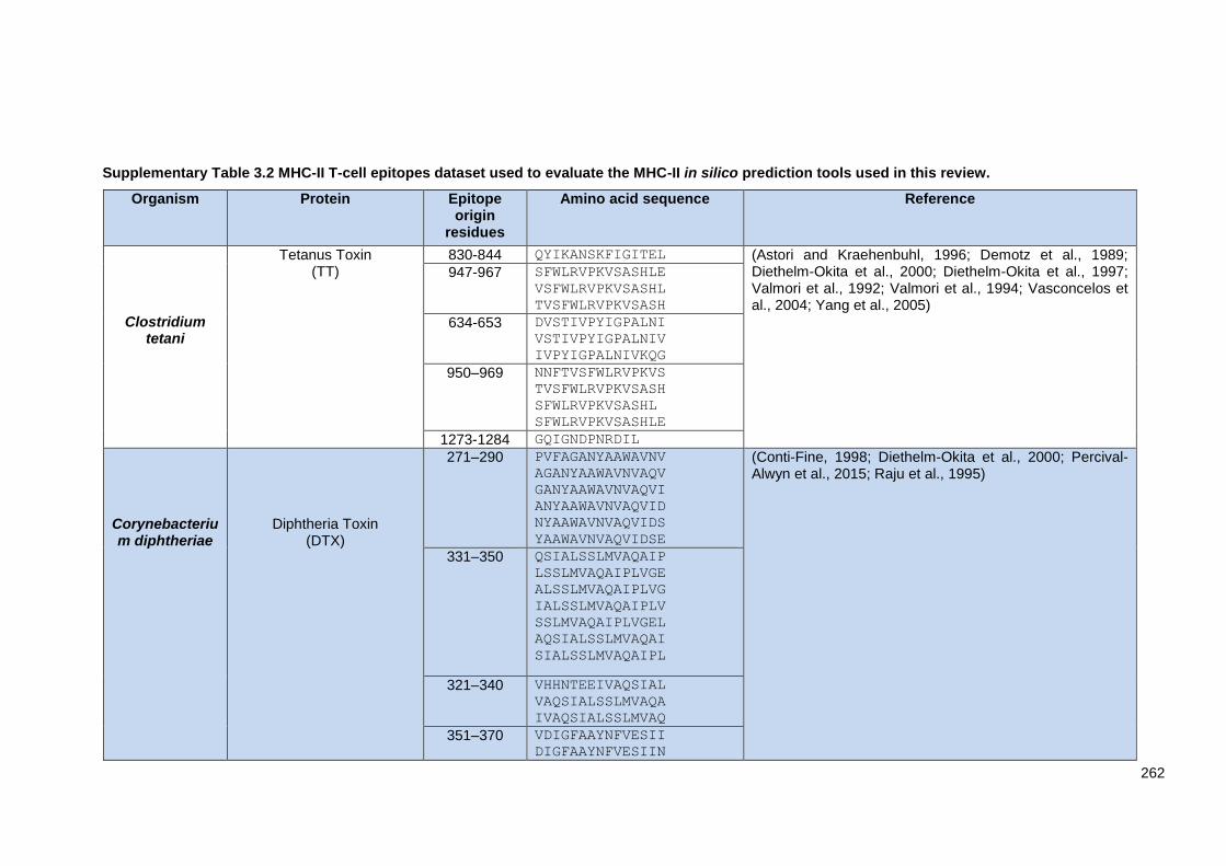
262
Supplementary Table 3.2 MHC-II T-cell epitopes dataset used to evaluate the MHC-II in silico prediction tools used in this review.
Organism Protein Epitope origin
residues
Amino acid sequence Reference
Clostridium tetani
Tetanus Toxin (TT)
830-844 QYIKANSKFIGITEL (Astori and Kraehenbuhl, 1996; Demotz et al., 1989; Diethelm-Okita et al., 2000; Diethelm-Okita et al., 1997; Valmori et al., 1992; Valmori et al., 1994; Vasconcelos et al., 2004; Yang et al., 2005)
947-967 SFWLRVPKVSASHLE
VSFWLRVPKVSASHL
TVSFWLRVPKVSASH
634-653 DVSTIVPYIGPALNI
VSTIVPYIGPALNIV
IVPYIGPALNIVKQG
950–969 NNFTVSFWLRVPKVS
TVSFWLRVPKVSASH
SFWLRVPKVSASHL
SFWLRVPKVSASHLE
1273-1284 GQIGNDPNRDIL
Corynebacterium diphtheriae
Diphtheria Toxin (DTX)
271–290 PVFAGANYAAWAVNV
AGANYAAWAVNVAQV
GANYAAWAVNVAQVI
ANYAAWAVNVAQVID
NYAAWAVNVAQVIDS
YAAWAVNVAQVIDSE
(Conti-Fine, 1998; Diethelm-Okita et al., 2000; Percival-Alwyn et al., 2015; Raju et al., 1995)
331–350 QSIALSSLMVAQAIP
LSSLMVAQAIPLVGE
ALSSLMVAQAIPLVG
IALSSLMVAQAIPLV
SSLMVAQAIPLVGEL
AQSIALSSLMVAQAI
SIALSSLMVAQAIPL
321–340 VHHNTEEIVAQSIAL
VAQSIALSSLMVAQA
IVAQSIALSSLMVAQ
351–370 VDIGFAAYNFVESII
DIGFAAYNFVESIIN

263
IGFAAYNFVESIINL
GFAAYNFVESIINLF
FAAYNFVESIINLFQ
411–430 QGESGHDIKITAENT
GHDIKITAENTPLPI
SGHDIKITAENTAEN
ESGHDIKITAENTPL
431–450 GVLLPTIPGKLDVNK
TIPGKLDVNKSKTHI
PTIPGKLDVNKSKTH
P. falciparum
Erythrocyte binding proteins (EBP3)
12-27 LVSEEIVTEEGSVAQE
VSEEIVTEEGSVAQE
(Ahlborg et al., 2000; Ahlborg et al., 1997; Calvo-Calle et al., 1997; Kironde et al., 1991; Sinigaglia, 1992; Vasconcelos et al., 2004) Circumsporozoite (CS) 379-398
326-345
AKMEKCSSVFNVVNS
IEKKIAKMEKCSSVF
EYLNKIQNSLSTEWS
NKIQNSLSTEWSPCS
Mycobacterium tuberculosis
6 kDa Early secretory antigenic target (ESAT-6)
1-20 71–90
QQWNFAGIEAAASAI
EQQWNFAGIEAAASA
TEQQWNFAGIEAAAS
MTEQQWNFAGIEAAA
QWNFAGIEAAASAIQ
WNFAGIEAAASAIQG
NLARTISEAGQAMAS
LARTISEAGQAMAST
ARTISEAGQAMASTE
RTISEAGQAMASTEG
(Brandt et al., 1996; Grover et al., 2014; Kanaujia et al., 2004; Kumar et al., 2010; Kumar and Raja, 2010; Myers et al., 2013; Olsen et al., 2000; Rapeah and Norazmi, 2006; Sørensen et al., 1995; Vogelzang et al., 2014; West et al., 2011; Winslow et al., 2003; Woodworth et al., 2014)
Antigen 85B (Ag85B)
240-254
154-173
164-183
FQDAYNAAGGHNAVF
SAMILAAYHPQQFIYAGSLS
AYHPQQFIYAGSLSA
QQFIYAGSLSALLDP
QFIYAGSLSALLDPS
FIYAGSLSALLDPSQ
IYAGSLSALLDPSQG
(Andersen et al., 1995; Brandt et al., 1996; Friscia et al., 1995; Geluk et al., 1998; Jurcevic et al., 1996; Smet, 1998; Vordermeier et al., 1995; Vordermeier et al., 1993)
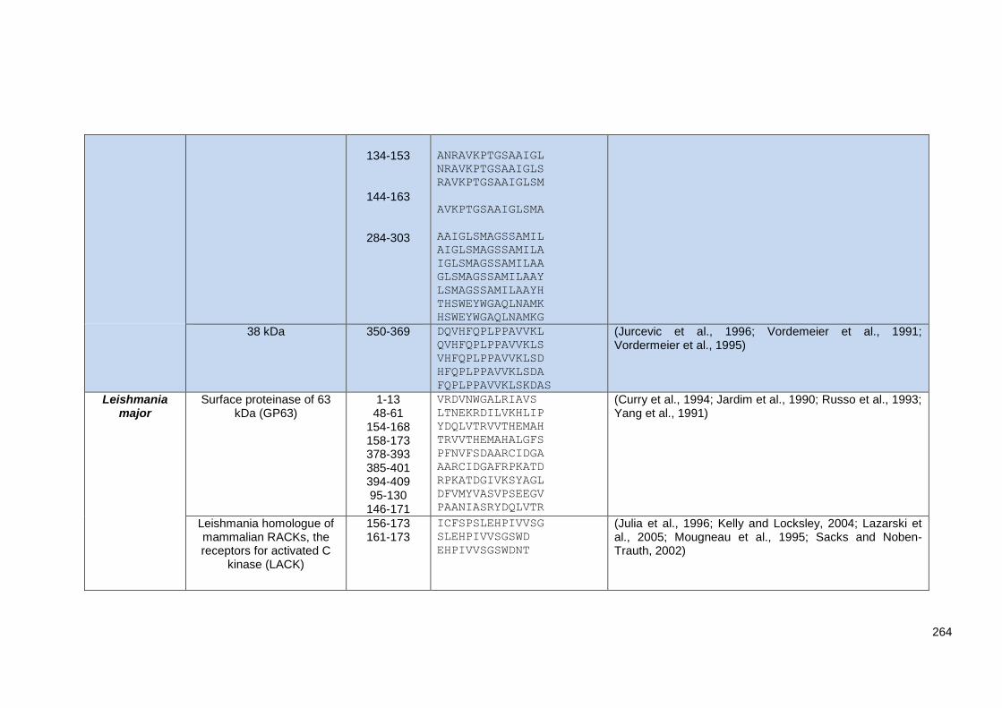
264
134-153
144-163
284-303
ANRAVKPTGSAAIGL
NRAVKPTGSAAIGLS
RAVKPTGSAAIGLSM
AVKPTGSAAIGLSMA
AAIGLSMAGSSAMIL
AIGLSMAGSSAMILA
IGLSMAGSSAMILAA
GLSMAGSSAMILAAY
LSMAGSSAMILAAYH
THSWEYWGAQLNAMK
HSWEYWGAQLNAMKG
38 kDa 350-369 DQVHFQPLPPAVVKL
QVHFQPLPPAVVKLS
VHFQPLPPAVVKLSD
HFQPLPPAVVKLSDA
FQPLPPAVVKLSKDAS
(Jurcevic et al., 1996; Vordemeier et al., 1991; Vordermeier et al., 1995)
Leishmania major
Surface proteinase of 63 kDa (GP63)
1-13 48-61
154-168 158-173 378-393 385-401 394-409 95-130 146-171
VRDVNWGALRIAVS
LTNEKRDILVKHLIP
YDQLVTRVVTHEMAH
TRVVTHEMAHALGFS
PFNVFSDAARCIDGA
AARCIDGAFRPKATD
RPKATDGIVKSYAGL
DFVMYVASVPSEEGV
PAANIASRYDQLVTR
(Curry et al., 1994; Jardim et al., 1990; Russo et al., 1993; Yang et al., 1991)
Leishmania homologue of mammalian RACKs, the receptors for activated C
kinase (LACK)
156-173 161-173
ICFSPSLEHPIVVSG
SLEHPIVVSGSWD
EHPIVVSGSWDNT
(Julia et al., 1996; Kelly and Locksley, 2004; Lazarski et al., 2005; Mougneau et al., 1995; Sacks and Noben-Trauth, 2002)

265
Leishmania infantum
Cysteine peptidase A (CPA)
149-163 246-260 114-128 257-271 32-46
312-326
MCGSCWAFATTGNIE
PHDEEEIAAYVGKNG
KDYKEHVHVDDSVRS
GKNGPVAVAVDATTW
GVDDFIASAHYGRFK
GSSWGEKGYIRLAMG
(Agallou et al., 2014; Coler and Reed, 2005)
Histone 1 like protein (H1) 1-15 27-41 32-46 52-56 2-16 26-40 31-45 29-43 30-44 25-39 24-38 28-42 5-19 4-18 48-62 33-47 49-63 35-49
MSSDSAVAALSAAMT
KTAAKKAAAKKAAAK
KAAAKKAAAKKAGAK
KAAAKKAAAKKAGAK
SSDSAVAALSAAMTS
KKTAAKKAAAKKAAA
KKAAAKKAAAKKAGA
AAKKAAAKKAAAKKA
AKKAAAKKAAAKKAG
PKKTAAKKAAAKKAA
SPKKTAAKKAAAKKA
TAAKKAAAKKAAAKK
SAVAALSAAMTSPQK
DSAVAALSAAMTSPQ
AGAKKAVRKVATPKK
AAAKKAAAKKAGAKK
GAKKAVRKVATPKKP
AKKAAAKKAGAKKAG
AAKKAAAKKAGAKKA
Leishmania eukaryotic initiation factor (LeIF)
100-114 166-180 140-154 102-116 103-117 101-115 139-153 99-113 98-112 141-155
LSPTRELALQTAEVI
ALRTESLRVLVLDEA
DLRKLQAGVIVAVGT
PTRELALQTAEVISR
TRELALQTAEVISRI
SPTRELALQTAEVIS
DDLRKLQAGVIVAVG
VLSPTRELALQTAEV
LVLSPTRELALQTAE
LRKLQAGVIVAVGTP
PSSIQQRAIAPFTRG
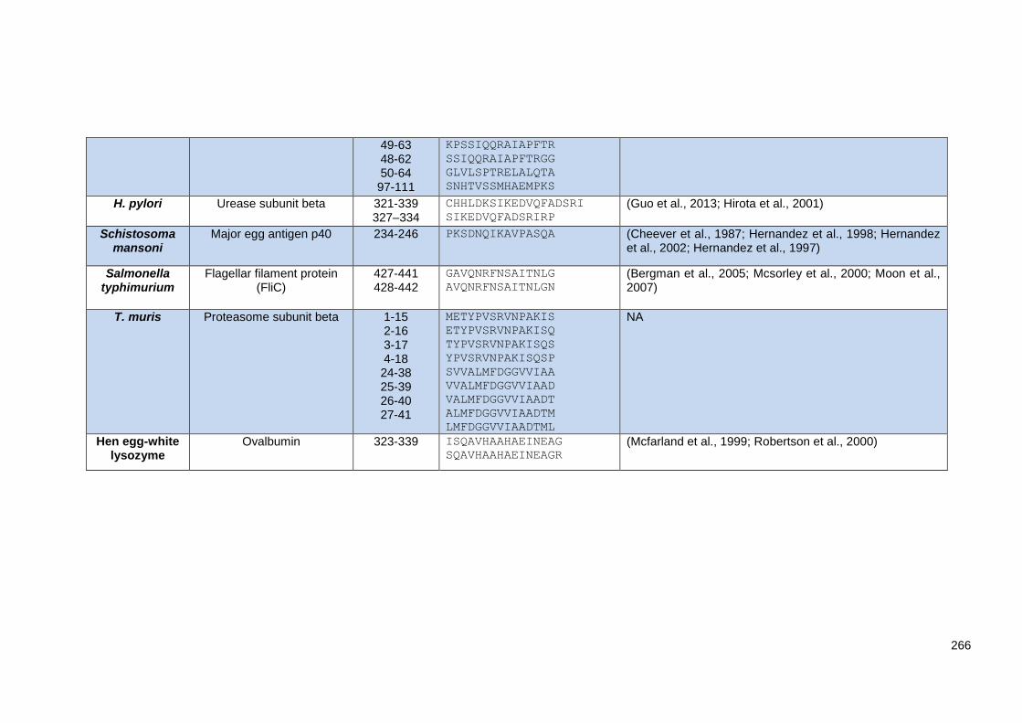
266
49-63 48-62 50-64 97-111
KPSSIQQRAIAPFTR
SSIQQRAIAPFTRGG
GLVLSPTRELALQTA
SNHTVSSMHAEMPKS
H. pylori Urease subunit beta 321-339 327–334
CHHLDKSIKEDVQFADSRI
SIKEDVQFADSRIRP
(Guo et al., 2013; Hirota et al., 2001)
Schistosoma mansoni
Major egg antigen p40 234-246 PKSDNQIKAVPASQA (Cheever et al., 1987; Hernandez et al., 1998; Hernandez et al., 2002; Hernandez et al., 1997)
Salmonella typhimurium
Flagellar filament protein (FliC)
427-441 428-442
GAVQNRFNSAITNLG
AVQNRFNSAITNLGN
(Bergman et al., 2005; Mcsorley et al., 2000; Moon et al., 2007)
T. muris Proteasome subunit beta 1-15 2-16 3-17 4-18 24-38 25-39 26-40 27-41
METYPVSRVNPAKIS
ETYPVSRVNPAKISQ
TYPVSRVNPAKISQS
YPVSRVNPAKISQSP
SVVALMFDGGVVIAA
VVALMFDGGVVIAAD
VALMFDGGVVIAADT
ALMFDGGVVIAADTM
LMFDGGVVIAADTML
NA
Hen egg-white lysozyme
Ovalbumin 323-339 ISQAVHAAHAEINEAG
SQAVHAAHAEINEAGR
(Mcfarland et al., 1999; Robertson et al., 2000)
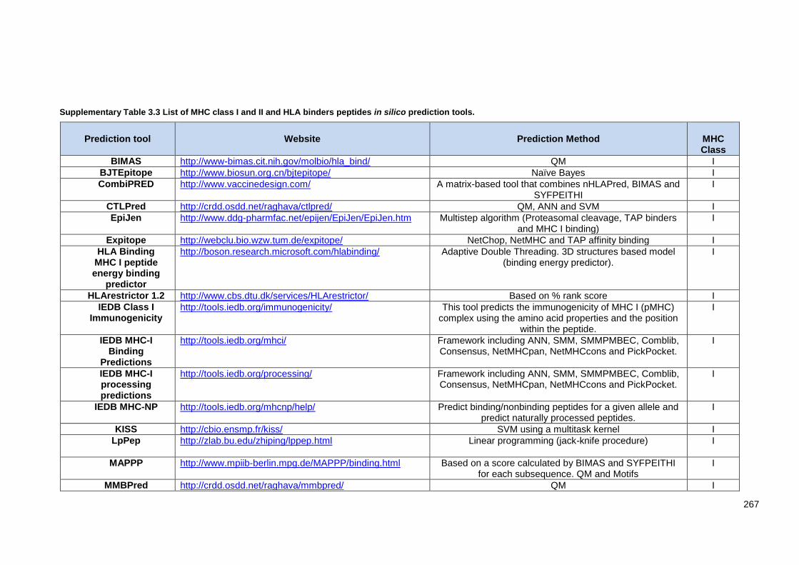
267
Supplementary Table 3.3 List of MHC class I and II and HLA binders peptides in silico prediction tools.
Prediction tool
Website
Prediction Method
MHC Class
BIMAS http://www-bimas.cit.nih.gov/molbio/hla_bind/ QM I
BJTEpitope http://www.biosun.org.cn/bjtepitope/ Naïve Bayes I
CombiPRED http://www.vaccinedesign.com/ A matrix-based tool that combines nHLAPred, BIMAS and SYFPEITHI
I
CTLPred http://crdd.osdd.net/raghava/ctlpred/ QM, ANN and SVM I
EpiJen http://www.ddg-pharmfac.net/epijen/EpiJen/EpiJen.htm Multistep algorithm (Proteasomal cleavage, TAP binders and MHC I binding)
I
Expitope http://webclu.bio.wzw.tum.de/expitope/ NetChop, NetMHC and TAP affinity binding I
HLA Binding MHC I peptide energy binding
predictor
http://boson.research.microsoft.com/hlabinding/ Adaptive Double Threading. 3D structures based model (binding energy predictor).
I
HLArestrictor 1.2 http://www.cbs.dtu.dk/services/HLArestrictor/ Based on % rank score I
IEDB Class I Immunogenicity
http://tools.iedb.org/immunogenicity/ This tool predicts the immunogenicity of MHC I (pMHC) complex using the amino acid properties and the position
within the peptide.
I
IEDB MHC-I Binding
Predictions
http://tools.iedb.org/mhci/ Framework including ANN, SMM, SMMPMBEC, Comblib, Consensus, NetMHCpan, NetMHCcons and PickPocket.
I
IEDB MHC-I processing predictions
http://tools.iedb.org/processing/ Framework including ANN, SMM, SMMPMBEC, Comblib, Consensus, NetMHCpan, NetMHCcons and PickPocket.
I
IEDB MHC-NP http://tools.iedb.org/mhcnp/help/ Predict binding/nonbinding peptides for a given allele and predict naturally processed peptides.
I
KISS http://cbio.ensmp.fr/kiss/ SVM using a multitask kernel I
LpPep
http://zlab.bu.edu/zhiping/lppep.html Linear programming (jack-knife procedure) I
MAPPP http://www.mpiib-berlin.mpg.de/MAPPP/binding.html Based on a score calculated by BIMAS and SYFPEITHI for each subsequence. QM and Motifs
I
MMBPred http://crdd.osdd.net/raghava/mmbpred/ QM I

268
NetChop 3.1 http://www.cbs.dtu.dk/services/NetChop/ ANN I
NetCTL 1.2 http://www.cbs.dtu.dk/services/NetCTL/ ANN I
NetCTLpan 1.1 http://www.cbs.dtu.dk/services/NetCTLpan/ ANN I
NetMHC 4.0 http://www.cbs.dtu.dk/services/NetMHC ANN and weighted matrices I
NetMHCcons 1.1 http://www.cbs.dtu.dk/services/NetMHCcons/ Integrating three methods: NetMHC (ANN), NetMHCpan (Pan specific ANN) and PickPocket (Matrix based)
I
NetMHCpan 2.8 http://www.cbs.dtu.dk/services/NetMHCpan/ Pan specific ANN I
NetMHCstab 1.0 http://www.cbs.dtu.dk/services/NetMHCstab-1.0/ ANN I
NetTepi 1.0 http://www.cbs.dtu.dk/services/NetTepi/ Integrates three methods: peptide-MHC binding affinity using NetMHCcons, peptide-MHC stability using the
NetMHCstab and T-cell propensity.
I
Pcleavage http://crdd.osdd.net/raghava/pcleavage/ SVM I
PEPVAC http://bio.dfci.harvard.edu/PEPVAC/ PSSM Profile-matrices I
PREDEP http://margalit.huji.ac.il/Teppred/mhc-bind/index.html Threading and Motif. I
PREDmafa http://cvc.dfci.harvard.edu/mafa/ QM I
ProPred1 http://www.imtech.res.in/raghava/propred1 Based on QM, proteasomal cleavage and promiscuous peptides.
I
SliDER http:// modlab-cadd.ethz.ch/software/slider/ ANN and SVM I
SMM http://zlab.bu.edu/SMM/ linear programming (SMM) I
SVMHC http://www-bs.informatik.unituebingen.de/Services/SVMHC http://svrmhc.biolead.org/
SVM I
TAPpred http://crdd.osdd.net/raghava/tappred/ SVM I
T-EPITOPE Designer
http://www.bioinformation.net/ted/index.html Structure modelling (Virtual pockets in 3D). I
WAPP
http://abi.inf.uni-tuebingen.de/Services/WAPP/information SVMHC I
IFNepitope http://crdd.osdd.net/raghava/ifnepitope/ Motif based SVM, and SVM hybrid. IFN-gamma inducing epitope
EpiDOCK http://epidock.ddg-pharmfac.net/ Structure molecular docking II
EpiTOP http://www.pharmfac.net/EpiTOP/ Proteochemometrics (QSAR) QM II

269
FDR4 http://crdd.osdd.net/raghava/fdr4/ SVMOT II
HLA-DR4Pred http://www.imtech.res.in/raghava/hladr4pred/ SVM and ANN II
IEDB MHC-II Binding
Predictions
http://tools.iedb.org/mhcii/ Consensus, CombLib, NN-align (netMHCII-2.2), SMM-align (netMHCII-1.1), Sturniolo, and NetMHCIIpan.
II
IL4pred http://crdd.osdd.net/raghava/il4pred/ SVM, Merci Motif based, Hybrid (SVM + Motif) based, and Swissprot based.
II
MetaMHCIIPan http://www.biokdd.fudan.edu.cn/Service/MetaMHC.html A pan-specific consensus II
MHC http://crdd.osdd.net/raghava/mhc/ Motifs Matrix Optimization Technique II
MHC2MIL http://datamining-iip.fudan.edu.cn/service/MHC2MIL/index.html
Pan-specific: PSSM, ANN, kernel based methods, and multiple instance learning based methods.
II
MHC2Pred http://crdd.osdd.net/raghava/mhc2pred/ SVM II
MHC2SKpan-1.0. http://datamining-iip.fudan.edu.cn/service/MHC2SKpan/index.html
MHC-II String Kernel II
MHCIIMulti http://etk.informatik.uni-tuebingen.de/acl_users/credentials_cookie_auth/require_login?came_from=http%3A//etk.informatik.uni-tuebingen.de/mhciimulti/index_html/ddocument_view
Machine learning method II
MHCMIR 1.0 http://ailab.ist.psu.edu/mhcmir/ Multiple instance regression II
MHC-Thread http://www.csd.abdn.ac.uk/~gjlk/MHC-Thread/ Structure peptide threading II
NetMHCII 2.2 http://www.cbs.dtu.dk/services/NetMHCII/ ANN II
NetMHCIIpan 3.1 http://www.cbs.dtu.dk/services/NetMHCIIpan/ ANN II
NNAlign http://www.cbs.dtu.dk/services/NNAlign/ ANN II
PREDIVAC http://predivac.biosci.uq.edu.au/ PSSM II
ProPred http://crdd.osdd.net/raghava/propred/ QM II
RTA http://bordnerlab.org/RTA/ Regularized Thermodynamic Average II
TEPITOPEpan http://datamining-iip.fudan.edu.cn/service/TEPITOPEpan/TEPITOPEpan.html
PSSM II
EpDis http://bioinfo.matf.bg.ac.rs/home/downloads.wafl?cat=Software.
NA I and II
EpiMatrix http://www.epivax.com/ Matrix-based and pocket Profile I and II
EpiToolKit 2.0 http://epitoolkit.de/ Syfpeithi, BIMAS, SVMHC, SMM, SMMPMBEC, UniTope, I and II

270
NetMHC, NetMHCpan, NetMHCII, NetMHCIIpan, TEPITOPEpan.
Epitopemap http://dmnfarrell.github.io/epitopemap/ The Tepitope method is the Python implementation of TEPITOPEPan, NetMHCIIpan and IEDB MHCI
I and II
FRED http://abi.inf.uni-tuebingen.de/Software/FRED/index.html Machine learning method I and II
HLAPRED http://crdd.osdd.net/raghava/hlapred/ QM
I and II
Hotspot Hunter http://antigen.i2r.a-star.edu.sg/hh/ ANN and SVM I and II
MetaMHC http://www.biokdd.fudan.edu.cn/Service/MetaMHC.html Local Alignment (LA) kernel I and II
MHCBench http://crdd.osdd.net/raghava/mhcbench/ Evaluate and compare the performance of the old/new prediction methods in terms of the threshold dependent
and independent parameters
I and II
MHC-BPS http://bidd.cz3.nus.edu.sg/mhc/ SVM I and II
MHCcluster 2.0 http://www.cbs.dtu.dk/services/MHCcluster/ Based on predicted binding specificity. MHC class I peptide binding predictions using
NetMHCpan, and MHC class II peptide binding predictions using NetMHCIIpan
I and II
MHCpred http://www.ddg-pharmfac.net/mhcpred/MHCPred/ QSAR I and II
MOTIF SCAN http://www.hiv.lanl.gov/content/immunology/motif scan/ Sequence Motifs I and II
MULTIPRED2 http://cvc.dfci.harvard.edu/multipred2/HTML/prediction2.php
netMHCpan and netMHCIIpan (ANN and HMM) I and II
nHLApred
http://crdd.osdd.net/raghava/nhlapred/comp.html ANN and QM I and II
OptiTope http://etk.informatik.uni-tuebingen.de/optitope Designing epitope-based vaccines by assessing the immunogenicity
I and II
PepCrawler http://bioinfo3d.cs.tau.ac.il/PepCrawler/about.html Docking and structure modelling I and II
PickPocket 1.1 http://www.cbs.dtu.dk/services/PickPocket/ Position specific weight matrix (SMM) I and II
POPI2.0 http://iclab.life.nctu.edu.tw/POPI/ SVM I and II
POPI2.0 http://iclab.life.nctu.edu.tw/POPI/ QM I and II
PREDBALB/c http://cvc.dfci.harvard.edu/balbc/ QM I and II
Predict http://research.i2r.a-star.edu.sg/fimm ANN I and II
RANKPEP http://imed.med.ucm.es/Tools/rankpep_help.html PSSM or profile I and II
SNEPv2 http://etk.informatik.uni-tuebingen.de/snep SYFPEITHI, Bimas/HLA_Bind, Epidemix, SVMHC, UniTope, TEPITOPE, MHCIIMulti
I and II

271
SVMHC http://abi.inf.uni-tuebingen.de/Services/SVMHC SVM I and II
SVRMHC http://c1.accurascience.com/SVRMHCdb/ SVR I and II
SYFPEITHI http://www.syfpeithi.de/ MM I and II
TEpredict http://tepredict.sourceforge.net/index.html QM I and II
TmhcPred http://www.imtech.res.in/raghava/tmhcpred/index.html Virtual and QM I and II
Vaxign http://www.violinet.org/vaxign/ PSSM I and II
Abbreviations: SMM: stabilized matrix method, SMMPMBEC: SMM with a Peptide: MHC Binding Energy Covariance matrix (SMMPMBEC), CombLib: Scoring Matrices derived from Combinatorial Peptide Libraries, QM: quantitative matrix, ANN: artificial network, PSSM: position specific scoring matrix, SDR: specificity-determining residue, SVRM: support vector machine regression, SVMOT: Support Vector Machine Optimization Technique, MM: Motif Matrix, ARB: Average relative binding, QSAR: Quantitative Structure Activity Relationship, HLA Peptide Binding Predictions: The Bioinformatics and Molecular Analysis Section of the National Institutes of Health (BIMAS), KISS: Kernel-based Inter-allele peptide binding prediction system, WAPP: Whole Antigen Processing Pathway. NA: Not available.
Appendices Chapter 4:
Supplementary Table 4.1 Percentage conservation of the T. muris MHC-II T-cell epitopes (CLSP222-237, CLSP433-450, CLSP143-158, CLSP424-443) to homologous T. trichiura proteins.
Epitope Peptide sequence T. trichiura homology proteins
A0A077Z1C2
A0A077ZKH1
A0A077ZLU4
A0A077YXV1
A0A077Z5M9
A0A077ZGC
8
A0A077ZDL8
A0A077ZLQ9
A0A077Z2Z8
A0A077YX43
A0A077ZCD9
A0A077ZN47
CLSP222-237 MEKKRKYLGSPLVCF 47 47 47 40 47 47 40 40 60 33 73 47 EKKRKYLGSPLVCFV 53 53 53 47 47 53 47 40 60 40 80 53
CLSP433-450 AKSKLRVYAGSPRFS 80 33 33 33 80 80 47 40 47 27 33 80 KSKLRVYAGSPRFSR 80 40 33 33 87 87 47 33 47 27 33 87 SKLRVYAGSPRFSRI 73 33 27 33 87 87 40 33 47 27 33 87 KLRVYAGSPRFSRIR 67 33 27 27 87 87 40 33 47 27 27 87
CLSP143-158 AILQLAKPVPFSNTV 40 40 27 80 40 40 40 27 33 27 40 27 ILQLAKPVPFSNTVR 33 40 27 80 33 33 33 27 33 27 33 27
CLSP398-416 SDHQEGYPVSPSVHI 27 27 33 87 33 33 33 33 40 27 33 33

272
DHQEGYPVSPSVHIV 27 27 33 87 33 33 33 33 33 27 40 33 HQEGYPVSPSVHIVS 27 27 33 87 33 33 33 33 33 27 40 33 QEGYPVSPSVHIVSL 33 27 33 87 33 33 33 27 40 27 40 33 EGYPVSPSVHIVSLA 33 27 33 93 27 33 33 27 40 27 40 33
CLSP424-443 CTGSLYASARANFTR 40 27 27 73 33 27 40 33 33 27 27 27 TGSLYASARANFTRT 33 33 27 73 40 27 40 27 33 27 27 27 GSLYASARANFTRTV 40 40 27 73 33 27 40 27 33 27 27 27 YASARANFTRTVLTS 40 40 27 73 27 33 33 27 27 27 33 27
Supplementary Table 4.2 Percentage conservation of the T. muris MHC-II T-cell epitopes (CBD36-52 and CBD1243-1259) to homologous T. trichiura proteins.
Epitope Peptide sequence T. trichiura homology proteins
A0A077ZFY4 A0A077ZC62 A0A077Z8B3 A0A077Z3K5 A0A077Z111 A0A077ZFK2
CBD36-52
RPRLLKIKWSPTAAS 33 27 33 100 47 33
PRLLKIKWSPTAAST 33 27 33 93 47 33
RLLKIKWSPTAASTA 33 27 33 87 47 33
CBD1243-1259
PAGVVYQCTMPRYTL 87 33 27 33 33 33
AGVVYQCTMPRYTLC 86 26 33 33 33 26
GVVYQCTMPRYTLCV 80 26 33 33 33 26

273
Supplementary Table 4.3 Percentage conservation of the T. muris MHC-II T-cell epitope (CBD241-257) to homologous T. trichiura proteins.
Peptide sequence T. trichiura homology proteins
O44397 Q27087 A0A077ZJX5 Q7KPX3 A0A077YWQ4 A0A077YYJ8
GRTTPVTSAPTTVTT 40.00 33.33 100.00 46.67 26.67 33.33 RTTPVTSAPTTVTTE 40.00 33.33 93.33 46.67 26.67 33.33 TTPVTSAPTTVTTER 33.33 33.33 86.67 46.67 26.67 33.33
Supplementary Table 4.4 Percentage conservation of the T. muris MHC-II T-cell epitope (Pfam256-273) to homologous T. trichiura proteins.
Peptide sequence T. trichiura homology proteins
A0A077ZFC1
A0A077ZQ60
A0A077ZKA9
A0A077ZEX9
A0A077Z7Q4
A0A077ZIP6
A0A077ZF51
A0A077ZGQ3
A0A077ZIE1
A0A077ZH26
NFGEYWFTKPVGSVS 73 73 73 80 73 73 73 73 73 60
FGEYWFTKPVGSVSY 73 73 73 80 80 80 80 80 80 67 GEYWFTKPVGSVSYK 67 67 67 73 73 73 73 73 73 60 EYWFTKPVGSVSYKV 67 67 67 73 67 67 67 67 67 53
Supplementary Table 4.5 Percentage conservation of the predicted T. muris MHC-II T-cell epitope (H112-128) to homologous T. trichiura proteins.
Peptide sequence T. trichiura homology proteins
A0A077Z2Q3 A0A077YYY3 A0A077Z3K3 A0A077ZN82 A0A077ZDH0 A0A077ZE95 A0A077ZIB0 A0A077ZMB7 GARKFLPIYQKAVAE 87 87 87 87 87 87 87 87 ARKFLPIYQKAVAEE 87 87 87 87 87 73 87 87 RKFLPIYQKAVAEEK 86 86 86 86 86 73 86 86

274
Appendices Chapter 7:
Appendix 7.1 Serum parasite-specific responses following vaccination of mice with VLPs
expressing Trichuris T-cell epitopes
To analyse parasite-specific antibody (IgM, IgG1, and IgG2c) responses of mice vaccinated with
(HBc-H112-128, HBc-CLSP143-158, HBc-CLSP398-416 and HBc-CBD241-257)/AddaVax, serial dilutions of
mice serum were assessed using an ELISA-based assay with ES as a target antigen at day -1 and
day 14 p.i. Parasite-specific IgG1 levels were only detected in the serum of mice vaccinated with
ES/AddaVax prior to Trichuris infection, as shown in Supplementary Figure 7.1.
Following vaccination and Trichuris infection, negligible levels of parasite-specific IgM were
detected in the serum of mice vaccinated with VLPs+ T-cell epitopes/AddaVax with a significant
difference compared to the PBS/AddaVax injected mice (Supplementary Figure 7.2 A).
Furthermore, ES vaccinated mice activated both Th1/Th2 immune responses as significant high
levels of parasite-specific IgG2c/IgG1 were measured in mice sera (Supplementary Figure 7.2 B
and C). However, no parasite-specific IgG1 and IgG2c antibodies were detected in the sera of mice
vaccinated with PBS/AddaVax, VLP (HBc-Ag)/AddaVax and VLPs+ T-cell epitopes/AddaVax
groups, as shown in Supplementary Figure 7.2 (B) and (C).
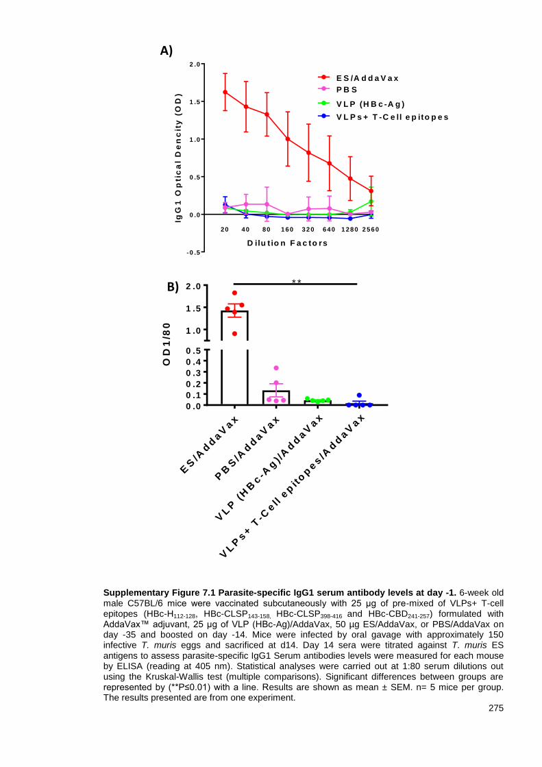
275
Supplementary Figure 7.1 Parasite-specific IgG1 serum antibody levels at day -1. 6-week old male C57BL/6 mice were vaccinated subcutaneously with 25 μg of pre-mixed of VLPs+ T-cell epitopes (HBc-H112-128, HBc-CLSP143-158, HBc-CLSP398-416 and HBc-CBD241-257) formulated with AddaVax™ adjuvant, 25 μg of VLP (HBc-Ag)/AddaVax, 50 µg ES/AddaVax, or PBS/AddaVax on day -35 and boosted on day -14. Mice were infected by oral gavage with approximately 150 infective T. muris eggs and sacrificed at d14. Day 14 sera were titrated against T. muris ES antigens to assess parasite-specific IgG1 Serum antibodies levels were measured for each mouse by ELISA (reading at 405 nm). Statistical analyses were carried out at 1:80 serum dilutions out using the Kruskal-Wallis test (multiple comparisons). Significant differences between groups are represented by (**P≤0.01) with a line. Results are shown as mean ± SEM. n= 5 mice per group. The results presented are from one experiment.
ES
/Ad
daV
ax
PB
S/A
dd
aV
ax
VL
P (
HB
c-A
g) /
Ad
daV
ax
VL
Ps+ T
-Cell e
pit
op
es/A
dd
aV
ax
0 .0
0 .1
0 .2
0 .3
0 .4
0 .5
1 .0
1 .5
2 .0
OD
1/8
0
* *B)
-0 .5
0 .0
0 .5
1 .0
1 .5
2 .0
D ilu t io n F a c to rs
IgG
1 O
pti
ca
l D
en
cit
y (
OD
)
P B S
E S /A d d a V a x
V L P (H B c -A g )
20 40 80 160 320 640 1 280 2 560
V L P s + T -C e ll e p ito p e s
A)

276
Supplementary Figure 7.2 Parasite-specific IgM, IgG1 and IgG2c serum antibodies levels at day 14 post-infection. 6-week old male C57BL/6 mice were vaccinated subcutaneously with 25 μg of pre-mixed of VLPs+ T-cell epitopes (HBc-H112-128, HBc-CLSP143-158, HBc-CLSP398-416 and HBc-CBD241-257) formulated with AddaVax™ adjuvant, 25 μg of VLP (HBc-Ag)/AddaVax, 50 µg ES/AddaVax, or PBS/AddaVax on day -35 and boosted on day -14. Mice were infected by oral gavage with approximately 150 infective T. muris eggs and sacrificed at d14 post-infection. Day 14 sera were titrated against T. muris ES antigens to assess parasite-specific IgM (A), IgG1 (B), and IgG2c (C) by ELISA (reading at 405 nm). Statistical analyses were carried out at 1:20 (IgM and gG2c) and 1:40 (IgG1) serum dilutions using the Kruskal-Wallis test (multiple comparisons). Significant differences between groups are represented by (*P≤0.05, **P≤0.01) with a line. Results are shown as mean ± SEM. n= 5 mice per group. The results presented are from one experiment.
ES
/Ad
daV
ax
PB
S/A
dd
aV
ax
VL
P (
HB
c-A
g) /
Ad
daV
ax
VL
Ps+ T
-Cell e
pit
op
es/
Ad
daV
ax
0 .0
0 .2
0 .4
0 .6
IgM
OD
1/4
0
*
0 .0
0 .2
0 .4
0 .6
0 .8
D ilu tio n F a c to rs
IgG
2c
O
pti
ca
l D
en
cit
y (
OD
)
20 40 80 1 6 0 3 2 0 6 4 0 1 2 8 0 2 5 6 0
P B S /A d d a V a x
E S /A d d a V a x
V L P (H B c -A g )/A d d a V a x
V L P s + T -C e ll e p ito p e s /A d d a V a x
-0 .2
0 .0
0 .2
0 .4
0 .6
D ilu tio n F a c to rs
IgM
Op
tic
al
De
nc
ity
(O
D)
20 40 80 1 6 0 3 2 0 6 4 0 1 2 8 0 2 5 6 0
P B S /A d d a V a x
E S /A d d a V a x
V L P (H B c -A g )/A d d a V a x
V L P s + T -C e ll e p ito p e s /A d d a V a x
0
1
2
3
4
D ilu tio n F a c to rs
Ig
G1
Op
tic
al
De
nc
ity
(O
D)
20 40 80 1 6 0 3 2 0 6 4 0 1 2 8 0 2 5 6 0
P B S /A d d a V a x
E S /A d d a V a x
V L P (H B c -A g )/A d d a V a x
V L P s + T -C e ll e p ito p e s /A d d a V a x
ES
/Ad
daV
ax
PB
S/A
dd
aV
ax
VL
P (
HB
c-A
g)/A
dd
aV
ax
VL
Ps+ T
-Cell e
pit
op
es/
Ad
daV
ax
0 .0
0 .5
1 .0
1 .5
2 .0
IgG
1 O
D 1
/40
* *
ES
/Ad
daV
ax
PB
S/A
dd
aV
ax
VL
P (
HB
c-A
g)/A
dd
aV
ax
VL
Ps+ T
-Cell e
pit
op
es/A
dd
aV
ax
0 .0
0 .5
1 .0
1 .5
IgG
2c
OD
1/4
0
*
*
A) Parasite-specific IgM
B) Parasite-specific IgG1
C) Parasite-specific IgG2c

277
Appendix 7.2 Serum VLP+ T-cell epitope-specific responses following vaccination of mice
with VLPs expressing Trichuris T-cell epitopes
To analyse VLPs+ T-cell epitopes specific-IgM antibody responses of vaccinated mice, serial
dilutions of mice sera were assessed using an ELISA-based assay against pre-mixed VLPs+ T-cell
epitopes (HBc-H112-128, HBc-CLSP143-158, HBc-CLSP398-416 and HBc-CBD241-257) as target antigens.
Significantly higher levels of HBc-CLSP143-158, HBc-CLSP398-416 and HBc-CBD241-257 -specific IgM
were measured in the sera of mice vaccinated with VLPs+ T-cell epitopes/AddaVax compared to
the ES/AddaVax, PBS/AddaVax and VLP (HBc-Ag)/AddaVax immunised mice, groups, day 14 p.i.
In contrast, only negligible levels of HBc-H112-128 -specific IgM were detected in the sera of
vaccinated mice groups, as shown in Supplementary Figure 7.3. Furthermore, only background
levels of VLPs+ T-cell epitopes-specific IgM levels were detected in the sera of mice immunised
with ES/AddaVax, PBS/AddaVax and VLP (HBc-Ag)/AddaVax.
Supplementary Figure 7.3 VLPs-specific IgM serum antibody levels at day 14 post-infection. 6-week old male C57BL/6 mice were vaccinated subcutaneously with 25 μg of pre-mixed of VLP+ T cell epitopes (HBc-H112-128, HBc-CLSP143-158, HBc-CLSP398-416 and HBc-CBD241-257) formulated with AddaVax™ adjuvant, 25 μg of VLP (HBc-Ag)/AddaVax, 50 µg ES/AddaVax, or PBS/AddaVax on day -35 and boosted on day -14. Mice were infected by oral gavage with approximately 150 infective T. muris eggs and sacrificed at d14 post-infection. Day 14 sera were titrated against VLPs+ T-cell epitopes proteins (HBc-Ag, HBc-H112-128, HBc-CLSP143-158, HBc-CLSP398-416 and HBc-CBD241-257) to assess VLPs specific IgM levels measured by ELISA (reading at 405 nm). Statistical analyses were carried out at 1:40 serum dilutions using Two-way ANOVA (multiple comparisons). Significant differences between groups are represented by (*P≤0.05, **P≤0.01, ***P≤0.001, ****P≤0.0001) with a line. Results are shown as mean ± SEM. n= 5 mice per group. The results presented are from one experiment.
HB
c-H
112-1
28
HB
c-C
LS
P 143-1
58
HB
c-C
LS
P 398-4
16
HB
c-C
BD 2
41-2
57
0 .0
0 .2
0 .4
0 .6
IgM
OD
1/4
0
***
*
P B S /A d d a V a x
V L P (H B c -A g )/A d d a V a x
V L P s + T -C e ll e p ito p e s /A d d a V a x
********
****
***
****
E S /A d d a V a x
* * * *

278
Appendix 7.3 The cellular immune responses following vaccination of mice with VLPs
expressing Trichuris T-cell epitopes and infection
To analyse the cellular immune responses of mice vaccinated with VLPs+ T-cell epitopes/AddaVax
compared to control ES/AddaVax, PBS/AddaVax and VLP (HBc-Ag)/AddaVax immunised mice
groups after T. muris infection, MLN cells were re-stimulated in vitro with 4 hours ES for 48 hours
and with ConA for 24 hours. As readout of cell activation, the supernatants were analysed for the
concentrations of IL-5, IL-13, IFN-γ, TNF, IL-6, IL-17, and IL-10 cytokines production by CBA.
Only MLN cells from ES/AddaVax vaccinated mice released higher levels of IL-5 (Supplementary
Figure 7.4 A), IL-13 (B) and IL-10 (G) compared to the VLPs+ T-cell epitopes/AddaVax, VLP (HBc-
Ag)/AddaVax and PBS/AddaVax immunised mice upon their re-stimulation in vitro with ES
antigens. In contrast, re-stimulated MLN cells taken from VLPs+ T-cell epitopes/AddaVax
vaccinated mice day 14 p.i. produced significantly higher levels of IFNϒ (Supplementary Figure7.4
C), TNF (D) and IL-6 (E) compared to the ES/AddaVax, PBS/AddaVax, and VLP (HBc-
Ag)/AddaVax immunised mice groups. Furthermore, there was no difference between the level of
IL-17 in all immunised mice groups (P>0.05), as shown in Supplementary Figure 7.4 (F).
Similar results were seen when the MLN cells from all immunised mice groups were re-stimulated
in vitro with ConA. Only MLN cells from ES/AddaVax vaccinated mice produced higher levels of
Th2 cytokines (IL-5 and IL-13) and anti-inflammatory cytokine (IL-10) compared to the other
immunised mice groups (Supplementary Figure 7.5 A, B and G). However, high levels of IFN-ϒ
(Supplementary Figure 7.5 C), TNF (D), IL-6 (E) and IL-17 (F) were seen on the re-stimulated MLN
cells taken from mice immunised with native VLP (HBc-Ag)/AddaVax compared to the other
immunised mice groups.
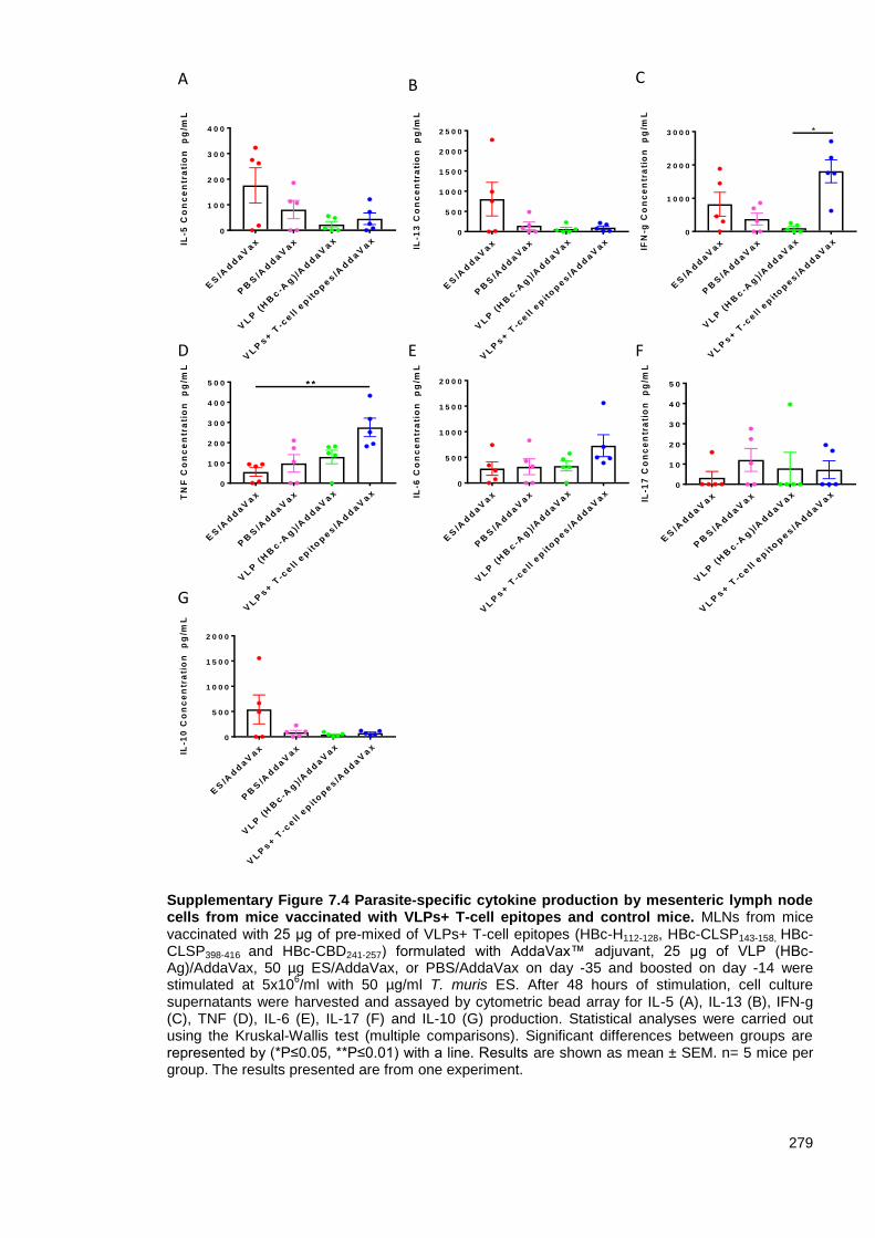
279
Supplementary Figure 7.4 Parasite-specific cytokine production by mesenteric lymph node cells from mice vaccinated with VLPs+ T-cell epitopes and control mice. MLNs from mice vaccinated with 25 μg of pre-mixed of VLPs+ T-cell epitopes (HBc-H112-128, HBc-CLSP143-158, HBc-CLSP398-416 and HBc-CBD241-257) formulated with AddaVax™ adjuvant, 25 μg of VLP (HBc-Ag)/AddaVax, 50 µg ES/AddaVax, or PBS/AddaVax on day -35 and boosted on day -14 were stimulated at 5x10
6/ml with 50 µg/ml T. muris ES. After 48 hours of stimulation, cell culture
supernatants were harvested and assayed by cytometric bead array for IL-5 (A), IL-13 (B), IFN-g (C), TNF (D), IL-6 (E), IL-17 (F) and IL-10 (G) production. Statistical analyses were carried out using the Kruskal-Wallis test (multiple comparisons). Significant differences between groups are represented by (*P≤0.05, **P≤0.01) with a line. Results are shown as mean ± SEM. n= 5 mice per group. The results presented are from one experiment.
ES
/Ad
daV
ax
PB
S/A
dd
aV
ax
VL
P (
HB
c-A
g) /
Ad
daV
ax
VL
Ps+
T-c
ell e
pit
op
es/A
dd
aV
ax
0
5 0 0
1 0 0 0
1 5 0 0
2 0 0 0
IL-6
Co
nc
en
tra
tio
n
pg
/mL
ES
/Ad
daV
ax
PB
S/A
dd
aV
ax
VL
P (
HB
c-A
g) /
Ad
daV
ax
VL
Ps+
T-c
ell e
pit
op
es/A
dd
aV
ax
0
1 0 0 0
2 0 0 0
3 0 0 0
IFN
-g C
on
ce
ntr
ati
on
p
g/m
L
*
ES
/Ad
daV
ax
PB
S/A
dd
aV
ax
VL
P (
HB
c-A
g) /
Ad
daV
ax
VL
Ps+
T-c
ell e
pit
op
es/A
dd
aV
ax
0
1 0
2 0
3 0
4 0
5 0
IL-1
7 C
on
ce
ntr
ati
on
p
g/m
L
ES
/Ad
daV
ax
PB
S/A
dd
aV
ax
VL
P (
HB
c-A
g) /
Ad
daV
ax
VL
Ps+
T-c
ell e
pit
op
es/A
dd
aV
ax
0
1 0 0
2 0 0
3 0 0
4 0 0
5 0 0
TN
F C
on
ce
ntr
ati
on
p
g/m
L
* *
ES
/Ad
daV
ax
PB
S/A
dd
aV
ax
VL
P (
HB
c-A
g) /
Ad
daV
ax
VL
Ps+
T-c
ell e
pit
op
es/A
dd
aV
ax
0
5 0 0
1 0 0 0
1 5 0 0
2 0 0 0
IL-1
0 C
on
ce
ntr
ati
on
p
g/m
L
ES
/Ad
daV
ax
PB
S/A
dd
aV
ax
VL
P (
HB
c-A
g) /
Ad
daV
ax
VL
Ps+
T-c
ell e
pit
op
es/A
dd
aV
ax
0
1 0 0
2 0 0
3 0 0
4 0 0
IL-5
Co
nc
en
tra
tio
n
pg
/mL
ES
/Ad
daV
ax
PB
S/A
dd
aV
ax
VL
P (
HB
c-A
g) /
Ad
daV
ax
VL
Ps+
T-c
ell e
pit
op
es/A
dd
aV
ax
0
5 0 0
1 0 0 0
1 5 0 0
2 0 0 0
2 5 0 0
IL-1
3 C
on
ce
ntr
ati
on
p
g/m
L
E
G
C
D F
A B
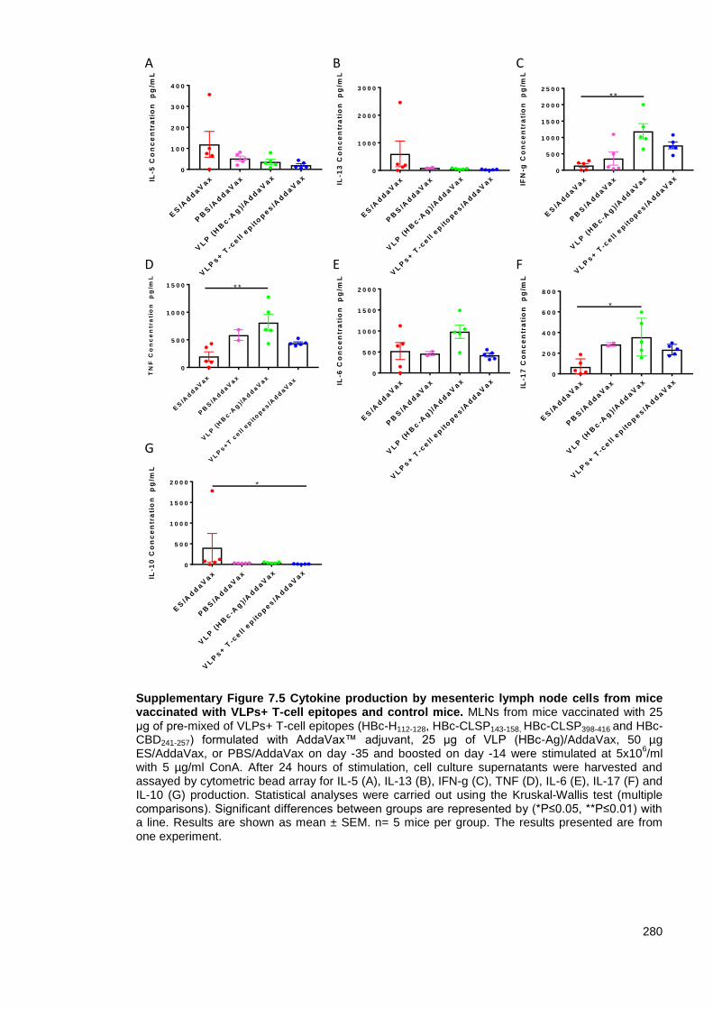
280
Supplementary Figure 7.5 Cytokine production by mesenteric lymph node cells from mice vaccinated with VLPs+ T-cell epitopes and control mice. MLNs from mice vaccinated with 25 μg of pre-mixed of VLPs+ T-cell epitopes (HBc-H112-128, HBc-CLSP143-158, HBc-CLSP398-416 and HBc-CBD241-257) formulated with AddaVax™ adjuvant, 25 μg of VLP (HBc-Ag)/AddaVax, 50 µg ES/AddaVax, or PBS/AddaVax on day -35 and boosted on day -14 were stimulated at 5x10
6/ml
with 5 µg/ml ConA. After 24 hours of stimulation, cell culture supernatants were harvested and assayed by cytometric bead array for IL-5 (A), IL-13 (B), IFN-g (C), TNF (D), IL-6 (E), IL-17 (F) and IL-10 (G) production. Statistical analyses were carried out using the Kruskal-Wallis test (multiple comparisons). Significant differences between groups are represented by (*P≤0.05, **P≤0.01) with a line. Results are shown as mean ± SEM. n= 5 mice per group. The results presented are from one experiment.
ES
/Ad
daV
ax
PB
S/A
dd
aV
ax
VL
P (
HB
c-A
g) /
Ad
daV
ax
VL
Ps+
T-c
ell e
pit
op
es/A
dd
aV
ax
0
1 0 0
2 0 0
3 0 0
4 0 0
IL-5
Co
nc
en
tra
tio
n
pg
/mL
ES
/Ad
daV
ax
PB
S/A
dd
aV
ax
VL
P (
HB
c-A
g) /
Ad
daV
ax
VL
Ps+
T-c
ell e
pit
op
es/A
dd
aV
ax
0
5 0 0
1 0 0 0
1 5 0 0
2 0 0 0
2 5 0 0
IFN
-g C
on
ce
ntr
ati
on
p
g/m
L
* *
ES
/Ad
daV
ax
PB
S/A
dd
aV
ax
VL
P (
HB
c-A
g)/
Ad
daV
ax
VL
Ps+
T c
ell e
pit
op
es/A
dd
aV
ax
0
5 0 0
1 0 0 0
1 5 0 0
TN
F C
on
ce
ntr
ati
on
p
g/m
L
* *
ES
/Ad
daV
ax
PB
S/A
dd
aV
ax
VL
P (
HB
c-A
g) /
Ad
daV
ax
VL
Ps+
T-c
ell e
pit
op
es/A
dd
aV
ax
0
2 0 0
4 0 0
6 0 0
8 0 0
IL-1
7 C
on
ce
ntr
ati
on
p
g/m
L
*
ES
/Ad
daV
ax
PB
S/A
dd
aV
ax
VL
P (
HB
c-A
g) /
Ad
daV
ax
VL
Ps+
T-c
ell e
pit
op
es/A
dd
aV
ax
0
5 0 0
1 0 0 0
1 5 0 0
2 0 0 0
IL-1
0 C
on
ce
ntr
ati
on
p
g/m
L
*
ES
/Ad
daV
ax
PB
S/A
dd
aV
ax
VL
P (
HB
c-A
g) /
Ad
daV
ax
VL
Ps+
T-c
ell e
pit
op
es/A
dd
aV
ax
0
1 0 0 0
2 0 0 0
3 0 0 0
IL-1
3 C
on
ce
ntr
ati
on
p
g/m
L
ES
/Ad
daV
ax
PB
S/A
dd
aV
ax
VL
P (
HB
c-A
g) /
Ad
daV
ax
VL
Ps+
T-c
ell e
pit
op
es/A
dd
aV
ax
0
5 0 0
1 0 0 0
1 5 0 0
2 0 0 0
IL-6
Co
nc
en
tra
tio
n
pg
/mL
E
G
C
D F
A B
281
Appendix 7.4 Crypt hyperplasia following vaccination of mice with VLPs expressing
Trichuris T-cell epitopes
The length of colonic crypts was measured day 14 p.i. in ES/AddaVax, PBS/AddaVax, VLP (HBc-
Ag)/AddaVax and VLPs+ T-cell epitopes/AddaVax immunised mice groups following Trichuris
infection as an index of crypt hyperplasia (Supplementary Figure 7.6 A).
Following a Trichuris infection, ES/AddaVax vaccinated mice exhibited crypt hyperplasia with a
significant difference compared to the PBS/AddaVax, and VLP (HBc-Ag)/AddaVax injected mice
groups (P<0.05), as shown in Supplementary Figure 7.6 (B).

282
Supplementary Figure 7.6 Changes in intestinal pathology in T. muris infected mice vaccinated with VLPs+ T-cell epitopes and control mice. (A) Representative photographs of gut sections stained with hematoxylin and eosin (H&E). Proximal colon was harvested from mice vaccinated with 25 µg of pre-mixed VLPs+ T-cell epitopes (HBc-H112-128, HBc-CLSP143-158, HBc-CLSP398-416 and HBc-CBD241-257) formulated with AddaVax™ adjuvant, 25 μg of VLP (HBc-Ag)/AddaVax, 50 µg ES/AddaVax, or PBS/AddaVax on day -35 and boosted on day -14. All photographs were taken with 20X magnification. Gut pathology assessed by measuring crypt lengths using Panoramic Viewer software. The yellow double-headed arrows show how the crypt lengths were measured. Results are shown as mean crypt length from 20 crypts units per mouse and shown as the mean values per mouse ±SEM. Statistical analyses were carried out using the Kruskal-Wallis test (multiple comparisons). Significant differences between groups are represented by (**P≤0.01, ***P≤0.001) with a line. n= 5 mice per group. The results presented are from one experiment.
ES
/Ad
daV
ax
PB
S/A
dd
aV
ax
VL
P (
HB
c-A
g) /
Ad
daV
ax
VL
Ps+ T
-cell e
pit
op
es/A
dd
aV
ax
0
5 0
1 0 0
1 5 0
Me
an
Cry
pts
le
ng
ht
µm * *
*
ES/AddaVax
VLPs+ T-cell epitopes/AddaVax
PBS/AddaVax
VLP (HBc-Ag) /AddaVax
A
B

283
Appendix 7.5 Goblet cells hyperplasia in mice vaccinated with VLPs expressing Trichuris
T-cell epitopes
PAS-stained goblet cells were measured in the mice vaccinated with ES/AddaVax, PBS/AddaVax,
VLP (HBc-Ag)/AddaVax and VLPs+ T-cell epitopes/AddaVax day 14 p.i. as an index of goblet cells
hyperplasia (Supplementary Figure 7.7 A).
Only ES/AddaVax vaccinated mice exhibited goblet cells hyperplasia with a significant difference
(P<0.05) compared to the mice vaccinated with VLPs+ T-cell epitopes/AddaVax, and
PBS/AddaVax injected mice, as shown in Supplementary Figure 7.7 (B).

284
Supplementary Figure 7.7 Quantification of goblet cell numbers in T. muris infected mice vaccinated with VLPs+ T-cell epitopes and control mice. (A) Representative photographs of gut sections stained with stained with Periodic acid–Schiff staining (PAS). Proximal colon was harvested from mice vaccinated with 25 μg of pre-mixed of VLPs+ T-cell epitopes (HBc-H112-128, HBc-CLSP143-158, HBc-CLSP398-416 and HBc-CBD241-257) formulated with AddaVax™ adjuvant, 25 μg of VLP (HBc-Ag)/AddaVax, 50 µg ES/AddaVax, or PBS/AddaVax on day -35 and boosted on day -14. The slides were scanned, and the goblet cells were quantified using Panoramic Viewer software. All photographs were taken with 20X magnification. (B) Goblet cells were quantified by counting the number of alcian blue-stained cells per crypt unit in three fields of view from each section and are shown as mean cell numbers per 20 crypt units (cu) ±SEM. Statistical analyses were carried out using the Kruskal-Wallis test (multiple comparisons). Significant differences between groups are represented by (*P≤0.05) with a line. n= 5 mice per group. The results presented are from one experiment.
ES/AddaVax
VLPs+ T-cell epitopes/AddaVax
PBS/AddaVax
VLP (HBc-Ag)/AddaVax
A
B
ES
/Ad
daV
ax
PB
S/A
dd
aV
ax
VL
P (
HB
c-A
g) /
Ad
daV
ax
VL
Ps+ T
-cell e
pit
op
es/
Ad
daV
ax
0
1 0 0
2 0 0
3 0 0
4 0 0
5 0 0
Go
ble
t c
ell
s p
er 2
0 c
ry
pt
un
its
**