development of a novel subunit vaccine against east coast
TRANSCRIPT

I
Development of a novel subunit vaccine againstEast Coast fever based on the Theileria parva
sporozoite surface protein p67
Stephen A. Kaba

II
Promotoren:prof. dr. J.M. VlakPersoonlijk Hoogleraar bij de Leerstoelgroep Virologie
prof. dr. R.W. GoldbachHoogleraar in de Virologie
Co-promoter:dr. M.M. van OersUniversitair Docent bij de Leerstoelgroep Virologie
Samenstelling promotiecommissie:dr. V. Nene (The Institute for Genomic Research, Rockville, USA)prof. dr. F. Jongejan (Utrecht Universiteit)prof. dr. H. Savelkoul (Wageningen Universiteit)dr. D. Schaap (Intervet International BV, Boxmeer)dr.ir. H.M.J. Udo (Wageningen Universiteit)

III
Development of a novel subunit vaccine againstEast Coast fever based on the Theileria parva
sporozoite surface protein p67
Stephen A. Kaba
proefschriftter verkrijging van de graad van doctor
op gezag van de rector magnificusvan Wageningen Universiteit
prof. dr. ir. L. Speelmanin het openbaar te verdedigenvrijdag op 17 oktober 2003
des namiddags te half twee in de Aula

IV
Kaba, S.A.
Development of a novel subunit vaccine against East Coast fever based on theTheileria parva sporozoite surface protein p67Thesis Wageningen University, the NetherlandsWith references – With summary in DutchISBN 90-5808-889-8Subject headings: Theileria parva, East Coast fever, p67, subunit vaccine

V
Contents
Chapter 1 Introduction and scope of the thesis 1
Chapter 2 Fusion to green fluorescent protein improves expression levelsof Theileria parva sporozoite surface antigen p67 in insectcells 11
Chapter 3 Baculovirus surface display of Theileria parva p67 antigenpreserves the conformation of sporozoite-neutralizing epitopes 25
Chapter 4 Improved secretion of Theileria parva sporozoite surfaceprotein p67 using a chitinase and cathepsin negative bacmid 37
Chapter 5 Enhanced immunogenicity of baculovirus-derived Theileriaparva p67 subunit antigens 49
Chapter 6 Improved protection against East Coast fever with novelbaculovirus-derived p67 subunit vaccines 63
Chapter 7 General discussion 79
References 91
Summary In English In Dutch (Samenvatting)
108110
Curriculum Vitae 113
List of publications 114
Acknowledgements 115

VI
Dedicated to my grand-mum, Madam Akakyeibire

Chapter 1
1
Chapter 1
Introduction and scope of the thesis
East Coast feverIn the early parts of the 20th century East Coast fever (ECF) was identified as a tick-borne disease of cattle in South Africa and associated with cattle imported from EastAfrica in an attempt to repopulate the livestock that had been devastated by rinderpest[148]. ECF is a lymphoproliferative disease characterized by a number of diverseclinical signs and pathological lesions, all of which are associated with invasion oflymphoid and non-lymphoid tissues with parasitized lymphoblasts [38, 61]. Thedisease is caused by a protozoan parasite called Theileria parva and is endemic in theeastern, central and southern parts of Africa. The distribution of ECF is strictlyassociated with the distribution of the vector tick species (Fig. 1), mainlyRhipicephalus appendiculatus.
Fig. 1: Distribution of ECF based on the probability of the presence of the vector, the brown eartick. [This picture originated from a poster designed and produced by Bruno Minjauw and others(ILRI) and presented jointly by DFID, AHP, FAO, ILRI and ICTTD. With permission from SophieCarver, AHP, the University of Edinburg, Scotland, UK].

Introduction and scope of thesis
2
Epidemics of ECF in cattle, defined as occurrence of Theileria parva infections inpreviously or recently unaffected areas [148], are renowned for their devastatingimpact on the cattle population. These epidemics often strike with high morbidity andmortality, causing major loss and significant social and economic distress to theindividual farmer. Extinction of entire herds is not uncommon especially when nomeasures are taken to mitigate the deadly impact of the outbreak [140]. As thelivelihood of smallholder farms, often managed by women depends on one or twocattle, the financial burden due to loss of income and livestock products impacts onthe quality of all aspects of family life. The disease is of considerable importancethroughout the region resulting in significant losses in terms of cattle deaths andincome, and represents a major constraint to the development of the livestock industryin eastern, central and southern Africa. For example T. parva costs farmers over US$170 million a year in direct losses [121, 130]. The spread of the infection is mainlythrough cattle movement, e.g. oxen working and/or feeding in neighboring or distantvillages, making migration for distances of 15 km or more not exceptional [15, 16].
Theileria parva parasites have the ability, unique among protozoan parasites, to‘transform’ the bovine lymphocytes they invade into cancer-like cells that proliferateuncontrollably, as leukaemia cells do, killing the animal within three weeks ofinfection. Pyrexia, generalized lymphodenopathy and terminal respiratory distressand/or diarrhea, due to pulmonary infiltration and edema, characterize ECF [114]. Aconsistent feature of the disease in its advanced stages is pronounced leukopoenia,which is reflected by extensive depletion of lymphocytes in the lymphoid organs. Theseverity of the disease in individual cattle is known to be influenced by a number ofparameters, including infection dose, breed, and individual variation [114, 148]. Thefirst clinical sign is persistent high fever that is used as a reliable indicator for earlydiagnosis. ECF exists in a number of epidemiological states in the field and exhibits aspectrum of syndromes ranging from those described above to sub-clinical infections,which are common, where heavy challenge occurs under conditions of endemicstability. The epidemiology is complicated by the presence of polymorphic traitsbetween T. parva isolates and stocks, associated primarily with different clinicalsymptoms, and the presence of a wildlife reservoir in the African Cape buffalo(Synerus caffer) [116].
In the past, T. parva was thought to comprise three distinct sub-species: T. parvaparva for parasites causing classical ECF; T. parva lawrencei for parasites causingCorridor disease and T. parva bovis for parasites causing January disease. It is nowknown that genetically T. parva is a single species and T. parva parasites are nowclassified according to their host species of origin, referred to as either cattle-derivedor buffalo-derived [148]. The closely related T. annulata, a parasite of the Asiatic orwater buffalo (Bubalus bubalis), is responsible for Mediterranean or tropicalTheileriosis in cattle in areas extending from the Mediterranean and North Africa

Chapter 1
3
through Asia to China [133]. Other Theileria species (e.g. T. buffeli, T. orientalis andT. sergenti) do not transform the host cells and cause diseases of lesser severity invarious parts of the world in particular the Asian and African continents [192].
Life Cycle of Theileria parvaTheileria parva is a member of the phylum Apicomplexa, which includes manyspecies of both medical (e.g. Plasmodium species; Toxoplasma) and veterinary (e.g.Babesia; Eimeria; Theileria) importance. The Apicomplexa are obligate intracellularparasites that, in general, have complex life cycles involving the invasion of a rangeof cells in one or more different hosts. For a detailed review of the classification andbiology of T. parva, see references [38, 39, 145, 148, 179, 181]. The life cycle of T.parva (Fig. 2) resembles that of the malaria parasite, P. falciparum, in that theseparasites undergo sequential development in nucleated cells and erythrocytes, theformer involving cells of the lymphoid system. However, unlike malaria, the diseasecaused by Theileria is attributed mainly to the schizogenous stage of development inleukocytes, although anaemia also occurs in case of T. annulata.
Theileria parva is transmitted to cattle primarily by infected nymphal or adult ticks ofthe genus Rhipicephalus appendiculatus (the brown-ear tick) when the tick injectsinfective sporozoites into the new host while taking a blood meal. The sporozoitesrapidly invade bovine lymphocytes by a receptor-mediated process and differentiateinto schizonts [38, 61, 179 - 181]. This event is associated with transformation of theinfected cell to a state of uncontrolled proliferation [61, 176]. Subsequent invasion ofnon-lymphoid tissues by parasitized cells and the associated immunopathologicaleffects usually result in death of the animal within two to four weeks of infection [38,114, 148]. During mitosis, the parasite body associates with the mitotic spindle anddivides synchronously with the host nucleus so that the schizonts are distributedrandomly to both daughter cells.
A proportion of the schizonts undergoes merogony to produce merozoites, whichinvade erythrocytes and develop into piroplasms, the form of T. parva infective to thetick. For the completion of the life cycle the piroplasms require to be ingested bylarval and nymphal stages of the tick while feeding on an animal carrying piroplasmsin the red blood cells. Infection in the tick is transmitted trans-stadially (i.e. from larvato nymph or nymph to adults) and unlike Babesia [67], there is no transovariantransmission.

Introduction and scope of thesis
4
Fig. 2: Life Cycle of Theileria parva [148].
The life cycle in the tick continues through production of gametes that fuse to form azygote. Zygotes penetrate the intestinal epithelium where they undergo reductiondivisions to form mobile kinetes. The kinetes in turn migrate to the salivary glandswhere they undergo nuclear divisions without accompanying cytokinesis to form acomplex syncytium, the sporoblast. Sporogony is completed when large numbers ofthe infective sporozoites are produced following four days of blood meal by theinfected nymphal or adult tick and are ready to be released into the mammalian host.
Treatment and prevention of ECFCurrently ECF is controlled by a combination of techniques including chemotherapy,immunisation and vector control by use of acaricides on cattle as spray or cattle dips.

Chapter 1
5
The first commercial drug against Theileria was halofuginone lactate (Terit®)followed by parvaquone (Clexon®). Subsequently, a more active analogue ofparvaquone, buparvaquone (Butalex®), was synthesised. The three drugs are availablebut are extremely expensive. Costs of treatment of small indigenous breeds of lowproductivity may equal the value of the animal and only early treatment has a highprobability of success.
Regular spraying or dipping of livestock in acaricide is quite effective in preventingECF but the chemicals are expensive and due to environmental concerns and possiblecontamination of the food chain not desirable. Many of the farmers in the affectedregions can hardly afford the cost of spraying and dipping even in communal dips andspray bays without any financial assistance. An addition to these problems is theability of the ticks to develop resistance to the acaricides.
The only immunisation method used on a routine scale for the control of ECF is whatis termed ‘Infection and Treatment’. This is a live vaccination regimen where animalsare given a needle injection of a lethal dose of T. parva and are simultaneously treatedwith a long-acting antibiotic, oxytetracycline. Immunity acquired can last for years,possibly even a lifetime, and resists homologous challenge, but not necessarily allstrains encountered in the field. Immunized animals become carriers, thus increasingthe infection rate in ticks and consequently the inoculation rate of other cattle.Although the techniques associated with the ‘infection and treatment’ method are farfrom being sophisticated and well within the reach of a reasonably equippedlaboratory, the method demands skill, meticulousness and a great sense ofresponsibility and flexibility on behalf of the operators [202]. As with other livevaccines, problems related to the necessary cold chain (sporozoite stabilates have tobe kept in liquid nitrogen until use in the field) are formidable obstacles in countrieswith poor infrastructure. In addition, there is a major concern over the introduction ofexogenous vaccine strains to resident tick populations. Furthermore, the immunityengendered by the ‘infection and treatment’ method is strain-specific [202].
Thus from economic, social and environmental perspectives, the need for analternative, such as a subunit vaccine against ECF, cannot be emphasised enough. Theultimate aim of the research described in this thesis therefore was to develop andoptimise a subunit vaccine for ECF based on p67, the Theileria parva sporozoitesurface protein. Besides, intensive efforts are focused on the identification ofschizont-specific components for incorporation in a second-generation multi-component product [113, 114, 124,127]. The completion of the genome sequence ofT. parva [145, 146] will further enhance the schizont antigen identification process. Inaddition, it offers new opportunities in combating infection and diseases byunderstanding the biology of these parasites and their hosts in greater detail.

Introduction and scope of thesis
6
Molecular basis of T. parva sporozoite-host cell interaction andapproaches to vaccine developmentA 20-25-nm thick surface coat covers sporozoites of T. parva and it is the interactionbetween this coat and the host cell surface molecules that are critical to parasiteinvasion [181]. For a detailed review on the biology and the morphological stepsinvolved in the T. parva sporozoite invasion and/or interactions with the host cells,see references [176, 179 - 181]. The surface coat may have other functions in thebiology of the sporozoites but the interest in its molecular composition has beendirected primarily towards the development of a vaccine to block sporozoite entry andsubsequent intracellular development [133, 135]. Sera from cattle repeatedlyimmunised with T. parva sporozoite lysates or from cattle that had been exposed toheavy parasite challenge in ECF endemic areas contain high titre antibodies capableof neutralizing T. parva sporozoite infectivity. These observations led to the hunt forsporozoite antigens that may be involved in the entry process [133]. Although thesesera recognised five antigens on immuno-blots, a 67-kDa protein on the surface ofsporozoites, designated p67, contained the major neutralising epitopes, as determinedin studies with a panel of neutralising mAb [133]. The gene that encodes p67 has beencloned and characterized and it exists as a single copy gene interrupted by a 29-bpintron [141]. It encodes a 709 amino acid product that contains seven potential sites ofN-linked glycosylation, although there is no evidence that the native protein is [141].The p67 antigen is uniformly distributed on the sporozoite surface and is synthesisedand assembled during the earlier stages of sporogony in the salivary glands [179].P67-specific antiserum neutralises sporozoite infectivity from a range of differentparasite stocks [143], indicating that p67 is conserved in cattle-derived parasites andunderscoring the potential of p67 as a broad-spectrum vaccine. Monoclonal antibodiesto both native and recombinant p67 also inhibit sporozoite invasion of target cells in-vitro [34, 141].
Anti-p67 specific antibodies block the initial binding event of the sporozoite to thehost cell [179]. Furthermore, purified native and recombinant p67 can competitivelyblock sporozoite entry, provided that the molecule is present in the incubationmedium [177]. While the addition of exogenous p67 significantly inhibited sporozoiteinvasion, sporozoites that could bind entered host cells at levels comparable to controlinfections suggesting that exogenous p67 was affecting only the initial bindingprocess [177]. These observation indicated that p67 may act as a ligand mediating theinitial parasite-host cell interaction. Shaw et al., [177] also reported that the inhibitionwas readily and rapidly reversed by removing p67 from the medium implying that thatat the molecular level, the interactions between individual p67 molecules and host cellsurface molecules are relatively weak. In cell-to-cell adhesion processes, the rate offormation and the stability of the molecular interactions are dependent on theinterplay of the association and dissociation rate constants. While at the molecular

Chapter 1
7
level these individual interactions may be weak, in vivo they may be highlymultimeric and involve large numbers of interactions between arrays of molecules oneach cell surface. As T. parva sporozoites are uniformly covered by p67, it seemsreasonable to conclude that parasite binding will involve the concerted participation ofmultiple interactions between, at least p67 and host of cell surface molecules. Thesestudies thus provided direct and convincing evidence that p67 plays a critical role insporozoite invasion.
The immunisation potential of p67 has been evaluated in cattle, using an E. coli-expressed recombinant form in which the p67 antigen was fused to the C-terminal 85residues of the influenza A virus NS1 antigen [134]. Immunised animals generatedhigh titres of p67-specific antibodies with strong in-vitro neutralising activity, and sixof nine cattle (67%) were protected against severe disease following homologouschallenge. Importantly, two of the immune animals developed a mild parasitosis,indicating that neutralisation was not complete. This has been a consistent feature insubsequent immunisation trials using recombinant p67 formulated in a number ofadjuvant preparations. Five inoculations of 600 µg protein per inoculation wererequired to achieve this level of protection. Similar results were obtained with abaculovirus derived recombinant p67 vaccine [142]. Of considerable significance tothe vaccine potential of p67 is the observation that the gene is highly conservedamong cattle-derived isolates and that protection extends to heterologous parasitestocks [143].
The baculovirus-insect cell expression vector systemSince the discovery of the Baculovirus Expression Vector System (BEVS) in the early1980s [157, 186, 187], the system has established itself as a powerful and versatileculture system for the production of numerous recombinant proteins in insect cells[96, 154]. Baculoviruses comprise a diverse group of arthropod viruses with high hostspecificity [120]. The best-studied member of this family, Autographa californicanucleopolyhedrovirus (AcMNPV), is a large enveloped virus with a double-stranded,circular DNA genome of about 130 kbp. The complete sequence of the viral genomehas been determined [8]. This virus is non-pathogenic to vertebrates and canencapsulate large pieces of foreign DNA.
BEVS is based on constructing recombinant baculoviruses by replacing the codingsequence of the polyhedrin or p10 gene with the foreign DNA and then using theseviruses as vectors for the infection of insect cells (Fig. 3). The polyhedrin gene has anextremely strong promoter, which is activated very late in the baculovirus infectioncycle, and the gene encodes the occlusion-body matrix protein polyhedrin. This viral-encoded protein is the major component of the occlusion body, which embeds thewild-type baculovirus inside a protective para-crystalline coating.
Introduction and scope of thesis
TleDacc
InTthpspthhmsk
Froosp
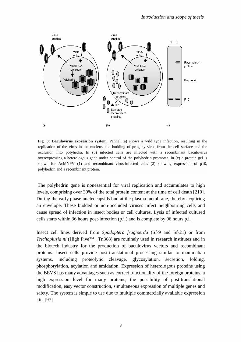
ig. 3: Baculovirus expression system. Pannel (a) shows a wild type infection, resulting in theeplication of the virus in the nucleus, the budding of progeny virus from the cell surface and thecclusion into polyhedra. In (b) infected cells are infected with a recombinant baculovirusverexpressing a heterologous gene under control of the polyhedrin promoter. In (c) a protein gel ishown for AcMNPV (1) and recombinant virus-infected cells (2) showing expression of p10,olyhedrin and a recombinant protein.
8
he polyhedrin gene is nonessential for viral replication and accumulates to highvels, comprising over 30% of the total protein content at the time of cell death [210].uring the early phase nucleocapsids bud at the plasma membrane, thereby acquiring
n envelope. These budded or non-occluded viruses infect neighbouring cells andause spread of infection in insect bodies or cell cultures. Lysis of infected culturedells starts within 36 hours post-infection (p.i.) and is complete by 96 hours p.i.
sect cell lines derived from Spodoptera frugiperda (Sf-9 and Sf-21) or fromrichoplusia ni (High Five™ , Tn368) are routinely used in research institutes and ine biotech industry for the production of baculovirus vectors and recombinant
roteins. Insect cells provide post-translational processing similar to mammalianystems, including proteolytic cleavage, glycosylation, secretion, folding,hosphorylation, acylation and amidation. Expression of heterologous proteins usinge BEVS has many advantages such as correct functionality of the foreign proteins, a
igh expression level for many proteins, the possibility of post-translationalodification, easy vector construction, simultaneous expression of multiple genes and
afety. The system is simple to use due to multiple commercially available expressionits [97].
Chapter 1
9
Baculovirus transfer vectorsTraditionally, recombinant baculoviruses were constructed in two steps. First, theheterologous gene was inserted into a transfer vector that contained a promoter,usually from the polyhedrin or p10 gene, flanked by baculovirus DNA. This plasmidwas then introduced into insect cells along with circular or linearized genomic viralDNA after which the generation of recombinants occurred. Several approaches havebeen developed to obtain recombinant baculoviruses easily and nowadays manymodified viral DNA’s [55, 90, 91, 109], and a huge variety of transfer plasmids areavailable [109, 117, 119, 164, 206]. Recent advances in the baculovirus expressionvector technology include improvements to methods for dominant selection ofrecombinant viruses [118, 166], development of the system for production of multi-subunit protein complexes and vectors for easy and improved heterologous proteinexpression and production (e.g. bacmid and baculovirus display technologies).Recombinant vectors have also been engineered for transient and stable genedelivery/transfer into a broad spectrum of primary and established mammalian andother cells [95, 97, 100]. The application of modified baculovirus for in vivo genedelivery has also been demonstrated [188].
The baculovirus surface display method, which mimics the phage display system [13]allows heterologous proteins expressed in the BEVS to be projected on the surface ofboth infected insect cells and budded virions by fusing the foreign protein to the majorbaculovirus envelope glycoprotein, GP64 [51]. This method has been usedsuccessfully for the expression of viral surface antigens [50, 105, 128, 194]. Thissystem has the capacity to produce near-native folding of recombinant protein [129].Bacmid technology provides a rapid and efficient method to generate recombinantbaculoviruses, and is based on site-specific transposition of a foreign gene into abaculovirus shuttle vector (bacmid) propagated in E. coli [108]. The generation ofrecombinant baculoviruses via the bacmid system eliminates the need for multiplerounds of plaque purification and reduces the time required to identify and purify arecombinant virus compared to the traditional methods involving homologousrecombination.
Scope of the thesisThe aim of this thesis is to obtain the Theileria parva sporozoite coat protein (p67) ina near-native form and to develop a recombinant p67-based subunit vaccine againstthe deadly cattle disease ECF. Previous reports have showed the potential of p67 as agood and broad-spectrum antigen for inclusion in the development of a subunitvaccine against ECF [134, 142, 143]. P67 has been expressed in BEVS before but theexpression level was very low and just a minor fraction of this product appeared to becorrectly folded [142]. In addition, p67 was not transported to the cell surface. In allthese reports, large amounts of the antigen ((300-600 µg)/inoculation) and more than

Introduction and scope of thesis
10
three inoculations were required to achieve a reasonable level of protection.Incorporating conformational epitopes may enhance the immunogenicity and efficacyof the p67-based subunit vaccine. Therefore, novel strategies have to be developedand incorporated into BEVS technology in order to express and produce heterologousproteins in more stable and correctly folded forms. In the first experimental chapter(Chapter 2) the aim is to overcome the low levels of p67 expression observed withfull-length p67. Two sets of constructs are analyzed, where p67 regions are expressedas non-fused entities or as fusions to the C-terminus of green fluorescent protein(GFP). With these constructs various regions of p67 are expressed as cytoplasmicpeptides. The idea behind this is that expressing full-length p67 via the secretorypathway may result in aggregation and bad processing of the recombinant protein.
In the next chapter the baculovirus surface display method is explored for theexpression of an N- and C-terminal domains of p67 (Chapter 3). The baculovirussurface display system was used to direct p67 through the export system to the outsideof the cell, hopefully with conservation of it’s native folding. To allow easygeneration and screening of recombinant viruses the surface display system iscombined with bacmid technology, enabling the generation of recombinant viralgenomes in bacterial cells.
Transmembrane proteins are in general produced in relatively low amounts in BEVS.Therefore, another way of improving expression levels of p67 is sought in producinga secretory, soluble form of p67 (Chapter 4) using baculovirus vectors. In order toachieve this a hydrophobic region is deleted from the C-terminus that most likelyserves as transmembrane domain, and the signal peptide is replaced by an insectsignal peptide, derived from honeybee melittin. Furthermore, the purification of largeamounts of p67 immunogens from the insect cell culture medium may be an easy-to-handle process for large-scale production of recombinant p67. In addition, baculovirusvectors with deletions in the chitinase and v-cathepsin genes are constructed in orderto prevent breakdown of the recombinant protein by viral proteases.
The next step is to test the p67 antigens that have good levels of expression and aproper folding for their immunogenicity (Chapter 5). Therefore, the various GFP-p67and GP64-p67 fusion proteins are tested in mice to select those antigens that give thehighest levels of sero-conversion and generate neutralizing antibodies. The bestimmunogens are subsequently investigated for their ability to induce neutralizingantibodies in cattle. Finally, based on the sero-conversion experiments, two vaccinecandidates are tested in an efficacy trial in cattle, which also includes two differenttypes of adjuvants, in order to determine the level of protection conferred (Chapter6). Finally the prospective of the new p67-based vaccines and additional questionsthat need to be addressed are discussed (Chapter 7).

Chapter 2
11
Chapter 2
Fusion to green fluorescent protein improves expressionlevels of Theileria parva sporozoite surface antigen p67
in insect cells
SUMMARY
East Coast fever (ECF) is a fatal disease of cattle caused by the protozoan parasiteTheileria parva. The development of a subunit vaccine, based on the sporozoite-specific surface antigen p67, has been hampered by difficulties in achieving highlevel expression of recombinant p67 in a near-authentic form. Therefore, two sets ofrecombinant baculovirus vectors were constructed. The first set, encoding variousregions of p67, produced low levels of the corresponding p67 domains in HighFiveTM cells, despite the presence of large amounts of p67 RNA. The second set,consisting of p67 domains fused to the carboxy-terminus of green fluorescent protein(GFP) expressed significantly higher levels of p67 protein. The GFP:p67 fusionproteins were recognised by a sporozoite-neutralising monoclonal antibody (TpM12)raised against native p67 whereas non-fused full length p67 was not recognised. GFP-tagging, therefore, appeared to enhance the stability of p67 and to conserve itsfolding. The high-level expression of p67 domains in a more authentic form is animportant step towards the development of an effective subunit vaccine against ECF.
This chapter has been published as : Kaba, S. A., Nene, V., Musoke, A. J., Vlak, J. M. andvan Oers, M. M., Parasitology (2002) 125, 497-505.
Improved expression levels of T. parva p67
12
IntroductionTheileria parva is a tick-transmitted protozoan parasite of cattle, which causes EastCoast fever (ECF). This disease is of major economic importance throughout east,central and southern Africa. The brown ear tick, Rhipicephalus appendiculatus,transmits the sporozoite stage of the parasite. The sporozoites enter host lymphocyteswhere they develop into intracellular multinucleate schizonts resulting in a fatallymphoproliferative and destructive disease [38, 148].
The sporozoite surface protein p67 synthesized during sporogony within the tick playsan essential role in the invasion process [35-37]. Monoclonal antibodies directedagainst p67 block [34] and soluble p67 competitively inhibits [177] invasion ofbovine lymphocytes by the sporozoites. P67 contains 709 amino acid residues and hasthe characteristics of a transmembrane protein. It has a secretory signal sequence atthe N-terminus, seven potential N-linked glycosylation sites and a hydrophobic C-terminal tail [141]. P67 is conserved among sporozoites isolated from different cattle-derived T. parva stocks [143] thus making it a good candidate for a broad-spectrumsubunit vaccine against ECF.
To investigate its vaccine potential, p67 has been cloned and expressed in Escherichiacoli [134] as a fusion to the non-structural (NS1) protein of influenza virus A. Cattleimmunised with NS1-p67 generated high titres of p67 specific antibodies with astrong in vitro neutralising activity against T. parva sporozoites. On challenge withLD70 of stabilated sporozoites, 70% of the immunised cattle were protected againstECF. A relatively high dose of NS1-p67 was required to achieve this level ofprotection. In addition NS1-p67 was not recognised by TpM12, a neutralisingmonoclonal antibody raised against native p67 [132] These observations indicate thatthe E. coli-expressed p67 was not in a native conformation or it lacked characteristicpost-translational modifications, such as glycosylation, which might be required forcomplete protective immune responses.
The baculovirus-insect cell expression system has been used in an attempt to expressp67 in more authentic forms with the proper folding and modifications [142].Attempts to generate a full-length p67 in insect cells were frustrated by the fact thatrecombinant p67 was produced at a low level, the product was only partiallyprocessed and not transported to the cell membrane. This insect cell-derived p67 wasessentially produced in in a non-native form. Cattle immunized with the baculovirus-derived p67 showed a response similar to animals immunized with the NS1-p67product. The lack of sufficient quantities of near-authentic recombinant p67 hampers

Chapter 2
13
the evaluation of the full potential of p67 as a vaccine [142]. Waldo et al. [209] firstsuggested that green fluorescent protein (GFP) might enhance stability of GFP fusionproteins. Indeed in prokaryotic expression systems, GFP-tagging increased bothstability and solubility of recombinant proteins [172]. Since the first expression ofGFP with recombinant baculoviruses [170], GFP has been widely used as afluorescence marker for gene expression, protein localization and trafficking, andprotein-protein interactions by fusing its coding sequence to that of the protein ofinterest [216].
In this study we sought to evaluate whether GFP fusion could enhance the stabilityand hence the expression levels of p67 in insect cells as well as induce/preserve itsnative conformation, by expressing various p67 sub-domains either as separate openreading frames (ORFs) or as C-terminal fusions to GFP. We report here the high-levelexpression of p67 domains in a more authentic form in insect cells.
Materials and Methods
Cells and virusesTrichoplasia ni High-FiveTM insect cells (Invitrogen) were maintained in Grace�ssupplemented insect medium (Invitrogen) with 10% foetal bovine serum (FBS) asmonolayer cultures at 27oC. For routine cell maintenance, virus infection andpropagation, standard procedures were followed [89, 193]. As control viruses theAutographa californica multicapsid nucleopolyhedrovirus (AcMNPV), strain E2[186], and the recombinant baculovirus, BEV-p67 [142] were used. BEV-p67 is arecombinant baculovirus carrying the complete p67 protein including its native signaland transmembrane sequences.
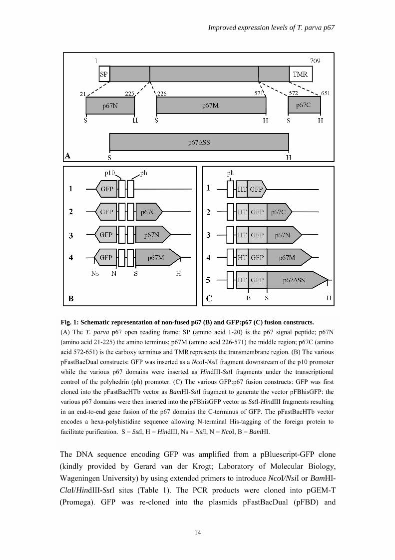
Construction of recombinant baculovirusesTheileria parva cDNA sequences (Fig 1A) encoding the N-terminus (p67N21-225), thecentral region (p67M226-571), the C-terminus (p67C572-651) as well as the full-lengthp67 protein (p67∆SS21-651) were PCR-amplified from a pMG1-p67 clone [134] usingextended primers that introduced BssHII/SalI/SstI or HindIII/ClaI restriction sites (seeTable 1). In contrast to the complete p67 protein expressed by recombinant BEV-p67[140], the p67∆SS domain excluded the authentic p67 signal and transmembraneanchor sequences. The PCR products were cloned into pGEM-T (Promega) andsequences of the inserts were verified by automated sequencing.
Improved expression levels of T. parva p67
14
The DNA sequence encoding GFP was amplified from a pBluescript-GFP clone(kindly provided by Gerard van der Krogt; Laboratory of Molecular Biology,Wageningen University) by using extended primers to introduce NcoI/NsiI or BamHI-ClaI/HindIII-SstI sites (Table 1). The PCR products were cloned into pGEM-T(Promega). GFP was re-cloned into the plasmids pFastBacDual (pFBD) and
Fig. 1: Schematic representation of non-fused p67 (B) and GFP:p67 (C) fusion constructs.(A) The T. parva p67 open reading frame: SP (amino acid 1-20) is the p67 signal peptide; p67N(amino acid 21-225) the amino terminus; p67M (amino acid 226-571) the middle region; p67C (aminoacid 572-651) is the carboxy terminus and TMR represents the transmembrane region. (B) The variouspFastBacDual constructs: GFP was inserted as a NcoI-NsiI fragment downstream of the p10 promoterwhile the various p67 domains were inserted as HindIII-SstI fragments under the transcriptionalcontrol of the polyhedrin (ph) promoter. (C) The various GFP:p67 fusion constructs: GFP was firstcloned into the pFastBacHTb vector as BamHI-SstI fragment to generate the vector pFBhisGFP: thevarious p67 domains were then inserted into the pFBhisGFP vector as SstI-HindIII fragments resultingin an end-to-end gene fusion of the p67 domains the C-terminus of GFP. The pFastBacHTb vectorencodes a hexa-polyhistidine sequence allowing N-terminal His-tagging of the foreign protein tofacilitate purification. S = SstI, H = HindIII, Ns = NsiI, N = NcoI, B = BamHI.

Chapter 2
15
pFastBacHTb (pFBhis) (Invitrogen). GFP was cloned as NcoI-NsiI fragmentdownstream of the p10 promoter in pFastBacDual generating the parental plasmid fornon-fused expression, pFBDp10GFP (Fig 1B; 1). In the pFastBacHT vector, GFP wascloned as BamHI-SstI fragment generating the parental plasmid for the GFP fusions,pFBhisGFP (Fig 1C; 1).
To generate non-fused constructs, p67C, p67N and p67M were cloned as SstI-HindIIIfragments downstream of the polyhedrin promoter in the pFBDp10GFP vector,generating the donor plasmids: pFBDp10GFP-php67C, pFBDp10GFP-php67N andpFBDp10GFP-php67M (Fig 1B; 2, 3, 4 respectively). Cloning of the p67 domains asSstI-HindIII fragments into the pFBhisGFP vector resulted in in-frame end-to-endgene fusion of the p67 domains to the C-terminus of the GFP, generating the GFP:p67fusion constructs pFBhisGFP:p67C, pFBhisGFP:p67N, pFBhisGFP:p67M andpFBhisGFP:p67∆SS (Fig 1C; 2, 3, 4, 5). Recombinant baculoviruses encoding thenon-fused p67 protein domains were generated via the Bac-to-BacTM baculovirusexpression system (Invitrogen), resulting in Ac-p67N, Ac-p67M, Ac-p67C and thecontrol virus Ac-GFP. The recombinant baculoviruses Ac-hisGFP:p67N, Ac-hisGFP:p67M, Ac-hisGFP:p67C, Ac-hisGFP:p67∆SS encoding the fusion proteinsand the control, Ac-hisGFP was also generated [108].
Protein analysisFor the analysis of recombinant protein expression, 1x10⁶ T. ni High FiveTM cellswere seeded into 35 mm2 Petri dishes and infected with the various recombinantviruses at a multiplicity of infection (MOI) of 10 plaque-forming units (PFU) per cell.At 72 h post infection (p.i.) cells were harvested and washed in 1-ml phosphatebuffered saline (PBS) at 4oC. Finally the pellet was resuspended in 100-µl ice-coldPBS. Protein concentrations were determined using Bradford reagent (BIO-RAD).Samples containing 2.5-µg total protein were analysed in 15% (non-fused samples) or10% (fusion proteins) SDS-PAGE gels as described by Laemmli, [98]. Proteins werevisualized by Coomassie Brilliant blue (CBB) staining or subjected to Westernanalysis. Monoclonal antibodies, ARIII 22.7 and ARIII 21.4 [144] specific for p67Nand C respectively, were used. The polyclonal antibodies, Rat 44 and bovine BJ 36[144] raised against the complete p67 protein were used for p67M and p67∆SSrespectively. All antibodies were used at a dilution of 1:200. For the GFP:p67 fusionproteins, monoclonal antibodies against the polyhistidine tag (Sigma) or a polyclonalantibody against GFP (both at a dilution of 1 : 5000) were used. As the secondantibody, rabbit anti-mouse immunoglobulins (for the monoclonals), anti-bovineimmunoglobulins (for BJ 36), or swine anti-rabbit immunoglobulins (for the αGFP)

Improved expression levels of T. parva p67
16
conjugated to horseradish peroxidase (HRP) (DAKO, A/S, Denmark) were used at adilution of 1 : 5000. The HRP substrate, 4-chloro-1-naphthol (Bio-Rad) was used todetect the p67 recombinant proteins.
Table 1: Oligonucleotides used for the amplification of p67 domains and GFP*
Name Sequence from 5’to 3’ Characteristics
SUN-1F: CGGATCCATCGATGCCATGG
GCAAAGGAGA
Forward primer for GFP.
Introduces BamHI and ClaI sites.
SUN-1R: CCAAGCTTGAGCTCTTCATC
CATGCATGTG
Reverse primer for GFP.
Introduces HindIII and SstI sites.
GFP/F: CATGCCATGGGCCATGGGCA
AAGGAGA
Forward primer for GFP.
Introduces NcoI site.
GFP/R: TGCATGCATTTCATCCATGCC
ATGTG
Reverse primer for GFP.
Introduces NsiI site.
TP67/21-F: GGCGCGCGTCGACGAGCTCA
TGCCTACGGAGGAACAACCAT
Forward primer for N-terminal
domain of p67. Introduces
BssHII, SalI and SstI sites.
TP67/225-R: CAAGCTTATCGATAAGATCTT
GGCCCGATGTAGTT
Reverse primer for N-terminal
domain of p67. Introduces
HindIII and Cla1 sites.
TP67/226-F: GGCGCGCGTCGACGAGCTCATG
AATTCAAAACAACAGCAAACTG
Forward primer for the middle
region of p67. Introduces
BssHII, SalI and SstI sites.
TP67/571-R: CAAGCTTATCGATTGCTGCTCGT
CCCGTACCTGAT
Reverse primer for the middle
region of p67. Introduces
HindIII, and Cla1 sites.
TP67/572-F: GGCGCGCGTCGACGAGCTCTGG
GAACGGGAGGGGGATCACTGAG
Forward primer for C-terminal
domain of p67. Introduces
BssHII, SalI and SstI sites.
TP67/651-R: CAAGCTTATCGATTCCAGCTGCT
ATTGTGGGCCCT
Reverse primer for C-terminal
domain of p67. Introduces
HindIII, and ClaI sites.
*Restriction sites are underlined

Chapter 2
17
Purification and dot blot analysisThe polyhistidine tag was exploited to purify the fusion proteins using TALONspinIMAC columns (CLONTECH Laboratories). High FiveTM insect cells were infectedwith the recombinant viruses at a MOI of 10 PFU per cell and collected at 72 h p.i.Cells were concentrated and clarified cytoplasmic extracts were applied to thecolumns. After washing with buffer (50 mM sodium phosphate, 300 mM NaCl, pH7.0) containing 25 mM imidazole to remove unbound and weakly bound proteins, thepolyhistidine-tagged GFP:p67 fusion proteins were eluted from the column byincreasing the imidazole concentration to 200 mM.
To examine the antigenic authenticity of the insect cell-derived GFP:p67 fusionproteins dot blot analysis was performed. Five micrograms of the purified GFP:p67fusion proteins and 10 µg of total cell lysate of infected T. ni High FiveTM insect cellswere spotted on a nitro-cellulose membrane and allowed to dry. Proteins weredenatured by adding 1/20 volume of β-mercaptoethanol and 1/4 volume of 4x samplebuffer (40 mM Tris-HCl pH 8.0, 4 mM EDTA, 8% SDS) and incubation at 95 oC for10-min. Duplicate blots were incubated with TpM12 (1:50) or ARIII 22.7 (1:200) for1 h at room temperature, washed and further incubated with goat anti-mouseimmunoglobulins conjugated with horseradish peroxidase (Amersham) at a dilution of1:5000. Enhanced chemiluminescence (ECL) (Amersham) was used for detection.The insect cell-derived p67 expressed by Nene et al. [142], indicated as BEV-p67,was compared with these GFP:p67 fusion proteins. The same amount (5 µg) ofpurified E. coli-expressed p67N, and his: GFP proteins as well as mock and Ac-wtinfected cell lysate containing 10 µg total protein were used as controls.
Transcript analysisTrichoplusia ni High FiveTM insect cells (1.0 x 10⁶) were infected with therecombinant viruses Ac-p67N, Ac-p67M, Ac-p67C, and Ac-GFP at a MOI of 10pfu/cell and harvested at 24, 48 and 72 h p.i. Total RNA was isolated using the single-step acid guanidinium thiocynate-phenol-chloroform RNA extraction methoddescribed by Chomczynski and Sacchi [24]. Five µg RNA were resolved in 1.4 %agarose gel and Northern blot analysis was carried out as described in Pellé &Murphy [156]. The RNA was fixed to the filter by a UV light cross-linking. The filterwas hybridized to a PCR-amplified p67 (full length) probe, labeled with α32P-dCTPby random primed labeling with Klenow (Promega).

Improved expression levels of T. parva p67
18
Results
Expression of p67 sub-domains in insect cellsTo enhance the expression levels of p67 in insect cells, sub-regions of p67 rather thanthe complete p67 protein [142] were expressed. The domains of p67 expressed in thisstudy were an N-terminal domain (p67N21-225), the central region (p67M226-571) and aC-terminal domain (p67C572-651) as well as the full-length protein without its nativesignal sequence and transmembrane region (p67∆SS) (Fig. 1A). The p67N, p67Mand p67C domains were cloned and expressed as separate open reading frames (non-fused constructs) (Fig. 1B). In a second experiment, each p67 domain was fused to theC-terminus of GFP and expressed as a fusion protein (Fig. 1C).
Trichoplusia ni High FiveTM cells infected with recombinant viruses encoding thesedifferent p67 sub-domains s (non-fused and fused) were analysed in SDS-PAGE andstained with Coomassie Brilliant blue (CBB) or subjected to immunoblot analysis. Inthe Coomassie-stained gel of the non-fused p67 recombinants the p67 sub-domainsp67N, p67M and p67C could not be detected (Fig. 2A, lanes 5-7). A protein with theexpected electrophoretic mobility for GFP was observed for all recombinants (lanes 4-7). The medium of cells infected with Ac-GFP, Ac-p67N, Ac-p67M and Ac-p67Cturned green as early as 24 h p.i. GFP expression was confirmed by the greenfluorescence of the protein under UV-light and by Western blot analysis using αGFPpolyclonal antibody (DAKO) (results not shown), indicating that the infection hadbeen successful. These results indicate that the expression levels for the sub-domainswere not significantly increased compared to the full-length p67 expressed in insectcells (lane 3).
In immuno-blot analysis A reaction with p67 antibodies was observed for BEV-p67which was absent in the mock, Ac-wt and Ac-GFP samples (Fig. 2B, lanes 1-4). Themonoclonal antibodies, ARIII 22.7, RAT 44 and ARIII 21.4, directed against p67N,p67M and p67C, respectively, showed that p67N, p67M and p67C proteins wereexpressed but at low levels and in unstable forms as indicated by the observation ofdegraded products (Fig. 2B, lanes 5-7). The major product of p67N was detected as a45.0 kDa protein instead of the expected 22.0 kDa (Fig. 2B, lane 5). In Ac-p67Minfected cells a 38-kDa protein was detected as expected for p67M protein (Fig. 2Blane 6). P67C was detected as two separate major proteins of 8.6 kDa, the expectedmobility for p67C, and of 28.0 kDa, three times the predicted size of p67C (Fig. 2B,lane 7).

Chapter 2
Fig. 2: SDS-PAGE and Western blot analysis of non-fused p67 proteins. Trichoplusia ni High FiveTM insectcells were mock-infected (lane 1) or infected with Ac-wt virus (lane 2) or recombinant viruses BEV-p67 (lane 3),Ac-GFP (lane 4), Ac-p67N (lane 5), Ac-p67M (lane 6), Ac-p67C (lane 7) and harvested at 72 h p.i. Total protein(2.5 µg) was resolved in 15% SDS-PAGE and stained with Coomassie Brilliant blue (A) or subjected toimmunoblot analysis using the p67N-specific monoclonal antibody ARIII 22.7, the p67M-specific polyclonalantibody Rat 44 or the p67C-specific monoclonal antibody ARIII 21.4 (B).
19
To investigate whether the low level of expression of p67 domains observed was dueto low transcription, we examined p67 transcripts at different times post infection.Total RNA was purified from mock-infected and cells infected with Ac-wt, Ac-p67N,Ac-p67M and Ac-p67C viruses and subjected to Northern blot analysis using a p67-specific probe. The p67 probe did not hybridise to mRNA purified from mock andAc-wt infected cells (Fig. 3 lanes 1 & 2). In the recombinant virus-infected cells, p67transcripts were detected at 24 h p.i. and the amount increased at 48 h p.i. Transcriptswere still observed at 72 h p.i. (Fig. 3, lanes 3-5). The large amount of transcripts was,however, not reflected in the level of p67 proteins observed (see Fig. 2A), suggestingthat the low level of expression of p67 sub-domains might be due to instability of theend products. To overcome this problem, p67 sub-domains were expressed ascarboxy-terminal fusion proteins to GFP (Fig. 1C). T.ni High FiveTM cells wereinfected with the recombinant baculoviruses, Ac-hisGFP, Ac-hisGFP:p67N, Ac-hisGFP:p67M, Ac-hisGFP:p67C, Ac-hisGFP:p67∆SS and total proteins from infectedcell lysates were resolved in SDS-PAGE and stained with CBB or subjected toimmunoblot analysis. In the control lanes no p67 specific proteins were observed (Fig.

Improved expression levels of T. parva p67
4A, lanes 1-4). A 31-kDa protein corresponding to the predicted size of hisGFP wasobserved for Ac-hisGFP (Fig. 4A, lane 4). With the fusion constructs, significantlyhigher-levels of expression of p67 recombinant fusion proteins were observed in theCBB-stained gel (Fig 4A, lanes 5-8) as compared to the non-fused proteins. ThehisGFP:p67N fusion protein (lane 5) migrated as a 75-kDa protein as opposed to apredicted size of 53-kDa. HisGFP:p67M was expressed in two forms, the expected69-kDa protein and a 110-kDa protein (Fig. 4B, lane 6). HisGFP:p67C was expressedas a 39 kDa protein (Fig 4B, lane 7) and hisGFP:p67∆SS as a 110 kDa protein(Fig. 4B, lane 8) as expected.
A(iopd
CTfcAa
Fig. 3: Northern blot analysis of non-fused p67 transcripts. Total RNA (5 µg) was extracted frommock-infected T. ni High FiveTM insect cells (lanes 1), or cells infected with Ac-wt virus (lanes 2) orthe recombinant viruses Ac-p67N (lanes 3), Ac-p67M (lanes 4), Ac-p67C (lanes 5) at 24 h, 48 h, or72 h p.i. Total RNA was resolved in a 1.4 % agarose gel and transferred to a Nylon filter. The filter washybridized to a radioactively labeled p67 DNA probe. The sizes of an RNA marker are indicated inkilobases.
20
monoclonal antibody directed against the N-terminal hexa-histidine tagCLONTECH) was used to confirm expression of all the fusion proteins in anmmuno-blot analysis. All the fusion proteins were detected at the same size asbserved in the protein gel (Fig.4B lanes 4-8). Western blot analysis using either aolyclonal antibody against GFP or monoclonal antibodies directed against theifferent p67 domains gave similar results (not shown).
onformation of p67 fusion proteinshe GFP-p67 fusion proteins were purified and the SDS-PAGE analysis of eluted
ractions showed that the polyhistidine tagged proteins were efficiently bound to theolumn and that the fractions contained highly purified proteins (results not shown).n immuno-dot blot analysis was performed to investigate whether the GFP-tagging
ffected the folding of the p67 domains in the fusion protein. A monoclonal antibody,

Chapter 2
21
TpM12, raised against native p67, and which does not recognise E.coli derived p67[132, 134], was used as the first antibody in this analysis. Purified proteins as well astotal cell lysate of cells infected with hisGFP:p67N and hisGFP:p67∆SS were spottedon the blot.
As controls, we used hisGFP, BEV-p67 and E. coli-derived p67N as well as a mock-infected cell lysate. Both denatured and non-denatured samples were analysed. As acontrol measure a duplicate blot was incubated with the monoclonal antibody, ARIII22.7, that is raised against denatured p67 and which recognises a linear epitope in theN-domain [144]. TpM12 did not react with proteins (both denatured and non-denatured) in the mock, the BEV-p67, E. coli-derived p67N and the hisGFP samples(Fig. 5A, lanes 1-4). TpM12 reacted with non-denatured but not with the denaturedsamples of hisGFP:p67N and hisGFP:p67∆SS (Fig. 5A, lanes 5 & 6), indicating that
Fig. 4: SDS-PAGE and Western blot analysis of GFP:p67 fusion proteins. T. ni High FiveTM insectcells (lane 1) were infected with the Ac-wt virus (lane 2), or recombinant viruses BEV-p67 (lane 3),Ac-hisGFP (lane 4), Ac-hisGFP:p67N (lane 5), Ac-hisGFP:p67M (lane 6), Ac-hisGFP:p67C (lane 7),Ac-hisGFP:p67∆SS (lane 8) and harvested at 72 h p.i. Proteins (2.5 µg) were resolved in 10% SDS-PAGE and stained with Coomassie Brilliant blue (A) or subjected to immunoblot analysis using amonoclonal antibody against the hexa-histidine tag (B). The dots (in A) indicate the various GFP fusionproteins.

Improved expression levels of T. parva p67
the GFP-p67 fusion proteins were expressed in a near-native conformation. All theprotein samples (both denatured and non-denatured) except for the negative controlsreacted with ARIII 22.7 (Fig. 5B, lanes 1-6), confirming that p67 specific proteins hadbeen spotted on all blots.
DLai1l
Fig. 5: Immuno-dot blot analysis of hisGFP:p67 fusion proteins. Both non-denatured anddenatured purified proteins (A and B) or total cell lysate (C) of hisGFP:p67N and hisGFP:p67∆SSwere spotted on a nitrocellulose membrane and subjected to immunoblot analysis using monoclonalTpM12 or ARIII 22.7. TpM12 was raised against native p67 while ARIII 22.7 was raised againstE.coli- derived p67.
22
iscussionow expression levels of T. parva p67 were observed by Nene et al. [142] in theirttempt to produce large amounts of p67 in a more authentic form via the baculovirus-nsect cell expression system. In the present study we expressed sub-domains (Fig.A) of T. parva sporozoite p67 instead of the complete p67 protein to obtain higherevels of expression. First, we expressed p67 sub-domains as non-fused (Fig. 1B)

Chapter 2
23
proteins and in a second experiment, as fusions to the C-terminus of GFP (Fig. 1C). Inboth groups the native p67 signal peptide and transmembrane region were eliminatedas this might limit expression levels [102]. Pepscan and computer analyses havepredicted that antigenic sites of p67 were located in the N- and C-terminal regions[92, 144]. Bovine antibody responses to p67 are restricted to these two domains [92,144]. Although the middle region of p67 does not appear to have linear B-cellepitopes, it contains several Th-cell epitopes [136]. The middle region was included inthis study since it could be used to define T-cell epitopes when it becomes necessaryto investigate the protective capacity of peptide-based vaccines in the future.
The non-fused proteins were expressed at the same low level (Fig. 2A) as the full-length p67 protein [142]. This may be due to inefficient translation of p67 transcriptsor the instability of nascent p67 proteins since abundant transcripts were detected byNorthern blot analysis (Fig. 3). A similar problem was seen in E. coli, where p67expressed as fusion to the glutathione-S-transferase of Schistosoma japonicum [141]and the non-structural protein-1 (NS1) of influenza virus A [134] have failed toproduce stable recombinant p67 protein.
A significantly higher level of expression of the p67 domains was obtained whenfused to GFP, relative to the non-fused as well as full-length p67 [142]. Fusion to GFPeither enhanced translation or, more likely, improved the stability of the p67 nascentprotein products, thereby increasing the yield of p67 expression in insect cells. GFPhas been shown to enhance the stability and increase the expression of fusion proteinsin prokaryotic expression systems [172, 209]. Another indication that GFP couldstabilise a fused protein was obtained by Akgul et al. [1] when they deleted astability-regulating motif (the PEST sequence) from the Mc-1 protein [173, 175] butdid not find any effect on the stability of a Mcl-GFP fusion protein.
The observed molecular mass of the hisGFP:p67N, hisGFP:p67M andhisGFP:p67∆SS recombinant fusion proteins in SDS-PAGE was considerably higherthan the calculated value. For example the apparent molecular mass of hisGFP:p67Nwas 75 kDa as opposed to the predicted size of 53 kDa. The exact reason for theanomalous behavior of these recombinant proteins in SDS-PAGE is not clear. It isknown that p67 undergoes N-glycosylation in insect cell [142], but this could notexplain the present observation since the signal peptide of p67 was eliminated in theseconstructs. The primary amino acid sequence of p67 may be responsible for theaberrant mobility since a similar difference between the observed and calculatedmolecular mass was observed for the non-fused p67 domains (Fig. 2B). Similarobservations have been made with recombinant p67 production in both E. coli and

Improved expression levels of T. parva p67
24
insect cells [141, 142]. A recombinant p67 in a near authentic form is highly desirableas it might function as a better immunogen than the previously tested NS1-p67 [134,142]. An immuno dot-blot analysis using a monoclonal specific for native p67indicated that GFP fusions of p67N and p67∆SS had a near native folding, in contrastto the full length p67 expressed in insect cells in a non-fused form.
GFP is being used extensively as a visible marker in cell biology because it operatesindependently of cofactors and can be detected rapidly and easily. Due to its smallsize, GFP does not significantly increase the size of the chimeric protein neither doesit interfere in general with the biological functions of even small proteins [23, 189,190, 201, 209]. In this case, GFP fusion not only facilitated direct detection andmonitoring of infected insect cells, and titration of recombinant viruses, it alsoincreased expression levels of a recombinant protein, and appeared to conserve itsnatural folding properties. The high-level expression of near-authentic p67 domainsis an important step towards the development of an effective subunit vaccine againstECF. Investigation of the immunogenic properties of these products and determiningthe level of protection against ECF in cattle will be the next step towards evaluatingthe vaccine potential of these GFP-p67 fusion proteins.
AcknowledgementThe authors are thankful to Arno Vermeulen and Dick Schaap (IntervetInternational BV) for their invaluable advice. We are thankful to Magda Usmany,Stephen Wanyoni, Julius Osasa and James Gachanja, for their technical assistance andAart van t' Oever for purifying the fusion proteins. Stephen A. Kaba was financed bythe Netherlands Foundation for the Advancement of Tropical Research (WOTRO).

Chapter 3
25
Chapter 3
Baculovirus surface display of Theileria parva p67antigen preserves the conformation of
sporozoite-neutralizing epitopes
AbstractTheileria parva is an intracellular protozoan parasite that causes East Coast fever, asevere lymphoproliferative disease in cattle. Previous attempts to producerecombinant sporozoite surface antigen (p67) in bacterial or insect cells for vaccinepurposes have not resulted in a correctly folded protein. Here, we report theexpression of N- and C-terminal domains of p67 fused to the baculovirus envelopeglycoprotein GP64 by cloning the appropriate p67 cDNA segments between the signalsequence and the major portion of GP64. To further advance the generation of suchrecombinants, existing surface display techniques were combined with bacmidtechnology. Chimeric proteins were present on the surface of budded viruses asjudged by immunogold labelling and were exposed on the surface of insect cells, asconcluded from immunofluorescence studies of infected, non-fixed insect cells. Innon-denaturing dot blot experiments, a strong reaction was obtained betweenmonoclonal TpM12 and baculovirus particles displaying the p67N-GP64 chimericprotein. This antibody, raised against native p67, also specifically recognised thesurface of recombinant-infected cells. Apparently, a more native conformation wasachieved than when p67 was expressed in E. coli or in conventional baculovirusexpression systems. The baculovirus surface expression system, therefore, provides animproved way of expressing this T. parva sporozoite surface protein.
This chapter has been published as Kaba, S. A., Hemmes, J. C, van Lent, J. W. M.,Vlak, J. M., Nene, V., Musoke, A. J., and van Oers, M. M. in Protein Engineering (2003) 16,73-78.

Baculovirus surface display of T. parva p67
26
IntroductionThe protozoan parasite Theileria parva is the causative agent of East Coast fever(ECF), a lymphoproliferative disease of cattle with a high mortality rate. The brownear tick, Rhipicephalus appendiculatus, transmits the parasite during feeding. Incattle, the infecting sporozoites develop into schizonts, which induce transformationof lymphocytes [38]. Clinical signs such as pyrexia, lymphoadenopathy andpanleukopenia are associated with the invasion of parasitized lymphoblasts intobovine tissues [75]. The current way of immunization of cattle involves vaccinationwith cryopreserved sporozoites and simultaneous treatment with long-acting antibiotictetracyclines. The immunity achieved in this way, though often strain-specific, lastsfor at least three years [124].
Immune responses against the infecting sporozoite and pathogenic schizont stagesplay a major role in mediating protection and immunity to ECF [114]. Cattle thatrecover from infection mount immunity to homologous strains. This immunity isthought to be dependent on a cellular immune response mediated by class I MHC-restricted CD8+ cytotoxic T-lymphocytes, and directed against schizont-infectedlymphoblasts [114, 124]. On a single exposure, animals do not mount a detectableanti-sporozoite response, but on repeated challenge with infected ticks cattle generatehigh levels of antibodies that neutralize sporozoites in vitro [132]. These antibodiesrecognize p67, the major surface protein of sporozoites. A recombinant form of thisantigen produced in bacteria has shown to provide protection in cattle [134]. Becausep67 is invariant in parasites isolated from cattle and exhibits 76-96% sequenceidentity with stocks from buffalo, the antigen has a clear potential for development ofa sub-unit vaccine against ECF. Pepscan analysis revealed that both murine andbovine epitopes cluster in the N- and C-terminal regions. Linear bovine B cellepitopes mapped within residues 25 to 296, and 577 to 591 while residues 105 to 221contained three epitopes defined by neutralizing monoclonal antibodies. A furthertwo epitopes were located in the C- terminal fragment between residues 617 and 631[144].
Expression of p67 in E. coli as a fusion protein to influenza NS1, resulted in aninsoluble protein [134]. In insect cells infected with a conventional recombinantbaculovirus, p67 was expressed at low levels and mainly in a non-nativeconformation, and was not transported to the cell surface [142]. The protectionachieved by these two recombinant proteins was only partial and relatively high dosesof antigen were needed for induction of protective immunity. We believe thatincorporating conformational epitopes would enhance the efficacy of the vaccine.
In this paper, we have expressed domains of p67 on the surface of buddedbaculovirus particles (BVs) in an attempt to achieve a more native folding of

Chapter 3
27
immunodominant epitopes. The baculovirus surface display method [18, 51] is basedon the expression of foreign proteins fused to the baculovirus GP64 protein. Thismajor BV envelope protein is responsible for fusion of the viral envelope withendosomes of the insect host and for virus budding from infected insect cells [122,152]. Baculovirus surface display has been used previously for the expression of viralsurface antigens, such as HIV GP41 and GP120, rubella virus spike proteins andFMDV structural proteins [18, 50, 128, 194]. Indications for a near-native folding ofrecombinant proteins produced in this system have been given by the display offunctional scFv fragments [129].
To allow easy generation and screening of recombinant viruses, we combined theexisting surface display system with bacmid technology [108], enabling thegeneration of recombinant viral genomes in bacterial cells. This is the first report ofthe expression of a parasite antigen on the surface of baculovirus particles and mayopen new venues to develop vaccines against parasitic diseases.
Materials and Methods
Construction of transfer vectorsThe coding sequence for enhanced GFP [25] was cloned as a NcoI-NsiI fragmentdownstream of the p10 promoter in the pFastBacDual vector (Invitrogen Breda, TheNetherlands). The KpnI restriction site in the multiple cloning site (MCS) downstreamof the p10 promoter was removed by digesting with NsiI and KpnI, and filling in withKlenow. After self-ligation, this resulted in the vector pFBD-GFP-∆KpnI. In this way,we were able to use a unique KpnI cloning site between the gp64-signal peptide andmajor domain (see Figure 1). A p67 N-terminal domain corresponding to amino acids21 to 225 was obtained by PCR from plasmid pMG1-p67 [134] with primers surf-3and surf-4, generating KpnI sites (Table I, Figure 1A). The C- terminal domain ofp67, encoding amino acids 572 to 651, was amplified with primers surf-1 and surf-2to introduce KpnI sites on both ends (Table I, Figure 1A). The PCR products wereverified by sequencing. The C-terminal domain was cloned into the KpnI site ofpBACSurf-1 (Novagen) to give plasmid pBACSurf-p67C. With this plasmid astemplate, the p67 C-domain flanked by the GP64 leader sequence (86 bp) and theGP64 major domain (1511 bp) was amplified using primers surf-5 and surf-6 (TableI) and the Expand Long Template PCR system (Roche). The PCR product was clonedbetween the EcoRI and HindIII restriction sites in the MCS of pFBD-GFP-∆KpnI,resulting in pFBD-GFP-GP64/p67C (Figure 1B). The sequence was verified.Removing the p67 C domain by KpnI digestion and replacing it with the N-domaingenerated plasmid pFBD-GFP-GP64/p67N. Self-ligation after KpnI digestion resultedin an empty vector, pFBD-GFP-GP64, which was used as a control in the experimentsdescribed below.

Baculovirus surface display of T. parva p67
28
Fig. 1: Schematic representation of gp64/p67 chimeric constructs. (A) The Theileria parva p67open reading frame in which the signal peptide (SP), and N and C-terminal domains, and atransmembrane region (TMR) are indicated. The N- and C-terminal domains were amplified as KpnIfragments by PCR. The numbers refer to the corresponding position in the p67 amino acid sequence.(B) The various pFastBacDUAL constructs, in which N- (1) and C-terminal (2) domains of T. parvap67 were cloned between the GP64 signal peptide (SP) and the GP64 mature domain locateddownstream of the polyhedrin (ph) promoter. An ‘empty’ construct was made to serve as a control (3).The green fluorescent protein (GFP) sequence was cloned in the opposite orientation under control ofthe p10 promoter. Restriction sites used for cloning are indicated as follows: E: EcoRI, H: HindIII;K: KpnI. (C) Predicted amino acid sequence of the primary translation products for the three constructsshown above. The first 20 amino acids represent the signal sequence and will be cleaved off by acellular signal peptidase. Small spacer sequences are present between the different domains, asindicated.
Generation of bacmids and recombinant virusesEscherichia coli DH10BAC cells (Invitrogen Life Technologies) were transformedwith the plasmids pFBD-GFP-gp64/p67N, pFBD-GFP-gp64/p67C or pFBD-GFP-gp64 to generate recombinant AcMNPV bacmids. Putative recombinant bacmids wereanalyzed by PCR using the M13 reverse and surf-5 primers. Isolated bacmid DNA was

Chapter 3
29
used to transfect Spodoptera frugiperda Sf21 cells [205], using Cellfectin (InvitrogenLife Technologies). This resulted in the recombinant viruses Ac-gp64/p67N, Ac-gp64/p67C and Ac-gp64. Recombinant viruses were grown to high titre stocks usingstandard procedure [89].
Table I. DNA sequence of oligonucleotides*Primername
Sequence 5’ to 3’ Details
Surf-1 CCGGTACCAGGAACGGGAGGGGGATC Forward primer for amplificationof p67-C. Introduces KpnI site.
Surf-2 CCGGTACCCCTTCTCCTCCAGCTGCTATTG
Reverse primer for amplificationof p67-C. Introduces KpnI site.
Surf-3 CGGTACCCATGCCTACGGAGGAACAACC Forward primer for amplificationof P67-N. Introduces KpnI site.
Surf-4 CGGTACCGGAAGATCTTGGCCCGATG Reverse primer for amplificationof p67-N. Introduces KpnI site.
Surf-5 GGGAATTCCAAGCAAGATGGTAAGC Forward primer for amplificationof gp64, anneals to gp64 signalsequence. Introduces EcoRI site.
Surf-6 CCAAGCTTAATAAATGTACTAATAACCC Reverse primer for amplificationof gp64, anneals to majordomain. Introduces HindIII site.
*Restriction sites are underlined.
Immunofluorescence studies with non-fixed cellsSf21 cells were grown in Grace’s supplemented medium containing 10% FBS(Invitrogen Life Technologies). Sf21 cells were infected with Ac-GP64, Ac-gp64/p67N and Ac-GP64/p67C at a multiplicity of infection (m.o.i.) of 10 tissueculture infection dose 50 (TCID50) units/cell. The cells were harvested and collectedin 2 ml of Grace’s supplemented medium with 10% FBS at 48 h post infection (p.i.).Infected non-fixed cells were incubated in this medium with monoclonal antibodiesARIII 22.7, recognising the N-domain of p67, or with ARIII 21.4, specific for the C-domain [144] at a dilution of 1 : 200 for 1 h at room temperature. Cells were washedthree times with Grace’s supplemented medium containing 10% FBS. Cells werefurther incubated with goat-anti-mouse IgG conjugated to Rhodamine Red X(Molecular Probes) for 1 h at a 1 : 200 dilution. Similar studies were also performedwith monoclonal TpM12 at a 1 : 50 dilution. After extensive washing, the insect cellswere viewed in a Zeiss LSM510 confocal laser-scanning Microscope (LSM510). GFPfluorescence was observed through excitation with blue laser light at 488 nm andemission through a 505-530 nm bandpass filter. Rhodamine was simultaneouslyvisualised using green laser light at 545 nm for excitation and a 560 nm longpass filterfor emmision.

Baculovirus surface display of T. parva p67
30
Analysis of budded virionsSf21 cells were infected with the various recombinant viruses at a m.o.i. of 10 TCID50
units/cell and harvested 48 h p.i. Cell debris was removed by centrifugation at 3000rpm for 5 min and filtration over a 0.45 µm non-pyrogenic filter. The filtrate wasoverlaid onto a 2.5 ml 25% sucrose cushion in 1 mM Tris, 0.01 mM EDTA, pH 8.0.(0.1 TE). Budded viruses (BVs) were pelleted by centrifugation at 25,000 rpm for 90min at 4°C in a SW41 rotor. The pellet was suspended in 0.1 TE. Purified BVs wereused for SDS-PAGE and dot blot analysis. Western blots were incubated with eithermonoclonal AcV5 recognising GP64 [63] diluted 1:1000, or monoclonals ARIII 22.7or ARIII 21.4, diluted 1 : 200 and recognising p67 N- (amino acids 201-215) and C-specific (609-623) peptides, respectively [144]. Rabbit-anti-mouse immunoglobulinsconjugated to horse-radish peroxidase (Dako A/S) were used as second antibody.
For immuno-dot blot analysis, a sample of the budded virus preparation of Ac-gp64/p67N or Ac-gp64 equivalent to 5 µg of total protein was spotted ontonitrocellulose membrane, either directly or after denaturation by boiling for 10 min in10 mM Tris.HCl pH 8.0, 1 mM EDTA, 2% SDS and 5% β-mercaptoethanol. Thefilters obtained were incubated as described above using either monoclonal TpM12 orARIII 22.7 [144]. These blots were developed using ECL (Pharmacia). As a control,Sf21 cells were infected with an AcMNPV recombinant expressing non-fused p67 and5 µg total cell protein was blotted under both denaturing and native conditions (datanot shown).
For immunogold labelling, Sf21 cells were infected with the various recombinants inGrace’s medium without any supplements and the culture supernatant was replaced at20 h p.i. The supernatant was collected at 36 p.i. and cleared from cell debris bycentrifugation at 1000 rpm. The virus suspension was attached to nickel grids. Afterblocking in 1% (w/v) BSA in PBS for 20 min, grids were incubated for 1.5 h with themonoclonal antibodies ARIII 22.7 (N-specific), ARIII 21.4 (C-specific) or AcV1(specific for GP64), all in a 1 : 200 dilution. The grids were then washed on 6 drops of20 µl PBS-BSA for 5 min each and further incubated for 1 h with RAM coupled to 7nm gold particles (Aurion). The grids were washed with PBS and negatively stainedwith 2% (w/v) uranyl acetate, pH 3.9 and examined in a Philips CM12 transmissionelectron microscope.
Results and Discussion
In the standard baculovirus expression system, the Theileria parva sporozoite surfaceprotein p67 was expressed in low amounts in intracellular compartments and largelyin a non-native form [142]. To overcome this hurdle, domains of p67 were inserted inframe into the open reading frame of the baculovirus GP64 protein between thecoding sequences for the GP64 signal peptide and the GP64 mature protein (Figure 1).

Chapter 3
31
The GP64 signal peptide is used to route the recombinant protein via the secretorypathway to the plasma membrane, while the GP64 portion directs the chimeric proteinto the envelope of budded viruses. Since the antibody responses of bovine serum top67 are restricted to N- and C-terminal domains [144], these two domains wereconsidered as good starting points to test the baculovirus surface display technique forT. parva p67. The p67 N-domain comprises amino acid residues 21-225, therebyexcluding the original p67 signal peptide (Figure 1A). The C-domain consists ofresidues 572-651 and does not contain the p67 transmembrane region, since the GP64portion provides a functional analogue.
To avoid tedious rounds of plaque purification, recombinant viruses were generatedusing bacmid technology [108]. To this end, the GP64 sequences were amplified fromthe pBacSurf-1 vector (Novagen) and cloned downstream of the polyhedrin promoterbetween the EcoRI and HindIII sites of the pFastBacDUAL vector (LifeTechnologies). A KpnI site, situated downstream of the signal sequence, was used tointroduce the p67 N-and C-terminal domains (Figure 1B, 1C). The coding sequencefor enhanced GFP was cloned downstream of the p10 promoter in order to followtransfection and infection processes, and to guarantee a simple read-out in virustitrations.
It has been reported that fusion of proteins to GP64 may adversely affect viralproduction or infectivity [18]. This appears to be dependent on the size of the protein,since larger proteins are more likely to interfere with GP64 trimer formation [151] andhence with incorporation into the virus particle. Apparently, since the titers obtainedfor the various viral stocks were within the normal range, the expression of GP64-p67chimeric proteins did not significantly disturb virus budding and membrane fusion.
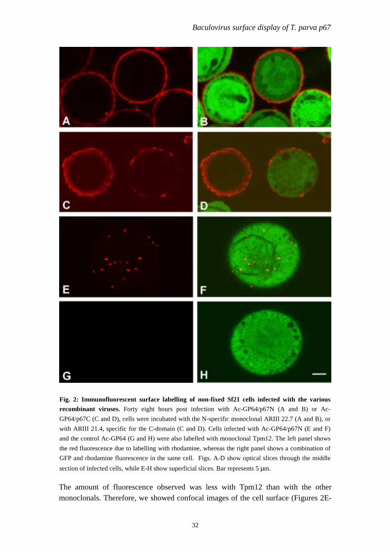
Immunofluorescence studies were performed in Grace’s medium on non-fixed, livingSf21 cells. In this way antisera could only reach p67 domains which were exposed onthe surface of the cells. With monoclonals specific for the N- and C-domains (ARIII22.7 and ARIII 21.4, respectively), and with a second antibody conjugated torhodamine Red X, a strong red peripheral fluorescence was observed (Figures 2A andC), which surrounded the cytoplasmic green GFP fluorescence (Figures 2B and D). Incells infected with the control Ac-GP64, only the green GFP fluorescence wasobserved with both monoclonal antibodies (data not shown). Cell surface expressionhas also been reported by Tami et al., [194], when expressing FMDV structuralproteins fused to GP64. To analyse the conformation of p67N on the cell surface,monoclonal TpM12 was used in immunofluorescence studies with recombinant-infected non-fixed cells. A red fluorescence was observed at the cell surface for cellsinfected with Ac-GP64/p67N (Figures 2E and F), indicating that a conformationalepitope in p67N was conserved.
Baculovirus surface display of T. parva p67
Tm
FrGwatGs
ig. 2: Immunofluorescent surface labelling of non-fixed Sf21 cells infected with the variousecombinant viruses. Forty eight hours post infection with Ac-GP64/p67N (A and B) or Ac-P64/p67C (C and D), cells were incubated with the N-specific monoclonal ARIII 22.7 (A and B), orith ARIII 21.4, specific for the C-domain (C and D). Cells infected with Ac-GP64/p67N (E and F)
nd the control Ac-GP64 (G and H) were also labelled with monoclonal Tpm12. The left panel showshe red fluorescence due to labelling with rhodamine, whereas the right panel shows a combination ofFP and rhodamine fluorescence in the same cell. Figs. A-D show optical slices through the middle
ection of infected cells, while E-H show superficial slices. Bar represents 5 µm.
32
he amount of fluorescence observed was less with Tpm12 than with the otheronoclonals. Therefore, we showed confocal images of the cell surface (Figures 2E-

Chapter 3
2F) instead of optical slides through the middle of the cell. TpM12 did not react withcells infected with the Ac-GP64 control (Fig. 2G and H).
TocTmswip83amoa
Fig. 3: Western blot analysis of AcMNPV recombinants. BVs were harvested from cells infectedwith either Ac-gp64, Ac-GP64/p67N or Ac-GP64/p67C and subjected to SDS-PAGE followed byWestern blot analysis. In panel A, the filter was incubated with a monoclonal antibody recognising theAcV5 epitope of GP64. In panel B and C, monoclonals specific for the N- (ARIII 22.7), and the C-terminal (ARIII 21.4) domains were used, respectively.
33
o determine whether the GP64-p67 chimeric proteins were incorporated into non-ccluded virus particles, budded virions were harvested from infected insect cellultures by centrifugation through a sucrose cushion (see Materials and Methods).he samples obtained were analysed by Western blotting (Figure 3). Labelling with aonoclonal antibody that is directed against the GP64 AcV5 epitope (Figure 3A)
howed a protein of approximately 67 kDa in all recombinant viruses representingild type GP64, expressed from the GP64 promoter. In the control Ac-GP64 this band
s stronger due to over-expression of GP64 (Figure 3A, lane 1) driven by theolyhedrin promoter. In Ac-gp64/p67N budded viruses an additional protein of about4 kDa was present, which was also observed with an N-specific monoclonal (FiguresA and B, lanes 2). The recombinant virus expressing the C-domain showed andditional band of approximately 70 kDa, that was also detected with the C-specificonoclonal ARIII 21.4 (Figures 3A and C, lanes 3 and 2, respectively). The amounts
f the recombinant p67-GP64 chimeric proteins were slightly less than that ofuthentic GP64 (Figure 3A, lanes 2 and 3).

Baculovirus surface display of T. parva p67
TiwGrTc(4icfdrb
TwbdtpT5B(
Fig. 4: Immuno-gold labelling of recombinant budded virions. Budded virions of recombinantsAc-gp64/p67N (A), Ac-GP64/p67C (B) were collected at 36 h p.i. and labelled with the N-specificmonoclonal ARIII 22.7 (A) or the C-specific monoclonal ARIII 21.4 (B). As a positive controlAcMNPV wild type was labelled with the GP64-directed monoclonal V1 (C). Rabbit-anti-mouseimmunoglobulins coupled to 7 nm gold particles were used to visualise the bound antibodies. Barrepresents 100 nm.
34
he presentation of chimeric proteins on the surface of budded virions was studied bymmunogold labelling (Fig. 4). To this aim virions budded between 20 and 36 h p.i.ere analysed for wild type AcMNPV, Ac-GP64, Ac-GP64/p67N and Ac-P64/p67C. N- and C epitopes were detected on the surface of budded virions of the
ecombinants Ac-GP64/p67N and Ac-GP64/p67C, respectively (Figures 4A and B).he labelling was less intense with monoclonal ARIII 21.4, directed against C, butonsistently present. Both monoclonals did not recognise the control virus Ac-GP64not shown). The GP64 epitope V1 was detected in all samples, as expected (FigureC for AcMNPV wild type). Recombinant virus preparations of earlier times postnfection showed a high proportion of virions that did not present the p67-GP64himeric proteins, but only native GP64 (not shown). This can be explained by theact that the first budding occurs much earlier than the onset of polyhedrin promoter-riven transcription. Since the polyhedrin promoter drove the synthesis of theecombinant proteins, the timing of expression is not optimal for incorporation inudded viruses.
he conformation of the p67 N-domain displayed on the surface of budded virusesas analysed by immunoblotting of budded virions directly, or after denaturation byoiling in SDS and β-mercaptoethanol-containing buffer. Both denatured and non-enatured virions reacted with monoclonal ARIII 22.7, but with a stronger signal inhe denatured sample (Figure 5A and B). This might indicated that the folding of therotein in non-denatured samples partially masks the availability of a linear epitope.pM12 reacted with the non-denatured virions, but not with denatured virions (FigureC and D), showing the preservation of a conformational epitope in the N-segment.oth monoclonals did not recognise budded virions of the control virus Ac-gp64
Figure 5E and F), indicating that the reaction was specifically against the N-domain

Chapter 3
35
of p67 and not against any other component of the budded virions. These resultsconfirm the data obtained with the immune fluorescence studies on infected cells.
When a lysate of insect cells infected with a non-fused baculovirus-p67 recombinant[142] was spotted under non-denaturing condition no reaction with TpM12 wasobserved (not shown). Previous experiments also showed that Tpm12 did not reactwith the majority of full- length e p67 expressed with conventional baculovirus vectors[142]. It was argued that the lack of recognition of the TpM12 epitope using thesevectors was due to the fact that p67, despite the presence of a signal peptide, was nottransported to the surface of insect cells but rather accumulated in intracellularcompartments, a process that might affect its folding or processing. It is unlikely thatthis was due to lack of glycosylation, since glycosylation in p67 isolated fromsporozoites has not been observed so far, although it contains seven potential sites ofN-linked glycosylation [141]. Bacterially expressed p67 completely failed to reactwith this monoclonal [134, 142]. Transfected COS cells and bovine or murine cellsinfected with p67 recombinant vaccinia virus also failed to express p67 in its nativeform, and again p67 remained intracellular in these systems [142, and referencestherein]. Here, we showed labelling of p67 with TpM12 on the surface of recombinantinfected cells, by immunofluoresent labelling of living cells (Fig. 2). Recombinantbudded virions also reacted with TpM12 as shown in immunodot blot assays (Fig. 5).Also the stability of the protein seems to be increased compared to the expression ofnon-fused p67. Thus, the baculovirus surface display system appears to provide animproved way of expressing sporozoite surface protein p67, compared to bacterial orother conventional expression systems. Besides the more natural folding, the
Fig. 5: Immuno-dotblot analysis of recombinant AcMNPV. Budded viruses were harvested 48 hpost infection of Sf21 cells with Ac-GP64/p67N (panels A and B) or with the controls Ac-GP64(panel C) and the equivalent of 5 µg of total protein was blotted onto a nitrocellulose membrane undernon-denaturing or denaturing conditions. The blots were incubated either with monoclonal ARIII 22.7(panel A), recognising a N-specific, linear epitope or with monoclonal TpM12, specific for native p67(panel B and C).

Baculovirus surface display of T. parva p67
36
particulate nature of the budded virus preparations is likely to enhance theimmunogenicity of p67. In addition, isolation of budded virions is a much more easyprocedure than purification of p67 from intracellular compartments.
The combination of surface display with bacmid technology as described in this paperhas greatly accelerated the procedure of generating recombinant viruses, therebyfacilitating the expression of antigenic domains for vaccine-related studies. The nextstep will be to test the immunogenic properties of the budded virions displaying thep67-GP64 chimeric proteins in cattle, and to determine whether it can protect againstECF and, if so, to evaluate the potential of p67-budded viruses under field conditions.
AcknowledgementsThe authors like to thank Magda Usmany for her assistance in insect cell culture andDegu Berhanu Abebe for constructing one of the plasmids. We thank ArnoVermeulen and Dick Schaap (Intervet International BV) for scientific advice. Thiswork was sponsored by a fellowship to Stephen Kaba from the NetherlandsFoundation for the Advancement of Tropical Research (WOTRO).

Chapter 4
37
Chapter 4
Improved secretion of Theileria parva sporozoite surfaceprotein p67 using a chitinase and v-cathepsin negative
baculovirus vector
AbstractEast Coast fever (ECF) is caused by the protozoan parasite Theileria parva and is aserious constraint to the development of cattle industry in Africa. Towards thedevelopment of a sub-unit vaccine based on the p67 sporozoite surface antigen,baculovirus vectors were engineered aimed at increasing expression levels andproducing a secreted form of p67. Therefore, the signal peptide of p67 was replacedby the honeybee mellitin (HBM) signal sequence and a putative membrane anchordomain (amino acids 659 to 709) was deleted. This resulted in secretion of p67 intothe culture medium. Deletion of the chitinase and v-cathepsin genes by ET-recombination from the baculovirus vector showed a remarkable effect on theconservation of the integrity of the secreted p67. A large fraction of the secreted p67recombinant protein was expressed in a glycosylated form as confirmed by infectingthe cells in the presence of tunicamycin. The modified bacmid vector AcBac∆CCdeveloped in these studies may be of great help for the expression of other secretedproteins in the baculovirus/insect cell system.
Manuscript in preparation by Kaba, S. A, Salcedo, A. M., Wafula, P. O., Musoke, A., Vlak,J. M., and van Oers, M. M.

Secretion of T. parva p67
38
IntroductionEast Coast fever (ECF) is an infectious disease of cattle caused by the protozoanparasite Theileria parva. The sporozoite stage of the parasite is transmitted to cattleby the brown ear tick Rhipicephalus appendiculatus. These sporozoites invade whiteblood cells and, after developing into schizonts, cause a massive proliferation oflymphocytes, leading to the death of the animals’ [38]. Previous experiments haveshown that the immunodominant sporozoite surface protein, p67, is a good candidatefor the development of a broad-spectrum subunit vaccine against ECF [34, 132, 143].T. parva p67 is entirely sequenced [141] and is conserved among various cattle-derived parasite stocks [143]. The exact function of p67 is not known but themolecule has been implicated in the attachment and subsequent entry of thesporozoites into bovine lymphocytes [178). Hydrophobicity analysis of the moleculerevealed hydrophobic N and C terminal tails [141] coinciding with a signal sequenceand a putative transmembrane domain (TMR), respectively.
Vaccination with recombinant p67 produced in E. coli has been reported to give up to50% protection in cattle against ECF but required high doses of recombinant protein[17, 134, 142]. The baculovirus expression system [89] was previously used toexpress full-length p67, but the expression levels obtained were very low. P67 wasalso not transported to the cell surface as in the parasite and it did not have a nativeform (BEV-p67) [142]. One possibility for the lack of transport of p67 in thebaculovirus/insect cell expression system could be an inefficient recognition of thenative p67 signal peptide by the protein translocation machinery in insect cells whencompared to sporozoites. Fusion of p67 to the baculovirus fusion protein GP64resulted in display of the GP64: p67 fusion protein on the surface of both infectedcells and recombinant-budded viruses [83], suggesting that further improvements intransporting p67 to the cell surface can be made. Enhancement of cytoplasmicexpression levels was achieved by fusing p67 domains to GFP [82]. In both cases p67was expressed in near-authentic forms as it was recognized by a monoclonal antibodyraised against native p67. This makes the recombinant p67 a better candidate as avaccine.
In this paper, we aimed at improved expression of a secreted form of p67. Wereplaced the native signal sequence of p67 by the honey bee mellitin (HBM) signalsequence [194] and deleted the putative TMR, in order to direct the protein to theexport pathway. Furthermore, the chitinase and v-cathepsin genes [8], encodingproteins which are involved in liquefaction of the insect at the end of the infectionprocess and which are stored in large amounts in the ER, were deleted from thebaculovirus genome to favor the secretion of p67 and to prevent degradation of the

Chapter 4
39
protein. To facilitate the engineering of recombinant baculoviruses for this and otherpurposes, these deletions were made in the AcMNPV bacmid system.
Materials and Methods
Generation of p67 recombinant virusesA p67 cDNA fragment encoding amino acid residues 18 to 651, thereby deleting thenative signal sequence (aa 1-17) and the putative TMR (aa 651-709) was amplified byPCR using primers p67/18F and p67/651R (Table 1). A StuI recognition site wasadded at the N-terminus of p67 (see Fig. 1A). The reverse primer contained thecoding sequence for a polyhistidine tag flanked by SacI and HindIII restriction sites.The obtained fragment was digested with StuI and HindIII and cloned intopFastBacMelit, (kindly provided by Intervet International, Boxmeer, the Netherlands;Fig.1). This plasmid was constructed by inserting the honeybee melittin signalsequence (aa 1-21) [196] as a BamHI–EcoRI fragment in pFastBac1 (Invitrogen). Theresulting clone was designated pFB-HBM-p67[TMR-] (Fig. 1B). From this clone,plasmid pFB-HBM-p67[TMR+] was generated by replacing the StuI-SacI (aa 18-651)fragment with a PCR-amplified p67 fragment encoding amino acid residues 18 to 709,which includes the hydrophobic C-terminus (Fig. 1B). DH10B cells containing thewild type AcMNPV bacmid [108] were transformed with these two plasmids andrecombinant bacmids were isolated. Standard PCR-based techniques were used toverify the nature of the bacmids, to transfect Sf21 insect cells [205] and to grow hightiter virus stocks.
ET recombinationThe neighboring chitinase and cathepsin genes (AcORF 126 and 127) [8] weredeleted from the AcMNPV bacmid [108] by a single ET recombination event, amethod originally described by Zhang et al. [219]. In our experiment thechloramphenicol resistance gene (CmR) was amplified by PCR from pBeloBac11(New England Biolabs) with CmR-specific oligonucleotides flanked at the 5’ regionsby 50 nucleotides, which were homologous to the regions flanking thechitinase/cathepsin deletion site (CC-flanking F and R primers, Table 1). The primerswere designed to delete the chitinase and cathepsin promoters together with largeportions of the chitinase and cathepsin ORFs, leaving the flanking lef-7 (acORF 125)and gp64 (AcORF 128) genes intact (Fig. 1C). Recombination of the PCR productwith the AcMNPV bacmid was performed in E. coli as described by Pijlman et al.[161] using the plasmid pBADαβγ [138] to supply the recombinase complex.Recombinant bacmid DNA was tested for insertion of the CmR resistance gene at thechitinase/cathepsin locus by PCR with the CC-forward and CC-reverse controlprimers (Table 1) followed by automated sequencing of the PCR product (BaseClear).The absence of the chitinase and cathepsin genes was verified with a CC-internalprimer (Table 1) in combination with the CC-forward control primer. DH10B cells

Secretion of T. parva p67
containing the resulting AcBac∆CC bacmid were transformed with the Tn7transposase helper plasmid pMON7124 [108]. A recombinant bacmid was generatedusing the pFB-HBMp67[TMR-] plasmid and used to produce recombinant virusesafter transfection of Sf21 cells.
Fig. 1: P67 recombinant baculovirus vectors: (A) Amino acid sequence of the native signal peptideof Theileria parva p67 and the honeybee mellitin signal peptide fused to p67. Arrows indicatepredicted sites for signal peptidase cleavage. In the case of HBM-p67, the EcoRI and StuI restrictionenzyme sites introduced the underlined amino acid residues. (B) Schematic representation of thevarious recombinant baculovirus vectors. All ORFs were placed into the AcMNPV genomedownstream of the polyhedrin promoter (POL). SSp67: native p67 signal sequence; HBM: honeybeemellitin signal sequence; TMR: putative transmembrane region of p67; HT: polyhistidine tag. Arrowsindicate the location and direction of PCR primers used to amplify p67 DNA fragments. (C) Genomicorganization of AcMNPV wild type and the chitinase and cathepsin deletion mutant AcBAC∆CCaround the site of the deletion. The chitinase and cathepsin genes were partially replaced by thechloramphenical resistance gene. The numbers refer to the AcMNPV sequence numbering used byAyres et al. [8].
40

Chapter 4
41
Table 1: OligonucleotidesName Sequence 5’ to 3’ Purposep67/18 F GAAGGCCTAGGGGACAAAATGCCTACGGAG Amplification of
p67 withoutnative signalpeptide. StuI site
p67/651 R GGAAGCTTATGGTGATGGTGATGGTGGAGCTCTCCAGCTGCTATTGTGGGCC CTGTTGGAG
Amplification ofp67 withoutTMR + histag.HindIII site.
p67/709 R CCGAGCTCGTGGACGATGCTGATAATTATT Amplification ofp67 with TMR.SacI site
CC-flanking F CCGAGTTCCAACTTGGCGCGTAGACTGTTGTTTGGTAGCCCAAATCCGTGCCTGAGGTTTAAGGGCACCAATAACTG
ETrecombination todelete chitinaseand cathepsin
CC-flanking R CCTGTTTATAGTTAACAATGTCGGCAGCGTCTATGGCCATAGGAATAGGGCCTCAGGTTCCTGTGCGACGGTTAC
ETrecombination todelete chitinaseand cathepsin
CC-control F CCGTACATGGCGACGCCCACAA Control PCRCC-control R CCGTCCTCTCCCCAATCCGTGC Control PCRCC-internalcontrol
GGCGTCGTCTCAAAGTCAC Control PCR
*Restriction sites are underlined
Protein expressionThe expression of recombinant p67 protein was tested in T. ni High Five insect cells(Invitrogen) infected with the various recombinant viruses at a multiplicity ofinfection of 10 TCID50 units/cell using serum free Sf900II medium (Invitrogen).Both total cell lysates and medium samples harvested 48 h post infection (p.i.) andcontaining equal amounts of total protein were resolved in SDS-PAGE and blotted toa PDVF membrane (Milipore). For Western blot analysis, a bovine polyclonal p67-specific antiserum (BP300) was used diluted 1:1000. Rabbit anti-bovineimmunoglobulins (Sanbio) conjugated to alkaline phosphatase were used as secondaryantibodies, which were detected with NBT-BCIP (Roche).
Tunicamycin treatmentIn vivo studies of the glycosylation status of recombinant p67 were carried out byaddition of 10 µg/ml of tunicamycin (Sigma) 1 h p.i. to the culture media of T. niHigh Five cells infected with AcHBMp67[TMR-] or Ac∆CC-HBMp67[TMR-].Duplicate cultures to which no tunicamycin was added were also set up. Cell lysatesas well as supernatants were prepared 48 h p.i. and subjected to Western blot analysis

Secretion of T. parva p67
using a monoclonal antibody directed against the histidine tag (ClontechLaboratories).
Dot blot analysisThe authenticity of the insect cell-derived p67 proteins was examined by dot blotanalysis as described by Kaba et al., [82]. Volumes of cell lysate and supernatantscontaining 5 µg and 10 µg of total protein respectively, were spotted on anitrocellulose membrane directly, and allowed to dry at room temperature. Duplicateblots were incubated with monoclonal antibodies TpM12 (1:50) or ARIII 22.7 (1:200)for 1 h at room temperature, washed and further incubated with goat anti-mouseimmunoglobulins conjugated with horseradish peroxidase (Amersham) at theconcentration of 1 : 5000. Enhanced chemiluminescence (ECL) (Amersham) was usedfor detection. The insect cell-derived p67 expressed previously, BEV-p67 [142] wascompared with the new recombinant proteins. As positive control we used 1.2 µg ofpurified hisGFP-p67N fusion protein [82]. Mock and AcMNPV wild type virusinfected cell lysates containing 5 µg total protein were used as negative controls.
FHWA
ig. 2: Expression, secretion and N-linked glycosylation of recombinant p67 produced in T. niigh Five cells. P67 was detected by Western blot analysis using the bovine antiserum BP300. (A).estern blot analysis of lysates of cells (lanes 1-4) infected with the recombinant viruses BEV-p67,cHBMp67[TMR+], AcHBMp67[TMR-] or Ac∆CC-HBMp67[TMR-], and in the culture medium
42

Chapter 4
43
Results
Expression of a secreted form of p67 in insect cellsIn order to direct p67 to the export pathway for expression on the surface of infectedcells or in the culture medium the two recombinant baculoviruses Ac-HBMp67[TMR-
] and HBMp67[TMR+] were constructed using the bacmid system. In both constructsthe mature p67 domain, so omitting the native p67 signal peptide, was fused to the C-terminal end of the HBM signal. In one of the construct the hydrophobic C-tail,containing the putative TMR, was deleted. Both constructs contained a C-terminalhis-tag to allow later purification (Fig. 1).
The expression of recombinant p67 in T. ni High Five cells infected with eitherconstruct was difficult to detect in standard Coomassie brilliant blue stained gels.Therefore, immunostaining was used to detect p67 on Western blots. In cells infectedwith either recombinant a multiplex of p67 specific polypeptides was found (Fig. 2A,lanes 2-3). These p67-specific polypeptides ranged in size between approximately 20and 140 kDa, whereas the predicted size for the recombinant p67 protein would be75.6 and 69.5 kDa for the plus and minus TMR constructs, respectively. The resultswere comparable with cells infected with recombinant BEV-p67 [142] Fig. 2A, lane1) encoding full-length p67 with its own signal peptide and with TMR. The positionof the bands in the gel was shifted about 10 kDa when the TMR was removed.
The higher mobility forms of p67 are most likely the result of proteolytic degradation.p67 specific peptides were also detected in the culture medium of cells infected withthe recombinant virus AcHBMp67[TMR-], but were not secreted from cells infectedwith AcHBMp67[TMR+] (Fig. 2A, lanes 5-6). This result strongly suggests that thehydrophobic C-terminus or part there off indeed forms a transmembrane region andshows that the HBM signal is able to direct p67 towards the export pathway.
Prevention of protein degradation by a chitinase/cathepsin negative bacmidTo overcome the breakdown of secreted recombinant protein in order to improve thelevel of full length secreted protein we deleted the neighboring chitinase and v-cathepsin genes from the AcMNPV bacmid by ET recombination and produced a newrecombinant virus Ac∆CC-HBMp67[TMR-] (Fig. 1B and C). Western blot analysis(Fig. 2) showed that p67 secreted from cells infected with Ac∆CC-HBMp67[TMR-]migrated as two separated forms with molecular masses of approximately 120 and130 kDa, whereas in the presence of chitinase and v-cathepsin, multiple p67-specificpolypeptides were found (Fig. 2A, lanes 6 and 7) differing in their migration in SDS-PAGE. This result showed that deletion of cathepsin and chitinase had a remarkableeffect on the integrity of secreted p67. In cell lysates, p67 was also better conserved inthe deletion mutant (Fig. 2A, lane 4).

Secretion of T. parva p67
44
Glycosylation of recombinant p67The fact that recombinant p67 was detected as two forms of 120 and 130,respectively, when produced with Ac∆CC-HBMp67[TMR-] kDa (Figure 2A, lane 7)prompted us to test whether the recombinant p67 protein was N-glycosylated in insectcells. Infections in the presence of tunicamycin, which blocks the first step in theformation of dolichol-linked precursors of N-linked oligosaccharides, resulted in thedisappearance of the lower mobility form of 130 kDa, while the 120 kDa formremained (Fig. 2B) both in cells and supernatants. The results indicate thatrecombinant p67 is glycosylated, but that glycosylation can only account for a smallshift in mobility.
Recognition by TpM12To determine the authenticity of the recombinant HBM-p67 fusion proteins, a non-denaturing immuno dot blot analysis was carried out using a monoclonal antibodyraised against native p67 (TpM12). A purified fusion protein containing the N-terminal domain of p67 fused to the C-terminus of GFP (hisGFP-p67N) [82] was usedas positive control, the full-length recombinant p67 protein (BEV-p67) [142] as anegative control. The HBM-p67 fusion proteins expressed by the various recombinantviruses all reacted with monoclonal antibody ARIII 22.7, but not with TpM12 (Fig.3), in contrast to hisGFP-p67N. The result was similar for cell lysates (Fig. 3A) andculture medium (Fig. 3B). This experiment showed that there is a difference betweennative p67 and the secreted recombinant form, which affects the TpM12 epitope. Thenature of this difference is not clear yet.
Fig. 3: Immuno-dotblot analysis ofrecombinant HBM-p67 fusion proteins. Theblots were incubated either with monoclonalantibodies ARIII 22.7 or TpM12. The epitopesrecognised by the antibodies are located in theN-terminal region of p67. ARIII 22.7 wasraised against recombinant p67 whereasTpM12 was raised against native p67. Fivemicrograms of total protein from cell lysates(A) and 10 µg of total protein from the culturemedium (B) were spotted onto nitrocellulosemembranes under non-denaturing conditions.Purified hisGFP-p67N fusion protein [81] wasused as a positive control for TpM12detection.

Chapter 4
45
Discussion
This is the first report of secretion of p67 using baculovirus expression vectors. Themost likely reason for the secretion of p67 into the culture medium as observed in thisstudy is the deletion of the hydrophobic C-terminal region. Our results clearlysuggests that the amino acid residues 652 to 709 (or part there off) function asmembrane anchor domain. HBM might further enhance the expression levels andsecretion of p67, but this cannot be determined from this study. The functionality ofthe HBM signal in insect cells has been shown before in T. ni High FiveTM cells [14,76, 102, 196].
A strategy described in literature to enhance the level of expression of secretoryproteins is the use of baculovirus vectors with a deleted chitinase gene [165].Chitinase is expressed in the late phase of virus replication and in conjunction with v-cathepsin (v-CATH) promotes liquefaction of the host in the latter stages of infection[57]. Chitinase translates into a secretory protein with an enormous burden on theprotein translocation machinery and probably present keen competition withrecombinant secretory proteins in entering the secretory pathway [165, 197]. V-CATHis synthesized as an inactive proenzyme, pro-v-CATH, that also accumulates in the ERof infected cells and is activated by proteolytic cleavage upon cell death [66].Chitinase may function as a chaperone for the proper folding of pro-v-CATH in theER [65]. Chaotropic agents like sodium dodecyl-sulfate (SDS) trigger processing ofpro-v-CATH to active v-CATH, which may result in proteolysis of recombinantprotein [64]. The chitinase and v-cathepsin genes are located in flanking positions inthe AcMNPV genome [8] and were deleted simultaneously to favor the secretion ofrecombinant p67.
In this study the AcBac∆CC vector was used to express p67 fused to HBM signalsequence (Ac∆CC-HBMp67[TMR-]). Our results showed no major increase in thelevel of expression and secretion of p67 protein from T. ni High Five cells infectedwith Ac∆CC-HBMp67[TMR-]HT compared to those infected with the non-deletionvirus AcHBMp67[TMR-]HT. A remarkable effect of the CC-deletion, however, wasseen on the conservation of the integrity of both the intracellular and the secreted p67protein (Fig. 2B). Addition of cysteine protease inhibitors (CPI, like E64) togetherwith the SDS sample buffer during cell lysis can inhibit the pro-v-CATH processing[64]. In our studies we did not add any CPI to the cells or culture media during thesample preparation. Hence, we could demonstrate the positive effect of the deletionon the integrity of secreted p67. In our experiments, v-CATH most likely isresponsible for the degradation of the recombinant protein as present in the extra-cellular medium at 48 h p.i. This observation contrast to what reported by Hom et al.[66] in which v-CATH appeared on the fourth day of infection. Intrinsic factors to the

Secretion of T. parva p67
46
experimental conditions such as different cell line, culture medium and incubationconditions used for the infections may have caused this discrepancy. The protectiveeffect of the v-cathepsin deletion was less prominent in cell lysates. This resultsuggests that p67 may be broken down intracellularly during synthesis by otherproteases, unless it is routed to the secretory pathway.
A prerequisite for the production of a highly immunogenic protein that matches thenatural p67 produced by T. parva sporozoite is the correct folding of the newlysynthesized polypeptide that results in the recognition by antibodies that specifytopological epitopes on the molecule. The loss of reactivity of recombinant p67expressed under the signaling of HBM signal peptide with the mAb TpM12 (Fig. 3)raised against native p67, is a strong indication that the antigenic properties may notbe totally conserved. In contrast, p67 expressed as a cytoplasmic GFP fusion protein[82] retained the capacity to react with this antibody (Fig. 3B). The difference inrecognition by TpM12 and the low mobility of the recombinant p67 in SDS-PAGE(Fig. 2) might indicate that recombinant p67 is post-translationally modified in thesecretory pathway of insect cells. These modifications may make the recombinantprotein different from the native p67 expressed on the surface of T. parva sporozoites.
Enzymatic deglycosylation of N-linked sugars with PNGase F failed to reduce thehigh mobility forms of purified p67 (data no shown). Our data seem to coincide with areport by Hsu et al., [69] showing 1,3-fucosylation of recombinant proteins in T. niHigh Five cells. Such fucosylation renders N-glycans resistant to hydrolysis withPNGase F [3]. Treatment with tunicamycin on the other hand showed that N-linkedglycosylation was responsible for a shift in mobility corresponding to 10 kDa(Fig. 2B). This observation showed that glycosylation of p67 is only partlyresponsible for the low mobility of recombinant p67. Although the amino acidsequence contains seven potential N-linked glycosylation sites, there is no evidencethat the native protein as expressed in the parasite is glycosylated (Nene, personalcommunication). The localization of these motifs on the amino acid sequence does notcoincide with epitopes of the characterized mAbs [132, 144] that are currently used toevaluate the in vitro potential of recombinant p67 proteins.
Secreted recombinant p67, had a much lower mobility in SDS-PAGE as expected,which was only partly due to glycosylation. Furthermore, secreted p67 was notrecognized by mAb TpM12, which was raised against native p67 (Fig. 3). The p67coding sequence contains multiple codons for serine and threonine residues (18.2 %)[141] that might constitute potential phosphorylation sites. Predicted phosphorylationsites, situated along the molecule, might be phosphorylated by several kinases duringthe intracellular transport of the protein through the lumen of ER and Golgicompartments [21]. The importance of phosphorylation for epitope recognition hasbeen described for several eukaryotic gene products [139, 158]. The mAb TpM12

Chapter 4
47
used in this study recognizes an epitope between positions 169 to 183(TKEEVPPADLSDQVP) [144]. This epitope contains the consensus motif TKEE forcasein kinase II (CK2). Steady state levels of CK2 phosphorylation have beenreported for the ER/SR glycoprotein GPR94 when expressed in the baculovirusexpression system, supporting the presence of functional protein kinases in the ERcompartment. The phosphorylation status of recombinant p67 is currently understudy in relation with TpM12 recognition and the mobility in SDS-PAGE.
AcknowledgementsThe authors like to thank Vish Nene (TIGR, Rockville, U.S.A) for his scientificsupport, and Arno Vermeulen and Dick Schaap (Intervet International BV) for theircontinuous interest and advice, and Paul Sondermeier from the same company for thepFastbac-Mellit vector. We thank Gorben Pijlman for his assistance in ETrecombination. This work was sponsored by a fellowship to Stephen Kaba from theNetherlands Foundation for the Advancement of Tropical Research (WOTRO).Adriana Salcedo and Paul Wafula were both sponsored by the NetherlandsOrganization for International Cooperation in Higher Education (NUFFIC).

Chapter 5
49
Chapter 5
Enhanced immunogenicity of novel baculovirus-derivedTheileria parva p67 subunit antigens
AbstractEast Coast fever (ECF) in cattle is caused by the tick-borne protozoan parasiteTheileria parva. The major sporozoite surface antigen of Theileria parva (p67) is animportant candidate for inclusion in a subunit vaccine. Recently, we reported theexpression and production of different parts of p67 as fusions to either GFP or to thebaculovirus GP64 envelope glycoprotein in insect cells, which resulted in well-folded,stable proteins. The immunogenicity of these fusion proteins was examined in out-bred mice and cattle. In mice, the full length p67 molecule without its signal peptideand transmembrane region, but fused to GFP (HisGFP:p67∆SS) was the bestimmunogen followed by the C-terminus of p67 fused to GP64 (GP64:p67C). Thesetwo immunogens also provoked a high level of sero-conversion in cattle whenformulated in a proprietary water-in-oil or saponin-derived adjuvant with only 100 µgof protein and a single booster. The vaccine-elicited antibodies inhibited theinfectivity of T. parva sporozoites in in-vitro neutralization assays. This studydemonstrated that these new baculovirus�derived p67 vaccines were highlyimmunogenic, and that in combination with a suitable adjuvant, they have a clearpotential to induce protective immunity in cattle.
Manuscript in preparation by Kaba, S. A., Schaap, D., Roode, E. C, Nene, V., Musoke, A. J.,Vlak, J. M., and van Oers, M. M.

Enhanced immunogenicity of recombinant T. parva p67
50
IntroductionEast Coast fever (ECF) is a lethal disease of cattle caused by the intracellularprotozoan parasite Theileria parva. The disease poses a major threat to developmentof the livestock industry in eastern, central and southern Africa and the financialburden due to loss of income and livestock product impacts on the quality of allaspects of family life [127]. The need for a protective vaccine against ECF cannottherefore be emphasized enough. The brown ear tick (Rhipicephalus appendiculatus)transmits the infective sporozoite stage of the parasite to the mammalian host duringblood feeding. These sporozoites rapidly invade bovine lymphocytes and, once inside,the sporozoites differentiate into schizonts. This differentiation event transforms theinfected cell into a state of uncontrolled proliferation. Subsequent invasion of non-lymphoid tissues by parasitized lymphocytes and associated immunopathologicaleffects usually result in death of the animal within 3 weeks after infection [38, 112].
Sera from cattle in areas endemic for ECF or from cattle hyper-immunized withsporozoite lysates contain sporozoite-neutralizing antibodies. Monoclonal antibodies(mAbs) to surface epitopes of this life-cycle stage can abrogate sporozoite infectivity,indicating that immune responses directed at the sporozoites stage may be protectivein vivo [34, 132]. Cattle immune to ECF also develop cell-mediated responsesdirected at schizont-containing lymphocytes [114]. Previous data showed that six outof a group of nine cattle (67%) were immune to ECF when inoculated with arecombinant form of the T. parva sporozoite surface protein p67 produced in E. coli.Similar protection levels have been reported for cattle immunized with insect cell-derived p67 [142] or fragments of p67, including an 80 amino acid region from the C-terminal end of the protein [17]. These results showed that p67 is a promisingcandidate for the development of a subunit vaccine against ECF. However, it is worthnoting that, in all these studies very high amounts of antigen (1.4 - 3.0 mg) wererequired and given in multiple inoculations (3-5) to provide this response [17, 134,142]. For a vaccine, which should be used on a large scale, these practical drawbackshave to be addressed prior to the introduction of a p67 subunit vaccine.
In recent papers, we have described the expression of recombinant p67 in moreauthentic forms in the baculovirus-insect cell system by making fusions to greenfluorescent protein (GFP) or to the baculovirus envelope glycoprotein GP64 [82, 83].GP64 is a trans-membrane protein found on the surface of budded baculovirus virions(BVs) and is responsible for the fusion of the viral envelope with endosomes of theinsect host, and involved in virus budding from infected insect cells [122, 152]. P67fused to GP64, is displayed on the surface of infected cells and BVS [83]. Fusion ofp67 fragments to the C-terminus of GFP resulted in a stable and high expression ofp67 in the insect cell cytosol and allowed easy titration of the recombinant virus [82].

Chapter 5
51
Our products, GFP fusion proteins or BVs, strongly bound to TpM12 [82, 83] amonoclonal antibody raised against native p67 [132] on immunoblots. TpM12discriminates between correctly folded and misfolded p67 proteins and only binds tothe former. Only a minute fraction of non-fused p67 expressed in insect cells weaklybound to TpM12 [142]. Other expression systems failed to produce TpM12-reactivep67 [17, 47, 60, 68, 112, 134]. Thus, fusion to GFP and GP64 represents a significantimprovement in p67 expression.
In this study we examined the immunogenicity of five novel baculovirus-derived p67fusion proteins by studying sero-conversion in out-bred mice induced by theseantigens. Based on the results a selection of p67 antigens was tested in cattle incombination with two different adjuvants: a saponin-based and a water-in-oilemulsion. The antibodies obtained in mice and cattle were tested for their in-vitroanti-parasite activity in neutralization assays. The most promising results wereobtained with the full length p67 (without the signal peptide and the transmembraneregion) fused to GFP and with a C-terminal domain of p67 fused to baculovirus GP64.
Materials and Methods
Production and purification of p67 antigensThe various p67 recombinant proteins used as vaccines in this study are depicted inFig. 1. The cloning, expression and analysis of these p67 fusion proteins have beenpreviously described [82, 83]. The GFP-fusion proteins contained an N-terminal his-tag to facilitate purification. TALONspinTM IMAC columns (CLONTECHLaboratories) were used to purify the GFP-p67 fusion proteins following theinstructions of the manufacturer. For mid-scale production of recombinant p67 fusionproteins, Trichoplusia ni High FiveTM insect cells (InVitrogen) in the log phase ofgrowth were seeded at a density of 2 x 10⁷ cells per 175 cm2 tissue culture flask, inGrace�s supplemented insect medium (InVitrogen) containing 10% fetal bovineserum. Cells were grown as a monolayer culture and infected with the variousrecombinant viruses at a multiplicity of infection (MOI) of 10 plaque-forming unitsper cell. Cells were harvested 48 hours post infection by centrifugation at 240 x g in aBiofuge 22R (Heraeus) for 15 min at 4oC. Cell pellets were washed with ice-cold PBSand re-suspended in 5 ml of cold wash buffer (50 mM NaH2PO4; 300 mM NaCl, pH7.0). The protease inhibitors Pefabloc (0.4 mg/ml; Boehringer Mannheim) and E-64(N-[N-(L-3-trans-carboxyoxirane-2-carbonyl)-L-leucyl]-agmatine; 30 mg/ml; Roche)were added to the cell suspension. Cells were disrupted by ultra-sonication for 6 times30 sec on ice. The sonicate was centrifuged for 20 min at 17300 x g in a SS-34 rotor(Sorvall) at 4oC and the supernatant applied to TALONspin columns and allowed toflow through by gravity. To remove any unbound and weakly bound proteins, thecolumns were treated first with 20 ml ice cold wash buffer followed by 1.2 ml elution

Enhanced immunogenicity of recombinant T. parva p67
buffer-I (50 mM NaH2PO4, 300 mM NaCl, 25 mM imidazole, pH 7.0). The proteinsbound with high affinity were eluted from the columns with elution buffer-II (50 mMNaH2PO4, 300 mM NaCl, 200 mM imidazole, pH 7.0). Glycerol and β-mercapto-ethanol were added to the eluted proteins to final concentrations of 10% and 0.5%,respectively, before storage at -80oC until further use. Before use, the proteins weredialyzed three times for 6 hours against 500 ml D100 buffer (10% glycerol, 20 mMTris-HCl, pH 7.6, 0.1 mM EDTA, 7-mM β-mercapto-ethanol and 100 mM KCl) at4oC. The Bradford protein assay (BIORAD) was used to determine the concentrationof purified proteins.
FinwapthtrTth2
FcawcG
ig. 1: Schematic representation of the various recombinant p67 constructs. (A) GFP fusiononstructs; HT: (His)6-Tag; GFP: green fluorescent protein; p67C: C-terminal region of p67 (aminocid residues 572-651); p67N: N-terminal region of p67 (aa 21-225) and p67∆SS: full�length p67ithout the sequences of its native signal and transmembrane domains (aa 21-651). For details on the
onstruction of these constructs, see chapter 2. (B) GP64:p67 fusion proteins. SP: signal peptide ofP64; GP64: baculovirus major envelope protein of 64 kDa. Details are described in chapter 3.
52
or the purification of BVs that carry the GP64:p67 fusion proteins, T. ni High FiveTM
sect cells were grown in SF900-II serum free medium (InVitrogen) and infectedith AcGP64:p67N and AcGP64:p67C, carrying N or C-terminal domains of p67
nd harvested 48 h post-infection. BVs were purified over a sucrose cushion asreviously described [83]. To obtain total cell extract for the sero-conversion in cattlee cell pellet was re-suspended in PBS and the cells were disrupted by ultrasoniceatment. The purified virus particles and total cell lysates were inactivated withriton X-100 and gamma irradiated prior to vaccination. Triton X-100 was added toe virus sample to a final concentration of 0.2% and the samples were incubated for
8 hours at 28oC and then stored at 4oC.

Chapter 5
53
Sero-conversion in miceTwenty-four 3-4 week old, WL Swiss out-bred SPF female mice were immunizedsubcutaneously with 50 µg of the purified GFP-p67 fusion proteins or 150 µg of thepurified, inactivated budded virus particles, each formulated with a proprietary water-in-oil emulsion designated �WOE�. As a positive control, 100 µg recombinant p67Cproduced in E. coli [134] was used. �Negative-control� mice received a formulationcontaining only the adjuvant. Five vaccine groups were categorized with three miceper group. All animals received a booster inoculation containing the amount of proteinas described for the priming. Blood samples were collected at weeks 0, 6 and 10 afterimmunization for immunoblot analysis and for testing the ability to neutralizesporozoite infectivity in vitro.
Sero-conversion in cattleFor this experiment, eighteen 12-24 month old immuno-competent Holstein-Friesianoxen were immunized with 100 µg purified GFP:p67∆SS protein (the full length p67molecule without its signal peptide and transmembrane region spanning amino acidsresidues 21-651, fused to GFP) or, with 90 µg of total protein of purified BVs or totalcell lysate prepared from insect cells infected with AcGP64:p67C after inactivationwith Triton-X-100 and gamma irradiation. Five vaccine groups of three animals pergroup were tested. Each vaccine was formulated in a proprietary saponin-derivativedesignated �SA� or in the water-in-oil emulsion (WOE) mentioned above. All animalsreceived one boost inoculation containing the same amount of protein as that used inthe prime inoculation, two weeks after the first immunization. Control cattle receiveda formulation containing only the adjuvant (SA + WOE). Serum samples werecollected at day 0, 7, 14, 21, 28, 35, 42, 49, 56 and 63 and tested for antibodyresponses by Western blot analysis and ELISA (as described below).
Western blot analysisSera separated from the blood samples of mice and cattle were tested for the presenceof p67-specific antibodies by determining their reactivity with total sporozoite lysatein a immunoblots. To this aim, the proteins of a total sporozoite lysate were resolvedby size in 15% SDS-PAGE and transferred to a PVDF membrane following standardprocedures [199]. Each membrane was then cut into 12 strips and each strip subjectedto immunoblot analysis using the different serum samples. Each serum sample wastested in a series of two-fold dilutions for the mice sera and in 1 : 200 dilutions for thecattle sera. HRP-conjugated secondary antibodies were used to develop theimmunoblots.
Direct ELISAELISA was used to measure the titers of serum antibodies elicited against the variousp67 subunit vaccines as previously described [85]. P67635, a recombinant form of p67lacking the 74 C-terminal residues, was used as the antigen for coating the plates. As a

Enhanced immunogenicity of recombinant T. parva p67
54
positive control, a standard bovine immune serum of known antibody titer was usedwhile a pre-immune serum was used as the negative control. The antibody titer wasdesignated as the highest dilution at which the mean absorbance, in duplicate wells,was three or more times the value of the negative control serum.
Sporozoite neutralization assaysAssays to measure the neutralization of sporozoite infectivity in vitro were performedas previously described [134]. Briefly, a suspension of sporozoites was incubated for30 min with pre- or post-immunization sera at a final dilution of 1:50, and thenincluded with bovine peripheral blood lymphocytes (PBMs). Cultures were examinedon days 7, 10 and 14 and assessed for the percentage of infected lymphocytes. As apositive control for assay, the bovine serum G151 [134] was used, whilst therespective pre-immunization serum samples from mice or cattle were used as negativecontrols. PBMs were purified from the blood of five donor animals and infected withsporozoites in vitro. The most susceptible donor animal was selected and used in allsubsequent experiments. Three independent assays were conducted for each sampleand the mean percent inhibition was calculated.
Results
Antibody responses to p67 antigens in miceTwo sets of recombinant baculovirus vectors were constructed (Fig. 1) as describedby Kaba et al. [82, 83]. The first set encoded various regions of p67 fused to the C-terminus of GFP and these antigens are designated �GFP fusions�. In the second set,the p67 regions were fused to the baculovirus (AcMNPV) envelope protein, GP64 andthese are designated �GP64 fusions�. To determine which of the p67 vaccineconstructs had the best potential to elicit high titers of sporozoite neutralizingantibodies, groups of three mice were immunized with purified proteins (GFP:p67N,GFP:p67C and GFP:p67∆SS) or purified budded virus particles displayingGP64:p67N and GP64:p67C antigens. The antisera obtained were tested for theirreactivity with sporozoite lysate in Western blots (data not shown) and scored as plus(+) for reactivity or as minus (-) for non-reactivity (Table 1). There were noobservable differences among the individual mice used in each group with regards tothe induction of antibodies as determined in Western blot analysis. Therefore the serafrom each group were pooled for subsequent analysis.
With the exception of the GFP:p67C fusion protein, all vaccine antigens inducedantibody responses to p67 in mice (Table 1). The bacterially produced recombinantp67 (p67C) product failed to generate high titer p67-specific antibodies in mice.GFP:p67∆SS and GP64:p67C induced p67 antibody responses with the highest titers(1:5000). GP64:p67N and GFP:p67N vaccines elicited antibodies with lower titers

Chapter 5
55
whereas GFP:p67C, GFP as well as adjuvant alone failed to induce any p67-specificantibody responses in mice as determined in Western blot analysis.
Table 1. Antibody titrations and sporozoite neutralization assays of mice sera
Dilution of serum used in Western blot analysis(Antibody titration)
Mean %inhibition(SNA)
Antigenused forimmunization
1:200
1:500 1:1000 1:2000 1:3000 1:5000
GFP:p67∆SS(P)
+ + + + + + 98.18 ± 0.11
Gp64:p67C(BVs)
+ + + + + + 90.00 ± 2.90
Gp64:p67N(BVs)
+ + + - not done
GFP:p67N (P) + + - not doneGFP:p67C (P) - - not doneP67C (B) - - not doneGFP only (P) - - 12.38 ± 1.00Adjuvant only - - 9.5 ± 1.25Medium N/A N/A N/A N/A N/A N/A 7.59 ± 1.04
Values are means ± standard deviations (results of three independent assays); P = purified protein;BVs = purified budded viruses; B = recombinant p67C produced in E coli; N/A = not applicable;
In vitro neutralization activity of murine seraTo determine whether the antisera produced in mice could neutralize the infectivity ofsporozoites in vitro, a serum neutralization assay (SNA) was conducted. Sinceantibodies raised against GFP:p67∆SS and GP64:p67C showed the highest serumtiters on Western blots (Table 1), these sera were selected for the SNA. Both serastrongly inhibited the invasion of bovine lymphocytes by T. parva sporozoites invitro. GFP:p67∆SS-elicited antiserum inhibited the sporozoite invasion oflymphocytes by 98%, whereas serum from mice immunized with the GP64:p67Cresulted in 90% inhibition of sporozoite invasion (Table 1). An inhibition of 12% and10% was observed for the GFP and adjuvant controls, respectively, and an inhibitionof 8% for the medium control.
Antibody responses to p67 antigens in cattleBased on the results of the seroconversion experiments in mice in combination withthe promising SNA results, purified GFP:p67∆SS and BVs containing GP64:p67Cantigens were selected for immunogenicity studies in cattle. Given the difficultiesinvolved in purifying large amounts of BVs for large scale immunization programs incattle the immunogenicity of total cell extract (CE) prepared from infected insect cells

Enhanced immunogenicity of recombinant T. parva p67
expressing GP64:p67C (GP64:p67C-CE) was also tested. GP64:p67C is abundantlyexpressed on the surface of insect cells [83]. Cattle were immunized withGFP:p67∆SS and GP64:p67C formulated either in a proprietary water-in-oil(designated �WOE�) or a saponin-based (designated �SA�) adjuvant. Sera werecollected weekly after the first immunization and analyzed by direct ELISA (Fig. 2)and Western blot analysis (Fig. 3). P67 antibody responses were determined by thereactivity of antibodies in each serum sample (1:200) with bacterial expressed p67635
coated on the ELISA plate or total sporozoite lysate on the immunoblot.
The ELISA results (Fig. 2) showed that all the antigens tested induced high-levels ofp67-specific antibody responses in immunized cattle. GFP:p67∆SS inducedsignificantly higher antibody titers than GP64:p67C (in either forms, BV or CE) withboth adjuvants (P < 0.05). Antibody titers induced by GFP:p67∆SS/WOE andGFP:p67∆SS/SA formulated vaccines were not significantly different (P < 0.05) incattle. Statistically, there was no difference between GP64:p67C-BV and GP64:p67C-CE, irrespective of the adjuvant used (P < 0.05). GP64:p67C-CE was the poorestimmunogen. All vaccine-elicited antibodies reached an initial peak two weeks afterthe first immunization and began to level off afterwards. The boost inoculation withthe same amount of protein (100 µg) resulted in a drastic increase in antibody titersattaining a new peak level around day 35. Cattle immunized with WOE-basedvaccines maintained peak levels for more than 5 weeks in contrast to those immunizedwith SA-based vaccines, which decreased after the peak
Fi(Cr2B
ig. 2: Antibody responses to p67 antigens in cattle. Antiserum raised with the variousmmunogens GFP:p67∆SS/WOE, GFP:p67∆SS/SA, GP64:p67C/WOE (BV), GP64:p67C-BV/SACE), GP64:p67C/ WOE (CE), were titered against p67635 (a recombinant form of p67 lacking the 74-terminal residues) by direct ELISA. Data are the average of three cattle with the error bars
epresenting the standard error. Arrows represent days of first immunization (day 0) and booster (day8). Pre-immune and adjuvant control sera did not react with p67635 (data not shown). CE: cell extract,V: budded virus, SA: saponin derivative, WOE: water in oil emulsion.
56

Chapter 5
57
Fig. 3: Immunoblot analysis of antibody responses to p67 antigens in cattle. Total sporozoitelysate was resolved in 12% SDS-PAGE and subjected to immunoblot analysis using the various seracollected weekly from cattle immunized at day 0, and boosted at day 28 (*) with the vaccine antigensGFP:p67∆SS and GP64:p67C. Sera were diluted 1 : 200. The negative control lane (-) was stainedwith serum from cattle immunized with a formulation containing only adjuvant whereas the positivecontrol lane was stained with a polyclonal bovine antiserum, BP300, against p67. All other lanes werestained with sera collected at days 0, 7, 14, 21, 28, 35, 42, 49, 56, and 63 respectively. PurifiedGFP:p67∆SS was formulated in SA (A) or WOE (B). AcGP64:p67C budded viruses (BV) wereformulated in SA (C). Extracts of cells (CE) infected with AcGP64:p67C were formulated in SA (D)or WOE (E). Molecular size markers (kDa) are indicated.

Enhanced immunogenicity of recombinant T. parva p67
58
In Western blot analysis, vaccine-elicited antibodies in sera bound to p67, revealingmultiple bands (Fig. 3) consistent with that observed in previous reports [17, 132, 134,141, 142, 144]. P67-specific antibody responses were observed at day 21 after firstimmunization in cattle immunized with GFP:p67∆SS/SA (Fig. 3A). In cattleimmunized with GFP:p67∆SS/WOE (Fig. 3B), p67-specific antibody responses wereobserved as early as day 14 after first immunization. Hence, WOE induced p67-specific responses seven days earlier than SA for GFP:p67∆SS. In cattle immunizedwith GP64:p67C/SA (BVs and cell extract; Figs. 3C and D) a similar response wasfirst observed one week after the boost on day 35. First responses for immunizationswith GP64:p67C/WOE (cell extract) were also seen one week after the boostinoculation (day 35, Fig. 3E). When animals were inoculated with cell extracts withGP64:p67C, there was no clear difference between WOE and SA with regards to thetiming of induction of p67-specific responses (Fig. 3D and E). Due to a limitedamount of purified budded virus we did not immunize animals with GP64:p67C-BVformulated in WOE. As expected, cattle immunized with GFP or adjuvant controlsdid not induce p67-specific antibody responses in mice or cattle (data not shown).Overall, GFP:p67∆SS was the best antigen followed by GP64:p67C-BV, andGP64:p67C-CE (Figs. 2 and 3). GFP:p67∆SS/WOE immunization led to a rapidantibody induction (14 days post-immunization) and a long sustained peak titer(>5 weeks; see Fig. 2).
Table 2: Neutralization of sporozoite infectivity by p67 vaccine-elicited antibodies in cattle
Antigen/Adjuvant Mean % inhibition (SNA)
GFP:p67∆SS/SA 95.00 ± 2.50GFP:p67∆SS/WOE 97.00 ± 0.25GP64:p67C/SA (CE) 69.90 ± 2.63GP64:p67C/WOE (CE) 69.75 ± 5.25GP64:p67C/SA (BV) 76.00 ± 1.25Adjuvant alone 14.67 ± 2.50Pre-immune serum 15.00 ± 1.50Medium 8.75 ± 0.90
Values are means ± standard deviations (results of three independent assays) CE = Cell extract; BV= Budded virus
Sporozoite neutralization activity of bovine seraTable 2 shows the SNA results obtained with sera from cattle immunizations. Serafrom animals immunized with GFP:p67∆SS/WOE caused an average inhibition of97%, whereas GFP:p67∆SS/SA-elicited antibodies resulted in 95% inhibitionfollowed by GP64:p67C-BV/SA (76%), GP64:p67C-CE/SA (70%) and GP64:p67C-CE/WOE (70%) in decreasing order. The positive control serum G151 gave around100% inhibition. Both pre-immune and sera from animals immunized with adjuvant

Chapter 5
59
alone resulted in only 15% inhibition whereas in medium alone it was 9% (Table 2).These new p67-based vaccine antigens, especially those for GFP:p67∆SS inducedantibodies with higher inhibition activity comparable to published data [134, 144] aswell as that observed with the positive control.
DiscussionPrevious research has implicated that the T. parva sporozoite surface coat protein p67is crucial for the attachment and subsequent entry of T. parva sporozoite into hostlymphocytes (for review see references 179 � 181]. The possibility of using p67 as asubunit vaccine against ECF has been shown before [17, 134, 142], but the amount ofantigen needed (>1 mg) and the number of boosts required was too high to make it asuitable vaccination regime. In recent papers we described the expression andproduction via the baculovirus expression system of more authentic recombinant p67proteins, which reacted with a monoclonal antibody raised against native p67 [82, 83],and we speculated on their potential as improved vaccines.
To select the most immunogenic recombinant p67 proteins, we first immunized micewith five baculovirus-derived p67 protein constructs, in which domains of p67 werefused to either GFP or to the baculovirus envelope protein GP64 (Fig 1). Thebaculovirus-derived p67 fusion proteins were immunogenic in mice with theexception of GFP:p67C (Table 1). GFP:p67∆SS and GP64:p67C, induced antibodieswith the highest titers. In contrast to GFP-p67C, p67C fused to the baculovirus majorenvelope protein GP64 and projected on the surface of the baculovirus particle washighly immunogenic and induced p67-specific antibody responses in mice with titerscomparable to that induced by GFP:p67∆SS (Table 1). Therefore, the baculovirussurface display system might be a better alternative with regard to the expression ofrelatively small immunogens than GFP-fusions. High levels of antibody were elicitedto the GFP portion of the GFP-p67C fusion protein in mice (data not shown). Sincep67C comprises only 80 amino acid residues and a small portion of the fusion protein,it seems that most antibodies are directed to the GFP and not p67C.
The fact that high antibody titers were provoked in mice, especially by GFP:p67∆SSand GP64:p67C, does not per se make these antigens good vaccine candidates if theseelicited antibodies cannot neutralize sporozoite infectivity. In the past, high antibodytiters did not always translate into high neutralization sporozoite infectivity and/orprotection [60, 68, 134, 142, 143]. Table 1 shows that sera elicited by GFP:p67∆SSand GP64:p67C had very strong sporozoite neutralizing activity in an in vitro assay.The percent inhibition obtained was remarkable since only small amount (2 x 50 µg)of antigen were used in a single prime and boost immunization. The failure of thebacterially produced p67C to elicit antibodies of comparable titers in mice despite thefact that at least twice (2 x 100 µg) as much of this protein was used, could be due tothe difference in conformation of these proteins effecting the quality of antibodies

Enhanced immunogenicity of recombinant T. parva p67
60
elicited. Thus, the present results show that, at least in mice, these p67 fusionconstructs not only provoke high levels of serum conversion but also induceantibodies with the potential to prevent sporozoite invasion of lymphocytes.
Based on the mice data, the immunogenicity of GFP:p67∆SS and GP64:p67C wastested in cattle, using two different adjuvants, WOE and SA. The levels of anti-p67antibodies raised were comparable to the levels observed in previous studies [17, 134,141, 142], but the amount of antigen used per inoculation was much lower (100 µgversus 300-600 µg) and only a single boost was given, compared to 2 or 4 boostsgiven before. One major reason for this superiority is likely to be due to properfolding in these new recombinant proteins resulting in the conservation of nativeepitopes [82, 83] and possibly to a longer half-life in vivo. Another reason could be animprovement in adjuvant and formulation. The positive correlation betweenneutralizing efficacy and the presence of conformational epitopes has beendemonstrated [43, 86, 99, 163]. The antibody levels obtained with GFP:p67∆SS weresignificant higher than with GP64:p67C (P < 0.05). This may be related to theabsolute amount of p67 present in the various samples used for immunization. Inaddition, even though the middle region (present in GFP:p67∆SS) has been reportedto have no B-cell activity it might contain some Th-cell epitopes that enhance B cellresponses [141, 144].
The strong in vitro anti-parasite activity observed for mice and cattle sera convertedwith GFP:p67∆SS and GP64:p67C (Tables 1-2) suggests that these novel antigenselicit high quality antibodies in high quantities. These are essential properties for avaccine and may be critical in protection, given that T. parva sporozoites are availablefor direct antibody-based intervention only for a short period of time. T. parvasporozoites, once deposited in the host rapidly invade host lymphocytes andinternalize within 10 minutes. It is also important to note that the severity of ECF isdose dependent [114] and the number of sporozoites exceeding the infectivitythreshold and the capacity of the animal to acquire immunity will influence this. Thuslarge amount of antibodies of high quality and neutralizing capacity is desirable.
GFP:p67∆SS formulated in WOE induced antibody responses within 14 days afterfirst immunization compared to 21 days for GFP:p67∆SS formulated with SA (Figs.3A and B). This accelerated development of antibody responses could beadvantageous in situations where ECF is endemic and rapid vaccination ofunprotected animals is required. In our studies WOE was a better adjuvant than SA inrapid induction and sustaining of high titer antibodies (Fig. 2). The exact mode ofaction of both WOE and SA are not known. But water-in-oil or oil-in-wateremulsifiers are known to enhance the induction of humoral responses better thansaponin-based adjuvants [2, 26, 88, 149].

Chapter 5
61
In conclusion, we have developed highly immunogenic baculovirus-derivedrecombinant p67 antigens that induce high levels of antibodies in cattle and can serveas a vaccine. Even more important, the vaccine-elicited antibodies producedsignificant anti-parasite activity as evidenced by high levels of inhibition of T. parvasporozoite infectivity in vitro. Overall, the GFP:p67∆SS fusion protein was the bestimmunogen, followed by GP64:p67C (BV). Our results indicate that WOE was abetter adjuvant than SA with respect to rapid induction and sustaining of anti-p67antibodies at peak level. The potential of these formulations to provoke protectiveimmunity against ECF will be analyzed in subsequent efficacy trials in cattle. Ifpositive, the improved expression of p67 in combination with an improved applicationmight lead to the first recombinant vaccine for the control of East Coast fever in Sub-Saharan Africa.
AcknowledgementsThis work was supported by a fellowship to Stephen A. Kaba from the NetherlandsFoundation for the Advancement of Tropical Research (WOTRO). The authors arethankful to Dr. Arno Vermeulen (Intervet International BV) for his invaluable adviceand comments. We thank James Gachanja and Luka Juma for their technicalassistance. Aart van �t Oever is acknowledged for his help in purifying proteins for themice vaccination experiments.

Chapter 6
63
Chapter 6
Improved protection against East Coast fever with novelbaculovirus-derived p67 subunit vaccines
SummaryThe Theileria parva sporozoite surface antigen p67 is the basis for the development ofan anti-sporozoite subunit vaccine for the control of East Coast fever (ECF) in cattle.The vaccine potential of two novel baculovirus-derived recombinant p67 constructswith a near-native conformation was evaluated in an efficacy trial. Boran calves wereimmunized with an affinity-purified his-GFP-p67 fusion protein (GFP:p67∆SS) orwith total insect cell extracts containing GP64-p67C, a protein fusion between a C-terminal domain of p67 and the baculovirus envelope protein GP64. The formervaccine was formulated either in a water-in-oil (WOE) or a saponin-based (SA)adjuvant and the later in SA. Both GFP:p67∆SS and GP64:p67C induced antibodieswith high ELISA titers, that neutralized sporozoite infectivity with high efficiency inin-vitro assays. Upon challenge with cryopreserved T. parva sporozoites, eleven ofthirteen (11/13) animals immunized with GFP:p67∆SS formulated in WOE wereprotected, while immunization with the GFP:p67∆SS/SA formulated in SA conferredprotection to ten of thirteen (10/13) calves. Nine of thirteen (9/13) animals immunizedwith GP64-p67C were protected. In comparison, 10/15 controls were unprotected andwere euthanased or treated. Level of protection correlated positively with the in-vitroneutralization assays of T. parva sporozoites. A significant level of protectionresulting in an average 67% reduction in incidence of severe ECF for the threeimmunized groups over the controls was achieved with only two inoculations using25 - 50 µg of recombinant p67 per inoculation. These ECF subunit vaccinesconsisting of insect cell-derived recombinant p67 in a near-native conformation, whenformulated in a suitable adjuvant, can be applied in a simple vaccination regime.
Manuscript in preparation by Kaba, S. A., Musoke, A. J., Schaap, D., Vermeulen, A.N.,Rowlands, J., Nene, V., Vlak, J. M. and van Oers, M. M.

Improved protection against ECF
64
IntroductionTheileria parva is a tick-borne protozoan parasite that infects T and B-lymphocytes ofcattle. Once inside the lymphocyte, the parasites rapidly develop into schizonts,transforming the target cells into clonally proliferating lymphoblasts. The host cellsand schizonts divide synchronously and the parasitized lymphocytes invade tissuesthroughout the body, resulting in a severe and often lethal disease known as EastCoast fever (ECF). A proportion of the schizonts differentiates into merozoites, whichinvade erythrocytes and give rise to piroplasms, the tick-infective stage [38]. Thedisease is of major economic importance because of the considerable losses caused ineastern, central and southern Africa [121]. Direct losses comprise mortality and lossof production (milk, draught power and growth). Indirect losses are costs incurred fordisease control, such as acaricides for tick control and theileriocides forchemotherapy. Currently, vaccination against the disease involves the simultaneoususe of a lethal dose of cryopreserved sporozoites and a long-acting oxytetracycline. Amajor limitation of this vaccination strategy is that it requires a cold chain, a rarefacility in endemic areas, and that the immunity engendered is often parasite strainspecific [202]. The quest for a subunit vaccine for the control of ECF thereforecontinues [114, 127].
Primary exposure of cattle to T. parva does not result in significant antibodyresponses to the sporozoites. However, sera from cattle repeatedly challenged orhyper-immunized with the parasite or sporozoite lysate contain high titers ofsporozoite-specific antibodies capable of neutralizing infectivity of sporozoites in-vitro [113, 114]. Although these sera react with an array of T. parva antigens inimmunoblots, the sporozoite surface coat protein p67 has been shown to bear theneutralizing epitopes [34, 36, 132, 134, 141]. Previously, the potential of p67 as abroad-spectrum vaccine against ECF has been demonstrated [134, 141-144]. Theleriaparva exists as different cattle-derived and buffalo-derived strains [143, 148].
The success in developing a p67-based subunit vaccine has been hampered byproblems associated with expressing sufficient amounts of recombinant p67 in a near-authentic form, protein stability and solubility in a variety of bacterial and eukaryoticprotein expression systems, including the baculovirus/insect cell system [47, 60, 68,134, 141, 142]. An 80 amino acid peptide corresponding to a C-terminal region of p67exhibited improved stability and the degree of protection was comparable to fulllength p67 protein produced in E. coli. However, this still required doses as high as450 µg per inoculation [17] in a multiple vaccination scheme with two boosters, twomonths apart.
Recently, we described the successful expression of p67 in near-native forms via thebaculovirus-insect cell expression system [82, 83, 89]. In those experiments, p67 was

Chapter 6
65
fused to green fluorescent protein (GFP) and expressed as a cytoplasmic protein ininsect cells. GFP fusion enhanced the stability and hence the expression levels of p67.Furthermore, the TpM12 epitope, a marker for the native form of p67, was preserved[81]. Similar results were obtained when domains of p67 were fused to thebaculovirus envelope protein GP64, resulting in the display of p67 on the surface ofinfected insect cells and budded baculovirus particle [82]. Amino acids 21 to 651 ofp67 fused to GFP (GFP:p67∆SS) and amino acids 572 to 651 fused to GP64(GP64:p67C) induced high titers of antibodies in mice and cattle and the seraobtained neutralized sporozoite infectivity very efficiently in in-vitro assays [84].
In this paper the efficacy of these two recombinant proteins as p67-based subunitvaccines in cattle is evaluated in detail in combination with a saponin-based (SA) or awater-in-oil (WOE) adjuvant. These novel baculovirus-derived p67 antigens needonly a single booster and much lower amounts of antigen than required before [17].
Fig. 1: Schematic representation of the best recombinant p67 construct used in efficacy trial incattle. (A). The full-length p67(21-651) protein without its signal and transmembrane domains, fused tothe C-terminus of GFP. HT: N-terminal polyhistidine tag; GFP: green fluorescent protein. (B). The C-terminus of p67(572-651) sandwiched between the signal peptide (SP) and the main domain of thebaculovirus major envelope protein, GP64. Numbers in brackets (subscript) represents the range of p67amino acid residues included in the construct. (C). GFP with N-terminal his-tag used as negativecontrol. (For detail description of these constructs, see ref: [82, for A & C and 83 for B).
Materials and Methods
Expression, purification and preparation of baculovirus-derived p67The construction and expression of recombinant baculoviruses encoding the two p67antigens (AcGFP:p67∆SS and AcGP64:p67C) as well as the non-fused GFP controlconstruct (AcGFP), have been described previously [82, 83]. GFP and GFP: p67∆SShave his-tags incorporated at their N-terminus to facilitate purification. For thepreparation of the GFP and GFP:p67∆SS vaccines, 2 x 10⁷ T. ni High FiveTM cellswere seeded into 75 cm2 tissue culture flasks in a monolayer and infected with the

Improved protection against ECF
66
recombinant baculoviruses AcGFP:p67∆SS or AcGFP at a multiplicity of infection of10 plaque-forming units per cell. At 48 h post-infection (p.i) cells were harvested andwashed once in cold phosphate-buffered saline (PBS) and re-suspended in 5 ml washbuffer (50 mM NaH2PO4; 300 mM NaCl, pH 7.0). To purify the GFP andGFP:p67∆SS proteins, the cells were disrupted by ultra-sonic treatment and purifiedover his-tag affinity purification columns as described previously [83].
To prepare the GP64:p67C vaccine, cells were infected with the recombinantbaculovirus AcGP64:p67C, as described above. After 48 h, the cells were harvested,washed and resuspended in PBS and then disrupted by ultra-sonication. To inactivateremaining baculoviruses according to current GMO regulation, the total cell extractwas treated with Triton X-100 as previously described [83], and gamma-irradiated.The protein concentration of the purified proteins (GFP and GFP: p67∆SS), as well asthe total protein content of the cell extract, were determined using Bradford reagent(Bio-Rad). The amount of recombinant p67C protein contained in the total cell extractwas estimated by resolving known amounts of affinity-purified GFP:p67C proteinalongside the GP64:p67C in SDS-PAGE, and then comparing the intensity of thebands detected in immunoblot analysis using a monoclonal specific for an epitope inthe p67C terminal portion (ARIII 21.4) [144]. The vaccines were formulated in twodifferent proprietary adjuvants, a saponin-derived adjuvant (SA) and a water-in-oilemulsion (WOE) (Intervet International B.V., Boxmeer, The Netherlands).
Experimental animals and parasitesFifty-four male Boran cattle (Bos indicus), aged 4-6 months, from the InternationalLivestock Research Institute�s (ILRI) Kapiti Ranch, an ECF-free region near Nairobi,were used in this study. The animals were kept under strict acaricide control of ticksat the ILRI Campus at Nairobi, Kenya, and given hay, supplementary calf pellets andwater ad libitum. All animals were tagged and confirmed to be free of antibodies toTheileria parva by ELISA as described by Katende et al., [85] prior to theimmunization experiments. The Muguga stock of T. parva, originally obtained fromthe East African Veterinary Research Organization, Muguga, Kenya, was used in thisstudy. A bulk stabilate (# 4133) of cryopreserved sporozoites was prepared followingthe procedure previously described by Morzaria et al. [126] from adult Rhipicephalusappendiculatus ticks infected with the T. parva (Muguga) stock.
Immunization, challenge and monitoring of cattleAnimals were randomly assigned to three vaccine groups of 13 animals each and twocontrol groups of 8 and 7 animals, respectively. Group 1 and 2 animals wereinoculated with 50 µg of purified GFP:p67∆SS protein formulated in SA or WOE,respectively. Group 3 animals were primed with total cell extract equivalent to 50 µgtotal protein from infected insect cells expressing GP64:p67C, formulated in SA. Thetotal cell extract contained about 23 µg of GP64:p67C per dose. Control Groups 4 and

Chapter 6
67
5 were inoculated with 50 µg of GFP in SA or WOE, respectively. Each animal in allthe five groups received a single booster, containing the same type and amount ofantigen as described for the prime, 28 days after the first immunization. Thus, eachanimal in groups 1, 2, 4 and 5 was immunized with a total (prime + boost) of 100 µgof GFP:p67∆SS (Group 1 and 2) or GFP (Group 4 and 5), whereas each animal inGroup 3 received a total of about 46 µg of GP64:p67C fusion protein. Immunizationswere administered sub-cutaneously. Prior to challenge, 31 days after the booster, allthe animals were given new tags and regrouped randomly. The code for this re-tagging/reshuffling was kept a secret till the experiment was terminated, in order thatthe investigation remained blind (Randomized double-blind placebo-controlled trial).All were given a syringe challenge of 1 ml of T. parva (Muguga) sporozoitescorresponding to a lethal dose 70 (LD70), administered sub-cutaneously over theparotid lymph node (local drainage lymph node). The challenge was given at that time(31 days after the booster) to reduce any possible effect of the adjuvant alone.
All experimental animals were monitored daily for changes in rectal temperatures andfor other clinical manifestations of ECF as previously described [171]. On day 5 afterchallenge, and daily thereafter, needle biopsies were made from the parotid lymphnode, and examined for the presence of schizonts. Biopsy smears were also preparedusing fluid from the pre-scapular lymph node and blood from the jugular vein, fromthe day after the parotid lymph node was found to be positive for schizonts. Bloodsmears were stained with Giemsa and examined for piroplasm parasitaemia bycounting the number of infected red blood cells (RBC) per 1000 cells. Total whiteblood cell counts (WBC) were determined from blood taken from the jugular veinbefore the challenge and 3 times per week, thereafter. Serum was collected from eachanimal every week for ELISA and immunoblot analysis. Animals were euthanased ifthey became severely affected by ECF, the predominant signs of which includedduration and level of pyrexia and parasitosis, severe changes in haematology andgeneral body condition affecting appetite and respiration rate [171]. Animals lessseverely affected were allowed to recover.
Analysis of anti-p67 antibody responses in cattleAnti-p67 specific antibody titers in serum were determined by direct ELISA usingp67(635) [134] as an antigen, as described previously by Kaba et al., [84]. Briefly,ELISA plates were coated with 150 ul (2.5 µg/ml) of antigen, blocked with 0.1%Tween20 in PBS, and tested with appropriate dilutions of sera, collected from bothimmunized and control cattle on day 0 and weekly thereafter. Antibody titers(geometric mean) for each animal, were determined based on the highest dilution ofthe samples that generated an optical density (OD) value greater than the cutoff value(geometric mean of pre-immune sera + 3 SD). The mean antibody titer for each groupwas then calculated using the titer determined from the sera collected a day before thesporozoite challenge.

Improved protection against ECF
68
Sporozoite neutralization assays (SNA) were performed using pre-immune sera andsera taken a day before the sporozoite challenge as previously described [134].Briefly, a suspension of sporozoites was incubated for 30 min with pre- or post-immunization sera at a final dilution of 1:50, and then included with bovine peripheralblood lymphocytes (PBMs). Cultures were examined on days 7, 10 and 14 andassessed for the percentage of infected lymphocytes. Percent inhibition wasdetermined as (average of three separate counts) the number uninfected lymphocytesof 400 counted, times 100. As a positive control for this analysis, the bovine serumG151 [134] was used, whilst the pre-immunization serum samples served as negativecontrols. Three independent assays were conducted for each animal and the meanpercent inhibition was calculated.
Detection of T. parva DNA by p104 gene PCR assaysNeedle biopsies from the parotid lymph node at the site of sporozoite inoculation werecollected from non-reacting animals into 1 ml of Dulbecco�s phosphate-bufferedsaline 8-10 days after the sporozoite challenge. In addition, blood samples werecollected into EDTA vacutainers (Becton Dickson, Oxford, UK). DNA was purifiedfrom lymphocytes using a tissue DNA purification kit (Promega) following themanufacturer�s protocol. Purified DNA (50 ng) was amplified in a 50 µl reactionvolume following the procedure described by Skilton et al. [183]. For a positivecontrol, 10 ng of piroplasm DNA was used. The dried blood spot p104 gene PCRtechnique described by Skilton et al., [183] was used to analyze the blood samples.Cleaned filters and/or filters spotted with blood from uninfected cattle were used asnegative controls. Blood from ECF-infected animals BT 169 [183] and TM 413 wasused as positive controls and spotted on two filters, respectively. TM 413 was ananimal from the current experiment showing schizont parasitaemia.
PCR amplification from both the lymphocyte DNA and blood samples was performedusing the p104 primer-pair IL755 (5�-TAAGATGCCGACTATTAATGACACC-3�)and IL3231 (5�-ATTTAAGGAACCTGACGTGACTGC-3�) following the proceduredescribed by Skilton et al. [183]. PCR products were resolved in a 2 % agarose geland transferred onto Hybond-Nylon membrane (Amersham Pharmacia Biotech). Thefilter was hybridized to a nested probe generated by PCR from T. parva Mugugapiroplasm DNA with p104 gene primers IL 4234 (5�-GGCCAAGGTCTCCTTCAGAATACG-3�) and IL3232 (5�-TGGGTGTGTTTCCTCGTCATCTGC-3�) and labeledwith α32[P]-dATP using the Mega-primer random priming kit (Amersham PharmaciaBiotech).
Statistical analysisA number of variables, derived from parasitological, haematological, and rectaltemperatures were used for the statistical analysis. Details of how each variable was

Chapter 6
69
derived, the statistical analysis that followed, and how the statistically derived ECFreaction indices in a range 0 - 10 were calculated as previously described [171]. ECFindices were then analyzed by a one-way analysis of variance for the five groups ofcattle. Animals were further divided into five disease categories based on the ECFindex scores [171] calculated from the pathological and clinical data collected foreach animal. The range of ECF index scores for four of these classification groupswas chosen as follows: 0.01 - 3.49, mild reactors (MR); 3.5 - 5.99, mild-moderatereactors (MR/MODR); 6.0 -7.99, moderate-severe reactors (MODR/SR), and 8.0 -10.0, severe reactors (SR). An animal showing a MODR/SR or SR reaction wasdefined as having severe ECF, a condition likely to result in the need for treatment forECF when the disease is contracted in the field. This is equivalent to an ECF indexscore between 6 and 10 as indicated above. An immunized animal with a score lessthan 6 was considered to be �protected�. Animals showing no parasitological reaction(score 0) were classified as NR (non-reactors). The proportions of severe ECF(MODR/SR or SR) in each of the immunized groups were compared with the averageproportions in the controls by a chi-square (χ2) test.
Results
Expression and purification of p67 recombinant proteinsBased on previous sero-conversion experiments in mice and cattle [84], two novelbaculovirus-derived p67 vaccines were chosen for efficacy tests in cattle. Bothrecombinant proteins, GFP:p67∆SS (Fig 1 A) and GP64:p67C (Fig 1 B) were capableof inducing highly effective neutralizing antibodies in both mice and cattle.GFP:p67∆SS contains amino acid residues 21 to 651 of p67 fused to the C-terminusof GFP. GP64:p67C encodes the C-terminus of p67 (amino acid residues 572 to 651)fused to the baculovirus envelope protein, GP64. Both GFP:p67∆SS and non-fusedGFP (negative control; Fig 1 C) were purified on his-tag affinity columns and thepurity was checked by SDS-PAGE and Western blot analysis (data not shown). Theaverage yield of both GFP and GFP:p67∆SS was approximately 300 µg protein per1.6 x 107 T. ni insect cells. The GP64-p67C vaccine was prepared from total insectcell extract from GP64-p67C-infected cells. The total cell extract was estimated tocontain 45% GP64:p67C. The vaccines were formulated in either SA or WOE.
Immunization and serological analysisSera collected on a weekly basis were analyzed by immunoblotting, ELISA andsporozoite neutralization assays. For immunoblotting, recombinant p67 or totalsporozoite lysate were resolved on SDS-PAGE and subjected to immunoblot analysisusing pooled sera from each group. The sera from the two control groups werecombined for this analysis. Sera from animals immunized with p67 recombinantproteins (Groups 1, 2 and 3) bound to both recombinant and sporozoite p67, whereas

Improved protection against ECF
70
sera from Groups 4 and 5, as expected, failed to bind to p67 in immunoblot analysis(data not shown).
Sera collected from all thirty-nine animals immunized with p67 recombinant proteinsa day before T. parva stabilated-sporozoite challenged (day 60 after firstimmunization) showed direct ELISA titers ranging between 1 : 8000 and 1 : 128000(Fig.2; Table 1). The mean antibody titers were compared amongst p67-immunizedgroups and that of the combined mean for the control groups by a two-tailed t-test.The mean titers of GFP:p67∆SS/SA or GFP:p67∆SS/WOE immunized groups weresignificantly different from that of GP64:p67C (P < 0.001). There was no significantdifference between the mean titers of GFP:p67∆SS/SA and GFP:p67∆SS/WOEimmunized groups (Table 1).
Sera from p67-immunized cattle had strong anti-parasite activity as evidenced by theneutralization of sporozoite infectivity in in-vitro assays (Table 1). Although notdifferent the other immunized groups, GFP:p67∆SS/WOE-immunized animalsinduced antibodies with the highest average of sporozoite neutralizing activity (89%)followed by GFP:p67∆SS/SA and GP64-p67C/SA in that order (Table 1). Pre-immune sera from all experimental animals and that from cattle immunized with non-fused GFP (GFP alone) failed to neutralize sporozoite infectivity in-vitro (Table 1).These observations are consistent with previous sero-conversion experiments [83]. Intotal 11 animals (5/15 controls and 6/39 immunized animals) were ethanased betweendays 20 and 22 post-challenge, so that the length of the period over whichmeasurements might have been taken for these animals was truncated. Monitoring
Fig 2: Mean antibody responses to vaccine formulations in p67-immunized and control cattle.Antibody titers for all animals were determine using the serum collected 60 days after firstimmunization (that is a day before animals were challenged with T. parva stabilated sporozoites). Thegraph was generated using the mean antibody titers. Antibody titers for the two control groups werecombined. G1: group 1, G2: group 2, G3: group 3 and G4+G5: group 4 plus group 5.

Chapter 6
71
was continued for the rest of animals to day 27. Two animals from the control groupwere treated after day 27.
Table 1: Mean antibody responses for vaccine formulations in immunized cattleand neutralization of sporozoite infectivity
ELISA SNA
Titer (x 103)
Group Vaccine
formulation
Total
number
of
animals
Mean Range
SEM
(x 103)
Mean %
inhibition*
1 GFP:p67∆SS/SA 13 87.4 16-128 ± 11.2 81.7 (± 14.1)2 GFP:p67∆SS/WOE 13 71.4 32-128 ± 10.9 88.9 (± 12.1)3 GP64-p67C/SA 13 27.7 8-64 ± 5.7 76.4 (± 15.6)4 GFP/SA+WOE 15 1.7 1-4 ± 2.6 22.4 (± 3.7)
Pre-immune 54 N/A N/A N/A 17.5 (± 1.0) * Value in brackets represents ± SEM. SNA: Sporozoite Neutralization Assay; SEM: standard error mean
Eleven of 13 animals immunized with GFP:p67∆SS/WOE (85%) were protectedwhereas 10 of 13 animals immunized with GFP:p67∆SS/SA (77%) were protected(Table 2). GP64-p67C/SA induced protection in nine of thirteen (69%). Eight animalsshowed no parasitological reaction (NR). Five of these were immunized withGFP:p67∆SS/WOE and three with GFP:p67∆SS/SA (Table 2). Between two and fourof the 13 animals in the three immunized groups (average 23%) had severe ECFcompared with six and four animals, respectively, from the two control-groups(average 67%). There was no significant difference between the proportions ofanimals with severe ECF in the two control groups. Hence, these two groups werecombined for further statistical comparison with the immunized groups. On theaverage, 67% of the controls had severe ECF. This corresponds exactly to theinfectivity expected with the LD70 sporozoite stabilate challenge given to the cattle,suggesting that inoculation with the adjuvants alone had no influence on level ofimmunity. The proportion of animals with severe ECF in each of the immunizedgroups was compared with the average proportion in the controls by a χ2 test (Table3). P-levels ranged from 0.06 to < 0.01. There were no significant differences amongthe three immunized groups when comparing either the proportions with severe ECFor the mean ECF indices shown in Table 2.
ECF indices were also analyzed by one-way analysis of variance. In this analysis eachof the GFP-p67∆SS-immunized groups (Groups 1 and 2) was significantly differentfrom the average of the controls when compared by a two-tailed t-test (P < 0.01) butnot the GP64:p67C group, which had no non-reactors. Nevertheless, there were nosignificant differences among the immunized groups. These data follow from the fact

Improved protection against ECF
72
that the level of protection achieved by GP64:p67C, as indicated by the mean ECFindex, was intermediate between the two GFP:p67∆SS groups and the control groups(Table 2).
Table 2: Distribution of ECF reactions, proportion of animals with severe ECFand mean ECF indices for all p67-immunized and control groups
Number of animalsVaccineformulation
MR MR/MODR
MODR/SR
SR* Total Severeb
ECFRange ofECFindices
MeanECFIndex
GFP:p67∆SS/SA 3 3 4 2 1 13 3/13 0.17-8.37 3. 72GFP:p67∆SS/WOE
5 2 4 0 2 13 2/13 0.21-8.80 3.44
GP64-p67C/SA 0 6 3 1 3 13 4/13 2.07-8.92 4.89Total 8 11 11 3 6 39 9/39 ----------- 4.02GFP/SA 0 2 0 5 1 8 6/8 2.95-7.28 6.01GFP/WOE 0 0 3 0 4 7 4/7 3.43-9.14 6.83Total 0 2 3 5 5 15 10/15 ---------- 6.39SEDa ---
----- -------- ---------
--------
------- ----------
---------- 0.96
a Standard error difference between each of the immunized group means (13 observations) and theaverage of the two control means (15 observations)b An individual animal with an ECF index score ≥6 (MODR/SR or SR) is defined to have severe ECF.*All animals falling in the SR category were euthanased.
Table 3: Comparison of animals immunized with p67 recombinant proteins withthe combined control groups in a chi-square test
GFP:p67∆SS in SA χ2 = 5.32 (P < 0.05)GFP:p67∆SS in WOE χ2 = 7.48 (P < 0.01)Gp64-p67C in SA χ2 = 3.59 (P = 0.06)
An apparent correlation between antibody titers in the serum-ELISA data, and level ofprotection as described by the ECF index could not be found among individual cattlein these studies, although mean antibody titers for the three immunized groupsfollowed the same trend as mean ECF indices (Table 1, 2). However, a positivecorrelation between the level of inhibition of sporozoite infectivity observed in in-vitro assays and the level of protection achieved in individual cattle was apparent.Immunization with full-length p67 antigen provided significant protection regardlessof the adjuvant used. Given an average LD of 0.7 in the controls, the averagereduction in incidence of severe ECF resulting from immunization of GFP:p67∆SS(19%) compared with controls (67%) was [(67-19)/67]% = 72% and the odds ratio0.12. Immunization with GFP:p67∆SS thus provided significant protection with bothtypes of adjuvant used.
NR

Chapter 6
Fig 3: Detection of T. parva DNA in lymph node biopsies and blood taken from non reactingp67- immunized cattle by p104 PCR assay. Upper panels: An ethidium bromide-stained agarose gelof amplified PCR DNA using p104-specific primers [181]. As template samples of lymphocyte DNAor blood were used derived from from animals TM388, TM395, TM399, TM404, TM412, TM413,TM425, TM431 and TM433, as well as T. parva piroplasm DNA, blood from the positive controlanimal BT169. As negative controls for blood samples, clean filters and filters spotted with uninfectedbovine blood, and for biopsies, clean water and DNA from uninfected bovine lymphocytes was used.Lower panels: Southern blots of the agarose gel probed with a PCR product generated by PCR fromT. parva piroplasm DNA with p104-specific primers. Bio: Biopsy (DNA); Bld: blood; c: clean blood;f: clean filter; w: water; i: piroplasm DNA from an infected animal; u: piroplasm DNA from anuninfected animal.
73
PCR detection of T. parva DNA in NR-cattleTo determine if the non-reacting animals were solidly immune and completely clearedthe experimentally infected parasites before they could develop, the Theileria parvap104 gene-based PCR assay for detecting parasite DNA in blood or in lymph nodebiopsies was employed. Theleria parva DNA was detected by PCR in the blood ofonly one NR-animal (TM 399; Fig. 3) but not in DNA extracted from lymphocytesobtained from the same animal, indicating that sporozoites had not extensivelyinvaded lymphocytes. As expected, T. parva DNA was also detected in a mild-reactor(animal number TM 413) that was included as a positive control for this analysis fromthe current study (Fig. 3, upper panels). All positive control samples produced astrong specific PCR signal while none of the negative control samples produced aPCR signal. The probe obtained by PCR with p104-specific oligonucleotides

Improved protection against ECF
74
hybridized to all the positive controls as well as TM 399 but not to the rest of the NR-animals and the negative controls in a Southern hybridization (Fig. 3, lower panels).
DiscussionIn previous papers, we described the development of two novel baculovirus-derivedp67-based fusion proteins [82, 83] capable of provoking efficient p67 specificneutralizing antibody responses in mice and cattle [84]. To evaluate the protectiveefficacy of these recombinant proteins, GFP-p67∆SS and GP64-p67C, Boran cattlewere immunized and subsequently challenged with a lethal dose (LD70) of T. parvasporozoite stabilate, and monitored for the manifestation of clinical ECF. Theinfluence of two proprietary adjuvants, SA (saponin-derivative) and WOE (water-in-oil emulsion) on the efficacy of the proteins as immunogens was examined.
The exact mode of action of the water-in-oil adjuvant is unclear but the resultsobtained here suggest that in cattle, it enhanced the induction of GFP:p67∆SS-specificantibodies of a similar if not better, neutralizing quality than SA. In addition, p67-specific responses induced by GFP:p67∆SS/WOE were seen earlier and weremaintained longer at a high level than GFP:p67∆SS/SA antibody responses (data notshown). Water-in-oil emulsions have been shown to provide short-term depots andinduce good antibody response [26]. It is possible that WOE influenced thegeneration of an immune response at several levels including the mobilization ofappropriate antigen presenting cells to the injected site, enhancing efficient antigenprocessing and presentation, and influencing the cytokine and co-stimulatory signalsnecessary for an optimal immune response. WOE might also have exerted itsinfluence by simply boosting relevant antibody responses to neutralizing epitopes onp67 [2].
PepscanTM data reported by Nene et al., [144] showed that the major linear bovine B-cell epitopes were mapped in residues 25-39, 73-175, 281-296, 577-591 and 617-671of p67. Three of the sporozoite neutralizing epitopes are located between 105-221 andtwo between 617-631 and antibodies to any one of these epitopes of p67 have beenshown to inhibit sporozoite entry into host cells [144]. It is therefore possible that thecombined effect of these adjuvants and the conformation of the recombinant p67protein greatly enhanced the bovine immune response to the neutralizing epitopeswhich are present in the GFP:p67∆SS vaccine, thereby optimizing its potentialcompared to the full-length p67 used in previous studies [17, 134, 142].
Immunized cattle generated high p67-specific antibody titers, both to his-tag affinitypurified GFP:p67∆SS or to a crude insect cell extract containing GP64:p67C protein(Fig. 1; Table 1). GFP:p67∆SS formulated in SA generated similar antibody titers toGFP:p67∆SS formulated in WOE (Table 1). GP64:p67C/SA induced a lower meanantibody titer. Nevertheless, there was wide variation in p67 specific antibody

Chapter 6
75
responses among p67 immunized cattle (1: 8000 to 1:128000) (Table 1) and noapparent correlation was observed between the levels of antibodies in individualanimals and their susceptibility to challenge. Qualitative differences in the antibodyresponses among individual animals, such as the ability to recognize neutralizingepitopes, most likely accounted for the variable protection. The wide variation inantibody responses is most likely due to differences resulting from the geneticbackground of the animals. Genetic variations or the ambient health stage of theanimal may further influence the levels of neutralizing antibodies induced inimmunized animals and the capacity of an animal to acquire immunity.
Upon challenge with T. parva sporozoites, an average 77% of immunized cattle fellinto the �protected� category, whereas 67% (in good accordance with LD70
expectation) of the control group succumbed to severe ECF and were eithereuthanased or treated (Table 2). Eleven of thirteen animals (85%) immunized withGFP:p67∆SS/WOE and 10/13 (77%) of GFP:p67∆SS/SA-immunised animals wereprotected (Table 2). GP64:p67C/SA induced protection in nine of thirteen animals(69%) (Table 2). Previous studies [17, 134] have reported that p67C produced in E.coli induced comparable antibody titers and protective immunity as the full-lengthp67 molecule. However, in this study we observed an apparent superiority of purifiedGFP:p67∆SS over GP64:p67C. This superiority might be due, at least in part, todifferences in purity and the amount of antigen used [207], or to difference in theconstructs themselves (GFP versus GP64 fusions) possibly effecting the folding of theprotein. It is also possible that when given with WOE, the antibody titers ofGP64:p67C might increase to levels comparable to GFP:p67∆SS. On the other hand,it is not unlikely that when properly folded and given with an optimal adjuvant,immunological responses might be improved when epitopes distributed over the full-length p67 are present [167].
Levels of protection obtained in previous studies using the full-length p67 protein orfragments of it [134, 142], as well as a p67 peptide vaccine incorporating an 80 aminoacid region from the C-terminal end of p67 [17] have averaged about 50%. Ourexperimental protocol reduced the number of inoculations to two from the threeinoculations employed in the previous studies. The amount of antigen required perinoculation was also reduced drastically from 300-600 µg used previously, to 25-50µg. The reduction in the amount of antigen as well as in the frequency ofimmunization needed to provide significant protective immunity to ECF is asignificant advancement over previous p67 vaccine efficacy trials [17, 134, 142].Further more, our data suggest a correlation between anti-p67 neutralizing antibodytiter and induction of protective immunity to ECF. This has not been observed inprevious studies with recombinant p67 [17, 134, 142]. The most likely explanation forthis difference may be found in the near-native authenticity/conformation of the p67protein used in this study in combination with the adjuvants.

Improved protection against ECF
76
The mechanism of the protective immunity induced by the GFP:p67∆SS orGP64:p67C is not clear but antibody responses appeared to have played a role inreducing the infectivity of sporozoites as evidenced by the lack of schizont parasitosisin the eight non-reacting cattle. Perhaps, by simply binding to the sporozoites andblocking attachment to host receptors (lymphocytes), thereby interfering withestablishment in the predilection site on the lymphocytes and hence with their entry[34, 141, 176 - 181, 215]. The fact that only one of the non-reactors showed thepresence of parasite DNA on PCR amplification, indicated that the parasites failed toestablish/develop in these animals. GFP:p67∆SS or GP64:p67C specific antibodiesmost likely contributed to sporozoite neutralization and clearance in vivo byenhancement of phagocytosis of sporozoites by opsonization [134]. It is known thatbovine IgG1 and IgG2 induce phagocytosis by homologous neutrophils andmacrophages [134]. It has been reported that antibody-dependent cell-mediatedcytotoxicity (ADCC) plays a role in the clearance of Trypanosoma theileri infectionin cattle [200] and it is possible that GFP:p67∆SS or GP64:p67C specific antibodiesoperate through ADCC as well to clear the sporozoites.
An important feature of a p67-based immunization strategy for ECF is that a limitedbreakthrough of infection may be desirable. The longevity of the antibody responsesgenerated against these novel recombinant p67 proteins remains to be defined and it isnot known whether p67-specific responses will be boosted by field challenge. Itsworth noting at this point, that p67-specific antibodies in a sero-conservation inHolstein Friesian by these novel recombinant p67 proteins persisted for more thanninety-six days after first immunization [84]. In the present study animals werechallenged thirty-one days after the booster, whereas in all the previous experimentsanimals have always been challenged 14 days after the final booster [17, 134, 142].
Cellular responses to the schizont stage of the parasite are believed to persist for up tothree years or more [202]. Thus a controlled development of schizonts in immunizedcattle (as seen with MR or MR/MODR ECF reactions) might be considered moredesirable and sustainable than complete protection. Whether this is achievable usingthese novel baculovirus-derived p67 subunit vaccines under field conditions willawait the results of further immunization and challenge experiments in the laboratoryand field. In addition, protection against heterologous parasites stocks and speciesremains to be evaluated. There is ample evidence that ticks in the field can deliverheterologous challenge [125].
In conclusion, we have developed novel recombinant p67-based vaccines,GFP:p67∆SS and GP64:p67C, against ECF. GFP:p67∆SS performed slightly thebetter (though not significantly so) resulting in a reduction of 72% in severe ECF forLD70 in challenge dose, compared to 50% achieved in earlier experiments [17, 134,
Chapter 6
77
142]. When combining all three immunized groups the reduction in severe ECF was67%. More importantly GFP:p67∆SS induced this significant protective immunity toimmunized cattle with much lower (12 - 15 fold lower) amounts of antigen requiredper dose and with just one booster. Our results also showed that the choice of adjuvantdid not significantly influence the performance of the GFP:p67∆SS vaccine.
AcknowledgementsWe thank Paul Spooner and Edward Okoth (Biological Services, ILRI), and StephenMwaura (Tick Unit, ILRI) for provision of parasite material. We are grateful toThomas Njoroge, Timothy Njoroge (Parasitology Unit, ILRI) and Nelson Nyagu(Animal Husbandry Unit, ILRI) and their respective teams for their invaluabletechnical assistance. We thank Sonal Nagda for her assistance in the statisticalanalysis and Rob Skilton (both from ILRI) for providing the p104 gene primers andassisting in the p104 PCR assay. We thank Paul Sondermeijer (Intervet InternationalB.V.) for his scientific advice. Stephen Kaba was sponsored by a fellowship from theNetherlands Foundation for the Advancement of Tropical Research (WOTRO) andIntervet International BV.

Chapter 7
79
Chapter 7
General Discussion
IntroductionHistorically, vaccines have been one of the most cost-effective and easilyadministered means of controlling infectious diseases in humans and animals. Inveterinary practice, they have prevented economic losses, increased life span oflivestock [4, 10] and have considerably improved animal welfare. Vaccinedevelopment had its roots in the work of Edward Jenner (1749-1823), who discoveredhow to protect people from smallpox by inoculation with cowpox [42], and LouisPasteur (1822-1895), who developed the first rabies vaccine. These pioneering effortssubsequently led to vaccines against diseases that had once claimed millions of livesworldwide [4]. Currently, most human and veterinary vaccines are based on killed orattenuated pathogens [10, 198].
Live vaccines can potentially replicate in the host, but are typically attenuated in theirpathogenicity in order not to cause disease. Live vaccines may elicit both humoral andcellular immunity, and only a single or few doses may give lifelong protection [54]One major drawback of attenuated live vaccines is the risk of reversion into theiroriginal pathogenic forms or recombinant virulent strains, especially in immuno-compromised individuals or young animals or infants. Moreover, it is possible thatsome live vaccine strains can be transmitted from the vaccine to a non-vaccinatedindividual or animal [10, 54].
Ideally, killed vaccines should not be capable of replicating in the host and cantherefore not multiply, or revert to pathogenicity, or transmit the disease [54]. Theimmunogenicity of a killed vaccine usually has to be enhanced using its presentationby an adjuvant system, and multiple doses are normally necessary for obtaining long-term protective immunity. The production of killed vaccines requires large-scaleculturing in vitro of the disease causing microorganism, which can be associated withboth safety risks and may not always be cost-effective [10, 54]. In the preparation ofkilled vaccines, one major problem is to achieve effective killing without completedestruction of the protective antigens [10, 54]. Thus, killed vaccines can still containfew copies of the live pathogen. In terms of safety killed vaccines are preferred. Theseproblems together with consumer demands for chemical-free food and concernsregarding the environment and animal welfare led to a growing need forimprovements of existing animal vaccines in terms of increased efficacy andimproved safety. Novel technological possibilities, combined with increased

General Discussion
80
knowledge in related areas, such as immunology and molecular biology, nowadaysallow for new vaccine development strategies. Recombinant DNA techniques are nowdominating the strive for ideal vaccines, which should be safe and cheap, heat-stableand easy to administer, preferably as a single-dose, and capable of inducing a broadimmune response with life-long memory [104]. Potential challenges in thedevelopment of subunit vaccines are that they are often poorly immunogenic and haveshort in vivo half-life [10, 54, 104]. Subunit vaccines are designed to include only theantigens required for protective immunization and to be safer than whole-inactivatedor live-attenuated vaccines. However, the purity of the subunit antigens and theabsence of the self-adjuvanting, immunomodulatory components associated withattenuated or killed vaccines often result in weaker immunogenicity.
Table 1. Veterinary parasite vaccines presently commercially available*Parasite Trade make ManufacturerAvian coccidiosisAvian coccidiosisAvian coccidiosisAvian coccidiosisToxoplasmosis in sheepGiardiosis in dogsAnaplasmosis in cattleLungwormLungwormBoophilus microplusBoophilus microplus
ParacoxCoccivaxLivacoxImmunocoxToxovaxGiardiaVaxAnaplazHuskvacDictolTickGardGavac
Schering PloughAnimal HealthSchering PloughAnimal HealthBiopharm (Czech Republic)VetechIntervetFort DodgeFort DodgeIntervetSchering PloughAnimal HealthBiotech AustraliaHeber Biotec S. A., Havana,Cuba
* Taken from Dalton and Mulcahy, (2001)
Thus far, only one subunit vaccine has been licensed for human use, the surfaceantigen of the hepatitis B virus produced in yeast [203], in contrast to the many suchvaccines developed against veterinary viruses and bacteria [12, 27, 29, 72]. Presently,there also exist a number of anti-parasite (endo-/ecto-parasites) subunit vaccines inveterinary practice, e.g. the recombinant Bm86-vaccine directed against the cattle tickvector, Boophilus microplus [22, 46, 195, 213] and the recombinant 45w and EG95oncosphere proteins against Taenia ovis and Echinococcus granulosis, respectively[103]. An overview of anti-parasite vaccines currently on the market is given inTable I. However, similar vaccines against protozoan parasites such as Plasmodium,Theileria, Babesia, Cryptosporidia are still elusive mainly due the complexity of theirpathogenesis and life cycles. Extensive research is going on in this area but mostvaccines are still in the earlier stages of development [7, 28, 150, 162, 214]. Similarly,until recently many veterinary pathogens and immune responses to these pathogens

Chapter 7
81
have been poorly characterized. As a result, it has been difficult to develop effectivevaccines [10].
The research described in this thesis aimed at the development of a subunit vaccineagainst East Coast fever, a fatal cattle disease caused by the protozoan parasiteTheileria parva. As described in the experimental chapters, this research led to thedevelopment of a recombinant vaccine against ECF based on the sporozoite surfaceprotein p67. Several aspects, which have not yet been experimentally addressed in thisthesis, will be discussed. These aspects include the longevity of the protection inducedby p67-based recombinant sub-units, the advantages and problems related to thebaculovirus-insect cell expression system and the contribution of this research to thefurther exploitation of the baculovirus expression vector system vis-à-vis otherreports. The chapter will also include safety aspects and will conclude with the futureperspectives of the p67-based vaccine development both from a technical point ofview as well as the impact that it may have on animal husbandry and povertyalleviation of smallholder and/or poor farmers in sub-Saharan Africa.
Prospect of novel p67-based ECF subunit vaccinesThe studies conducted in this thesis led to the development of p67-based recombinantvaccine candidates of which GFP:p67∆SS is the most promising giving 85%protection against ECF when cattle were challenged with T. parva sporozoites byneedle injection in an efficacy trial (Chapter 6). Important remaining questions regardthe likely performances of this vaccine under field conditions. In particular, thelongevity of the induced protective immunity will be crucial to its sustainability.Ideally vaccines should be capable of stimulating a potent and long-lasting immunityafter a single vaccination. In this respect it is important to understand for how longperiod the antibodies generated against recombinant p67 remain in the serum and areable to stand guard against T. parva sporozoites during re-infection.
This is certainly true for cattle, which reach a relative long lifetime. Nearly all studiesthat have been conducted before on recombinant p67-based vaccines have beendirected at examining the early immune events and were frequently terminated after28 days [17, 47, 60, 68, 134, 142, 143]. Results from all these earlier studies indicatethat under laboratory conditions anti-p67 specific antibody titers and protectionappear to decline relatively quickly, within 2-3 weeks after immunization [113].
In many acute viral infections, individuals induce a protective humoral response thatis characterized by pre-existing antibodies, which can persist for a long time - in therange of a few months to years [185]. Despite the extensive documentation ofprolonged antibody responses following vaccination or acute infection, themechanisms behind long-term antibody production are not fully understood. Thesimplest explanation is that antibody levels are boosted by repeated exposure to the

General Discussion
82
pathogen. Although such sustained antibody production has not been studied for ECFwith regard to animals immunized with the new baculovirus-derived recombinantp67-based vaccines developed in this thesis, in our studies anti-p67 antibodiesremained at peak titers for at least two months (>96 days after first immunization)both in the Holstein-Friesian cattle used in sero-conversion experiments (Chapter 5)and in the Boran cattle used in the efficacy trial (Chapter 6). In general, the half-lifeof serum immunoglobulins is less than 2-3 weeks [184]. Therefore the antibody levelscould only be maintained by continuous antibody production. Foot-and-mouth diseasevirus (FMDV) subunit vaccines with various types of formulations in oil havepreviously been shown to promote good protection in ruminants and pigs withimmunity lasting for more than six months [12, 27].
A p67-based immunization strategy against ECF is supposed to be based onneutralizing antibodies limiting the severity of the infection by reducing the numbersof sporozoites capable to invade lymphocytes. This will provide a controlleddevelopment of schizonts in immunized cattle, as seen in animals with mild ormoderate ECF reactions. Although a sustained antibody production to p67 has notbeen reported yet for ECF, animals immunized by infection and treatment orrecovering from natural infections, have showed that cellular responses to the schizontstage of the parasite persist for over 3½ years after immunization [113]. Thus, if theimmunized animals in the field are exposed to ticks carrying the parasites, it can beassumed that these animals will experience additional exposure to sporozoites beforethe vaccine based immunity wanes completely. In other words, if the field challengecaused by the ticks boost ECF immunity, these animals might be expected to resistfurther infection, removing or at least minimizing the need for re- or multiple-component vaccinations.
This effect has been demonstrated under laboratory conditions, where animals thatunderwent mild-moderate clinical reaction generated potent parasite specific CTLresponses and were found to be solidly immune to ECF following re-challenge with alethal dose (LD100) of T. parva sporozoites [134, 135]. In the absence of repetitivenatural infections that can function as boosts, repeated booster injections withrecombinant p67 may be required to maintain an adequate level of neutralizingantibodies; the frequency of which will depend on the longevity of the vaccine elicitedantibodies and the life-span of farm animals. In the case that animals are not boostedby field challenge or by re-injection, exposure may eventually result in theestablishment of infection, the severity of which will depend on the remaining level ofimmunity. In the case of sufficient field boosters endemic stability may establish, atleast in the short term, and only new calves and replacement stock would requirevaccination. In contrast, under conditions of infrequent challenge, at the extreme ofendemic instability, it is more likely that those animals that withstand primary

Chapter 7
83
exposure in the field will be susceptible to subsequent infections and such a situationwill necessitate a more frequent vaccination regimen [113].
Another explanation for long-term antibody production may be intermittent re-exposure to the antigen, as caused for example by a low-grade persistingpathogen/virus infection, or by persisting antigen itself, in the form of antigen-antibody complexes that are sequestered and retained on the surface of folliculardendritic cells (FDC) [94, 185]. Antigen trapped on the surface of FDC can lead tocontinuous antibody production by stimulating memory B cells to become antigen-secreting cells (ASC).
In a p67-based subunit vaccination optimal adjuvants should trap p67 antigen forinstance in the form of immune-stimulating complexes exposing the antigenintermittently over a longer period [11, 30, 31, 48, 147, 167, 168]. Two different kindsof proprietary adjuvants (Intervet International B.V., The Netherlands) were used inthis study; a saponin-based and a water-in-oil emulsion adjuvant. The exact modes ofaction of these adjuvants are not known but water-in-oil emulsions in general providegood short-term depots and induce good antibody responses while saponin-basedadjuvants are known to induce strong Th1 and Th2 and excellent CTL responses [26].Useful reviews of classification and the mode of action of adjuvants can be found inCox and Coulter, [26], Vogel, [207] and O’Hagan et al., [149]. The immunologicalbasis for the enhanced ECF protection achieved in this study is not completely clear.It is tempting to speculate that the improved conformation of p67 we obtainedcontributed to the improved efficacy of the vaccine. Finally, long-term antibodyproduction we observed (Chapter 6) may also be maintained by long-lived plasmacells [185]. It is not known whether plasma cells relevant for protection against ECFhave a long life span, but if so, the requirement for long-term antigen retention may beless critical in sustaining immunological memory.
An important topic for further study is whether or not animals immunized with p67can withstand a tick-delivered challenge. Since sporozoite infection rates in ticks areextremely variable [113, 115, 169], it is essential that p67-induced protection issufficiently robust to accommodate the most likely challenge under different fieldconditions. In addition, the success of a p67 subunit vaccine will depend not only onits protective capacity but also on how it performs under different geographiclocations and epidemiological states of the disease. Its efficacy may also be affectedby changes in management practices arising from a reduced threat of disease, thatalter the intensity of parasite challenge. For example, in contrast to intensivemanagement systems, the establishment of endemic stability will be desirable inextensive management systems, where tick challenge is maintained by carrier animalsand migrating wildlife. The frequency of exposure to infected ticks will diminish asintensive management systems (e.g. zero grazing) spread within endemic areas. Under

General Discussion
84
these conditions, maintenance of immunity will depend entirely on vaccination, thefrequency of which will be determined by the longevity of vaccine-elicited antibodiesand immunity engendered by the subunit vaccines. Resolution of these issues willrequire careful evaluation of the vaccines under different circumstances, applyingmolecular diagnostic tools and epidemiological methods [113, 115, 202].
Given the information regarding the immunnological mechanisms deployed by T.parva-immune cattle, current research strategies for the development of improvedvaccines against the parasite are based on a multi-component formulation targeted atboth the sporozoites and schizont stages [114]. Since p67 vaccines produced in earlierstudies may not meet expectations as a stand-alone product. Research on arecombinant vaccine for ECF also focuses on stimulating cell-mediated immunityusing a schizont-based approach. Some efforts have also been directed to developingan anti-tick vaccine based on the so-called ‘concealed’ antigens present in the gut ofticks [153, 212]. Typical examples and proof of principle, are the TickGard (Biotech,Australia) and Gavac (Heber Biotec S.A., Cuba) vaccines presently on the marketbased on the 89 kDa glycoprotein from Boophilus microplus [22, 46, 195, 213].However, from the studies described in this thesis, it is tempting to speculate that,assuming on the performance of the novel recombinant vaccine in the field mimics theone in the laboratory (Chapter 6), this p67-based vaccine could provide effectiveprotection against T. parva as a standalone vaccine for ECF.
Exploiting the benefits of the baculovirus expression system toimprove the Theileria parva subunit vaccineThe possibility of using a recombinant protein subunit as a vaccine depends on theability to produce large quantities of this antigen in a form, which is immunogenic andcomparable to the native antigen. The baculovirus expression system (BEVS) offersan excellent tool for the high-level production of heterologous immunogens. Thesystem can very well serve as production system for human and animal vaccines [32,89, 206]. It is also very versatile and can be easily tailored towards specifictechnological needs. However, the expression of complex secretory or membrane-bound glycoproteins, such as T. parva p67, are generally expressed at low-levelcompared to cytoplasmic or nuclear proteins in the baculovirus-insect cell system.The exact reasons for this are unclear, but a widely discussed possibility is thatheterologous signal peptides might be inefficiently recognized by the proteintranslocation machinery in host cells [76] or by preferential synthesis and clogging ofthe ER by host secretory proteins such as the viral chitinase [57]. This could be one ofreasons for the low expression levels of p67 observed by Nene et al., [142]. Otherpossible reasons could be rates of transcription and translation as well as post-translational modification [80].

Chapter 7
85
In the presented studies the bacmid technology, which allows the generation andtesting of various recombinant baculoviruses within a workable time frame, wascombined with application of GFP-fusion of the foreign gene and the baculovirussurface display. In the latter strategy, the N- and C-terminal domains of p67 werefused to the baculovirus envelope protein GP64. Fusion of different regions of thep67gene to GFP led to significantly higher expression levels, probably by increasingthe stability of the expressed protein (Chapters 2 and 3). GFP-fusion also allowed foreasy monitoring of expression levels, targeting and titration of recombinant viruses.Furthermore, fusion to GFP and GP64 was beneficial a better folding of the protein,as evidenced by the conservation of immunological properties of the recombinant p67proteins (Chapter 2, 3, 5). Recombinant p67 proteins were also tagged withpolyhistidine to facilitate purification of the expressed proteins.
Further improvements of the BEVS to allow a better secretion of recombinantproteins from insect cells may be the use of insect secretion signals such as thehoneybee melittin signal sequence [196] which was exploited in Chapter 4. The nativesignal peptide of full-length p67 without its transmembrane region was replaced withthe honeybee melittin signal sequence, resulting in the secretion of recombinant p67into culture medium. Deletion of the viral chitinase and v-cathepsin genes for the viralgenome further improved the integrity of the recombinant p67 (Chapter 5). Low levelsof expression can sometimes be increased with optimization of time of expression andmultiplicity of infection (MOI) and other relative adjustments [70, 220]. Improperfolding of proteins and the occurrence as intracellular aggregates may result from highexpression, late in the baculovirus infection process and may be solved by harvestingcells at earlier times after infection. Alternatively, early baculovirus promoters insteadof very late promoters might be used to drive foreign gene expression [77, 123].
Health risk and environmental concernsDue to consumer demands for chemical-free food and concerns regarding theenvironment and human and animal welfare coupled with the present requirementsfrom the regulatory agencies e.g. U.S. Food and Drug Administration (FDA) and theWorld Health Organization (WHO) for the exact specifications of vaccinepreparations and the mechanisms to obtain immunity, it will probably be increasinglydifficult to get new classical (live-attenuated or killed) vaccines accepted, inparticular, for human use. Subunit vaccines take the advantage of the possibility ofusing only parts of the infectious microorganism to raise a protective immuneresponse, and since subunit vaccines cannot replicate in the host, there is no risk ofpathogenicity provided that the subunit is not a toxin. The composition of a subunitvaccine can be defined more specifically, which is a significant step in terms of safetyconsiderations and minimization of side effects. Recombinant-DNA technologyallows controlled production and purification of recombinant protein subunits (to beused as vaccines) in heterologous hosts including insect cells via baculovirus vectors

General Discussion
86
[9, 10, 32, 40, 45, 54, 104, 106]. As already mentioned above, baculoviruses do notaffect human and other vertebrates as reviewed by Kost and Condreay [97] includingfish [101]. So far, over 3000 different proteins have been expressed using BEVSincluding several presenting interest as diagnostic tools (e.g. Puumala virus, Herpessimplex virus, Parvovirus, Hantaan virus, Ebola), therapeutic treatment and humanand veterinary vaccines against for instance Hog Cholera virus, Canine Parvovirus,dengue, flu, malaria, and production of anti-Rhesus immunoglobulines [6, 29, 33, 40,52, 71, 72, 74, 81, 107, 211].
BEVS based-products are allowed [155] and recombinant subunit proteins producedin via BEVS and purified over affinity columns have been tested in humans [59].However, it is essential to obtain more information on insect cells before crudepreparations derived from the baculovirus-insect cell expression system may becomeacceptable in human vaccines, if ever [56]. Adding to cautious move is the findings ofWu et al., [217], that baculovirus replication in the insect cell nucleus is prone to thegeneration of defective viral genomes by deletion. Similar findings have beenreported by Pijlman et al. [160, 161]. The replication behavior of these defectiveviruses may be unpredictable. These findings are especially important when usingrecombinant baculoviruses as insecticides or gene therapy vectors [218]. Analternative approach could be to identify the capsid proteins involved in intracellularand nuclear transport and to use this knowledge to develop synthetic gene transfersystem that will not only target a transgene to the nucleus but also transport it into thenucleus. However, for the production of veterinary vaccines, the BEVS may be anideal system as the registration requirements are less stringent than for humans.Various successful veterinary vaccines have been produced and a few are marketedinternationally, for example the vaccine against the Hog Cholera virus in swine andthe Canine Parvovirus in dogs [29, 72].
Social impact of a successful p67-based vaccine against ECFThe immediate beneficiaries of this research will be laboratories, companies, andother agencies engaged in animal health research program developing vaccinesagainst intracellular pathogens. In addition to developing an effective vaccine againstEast Coast fever, other economically important parasitic infections of animals forwhich technologies developed in this study could be applied include anaplasmosis,babesiosis, and cowdriosis as well as related diseases in humans like malaria. Thedevelopment of these novel vaccines for East Coast fever will contribute to improvedhealth of cattle and as a consequence increased productivity of both local cattle, andenable use of more highly productive exotic and crossbred animals. Increasedlivestock production contributes to poverty alleviation through sale of livestockproducts such as meat and milk, which are the only sources of income, many landlesspoor African smallholders have available to them [121]. In Kenya smallholderfarmers produce between 75% and 80% of the total marketed milk [49, 131].

Chapter 7
87
Removing disease constraints to livestock production makes an important contributionto food security because livestock products contain micronutrients such as vitaminsB12, which are necessary for health, and in particular for children’s growth anddevelopment of their cognitive capabilities. Livestock are the only assurance againstcrop failure and thus make another critical contribution to food security. In addition,livestock provide draft power without which crop production would be severelycompromised. Because women account for 80% of smallholder livestock productionin sub-Saharan Africa, the impact of improved animal health afforded by thedevelopment of the East Coast fever vaccine, will be of particular benefit to them[121]. Furthermore this research has a positive environmental impact resulting fromreduced reliance on the use of chemicals (acaricides) for disease (tick) control.
Future perspectives of p67-based researchFurther research involving the longevity of the protective immunity induced by p67-based vaccination and performance of the vaccine in the field has been discussedabove. In relation to the characterization of the antibodies generated, it has beenreported for earlier experiments, that protection in animals immunized withrecombinant p67 formulated in adjuvant did not correlate with titer, avidity, isotype,or peptide specificity of the antibody response. It has therefore been suggested thatcell-mediated mechanisms play an important role, possibly through the elaboration ofcytokines [136]. In the study described in this thesis, a positive correlation was foundbetween the results of sporozoite neutralization assays and the protection. In otherwords the titers of neutralizing antibodies elicited by these new recombinant p67antigens correlated positively with the level of protection they conferred toimmunized animals. It will be interesting therefore to characterize the antibodiesgenerated by the new baculovirus recombinant p67 antigens. For example to map-outall the epitopes on the new recombinant p67, linear and/or conformational, anddetermine which of these are neutralizing. Determine the avidity of these antibodiesand identify (classify) the various isotypes of immunoglobulins. Knowledge aboutcharacteristics and kind of antibodies generated by these antigens will shed more lighton mechanism of immunity underlying the protection.
Most if not all studies about p67, have concentrated on vaccine development aspects.Knowledge about the exact function and biochemical properties of p67 willenlightened us about the biology of T. parva and may be also of other protozoanparasites of medical importance. Detection of recombinant p67 proteins produced inboth prokaryotic and eukaryotic expression systems has always presented acharacteristic multiplex banding pattern in SDS-PAGE [17, 82, 83, 134, 142] oftendifficult to interpret. The p67 domain fused to GFP gives rise to two bands, one ofwhich could be a dimer (Chapter 2) [82]. Secreted p67 migrates in SDS-PAGE as a130 kDa band as opposed to the expected 85kDa band (Chapter 4). It would beinteresting to analyze the post-translational modifications (e.g. glycosylation,

General Discussion
88
phosporylation) of p67 produced in the baculovirus system in more detail, both in thecytoplasmic and secreted forms in comparison to native p67 present in sporozoitesand also study the possible dimerization of this protein. These types of studies shouldbe performed by biochemical analysis and modifications should be analyzed inrelation to epitope recognition.
The nature of the binding of sporozoites to lymphocytes is still unclear, but we arenow nearer to identifying the host cell molecule(s) recognized by the invadingparasite [179 - 181]. The GFP-p67 fusion peptides developed in this study may help inelucidating a possible receptor. The identification of other sporozoite surfacemolecules will not only be important for vaccine development, but will also help us tounderstand the invasion process. Preliminary data using FACS analysis shows thatGFP-p67 fusion peptides bind to bovine lymphocytes in a measurable way. Proof ofbinding phenomenon will not only be interesting in terms of localizing the receptorbinding site and the invasion domain on p67, but could also led to the identification ofthe receptor(s) on bovine lymphocytes. The binding of GFP-p67 fusion tolymphocytes could be done using direct fluorescence with non-bovine lymphocytesserving as negative controls. To this aim the existing GFP-fusion domains viz.;GFP:p67N, GFP:p67M and GFP:p67C, could be used to test the binding and abilityto block invasion. Using site-directed mutagenesis, new mutants could then begenerated to study and to identify motifs or amino acid residue(s) that may benecessary for the binding process. The use of monoclonal antibodies with knownepitopes to block the binding could also help the identification of the binding sites onthe lymphocytes. Pull-down experiments such immunoprecipitation with GFP-p67purified fusion proteins or possibly purified BVs of the GP64:p67N or C fusion couldalso help in the identification of the receptor on the bovine lymphocytes. Similarexperiments could be carried out with T. annulata SPAG-1 protein [19, 20] to searchfor conserved domains and possible binding sites.
A model for vaccines against animal and human parasitesThese experiments if successful will not only contribute more to recent advancesmade in the understanding of the biology of T. parva but also other Theileria species,in particular, Theileria annulata and Babesia as well as other related parasites ofmedical importance, Plasmodium for example (19, 137]. Although a very effectivecell-culture derived vaccine exists for T. annulata [162], the search for a subunitvaccine continues [20] and findings in this research may be applicable to T. annulataresearch. The prime candidate for inclusion in a subunit vaccine against T. annulata,is the major sporozoite surface antigen (SPAG-1) [19, 20]. The nucleotide sequencesof SPAG-1 and p67 have about 54% homology [141] and could even be linked togenerate a common vaccine against the two pathogens.

Chapter 7
89
An equally important application might the possible use of this animal parasite modelfor human health. Tremendous effort and significant investments are currently beingmade to develop vaccines against major human parasites such as Plasmodium andSchistosomia [53]. Time and again, researchers are confronted with a basic lack ofunderstanding of the ecological, epidemiological and immunological mechanisms thatdetermine host-parasite relationships. Methodological and ethical constraints oftenlimit the study of complex interactions in the human host using an integrated, holisticapproach. Several of the malaria vaccine candidates which progressed to the intophase I/II clinical trials are based on the circumsoporozoite and merozoites [5, 191]surface proteins. Successful in this research and better understanding of the biology ofT. parva could be beneficial to the quest for a malaria vaccine. In addition, an in-depth knowledge about the biology of T. parva might also be of special interest tocancer researchers, because of its ability, unique among protozoan parasites to‘transform’ the cattle cells it invade into cancer-like cells that proliferate wildly, asleukemia cells do, killing the animal within three weeks of infection.
Finally, it should be noted that T. parva is only one of the problems cattle owners (inthe affected regions in Africa, or worldwide) have to deal with, although in manysituations its is a factor of major economic importance. Theileriosis cannot be seen incomplete isolation from other tick-borne associated diseases like babesiosis forexample, and immunization against ECF in the end should be part of an integratedcontrol program for the whole package. Control strategies depend on several factors,including the value of the cattle, their susceptibility to theileriosis and to other tick-borne and tick-associated diseases, the level of infestation by the tick species in thearea, and the percentage of pathogen carrying ticks. The type of theileriosis (ECF,January disease, and Corridor disease) and the presence or absence of buffaloes, thenatural reservoir of these diseases also determines control strategies [202]. Forinstance in South Africa wild buffaloes may mix up with domestic cattle. And this iscertainly the case with the Massai cattle in MaSai Mara National Park in Kenya, theMassai’s are allowed to graze their cattle within the park. Hence, direct contact evenoccurs allowing very easy transmission of new infectious diseases. In addition, thecontrol strategy in an endemic area is different from the approach to preventexpansion of theileriosis and/or its vector into a normally theileriosis free area.
Concluding RemarksThe new recombinant p67 molecule GFP-p67∆dSS produced in this study is verysimilar, if not identical, to the authentic sporozoite p67, is highly immunogenic and isable to induce a high level of protective immunity to cattle against ECF. The workdescribed in this thesis represents a significant advancement over earlier experimentsusing either NS1-p67 [134], or baculovirus-p67 [142] as well as peptides encoding 80(p67C) and 250 (p67N) amino acid fragments of p67 [17]. For the first time a simple

General Discussion
90
protocol in p67-vaccination has been described requiring only a single booster andsmall amounts of antigen.
The advantages/benefits of the baculovirus-insect cell expression system have beenoptimally exploited to drastically improve the expression levels and the fidelity ofrecombinant p67 produced via the system. In addition further development of newtransfer vectors tailored for easy and optimal expression, secretion andcharacterization of heterologous proteins with high fidelity.
Progress towards the development of a p67-based subunit vaccine against T. parvahas never been closer and more promising, but a few questions need to be answeredbefore the kick-off. For example, efficacy trials under field conditions as well asstudies towards the longevity of the protection engendered. This work has also shownthat conformational epitopes, optimal adjuvants and formulations are essential to theperformance and success of a p67-based subunit vaccine, at least under laboratoryconditions. GFP-p67∆SS has the potential to constitute a standard-alone p67-basedsubunit vaccine against ECF.

References
91
References:1. AKGUL, C., D. A. MOULDING, M. R. H. WHITE, AND S. W. EDWARDS
(2000). In vivo localization and stability of human Mc-1 using green fluorescentprotein (GFP) fusion proteins. FEBS Letters 478: 72 - 76.
2. ALLISON, A. C. (1998). The mode of action of immunological adjuvants. Dev.Biol. Stand. 92: 3 - 11.
3. ALTMANN, F., E. STAUDACHER, I. B. H. WILSON, AND L. MARX (1999).Insect cells as hosts for the expression of recombinant glycoproteins.Glycoconjugate J. 16: 109 - 123.
4. ANDRÉ, F. E. (2003). Vaccinology: past achievements, present roadblocks andfuture promises. Vaccine 21: 593 - 595.
5. ANGOV, E., B. M. AUFIERO, A. M. TURGEON, M. VAN HANDENHOVE, C.F. OCKENHOUSE, K. E. KESTER, D. S. WALSH, J. S. MCBRIDE, M-C.DUBOIS, J. COHEN, J. D. HAYNES, K. H. ECKELS, D. G. HEPPNER, W. R.BALLOU, C. L. DIGGS, AND J. A. LYON. (2003). Development and pre-clinical analysis of a Plasmodium falciparum merozoite surface protein-142malaria vaccine. Mol. Biochem. Parasitol. 128: 195 - 204.
6. ASHLEY, R. L. (2002). Performance and use of HSV type-specific serology testkits. Herpes 9: 38 - 45.
7. AUGUSTINE, P. (2001). Cell: sporozoite interactions and invasion byapicomplexan parasites of the genus Eimeria. Int. J. Parasitol. 31: 1 - 8.
8. AYRES, M. D., S. C. HOWARD, J. KUZIO, M. LOPEZ-FERBER, AND R. D.POSSEE (1994). The complete DNA sequence of Autographa californica nuclearpolyhedrosis virus. Virol. 202: 586 - 605.
9. BABIUK, L. A. (1999). Broadening the approaches to developing more effectivevaccines. Vaccine. 17: 1587 - 1595.
10. BABIUK, L. A. (2002). Vaccination: A management tool in veterinary medicine.Vet. J. 164: 188 - 201.
11. BARR, T. A., A. L. McCORMICK, J. CARLING, AND A. W. HEATH (2003). Apotent adjuvant effect of CD40 antibody attached to antigen. Immunology 109: 87- 92.
12. BARTELING, S. J. AND J. VREESWIJK (1991). Developments in foot-and-mouth disease vaccines.Vaccine 9: 75 - 88.
13. BENHAR, I. (2001). Biotechnological applications of phage and cell display.Biotech. Advances 19: 1 - 33.
14. BIELEFELDT-OHMANN, H., D. BEASLEY, D. FITZPATRICK, AND J.AASKOV (1997). Analysis of a recombinant dengue-2 virus-dengue-3 virushybrid envelope protein expressed in a secretory baculovirus system. J. Gen.Virol. 78: 2723 - 2733.
15. BILLIOUW, M., L. MATAA, T. MACOTTY, G. CHAKA, J. BRANDT, AND D.BERKVENS (1999). The current epidemiological status of bovine theileriosis ineastern Zambia. Trop. Med. Int. Health. 4: A28 - A33
References
92
16. BILLIOUW, M., J. VERCRUYSEE, T. MACOTTY, N. SPEYBROECK, G.CHAKA, AND D. BERKVENS (2002). Theileria parva epidemics: a case studyin eastern Zambia. Vet. Parasitol. 107: 51 - 63.
17. BISHOP, R., NENE, V., J. STAEYERT, J. ROWLANDS, J. NYANJUI, J.OSASO, S. MORZARIA, AND A. MUSOKE (2003). Immunity to East Coastfever in cattle induced by a polypeptide fragment of the major surface coat proteinof Theileria parva sporozoites. Vaccine 21: 1205 - 1212.
18. BOUBLIK, Y., P. DI BONITO, AND U. K. JONES (1995). Eukaryotic virusdisplay: Engineering the major surface glycoprotein of the Autographa californicanuclear polyhedrosis virus (AcNPV) for the presentation of foreign proteins on thevirus surface. Biotechnol. 13: 1079 - 1084.
19. BOULTER, N. AND R. HALL (1999). Immunity and vaccine development in thebovine theilerioses. Adv. Parasitol. 44: 41 - 97
20. BOULTER, N., D. BROWN, G. WILKIE, S. WILLIAMS, E. KIRVAR, P.KNIGHT, E. GLASS, J. CAMPBELL, S. MORZARIA, V. NENE, A. MUSOKE,C. D�OLIVEIRA, M-J. GUBBELS, F. JONGEJAN, AND R. HALL (1999).Evaluation of recombinant sporozoite antigen SPAG-1 as a vaccine candidateagainst Theileria annulata by the use of different delivery systems. Trop. Med.Int. Health 4: A71 - A77.
21. CALA S. E. (2000). GRP94 hyperglycosylation and phosphorylation in Sf21 cells.Biochem. Biophys. Acta. 1496: 296 - 310.
22. CANALES, M., A. ENRÍQUEZ, E. RAMOS, D. CABRERA, H. DANDIE, A.SOTO, V. FALCÓN, M. RODRÍGUEZ, AND J. DE LA FUENTE (1997). Large-scale production in Pichia pastoralis of the recombinant vaccine GavacTM againstcattle tick. Vaccine. 15: 414 - 422.
23. CARTER, R. E. AND A. SORKIN (1998). Endocytosis of functional epidermalgrowth factor receptor-green fluorescent protein chimera. J. Biol. Chem. 273:35000-35007.
24. CHOMCZYNSKI, P. AND N. SACCHI (1987). Single-step method of RNAisolation by acid guanidinium thiocyanate-phenol-chloroform extraction. Anal.Biochem. 162: 156 - 159.
25. CORMACK, B. P., R. H. VALDIVIA, AND S. FALKOW (1996). FACS-optimized mutants of the green fluorescent protein (GFP). Gene 173: 33 - 38.
26. COX, J. C. AND A. R. COULTER (1997). Adjuvants - a classification and reviewof their modes of action. Vaccine 15: 248 - 256.
27. COX, S. J., N. AGGARWAL, R. J. STATHAM, AND P. V. BARNETT (2003).Longevity of antibody and cytokine responses following vaccination with highpotency emergency FMD vaccines. Vaccine 21: 1336 - 1347.
28. DALTON, J.P. AND G. MULCAHY (2001). Parasite vaccines � a reality. Vet.Parasitol. 98: 149 - 167.

References
93
29. DE TURISO, J. A. L., E. CORTÉS, C. MARTÍNEZ, R. R. DE YBÁÑEZ, I.SIMARRO, C. VELA, AND I. CASAL (1992). Recombinant vaccine for canineparvovirus in dogs. J. Virol. 66: 2748 - 2753.
30. DEL GIUDICE, G. (2002). Vaccination strategies, An overview. Vaccine 21:S2/83 - S2/88.
31. DEN BOER, A. T., L. DIEHL, G. J. VAN MIERLO, E. I. VAN DER VOORT,M. F. FRANSEN, P. KRIMPENFORT, C. J. MELIEF, R. OFFRINGA, AND R.E. TOES (2001). Longevity of antigen presentation and activation status of APCare decisive factors in the balance between CTL immunity versus tolerance. J.Immunol. 167: 2522 - 2528.
32. DERTZBAUGH, M. T. (1998). Genetically engineered vaccines: an overview.Plasmid 39: 100 - 113.
33. DEVAUCHELLE, G., AND M. CERUTTI (1999). Baculovirus: an example of aninsect virus of use to humans. (Article in French). Med. Trop. 59: 517 - 521.
34. DOBBELAERE, D. A. E., P. R. SPOONER, W. C. BARRY, AND A. D. IRVIN(1984). Monoclonal antibody neutralizes the sporozoite stage of differentTheileria parva stocks. Parasite Immunol. 6: 361 - 370.
35. DOBBELAERE, D. A., S. Z. SHAPIRO, AND P. WEBSTER (1985a).Identification of a surface antigen on Theileria parva sporozoites by monoclonalantibody. Proc. Natl. Acad. Sci. USA. 82: 1771 - 1775.
36. DOBBELAERE, D. A. E. AND P. R. SPOONER (1985). Production in ascitesfluid of biosynthetically labelled monoclonal antibody to Theileria parvasporozoites. J. Immunol. Methods. 82: 209 - 214.
37. DOBBELAERE, D. A. E., P. WEBSTER, B. L. LEITCH, W. P. VOIGT, AND A.D. IRVIN (1985b). Theileria parva: expression of a sporozoite surface coatantigen. Expl. Parasitol. 60: 90 - 100.
38. DOBBELAERE, D. AND V. HEUSSLER (1999). Transformation of leukocytesby Theileria parva and T. annulata. Ann. Rev. Microbiol. 53: 1 - 42.
39. DOBBELAERE, D. A. E. AND D. J. McKEEVER, eds. (2002). Theileria. KluwerAcademic Publishers.
40. ELLIS, R. W. (1999). New technologies for making vaccines. Vaccine 17: 1596 -1604.
41. ENNIS, O., A. CORCORAN, K. KAVANAGH, B. P. MAHON, AND S. DOYLE(2001). Baculovirus expression of parvovirus B19 (B19V) NS1 utility inconfirming recent infection. J. Clin. Virol. 22: 55 - 60.
42. FENNER, F. (2000). Adventures with poxviruses of vertebrates. FEMS MicrobiolRev. 24: 123 - 133.
43. FRANGIONE-BEEBE, M., B. ALBRECHT, N. DAPAKAGARI, T. R. ROSE, C.L. BROOKS, S. P. SCHWENDEMAN, M. D. LAIRMORE, AND P. T. P.KAUMAYA (2001). Enhanced immunogenicity of a conformational epitope ofhuman T-lymphotropic virus type 1 using a novel chimeric peptide. Vaccine 19:1068 - 1081.

References
94
44. FRANK, B. (1998). Perspectives of baculovirus expression systems. OMRFResearch Technology Forum, Arthitis and Immunology Program.
45. GAO, R., C. J. McCORMICK, M. J. ARTHUR, R. RUDDELL, F. OAKLEY, D.E. SMART, F. R. MURPHY, M. P. HARRIS, AND D. A. MANN (2002). Highefficiency gene transfer into cultured primary rat and human hepatic stellate cellsusing baculovirus vectors. Liver 22: 15 - 22.
46. GARCÍA-GARCÍA, J. C., C. MONTERO, M. REDONDO, M. VARGAS, M.CANALES, O. BOUE, M. RODRÍGUEZ, M. JOGLAR, H. MACHADO, I. L.GONZÁLEZ, M. VALDÉS, L. MÉNDEZ, AND J. LA FUENTE (2000). Controlof ticks resistant to immunization with Bm86 in cattle vaccinated with therecombinant antigen Bm95 isolated from the cattle tick, Boophilus microplus.Vaccine. 18: 2275 - 2287.
47. GENTSHEV, I., I. GLASER, W. GOEBEL, D. J. McKEEVER, A. MUSOKE,AND V. HEUSSLER (1998). Delivery of p67 sporozoite antigen of Theileriaparva by using recombinant Salmonella dublin: Secretion of the product enhancesspecific antibody responses in cattle. Infect. Immun. 66: 2060 - 2064.
48. GILBERT, S. C. J. SCHNEIDER, C. M. HANNAN, J. T. HU, M. PLEBANSKI,S. SINDEN, AND A. V. S. HILL (2002). Enhanced CD8 T cell immunigenicityand protective efficacy in a mouse malaria model using a recombinant adenoviralvaccine in heterologous prime-boost immunisation regimes. Vaccine 20: 1039 -1045.
49. GITAU, G. K. (1992). Health and productivity of calves in dairy farms in Kiambudistrict, Kenya. MSc Thesis, University of Guelgh, Canada.
50. GRABHERR, R., W. ERNST, O. DOBLHOFF-DIER, M. SARA, AND H.KATINGER (1997). Expression of foreign proteins on the surface of Autographacalifonica nuclear polyhedrosis virus. BioTechn. 22: 730 - 735.
51. GRABHERR, R., W. ERNST, C. OKER-BLOM, AND I. JONES (2001).Developments in the use of baculoviruses for the surface display of complexeukaryotic proteins. Trends in Biotechnol. 19: 231 - 236.
52. GROEN, J., B. G. VAN DEN HOOGEN, C. P. BURGHOORN-MAAS, A. R.FOOKS, J. BURTON, C. J. S. C. CLEGG, H. ZELLER, AND A. D. M. E.OSTERHAUS (2003). Serological reactivity of baculovirus-expressed Ebola virusVP35 and nucleoproteins. Microbes Infect. 5: 379 - 385.
53. GRYSEELS, B. AND D. BERKVENS (1999). Theileriosis- A model for vaccinesagainst animals and parasites. Trop. Med. Int. Health 4: 595.
54. HANSSON, M., P. A. NYGREN, AND S. STÅHL (2000). Design and productionof recombinant subunit vaccines. Biotechn. Appl. Biochem. 32: 95 - 107.
55. HARTIG, P. C. AND M. C. CARDON (1992). Rapid efficient production ofbaculovirus expression vectors. J. Virol. Meth. 38: 61 - 70.
56. HARTIG, P. C., M. C. CARDON, AND C. Y. KAWANNISHI (1992). Effect ofbaculovirus on selected vertebrate cells. Dev. Biol. Stand. 76: 313 - 317.

References
95
57. HAWTIN, R. E, T. ZARKOWSKA, K. ARNOLD, C. J. THOMAS, G. W.GOODAY, L. A. KING, J. A. KUZIO, AND R. D. POSSEE (1997). Liquefactionof Autographa californica nucleopolyhedrovirus-infected insects is dependent onthe integrity of virus-encoded chitinase and cathepsin genes. Virology. 238: 243 -253.
58. HERNIOU, E.A., J. A. OLSZEWSKI, J. S. CORY, AND D. R. O�REILLY(2003). The genome sequence and evolution of baculoviruses. Annu. Rev.Entomol. 48: 211 - 234.
59. HERRINGTON, D. A., G. A. LOSONSKY, G. SMITH, F. VOLVOVITZ, M.COCHRAN, K. JACKSON, S. L. HOFFMAN, D. M. GORBEN, M. M. LEVINEAND R. EDELMAN (1992). Safety and immunogenicity in volunteers of arecombinant Plasmodium falciparum circumsporozoite protein malaria vaccineproduced in Lepidopteran cells. Vaccine 10: 841 - 846.
60. HEUSLER V. T., E. L. N. TARACHA, A. MUSOKE, L. DUCHATEAU, ANDD. J. MCKEEVER (1998). Immunisation with live attenuated Salmonella dublinexpressing a sporozoite protein confers partial protection against Theileria parva.Vaccine 16: 834 - 841.
61. HEUSSLER, V. T. (2002). Theileria survival strategies and host celltransformation. In: Theileria. (Dobbelaere, D. A. E. and D. J. McKeever, eds.)(2002). Kluwer Academic Publishers, Boston, USA. p69 - 84.
62. HOFMANN, C., V. SANDIG, G. JENNINGS, M. RUDOLPH, P. SCHLAG,AND M. STRAUSS (1995). Efficient gene transfer into human hepatocytes bybaculovirus vectors. Proc. Natl. Acad. Sci. USA 92: 10099 - 10103.
63. HOHMANN, A. W. AND P. FAULKNER (1983). Monoclonal antibodies tobaculovirus structural proteins: determination of specificities by Western blotanalysis. Virology 125: 432 - 444.
64. HOM, L. G. AND L. E. VOLKMAN (1998). Preventing proteolytic artifacts inthe baculovirus expression system. BioTechn. 25: 18 - 20.
65. HOM, L. G. AND L. E. VOLKMAN (2000). Autographa californica Mnucleopolyhedrovirus chiA is required for processing of V-CATH. Virology 277:178 - 183.
66. HOM, L. G., T. OHKAWA, D. TRUDEAU, AND L. E. VOLKMAN (2002).Autographa californica M nucleopolyhedrovirus Pro-V-CATH is activated duringinfected cell death. Virology. 296: 212 - 218.
67. HOMER, M. J., I. AGUILAR-DELFIN, S. R., TELFORD III, P. J. KRAUSE,AND D. H. PERSING (2000). Babesiosis. Clin. Mirobiol. Rev. 13: 451 - 469.
68. HONDA Y, M. WAITHAKA, E. L. N. TARACHA, L. DUCHATEAU, A. J.MUSOKE, AND D. J. McKEEVER (1998). Delivery of the Theileria parva p67antigen to cattle using recombinant vaccinia virus: IL-2 enhances protection.Vaccine 16: 1276 - 1282.
69. HSU, T. A., N. TAKAHASHI, Y. TSUKAMOTO, K. KATO, I. SHIMADA, K.MASUDA, E. M. WHITELEY, J. Q. FAN, Y. C. LEE, AND M. J.

References
96
BETENBAUGH (1997). Differential N-glycan patterns of secreted andintracellular IgG produced in Trichoplusia ni cells. J. Biol. Chem. 272: 9062 -9070.
70. HU, Y. C. AND W. E. BENTLEY (1999). Enhancing yield of infectious bursaldisease virus structural proteins in baculovirus expression systems: focus onmedia, protease inhibitors, and dissolved oxygen. Biotechnol. Prog. 15: 1065 -1071.
71. HUJAKKA, H., V. KOISTINEN, P. EERIKÄINEN, I. KURONEN, I.MONONEN, M. PARVIAINEN, Å. LUNDKVIST, A. VAHERI, A.NÄRVÄNEN, AND O. VAPALAHTI (2001). New immunochromatographicrapid test for diagnosis of acute puumala virus infection. J. Clin. Microbiol. 39:2146 - 2150.
72. HULST, M. M., D. F. WESTRA, G. WENSVOORT, AND R. J. M.MOORMANN (1993). Glycoprotein E1 of hog cholera virus expressed in insectcells protects swine from hog cholera. J. Virol. 67: 5435 - 5442.
73. HUSER, A. AND C. HOFMANN (2003). Baculovirus vectors: novel mammaliancell gene-delivery vehicles and their applications. Am. J. Pharmacogenomics. 3:53 - 63.
74. IKOMA, M., J.-A. LILJEQVIST, J. GROEN, K. L. GLAZENBURG, T. H. THE,AND S. WELLING-WESTER (2002). Use of a fragment of glycoprotein G-2produced in the baculovirus expression system for detecting herpes simplex virustype 2-specific antibodies. J. Clin. Microbiol. 40: 2526 - 2532.
75. IRVIN, A. D. AND W. I. MORRISON (1987). In E. J. L. Soulsby (ed), Immuneresponses in parasitic infections: immunology, immunopathology andimmunoprophylaxis. CRC Press, Boca Raton, Florida.
76. JARVIS, D. L., M. D. SUMMERS, A. J. GARCIA, AND D. A. BOHLEMEYER(1993). Influence of different signal peptides and prosequences on expression andsecretion of human tissue plasminogen activator in the baculovirus system. J. Biol.Chem. 268: 16754 - 16762.
77. JARVIS, D. L., C. WEINKAUF, AND L. A GUARINO (1996). Immediate-earlybaculovirus vectors for foreign gene expression in transformed or infected insectcells. Prot. Expr. Purif. 8: 191 - 203.
78. JARVIS, D. L., D. HOWE, AND J. J. AUMILLER (2001). Novel baculovirusexpression vectors that provide sialylation of recombinant glycoproteins inlepidopteran insect cells. J. Virol. 75: 6223 - 6227.
79. JARVIS, D. L. (2003). Developing baculovirus-insect cell expression systems forhumanized recombinant glycoprotein production. Virology 310: 1 - 7
80. JONES, I. AND Y. MORIKAWA. (1996). Baculovirus vectors for expression ininsect cells. Curr. Opin. Biotechnol. 7: 512 - 516.
81. JORDAN, J. A. (2000). Comparison of a baculovirus-based VP2 immunoassay(EIA) to an Escherichia coli-based for detection of Human Parvirus B19

References
97
immunoglobulin M and immunoglobulin G in sera of prgnant women. J. Clin.Microbiol. 38: 1472 - 1475.
82. KABA, S. A., V. NENE, A. J. MUSOKE, J. M. VLAK, AND M. M. VAN OERS(2002). Fusion to green fluorescent protein improves expression levels ofTheileria parva sporozoite surface antigen p67 in insect cells. Parasitol. 125: 497 -505.
83. KABA, S. A., J. C. HEMMES, V. NENE, A. J. MUSOKE, J. M. VLAK, AND M.M. VAN OERS (2003a). Baculovirus surface display of Theileria parva antigenpreserves the conformation of sporozoite-neutralizing epitopes. Prot. Engin. 16:73 - 78.
84. KABA, S. A., D. C. SCHAAP, E. C. ROODE, V. NENE, A. J. MUSOKE, J. M.VLAK, AND M. M. VAN OERS (2003b). Enhanced immunogenicity ofbaculovirus-derived Theileria parva p67 sub-unit antigens folded in near-nativeconformation. Submitted to Infect. Immun.
85. KATENDE, J., S. MORZARIA, P. TOYE, R. SKILTON, V. NENE, C.NKONGE, AND A. MUSOKE (1998). An enzyme-linked immunosorbent assayfor detection of Theileria parva antibodies in cattle using a recombinantpolymorphic immunodominant molecule. Parasitol. Res. 84: 408 - 416.
86. KAUMAYA, P. T. P. (1996). Sythetic peptide vaccines: dream or reality. In:Schneider, C. H., ed. European peptide symposium. Interlaken, Switzerland:Wiley, p117 - 148.
87. KEDEES, M. H., N. AZZOUZ, P. GEROLD, H. SHAMS-ELDIN, J. IQBAL, V.ECKERT, AND R. T. SCHWARZ (2002). Plasmodium falciparum: glycosylationstatus of Plasmodium falciparum circumsporozoite protein expressed in thebaculovirus system. Exp Parasitol. 101: 64 - 68.
88. KENNEY, R. T., R. RABINOVICH, S. PICHYANGKUL, V. L. PRICE, AND H.D. ENGERS (2002). Meeting Report: 2nd meeting on novel adjuvants currentlyin/close to human clinical testing: World Health Organization - OrganizationMondiale de la Santé Fondation Mérieux, Annecy, France, 5 - 7 June 2000.Vaccine 20: 2155 - 2163.
89. KING, L. A., AND R. D. POSSEE (ed.) (1992). The baculovirus expressionsystem: A laboratory guide. Chapman and Hall, London.
90. KITTS, P. A. AND R. D. POSSEE (1993). A method for producing recombinantbaculovirus expression vectors at high frequency. BioTechn. 14: 810 - 817.
91. KITTS, P. A. (1995). Production of recombinant baculoviruses using linearizedviral DNA. Meth. Mol. Biol. 39: 129 - 142.
92. KNIGHT, P., A. J. MUSOKE, J. N. GACHANJA, V. NENE, F. KATZER, N.BOULTER, R. HALL, C. G. D. BROWN, S. WILLIAMSON, E. KIRVAR, L.BELL-SAKYI, K. HUSSAIN, AND A. TAIT (1996). Conservation ofneutralizing determinants between the sporozoite surface antigens of Theileriaannulata and Theileria parva. Expl. Parasitol. 82: 229 - 241.

References
98
93. KOCH, J., M. LIANG, I. QUEITSCH, A. A. KRAUS, AND E. K. F. BAUTZ(2003). Human recombinant neutralizing antibodies against Hantaan virus G2protein. Virology. 308: 64 - 73.
94. KOHLER, J. J., L. PATHANGEY, A. HASONA, A. PROGULSKE-FOX, ANDT. A. BROWN (2000). Long-term immunological memory induced byrecombinant oral Salmonella vaccine vectors. Infect. Immun. 68: 4370 - 4373.
95. KOST, T. A. AND J. P. CONDREAY. (1999). Recombinant baculoviruses asexpression vectors for insect and mammalian cells. Curr. Opin. Biotechnol. 10:428 - 433.
96. KOST, T. A. (2000). Application of recombinant baculovirus inbiopharmaceutical research. In Cell Engineering (Vol. 2) (Al-Rubeai, M., ed.) pp1 - 28, Kluwer Academic Publishers, Boston, USA.
97. KOST, T. A. AND J. P. CONDREAY (2002). Recombinant baculoviruses asmammalian cell gene-delivery vectors. Trends Biotechnol. 20: 173 - 180.
98. LAEMMLI, U. K. (1970). Cleavage of structural protein during the assembly ofthe head of bacteriophage T4. Nature 227: 680 - 685.
99. LAIRMORE, M. D., A. M. DIGEORGE, S. F. CONRAD, A. V. TREVINO, R. B.LAL, AND P. T. P. KAUMAYA (1995). Human T-lymphotropic virus type1peptides in chimeric and multivalent cnstructs with promiscuous T-cell epitopesenhance immunogenicity and overcome genetic restriction. J. Virol. 69: 6077 -6089.
100. LEE, D.-F, C.-C. CHEN, T.-A. HSU AND J.-L JUANG (2000). A baculovirussuperinfection system: Efficient vehicle for gene transfer into Drosophila S2 cell.J. Virol. 74: 11873 - 11880.
101. LEISY, D. J., T. D. LEWIS, J. C. LEONG, AND G. F. ROHRMANN (2003).Transduction of cultured fish cells with recombinant baculoviruses. J. Gen. Virol.84: 1173 - 1178.
102. LI, Y., L. LUO, D. THOMAS, AND Y. KANG (1994). Control of expression,glycosylation and secretion of HIV-1 gp120 by homologous and heterologoussignal sequences. Virology. 204: 266 - 278.
103. LIGHTOWLERS, M. W., A. FLISSER, C. G. GAUCI, D. D. HEATH, O.JENSEN, AND R. ROLFE (2000). Vaccination against cysticercosis and hydatiddisease. Parasitol. Today. 16: 191 - 196.
104. LILJEQVIST, S. AND STÅHL, S. (1999). Production of recombinant subunitvaccines: protein immunogens, live delivery systems and nucleic acid vaccines. J.Biotechnol. 73: 1 - 33.
105. LINDLEY, K. M., J. SU, P. K. HODGES, G. B. WISELY, R. K. BLEDSOE, J. P.CONDREAY, D. A. WINEGAR, J. T. HUTCHINS, AND T. A. KOST (2000).Production of monoclonal antibodies using recombinant baculovirus displayinggp64-fusion proteins. J. Immunol. Meth. 234: 123 - 135.

References
99
106. LIU, Z., L. M. CASHION, AND H. PU (1998). Protein expression both inmammalian cell lines and in yeast Pichia pastoris using a single expressionplasmid. Biotechn. 24: 266 - 271.
107. LIVINGSTON, R. S., D. G. BESSELSEN, E. K. STEFFEN, C. L. BESCH-WILLIFORD, C. L. FRANKLIN, AND L. K. RILEY (2002). Serodiagnosis ofmice minute virus and mouse parvovirus infections in mice by enzyme-linkedimmunosorbent assay with baculovirus-expression recombinant VP2 proteins.Clin. Diagn. Lab. Immunol. 9: 1025 - 1031.
108. LUCKOW, V.A., S. C. LEE, G. F. BARRY, AND P. O. OLINS (1993). Efficientgeneration of infectious recombinant baculoviruses by site-specific transposon-mediated insertion of foreign genes into a baculovirus genome propagated inEscherichia coli. J. Virol. 67: 4566 - 4579.
109. LUQUE, T. AND R. D. O�REILLY (1999). Generation of baculovirus expressionvectors. Mol. Biotechnol. 13: 153 - 163.
110. MA, L., N. TAMARINA, Y. WANG, A. KUZNETSOV, N. PATEL, C.KENDING, B. J. HERING, AND L. H. PHILIPSON (2000). Baculovirus-mediated gene transfer into islet cells. Diabetes 49: 1986 - 1991.
111. MARANGA, L., P. RUEDA, A. F. G. ANTONIS, C. VELA, J. P. M.LANGEVELD, J. CASAL, AND M. J. T. CARRONDO (2002). Large scaleproduction and downstream processing of a recombinant parvovirus vaccine.Appl. Microbiol. Biotechnol. 59: 45 - 50.
112. MCKEEVER, D. J., AND A. J. MUSOKE (1990). Immunizing capacity ofrecombinant vaccinia virus encoding the 67-kDa surface antigen of Theileriaparva. ILRAD Ann. Sci. Rep. 28.
113. McKEEVER, D. J. AND W. I. MORRISON (1998). Novel vaccines againstTheileria parva: Prospects for Sustainability. Int. J. Parasitol. 28: 693 - 706.
114. McKEEVER, D. J., E. L. N. TARACHA, W. I. MORRISON, A. J. MUSOKE,AND S. P. MORZARIA (1999). Protective immune mechanisms againstTheileria parva: Evolution of vaccine development strategies. Parasitol. Today15: 263 - 267.
115. McKEEVER, D. J. (2001). Cellular immunity against Theileria parva and itsinfluence on parasite diversity. Res. Vet. Sci. 70: 77 - 81.
116. McKEEVER, D. J. AND W. I. MORRISON (2002). Epidemiological significanceof strain-specific immunity to Theileria parva. In: Theileria. (Dobbelaere, D. A.E. and D. J. McKeever, eds.). Kluwer Academic Publishers, Boston, USA p41 -54.
117. MERRINGTON, C. L., M. J. BAILEY, AND R. D. POSSEE (1997).Manipulation of baculovirus vectors. Mol. Biotechnol. 8: 283 - 297.
118. MICHEL, P., T. TORKKELI, M. KARP, AND C. OKER-BLOM (2001).Expression and purification of polyhistidine-tagged firefly luciferase in insectcells - a potential alternative for process scale-up. J. Biotechnol. 85: 49 - 56.

References
100
119. MILLER, L. K. (1993). Baculoviruses: high-level expression in insect cells. Curr.Opin. Genet. Dev. 3: 97 - 101.
120. MILLER, L. K. ed. (1997). The Baculoviruses. Plenum Press.121. MINJAUW, B. AND A. McLEOD (2001). Epidemiology and economics of tick-
borne diseases: Their effects on the live-hood of the in East and Southern Africaand India. DFID AHP Report, 94 pp.
122. MONSMA, S. A., A. G. P. OOMENS, AND G. W. BLISSARD (1996). TheGP64 envelope fusion protein is an essential baculovirus protein required for cellto cell transmission of infection. J. Virol. 70: 4607 - 4616.
123. MORRIS, T. D., AND L. K. MILLER. (1992). Promoter influence onbaculovirus-mediated gene expression in permissive and non-permissive insectcell lines. J. Virol. 66: 7397 - 7405.
124. MORRISON, W. I., E. L. N. TARACHA, AND D. J. McKEEVER (1995).Theileriosis: progress towards vaccine development through understanding theimmune responses to the parasite. Vet. Parasitol. 57: 177 - 187.
125. MORZARIA, S. P., P. R. SPOONER, R. P. BISHOP, A. J. MUSOKE, AND J. R.YOUNG (1990). SfiI and NotI polymorphisms in Theileria stocks detected bypulsed-field electrophoresis. Mol. Biochem. Parasitol. 40: 203 - 211.
126. MORZARIA S. P., T. T. DOLAN, R. A. I. NORVAL, R. BISHOP, AND P.SPOONER (1995). Generation and characterization of cloned Theileria parvaparasites. Parasitol. 111: 39 - 49.
127. MORZARIA, S., V. NENE, R. BISHOP, AND A. MUSOKE (2000). Vaccinesagainst Theileria parva. Ann. NY Acad. Sci. 916: 464 - 473.
128. MOTTERSHEAD, D., I. VAN DER LINDEN, C. H. VON BONSDORFF, K.KEINANEN, AND C. OKER-BLOM (1997). Baculoviral display of the greenfluorescent protein and rubella viral envelope proteins. Biochem. Biophys. Res.Comm. 238: 717 - 722.
129. MOTTERSHEAD, D. G., K. ALFTHAN, K. OJALA, K. TAKKINEN, AND C.OKER-BLOM (2000). Biochem. Biophys. Res. Comm. 275: 84 - 90.
130. MUKHEBI A. W., B. D. PERRY, AND R. KRUSKA (1992). Estimatedeconomics of theileriosis control in Africa. Prev. Vet. Med. 12: 73 - 85.
131. MURAGURI, G. R., S. K. MBOGO, N. MCHARDY, AND D. P. KARIUKI(1998). Cost analysis of immunisation against East Coast fever on smallholderdairy farms in Kenya. Prev. Vet. Med. 34: 307 - 316.
132. MUSOKE, A. J., V. M. NANTULYA, F. R. RURANGIRWA, AND G.BUSCHER (1984). Evidence for a common protective antigenic determinant onsporozoites of several Theileria parva strains. Immunol. 52: 231 - 238.
133. MUSOKE, A. J., AND V. NENE (1990). Development of recombinant antigenvaccines for the control of theileriosis. Parasitologia 32: 73 - 85.
134. MUSOKE, A. J, S. MORZARIA, C. NKONGE, E. JONES, AND V. NENE(1992). A recombinant sporozoite surface antigen of Theileria parva inducesprotection in cattle. Proc. Natl. Acad. Sci. USA 89: 514 - 518.

References
101
135. MUSOKE, A. J., V. NENE, AND S. P. MORZARIA. (1993). A recombinant-based vaccine for Theileria parva. Parasitol. Today 9: 385 - 388.
136. MUSOKE, A. J., V. NENE, AND D. J. McKEEVER (1995). Epitope specificityof bovine immune responses to major surface antigen of Theileria parvasporozoites. In: R. M. Chanock, F. Brown, H. S. Ginsberg, and E. Norry (ed),Molecular approaches to the control of infectious diseases. Cold Spring HarbourLaboratory Press, Cold Spring Harbour, N.Y. p57 - 61.
137. MUSOKE, A. J., G. H. PALMER, T. F. McELWAIN, V. NENE, AND D. J.McKEEVER. (1996). Prospects for subunit vaccines against tick-borne diseases.Br. Vet. J. 152: 617 - 619.
138. MUYRERS, J. P. P., Y. ZHANG, G. TESTA, AND A. F. STEWART (1999).Rapid modification of bacterial artificial chromosomes by ET recombination.Nucl. Acids Res. 27: 1555 - 1557.
139. NAGY, J. I., W. E. LI, C. ROY, B. W. DOBLE, J. S. GILCHRIST, E.KARDAMI, AND E. L. HERTZBERG (1997). Selective monoclonal antibodyrecognition and cellular localization of an unphosphorylated form of connexin43.Exp. Cell Res. 236: 127 - 136.
140. NAMBOTA, A., K. SAMUI, C. SUGIMOTO, C. KAKUTA, AND M. ONUMA(1994). Theileriosis in Zambia: etiology epidemiology and control measures. Jpn.J. Vet. Res. 42:1 - 18.
141. NENE, V., K. P. IAMS, E. GOBRIGHT, AND A. J. MUSOKE (1992).Characterization of the gene encoding a candidate vaccine antigen of Theileriaparva sporozoites. Mol. Biochem. Parasitol. 51:17 - 28.
142. NENE, V., S. INUMARU, D. MCKEEVER, S. MORZARIA, M. SHAW, ANDA. MUSOKE (1995). Characterization of an insect cell-derived Theileria parvasporozoite vaccine antigen and immunogenicity in cattle. Infect. Immun. 63: 503 -508.
143. NENE, V., A. J. MUSOKE, E. GODBRIGHT, AND S. MORZARIA (1996).Conservation of the sporozoite p67 vaccine antigen in cattle-derived Theileriastocks with different cross-immunity profiles. Infect. Immun. 64: 2056 - 2061.
144. NENE, V., E. GOBRIGHT, R. BISHOP, S. MORZARIA, AND A. MUSOKE(1999). Linear peptide specificity of bovine antibody responses to p67 of Theileriaparva and sequences diversity of sporozoite-neutralizing epitopes: Implication fora vaccine. Infect. Immun. 67: 1261 - 1266.
145. NENE, V., R. BISHOP, S. MORZARIA, M. GARDNER, C. SUGIMOTO, O.OLE-MOI-YOI, C. FRASER, AND A. IRVIN (2000). Theileria parva genomicsreveals an atypical apicomplexan genome. Int. J. Parasitol. 30: 465 - 474.
146. NENE, V., R. BISHOP, J. QUACKENBUSH, M. PERTEA, S. L. SALZBERG,E. TARACHA, S. MORZARIA, C. M. FRASER, AND M. GARDNER (2002).Genomics of Theileria parva. In: Theileria. (Dobbelaere, D. A. E. and D. J.McKeever, eds.). Kluwer Academic Publishers, Boston, USA p85 - 92.

References
102
147. NEUZIL, K. M., J. E. JOHNSON, Y. W. TANG, J. P. PRIEELS, M. SLAOUI,N. GAR, AND B. S. GRAHAM (1997). Adjuvants influence the quantitative andqualitative immune response in BALB/c mice immunized with respiratorysyncytial virus FG subunit vaccine. Vaccine. 15: 525 - 532.
148. NORVAL, R. A. I., PERRY, B. D. AND A. S. YOUNG (1992). Theepidemiology of Theileria in Africa. Academic Press Ltd, London. p41 - 154.
149. O'HAGAN, D. T., M. L. MACKICHAN, AND M. SINGH (2001). Recentdevelopments in adjuvants for vaccines against infectious diseases. Biomol. Eng.18: 69 - 85.
150. OLSON, M. E., H. CERI, AND D. W. MORCK (2000). Giardia vaccination.Parasitol. Today. 16: 213 - 217.
151. OOMENS, A. G. P., S. A. MONSMA, AND G. W. BLISSARD (1995). Thebaculovirus GP64 envelope fusion protein: Synthesis, oligomerization, andprocessing. Virology. 209: 592 - 603.
152. OOMENS, A. G. P., AND G. W. BLISSARD (1999). Requirement for GP64 todrive efficient budding of Autographa califonica multicapidnucleopolyhedrovirus. Virology 254: 297 - 314.
153. OPDEBEEK, J. P., J. Y. M. WONG, L. A. JACKSON, AND C. DOBSON(1988). Hereford cattle immunized and protected against Boophilus micropluswith soluble and membrane associated antigens from the midgut of ticks. ParasiteImmunol. 10: 405 - 410.
154. O�REILLY, D. R. (1997). Use of baculovirus expression vectors. Methods Mol.Biol. 62: 235 - 246.
155. PATTERSON, R. M., J. K. SELKIRK, AND B. A. MERRICK (1995).Baculovirus and insect cell gene expression: review of baculovirus biotechnology.Environ. Health Perspect. 103: 756 - 759.
156. PELLÉ, R. and N. B. MURPHY (1993). Northern hybridization: rapid and simpleelectrophoretic conditions. Nucl. Acids Res. 21: 2783 - 2784.
157. PENNOCK, G. D., C. SHOEMAKER, AND L. K. MILLER (1984). Strong andregulated expression of Escherichia coli β-galactosidase in insect cells with abaculovirus vector. Mol. Cell. Biol. 4: 399 - 406.
158. PERICH J. W., C. L BLACK, N. L. HUQ, AND E. C REYNOLDS (1999).Epitope analysis of the multiphosphorylated peptide alpha s1-casein (59-79). J.Pept. Sci. 5: 221 - 233.
159. PIERONI, L. AND N. LA MONICA (2001). Towards the use of baculovirus asgene therapy vector. Curr. Opin. Mol. Ther. 3: 464 - 467.
160. PIJLMAN, G. P., E. VAN DEN BORN, D. E. MARTENS, AND J. M. VLAK(2001). Autographa californica baculoviruses with large genomic deletions arerapidly generated in infected insect cells. Virology 283: 132 - 138.
161. PIJLMAN, G. P., J. C., DORTMANS, A. M., VERMEESCH, K. YANG, D. E.MARTENS, R. W. GOLDBACH, AND J. M. VLAK (2002). Pivotal role of the

References
103
non-hr origin of DNA replication in the genesis of defective interferingbaculoviruses. J. Virol. 76: 5605 - 5611.
162. PIPANO, E. (1995). Live vaccines against hemoparasitic diseases in livestock.Vet. Parasitol. 57: 213 - 231.
163. PIQUE, C., F. CONNAN, J. P. LEVILAIN, J. CHOPPIN, AND M. C.DOKHELAR (1996). Among all human T-cell leukemia virus type 1 proteins, tax,polymerase, and envelope proteins are predicted as preferential targets for theHLA-A2-restricted cytotoxic T-cell response. J. Virol. 70: 4919 - 4926.
164. POSSEE, R. D. (1997). Baculoviruses as expression vectors. Current Opin.Biotechnol. 8: 569 - 572.
165. POSSEE, R. D., C. J. THOMAS, AND L. A. KING (1999). The use ofbaculovirus vectors for the production of membrane proteins in insect cells.Biochem. Society Transcipts 27: 928 - 932.
166. POUL, M.-A., M. CERUTTI, H. CHAABIHI, G. DEVAUCHELLE, M.KACZOREK, AND M.-P. LEFRANC (1995). Design of cassette baculovirusvectors for the production of therapeutic antibodies in insect cells. Immunitechnol.1: 189 - 196.
167. RAFI-JANAJREH, A., J. E. TONGREN, C. KENSIL, C. HACKETT, F.CANDAL, A. LAL, AND V. UDHAYAKUMAR (2002). Influence of adjuvantsin inducing immune responses to different epitopes included in a multi-epitope,multi-valent, multistage Plasmodium falciparum candidate vaccine (FALVAC-1)in out-bred mice. Exp. Parasitol. 101: 3 - 12.
168. RAGUPATHI, G., S. CAPPELLO, S. S. YI, D. CANTER, M. SPASSOVA, W.G. BORNMANN, S. J. DANISHEFSKY, AND P. O. LIVINGSTON (2002).Comparison of antibody titers after immunization with monovalent or tetravalentKLH conjugate vaccines. Vaccine 20: 1030 - 1038.
169. RANDOLPH, S. (1999). Epidemiological uses of a population model for the tickRhipicephalus appendiculatus. Trop. Med. Int. Health 4: A34 - A42.
170. REILANDERS, H., W. HAASE, AND G. MAUL (1996). Functional expressionof the Aquorea victoria green fluorescent protein in insect cells using thebaculovirus expression system. Biochem. Biophys. Res. Comm. 219: 14 - 20.
171. ROWLANDS, G. J., A. J. MUSOKE, S. P. MORZARIA, S. M. NAGDA, K. T.BALLINGALL, AND D. J. McKEEVER (2000). A statistically derived index forclassifying East Coast fever reactions in cattle challenged with Theileria parvaunder experimental conditions. Parasitol. 120: 371 - 381.
172. RUCKER, E., G. SCHNEIDER, K. STEINHAUSER, R. LOWER, J. HAUBER,AND R. H. STAUBER (2001). Rapid evaluation and optimization of recombinantprotein production using GFP tagging. Prot. Exp. Purif. 21: 220 - 223.
173. SATO, T., HANADA, M., BODRUG, S., IRIE, S., IWANA, N., BOISE, L. H.,THOMPSON, C. B., S. E. GOLEMI, L. FONG, H. G. WANG, AND J. C. REED(1994). Interactions among members of the Bcl-2 protein family analyzed with ayeast two-hybrid system. Proc. Natl. Acad. Sci. USA 91: 9238 - 9242.
References
104
174. SCANLEN, M., J. T. PAWESKA., J. A. VERSCHOOR, AND A. A. VAN DIJK(2002). The protective efficacy of a recombinant VP2-based Africanhorsesickness subunit vaccine candidate is determined by adjuvant. Vaccine 20:1079 - 1088.
175. SEDLAK, T. W., Z. N. OLTVAI, E. YANG, K. WANG, L. H. BOISE, C. B.THOMPSON, AND S. J. KORSMEYER (1995). Multiple Bcl-2 family membersdemonstrate selective dimerizations with Bax. Proc. Natl. Acad. Sci. USA 92:7834 - 7838.
176. SHAW, M. K. (1995). Mobilization of intrasporozoite Ca2+ is essential forTheileria parva sporozoite invasion of bovine lymphocytes. Eur. J. Cell Biol. 68:78 - 87.
177. SHAW, M. K., L. G. TILNEY, A. J. MUSOKE, AND A. J. TEALE (1995). MHCclass I molecules are an essential cell surface component involved in Theileriaparva sporozoite binding to bovine lymphocytes. J. Cell Sci. 108: 1587 - 1596.
178. SHAW, M.K. (1996). Characterization of the parasite-host cell interactionsinvolved in Theileria parva sporozoite invasion of bovine lymphocytes. Parasitol.113: 267 - 277.
179. SHAW, M. K. (1997). The same but different: the biology of Theileria sporozoiteentry into bovine cells. Int. J. Parasitol. 27: 457 - 474.
180. SHAW, M. K. (2002). Theileria development and host cell invasion. In: Theileria.(Dobbelaere, D. A. E. and D. J. McKeever, eds.). Kluwer Academic Publishers,Boston, USA p1 -22.
181. SHAW, M. K. (2003). Cell invasion by Theileria sporozoites. Trends in Parasitol.19: 2 - 6.
182. SHI, Y. P., P. DAS, B. HOLLOWAY, V. UDHAYAKUMAR, J. E.TONGREN, F. CANDAL, S. BISWAS, R. AHMAD, S. E. HASNAIN, AND A.A. LAL (2000). Development, expression, and murine testing of a multistagePlasmodium falciparum malaria vaccine candidate. Vaccine 18: 2902 - 2914.
183. SKILTON, R. A., R. P. BISHOP, J. M. KATENDE, S. MWAURA, AND S. P.MORZARIA (2001). The persistence of Theileria parva infection in cattleimmunized using two stocks, which differ in their ability to induce a carrier state:analysis using a novel blood spot PCR assay. Parasitol. 124: 265 - 276.
184. SLIFKA, M. K. AND R. AHMED (1996). Long-term humoral immunity againstviruses: revisiting the issue of plasma cell longevity. Trends in Microbiol. 4: 394 -400.
185. SLIFKA, M. K. AND R. AHMED (1998). Long-lived plasma cells: a mechanismfor maintaining persistent antibody production. Curr. Opin. Immunol 10: 252 -258.
186. SMITH, G. E., J. M. VLAK, and M. D. SUMMERS (1983a). Physical analysis ofAutographa califonica nuclear polyhedrosis virus transcripts for polyhedrin and10000 molecular-weight protein. J. Virol. 45: 215 - 225.

References
105
187. SMITH, G. E., M. D. SUMMERS, AND M. J. FRASER (1983b). Production ofbeta interferon in insect cells infected with a baculovirus expression vector. Mol.Cell. Biol. 3: 2156 - 2165.
188. SOLLERBRANT, K., J. ELMEN, C. WAHLESTEDT, J. ACKER, H. LEBLOIS-PREHAUD, M. LATTA-MAHIEU, P. YEH, AND M. PERRICAUDET (2001).A novel method using baculovirus-mediated gene transfer for production ofrecombinant adeno-associated virus vectors. J. Gen. Virol. 82: 2051 - 2060
189. STAUBER, R. H., E. AFONINA, S. GULNIK, J. ERICKSON, AND G. N.PAVLAKIS (1998a). Analysis of intracellular trafficking and interactions ofcytoplasmic HIV-1 rev. mutants in living cells. Virology 251: 38 - 48.
190. STAUBER, R. H., K. HORIE, P. CARNEY, E. A. HUDSON, N. I. TARASOVA,G. A. GAITANARIS, AND G. N. PAVLAKIS (1998b). Development of andapplications of enhanced green fluorescent protein mutants. BioTechn. 24: 462 -471.
191. STOUTE, J. A., M. SLAOUI, D. G. HEPPNER, P. MOMIN, K. E. KESTER, P.DESMONS, B. T. WELLDE, N. GARÇON, U. KRZYCH, M. MARCHAND, W.R. BALLOU, AND J. D. COHEN (1997). A preliminary evaluation of arecombinant circumsporozoite protein vaccine against Plasmodium falciparummalaria. New Eng. J. Med. 336: 86 - 91.
192. SUGIMOTO, C. AND K. FUJISAKI (2002). Non-transforming Theileriaparasites of ruminants. In: Theileria. (Dobbelaere, D. A. E. and D. J. McKeever,eds.). Kluwer Academic Publishers, Boston, USA p93 - 106.
193. SUMMERS, M. D. AND G. E. SMITH (1987). A manual of methods forbaculovirus and insect cell culture procedures. Texas Agric. Exp. Stat. Bull. 1555.
194. TAMI, C., M. FARBER, E. L. PALMA, AND O. TABOGA (2000). Presentationof antigenic sites from foot-and-mouth disease virus on the surface of baculovirusand in the membrane of infected cells. Arch. Virol. 145: 1815 - 1828.
195. TELLAM, R., L., D. SMITH, D. H. KEMP, AND P. WILLADSEN (1992).Vaccination against ticks. In: Animal Parasite Control Utilizing Biotechnology,ed. W. K. Yong, Chapter 12, CRC Press, Boca Raton. pp 303 - 331.
196. TESSIER, D. C., D. Y. THOMAS, H. E. KHOURI, F. LALIBERTE, AND T.VERNET (1991). Enhanced secretion from insect cells of a foreign protein fusedto the honeybee melittin signal peptide. Gene 98: 177 - 183.
197. THOMAS, C. J., H. L. BROWN, C. R. HAWES, B.Y. LEE, M. K. MIN, L. A.KING, AND R. D. POSSEE (1998). Localization of a baculovirus-inducedchitinase in the insect cell endoplasmic reticulum. J. Virol. 72: 10207 - 10212.
198. TIZARD, I. (1999). Grease, anthraxgate and kennel cough: A revisionist historyof early veterinary vaccines. Adv. Vet. Med. 41: 7- 24.
199. TOWBIN, H., T. STAEHELIN, AND J. GORDON (1992). Electrophoretictransfer of proteins from polyacrylamide gels to nitrocellulose sheets procedureand some applications, 1979. Biotechnol. 24:145 - 149.

References
106
200. TOWNSHED, J., AND W.P.H. DUFFUS (1982). Trypanosoma theileri:antibody-dependent killing by purified populations of bovine leucocytes. Clin.Exp. Immunol. 48: 289 - 299.
201. TSIEN, R. (1998). The green fluorescent protein. Ann. Rev. Biochem. 67: 507 -544.
202. UILENBERG, G. (1999). Immunization against diseases caused by Theileria: areview. Trop. Med. Int. Health 4: A12 - A20.
203. VALENZUELA, P., A. MEDINA, W. J. RUTTER, G. AMMERER AND B. D.RUTTER. (1982). Synthesis and assembly of hepatitis B surface antigen particlesin yeast. Nature 298: 347 - 350.
204. VAN LOO, N.D., E. FORTUNATI, E. EHLERT, M. RABELINK, F.GROSVELD, AND B. J. SCHOLTE (2001). Baculovirus infection ofnondividing mammalian cells: mechanisms of entry and nuclear transport ofcapsids. J. Virol. 75: 961 - 970.
205. VAUGHN, J. L., R. H. GOODWIN, G. J. THOMPKINS, AND P. MCCAWLEY(1977). The establishment of two insect cell lines from the insect Spodopterafrugiperda (Lepidoptera:Noctuidae). In Vitro 13: 213 - 217.
206. VLAK, J. M. AND R. J. KEUS (1990). Baculovirus expression vectors system forproduction of viral vaccines. Adv. Biotechnol. Processes 14: 91 - 128.
207. VOGEL, F. R. (2000). Improving vaccine performance with adjuvants. Clin.Infect. Dis. 30, Suppl. 3: S266 - S270.
208. VOGEL, F. R. (2003). Adjuvants in HIV vaccine research.[htt://www.niaid.nih.gov/daids/ vaccine/adjuvants.htm].
209. WALDO, G. S., B. M. STANDISH, J. BERENDZEN, AND T. C.TERWILLIGER (1999). Rapid protein-folding assay using green fluorescentprotein. Nat. Biotechnol. 17: 691 - 695.
210. WEB, N. R. AND M. D. SUMMERS (1990). Expression of proteins usingrecombinant baculoviruses. J. Meth. Cell Mol. Biol. 4: 173 - 188.
211. WHITE, C. A., N. D. STOW, A. H. PATEL, M. HUGHES, AND V. G.PRESTON (2003). Herpes simplex virus type-1 portal protein UL6 interacts withthe putative terminase subunits UL15 and UL28. J. Virol. 77: 6351 - 6358.
212. WILLADSEN, P. AND D. H. KEMP (1988). Vaccination with �concealed�antigens for tick control. Parasitol. Today 4: 196 - 198.
213. WILLADSEN, P., P. E. BIRD, G. S. CODON, AND J. HUNGERFORD (1995).Commercialization of a recombinant vaccine against Boophilus microplus.Parasitol. 110: S43 - S50.
214. WILLADSEN, P. (1997). Novel vaccines for ectoparasites. Vet. Parasitol. 71: 209- 222.
215. WILLIAMS, D. J. L., K. A. TAYLOR, J. NEWSON, AND B. GICHUKI (1996).The role of anti-variable glycoprotein antibody responses in bovinetrypanotolerance. Parasite. Immunol. 18: 209 - 218.
References
107
216. WILSON, L. E., N. WILKINSON, S. A. MARLOW, R. D. POSSEE, AND L. A.KING (1997). Identification of recombinant baculoviruses using greenfluorescent protein as a selectable marker. BioTechn. 22: 674 - 681.
217. WU, Y., G. LUI, AND E. B. CARSTENS (1999). Replication, integration, andpackaging of plasmid DNA cotransformation with baculovirus viral DNA. J.Virol. 73: 5473 - 5480.
218. XIAO, H. Z., Y. P. QI, AND F. H. YANG (2001). Estimate of the safety ofbaculovirus insecticides: the history and the status. (in chinese) Sheng Wu GongCheng Xue Bao. 17: 236 - 240.
219. ZHANG, Y., F. BUCHHOLZ, J. P. P. MUYRERS, AND F. A. STEWART.(1998). A new logic for DNA engineering using recombination in Escherichiacoli. Nature Genet. 20: 123 - 128.
220. ZHENG, Y. Z., P. F. GREENFIELD, AND S. REID (1999). Optimizedproduction of recombinant bluetongue core-like particles produced by thebaculovirus expression system. Biotechnol. Bioeng. 65: 600 - 604.

Summary
108
SummaryTheileria parva is an intracellular protozoan parasite and the causative agent of alethal cattle disease, called East Coast fever (ECF). This disease poses a majorconstraint on improvement of cattle production in Eastern, Central and SouthernAfrica, especially for smallholder farmers. The protozoa are transmitted to cattle inthe form of sporozoites by the brown-ear tick, Riphicephalus appendiculatus. Thesporozoites invade lymphocytes, where they develop into schizonts. In addition, theyinduce a large-scale uncontrolled proliferation of the lymphocytes, leading to severeclinical symptoms, like weight loss, pyrexia, anaemia, terminal respiratory distressand finally death ensues within two to three weeks, if the animal is not treated. Thedisease can be cured with antibiotics, and this is the basis for the current method ofvaccination, called �Infection and Treatment�, where animals are injected with T.parva sporozoites and are, simultaneously, treated with antibiotics over a longerperiod of time. This vaccination method, however, is far from convenient, since theproduction of large amounts of sporozoites is very time consuming and a coldenvironment is needed to keep the sporozoites alive. In addition, the use of a livevaccine has pertinent risks, especially when the instructions for antibiotic treatmentare not strictly followed and the immunity engendered is strain-specific.
The research described in this thesis was aimed at the possibilities of developing asubunit vaccine against East Coast fever, based on the production of T. parvasporozoite surface major protein p67. This protein is present on the outside ofsporozoites and plays a crucial role in the entry of sporozoites into lymphocytes and isthe major antigen producing neutralising antibodies. The first objective was toproduce large amounts of p67 in a near-authentic conformation. Production ofrecombinant p67 in bacterial expression systems had failed to produce correctlyprocessed protein and large amounts were needed to achieve a reasonable (70 %) levelof protection. The baculovirus-insect cell expression system forms a valuablealternative for the expression of large amounts of near-authentic and immunologicallyactive proteins. Previous attempts, however, to produce p67 in insect cells resulted inlow levels of recombinant protein, which had a conformation different from the nativep67 protein. Again large quantities were needed to protect cattle against ECF.
In the research described in thesis several types of novel baculovirus vectors wereconstructed to produce different regions of p67 in insect cells. In the first set ofvectors, various domains of p67 were expressed as separate entities, but this resultedin low levels of expression. For the second set, domains of p67 were fused to thecarboxy-terminus of the �green fluorescent protein� (GFP), a visible marker, leadingto a considerable increase in yield of recombinant p67. In addition, GFP:p67 fusionpolypeptides were recognised by a monoclonal antibody (TpM12), which was raisedagainst native p67 and capable of neutralising sporozoites. On the contrary, only asmall portion of full length, non-fused p67 expressed in insect cells was recognised by

Summary
109
this antibody. Fusion to GFP, thus, appeared to increase the stability of p67 and toresult in a more native configuration of the recombinant protein. In a third set ofbaculovirus vectors, N and C terminal domains of p67 were fused to the baculovirusenvelope protein GP64. This resulted in the display of recombinant p67 on the outsideof insect cells as well as on the surface of budded baculovirus particles. The TpM12epitope was also conserved when p67 was fused to GP64.
P67 could also be expressed as a secreted soluble protein. The rationale behind thisexperiment was to ultimately facilitate the purification of the recombinant protein.This was achieved by removal of a putative transmembrane domain and fusion of p67to a specific signal peptide derived from honeybee melittin. Deletion of the viralgenes, chitinase and v-cathepsine from the baculovirus genome enhanced the integrityand increased the stability of this secreted p67 protein. Unfortunately, the secretedform was no longer recognised by TpM12, and hence, had a conformation differentfrom p67 in sporozoites. Therefore, the secreted p67 was not tested in furtherimmunological studies.
In order to select the best recombinant p67 products for extensive vaccine trials, thevarious fusion proteins combining domains of p67 with GFP or GP64 were tested inmice for their immunogenicity and, especially, the ability to induce neutralisingantibodies. In mice, the p67 molecule, lacking both its signal peptide andtransmembrane region, and fused to GFP (GFP:p67∆SS) gave the best humoralimmune response, followed by the p67 C-terminal domain coupled to GP64(GP64:p67C). These two immunogens were tested in cattle, in combination with awater-in-oil or a saponin-based adjuvant. Also in cattle, a high level of sero-conversion was obtained using a total of 100 µg recombinant p67 for immunisationdivided over two needle injections. Moreover, the antisera raised in mice and cattleneutralised the infectivity of T. parva sporozoites in an in-vitro assay. Subsequently,in Kenya Boran cattle were vaccinated with GFP:p67∆SS or with GP64-p67C. After aprimary immunisation followed by a single booster, T. parva stabilated sporozoiteswere injected to test whether the vaccines protected the animals from ECF. Eightyfive percent of the animals was protected from the lethal disease (ECF) using a muchlower dose of recombinant protein than was used in the earlier studies.
The research described in this thesis exploited the versatility of the baculovirus-insectcell expression system and showed that an ECF subunit vaccine based on recombinantp67, in a better conformation and formulated in an optimal adjuvant, can be usedeffectively in a vaccination program. Both of the proteins tested are good candidatesfor the development of a commercial ECF subunit vaccine and may contributesubstantially to improvement in cattle productivity and poverty alleviation in sub-Saharan Africa.

Summary in Dutch
110
SamenvattingTheileria parva is een ééncellige, intracellulaire parasiet en de veroorzaker van eenlethale aandoening in koeien. Deze ziekte wordt in het Engels aangeduid als EastCoast fever (ECF) en vormt een belangrijke beperkende factor voor de veeteelt inCentraal en Zuidelijk Afrika, vooral onder arme boeren. De protozoa wordenovergedragen in de vorm van z.g. sporozoïten door de teek Riphicephalusappendiculatus. Deze sporozoïten dringen witte bloed cellen (lymfocyten) binnen,waar ze zich ontwikkelen tot schizonten. Tevens zetten ze de lymfocyten aan om zichongebreideld te delen, hetgeen leidt tot ernstige klinische symptomen, zoalsvermagering, koorts, bloedarmoede, en uiteindelijk sterfte. De ziekte kan bestredenworden met antibiotica, en daarop is het huidige vaccinatiebeleid gestoeld, doortegelijk met een dosis T. parva een langdurige antibioticumkuur te geven. Dezemethode van vaccineren is echter niet practisch omdat het leveren van grotehoeveelheden sporozoïten erg tijdrovend is en ze bovendien in een koude omgevingmoeten worden bewaard om in leven te blijven. Bovendien neemt het gebruik van eenlevend vaccin risico�s met zich mee, zeker wanneer het antibioticum-regime niet striktnageleefd wordt.
Het in dit proefschift beschreven onderzoek was gericht op de mogelijkheden van deontwikkeling van een zogenaamd �subunit� vaccin, gebaseerd op het transmembraaneiwit p67 van T. parva en de expressie in insektencellen. Dit eiwit bevindt zich aan debuitenkant van de sporozoïten en speelt een cruciale rol bij het binnendringen van delymfocyten en het oproepen van neutraliserende antilichamen. De eerste hindernis diegenomen moest worden was het in grote hoeveelheden tot expressie brengen van p67in een meer authentieke vorm. Productie van recombinant p67 in bacteriele cellen hadnamelijk niet geleid tot een correct gevouwen eiwit en bovendien was er erg veel vannodig om een redelijke mate van bescherming te krijgen. Het baculovirusinsectencelsysteem vormt een alternatief expressiesyseem, dat in het bijzondergeschikt is voor de expressie van grote hoeveelheiden eiwit in de juisteimmunologische conformatie. Eerdere pogingen om het volledige p67 eiwit ininsectencellen te produceren leverde echter geen hoge expressieniveaus en deconformatie van het recombinante p67 eiwit was anders dan van het natieve eiwit.Ook hiervan waren grote hoeveelheden nodig om koeien te beschermen tegen ECF.
In dit promotieonderzoek werden verschillende nieuwe typen baculovirusvectorengeconstrueerd om delen van het p67 eiwit in insectencellen te maken. In de eerste setwerden verschillende domeinen van p67 tot expressie gebracht, maar dit resulteerde inlage expressieniveaus. Voor de tweede set werden delen van p67 gefuseerd aan hetcarboxy-einde van het �green flourescent protein� (GFP), een zichtbare marker, endit gaf een aanzienlijk hogere opbrengst aan p67 eiwit. De GFP:p67 fusie-eiwittenwerden bovendien herkend door een monoclonaal antilichaam (TpM12), datopgewekt is tegen natief P67 en in staat is sporozoïten te neutraliseren. De fusie met

Summary in Dutch
111
GFP leek dus niet alleen de stabiliteit van het eiwit te vergroten maar ook te leiden toteen natuurlijkere conformatie. In een derde set baculovirusvectoren werd een N- ofeen C-terminaal domein van p67 gefuseerd aan het baculovirus envelopeiwit GP64om een nog betere conformatie te bewerkstelligen via intracellulaire processing. Ditresulteerde in expressie van p67 op het oppervlak van zowel geïnfecteerde cellen alsvan virusdeeltjes. Ook bij fusie aan GP64 bleef de TpM12 epitoop intact.
In de laatste serie recombinanten werd p67 tot expressie gebracht in een vorm, diesecretie van dit eiwit in het celkweekmedium mogelijk maakte. Het idee hierachterwas dat op deze manier het eiwit eenvoudiger te zuiveren zou zijn. Verwijderen vanhet transmembraandomein van p67 en fusie aan een specifiek signaalpeptide ontleendaan honingbij melittine leverde inderdaad een secretoire vorm van p67. Deletie vantwee virale genen, chitinase en v-cathepsine, uit het baculovirusgenoom voorkwam deafbraak en verhoogde de stabiliteit van het uitgescheiden p67 eiwit. Helaas bleek desecretoire vorm niet herkend te worden door TpM12, en dus een afwijkendeconformatie te hebben vergeleken met het sporozoït-eiwit, en daarom werd ditproduct niet verder getest.
Om uit de verschillende fusie-producten van p67 met GFP en GP64 de bestekandidaten te kiezen voor uitgebreide vaccinatiestudies werden alle fusie-constructeneerst in muizen getest op immunogeniciteit, en in het bijzonder op hun vermogenneutraliserende antilichamen op te wekken. In muizen bleek het p67 molecuul zondersignaalpeptide en ontdaan van het transmembraandomein als fusie met GFP(GFP:p67∆SS) de beste humorale immuunrespons te geven, gevolgd door de C-terminus van p67 gefuseerd aan GP64 (GP64:p67C). Vervolgens werden deze laatstetwee immunogenen ook in koeien getest, in combinatie met een water-in-olie of eensaponine adjuvant. Ook in koeien werd een hoge mate van sero-conversie gevondenbij immunisatie met 100 µg recombinant P67 eiwit verdeeld over twee toedieningenin de vorm van injecties. De antilichamen opgewekt in muizen en koeien blekenbovendien de infectiositeit van T. parva sporozoïten in een in-vitro test teneutraliseren.
Vervolgens werd in Kenia een vaccinatie-experiment uitgevoerd, waarbij Borankoeien gevaccineerd werden met GFP:p67∆SS of GP64-p67C. Na een primaireimmunisatie gevolgd door een booster, werden sporozoïten geïnjecteerd om te zien ofde immunisatie de dieren zou beschermen tegen ECF. Vijfentachtig procent van dedieren werd beschermd en de hoeveelheid p67 antigen die hiervoor nodig was veelgeringen dan bij in het verleden geteste recombinant P67 preparaten (geproduceerd inE. coli of in insectencellen).
Dit onderzoek heeft gebruik gemaakt van de uitgebreide mogelijkheden van hetbaculovirusexpressiesysteem en aangetoond dat een ECF subunit vaccin gebaseerd op

Summary in Dutch
112
recombinant p67 met een natuurlijke conformatie en bij formulering in een geschiktadjuvant, doeltreffend gebruikt kan worden in een vaccinatieprogramma. Beidegeteste eiwitten zijn goede kandidaten voor de ontwikkeling van een commercieelECF subunit vaccin en kunnen een belangrijke bijdrage leveren aan de armoedebestrijding in Afrika.

Curriculum vitae
113
CurriculumVitae:Stephen A. Kaba was born one of five brothers and two sisters to Mr. and Mrs. Kabain Navrongo, Ghana, on Monday, November 10th, in the year of our Lord, onethousand nine hundred and sixty-five. He graduated from the Tamale SecondarySchool (TASMASCO) with the West African Examination Council Certificates of(both Ordinary and Advanced levels), in 1985 and 1987 respectively. He worked as aclassroom teacher under the National Service Scheme for three years (1987 - 1990)before enrolling for his first-degree program in Agricultural Sciences in the KwameNkrumah University of Science and Technology Kumasi, Ghana. He obtained hisBachelor of Science degree [B.Sc. (Hon.) Agric.] in March 1995. He specialized inAnimal Science and Husbandry.
He then joined the Ghana Education Service as a General Science and Mathematicstutor at St Jerome Senior Secondary School, Abofour in the Offinso District, Ghana.From August 1996 to January 1998 he studied for his Master of Science degree inBiotechnology (MSc. Biotechnology) in the Wageningen University under thesponsorship of the Netherlands Fellowship program (NFP). Mr. Kaba undertook hisMSc. thesis research work in the Laboratory of Virology in the MolecularVirology/Biotechnology of insect viruses (Baculovirus) group where he first workedon the Theileria parva sporozoite surface protein, p67. He also worked on plantmolecular biology including Agrobacterium based -transformation of Arabidopsis inthe Laboratory of Molecular Biology, Wageningen University.
In April 1999 he was awarded a 4-year research grant by the to undertake a PhDresearch by the Netherlands Foundation for the Advancement of Tropical Research(WOTRO) in field molecular Parasitology and vaccine development. This project wasin the framework of collaboration between the Laboratory of Virology, WageningenUniversity, the Netherlands and the International Livestock Research Institute (ILRI),Nairobi, Kenya.

List of publications
114
List of publicationsKaba, S.A., J.C. Hemmes, V. Nene, A.J. Musoke, , J.M. Vlak and M.M. van Oers(2003). Baculovirus surface display of Theileria parva p67 antigen preserves theconformation of sporozoite-neutralizing epitopes. Protein Engineering16, 73-78.
Kaba, S.A., A.M. Salcedo, P.O. Wafula, A.J. Musoke, J.M. Vlak and M.M. vanOers (2003). Improved secretion of Theileria parva sporozoite surface protein p67using a chitinase and cathepsin negative baculovirus vector. In preparation.
Kaba, S.A., Schaap, D., Roode, E.C., Nene, V., Musoke, A.J., Vlak, J.M., andVan Oers, M.M. (2003). Enhanced immunogenicity of novel baculovirus-derivedTheileria parva p67 subunit antigens. Veterinary Parasitology, submitted.
Kaba, S.A., A.J. Musoke, D.C. Schaap, A.N. Vermeulen, J. Rowlands, V. Nene,J.M. Vlak, and M.M. van Oers (2003). Improved protection against East Coastfever with novel baculovirus-derived p67 subunit vaccines. In preparation.
Kaba, S.A., V. Nene, A.J. Musoke, J.M. Vlak, and M.M. van Oers (2002). Fusionto green fluorescent protein improves expression levels of Theileria parva sporozoitesurface antigen p67 in insect cells. Parasitology 125, 497-505.
Kaba, S.A., A.J. Musoke, V. Nene, J.M. Vlak and M.M. van Oers (2002).Enhanced expression of the Theileria parva sporozoite surface antigen p67 in thebaculovirus-insect cell system as a fusion to GFP. 2002. Abstract book of the XIIthInternational Congress of Virology, Paris, France, p. 315.
Kaba, S.A., V. Nene, M.M. van Oers and J.M. Vlak (2001). Fusion to greenfluorescent protein improves expression of the Theileria parva sporozoite surfaceantigen p67 in insect cells. Abstract Book of the 34th Annual Meeting of the Societyfor Invertebrate Pathology, Noordwijkerhout, The Netherlands, p. 41

Acknowledgments
115
Acknowledgements
‘Then Samuel took a stone and set it up between Mizpah and Shen, and called itsname EBENEZER, saying “Thus far the Lord has helped us” [I Samuel 7: 12]. I givethanks to God for seeing me, not only through four years of relentless effort to acquireacknowledge but also a frantic race against time and resources. Thus far the Lord hasbrought me! I am grateful!!
The work presented in this thesis would not have been accomplished without thesupport of and assistance of many people. It is impossible to adequately acknowledgeeveryone who contributed to this work, in one way or the other, within the spaceprovided here. Should I miss your name please do not consider it as an act ingratitude.I consider this achievement yours too and I am grateful to all and sundry whosecontribution(s) has enabled this work to be concluded fruitfully.
This work has benefited immensely from the support, guidance and generosity of myprincipal supervisor (Promotor), Professor Just Vlak. His constructive critique,comments, stimulating discussions and intellectual advice throughout the years havebeen of great value to me. Very few times in life is it possible to find a person withsuch a good combination of professional capacity and human qualities. It has beennice experience to work with you, Just! May God bless you. I also wish to take thisopportunity to acknowledge the hospitality and friendship of Ellen (his Better Half).In others there will no Just without Ellen! I am grateful to you for all the wonderfulmoments and special times I we (the foreign workers in the baculovirus group, inparticular) have enjoyed, especially during Christmas holidays, and other specialoccasions, with you and your family. These special moments would always beremembered.
I am very grateful to Professor Rob Goldbach, my second promotor and head of theLab. of Virology, for his able leadership and management enabling a conducive andfriendly atmosphere for research in the Lab. I am also thankful for your specialinterest in my work and for your indepth scientific advice, critique on the all themanuscripts, comments and suggestions. Thank you very much!
The encouragement, guidance, hospitality, scientific discussions, advice andassistance of my co-promotor, Dr. Monique van Oers were great and indispensable.Thank you for providing your invaluable guidance and experience and always beingthere when I needed your help. Your return to the Lab. of Virology couldn’t be timedbetter and I glad you came in at the time you came. I have enjoyed working with youand I would always be grateful to you. Thank you!!

Acknowledgments
116
Dr. Jan van Lent, I am indebted to you for your assistance in all the electronmicroscopy work and the nice photos. I am also thankful to Dr. Douwe Zuidema forhis scientific advice and suggestions during our group meetings. My special thanks toall the staff and students of the Lab. of Virology for your co-operation andfriendliness. You are a very valuable group of professionals and I admire yourteamwork capacity and high productivity and continuos concerns for providing anexcellent work environment. My sincere appreciation to Dhr. Wout Rozeboom forbeing such an excellent custodian and distributor of research consumables in the lab.He’s such a well-organized and meticulous person, keeping the records so well,making it easy for every one to locate reagents and materials amidst the hosts. Youcan’t mess around with Wout’s time!! Mw Thea van Bemmel, I would ever remainindebted to you for always being there for us and willing to help us sort out oursecretarial problems. I will also miss the DROPS on your desk for which I visit youroffice often. I would like to express my gratitude to Dr. Marcel Prins for taking one ofthe photos used for the cover page of this thesis.
To my colleagues and fellow PhD-students in the lab, especially Marcel, Gorben,Juliette and Liljana, its been great working with all of you and I am thankful to youfor all the scientific, social and friendly discussion and ideas that we shared. MagdaUsmany and Els Roode, accept my special thanks and appreciation for yourinvaluable technical assistance and ever-willing desire to help and to offer your adviceand expertise. Worthy of mentioning is my profound gratitude to all the students-Degu B. Abebe, Hans Hemmes, Aart G. van t’ Oever, Paul O. Wafula and Adriana M.Salcedo, who undertook their MSc thesis research work in the Theileria parva project.It was wonderful working with all of you and your invaluable contribution to thisthesis will always be remembered and appreciated. To Anu Kariath and NicoleLefrink, who have just joined the Theileria ‘club’ in the on-going efforts todevelopment a vaccine against brown ear tick, I say thank you for your interest in theTheileria parva project. I wish you success in your work.
Part of the studies reported in this thesis was carried out under the auspices of theInternational Livestock Research Institute (ILRI), Nairobi, Kenya and the IntervetInternational B.V, Boxmeer, the Netherlands. I am grateful to ILRI for the assistanceboth in cash and kind, and the enthusiasm showed for my program. Worthy ofdistinctive recognition are the enormous contributions of Dr Anthony J. Musoke, Dr.Vish Nene and Dr Subhash Morzaria. I am very grateful to you for not only being thepioneers of the Theileria parva p67 subunit vaccine work but also for forging thecollaboration with the Lab. of Virology, Wageningen, from where I joined the ‘club’.I thank you for all the scientific guidance and advice to me without which this workwould not have been accomplished. My profound gratitude goes to Dr. Musoke for

Acknowledgments
117
your immense help in planning, organizing and executing the last part of my researchwork-the challenge experiments. I am also thankful to the different research teams fortheir indispensable contribution and co-operation. Paul Spooner and Edward Okoth(Biological Services), Parasitology & and Hematology units led by Thomas Njorogeand Timothy Njoroge, Nelson Nyagu and his team from the of large animal unit(Farms), Stephen Mwaura of the Tick unit and John Rowlands and Sonal Nagda(Biometrics Units). I am thankful to Dr. Duncan Mwangi, Dr Evans Taracha, DrPellé and Dr Simon Graham for your scientific advice and willingness to help anytime I approached you. Thanks to Mrs. Jeniffer Owino and all housing staff for thewonderful services and assistance during my stay in the ILRI hostel. I am grateful Mr.James Gachanja for teaching how to run the Theileria parva sporozoite neutralizationassay and Lucy Gichuru for isolation of PBMCs and many more. To all the wonderfulfriends in Lab 1, 2, 5 and 6, I say ‘ASANTE SANA’ for your love, friendship andassistance during my visits to ILRI. Judy Malu I am grateful to you for yourassistance and friendship. The last but by no means the least, I wish to express myprofound appreciation to Mrs. Beatrice Anyona for your immense help andorganization (accommodation, transportation etc) which always made my lifecomfortable at ILRI. You were always ready to listen and help me when I neededyour help. I really appreciated that very much. May God bless you!
My deepest appreciation also goes to Intervet International, for financial assistanceand the interest and immense scientific contribution to this work. My special thanksgoes to Prof. Arno Vermeulen and Dr. Dick Schaap (Parasitology Unit) for you’reyour keen interest and invaluable scientific advice, constructive criticism andsuggestions. Dr. Paul Sondermeijer and Theo Schetters, I am grateful for your interestand your scientific contributions. I would miss you Dick! I admire your very criticaland often ‘microscopic’ look and careful examination of everything,- samples, write-ups, what have you! For the all the blood samples and other biological materials thatyou always had to carry along with you on your way home from Boxmeer, I thankyou ‘elephantly’, dear Dr. Sjo Koumas. May God bless you!
Thanks to the Graduate School, Production Ecology and Resource Conservation(PE&RC) for your interest in my academic work and project. I am especially gratefulto Dr. Claudius van de Vijver for all the assistance and advice. I am also indebted tothe Netherlands Foundation for the Advancement of Tropical Research (WOTRO) forthe 4-year fellowship. I am grateful to the Dr. Judith Zwartz Foundation for thefinancial support for the printing of this thesis.
Studying in a foreign country can be lonely and one often misses home and thetraditions that go with it. However, in Wageningen I was fortunate to have thecompany of some Ghanaian and/or Ghanaian-Dutch families around who made myweekends and some weekdays real ‘Ghanaian’. The Banku, Fufu, Kenkey and Tuo

Acknowledgments
118
Zafi (T.Z) were frequent ‘encouters’ in these homes. I am profoundly grateful to theAkanyite-Hoek’s, the Nibi’s and the Kuwornu’s. Thank you for being such wonderfulfriends and families to me. I am deeply indebted to my Dutch-family, the Van denBornes, at Laage/Hooge Mierede. I am grateful to the entire family for your love andsupport for my family back in Ghana and also me. My special gratitude to Oma,Francine and Johannes for the special times that we had together.
Living in Wageningen also gave me the opportunity to meet an amazing number ofpeople from many different corners of the world. We may know this, but it is alwaysgood to reaffirm that nationality, language and colour of the skin are no specificboundaries to goodwill, friendship, kindness, affection and fraternity. Special thanksto all the nice and wonderful people with whom I had the privilege to live with in thesame corridors (Dijkgraaf 4- BGA, 2B and 4A) during my stay in Wageningen. Dearfriends, I learnt a lot from you. You would always be remembered. I would like toextend my sincere thanks and appreciation to the International Christian Fellowship(ICF), Wageningen, the Seventh Day Adventist Churches in Benekom and Ede, andthe Dutch student Christians groups in Wageningen, for the spiritual nourishment. Inparticular I appreciate the work of the ICF leadership and organization as well as theChakanda and Ware families for making your homes God’s for the prayer meetings.Sylvestre Owona and Saskia, Joseph and Rianne Muyeti, Michael and MonicaMasanza, I am grateful to you to for your love and friendship.
To everyone there is always this special person in life with whom one shares the goodtimes and the bad times. To Stephen, this special person is Ms Ruth Kiraka. Ruth, Ilove you! and I thank you for your immense support, encouragement, prayers andlove. I am also grateful to Rev. & Mrs, Kiraka, Lydia, Beth, Emily, Samwel, Davidand Fred, for your love and the warm hospitality you have always accorded me whilstin Kenya. I sincerely appreciate that and I wish to say may the Lord richly bless you!Asana sana!!
Finally, I am deeply indebted to my family- Aveh (Mum), Damorah, Nakwadia,Wemoye and Komkia (brothers), Adjeh (my lovely sister) and Lamisi and Juliette(sisters-in-law) and not forgetting of my wonderful nieces and nephews. You arewonderful people and I thank you all you for your love and immense support, evenfrom afar. Akadi and Dad, we love you and we miss you. You are always in ourminds. Thanks to you Joseph (my first cousin) and Christine (his wife) Azemba, myclosest biological family in the Netherlands. Josephine, Eric and Jessie Azemba, Igrateful to all of you and your families for your love and support. Mr. EdwardAlupungu and Judith, I am grateful to you for your love and support.

Acknowledgments
119
I owe my education to the Mr. and Mrs. Azemba (my uncle), my Grand-mum and myeldest brother, Dr. J. D. Kaba. I am very grateful to all of you for the immense moraland financial support and encouragement without which I would not have made it tothe height I have reached today on the academic ladder. My message will not becomplete without my profound gratitude and respect to you. May God richly bless allof you!
With Love, Abanega