towards single molecule imaging - understanding structural ...170359/fulltext01.pdf ·...
TRANSCRIPT
ACTA
UNIVERSITATIS
UPSALIENSIS
UPPSALA
2007
Digital Comprehensive Summaries of Uppsala Dissertationsfrom the Faculty of Science and Technology 315
Towards Single Molecule Imaging -Understanding StructuralTransitions Using Ultrafast X-raySources and Computer Simulations
CARL CALEMAN
ISSN 1651-6214ISBN 978-91-554-6911-5urn:nbn:se:uu:diva-7915

Don’t quote Jordan’s lemma...


List of Pubications
Publications included in this thesis
This thesis is based on the following papers, which are referred to in the textby their numbers.
1 Tîmneanu, N., Caleman, C., Hajdu, J. and van der Spoel, D.(2004) Auger electron cascades in water and ice. ChemicalPhysics, 299:277-283
2 Harbst, M., Hansen, T. N., Caleman, C., Fullagar, W. K., Jönsson,P., Sondhauss, P., Synnergren, O. and Larsson, J. (2005) Stud-ies of resolidification of non-thermally molten InSb using time-resolved X-ray diffraction. Applied Physics A, 81:893-900
3 Caleman, C. and van der Spoel, D. (2006) Temperatureand structural changes of water clusters in vacuum due toevaporation. Journal of Chemical Physics, 125:154508
4 Caleman, C. and van der Spoel, D. (2007) Picosecond Meltingof Ice by an Infrared Laser Pulse. Submitted for publication.
5 Ortiz, C. and Caleman, C. (2007) Secondary electron cascadedynamics in KI and CsI. Submitted for publication.
6 Caleman, C. and van der Spoel, D. (2007) Evaporation from wa-ter clusters containing singly charged ions. Submitted for publi-cation.
Reprints were made with permission from the publishers.
5
Supporting publications
7 Cavalieri, A. L., Fritz, D. M., Lee, S. H., Bucksbaum, P. H.,Reis, D. A., Rudati, J., Mills, D. M., Fuoss, P. H., Stephenson,G. B., Kao, C. C., Siddons, D. P., Lowney, D. P., MacPhee, A. G.,Weinstein, D., Falcone, R. W., Pahl, R., Als-Nielsen, J., Blome,C., Düsterer, S., Ischebeck, R., Schlarb, H., Schulte-Schrepping,H., Tschentscher, Th., Schneider, J., Hignette, O., Sette, F.,Sokolowski-Tinten, K., Chapman, H. N., Lee, R. W., Hansen,T. N., Synnergren, O., Larsson, J., Techert, S., Sheppard, J.,Wark, J. S., Bergh, M., Caleman, C., Huldt, G., van der Spoel, D.,Tîmneanu, N., Hajdu, J., Akre, R. A., Bong, E., Emma, P., Krejcik,P., Arthur, J., Brennan, S., Gaffney, K. J., Lindenberg, A. M.,Luening, K. and Hastings, J. B. (2005) Clocking Femtosecond XRays. Physical Review Letters, 94:114801
8 Lindenberg, A. M., Larsson, J., Sokolowski-Tinten K., Gaffney,K. J., Blome, C., Synnergren, O., Sheppard, J., Caleman, C.,MacPhee, A. G., Weinstein, D., Lowney, D. P., Allison, T. K.,Matthews, T., Falcone, R. W., Cavalieri, A. L., Fritz, D. M.,Lee, S. H., Bucksbaum, P. H., Reis, D. A., Rudati, J., Fuoss,P. H., Kao, C. C., Siddons, D. P., Pahl, R., Als-Nielsen, J.,Duesterer, S., Ischebeck, R., Schlarb, H., Schulte-Schrepping,H., Tschentscher, Th., Schneider, J., von der Linde, D.,Hignette, O., Sette, F., Chapman H. N., Lee, R. W., Hansen, T.N., Techert S., Wark, J. S., Bergh, M., Huldt, G., van der Spoel,D., Tîmneanu, N., Hajdu, J., Akre, R. A., Bong, E., Krejcik, P.,Arthur, J., Brennan, S., Luening, K. and Hastings, J. B. (2005)Atomic-Scale Visualization of Inertial Dynamics. Science,308:392-395
9 Gaffney, K. J., Lindenberg, A. M., Larsson, J., Sokolowski-TintenK., K. J., Blome, C., Synnergren, O., Sheppard, J., Caleman, C.,MacPhee, A. G., Weinstein, D., Lowney, D. P., Allison, Matthews,T., Falcone, R. W., Cavalieri, A. L., Fritz, D. M., Lee, S. H., Bucks-baum, P. H., Reis, D. A., Rudati, J., Macrander, A. T., Fuoss, P.H., Kao, C. C., Siddons, D. P., Pahl, R., Moffat, K., Als-Nielsen,J., Duesterer, S., Ischebeck, R., Schlarb, H., Schulte-Schrepping,H., Schneider, J., von der Linde, D., Hignette, O., Sette, F., Chap-man H. N., Lee, R. W., Hansen, T. N., Wark, J. S., Bergh, M.,Huldt, G., van der Spoel, D., Tîmneanu, N., Hajdu, J., Akre, R.A., Bong, E., Krejcik, P., Arthur, J., Brennan, S., Luening, K. and
6

Hastings, J. B. (2005) Observation of Structural Anisotropy andthe Onset of Liquidlike Motion During the Nonthermal Meltingof InSb. Physical Review Letters, 95:125701
10 Larsson, J., Synnergren, O., Hansen, T. N., Sokolowski-Tinten,K., Werin, S., Caleman, C., Hajdu, J., Shepherd, J., Wark, J. S.,Lindenberg, A. M., Gaffney, K. J. and Hastings, J. B. (2005)Opportunities and challenges using short-pulse X-ray sources.Journal of Physics: Conference Series, 21:87-94
11 Chapman, H. N., Barty, A., Bogan, M. J., Boutet, S., Frank, M.,Hau-Riege, S. P., Marchesini, S., Woods, B. W., Bajt, S., Benner,W. H., London, R. A., Plönjes, E, Kuhlmann, M., Treusch, R.,Düsterer, S., Tschentscher, Th., Schneider, J. R., Spiller, E.,Möller, T., Bostedt, C., Hoener, M., Shapiro, D. A., Hodgson, K.O., van der Spoel, D., Burmeister, F., Bergh, M., Caleman, C.,Huldt, G., Seibert, M. M., Maia, F. R. N. C., Lee, R. W., Szoke, A.,Tîmneanu, N. and Hajdu, J. (2006) Femtosecond diffractiveimaging with a soft-X-ray free-electron laser. Nature Physics,2:839-843
12 Hau-Riege, S. P., Chapman, H. N., Krzywinski, J., Sobierajski,R., Bajt, S., London, R. A., Bergh, M., Caleman, C., Nietubyc,R., Juha, L., Kuba, J., Spiller, E., Baker, S., Bionta, R.,Sokolowski-Tinten, K., Stojanvic, N., Kjornrattanawanich, B.,Gullikson, E., Plönjes, E., Toleikis, S. and Tschentscher, Th.(2007) Subnanometer-scale Measurements of the Interactionof Ultrafast Soft X-ray Free-Electron-Laser Pulses with Matter.Physical Review Letters, 98:145502
13 Hau-Riege, S. P., London, R. A., Bionta, R. M, McKernan,M. A., Baker, S. L., Krzywinski, J., Sobierajski, R., Nietubyc,R., Pelka, J. B., Jurek, M., Juha, L., Chalupský, J., Cihelka,J., Hájková, V., Velyhan, A., Krása, J., Kuba, J., Tiedtke, K.,Toleikis, S., Tschentscher, Th., Wabnitz, H., Bergh, M.,Caleman, C., Sokolowski-Tinten, K., Stojanic, N. and Zastrau,U. (2007) Damage threshold of inorganic solids underfree-electron-laser irradiation at 32.5 nm wavelength. AppliedPhysics Letters, 90:173128
14 Chalupský, J., Juha, L., Kuba, J., Cihelka, J., Hájková, V.,Koptyaev, S., Krása, J., Velyhan, A., Kuba, J., Bergh, M.,Caleman, C., Hajdu, J., Bionta, R. M., Chapman, H., Hau-Riege,S., London, R. A., Jurek, M., Krzywinsk, J., Nietubyc, R., Pelka,J. B., Sobierajski, R., Meyer-ter-Vehn, J., Krenz-Tronnier, A.,Sokolowski-Tinten, K., Stojanovic, N., Tiedtke, K., Toleikis,S., Tschentscher, T., Wabnitz, H. and Zastrau, U. (2007)
7

Characteristics of focused soft X-ray freeelectron laser beamdetermined by ablation of organic molecular solids. OpticsExpress, 15:6036-6043
15 Chapman, H. N., Hau-Riege, S. P., Bogan, M. J., Bajt, S., Barty, A.,Boutet, S., Marchesini, S., Frank, M., Woods, B. W., Benner, W.H., London, R. A., Rohner, U., Szoke, A., Spiller, E. A., Möller, T.,Bostedt, C., Shapiro, D. A., Kuhlmann, K., Treusch, R., Burmeis-ter, F., Bergh, M., Caleman, C., Huldt, G., Seibert, M. M., and Ha-jdu, J. (2007) Femtosecond Time-Delay X-ray Holography. Sub-mitted for publication
16 Chalupský, J., Juha, L., Kuba, J., Cihelka, J., Hájková, V., Bergh,M., Bionta, R. M., Caleman, C., Chapman, H., Hajdu, J.,Hau-Riege, S., Jurek, M., Koptyaev, S., Krása, J., Krenz-Tronnier,A., Krzywinsk, J., London, R., Meyer-ter-Vehn, J., Nietubyc, R.,Pelka, J. B., Sobierajski, R., Sokolowski-Tinten, K., Stojanovic,N., Tiedtke, K., Toleikis, S., Tschentscher, T., Velyhan, A.,Wabnitz, H. and Zastrau, U. (2007) Ablation of organicmolecular solids by focused soft X-ray free-electron laserradiation, X-Ray Lasers 2006 Proceedings of the 10th InternationalConference, Springer Proceedings in Physics, vol 115, Eds. Nickles, P.V.and Janulewicz, K.A. Springer-Verlag, Berlin-Heidelberg-New York,pp. 503-510
8
Contents
1 Introduction . . . . . . . . . . . . . . . . . . . . . . . . . . . . . . . . . . . . . . . . . . . . . . . . 131.1 Diffraction imaging . . . . . . . . . . . . . . . . . . . . . . . . . . . . . . . . . . . . . . 151.2 Scope of the thesis . . . . . . . . . . . . . . . . . . . . . . . . . . . . . . . . . . . . . . . 17
2 X-ray sources . . . . . . . . . . . . . . . . . . . . . . . . . . . . . . . . . . . . . . . . . . . . . . . 192.1 MAX II, Lund, Sweden . . . . . . . . . . . . . . . . . . . . . . . . . . . . . . . . . . . . 192.2 SPPS, Standford, California, USA . . . . . . . . . . . . . . . . . . . . . . . . . . . 212.3 FLASH, Hamburg, Germany . . . . . . . . . . . . . . . . . . . . . . . . . . . . . . . 22
3 Molecular Dynamics . . . . . . . . . . . . . . . . . . . . . . . . . . . . . . . . . . . . . . . . . 253.1 Molecular models . . . . . . . . . . . . . . . . . . . . . . . . . . . . . . . . . . . . . . . . 28
4 Handling hydrated samples . . . . . . . . . . . . . . . . . . . . . . . . . . . . . . . . . . . 314.1 Studies on water droplets in vacuum . . . . . . . . . . . . . . . . . . . . . . . 32
5 Interaction of photons with material . . . . . . . . . . . . . . . . . . . . . . . . . . . 355.1 Secondary electron cascades . . . . . . . . . . . . . . . . . . . . . . . . . . . . . . 375.2 Calculations of electron inelastic cross sections . . . . . . . . . . . . . . 38
6 Time resolved studies . . . . . . . . . . . . . . . . . . . . . . . . . . . . . . . . . . . . . . . . 437 Laser induced phase transitions . . . . . . . . . . . . . . . . . . . . . . . . . . . . . . . 47
7.1 Thermal phase transitions . . . . . . . . . . . . . . . . . . . . . . . . . . . . . . . . 477.1.1 Laser induced melting of ice studied by MD . . . . . . . . . . . . . 47
7.2 Non-thermal melting . . . . . . . . . . . . . . . . . . . . . . . . . . . . . . . . . . . . . 487.2.1 Time-resolved x-ray diffraction experiments . . . . . . . . . . . . 487.2.2 Resolidification of non-thermally molten InSb . . . . . . . . . . 49
8 Outlook . . . . . . . . . . . . . . . . . . . . . . . . . . . . . . . . . . . . . . . . . . . . . . . . . . . . 539 Sammanfattning på svenska . . . . . . . . . . . . . . . . . . . . . . . . . . . . . . . . . . 57Acknowledgements . . . . . . . . . . . . . . . . . . . . . . . . . . . . . . . . . . . . . . . . . . . . . 61The author’s contribution . . . . . . . . . . . . . . . . . . . . . . . . . . . . . . . . . . . . . . . 63


Abbreviations
EOS Electro Optic Sampling
ESI Electrospray Ionization
FEL Free Electron Laser
FLASH Free Electron Laser in Hamburg
fs femtosecond
GROMACS Groningen Machine for Chemical Simulations
IR Infra Red
LCLS Linac Coherent Light Source
LJ Lennard-Jones
MD Molecular Dynamics
MS Mass Spectroscopy
PBC Periodic Boundary Conditions
PME Particle-mesh Ewald
PPPM Particle-Particle Particle-Mesh
QM Quantum Mechanic
SASE Self Amplified Spontaneous Emission
SCSS Spring-8 Compact Sase Source
SLAC Stanford Linear Accelerator Center
SPC Single Point Charge
SPPS Sub Pico-second Pulse Source
TPP Tanuma, Powell and Penn
TTF Tesla Test Facility
VUV Vacuum Ultra Violet
XFEL X-ray Free Electron Laser
’
11

1. Introduction
Much of what we know about the detailed structure of biomolecules, in-cluding proteins, DNA and RNA, has come through the use of x-ray diffrac-tion. Synchrotron radiation has revolutionized this field, enabling studiesof larger and more complex systems at increasingly high levels of resolutionon smaller (often micron-sized) samples. The key to this success has beenthe use of Bragg diffraction from multiple copies of oriented molecules in asingle crystal. However, there are classes of proteins (as well as many othertypes of materials) that are difficult or impossible to crystallize, includingmembrane proteins and many glycoproteins, for which a high resolutionmeans of structure determination would be invaluable (Neutze et al., 2000,2004).
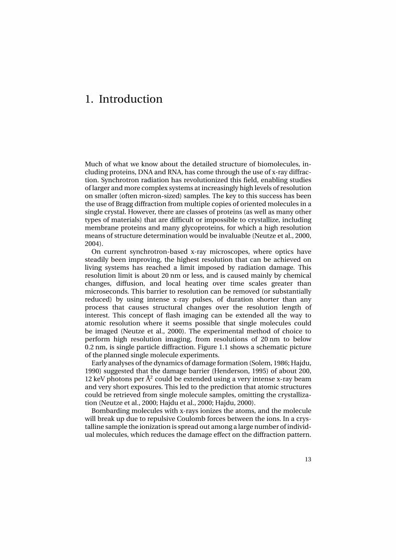
On current synchrotron-based x-ray microscopes, where optics havesteadily been improving, the highest resolution that can be achieved onliving systems has reached a limit imposed by radiation damage. Thisresolution limit is about 20 nm or less, and is caused mainly by chemicalchanges, diffusion, and local heating over time scales greater thanmicroseconds. This barrier to resolution can be removed (or substantiallyreduced) by using intense x-ray pulses, of duration shorter than anyprocess that causes structural changes over the resolution length ofinterest. This concept of flash imaging can be extended all the way toatomic resolution where it seems possible that single molecules couldbe imaged (Neutze et al., 2000). The experimental method of choice toperform high resolution imaging, from resolutions of 20 nm to below0.2 nm, is single particle diffraction. Figure 1.1 shows a schematic pictureof the planned single molecule experiments.
Early analyses of the dynamics of damage formation (Solem, 1986; Hajdu,1990) suggested that the damage barrier (Henderson, 1995) of about 200,12 keV photons per Å2 could be extended using a very intense x-ray beamand very short exposures. This led to the prediction that atomic structurescould be retrieved from single molecule samples, omitting the crystalliza-tion (Neutze et al., 2000; Hajdu et al., 2000; Hajdu, 2000).
Bombarding molecules with x-rays ionizes the atoms, and the moleculewill break up due to repulsive Coulomb forces between the ions. In a crys-talline sample the ionization is spread out among a large number of individ-ual molecules, which reduces the damage effect on the diffraction pattern.
13

In a single molecule sample this is not the case. To image a non-crystallinesample, the photon pulse therefore must be shorter than the time it takesthe molecule to explode, and the pulse has to be intense enough to generatea interpretable diffraction pattern from a single pulse.
The question is how short and intense a pulse has to be to achieve that.This problem has been addressed in a number of theoretical publication,by simulating the photon material interaction and the dynamics of highlyionized, biologically relevant samples (Neutze et al., 2000; Bergh et al., 2004;Hau-Riege et al., 2004; Jurek et al., 2004b,a). The conclusion from these cal-culations seems to be that pulse lengths shorter than 100 fs and intensitieshigher than 1011 photons per pulse are needed, focused to a 100 nm spot.
A more recent estimation of the maximum admissible pulse length, in-cluding image classification and structure reconstruction from diffractiondata (Huldt et al., 2003), was published by Hau-Riege et al. (2005), who con-cluded that an x-ray pulse shorter than ten femtoseconds is needed to getatomic resolution images of biological samples with 12 keV photons.
Figure 1.1: Scheme of the planned single molecule imaging experiment (www-ssrl.slac.stanford.edu/lcls/). A large number of challenges have to be overcome to solve thefirst structure using this technique.
Techniques for image alignment and averaging have been developedfor cryoelectronmicroscopy (Frank, 1996; van Heel et al., 2000), and thesemethods could be adopted for aligning and averaging diffraction images.A 3D diffraction reconstruction would then lead to the atomic-level
14

structure of the molecule. However, the challenges in carrying out suchan experiment are formidable. Among the challenges is the need for”containerless packaging” so that only the sample will be imaged andthe development of advanced reconstruction algorithms (Huldt et al.,2003) for averaging and inverting the diffraction images into the realspace molecule. In principle, there is a direct way to determine the phasesrequired for the inversion using an approach called ”oversampling” (Sayre,2002) but again much remains to be done to make this approach practicalin real circumstancess (Miao et al., 2001, 2006, 2003; Robinson et al., 2001;Marchesini et al., 2003a,b).
1.1 Diffraction imagingThe diffraction pattern of an object is proportional to the squared modulusof the molecular transform (the three-dimensional Fourier transform of theelectron density). The coordinates of the diffraction space, usually calledreciprocal space, are those of the scattering vector (or momentum transfervector) between the incident and scattered x-rays.
Conventional macromolecular crystallography requires high qualitysingle crystals, and in an x-ray diffraction experiment, crystals areexposed to a parallel beam of x-rays with a planar wave front to obtaina three-dimensional diffraction data set. This is done by rotating thecrystal in the beam, and bringing all possible lattice planes into diffractingposition, where the Bragg condition is satisfied. Data points from suchan experiment provide a discrete sampling of the Fourier transform ofthe object at points where the crystal lattice permits us to observe thistransform through the Bragg peaks (Figure 1.2).
In order to reconstruct the electron density, reciprocal space must besampled with sufficient density (Shannon, 1949) and the diffracted intensi-ties must be known with an acceptable accuracy. Bragg diffraction peaks ofa crystal provide a critical sampling of the Fourier transform (Sayre, 1952),and hence the phase problem of crystallography.
When a coherent diffraction pattern is sampled at spacings sufficientlyfiner than the Bragg peak frequency (i.e. the inverse of the sample size),the phase information is in principle encoded inside the diffraction inten-sities (Bernal et al., 1938; Bragg and Perutz, 1952), and can be directly re-trieved by using an iterative process (Gerchberg and Saxton, 1972). In com-bination of this oversampling phasing method with either coherent x-raysor electrons, a novel form of diffraction microscopy has recently been de-veloped to image nanoscale materials and biological structures.
Today, there are over 40,000 entries in the Protein Data Bank(http://www.pdb.org), 85% of which are x-ray crystal structures. This
15
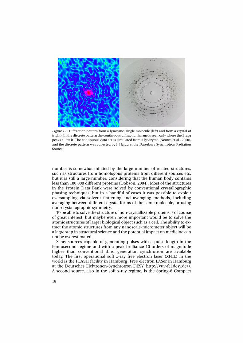
Figure 1.2: Diffraction pattern from a lysosyme, single molecule (left) and from a crystal of(right). In the discrete pattern the continuous diffraction image is seen only where the Braggpeaks allow it. The continuous data set is simulated from a lysozyme (Neutze et al., 2000),and the discrete pattern was collected by J. Hajdu at the Daresbury Synchrotron RadiationSource.
number is somewhat inflated by the large number of related structures,such as structures from homologous proteins from different sources etc,but it is still a large number, considering that the human body containsless than 100,000 different proteins (Dobson, 2004). Most of the structuresin the Protein Data Bank were solved by conventional crystallographicphasing techniques, but in a handful of cases it was possible to exploitoversampling via solvent flattening and averaging methods, includingaveraging between different crystal forms of the same molecule, or usingnon-crystallographic symmetry.
To be able to solve the structure of non-crystallizable proteins is of courseof great interest, but maybe even more important would be to solve theatomic structures of larger biological object such as a cell. The ability to ex-tract the atomic structures from any nanoscale-micrometer object will bea large step in structural science and the potential impact on medicine cannot be overestimated.
X-ray sources capable of generating pulses with a pulse length in thefemtosecond regime and with a peak brilliance 10 orders of magnitudehigher than conventional third generation synchrotron are availabletoday. The first operational soft x-ray free electron laser (XFEL) in theworld is the FLASH facility in Hamburg (Free electron LASer in Hamburgat the Deutsches Elektronen-Synchrotron DESY, http://vuv-fel.desy.de/).A second source, also in the soft x-ray regime, is the Spring-8 Compact
16
Sase Source (SCSS) (http://www-xfel.spring8.or.jp/) in Japan. In thenear future the Linac Coherent Light Source (LCLS) (http://www-ssrl.slac.stanford.edu/lcls/) at the Stanford Linear Accelerator Centerwill be operational as the first hard x-ray FEL with wavelengths down to1.0 Å). The interest in FELs is large and a considerable number of new FELfacilities are being planned around the world; the FERMI facility in Trieste,the BESSY-FEL in Berlin, SPARC in Rome, and DUVFEL in Brookhaven.
There are many interesting problems left that will have to be solved be-fore the first atomic structures can be determined using this technique.
1.2 Scope of the thesisThe work presented in this thesis addresses some of the problems facingpeak brightness experiments at FEL sources. These experiments exploit theextreme brightness and the high peak brilliance of a focused FEL pulse.The list of challenges can of course be made very long, but neverthelessthe hope is that this thesis will add some pieces to the puzzle. The workis relevant to a broad range of fields where high resolution structural andtemporal information is valued.
Understanding of photon-material interaction is fundamental in manyaspects of structural studies. Not only to answer the question whether onecan rapidly image an exploding sample before the effects of the x-ray dam-age destroy all useful structural information, but also in aspects such aspump-probe synchronization and the use of x-ray optics. A majority of thepapers in the list of publications deals with such topics.
The experiments will be done in vacuum to assure that the data recordedfrom a single shot image is as noise free as possible. Therefore understand-ing of the behavior of molecules under such conditions is essential for sin-gle particle imaging.
This thesis is structured as follows. The first part presents the tools, thex-ray sources used in this work (Chapter 2) and the basis of the moleculardynamics calculations used here (Chapter 3).
Chapter 4 presents sample handling. Papers 3 and 6 are put into the con-text of single molecule imaging, and the results are related to the knowledgeabout the behavior of biomolecules in vacuum.
Chapter 5 treats high energy photon interaction and is directly relatedto Papers 1 and 5. It gives the background to the studies and discusses therelevance of the results to damage calculations.
Chapter 6 describes time resolved x-ray studies, and the importance ofthe timing between the pump pulse (for example from an optical laser) andthe probe pulse (from the x-ray source). The chapter treats experimentspresented in Papers 2 and 7-10. Paper 5 presents calculations of the ex-
17

pected time resolution of two substances used for making photocathodes,and is also related to time resolved studies using pump-probe techniques.
Chapter 7 introduces phase transitions, and presents Papers 2 and 4. Bothpapers treat laser induced structural changes in crystals, one is an experi-mental study and the other is based on computer simulations.
The final chapter gives an outlook.
18

2. X-ray sources
This chapter gives a short overview of three x-ray sources relevant for thiswork. A number of different x-ray sources are utilized for structural studies.These sources differ in many ways, such as photon energy and intensity,and therefore, they are appropriate for different types of research. In gen-eral the pulse length limits the time resolution, the photon energy limits thelength resolution, and the photon intensity and the timing accuracy limitsthe signal to noise ratio. All these factors are important for the studies pre-sented here.
Table 2.1: Performance of x-ray sources used in this work. The data presented are taken fromPapers 2, 8 and 11, and from references (Ayvazyan et al., 2006; Synnergren, 2005; Düstereret al., 2006). † estimated numbers, ∗ after the monochromator (the repetition rate of MAX IIis 100 MHz)
Machine Photons/pulse Pulse length Photon energy
(FWHM) eV Å
MAX II (D611) 5×103∗ 100 ps 3-8×10³ 1.55-4.00
SPPS 2×10⁶ 80 fs 9×10³ 1.4
FLASH 1013 10-30 fs 12-200 62-103
LCLS† 3×10¹² 100 fs 8×10³ 1.55
2.1 MAX II, Lund, SwedenMAX-lab is a National Synchrotron Radiation Facility in Lund, and it housesa linear accelerator (LINAC) injector and three electron-storage rings. In thework described here, the 1.5 GeV MAX II ring was used (Figure 2.1). MAX IIis a third generation electron storage ring, producing synchrotron radiationin the x-ray domain. Experiments presented in Paper 2 were performed atbeam line D611. In a synchrotron, electrons circulate in the storage ring,emitting radiation as their trajectories bend. This way they lose energy and
19

to compensate for this, a radio frequency accelerating cavity gives the elec-trons a small momentum impulse to replenish lost energy each time theypass. The radiator used at beam line D611 is a dipole bending magnet. Atsynchrotrons undulators and wigglers are also commonly used radiators.The magnets of bending magnets, undulators or wigglers force the electronbeam on curved trajectories, and the change of velocity generates electronmagnetic radiation.
Figure 2.1: Layout of MAX-lab with its three storage rings. Beamline D611 at MAX II was usedin this thesis. MAX II is 1.5 GeV third generation electron storage ring for producing syn-chrotron radiation. The figure is adopted from www.maxlab.lu.se.
Synchrotrons are excellent light sources for producing a stable and in-tense x-ray beam with a large number of photons per second and very lowdivergence. Over the past 30 years, synchrotron radiation became standardin many research areas, and are routinely used for the determination of pro-tein structures from crystalline samples, for solution and gas phase scat-tering experiments on a variety of substances, and for a range of spectro-scopic studies. Synchrotrons have long pulses with duration in the rangeof 10-400 ps. This makes synchrotron sources suitable for studies on dy-namic phenomena with nanosecond time resolution, as in Paper 2. How-ever, synchrotrons are not particularly useful to study dynamics on a subpicosecond time scale (or for single particle imaging), since sample will bedestroyed way before enough photons have reached the detector.
20

2.2 SPPS, Standford, California, USAThe Sub Picosecond Pulse Source (SPPS), at the Stanford Linear AcceleratorCenter (SLAC) in California, was the first accelerator-based femtosecond x-ray source, operating in the hard x-ray domain. SPPS connects femtosecondspectroscopy with femtosecond structural methods. It was active between2003 and 2006 and produced pulses of spontaneous x-ray radiation utiliz-ing the electron beams from the LINAC at SLAC (Figure 2.2). The electrons
Figure 2.2: Layout of the Sub-Picosecond Pulse Source (SPPS). SPPS is an accelerator-basedhard x-ray source to produce femtosecond x-ray pulses. It uses the full length of the SLAClinac and injects 28 GeV electron bunches into an undulator magnet. SPPS is not a FEL, itproduces spontaneous radiation from the undulator. FFTB: Final Focus Test Beam. (Image,courtesy SLAC.)
were injected into an undulator with a fundamental energy of 9 keV (1.4 Å)at 28.5 GeV electron energy (Emma, 2002; Bentson et al., 2002; Akre et al.,2002). Two sets of multilayer mirrors directed the beam to the experimen-tal hutch, reducing the energy bandwidth (∆E/E) to about 1%. One of thesemirrors was resonant for the fundamental energy, and the other for the thirdharmonic. SPPS had all components of a free-electron laser but the undu-lator was too short bo be suitable for lasing. As a consequence, the numberof photons per pulse at SPPS was not sufficiently high for single moleculeimaging, but SPPS was eminently suitable for studying ultrafast structuraltransitions, including phase transition processes (resulting in Papers 8, 9,10 and Fritz et al. (2007)). SPPS was also an important ground for testingand developing some of the principles of the XFEL.
2.3 FLASH, Hamburg, GermanyFLASH (Figure 2.3) is the first free electron laser (FEL) to reach the soft x-raydomain. It operates on the principle of self amplified spontaneous emission(SASE). An electron bunch accelerated in a LINAC passes through an un-
21

Figure 2.3: Outline of the FLASH soft x-ray laser at DESY, Hamburg. FLASH is the first free-electron laser to produce ultrashort photon pulses with wavelength reaching the x-ray do-main.
dulator, consisting of a series of magnets with alternating field direction. Inthe undulator the electron bunch starts radiating due to acceleration pro-duced by periodically changing the velocity of electrons as the electronsswing in and out horizontally when passing along the undulator magnet. Atwavelengths longer than or comparable to the bunch length, the radiationgenerated from the electron bunch is coherent.
The electromagnetic field from the radiating electrons modulates thestructure of the electron bunch, such that the electrons that are locatedbefore the phase of the field lose energy while the electrons locatedafter the phase gain energy. This leads to a micro bunching within theelectron bunch. Since the radiating micro bunches of electrons are spacedwith the distance one wavelength of the light they radiate, whey will allradiate in phase. In other words, they are lasing, hence the name “freeelectron laser”. The coherent radiation intensity is proportional to thesquare of the number of electrons in the bunch. The amplification of theemitted radiation takes place in three stages, illustrated by Figure 2.4. Inthe beginning of the undulators (I) the microbunches start to form. Thewave grows roughly as the cube of the distance. In region II the gain isexponential and in the third region (III) the amplification saturates and the
22

energy will oscillate between the electron and the photon beam. This isbecause the electrons will stop gaining energy from the wave.
Figure 2.4: Evolution of the Self Amplified Spontaneous Emission effect along the undulator.
The SASE FEL principle at short wavelengths was first demonstrated towork at the Tesla Test Facility (TTF) in Hamburg (Andruszkow et al., 2000;Ayvazyan et al., 2002b,a), at a wavelength of 100 nm. TTF was a pilot fa-cility mainly used for machine development. Later TTF was closed and re-built as TTF-2, which later got the catchy name of Free Electron Laser inHamburg (FLASH). FLASH is, in contrast to TTF, a user facility where re-search communities from many different fields can take advantage of theintense beam. Originally operating at a wavelength of 32 nm, FLASH hascontinuously been modified to work at shorter and shorter wavelengths. Asthis is written the wavelength is down to around 13 nm, and the machinewill reach 6 nm by autumn 2007. These wavelengths are not yet suitable foratomic resolution structural studies, but they are suitable of imaging largerobjects with nanometer resolutions. There are still a lot of steps toward sin-gle particle imaging that have and will be taken at FLASH. Recent publica-tions based on experiments executed at FLASH, have provided knowledgeabout damage by FEL pulses in the soft x-ray regime (Papers 12-14 and 16).The principle of reconstruction of an image of a sample that is completelydestroyed during the x-ray exposure was also proven at FLASH, illustratedby Figure 9.1 (Paper 11).
23

3. Molecular Dynamics
The main theoretical tool used in this thesis is Molecular Dynamics (MD),implemented in the GROMACS software package (Berendsen et al., 1995;Lindahl et al., 2001; van der Spoel et al., 2005). MD is a powerful instrumentfor describing the dynamics of molecular systems. MD is based on simpleprinciples, but it can be used to describe nature quite accurately on a meso-scopic scale if handled with care (Allen and Tildesley, 1987). In MD atoms,ions and sometimes small groups of atoms are considered as soft sphereswhose interactions are described using force fields calculated from poten-tials. At every time step in a simulation the pairwise forces between all inter-action points (be it an atom, an ion or a group of such) are calculated. Whenthis is done for the complete system Newton’s equation of motion (Newton,1687) are integrated and all interaction points are moved to new positions.The procedure is then performed with the atoms at the new positions, andso forth. By keeping the time step short enough, such that each atom onlymoves a very short distance (compared to the distance between two atomsin the system), no unrealistic forces will occur and the algorithm is stable.
Newton’s equation of motion:
mi∂2ri
∂t 2 = Fi , i = 1,2, ....N , (3.0.1)
is solved at every time step for each of the N interaction points i , ri is theposition of interaction point i and mi its mass, and t time and, where theforces Fi are the negative derivative of the potential V (r1,r2, ...rN )
Fi =−∂V
∂ri. (3.0.2)
The potential V is described by the Coulomb potential (treating interac-tions due to charged particles)
V cr = 1
4πε0
(qi q j
ri j
), (3.0.3)
(where ri j is the distance between the two charges qi and q j ) and theLennard-Jones (LJ) potential, which is repulsive at short distances (as aresult of overlapping electron orbitals, due to Pauli repulsion (Massimi,
25
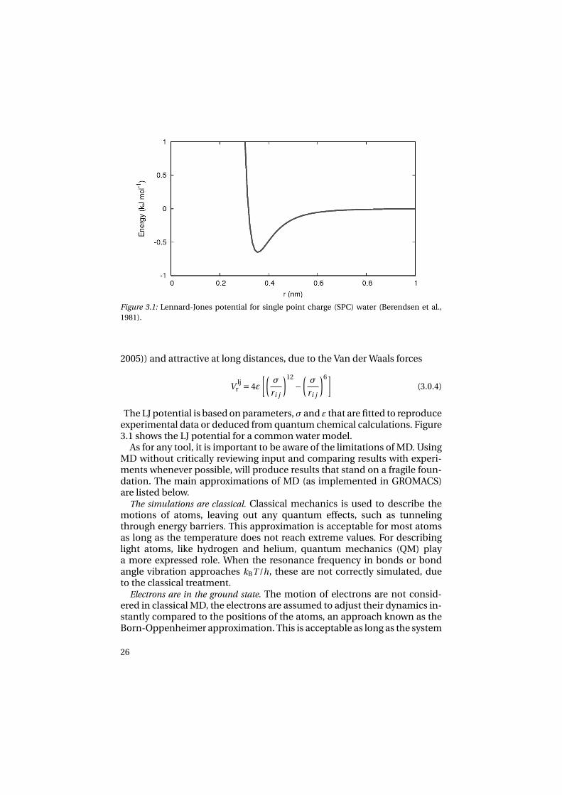
Figure 3.1: Lennard-Jones potential for single point charge (SPC) water (Berendsen et al.,1981).
2005)) and attractive at long distances, due to the Van der Waals forces
V ljr = 4ε
[(σ
ri j
)12
−(σ
ri j
)6](3.0.4)
The LJ potential is based on parameters, σ and ε that are fitted to reproduceexperimental data or deduced from quantum chemical calculations. Figure3.1 shows the LJ potential for a common water model.
As for any tool, it is important to be aware of the limitations of MD. UsingMD without critically reviewing input and comparing results with experi-ments whenever possible, will produce results that stand on a fragile foun-dation. The main approximations of MD (as implemented in GROMACS)are listed below.
The simulations are classical. Classical mechanics is used to describe themotions of atoms, leaving out any quantum effects, such as tunnelingthrough energy barriers. This approximation is acceptable for most atomsas long as the temperature does not reach extreme values. For describinglight atoms, like hydrogen and helium, quantum mechanics (QM) playa more expressed role. When the resonance frequency in bonds or bondangle vibration approaches kBT /h, these are not correctly simulated, dueto the classical treatment.
Electrons are in the ground state. The motion of electrons are not consid-ered in classical MD, the electrons are assumed to adjust their dynamics in-stantly compared to the positions of the atoms, an approach known as theBorn-Oppenheimer approximation. This is acceptable as long as the system
26
is not perturbed, e.g. ionized or otherwise electronically excited. The limitfor laser-matter interactions is discussed in Paper 4, where infrared (IR)laser radiation is absorbed in ice through stimulating bond vibrations. Todescribe x-ray induced ionization effects using MD simulations, as is donein references (Bergh et al., 2004; Neutze et al., 2000; Jurek et al., 2004b), atreatment of electron trajectories and free electron ejection as well as crosssections for photon and electron interaction must be included. One way todo so is by treating the electrons as an electron gas (Bergh et al., 2004).
Force fields are approximate. Forces in MD are described by force fields.These force fields are adapted to fit experimental data as well as possible,but there will always be some discrepancy between the experimental dataand the fit.
The force field is pair-additive. Force interactions are calculated pair wiseand then added together, which means that any multi-body effects are as-sumed to be the sum of multiple pair interaction.
Long-range interactions are cut-off. GROMACS normally uses a cut-off forthe LJ interactions and Coulomb interactions. The cut-off range can not ex-ceed the half of the simulation box side for a system with periodic bound-ary conditions (PBC), since the same particle should only be accountedfor once. For large neutral systems this is not a problem, but for polariz-able systems or systems containing charges this is expected to become aproblem. In such cases long-range electrostatic algorithms, such as Ewaldsummation (Ewald, 1921), the Particle-mesh Ewald (PME) method (Dardenet al., 1993) or the Particle-Particle Particle-Mesh (PPPM) method (Hock-ney and Eastwood, 1981) can be used. Using PBC the dipole from polariz-able molecule models in an external electric field might induce an effectiveelectric field that is not realistic. In Paper 4 such effects are discussed. In thecluster evaporation simulations (Papers 3 and 6) no cut-offs are used at all.
Boundary conditions are unnatural. Since only a finite number of particlescan be simulated the system will be spatially defined by unnatural bound-aries. Either a cluster of molecules can be put in vacuum, which might be anundesirable feature, or when simulating material in bulk, PBC can be used.PBC may induce an artificial order to the system (van der Spoel and vanMaaren, 2006) , but for a large system the errors are small. In some casesthough, as in Papers 3 and 6, the vacuum environment might actually bedesirable.
3.1 Molecular modelsIn MD molecules are constructed by putting rigid or flexible bondsbetween atoms. Bond lengths and angles are usually taken fromcrystallographic data and force constants from spectroscopy. The
27

Figure 3.2: The structure of hexagonal ice, the most common type of ice in nature. The struc-ture is taken form reference (Hayward et al., 1997).
charges are fitted to QM electrostatic potentials or treated as empiricalparameters (Allen and Tildesley, 1987). LJ parameters and charges aretuned such that simulations reproduce experimental data. A specificmolecule model is constructed such that it reproduces a limited set ofphysical parameters. It is therefore crucial to consider what physicalparameters a molecular model is tuned to, before any conclusions aboutthe results of a simulation are drawn (Leach, 2001).
Water is the main molecule simulated in Papers 1, 3, 4 and 6. Even for amolecule of such simple structure as water none of the over 50 water mod-els presented in the literature can reproduce all the physical properties ofwater (Guillot, 2002). In Paper 3, this fact is discussed and three differentcommonly used water models are compared; SPC (Berendsen et al., 1981),TIP4P (Jorgensen et al., 1983) and TIP5P (Mahoney and Jorgensen, 2000).The simulations in Paper 4 include absorption of an external electric field,it was therefore necessary to employ a flexible water model developed to doso (Lawrence and Skinner, 2003).
Very recently, a force field was presented for water, developed entirelyfrom first order calculations (Bukowski et al., 2007). This forcefieldpredicted successfully the properties of a water dimer, as well as of liquidwater, in good agreement with experiments. This raises hope for models,which could be particularly use full for detailed studies of laser-matterinteraction.
28

4. Handling hydrated samples
With ultrasmall samples, standard procedures for sample selection,characterization and handling can not be applied. Everything within thebeam-path will interact with x-rays, and contribute to the image, includinggas molecules and the sample holder. It is therefore necessary to developcontainer-free new methods, and inject samples directly into a vacuumchamber to assure a continuous flow of molecules into the interactionzone with the x-ray pulse (Hajdu et al., 2000; Neutze et al., 2000, 2004).Figure 1.1 shows the suggested experimental arrangement.
Sample injection can be done by spraying techniques. One such tech-nique is electrospray ionization (ESI). ESI was developed in the 1960s toinject biomolecules into the gas phase for mass spectrometry (MS) (Doleet al., 1968; Yamashita and Fenn, 1984a,b), and it was awarded with the No-bel Prize in Chemistry in 2002. Biomolecules sprayed into vacuum will bein random orientation, encapsulated in a micro-droplet (Kebarle and Tang,1993). The micro-droplets can be dried to a desired level before interact-ing with the x-ray beam. Ionization and dehydration can interfere with theconformation of the sample, and it is thus important to understand theseprocesses in detail. It is known, for instance, that unfolding of proteins invacuum is possible, especially when they are highly charged (Jarrold, 1999;Velazquez et al., 1999; Iavarone and Parks, 2005). It is also known, that con-ditions exist under which unfolding does not take place (Tito et al., 2000).In a recent paper, Patriksson et al. (2007) have shown that it is sufficient tohave a thin water layer (consisting of only a few water molecule) around theprotein to maintain the solvent structure in the gas phase.
Both Bergh et al. (2004) and Hau-Riege et al. (2004) predict that clustersexposed with an XFEL pulse will explode in shells, where outer shells ex-pand very fast compared to the rest of the cluster due to higher net charge,leaving an inner core, which is initially nearly stationary. This leads to theidea of keeping sample molecules in a protecting water cluster, not only be-cause its is an efficient way to deliver the sample to the x-ray beam, but alsoto help with slowing down the explosion of the molecule of interest, whichmight be necessary.
29
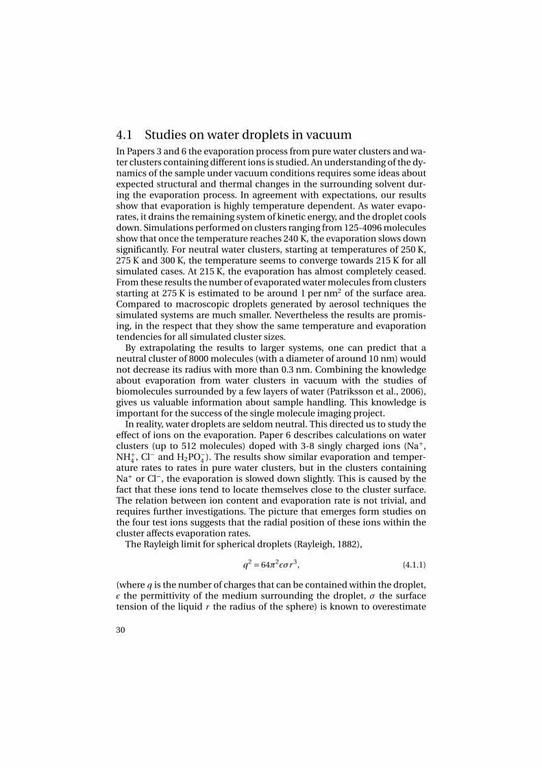
4.1 Studies on water droplets in vacuumIn Papers 3 and 6 the evaporation process from pure water clusters and wa-ter clusters containing different ions is studied. An understanding of the dy-namics of the sample under vacuum conditions requires some ideas aboutexpected structural and thermal changes in the surrounding solvent dur-ing the evaporation process. In agreement with expectations, our resultsshow that evaporation is highly temperature dependent. As water evapo-rates, it drains the remaining system of kinetic energy, and the droplet coolsdown. Simulations performed on clusters ranging from 125-4096 moleculesshow that once the temperature reaches 240 K, the evaporation slows downsignificantly. For neutral water clusters, starting at temperatures of 250 K,275 K and 300 K, the temperature seems to converge towards 215 K for allsimulated cases. At 215 K, the evaporation has almost completely ceased.From these results the number of evaporated water molecules from clustersstarting at 275 K is estimated to be around 1 per nm2 of the surface area.Compared to macroscopic droplets generated by aerosol techniques thesimulated systems are much smaller. Nevertheless the results are promis-ing, in the respect that they show the same temperature and evaporationtendencies for all simulated cluster sizes.
By extrapolating the results to larger systems, one can predict that aneutral cluster of 8000 molecules (with a diameter of around 10 nm) wouldnot decrease its radius with more than 0.3 nm. Combining the knowledgeabout evaporation from water clusters in vacuum with the studies ofbiomolecules surrounded by a few layers of water (Patriksson et al., 2006),gives us valuable information about sample handling. This knowledge isimportant for the success of the single molecule imaging project.
In reality, water droplets are seldom neutral. This directed us to study theeffect of ions on the evaporation. Paper 6 describes calculations on waterclusters (up to 512 molecules) doped with 3-8 singly charged ions (Na+,NH+
4 , Cl− and H2PO−4 ). The results show similar evaporation and temper-
ature rates to rates in pure water clusters, but in the clusters containingNa+ or Cl−, the evaporation is slowed down slightly. This is caused by thefact that these ions tend to locate themselves close to the cluster surface.The relation between ion content and evaporation rate is not trivial, andrequires further investigations. The picture that emerges form studies onthe four test ions suggests that the radial position of these ions within thecluster affects evaporation rates.
The Rayleigh limit for spherical droplets (Rayleigh, 1882),
q2 = 64π2εσr 3, (4.1.1)
(where q is the number of charges that can be contained within the droplet,ε the permittivity of the medium surrounding the droplet, σ the surfacetension of the liquid r the radius of the sphere) is known to overestimate
30

Figure 4.1: A water cluster containing four Cl− ions and 210 water molecules being rippedapart due to the Coulomb forces between the ions. The Cl− ions are green in the picture.
the maximum number of charges in a stable nanoscale system (Margineanet al., 2006), which our simulations verify. In simulations, the surfacetension is underestimated, and different water models show quitedifferent surface tension values. In bulk SPC water, the surface tensionis 59×10−3N/m (Abbas et al., 1995), compared to the experimental value72×10−3N/m (Weast, 1977). It is also known that the surface tensiondepends on the size of the simulated cluster (Zakharov et al., 1998). In thecase of the two anions, the Rayleigh limit seems to give a better estimationof the maximum charges than in the case of the positive ions. This wasfound to be related to the hydration energy (Marcus, 1994), and to theion-ion structure within the water cluster.
In clusters that carry more charges than the Rayleigh limit, the ions ripthe cluster apart, resulting in smaller satellite clusters, containing only a fewions below the Rayleigh limits. Figure 4.1 shows a water cluster containingCl− above the limit, being ripped apart. The simulations do not reveal anysignificant structural changes in the water. At temperatures well below 0 ˚C,it is tempting to believe that the water forms structured ice. However, ex-periments have shown that structural transitions from water to structuredice (in contrast to amorphous ice) in small droplets occurs at temperaturesbelow 200 K (Huang and Bartell, 1995; Bartell and Huang, 1994; Stein andArmstrong, 1973), and our clusters never reach temperatures below 210 K.To simulate the transformation from the amorphous phase to the struc-tured ice using MD have proven to be hard. Successful simulations on freez-ing water into crystalline ice are rare, and these have not been done undernatural conditions (Matsumoto et al., 2002; Svishchev and Kusalik, 1994;Zangi and Mark, 2003). Evaporation, on the other hand is something that iswell described by MD, and the simulations presented in Paper 3 are in good
31

agreement with a phenomenological expression for the evaporation, basedon experimental results (Tang and Kebarle, 1993).
32

5. Interaction of photons withmaterial
To understand what happens to a microdroplet, a cell, a sample moleculewhen it meets the FEL pulse, we need to comprehend photon material in-teraction.
X-rays interact with matter through absorption and scattering. As men-tioned in the introduction, one of the limiting factors concerning structuralstudies using intense x-ray sources is the damage of the sample due to ab-sorption.
In photo absorption, the energy of the photon is transferred to an elec-tron, which is then ejected from the atom, leaving a positively charged ionbehind. This is the main process that leads to damage in the sample.
Photon scattering can either be elastic, where the photon energy is con-served, or inelastic, where some of the photon energy is transferred to theatom. Generally, elastic scattering contributes to the recordable informa-tion in the diffraction pattern, while inelastic scattering does not carry eas-ily decipherable structural information.
Inelastic scattering is a relatively rare event at x-ray frequencies althoughit is the main source of energy deposition with hydrogen, and representsabout 3% of all interactions between x-rays and a biological sample at 1 Åwavelength. During inelastic scattering, the incoming photon excites anelectron to some virtual level and when the electron relaxes emitting a pho-ton, it does not come back to the ground state. The photon emitted hastherefore a different frequency from the photon absorbed, and it also hasan altered phase. When the photoelectron ejected from a core level leavesbehind a vacancy, an electron from a higher energy level may fall into theempty orbit, resulting in a release of energy. This energy may be emitted inthe form of a photon (dominant process with high-Z elements), but it canalso be transferred to an outer shell electron which is then ejected from theatom in a process called Auger decay (Auger et al., 1939). The Auger elec-tron carries the kinetic energy corresponding to the difference between theshell binding energy and the energy of the initial electronic transition, asillustrated by Figure 5.1. Compared to the photoelectron generated by anx-ray photon, the energy of the Auger electron is significantly lower, andit is ejected at a later time than the photoelectron. In biologically relevantmaterial Auger electrons have energies between 250 eV-2 keV (Thompson
33

Figure 5.1: The Auger decay. This is one of two principal processes for the relaxation of aninner shell electron vacancy in an excited or ionized atom. The Auger effect is a two electronprocess in which an electron makes a discrete transition from a higher shell to fill a vacantinner shell hole. The energy gained in this process is transferred to another bound electronwhich then escapes from the atom. This outgoing electron is referred to as an Auger electron.The photoelectron and the Auger electron are released at different times, and have differentenergies. The figure on the left shows a high energy photoelectron leaving the inner shell. Thefigure on the right shows the release of the low energy Auger electron as the system relaxes.Auger decay is the dominant relaxation process for filling K-hole vacancies in light elements,like C, N, O, S, P.
and Vaughan, 2001), compared to the photoelectron carrying the energyof the incoming photon minus the shell binding energy (typically between3-20 keV with x-rays). The physics of this decay is well understood (Hubbellet al., 1975; Veigele, 1973).
The life time of the inner level vacancy can be determined by measure-ments of the Auger line widths (Krause and Oliver, 1979). For atoms abun-dant in biological samples (such as C, N, O and S), the K-hole lifetimes areup to around 10 fs. During the photoionization process, the photoelectronmay interact with valence electrons, leading to the so called shake-up andshake-off effects (Siegbahn et al., 1969). For light elements of biological sig-nificance, electrons emissions from these effects are in order 10-30% of theevents where a low energy electron (10-100 eV) is emitted (Persson et al.,2001).
5.1 Secondary electron cascadesAs atoms are photo-ionized, the electrons ejected from the atom will inter-act with the surrounding sample. In a macroscopic sample, both the pho-
34

Figure 5.2: Secondary electron cascades. The initial electron, emitted through the Auger de-cay, leads to an avalanche of electrons due to inelastic scattering.
toelectrons and the secondary Auger electrons become thermalised andtrapped inside the sample. Thermalisation is based on inelastic electron-electron interactions and, to a lesser degree, on electron-nuclear interac-tions. An electron scattering inelastically on an atom may cause a secondionization of an outer shell electron. This mechanism leads to an avalancheof electrons generated from one single photoionization event, as illustratedby Figure 5.2. Thermalisation produces a large number of such secondary”cascade electrons” (Ziaja et al., 2001, 2002, 2005). The number of thesecascade electrons is roughly proportional to the energy of the impact elec-tron triggering the cascade. These electrons are redistributed in the sampleand can recombine with atoms. On the ∼100 fs time scale, considered rel-evant to damage formation in single molecule imaging, the recombinationis probability very low (Landau and Lifschitz, 1977). Collisions with atomsare highly relevant for the understanding of x-ray induced damage.
Paper 1 describes secondary electron cascades generated from anAuger decay in water (Auger energy for oxygen in a water molecule is507.9 eV (Wagner et al., 1980)). The goal is to map the characteristics ofelectron cascades to support further development of the model usedby Neutze et al. (2000), where the effects of the secondary ionization wasnot included. The studies of radiation damage in water are highly relevant,since water has a density comparable to that of biosamples and it has
35

been predicted that water would be used a carrier liquid containing thesamples as they are exposed to the x-ray pulse. The calculations show thatthe number of electrons generated from a single Auger decay in liquidwater is around 20. The thermalization of the secondary electrons throughcollisions occurs after only a few femtoseconds.
The results were later used by Bergh et al. (2004), where electrons whereincorporated into a classical MD scheme as a continuous electron gas. Inthe electron gas model the electrons are assumed to thermalize immedi-ately, compared to atoms, and can therefore be regarded as Maxwellian.Conclusions drawn from Bergh et al. (2004) show that free electrons in thesample shield positive charges on the ions, and slow down the explosion.
Our computational results give around 20 electrons per Auger decay inwater (energy of Auger electron from oxygen: 507.9 eV), and this is in goodagreement with results from radiolysis experiments on water (Muroya et al.,2002) where around 25 electrons are detected per 500 eV of an initial elec-tron. The time scale of the experiments is much longer than in our calcu-lations. It is therefore expected that effects not included in the model, suchas recombination, should be relevant. Still the fact that experiments andtheory agrees is ensuring.
5.2 Calculations of electron inelastic cross sectionsThe calculations of secondary electron cascades in materials, presented inPapers 1 and 5, use empirical models based on optical data to calculate theinelastic cross sections for electron scattering on atoms. Two different mod-els, the Ashley model (Ashley, 1990, 1991) and the Tanuma, Powell and Pennmodel (TPP) (Penn, 1976, 1987; Tanuma et al., 1988, 1991a,b, 1993a,b) wereemployed in Paper 1.
Calculations based on the Ashley model are believed to underestimatethe number of electrons generated (Ziaja et al., 2005), therefore only theTPP model was used in the studies described in Paper 5.
Silkin et al. (2003) have presented first-principles calculation of theelectron inelastic mean free path in beryllium metal. They used the socalled GW1 approximation (Hedin, 1965) of the Density Functional Theory(DFT) (Kohn and Sham, 1965).
For cross sections of electron scattering of free atoms, a couple of analyt-ical expressions, adapted to fit experimental data, are also available, see forexample (Lennon et al., 1988) and (Kim and Rudd, 1994). For cross sectionsin solids, two different methods have been employed in this thesis. Bothfollow an approach developed by Fermi (Fermi, 1924; Bethe, 1930) wherethe passage of a fast charged particle is treated through the linear pertur-
1G stands for Green’s function, W for the screened potential.
36

Figure 5.3: Electron impact ionization cross sections for carbon. Kim and Rudd (1994) is asemi-empirical model for free atoms, with parameters used in the explosion dynamics cal-culations by Jurek et al. (2004b). Lennon et al. (1988) is another semi-empirical approachagain for carbon, used by (Hau-Riege et al., 2004). Ashley and TPP are two solid state models,applied to diamond (Ashley, 1990; Tanuma et al., 1988). This was used in Papers 1 and 5, andthe results were used in Bergh et al. ( 2004). Experimental data are from free atoms (Brooket al., 1978).
bation caused by its electric field in the solid. We used two models here(Ashley and TPP) together with the Lindhard dielectric function (Lindhard,1954) to estimate the number of electrons generated. The models have dif-ferent ways to compensate for the fact that the dielectric function is onlyknown for photons, where the momentum transfer is zero. Ashley includesthe exchange between the incoming electron and the electrons in the solidin accordance with the non-relativistic Møller cross section (Kaku, 1993).The TPP approach on the other hand, is developed for calculating inelasticmean free paths for electrons in a solid. Figure 5.3 illustrates the differencebetween the electron cross sections based on two semi-empirical free elec-tron approaches (for carbon) and the two models for electron cross sectionsin solids, in this case diamond. As mentioned, the two models are depen-dent on the knowledge of the dielectric function, which typically have beentaken from experimental data.
In Paper 5, a new strategy is presented and tested. The energy loss func-tion was determined using GW calculations, and the secondary electron dy-namics in two alkali-iodides were than calculated and compared to calcu-
37

lations based on experimental data (Creuzburg, 1966). We made a compar-ison of results based on the GW calculations and experiment, concludingthat in the two test cases (KI and CsI) the calculations gave comparableoutputs. This indicates that the energy loss function based on GW calcu-lations can be used where no experimental data are available. The resultsare further discussed in Chapter 6.
Figure 5.4: Water cluster exploding under the influence of a 15 fs XFEL pulse at 1 Å wave-
length and with 1012 photons per pulse focused to a 100 nm diameter spot. Snapshots weretaken at -10, 0 and 15 fs, with the pulse centered at t=0 fs. These snapshots are from Berghet al. (2004), using results described in Paper 1. The dynamics of the explosion is influencedby the electron gas.
The hard x-ray damage calculations on bio relevant material includingsecondary electrons (Hau-Riege et al., 2004; Jurek et al., 2004b; Bergh et al.,2004) treat the generation of secondary electrons differently. Hau-Riegeet al. and Jurek et al. use cross sections from free atoms to calculate the lowenergy productions, whereas Bergh et al. use electrons calculated in a solidstate approach (snapshots from the calculations of Bergh et al. (2004) canbe seen in Figure 5.4). Which of these approaches is better, depends on awhole range of factors. Generally the solid state approach is more accuratebefore the sample starts exploding, and the single particle approach isbetter to describe the explosion while it is happening. As Figure 5.3 shows,the difference between cross sections is large, specially at low energies,yet the damage studies using the different models (Hau-Riege et al., 2004;Jurek et al., 2004b; Bergh et al., 2004) give fairly similar results.
Ionization in the vacuum ultra violet (VUV) regime is significantlydifferent from ionization in the hard x-ray regime (Ziaja et al., 2006). Thehigh field limits with hard x-ray are very high, and multiphoton processescan be neglected with photon intensities expected from XFELs. Hard x-raystrigger inner shell processes. In contrast, absorption of a VUV photoncauses ionization through outer shell processes. Samples exposed to ahighly focused VUV FEL pulse experience multiphoton processes andinverse Bremsstrahlung is significant in the sample exposed to a focused
38
VUV pulse. As a result, photoionization is significantly different with softx-rays than with hard x-rays. In a recent paper Ziaja et al. (2006), proposea model for describing ionization and ionization dynamics in the VUVregime, using Boltzmann equations. This model was successfully appliedto describe the behavior of xenon clusters in a focused VUV pulse from theVUV-FEL, and the results showed qualitative agreement with experimentaldata.
39


6. Time resolved studies
Ultrafast phenomena are typically studied using pump-probe techniqueswhere the dynamics are initiated by an ultrafast laser (the pump pulse) andprobed after some time with a second pulse (the probe pulse). If the probepulse originates from a high harmonic laser source, the pump pulse andthe probe pulse can both be generated from the same source, using a delayline which creates different optical path lengths for the two pulses. In suchcases, the time resolution is limited by the overlap of the pump pulse withthe probe pulse, and can be less than a femtosecond (Hentschel et al., 2001).
Figure 6.1: Demonstration of the principle of measuring the time delay between pump andprobe by after the experiment is carried out. Cross beam geometry (Hajdu, 1994; Neutzeand Hajdu, 1997; Synnergren et al., 2002); the upper picture is the x-ray diffration form theunperturbed InSb crystal, in the lower picture the laser pulse have reached the sample andmelted the crystal such that it does not diffract any longer (the dark line). The experimentshown was performed at SPPS, by Jörgen Larsson and coworkers.
At a typical accelerator based x-ray source, the pump and the probepulses come from different sources. Timing jitter (a random time delay)between the pump and the probe pulses affects temporal resolution, andsynchronization of these pulses becomes a primary concern in ultrafasttime-resolved studies. Requirements for tight synchronization can ofcourse be relaxed in studies of slower processes. This was the case forexperiments presented in Paper 2, where recrystallization was followedover nanoseconds, and speed was primarily limited by the detectionsystem.
41

A way to reduce the problem of jitter at accelerator-based light sources isoffered by measuring it. This can be done by electro optical sampling (EOS),a method developed at SPPS by Cavalieri et al. (Paper 7). EOS measures thearrival time of each high energy electron bunch to the undulator. The arrivaltime of each x-ray pulse can this way be determined relative to an externallaser pulse. Results from SPPS show time resolutions better than 60 fs rms.EOS is thoroughly explained in the reference.
Figure 6.2: Layout of a conventional streak camera. Image reproduced with permission fromThe Max Planck Institute for Quantum Optics/Vienna University of Technology.
A second way to handle the jitter is to deal with it after the experiment iscompleted, simply by interpreting the read out from the detector. By know-ing the pulse lengths and the geometry of the two sources, the timing can bedetermined by analyzing the data, using the so called crossed-beam topog-raphy geometry (Hajdu, 1994; Neutze and Hajdu, 1997; Synnergren et al.,2002). Figure 6.1 illustrates this approach, which was applied in the exper-iments presented in Papers 8, 9 and 10. By crossing the pump and probebeams on the sample and imaging the diffracted x-rays, one can map thetemporal information into spatial information, enabling collection of thecomplete time history around time zero in a single shot. In the figure, thex-ray pulse is diffracted by an InSb crystal. The laser pulse melts the crystal,and where the sample melted it does not diffract. Hence the dark line in themelted area of the crystal.
42

A more well established way to measure the delay between the pump andthe probe can be done by using a streak camera (Key et al., 1980). Figure 6.2shows the layout of a conventional streak camera. The x-ray photons hit thecathode, where they generate an electron bunch that is accelerated towardsa detector. These electrons are exposed to a high voltage electric field that istriggered by the laser pulse. This results in an image on the detector wherethe overlap of the two pulses can be interpreted by the spatial spread of thestreak image. One of the main parameters that affect the time resolution isthe ability of the cathode to generate electrons rapidly with a narrow energyand spatial distribution (Lowney et al., 2004).
Paper 5 provides a purely theoretical study on the development ofsecondary electron cascades in two photocathode materials (KI andCsI). These studies employed a method described in Paper 1 (furtherexplained in Chapter 5). This method allowed us to explicitly followelectron trajectories, which was not possible in earlier investigations. Bothmaterials are stable, and possess good photocathode properties (Verma,1973). They were also studied earlier experimentally (Lowney et al., 2004;Henke, 1972) and theoretically (Kane, 1966; Henke et al., 1979). Oursimulations consider impact electrons with a kinetic energy of 500 eV,and the results show that both materials release a similar number ofsecondary electrons. The number of electrons liberated by electroncascades seems to saturate slightly faster in CsI than in KI, but KI showsa significantly narrower secondary electron distribution than CsI. Thesecharacteristics indicate that a streak camera with a KI photocathode couldgive a higher temporal and spatial resolution than a streak camera a CsIwith photocathode.
43

7. Laser induced phase transitions
This chapter summarizes results on laser induced structural transitions re-ported in Papers 2 and 4.
7.1 Thermal phase transitionsHeating a material makes atoms vibrate. If the added energy is largeenough, the material may go through a phase transition into a lowerordered state. In a solid, the free energy is defined as the F =U −T S,where U is the internal energy of the system, T the temperature and S theentropy (Kittel, 1996). When two phases (L and H) are allowed at the sametemperature, TC, such that FL(TC) = FH(TC), the material may change fromits higher structured order phase to a lower order phase.
7.1.1 Laser induced melting of ice studied by MDIn classical MD simulations electrons are not considered explicitly. As longas the system is not ionized, structural changes induced by an external elec-tric field can still be studied. Furthermore, MD is an eminently suitable toolto do so, since it gives the exact position of each atom at every time step. Asmentioned in Chapter 3, the choice of molecular model (in this case the wa-ter model) is crucial for the result of a simulation. In the study presented inPaper 4, we used a water model developed to reproduce IR absorption. Themodel (flexible TIP4P by Lawrence and Skinner (2003)) includes a Morsepotential (Morse, 1929) to describe oxygen-hydrogen vibrations. The Morsepotential is described as
V (r ) = De[1−e−a(r−re)]2, (7.1.1)
where r is the distance between the atoms, re is the equilibrium bond dis-tance, De is the well depth (defined relative the dissociated atoms) and a isa constant controlling the width of the potential.
The simulations presented in Paper 4 provide a deeper understandingof experimental results on ultrafast superheating of ice performed by Iglevet al. (2006). We find that when heating ice to temperatures above 290 Kwith a short pulsed IR laser, the ice stays in a super-heated state for only a
45

few picoseconds before melting, whereas upon heating to T<290 K the sys-tem can stay superheated for nanoseconds. In the latter case the meltingof the crystal structure did not show a linear dependence on the amountof energy deposit in the system, but seemed to be governed by a statisti-cal process. We found that thermal melting follows a nucleation process,where the break down of the ice structure starts at highly localized pointsand spreads out through the crystal. Pockets of intact crystalline structurescould be found a couple of picoseconds after the melting process had beeninitiated.
The upcoming hard x-ray FELs will provide a revolutionary experimen-tal tool to study rapid structural transitions, such as melting of ice usinga short-pulse laser. Simulations, as those presented in Paper 4, serve as auseful guideline in the design of future experiments.
7.2 Non-thermal meltingIn a non-thermal melting process, the electrons and the crystal lattice are
not in equilibrium. Equilibrium between the electrons and the lattice isestablished on a picosecond timescale, which corresponds to the typicaltimescale of the electron-phonon coupling. In such processes the conceptof temperature breaks down, hence the free energy is not defined. Todefine a phase transition in such extreme cases the Lindemann criterioncan be used (Lindemann, 1919). The Lindemann criterion states that whenthe atomic displacement reaches 10% of the nearest neighbor distance,the material melts. During laser induced non-thermal ultrafast processesthe electron pressure and temperature can reach extremely high values,since these processes occur much faster than the typical phonon electroncoupling time.
7.2.1 Time-resolved x-ray diffraction experimentsX-ray diffraction is a very sensitive instrument to determine the order of asample and therefore it is an excellent probe for investigating phase tran-sitions. Generally the x-ray diffraction efficiency is much higher for a crys-talline sample than for an amorphous sample, which makes x-rays idealfor structure determination of crystals. Ultra short x-ray pulses permit hightemporal resolutions in x-ray diffraction experiments. In the experimentspresented in Papers 8, 9 and 10 the time resolution was set only by the x-raypulse duration, achieving a resolution of around 100 fs.
46

Figure 7.1: Experimental setup at the beam-line D611, MAX-lab
7.2.2 Resolidification of non-thermally molten InSbExperiments described in Paper 2 were performed to investigate the crys-tal regrowth of ultrafast melted InSb. Experiments were carried out at theD611 beamline at the MAX-lab synchrotron radiation facility in Lund (Swe-den). The general experimental set up is shown in Figure 7.1. An opticallaser pulse (800 nm, 25 fs, < 55 mJ/cm2) was used as the pump pulse, andthe x-ray beam from the MAX II synchrotron ring was used as the probepulse. This experiment required a temporal resolution of a few nanosec-onds, and therefore, the laser was not locked to the radio frequency sig-nal from the cavities of the synchrotron. Instead, the temporal resolutionwas given only by the detection system. This method is preferable in caseswhere low temporal resolution but long recording times are needed.
A requirement for the pump-probe experiments performed here is tomatch the penetration depth of the x-ray pulse with the penetration depthof the optical laser pulse. This could be achieved with an asymmetricallycut InSb wafer, such that the (1 1 1) plane was tilted 19˚towards the (1 1 0)direction.
The bandgap of InSb is 0.417 eV (Vurgaftmana et al., 2001) and the energyof the optical laser photons was 1.55 eV (800 nm), leading to direct ioniza-tion and non-thermal melting of the sample. The detected x-ray signal givesan intuitive understanding of the melting and resolidification process (Fig-ure 7.2) where the recovering x-ray signal is a result of crystal regrowth.
In an x-ray experiment involving a pump laser, there are a number of fac-tors that can smear out the detection signal, which in this specific case blurthe distinction between the molten and crystalline phase. Semiconductors
47
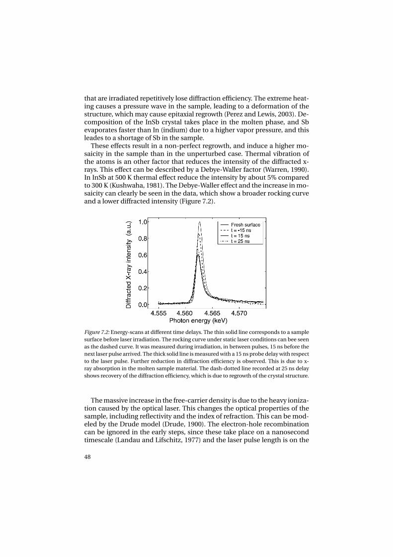
that are irradiated repetitively lose diffraction efficiency. The extreme heat-ing causes a pressure wave in the sample, leading to a deformation of thestructure, which may cause epitaxial regrowth (Perez and Lewis, 2003). De-composition of the InSb crystal takes place in the molten phase, and Sbevaporates faster than In (indium) due to a higher vapor pressure, and thisleades to a shortage of Sb in the sample.
These effects result in a non-perfect regrowth, and induce a higher mo-saicity in the sample than in the unperturbed case. Thermal vibration ofthe atoms is an other factor that reduces the intensity of the diffracted x-rays. This effect can be described by a Debye-Waller factor (Warren, 1990).In InSb at 500 K thermal effect reduce the intensity by about 5% comparedto 300 K (Kushwaha, 1981). The Debye-Waller effect and the increase in mo-saicity can clearly be seen in the data, which show a broader rocking curveand a lower diffracted intensity (Figure 7.2).
Figure 7.2: Energy-scans at different time delays. The thin solid line corresponds to a samplesurface before laser irradiation. The rocking curve under static laser conditions can bee seenas the dashed curve. It was measured during irradiation, in between pulses, 15 ns before thenext laser pulse arrived. The thick solid line is measured with a 15 ns probe delay with respectto the laser pulse. Further reduction in diffraction efficiency is observed. This is due to x-ray absorption in the molten sample material. The dash-dotted line recorded at 25 ns delayshows recovery of the diffraction efficiency, which is due to regrowth of the crystal structure.
The massive increase in the free-carrier density is due to the heavy ioniza-tion caused by the optical laser. This changes the optical properties of thesample, including reflectivity and the index of refraction. This can be mod-eled by the Drude model (Drude, 1900). The electron-hole recombinationcan be ignored in the early steps, since these take place on a nanosecondtimescale (Landau and Lifschitz, 1977) and the laser pulse length is on the
48

order of femtoseconds. In this approximation the total refractive index dueto the free electrons is given by
n = n0
√1+ (
ωp
n0ω)2(−1+ i
1
ωτe), (7.2.1)
where n0 is the complex lattice index of refraction, ωp the plasma frequency,ω the laser frequency and τe the electron collision time. From Equation 7.2.1it follows that the electron plasma acts as a mirror at the plasma frequency,when ωp = n0ω. Since the plasma frequency is dependent on the numberof free charge carriers, it is complicated to calculate the reflectivity dur-ing the laser radiation. The ionization of the sample is not proportional tothe laser fluence, due to multi-photon ionization and electron impact ion-ization, which both make the reflectivity hard to model. For studies of re-growth as presented in Paper 2, knowledge of the reflectivity is not crucial.However, the damage aspect is interesting, and measuring the change of re-flectivity in the sample is a way to determine the number of free electronsthrough the Drude model. This could be a way to perform experiments thatcould support, or overthrow, the secondary electron cascade calculationspresented in Papers 1 and 5, as well as (Ziaja et al., 2001, 2002, 2005), wherefew affirmative experiments are yet available.
49


8. Outlook
The impact of imaging non-crystalline samples at high resolutions iswidely expected to be substantial in a broad range of sciences andtechnologies. Advances made here have the potential to revolutionizestructural studies in many disciplines of science, similar to whatsynchrotron radiation once did. Experimental verification of the principleof single molecule diffraction imaging has now been performed. This wasdone at the first soft x-ray FEL in the world, the FLASH facility at DESY inHamburg. Experiments performed at FLASH taught us much about imagereconstruction, sample handling, and the damage caused by intense softx-ray pulses. The results fully support predictions, and indicate that aninterpretable diffraction pattern can be obtained before the sample turnsinto a plasma as a consequence of an exposure to an extremely intense andvery short soft x-ray pulse. This provides the first experimental proof forthe basic principle of single molecule imaging, showing that one can get aninterpretable diffraction pattern before sample explosion. The results haveimplication for imaging non-periodic molecular structures in biology andin any other area of science and technology where structural informationon the nanoscale is valuable.
Atomic resolution structural studies will require hard x-rays. LCLS, thefirst hard x-ray free-electron laser, will become operational in 2009, and willproduce intense hard x-ray pulses with wavelengths reaching about 1.0 Å.This XFEL will enable us to perform structural studies on single viral parti-cles or on large macromolecules at or near atomic resolutions for the firsttime. We will also be able to test theoretical predictions for radiation dam-age with hard x-rays, leading to a deeper understanding of the damage pro-cess and refinement of the theoretical models.
Studies on the behavior of the sample in vacuo are guiding thedevelopment of novel procedures and instruments for sample injection.For this end, it is necessary to gain a deeper understanding of thebehavior of biomolecules when injected into the vacuum chamber, and tounderstand and predict structural changes. There are a number of openquestions here, and these will require attention.
An important aspect of the single molecule imaging project is the fact thatit is multifaceted. One example of this relates to studies on structural tran-sitions in water, and certain aspects if this are presented in this thesis. The
51

Figure 8.1: The first photo-electron beam produced by the LCLS injector on April 5, 2007(www-ssrl.slac.stanford.edu/lcls/). The image shows the beam on a diagnostic viewer ap-proximately 80 cm from the electron gun. Beam energy is 5.5 MeV, pulse length is 6 ps. Thisa first step towards the first hard x-ray FEL in the world.
hard x-ray FEL will provide just the right tool to experimentally investigatefast structural changes induced by photons, and give us a deeper knowledgeof the properties of the most important liquid on Earth - water.
An example of an exciting spin-off of the single molecule imaging projectis the prospect of obtaining nuclear fusion with FEL radiation. From dam-age modeling for biomolecule imaging, it emerged that protons could beaccelerated to keV energies under certain conditions (Carlson, 2006). Bi-ological imaging with XFEL is at the front-end of high-energy density sci-ence, and spin-off are bound to appear and give rise to exciting new areasof research in natural sciences.
We can only speculate about the future possibilities of single particleimaging. Everything has worked so far, but will it work all the way? And if itdoes work all the way, and we have refined the method and reached atomicresolution, where do we go next? Femtosecond time-resolved imaging ofthe chemical processes? High-resolution structures of cells? Pump-probeexperiments on photosynthesis in situ?
52

This is the first thesis from the project on single molecule diffractiveimaging, and it shows that some parts of the big puzzle are fitting together,and the motif that is appearing is really starting to look promising!
53


9. Sammanfattning på svenska
För att kunna förstå hur någonting fungerar är det en stor fördel att veta hurdet ser ut. Detta gäller såväl i en klocka som i människokroppen. Ett stortområde inom biokemi är därför strukturbiologi, inom vilket man försökerbestämma hur viktiga biomolekyler ser ut och hur de fungerar.
Idag strukturbestäms biomolekyler till stor utsträckning med hjälp av enteknik som heter röntgenkristallografi. Tekniken bygger på att molekylensom ska strukturbestämmas, till exempel ett protein, kristalliseras så attett stort antal identiska molekyler sitter ordnade i ett tredimesionell mön-ster så att de bildar en kristallstruktur. Kristallen placeras i en röntgenstrålefrån en synkrotronanläggning och en diffraktionsbild av kristallen gener-eras. I det ideala fallet innehåller denna bild all nödvändig information föratt strukturbestämma molekylen.
Att utsätta ett material för röntgenljus medför att man joniserardet. Genom att fotonerna i röntgenpulsen slår ut elektronerna runtatomkärnorna joniseras dessa. I ett starkt joniserat prov förstörs molekyl-strukturen på grund av de krafter som uppstår mellan de laddade jonerna.Då man använder sig av en kristall blir effekten av joniseringsskadan inteså allvarlig. Detta beror på att skadan sker slumpmässigt över ett stort antalmolekyler och diffraktionsbilden är ett medelvärde över dessa molekyler.
Flera av stegen i röntgenkristallografi är svåra, och ibland omöjliga, attutföra. Ett av de största problemen är att det är svårt att kristallisera mångatyper av biomolekyler. En teknik som tillåter strukturbestämning utankristallisering skulle således vara till stor nytta.
Det har förutspåtts att det med hjälp av en mycket kort och intensivröntgenpuls kan vara möjligt att generera en diffraktionsbild från, ochdärmed också strukturbestämma, ett icke-kristallint prov. Vill man undvikakristalliseringen måste man dock hitta ett sätt att komma runt problemetmed att röntgenjoniseringen förstör molekylstrukturen. Idén är att ompulsen är tillräckligt kort kommer hela pulsen att kunna passera provetinnan det exploderar på grund av joniseringen. Man kan se det somatt man använder sig av en kamera med väldigt kort slutartid: vill manfotografera ett föremål i rörelse gäller det att använda en slutartid som ärså kort att föremålet inte hinner röra sig såmycket att bilden blir suddig.Figur 5.4, i kapitel 5 illustrerar en vattendroppe som joniseras av en kortröntgenpuls. Om pulsen hinner gå igenom provet innan det explodera
55

(bilden till vänster) får man en tydlig bild. Använder man sig istället av enlängre puls kommer bilden att bli suddig.
Idén om strukturbestämning av icke-kristallina molekyler med hjälp avkorta röntgenpulsar är bakgrunden till denna avhandling. Möjlighetenatt kunna strukturbestämma vilken biomolekyl som helst skulle kunna fåen enorm betydelse för biokemin och i förlängningen för den medicinskavetenskapen. Det är ett stort projekt att utveckla en teknik vilken görstrukturbestämning av molekyler utan att kristallisering möjlig. Enhel rad problem återstår innan vi kommer att kunna lösa den förstamolekylstrukturen. Bild 1.1 illustrerar hur det tänkta experimentet skullekunna gå till.
Vad denna avhandling presenterar är ett par pusselbitar, vilka kommeratt bidra till den slutgiltiga förståelsen för hur en sådan metod skallfungera. Två områden inom ämnet presenteras i denna avhandling:hantering av biomolekyler i vakuum och fotoners (från röntgen- ellerlaserkällor) påverkan på materia.
För att kunna genomföra strukturbestämning av icke-kristallinamolekyler behöver man leverera provmolekyler från en buffertlösningtill röntgenstrålen på ett kontrollerbart och repetativt sätt. Eftersom alltsom kommer i röntgenljusets väg bidrar till diffraktionsbilden, utförsdiffraktionsexperimentet i vakuum. Ett sätt att leverera prov från lösning ini vakuum är att använda sprayteknik, vilket innebär att man sprayar smådroppar som innehåller provet (molekylen) innesluten i buffertlösning,t.ex. vatten.
Att använda en sådan teknik har flera fördelar. Dels är det troligt att vat-tendroppen mer liknar molekylens naturliga miljö än vad vakuum gör. Delshar teoretiska beräkningar visat att ett omslutande vattenlager skulle kunnaha en fördröjande effekt på explosionen orsakad av joniseringen. Två delar-beten inkluderade i denna avhandling (artikel 3 och 6) undersöker hur småvattendroppar beter sig i vakuum. Detta är intressant eftersom förändringari det omslutande vattnet även påverkar molekylen. Våra resultat visar attavdunstningen från dropparna sänker temperaturen (eftersom det är devarma vattenmolekylerna i vatten droppen som avdunstar). Då tempera-turen sänks upphör avdunstningen. Detta innebär att man genom att vetatemperaturen på vattendropparna då de sprayas in i vakuum kan förutspåhur mycket vatten som kommer att finnas kvar i provet då avdunstningenupphör.
Den typen av röntgenkälla, hårdröntgen-frielektronlaser, som behövs föratt kunna strukturbestämma icke-kristalliserade molekyler, finns ännu intetillgänglig. Två anläggningar är dock under konstruktion, en i Stanford iKalifornien och en i Hamburg, Tyskland. Dessa kommer, om allt går enligtplanerna, stå klara 2009 respektive 2013. Detta innebär att det hittills harvarit svårt att utföra testexperiment för att kunna förstå vad som händermed ett prov då det placeras i en kort intensiv röntgenpuls. För att kunna
56
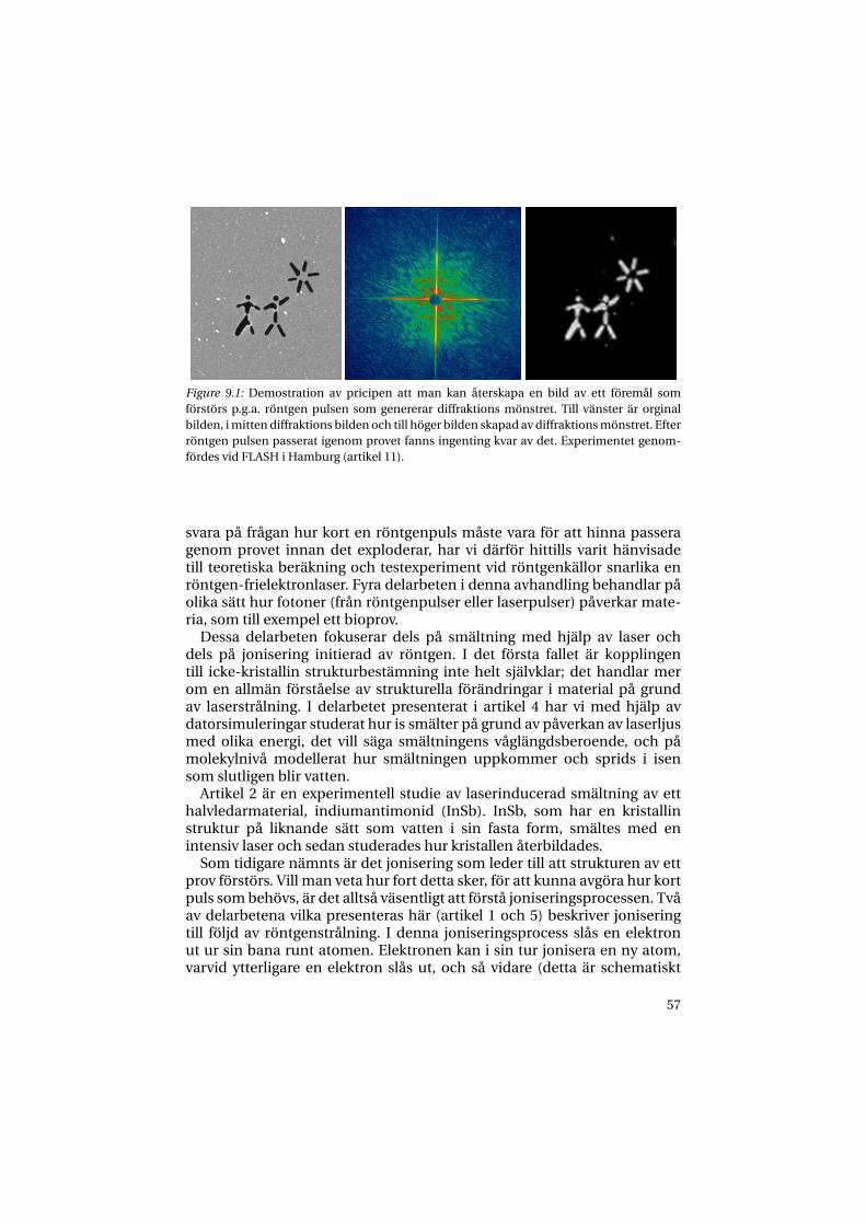
Figure 9.1: Demostration av pricipen att man kan återskapa en bild av ett föremål somförstörs p.g.a. röntgen pulsen som genererar diffraktions mönstret. Till vänster är orginalbilden, i mitten diffraktions bilden och till höger bilden skapad av diffraktions mönstret. Efterröntgen pulsen passerat igenom provet fanns ingenting kvar av det. Experimentet genom-fördes vid FLASH i Hamburg (artikel 11).
svara på frågan hur kort en röntgenpuls måste vara för att hinna passeragenom provet innan det exploderar, har vi därför hittills varit hänvisadetill teoretiska beräkning och testexperiment vid röntgenkällor snarlika enröntgen-frielektronlaser. Fyra delarbeten i denna avhandling behandlar påolika sätt hur fotoner (från röntgenpulser eller laserpulser) påverkar mate-ria, som till exempel ett bioprov.
Dessa delarbeten fokuserar dels på smältning med hjälp av laser ochdels på jonisering initierad av röntgen. I det första fallet är kopplingentill icke-kristallin strukturbestämning inte helt självklar; det handlar merom en allmän förståelse av strukturella förändringar i material på grundav laserstrålning. I delarbetet presenterat i artikel 4 har vi med hjälp avdatorsimuleringar studerat hur is smälter på grund av påverkan av laserljusmed olika energi, det vill säga smältningens våglängdsberoende, och påmolekylnivå modellerat hur smältningen uppkommer och sprids i isensom slutligen blir vatten.
Artikel 2 är en experimentell studie av laserinducerad smältning av etthalvledarmaterial, indiumantimonid (InSb). InSb, som har en kristallinstruktur på liknande sätt som vatten i sin fasta form, smältes med enintensiv laser och sedan studerades hur kristallen återbildades.
Som tidigare nämnts är det jonisering som leder till att strukturen av ettprov förstörs. Vill man veta hur fort detta sker, för att kunna avgöra hur kortpuls som behövs, är det alltså väsentligt att förstå joniseringsprocessen. Tvåav delarbetena vilka presenteras här (artikel 1 och 5) beskriver joniseringtill följd av röntgenstrålning. I denna joniseringsprocess slås en elektronut ur sin bana runt atomen. Elektronen kan i sin tur jonisera en ny atom,varvid ytterligare en elektron slås ut, och så vidare (detta är schematiskt
57

illustrerat i figur 5.2 i kapitel 5). Denna kaskad av joniseringar (utlösta avelektroner) är en viktig del av den röntgeninducerade skadan i provet. Vihar gjort beräkningar på fyra testsystem kaliumjodid (KI) och cesiumjodid(CsI), samt is och vatten. Vatten och is valdes för att det har en densitet somliknar biomolekylers och för att vatten är, som tidigare nämnts, en poten-tiell provbärare i vakuum. KI och CsI valde vi för att de har relevans för enannan del av projektet, de är nämligen båda goda fotokatoder.
Våra beräkningar för KI och CsI visar resultat som pekar på att KI är ennågot bättre fotokatod för att nå en hög tidsupplösning. KI genererar näm-ligen elektroner med en smalare energifördelning, vilket är en viktig egen-skap hos en snabb fotokatod.
För vatten visar våra resultat att varje röntgenfotonjonisering leder till 20elektronjoniseringar. Vi ser också att elektronjonisering sker inom loppetav 0,1 ps1. Dessa resultat ligger till grund för beräkningar av hur kort rönt-genpulsen måste om man ska kunna hinna ta en bild av molekylen innanden förstörs p.g.a. joniseringen. Resultaten pekar på att en puls kortare än0,1 ps behövs för att det ska vara möjligt.
Hurvida våra beräkningar stämmer och vi en dag kommer att kunna lösamolekylstrukturer med hjälp av röntgen-frielektronlasrar får framtidenutvisa. De steg vi tagit hittills, av vilka denna avhandling är en del, visar allaatt vi är på rätt väg. Vilken effekt möjligheten att kunna strukturbestämmaicke-kristallina prov kommer att få på naturvetenskapen kan vi bara siaom. Kanske kommer vi i framtiden att kunna skapa bilder av celler medatomär upplöning? Eller i detalj kunna studera hur kemin i kroppenfungerar och på så sätt kunna skapa bättre läkemedel? Det enda vi vetsäkert är att vi står inför en mycket spännande framtid.
1d.v.s. 0,0000000000001 sekund
58

Acknowledgements
It has been four years, eight months and twentysix days since I first en-tered the building known for its ugliness, BMC, and met Prof. J. Hajdu, whogreeted me with an open hand and a smile, and said ”We need people withyour experience and we’ve got money...”.
It has really been a privilege to work in a project as exiting as this and to besurrounded by such wonderful people. I would like to thank you all, those ofyou who supported me in science or in friendship or who just smiled at mein the BMC corridors or at the badge office. To make the acknowledgementschapter shorter than the thesis itself, I can not mention you all. However,by starting to thank the Swedish and the American taxpayers for financialsupport I guess I covered most of you. Nic once calculated how much of hispaycheck that went into mine, and he concluded that it was not much to bethankful for, but that’s Nic.
So here we go. Time for the most read part of any thesis, but also the partthat most authors spend the least time to write - since it is written hoursbefore submission. Thank you for:
59

60
The author’s contribution
Publications included in this thesis
Paper 1:N. Tîmneanu et al. “Auger electron cascades in water and ice”I was deeply invloved in all parts of this project: planning, calulations, anal-ysis and writing of the paper.
Paper 2:M. Harbst et al. “Studies of resolidification of non-thermally molten InSb usingtime-resolved X-ray diffraction”This project was M. Harbst’s and J. Larsson’s idea and planning. It was myfirst experimental work and I was involved in the set up and performing ofthe actual experiment.
Paper 3:C. Caleman and D. van der Spoel ” Temperature and structural changes of waterclusters in vacuum due to evaporation”This project originates from an idea of mine that D. van der Spoel believedin. I did almost all of the work.
Paper 4:C. Caleman and D. van der Spoel ”Picosecond Melting of Ice by an Infrared LaserPulse” The idea for this project originates form an experiment we wanted todo at SPPS. I did all the simulations, most of the data analysis and most ofthe writing of the paper.
61

Paper 5:C. Ortiz and C. Caleman “Secondary electron cascade dynamics in KI and CsI”During the work with Paper 1 I realized it was a limitation of the methodto be dependent on experimental data. Talking to C. Ortiz I realized that hecould provide calculated energy loss functions. At the same time I was askedabout the secondary electron cascades in photo cathode materials, so wedecided on performing the calculations. The work behind the final paperis equally divided between the authors, but my focus was on the electroncalculations.
Paper 6:C. Caleman and D. van der Spoel ”Evaporation from water clusters containingsingly charged ions”This is a continuation of Paper 3. I did all the simulations, most of the dataanalysis and most of the writing.
Supporting publications
Paper 7:A. L. Cavalieri et al. “Clocking Femtosecond X Rays”I was at SPPS doing mainly other experiments and I help A. L. Cavalieri ex-perimentally when that was required.
Paper 8:A. M. Lindenberg et al. “Atomic-Scale Visualization of Inertial Dynamics”I was involved in the preparation and implementation of the experiment atSPPS.
Paper 9:K. J. Gaffney et al. “Observation of Structural Anisotropy and the Onset of Liquid-like Motion During the Nonthermal Melting of InSb”Based on the same experiment as Paper 8, and my involvement in this paperwas the same.
62
Paper 10:J. Larsson et al. “Opportunities and challenges using short-pulse X-ray sources”Based on the same experiment as Paper 8, and my involvment in this paperwas the same.
Paper 11:H. N. Chapman et al. “Femtosecond diffractive imaging with a soft-X-ray free-electron laser”I was involved in the early stages of the sample preparations and the actualexperiment.
Paper 12:S. P. Hau-Riege et al. “Subnanometer-scale Measurements of the Interaction of Ul-trafast Soft X-ray Free-Electron-Laser Pulses with Matter”I was involved in the preparation and execution of the experiment, and Iwas also somewhat involved in the writing process.
Paper 13:S. P. Hau-Riege et al. “Damage threshold of inorganic solids under free-electron-laser irradiation at 32.5 nm wavelength”I was involved in the preparation and execution of the experiment, and Iwas also somewhat involved in the writing process.
Paper 14:J. Chalupsky et al. “Characteristics of focused soft X-ray freeelectron laser beamdetermined by ablation of organic molecular solids”I was involved in the preparation and execution of the experiment.
Paper 15:H. N. Chapman et al. “Femtosecond Time-Delay X-ray Holography”I was involved in the early stages of the sample preparations and the actualexperiment.
Paper 16:J. Chalupsky et al. “Ablation of organic molecular solids by focused soft X-ray free-electron laser radiation”
63

This work is based on the same experiment as Paper 16. I was involved inthe preparation and execution of the experiment.
64

References
Abbas, S., Ahlström, P., Nordholm, S., 1995. Molecular dynamnics simula-tions of surface tension for polar molecules. Correction for long-rangeinteraction by generalized van der waals theory. Acta Chem. Scand. 49,182–188.
Akre, R., Bentson, L., Emma, P., Krejcik, P., 2002. Bunch length measure-ments using a transverse rf deflecting structure in the slac linac. In: Gar-vey, T., Duff, J. L., Roux, P. L., Petit-Jean-Genaz, C., Poole, J., Rivkin, L.(Eds.), Proceedings of European Particle Accelerator Conference, 2002,Paris, France. CERN, Geneve, pp. 1883–1884.
Allen, M. P., Tildesley, D. J., 1987. Computer Simulations of Liquids. OxfordScience Publications, Oxford.
Andruszkow, J., Aune, B., Ayvazyan, V., Baboi, N., Bakker, R., Balakin, V.,Barni, D., Bazhan, A., Bernard, M., Bosotti, A., Bourdon, J. C., Brefeld,W., Brinkmann, R., Buhler, S., Carneiro, J.-P., Castellano, M., P. Castro,L. C., Chel, S., Cho, Y., Choroba, S., Colby, E. R., Decking, W., Hartog,P. D., Desmons, M., Dohlus, M., Edwards, D., Edwards, H. T., Faatz, B.,Feldhaus, J., Ferrario, M., Fitch, M. J., Flöttmann, K., Fouaidy, M., Gamp,A., Garvey, T., Gerth, C., Geitz, M., Gluskin, †. E., Gretchko, V., Hahn, U.,Hartung, W. H., Hubert, D., Hning, M., Ischebek, R., Jablonka, M., Joly,J. M., Juillard, M., Junquera, T., Jurkiewicz, P., Kabel, A., Kahl, J., Kaiser,H., Kamps, T., Katelev, V. V., Kirchgessner, J. L., Körfer, M., Kravchuk, L.,Kreps, G., Krzywinski, J., Lokajczyk, T., Lange, R., Leblond, B., Leenen, M.,Lesrel, J., Liepe, M., Liero, A., Limberg, T., Lorenz, R., Hua, L. H., Hai, L. F.,Magne, C., Maslov, M., Materlik, G., Matheisen, A., Menzel, J., Michelato,P., Möller, W.-D., Mosnier, A., Müller, U.-C., Napoly, O., Novokhatski, A.,Omeich, M., Padamsee, H. S., Pagani, C., Peters, F., Petersen, B., Pierini,P., Pfülger, J., Piot, P., Ngoc, B. P., Plucinski, L., Proch, D., Rehlich, K., Re-iche, S., Reschke, D., Reyzl, I., Rosenzweig, J., Rossbach, J., S. Roth, E. L. S.,Sandner, W., Sanok, Z., Schlarb, H., Schmidt, G., Schümser, P., Schnei-der, J. R., Schneidmiller, E. A., Schreiber, H.-J., Schreiber, S., P. Schütt, .J. S., Serafini, L., Sertore, D., Setzer, S., Simrock, S., Sonntag, B., Sparr, B.,Stephan, F., Sytchev, V. A., Tazzari, S., Tazzioli, F., Tigner, M., Timm, M.,Tonutti, M., Trakhtenberg, E., Treusch, R., Trines, D., Verzilov, V., Vielitz,
65

T., Vogel, V., v. Walter, G., Wanzenberg, R., Weiland, T., Weise, H., Weisend,J., Wendt, M., Werner, M., White, M. M., Will, I., Wolff, S., Yurkov, M. V.,Zapfe, K., Zhogolev, P., Zhou, F., 2000. First observation of self-amplifiedspontaneous emission in a free-electron laser at 109 nm wavelength.Phys. Rev. Lett. 85, 3825–3829.
Ashley, J. C., 1990. Energy loss rate and inelastic mean free path of low-energy electrons and positrons in condensed matter. J. Electron Spec-trosc. Relat. Phenom 50, 323–334.
Ashley, J. C., 1991. Energy-loss probabilities for electrons, positrons, andprotons in condensed matter. J. Appl. Phys 69, 674–678.
Auger, P., Ehrenfest, P., Maze, R., Daudin, J., Fréon, R. A., 1939. Extensivecosmic-ray showers. Rev. Mod. Phys. 11, 288–291.
Ayvazyan, V., Baboi, N., Bhr, J., Balandin, V., Beutner, B., Brandt, A., Bohnet,I., Bolzmann, A., Brinkmann, R., Brovko, O., Carneiro, J., Casalbuoni, S.,Castellano, M., Castro, P., Catani, L., Chiadroni, E., Choroba, S., Cianchi,A., Delsim-Hashemi, H., Pirro, G. D., Dohlus, M., Düsterer, S., Edwards,H., Faatz, B., Fateev, A., Feldhaus, J., Flöttmann, K., Frisch, J., Fröhlich,L., Garvey, T., Gensch, U., Golubeva, N., Grabosch, H.-J., Grigoryan, B.,Grimm, O., Hahn, U., Han, J., v. Hartrott, M., Honkavaara, K., Hüning,M., Ischebeck, R., Jaeschke, E., Jablonka, M., Kammering, R., Katalev,V., Khodyachykh, B. K. S., Kim, Y., Kocharyan, V., Körfer, M., Kollewe,M., Kostin, D., Krämer, D., Krassilnikov, M., Kube, G., Lilje, L., Limberg,T., Lipka, D., Löhl, F., Luong, M., Magne, C., Menzel, J., Michelato, P.,Miltchev, V., Minty, M., Möller, W., Monaco, L., Müller, W., Nagl, M.,Napoly, O., Nicolosi, P., Nölle, D., Nuñez, T., Oppelt, A., Pagani, C., Pa-parella, R., Petersen, B., Petrosyan, B., Pflüger, J., Piot, P., Plönjes, E., Po-letto, L., Proch, D., Pugachov, D., Rehlich, K., Richter, D., Riemann, S.,Ross, M., Rossbach, J., Sachwitz, M., E.L.Saldin, Sandner, W., Schlarb, H.,Schmidt, B., Schmitz, M., Schümser, P., Schneider, J., Schneidmiller, E.,Schreiber, H.-J., Schreiber, S., Shabunov, A., Sertore, D., Setzer, S., Sim-rock, S., Sombrowski, E., Staykov, L., Steffen, B., Stephan, F., Stulle, F.,Sytchev, K., Thom, H., Tiedtke, K., Tischer, M., Treusch, R., Trines, D.,Tsakov, I., Vardanyan, A., Wanzenberg, R., Weiland, T., Weise, H., Wendt,M., Will, I., Winter, A., Wittenburg, K., Yurkov, M., Zagorodnov, I., Zam-bolin, P., Zapfe, K., 2006. First operation of a free-electron laser generatinggw power radiation at 32 nm wavelength. Eur. J. Phys. D. 37, 297–303.
Ayvazyan, V., Baboi, N., Bohnet, I., Bolzmann, A., Brinkmann, R., Castro,P., Catani, L., Choroba, S., Cianchi, A., Dohlus, M., Edwards, H., Faatz,B., Fateev, A., Feldhaus, J., Flöttmann, K., Gamp, A., Garvey, T., Genz, H.,Gerth, C., Gretchko, V., Grigoryan, B., Hahn, U., Hessler, C., Honkavaara,
66

K., Hüning, M., Ischebeck, R., Jablonka, M., Kamps, T., Körfer, M., Kras-silnikov, M., Krzywinski, J., Liepe, M., Liero, A., Limberg, T., Loos, H., Lu-ong, M., Magne, C., Menzel, J., Michelato, P., Minty, M., Müller, W., Nölle,D., Novokhatski, A., Pagani, C., Peters, F., Pflüger, J., Piot, P., Plucinski,L., Rehlich, K., Reyzl, I., Richter, A., Rossbach, J., E.L.Saldin, Sandner, W.,Schlarb, H., Schmidt, B., Schmitz, M., Schümser, P., Schneider, J., Schnei-dmiller, E., Schreiber, H.-J., Schreiber, S., Sertore, D., Setzer, S., Simrock,S., Sobierajski, R., Sonntag, B., Steed, B., F. Stephan, K. S., Tiedtke, K.,Tonutti, M., Treusch, R., Trines, D., Türke, D., Verzilov, V., Wanzenberg, R.,Weiland, T., Weise, H., Wendt, M., Will, I., Wolff, S., Wittenburg, K., Yurkov,M., Zapfe, K., 2002a. Generation of gw radiation pulses from a vuv free-electron laser operating in the femtosecond regime. Phys. Rev. Lett. 88,104802.
Ayvazyan, V., Baboi, N., Bohnet, I., Brinkmann, R., Castellano, M., Castro, P.,Catani, L., Choroba, S., Cianchi, A., Dohlus, M., Edwards, H. T., Faatz, B.,Fateev, A. A., Feldhaus, J., Flöttmann, K., Gamp, A., Garvey, T., Genz, H.,Gerth, C., Gretchko, V., Grigoryan, B., Hahn, U., Hessler, C., Honkavaara,K., Hüning, M., Ischebeck, R., Jablonka, M., Kamps, T., Körfer, M., Krassil-nikov, M., Krzywinski, J., Liepe, M., Liero, A., Limberg, T., Loos, H., Luong,M., Magne, C., Menzel, J., Michelato, P., Minty, M., Müller, U.-C., Nölle,D., Novokhatski, A., Pagani, C., Peters, F., Pflüger, J., Piot, P., Plucinski,L., Rehlich, K., Reyzl, I., Richter, A., Rossbach, J., Saldin, E. L., Sandner,W., Schlarb, H., Schmidt, G., Schmüser, P., Schneider, J. R., Schneidmiller,E. A., Schreiber, H.-J., Schreiber, S., Sertore, D., Setzer, S., Simrock, S., So-bierajski, R., Sonntag, B., Steeg, B., Stephan, F., Sytchev, K. P., Tiedtke, K.,Tonutti, M., Treusch, R., Trines, D., Türke, D., Verzilov, V., Wanzenberg,R., Weiland, T., Weise, H., Wendt, M., Wilhein, T., Will, I., Wittenburg, K.,Wolff, S., Yurkov, M. V., Zapfe, K., 2002b. A new powerful source for coher-ent vuv radiation: Demonstration of exponential growth and saturationat the ttf free-electron laser. Eur. J. Phys. D. 20, 149–156.
Bartell, L. S., Huang, J., 1994. Supercooling of water below the anomalousrange near 226 k. J. Phys. Chem. 98, 7455–7457.
Bentson, L., Emma, P., Krejcik, P., 2002. A new bunch compressor chicanefor the slac linac to produce 30-fsec, 30-ka, 30-gev electron bunches. In:Garvey, T., Duff, J. L., Roux, P. L., Petit-Jean-Genaz, C., Poole, J., Rivkin,L. (Eds.), Proceedings of European Particle Accelerator Conference, 2002,Paris, France. CERN, Geneve, pp. 683–685.
Berendsen, H. J. C., Postma, J. P. M., van Gunsteren, W. F., Hermans, J., 1981.Interaction models for water in relation to protein hydration. In: Pull-man, B. (Ed.), Intermolecular Forces. D. Reidel Publishing Company, Dor-drecht, pp. 331–342.
67
Berendsen, H. J. C., van der Spoel, D., van Drunen, R., 1995. GROMACS:A message-passing parallel molecular dynamics implementation. Comp.Phys. Comm. 91, 43–56.
Bergh, M., Tîmneanu, N., van der Spoel, D., 2004. A model for the dynamicsof a water cluster in a X-ray FEL beam. Phys. Rev. E 70, 051904.
Bernal, J. D., Fankuchen, I., Perutz, M. F., 1938. An x-ray study of chy-motrypsin and haemoglobin. Nature 141, 523–524.
Bethe, H., 1930. Zur theorie des durchgangs schneller korpuskularstrahlendurch materie. Ann. Phys. 5, 325–400.
Bragg, L., Perutz, F. M., 1952. The structure of haemoglobin. Proc. R. Soc.London Ser. A 213, 425–435.
Brook, E., Harrison, M. F. A., Smith, A. C. H., 1978. Measurements of theelectron impact ionisation cross sections of he, c, 0 and n atoms. btjphb11, 3115–3132.
Bukowski, R., Szalewicz, K., Groenenboom, G. C., van der Avoird, A., 2007.Predictions of the properties of water from first principles. Science 315,1249–1252.
Carlson, L.-A., 2006. On the feasibility of nuclear fusion experiments withxuv and x-ray free-electron lasers (UPTEC X 06 024). Master’s thesis, Up-psala University.
Cavalieri, A. L., Fritz, D. M., Lee, S. H., Bucksbaum, P. H., Reis, D. A., Rudati,J., Mills, D. M., Fuoss, P. H., Stephenson, G. B., Kao, C. C., Siddons, D. P.,Lowney, D. P., MacPhee, A. G., Weinstein, D., Falcone, R. W., Pahl, R., Als-Nielsen, J., Blome, C., Düsterer, S., Ischebeck, R., Schlarb, H., Schulte-Schrepping, H., Tschentscher, T., Schneider, J., Hignette, O., Sette, F.,Sokolowski-Tinten, K., Chapman, H. N., Lee, R. W., Hansen, T. N., Syn-nergren, O., Larsson, J., Techert, S., Sheppard, J., Wark, J. S., Bergh, M.,Caleman, C., Huldt, G., van der Spoel, D., Timneanu, N., Hajdu, J., Akre,R. A., Bong, E., Emma, P., Krejcik, P., Arthur, J., Brennan, S., Gaffney, K. J.,Lindenberg, A. M., Luening, K., Hastings, J. B., 2005. Clocking femtosec-ond x rays. Phys. Rev. Lett. 94, 114801.
Creuzburg, M., 1966. Energieverlustspektren der alkalihalogenide und dermetalle cu, ag und au und vergleich mit optischen messungen. Z. Phys.196, 433–463.
Darden, T., York, D., Pedersen, L., 1993. Particle mesh Ewald: An N-log(N)method for Ewald sums in large systems. J. Chem. Phys. 98, 10089–10092.
Dobson, C. M., 2004. Chemical space and biology. Nature 432, 824–828.
68

Dole, M., Mach, L. L., Hines, R. L., Ferguson, L. P., Alice, M. B., 1968. Molec-ular beams of macroions. J. Chem. Phys. 49, 2240–2249.
Drude, P. K. L., 1900. Zur elektronentheorie der metalle. Annalen der Physik306, 566–613.
Düsterer, S., Radcliffe, P., Geloni, G., Jastrow, U., Kuhlmann, M., Plönjes,E., Tiedtke, K., Treusch, R., a nd P. Nicolosi, J. F., Poletto, L., Yeates, P.,Luna, H., Costello, J. T., Orr, P., Cubaynes, D., Meyer, M., 2006. Spectro-scopic characterization of vacuum ultraviolet free electron laser pulses.Opt. Lett. 31, 1750–1752.
Emma, P., 2002. Accelerator physics challenges of x-ray fel sase sources. In:Garvey, T., Duff, J. L., Roux, P. L., Petit-Jean-Genaz, C., Poole, J., Rivkin,L. (Eds.), Proceedings of European Particle Accelerator Conference, 2002,Paris, France. CERN, Geneve, pp. 49–53.
Ewald, P. P., 1921. Die Berechnung optischer und elektrostatischer Gitterpo-tentiale. Ann. Phys. 64, 253–287.
Fermi, E., 1924. Z. Phys. 29, 315.
Frank, J., 1996. Three-Dimensional Electron Microscopy of Macromolecu-lar Assemblies. Academic Press, San Diego.
Fritz, D. M., Reis, D. A., Adams, B., Akre, R. A., Arthur, J., Blome, C., Bucks-baum, P. H., Cavalieri, A. L., Engemann, S., Fahy, S., Falcone, R. W., Fu-oss, P. H., Gaffney, K. J., George, M. J., Hajdu, J., Hertlein, M. P., Hillyard,P. B., von Hoegen, M. H., Kammler, M., Kaspar, J., Kienberger, R., Krej-cik, P., Lee, S. H., Lindenberg, A. M., McFarland, B., Meyer, D., Montagne,T., Murray, E. D., Nelson, A. J., Nicoul, M., Pahl, R., Rudati, J., Schlarb,H., Siddons, D. P., Sokolowski-Tinten, K., Tschentscher, T., von der Linde,D., Hastings, J. B., 2007. Ultrafast bond softening in bismuth: Mapping asolid’s interatomic potential with x-rays. Science 315, 633–636.
Gerchberg, R. W., Saxton, W. O., 1972. A practical algorithm for the deter-mination of phase from image and diffraction plane pictures. Optik 35,237–246.
Guillot, B., 2002. A reappraisal of what we have learnt during three decadesof computer simulations on water. J. Mol. Liq. 101, 219–260.
Hajdu, J., 1990. Laue crystallography of macromolecules. In: Jensen, B., Jør-gensen, F. S., Kofod, H. (Eds.), Frontiers in drug research, Alfred BenzonSymposium 28. Munksgaard, Copenhagen, pp. 375–395.
69

Hajdu, J., 1994. Towards femtosecond time resolution in broad-band lauediffraction studies - a possibility. In: Arthur, J., Materlik, G., Winnick,H. (Eds.), Scientific Applications of Coherent X-rays. SLAC/SSRL-0066,SLAC, pp. 91–94.
Hajdu, J., 2000. Single-molecule x-ray diffraction. Curr. Op. Struct. Biol. 10,569–573.
Hajdu, J., Hodgson, K., Miao, J., van der Spoel, D., Neutze, R., Robinson,C. V., Faigel, G., Jacobsen, C., Kirz, J., Sayre, D., Weckert, E., Materlik, G.,Szoke, A., 2000. Structural studies on single particles and biomolecules.In: LCLS: The First Experiments. SSRL, SLAC, Stanford, USA, pp. 35–62.
Hau-Riege, S. P., London, R. A., Huldt, G., Chapman, H. N., 2005. Pulse re-quirements for x-ray diffraction imaging of single biological molecules.Phys. Rev. E 71, 061919.
Hau-Riege, S. P., London, R. A., Szoke, A., 2004. Dynamics of biological mo-lecules irradiated by short x-ray pulses. Phys. Rev. E 69, 051906.
Hau-Riege, S. P., London, R. A., Szoke, A., 2004. Dynamics of biological mo-lecules irradiated by short x-ray pulses. Phys. Rev. E 69, 051906.
Hayward, S., Kitao, A., Berendsen, H. J. C., 1997. Model-free method of ana-lyzing domain motions in proteins from simulation: a comparison of nor-mal mode analysis and molecular dynamics methods. PROTEINS: Struct.Funct. Gen. 27, 425–437.
Hedin, L., 1965. New method for calculating the one-particle green’s func-tion with application to the electron-gas problem. Phys. Rev. 139, A796–A823.
Henderson, R., 1995. The potential and limitations of neutrons, electronsand X-rays for atomic resolution microscopy of unstained biological mo-lecules. Quart. Rev. Biophys. 28, 171–193.
Henke, B. L., 1972. Ultrasoft-x-ray reflection, refraction, and production ofphotoelectrons (100-1000-ev region). Phys. Rev. A 6, 94–104.
Henke, B. L., Liesegang, J., Smith, S. D., 1979. Soft-x-ray-induced secondary-electron emission from semiconductors and insulators: Models and mea-surements. Phys. Rev. B 19, 3004–3021.
Hentschel, M., Kienberger, R., Spielmann, C., Reider, G. A., Milosevic, N.,Brabec, T., Corkum, P., Heinzmann, U., Drescher, M., Krausz, F., 2001. At-tosecond metrology. Nature 414, 509–513.
Hockney, R. W., Eastwood, J. W., 1981. Computer simulation using particles.McGraw-Hill, New York.
70

Huang, J., Bartell, L. S., 1995. Kinetics if homogeneous nucleation in thefreezing of large water clusters. J. Phys. Chem. 99, 3924–3931.
Hubbell, J. H., Veigele, W. J., Briggs, E. A., Brown, R. T., Cromer, D. T., Hower-ton, R. J., 1975. Atomic form factors, incoherent scattering functions, andphoton scattering cross sections. J. Phys. Chem. Ref. Data 4, 471–494.
Huldt, G., Szoke, A., Hajdu, J., 2003. Diffraction imaging of single particlesand biomolecules. J. Struct. Biol. 144, 219–227.
Iavarone, A., Parks, J., 2005. Conformational change in unsolvated trp-cageprotein probed by fluorescence. J. Am. Chem. Soc. 127, 8606–8607.
Iglev, H., Schmeisser, M., Simeonidis, K., Thaller, A., Laubereau, A., 2006.Ultrafast superheating and melting of bulk ice. Nature 439, 183–186.
Jarrold, M. F., 1999. Unfolding, refolding and hydration of protein ions inthe gas phase. Acc. Chem. Res. 32, 360–367.
Jorgensen, W. L., Chandrasekhar, J., Madura, J. D., Impey, R. W., Klein, M. L.,1983. Comparison of simple potential functions for simulating liquid wa-ter. J. Chem. Phys. 79, 926–935.
Jurek, Z., Faigel, G., Tegze, M., 2004a. Dynamics in a cluster under the in-fluence of intense femtosecond hard X-ray pulses. Eur. Phys. J. D 29, 217–229.
Jurek, Z., Oszlányi, G., Faigel, G., 2004b. Imaging atom clusters by hard x-rayfree-electron lasers. Europhys. Lett. 65, 491–497.
Kaku, M., 1993. Quantum Field Theory. Oxford University Press, New York.
Kane, E. O., 1966. Simple model for collision effects in photoemission. Phys.Rev. 147, 335.
Kebarle, P., Tang, L., 1993. From ions in solution to ions in the gas-phase- the mechanism of electrospray mass-spectrometry. Anal. Chem. 65,972A–986A.
Key, M. H., Lewis, C. L. S., Lunney, J. G., Moore, A., Ward, M., Thareja, R. K.,1980. Time-resolved x-ray spectroscopy of laser-produced plasmas. Phys.Rev. Lett. 44, 1669–1672.
Kim, Y.-K., Rudd, M. E., 1994. Binary-encounter-dipole model for electron-impact ionization. Phys. Rev. A 50, 3954.
Kittel, C., 1996. Introduction to Solid State Physics. John Wiley & Sons Inc.,New York.
71

Kohn, W., Sham, L. J., 1965. Self-consistent equations including exchangeand correlation effects. Phys. Rev. 140, a1133.
Krause, M. O., Oliver, J. H., 1979. Natural width of atomic K and L level Kα
X-ray lines and several KLL Auger lines. J. Phys. Chem. Ref. Data 8, 329–338.
Kushwaha, M. S., 1981. Compressibilities, debye-waller factors, and melt-ing criteria for ii-vi and iii-v compound semiconductors. Phys. Rev. B 24,2115–2120.
Landau, L. D., Lifschitz, E. M., 1977. Quantum Mechanics (Non-relativisticTheory), 3rd Edition. Pergamon Press, Oxford.
Lawrence, C. P., Skinner, J. L., 2003. Flexible tip4p model for molecular dy-namics simulation of liquid water. Chem. Phys. Lett. 372, 842–847.
Leach, A. R., 2001. Molecular modelling: principles and applications. Pren-tice Hall, New York.
Lennon, M. A., Bell, K. L., Gilbody, H. B., Hughes, J. G., Kingston, A. E., Mur-ray, M. J., Smith, F. J., 1988. Recommended data on electron impact ion-ization of atoms and ions: Fluorine to nickel. J. Phys. Chem. Ref. Data 17,1285–1363.
Lindahl, E., Hess, B. A., van der Spoel, D., 2001. GROMACS 3.0: A packagefor molecular simulation and trajectory analysis. J. Mol. Mod. 7, 306–317.
Lindemann, F. A., 1919. über die berechnung molekularer eigenfrequenzen.Z. Phys. 11, 609.
Lindhard, J., 1954. K. Dan. Vidensk. Selsk. Mat. Fys. Medd. 28, 1.
Lowney, D. P., Heimann, P. A., Padmore, H. A., Gullikson, E. M., MacPhee,A. G., Falcone, R. W., 2004. Characterization of csi photocathodes at graz-ing incidence for use in a unit quantum efficiency x-ray streak camera.Rev. Sci. Instrum. 75, 3131–3137.
Mahoney, M. W., Jorgensen, W. L., 2000. A five-site model for liquid waterand the reproduction of the density anomaly by rigid, nonpolarizable po-tential functions. J. Chem. Phys. 112, 8910–8922.
Marchesini, S., Chapman, H. N., Hau-Riege, S. P., London, R. A., Szoke, A.,2003a. Coherent x-ray diffractive imaging: applications and limitations.Opt. Express 11, 2344–2353.
Marchesini, S., He, H., Chapman, H. N., Hau-Riege, S. P., Noy, A., Howells,M. R., Weierstall, U., Spence, J. C. H., 2003b. X-ray image reconstructionfrom a diffraction pattern alone. Phys. Rev. B 114, 140101.
72

Marcus, Y., 1994. A simple empirical model describing the thermodynamicsof hydration of ions of widely varying charges, sizes, and shapes. Bioph.Chem. 51, 111–127.
Marginean, I., Znamenskiy†, V., Vertes, A., 2006. Charge reduction in elec-trosprays: Slender nanojets as intermediates. J. Phys. Chem. B. 110, 6397–6404.
Massimi, M., 2005. Pauli’s Exclusion Principle. Cambridge University Press,Cambridge, United Kingdom.
Matsumoto, M., Saito, S., Ohmine, I., 2002. Molecular dynamics simulationof the ice nucleation and growth process leading to water freezing. Nature416, 409–413.
Miao, J., Hodgson, K., Sayre, D., 2001. An approach to three-dimensionalstructures of biomolecules by using single-molecule diffraction images.Proc. Natl. Acad. Sci. U.S.A. 98, 6641–6645.
Miao, J., Hodgson, K. O., Ishikawa, T., Larabell, C. A., LeGros, M. A., Nishino,Y., 2003. Imaging whole escherichia coli bacteria by using single particlex-ray diffraction. Proc. Natl. Acad. Sci. U.S.A. 100, 110–112.
Miao, J., Ishikawa, T., Johnson, B., Anderson, E. H., Lai, B., Hodgson, K. O.,2006. High resolution 3d x-ray diffraction microscopy. Phys. Rev. Lett. 89,088303.
Morse, P. M., 1929. Diatomic molecules according to the wave mechanics.II. vibrational levels. Phys. Rev. 34, 57–64.
Muroya, Y., Meesungnoen, J., Jay-Gerin, J.-P., Filali-Mouhim, A., Goulet, T.,Katsumura, Y., Mankhetkorn, S., 2002. Radiolysis of liquid water: An at-tempt to reconcile monte-carlo calculations with new experimental hy-drated electron yield data at early times. Can. J. Chem. 80, 1367–1374.
Neutze, R., Hajdu, J., 1997. Femtosecond time resolution in X-ray diffractionexperiments. Proc. Natl. Acad. Sci. U.S.A. 94, 5651–5655.
Neutze, R., Huldt, G., Hajdu, J., van der Spoel, D., 2004. Potential impact ofan X-ray free electron laser on structural biology. Radiat. Phys. Chem. 71,905–916.
Neutze, R., Wouts, R., van der Spoel, D., Weckert, E., Hajdu, J., 2000. Poten-tial for biomolecular imaging with femtosecond X-ray pulses. Nature 406,752–757.
Newton, I., 1687. Philosophiae naturalis principia mathematica. London.
73

Patriksson, A., Adams, C., Kjeldsen, F., Raber, J., van der Spoel, D., Zubarev,R. A., 2006. Prediction of N-CO bond cleavage frequencies in electron cap-ture dissociation of Trp-cage dications by force-field molecular dynamicssimulations. Int. J. Mass Spectrom. 248, 124–135.
Patriksson, A., Marklund, E., van der Spoel, D., 2007. Protein structures un-der electrospray conditions. Biochemistry 46, 933–945.
Penn, D. R., 1976. Electron mean free paths for free-electron-like materials.Phys. Rev. B 13, 674.
Penn, D. R., 1987. Electron mean-free-path calculations using a model di-electric function. Phys. Rev. B 35, 483.
Perez, D., Lewis, L. J., 2003. Molecular-dynamics study of ablation of solidsunder femtosecond laser pulses. Phys. Rev. B 67, 184102.
Persson, P., Lunell, S., Szoke, A., Ziaja, B., Hajdu, J., 2001. Shake-up andshake-off excitations with associated electron losses in x-ray studies ofproteins. Prot. Sci. 10, 2480–2484.
Rayleigh, L., 1882. On the equilibrium of liquid conducting masses chargedwith electricity. Phil. Mag. 14, 184–186.
Robinson, I. K., Vartanyants, I. A., Williams, G. J., Pfeifer, M. A., Pitney, J. A.,2001. Reconstruction of the shapes of gold nanocrystals using coherentx-ray diffraction. Phys. Rev. Lett. 87, 195505.
Sayre, D., 1952. Some implications of a theorem due to shannon. Anal.Chem. 5, 843.
Sayre, D., 2002. X-ray crystallography. Struct. Chem. 13, 81–96.
Shannon, C. E., 1949. Communications in the presence of noise. Proceed-ings of the Institute of Radio Engineers 37, 10–21.
Siegbahn, K., Nordling, C., Johansson, G., Hedman, J., Heden, P. H., Hamrin,K., Gelius, U., Bergmark, T., Werme, L. O., Manne, R., Bear, Y., 1969. ESCAapplied to free molecules. North Holland, Amsterdam.
Silkin, V. M., Chulkov, E. V., Echenique, P. M., 2003. First-principles calcula-tion of the electron inelastic mean free path in be metal. Phys. Rev. B 68,205106.
Solem, J. C., 1986. Imaging biological specimens with high-intensity soft X-rays. J. Opt. Soc. Am. B. 3, 1551–1565.
Stein, G. D., Armstrong, J. A., 1973. Structure of water and carbon dioxideclusters formed via homogeneous nucleation in nozzle beams. J. Chem.Phys. 58, 1999–2003.
74

Svishchev, I. M., Kusalik, P. G., 1994. Crystallisation of liquid water in amolecular dynamics simulation. Phys. Rev. Lett. 73, 975–978.
Synnergren, O., 2005. Time-resolved x-ray diffraction studies of phononsand phase transitions. Ph.D. thesis, Malmö Universitet, Malmö Sweden.
Synnergren, O., Harbst, M., Missalla, T., Larsson, J., Katona, G., Neutze, R.,Wouts, R., 2002. Projecting picosecond lattice dynamics through x-ray to-pography. Appl. Phys. Lett. 80, 3727–3729.
Tang, L., Kebarle, P., 1993. Dependence of ion intensity in electrospray massspectrometry on the concentration of the analytes in the electrosprayedsolution. Anal. Chem. 65, 3654–3668.
Tanuma, S., Powell, C. J., Penn, D. R., 1988. Calculations of electron inelasticmean free paths for 31 materials. Surf. Interface Anal. 11, 577–589.
Tanuma, S., Powell, C. J., Penn, D. R., 1991a. Calculations of electron inelas-tic mean free paths ii. data for 27 elements over the 50-2000 ev range. Surf.Interface Anal. 17, 911–926.
Tanuma, S., Powell, C. J., Penn, D. R., 1991b. Calculations of electron inelas-tic mean free paths iii. data for 15 inorganic compounds over the 50-2000ev range. Surf. Interface Anal. 17, 927–939.
Tanuma, S., Powell, C. J., Penn, D. R., 1993a. Calculations of electron inelas-tic mean free paths (imfps) iv. evaluation of the calculated imfps and ofthe predictive imfp formula tpp-2 for electron energies between 50 and2000 ev. Surf. Interface Anal. 20, 77–89.
Tanuma, S., Powell, C. J., Penn, D. R., 1993b. Calculations of electron inelas-tic mean free paths v. data for 14 organic compounds over the 50-2000 evrange. Surf. Interface Anal. 21, 165–176.
Thompson, A. C., Vaughan, D. (Eds.), 2001. X-ray Data Booklet, 2nd Edition.Lawrence Berkeley National Laboratory, Berkeley.
Tito, M. A., Tars, K., Valegård, K., Hajdu, J., Robinson, C. V., 2000. Electro-spray time-of-flight mass spectrometry of the intact ms2 virus capsid. J.Am. Chem. Soc. 122, 3550–3551.
van der Spoel, D., Lindahl, E., Hess, B., Groenhof, G., Mark, A. E., Berend-sen, H. J. C., 2005. GROMACS: Fast, Flexible and Free. J. Comp. Chem. 26,1701–1718.
van der Spoel, D., van Maaren, P. J., 2006. The origin of layer structure arti-facts in simulations of liquid water. J. Chem. Theor. Comp. 2, 1–11.
75
van Heel, M., Gowen, B., Matadeen, R., Orlova, E. V., Finn, R., Pape, T., Co-hen, D., Stark, H., Schmidt, R., Schatz, M., Patwardhan, A., 2000. Single-particle electron cryo-microscopy: towards atomic resolution. Quart. Rev.Biophys. 33, 307–269.
Veigele, W. J., 1973. Photon cross sections from 0.1 keV to 1 MeV for ele-ments with Z=1 to Z=94. Atomic Data 5, 51–111.
Velazquez, I., Reimann, C. T., Tapia, O., 1999. Proteins in vacuo: Relaxationof unfolded lysozyme leads to folding into native and non-native struc-tures. A molecular dynamics study. J. Am. Chem. Soc. 121, 11468–11477.
Verma, R. L., 1973. Secondary electron emission of caesium iodide. J. Phys.D: Appl. Phys. 6, 2137–2141.
Vurgaftmana, I., Meyer, J. R., Ram-Mohan, L. R., 2001. Band parameters foriii – v compound semiconductors and their alloys. J. Appl. Spectrosc. 89,5815–5875.
Wagner, C. D., Zatko, D. A., Raymond, R. H., 1980. Use of the oxygen KLLAuger lines in identification of surface chemical states by electron spec-troscopy for chemical analysis. Anal. Chem. 52, 1445–1451.
Warren, B. E., 1990. X-ray Diffraction. Dover Publications Inc., New York.
Weast, R. C., 1977. Handbook of Chemistry and Physics. CRC Press, Cleve-land, Ohio.
Yamashita, M., Fenn, J. B., 1984a. Electrospray ion source. another variationon the free-jet theme. J. Phys. Chem. 88, 4451–4459.
Yamashita, M., Fenn, J. B., 1984b. Negative ion production with the electro-spray ion source. J. Phys. Chem. 88, 4671–4675.
Zakharov, V. V., Brodskaya, E. N., Laaksonen, A., 1998. Surface properties ofwater clusters: a molecular dynamics study. Mol. Phys. 95, 203–209.
Zangi, R., Mark, A. E., 2003. Monolayer ice. Phys. Rev. Lett. 91, 025502.
Ziaja, B., de Castro, A., Weckert, E., Möller, T., 2006. Modelling dynamics ofsamples exposed to free-electron-laser radiation with boltzmann equa-tions. Eur. J. Phys. D. 40, 465–480.
Ziaja, B., London, R. A., Hajdu, J., 2005. Unified model of secondary electroncascades in diamond. J. Appl. Phys. 97, 064905.
Ziaja, B., Szoke, A., van der Spoel, D., Hajdu, J., 2002. Space-time evolutionof electron cascades in diamond. Phys. Rev. B 66, 024116.
76

Ziaja, B., van der Spoel, D., Szoke, A., Hajdu, J., 2001. Radiation inducedelectron cascade in diamond and amorphous carbon. Phys. Rev. B 64,214104.
77

Acta Universitatis UpsaliensisDigital Comprehensive Summaries of Uppsala Dissertationsfrom the Faculty of Science and Technology 315
Editor: The Dean of the Faculty of Science and Technology
A doctoral dissertation from the Faculty of Science andTechnology, Uppsala University, is usually a summary of anumber of papers. A few copies of the complete dissertationare kept at major Swedish research libraries, while thesummary alone is distributed internationally through theseries Digital Comprehensive Summaries of UppsalaDissertations from the Faculty of Science and Technology.(Prior to January, 2005, the series was published under thetitle “Comprehensive Summaries of Uppsala Dissertationsfrom the Faculty of Science and Technology”.)
Distribution: publications.uu.seurn:nbn:se:uu:diva-7915
ACTA
UNIVERSITATIS
UPSALIENSIS
UPPSALA
2007