topographic maps. macaque retinotopy source: tootell et al., 1982
TRANSCRIPT


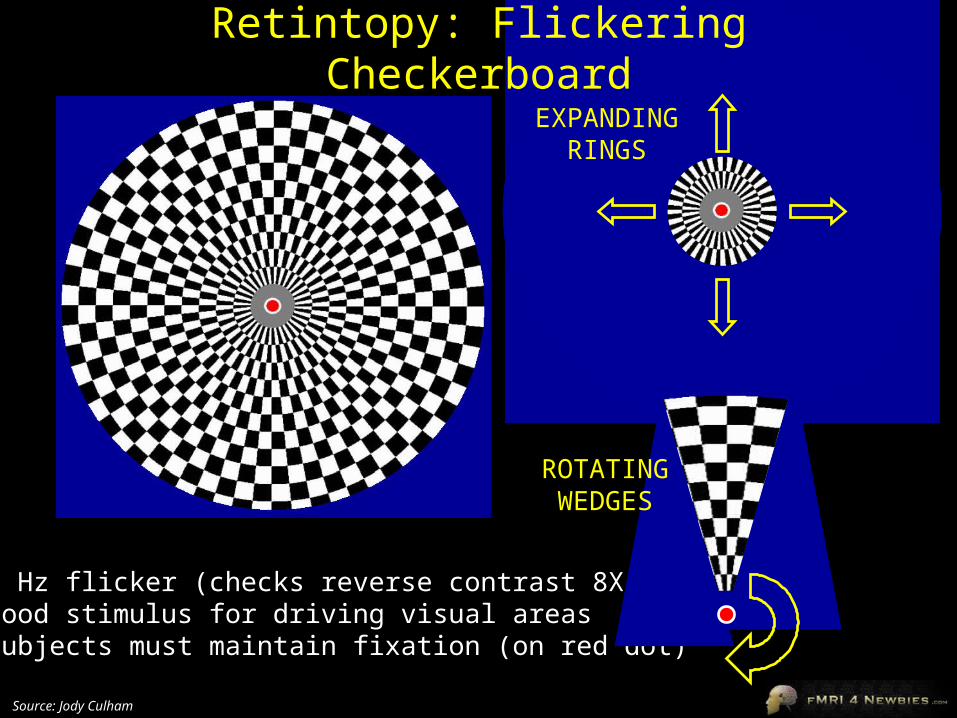
EXPANDINGRINGS
Retintopy: Flickering Checkerboard
•8 Hz flicker (checks reverse contrast 8X/sec)•good stimulus for driving visual areas•subjects must maintain fixation (on red dot)
ROTATINGWEDGES
Source: Jody Culham

Source: Jody Culham
time = 0
time = 20 sec
time = 60 sec
time = 40 sec
0 20 40 60
TIME
STIMULUSEXPECTED RESPONSE PROFILE OF AREA
RESPONDING TO STIMULUSTo analyze retinotopic data:
Analyze the data with a set of functions with the same profile but different phase offsets.
For any voxels that show a significant response to any of the functions, color code the activation by the phase offset that yielded maximum activation (e.g., maximum response to foveal stimulus = red, maximum response to peripheral stimulus = green)

Retintopy: Eccentricity
calcarinesulcus
left occipitallobe
right occipitallobe
•foveal area represented at occipital pole•peripheral regions represented more anteriorly
Source: Jody Culham

Retintopy on Flattened Occipital Lobe
2) cut along calcarinesulcus
left occipitallobe
Source: Jody Culham
3) unfold and flatten the cortical surface
uppe
r ca
lcar
ine
sulc
uslo
wer
cal
carin
e su
lcus
lateral surface (note: retinotopic areas do extend onto the lateral surface but are not shown here in this schematic)
1) virtually cut off the occipital lobe (remember, it’s a cup shape and the lateral surface is on the side we can’t see from this viewpoint)
occipitalpole
occipitalpole

Retintopy: Eccentricity Movie
occipital pole
calcarine sulcus
Movie: eccentricity.mpeghttp://cogsci.ucsd.edu/~sereno/phasemovie2.mpg
Source: Marty Sereno’s web page
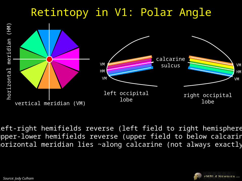
Retintopy in V1: Polar Angle
calcarinesulcus
left occipitallobe
right occipitallobe
•left-right hemifields reverse (left field to right hemisphere)•upper-lower hemifields reverse (upper field to below calcarine)•horizontal meridian lies ~along calcarine (not always exactly)
HM
VM
VM
vertical meridian (VM)
horiz
onta
l mer
idia
n (H
M)
HM
VM
VM
Source: Jody Culham

Polar Angle and Eccentricity in V1
calcarinesulcus
left occipitallobe
right occipitallobe
•retinotopic areas are like polar coordinates: eccentricity and polar angle
Source: Jody Culham

Polar Angle in V1, V2 and beyond
left occipitallobe
•V2 is mirror image map of V1•V1-V2 border occurs at vertical meridian•V2-V3 border occurs at horizontal meridian•situation gets more complex in higher-tier areas (V4v, V3A) that have representations of whole hemifield
HM
VM
VM
vertical meridian (VM)
horiz
onta
l mer
idia
n (H
M)
} V1 lower
HM
} V2 lower
VM
} VP
} V1 upperHM
} V2 upperVM
} V3
calcarinesulcus
Source: Jody Culham


Retinotopy: Polar Angle Movie
occipital pole
calcarine sulcus
Movie: phase.mpeghttp://zakros.ucsd.edu/~sereno/movies/phasemovie1b.mpg
Source: Marty Sereno’s web page

Getting Better Retinotopy
•use stimuli appropriate to the area (e.g., motion in MT, color in V4v)•use stimuli that are attentionally engaging•Marty Sereno: Buffy-o-topy
•UWO: chicken-o-topy

Other Sensory “-topies”
Touch: Somatotopy
Servos et al., 1998red = wrist; orange = shoulder
Audition: Tonotopy
cochlea
Sylvian fissure
temporal lobe
Movie: tonotopy.mpeghttp://cogsci.ucsd.edu/~sereno/downsweep2.mpg
Source: Marty Sereno’s web page

Face/Place-o-topy
Source: Levy et al., 2001
•faces activate foveal area (more for foveal than peripheral faces)
•places activate peripheral area (more for peripheral than foveal places)

Saccadotopy
Source: Sereno et al., 2001
•delayed saccades
•move saccadic target systematically around the clock
http://kamares.ucsd.edu/~sereno/LIP/both-closeup+stim.mpgMarty Sereno’s web page

Sulcal Formation
Source: Van Essen, 1997
Although sulci vary considerably from person to person (even in identical twins), there is considerable regularity in where the folds occur… Why?
David Van Essen proposes that as the brain develops, areas that are richly interconnected will be pulled together to form a gyrus (and those that are weakly interconnected form sulci).

Development of Sulci
Source: Ono, 1990
Sulci appear at predictable points in fetal development with the most prominent sulci (e.g., Sylvian fissure) appearing first.

Sulcal Formation: V1-V2
Source: Van Essen, 1997
The V1/V2 border provides one example of two richly interconnected areas that form a gyrus.
This arrangement also explains why maps in V1 and V2 are mirror images of each other!
calcarinesulcus

Comparative Neuroanatomy
Source: Comparative Mammalian Brain Collection
The complexity of sulci increased throughout evolution

Interspecies Comparisons
Figure H shows the macaque monkey visual areas morphed onto human cortex based on the placement of sulcal landmarks (Van Essen et al., 2001)
Can we assume humans are just morphed monkeys?
In some areas the human cortical surface area is slightly larger than in the macaque (e.g., visual cortex: 2X); in others it is considerably larger (e.g., parietal cortex: 20X)
Are individual areas larger? Are there more areas?