tools to kill: genome of one of the most destructive plant pathogenic fungi macrophomina phaseolina
TRANSCRIPT
Islam et al. BMC Genomics 2012, 13:493http://www.biomedcentral.com/1471-2164/13/493
RESEARCH ARTICLE Open Access
Tools to kill: Genome of one of the mostdestructive plant pathogenic fungi MacrophominaphaseolinaMd Shahidul Islam1, Md Samiul Haque1, Mohammad Moinul Islam1, Emdadul Mannan Emdad1, Abdul Halim1,Quazi Md Mosaddeque Hossen1, Md Zakir Hossain1, Borhan Ahmed1, Sifatur Rahim1, Md Sharifur Rahman1,Md Monjurul Alam1, Shaobin Hou2, Xuehua Wan2, Jennifer A Saito3 and Maqsudul Alam1,2*
Abstract
Background: Macrophomina phaseolina is one of the most destructive necrotrophic fungal pathogens that infectmore than 500 plant species throughout the world. It can grow rapidly in infected plants and subsequentlyproduces a large amount of sclerotia that plugs the vessels, resulting in wilting of the plant.
Results: We sequenced and assembled ~49 Mb into 15 super-scaffolds covering 92.83% of the M. phaseolinagenome. We predict 14,249 open reading frames (ORFs) of which 9,934 are validated by the transcriptome. Thisphytopathogen has an abundance of secreted oxidases, peroxidases, and hydrolytic enzymes for degrading cellwall polysaccharides and lignocelluloses to penetrate into the host tissue. To overcome the host plant defenseresponse, M. phaseolina encodes a significant number of P450s, MFS type membrane transporters, glycosidases,transposases, and secondary metabolites in comparison to all sequenced ascomycete species. A strikingly distinctset of carbohydrate esterases (CE) are present in M. phaseolina, with the CE9 and CE10 families remarkably higherthan any other fungi. The phenotypic microarray data indicates that M. phaseolina can adapt to a wide range ofosmotic and pH environments. As a broad host range pathogen, M. phaseolina possesses a large number ofpathogen-host interaction genes including those for adhesion, signal transduction, cell wall breakdown, purinebiosynthesis, and potent mycotoxin patulin.
Conclusions: The M. phaseolina genome provides a framework of the infection process at the cytological andmolecular level which uses a diverse arsenal of enzymatic and toxin tools to destroy the host plants. Furtherunderstanding of the M. phaseolina genome-based plant-pathogen interactions will be instrumental in designingrational strategies for disease control, essential to ensuring global agricultural crop production and security.
Keywords: Genome sequencing, Phytopathogens, Charcoal rot, Phenotypic microarray
BackgroundMacrophomina phaseolina, a global devastating necro-trophic fungal pathogen, infects more than 500 planthosts [1]. It includes major food crops (maize, sorghum[2]), pulse crops (common bean [3], green gram [4]),fiber crops (jute [5], cotton [6]), and oil crops (soybean
* Correspondence: [email protected] and Applied Research on Jute Project, Bangladesh Jute ResearchInstitute, Manik Mia Avenue, Dhaka 1207, Bangladesh2Advanced Studies in Genomics, Proteomics and Bioinformatics, University ofHawaii, 2565 McCarthy Mall, Keller 319, Honolulu, Hawaii 96822, USAFull list of author information is available at the end of the article
© 2012 Islam et al.; licensee BioMed Central LCommons Attribution License (http://creativecreproduction in any medium, provided the or
[1], sunflower [7], sesame [8]). Despite its wide hostrange, Macrophomina is a monotypic genus [9].Diseases caused by M. phaseolina (e.g., seedling blight,
charcoal rot, stem rot, and root rot) are favored withhigher temperatures (30-35°C) and low soil moisture[10]. It is difficult to control M. phaseolina due to itspersistence as sclerotia in the soil and plant debris [11].Recently, increased incidence of the pathogen on diversecrop species has been reported worldwide [12-14], high-lighting the importance of this disease to crop produc-tion in drought prone regions.
td. This is an Open Access article distributed under the terms of the Creativeommons.org/licenses/by/2.0), which permits unrestricted use, distribution, andiginal work is properly cited.

Islam et al. BMC Genomics 2012, 13:493 Page 2 of 16http://www.biomedcentral.com/1471-2164/13/493
The fungus has a wide geographical distribution, andis especially found in tropical and subtropical countrieswith arid to semi-arid climates in Africa, Asia, Europe,and North and South America [15-17]. This pathogencan result in severe crop losses. For example, charcoalrot is a serious problem of soybean, which accounted fora total yield loss of $173.80 million in the United Statesduring 2002 [18]. In Bangladesh, the fiber yield of jute isreduced by 30% due to this pathogen.M. phaseolina is an anamorphic fungus in the asco-
mycete family Botryosphaeriaceae. The fungus can re-main viable for more than 4 years in soil and cropresidue as sclerotia (Figure 1a) [11]. The M. phaseolinahyphae initially invade the cortical tissue of jute plants,followed by sclerotia formation, causing stem rot disease(Figure 1b, c). Gray-black mycelia and sclerotia are pro-duced (Figure 1c) and the infected area exhibits diseasesymptoms (Figure 1d). The conidia are hyaline, aseptate,
Figure 1 Infection of jute by M. phaseolina. (a) Stereomicrograph of scleand profuse aerial hyphae to invade the stem bark. (c) Longitudinal section oDuring early rainy season, hyphae penetrate the plant cell wall and produce dspores of M. phaseolina. (f) Diseased plants showing infection of the stem, wh
thin-walled, and elliptical (Figure 1e). Under favorableconditions, hyphae germinate from the sclerotia and in-fect the roots of the host plant by penetrating the plantcell wall through mechanical pressure and/or chemicalsoftening [19]. The disease progresses from leaf yellow-ing to wilting and ultimately plant death (Figure 1f ).Currently, genetic information on M. phaseolina is
scarce with only 176 expressed sequence tags (ESTs) and903 nucleotide sequences in the National Center for Bio-technology Information (NCBI). Here we report thedraft genome sequence of highly destructive plantpathogen M. phaseolina to gain insight into the molecu-lar basis of pathogenesis.
Results and discussionGenome sequencing and assemblyThe genome of M. phaseolina was sequenced using awhole-genome shotgun approach. A total of 6.92 Gb of
rotia that exists in soil and crop residue. (b) Pathogen produces extensivef stem bark showing inter- and intracellular mycelium and sclerotia. (d)isease symptoms. (e) Light micrograph of globose ostiolate pycnidia andich eventually wilt and prematurely die (Inset).

Islam et al. BMC Genomics 2012, 13:493 Page 3 of 16http://www.biomedcentral.com/1471-2164/13/493
raw sequence was generated from a combination of 454and Illumina platforms (Additional file 1: Table S1). Theresulting assembly is 49.29 Mb of which 98.53% is non-gapped sequence (Table 1; Additional file 1: Table S2).Mapping with Newbler GS Reference Mapper (v2.5.3)showed 96.50% reads and 99.11% bases mapped to thereference assembly. The draft genome sequence consistsof 94 scaffolds, with 15 super scaffolds covering 92.83%of the total assembled length (Additional file 1: TableS2). We predicted 14,249 protein-coding genes and9,934 were validated by the transcriptome (Additionalfile 1: Table S3).We examined the homology between M. phaseolina
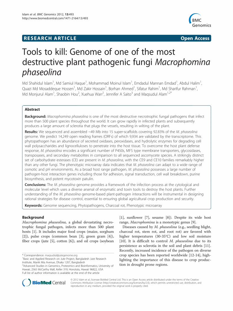
and 12 other fungal genomes under the classes of Sac-charomycetes, Sordariomycetes, Agaricomycetes, andEurotiomycetes. The results revealed that 71% of thegenes in the M. phaseolina genome have homologs inother fungal genomes and the remaining 29% are orphangenes (Figure 2a). Among the orphan genes, 51.11% arefound in the transcriptome.The comparison of M. phaseolina protein families with
other ascomycete fungal species is shown in Table 2(also see Additional file 1: Table S4). The genome contains13.07% (1,863) secreted proteins as compared to 7-10% inother plant pathogens [20]. Surprisingly, M. phaseolinahas the lowest number of proteases among ascomycetefungal species [21] (Table 2). On the other hand, thegenome is distinct from other fungi by having the highestnumber of cytochrome P450, glycosidase, and secondarymetabolite backbone genes. We predict that this might beone of the main strategies of M. phaseolina to overcomethe host plant defense response by using various second-ary metabolites.
Table 1 Genome assembly and annotation statistics
Genome features
Strain MS6
Sequence coverage (fold) 13
Genome Size (Mb) 49.295
Total scaffolds 94
No. of scaffolds (≥ 1 Mb) 15
N50 scaffold length (Mb) 3.39
Number of N50 scaffolds 6
Number of genes 14,249
No. of genes in 15 scaffolds (≥ 1 Mb) 14,071
Number of genes found in cDNA 9,934
Median gene length (bp) 1,265
Repetitive sequence (%) 2.84
Transposable elements (%) 3.98
NCBI accession AHHD00000000
Conserved syntenic and phylogenetic relationshipPairwise comparison revealed that the genome struc-tures of M. phaseolina and Fusarium oxysporum, one ofthe most important phytopathogenic and toxigenicfungi, have large areas of synteny (Figure 2b). Amongthe 14,249M. phaseolina genes, 7,767 (54.10%) areshared with F. oxysporum (Additional file 2: Figure S1).The large number of shared genes may reflect commonstrategies for infecting a remarkably broad host range.Ninety seven percent of the M. phaseolina genome com-prises non-repetitive sequences, and the orthologsshared with the F. oxysporum genome display an average52% identity. A phylogenetic analysis revealed the evolu-tionary relationship among fungal taxa and the position-ing of M. phaseolina within the pezizomycotina(Figure 2c).
Global paralog networkOf the 14,249 predicted proteins, we found homologsfor 7,999 proteins in nine fungal genomes. Tocharacterize functional protein families in the M. pha-seolina genome, we constructed a database in PathwayStudio (Ariadne Genomics Inc.) containing M. phaseo-lina proteins with functional annotation from the ortho-logs in nine other fungal genomes, predicted interologs,predicted pathways, and paralog network. The largestparalog families were identified by clustering the paralognetwork consisting of 6,210 paralog links representingparalog pairs with sequence homology above 30%. Weidentified 77 paralog families having more than six pro-teins (Additional file 2: Figure S2). The larger paralogfamilies were the cytochrome P450 (151), MFS typemembrane transporters (222), and transposases (101).The large number of transposons in the M. phaseolinagenome suggests that they could be the primary mech-anism for mutagenesis and gene duplications, which inturn may promote the ability of M. phaseolina to infectnew plant species.There was also a high number of paralogs for the
oxido-reductase class of enzymes which includes dehy-drogenases, aldehyde dehydrogenases, choline dehydro-genases, cytochrome P450 and aldose reductases. Theseoxido-reductase enzymes produce and utilize a large var-iety of secondary metabolites [22] which may triggerM. phaseolina to survive in a wide range of physicalenvironments and facilitate to infect new plant species.The PTH11 paralog family consists of a large number ofG-protein coupled receptors (GPCR; 44), which containa cysteine-rich fungal extracellular membrane domain.This may facilitate M. phaseolina to be more virulent.
Repetitive DNA and transposable elementsWide virulence capabilities of a genome are often asso-ciated with transposon-mediated inactivation or deletion
Figure 2 Homology, syntenic, and phylogenetic relationship of M. phaseolina. (a) Comparative analyses of orthologous and paralogousgene families of 13 fungal species. Number of genes are presented for each component. Clustering was done by using OrthoMCL (MCL-10-201).(b) Synteny of M. phaseolina and Fusarium oxysporum using whole genome data. The reference genome (Aspergillus fumigatus) is broken up intoeight chromosomes and syntenic regions are represented by two vertical columns with color. (c) Phylogenetic tree showing the positioning of M.phaseolina within the pezizomycotina.
Islam et al. BMC Genomics 2012, 13:493 Page 4 of 16http://www.biomedcentral.com/1471-2164/13/493
of pathogen-associated molecular pattern (PAMP)-encoding genes whose products trigger the plant adap-tive immune system [23]. The M. phaseolina genomecomprises 2.84% repetitive DNA and 3.98% transposableelements. The transposable elements are classified into11 families (Table 3). Most of them are DNA transpo-sases, with particular abundance of the subclasses gypsy(918), Ty1_Copia (331), and DDE_1 (242). LINE (184)and hAT (136) are also relatively abundant. Transposableelements appear to be tightly clustered in the genome(Additional file 2: Figure S3). Evidence for repeat-inducedpoint mutation (RIP) within the transposable elementswas searched using RIPCAL [24], but this mutationalbias was not observed in the M. phaseolina genome(RIP index 0.87). The genome contains more fragmen-ted pseudogenes (894) than processed pseudogenes(31) caused by mobile elements, but lacks duplicatedpseudogenes. This is consistent with transposons mak-ing a greater contribution to genetic instability inM. phaseolina.
Carbohydrate degrading enzymesPhytopathogenic fungi secrete a cocktail of hydrolyticenzymes (including carbohydrate-active enzymes; CAZymes)for degrading the plant cell wall and penetrating into thehost tissue [25]. The M. phaseolina genome encodes 362putative CAZymes including 219 glycoside hydrolases(GH), 56 glycosyltransferases (GT), 65 carbohydrateesterases (CE), 6 carbohydrate binding modules (CBM),and 16 polysaccharide lyases (PL) comprising more than80 distinct families. These enzymes do not appear to betightly clustered, but are distributed throughout the gen-ome (Additional file 2: Figure S4).The number of GHs possessed by M. phaseolina is
higher than the average for plant pathogenic fungi and isnearly four times more abundant than the GTs, consist-ent with the importance of carbohydrate degradationrather than formation. There are 25 putative endogluca-nases (GH5, GH12, GH45, GH61), 7 exocellobiohydro-lases (GH6, GH7, GH81), and 28 β-glucosidases (GH1,GH3, GH17) for the hydrolysis of cellulose. The
Table 2 Sizes of selected protein families in M. phaseolina and other fungi
Protein familya MPb FO FG MO BC SS NC AN AF PCH PP
Fungal specific transcription factors 156 101 192 95 118 90 89 209 169 65 63
C2H2 zinc finger transcription factors 66 73 85 58 48 54 63 58 51 77 42
Zn2/Cys6 transcription factors 113 370 376 155 142 108 110 307 230 146 118
Major facilitator superfamily 270 352 274 198 225 167 110 279 232 141 184
Cytochrome P450 256 178 112 137 129 93 40 116 74 155 236
Pth11-like G-protein coupled receptor 44 55 51 60 22 23 28 39 15 14 26
Protein kinases 140 160 129 129 124 164 111 127 131 106 56
Histidine kinase 1 37 20 6 3 5 8 12 6 19 24
Heterokaryon incompatibility 65 82 88 41 59 34 45 7 8 3 2
Serine proteases 1 12 60/150c 56/91 19/34 20/33 32/74 53/136 29/46 0 2
Subtilisin 19 36 16/24 26/29 4/7 4/6 6/10 3/4 3/7 12 33
Trypsin 2 3 2/3 3/3 1/1 1/1 0/2 1/2 0/0 0 0
Carboxypeptidase 19 31 12/21 7/8 7/9 8/ 11 6/9 5/12 14/15 24 22
Aspartic protease 4 0 15/18 14/19 11/14 9/21 15/19 7/16 7/9 38 18
Threonine protease 0 0 3/18 2/18 2/13 2/13 2/20 0/20 1/17 0 0
Cysteine protease 3 0 5/57 4/31 3/24 1/27 4/41 6/57 3/31 0 0
Metalloprotease 8 26 32/111 38/91 6/50 7/48 21/81 22/105 20/77 0 0
All proteases 113 261 354 250 135 142 235 334 180 228 325
Lipase 53 61 4/31 2/23 3/28 2/25 0/16 2/27 3/25 23 40
Esterase/thioesterase 108 95 70 64 70 58 42 63 52 74 69
Glycoside hydrolase related 219 168 159 198 120 126 137 200 165 180 144
Transposases 101 19 17 15 73 426 15 15 109 12 11
Cutinase 10 12 12 18 11 8 3 4 5 0 0
Polysaccharide lyase 16 23 25 9 25 20 5 24 27 4 6
Secondary metabolite backbone genes 75 34 37 32 37 29 15 58 40 51 39aCorresponding InterPro codes are listed in Additional file 1: Table S4.bFungal species are MP, Macrophomina phaseolina; FO, Fusarium oxysporum; FG, Fusarium graminearum; MO, Magnaporthe oryzae; BC, Botrytis cinerea; SS,Sclerotinia sclerotiorum; NC, Neurospora crassa; AN, Aspergillus nidulans; AF, A. fumigatus; PCH, Phanerochaete chrysosporium, and PP, Postia placenta.cFractions indicate the number of total proteins in each family that are secreted.
Table 3 Families of transposable elements in theM. phaseolina genome
Family name Class Number
LTR roo Class I 4
DDE_1 Class I 242
gypsy Class I 918
Ty1-Copia Class I 331
LINE Class I 184
hAT Class II 136
helitron Class II 15
cacta Class II 4
Mariner Class II 76
MuDR_A_B Class II 57
piggybac Class II 9
Total 1976
Islam et al. BMC Genomics 2012, 13:493 Page 5 of 16http://www.biomedcentral.com/1471-2164/13/493
cellulolytic activity of M. phaseolina was shown to besignificantly higher than that of other fungal species(i.e., Aspergillus niger and Trichoderma reesei) [26],reflecting the pathogenicity potency of this fungus.The M. phaseolina genome also has the highest num-
ber of CEs than any other sequenced fungal genome sofar (Table 4), with particular expansion of families CE9(chitin metabolism) and CE10 (sterol esterases). Thirtytwo CE10 members are found in the M. phaseolina gen-ome, but is absent in eight other species (Table 4). NineCE5 candidate cutinases were found in the genome, sug-gesting that these enzymes are critical for initial penetra-tion through the plant cuticle. The complement ofpectin lyases (PL1, PL3, PL4), pectin hydrolases (GH28,GH88), and pectin esterases allows M. phaseolina tofully saccharify pectin. Other polysaccharide degradingenzymes predicted in the genome include catalytic activ-ities for degrading starch and glycogen, hemicellulose,chitin, and β-glucans.
Table 4 Comparison of the number of carbohydrate esterases of M. phaseolina with other fungi
Name CE1 CE2 CE3 CE4 CE5 CE8 CE9 CE10 CE12 CE14 CE15 CE16 NC
C. neoformans var. neoformans 2 0 0 4 0 0 1 0 0 0 0 0 0
M. grisea 10 1 6 8 15 1 1 0 2 0 1 1 1
S. cerevisiae 1 0 0 2 0 0 0 0 0 0 0 0 0
P. anserina 14 0 8 5 7 1 1 0 1 0 3 1 0
A. nidulans 3 0 6 7 4 3 1 0 2 0 0 3 4
A. niger 3 0 1 5 5 3 1 0 2 0 0 2 3
A. oryzae 5 0 3 3 5 5 1 0 4 0 0 3 1
P. chrysogenum 2 0 4 5 4 2 1 0 2 0 1 1 0
M. phaseolina 1 0 0 8 9 4 10 32 0 1 0 0 0
Islam et al. BMC Genomics 2012, 13:493 Page 6 of 16http://www.biomedcentral.com/1471-2164/13/493
Genes involved in lignin degradationMajor components of the lignin depolymerization systemin M. phaseolina include laccases, lignin peroxidases,galactose oxidases, and chloroperoxidases, haloperoxi-dases, and heme peroxidases. M. phaseolina strainMS6 indeed demonstrates ligninolytic activity (Add-itional file 2: Figure S5). In comparison to seven otherfungal species, M. phaseolina possesses the highestnumber of laccases (Table 5). Lignin peroxidase hasbeen reported only in Phanerochaete chrysosporiumthus far [27] and interestingly, this study revealed thesecond occurrence in M. phaseolina. Six extracellularclass II heme peroxidases (IPR002016), 6 chloroperoxi-dases, and 7 haloperoxidases also contribute to the lig-ninolytic activity. In addition, we found a significantnumber of GMC oxidoreductases (40), which includesalcohol oxidases and cellobiose dehydrogenases. Theseenzymes are known to be directly involved in lignocel-lulosic degradation [27].
Virulence associated genesAs a wide host ranged pathogenic fungus, M. phaseolinais expected to possess a significant number of pathogen-host interaction genes. We searched the genome usingthe pathogen-host interaction database (PHI-base) [28]and identified 537 putative PHI genes (Additional file 1:Table S5). These genes play diverse roles in pathogenesis
Table 5 Comparison of the number of lignin degrading enzym
Fungal species Laccase Galactose oxidase
P. placenta 2 0
P. chrysosporium 0 0
C. neoformans 0 0
U. maydis 0 1
S. cerevisiae 0 0
A. nidulans 1 0
N. crassa 5 1
M. phaseolina 22 7
including adhesion, signal transduction, cell wall break-down, purine biosynthesis, and biosynthesis of the po-tent mycotoxin patulin. ATP-binding cassette (ABC)transporters aid in defending the pathogen from host-produced secondary metabolites as well as provide es-sential nutrients [29]. A number of detoxification genesare present, such as those encoding cytochrome P450(IPR001128), Cof protein (IPR000150), and superoxidedismutase, Cu/Zn binding (IPR001424). In addition, sev-eral beta-ketoacyl synthases (IPR000794) involved in thebiosynthesis of a polyketide antibiotic [30] as well assome tetracycline resistance genes have been identified inM. phaseolina. These data suggest that the M. phaseolinagenome encodes a large repertoire of pathogenicity-associated genes which may be involved in the pathogen-esis of this organism.
Signal transductionThe perception of environmental cues through cell-surface receptors and relaying the information to intra-cellular signaling pathways is essential for pathogenicity.The PTH11-like GPCR is a PHI protein shown to regu-late Magnaporthe grisea appressorium differentiation inresponse to the plant surface [31]. The M. phaseolinagenome has 44 putative PTH11-like GPCRs comparedto an average of 34 in other pathogenic fungi (Table 2).The putative PTH11-like GPCRs are grouped into seven
es of M. phaseolina with other fungi
s Lignin peroxidase Chloroperoxidase
0 5
10 3
0 0
0 0
0 0
0 0
0 0
3 6
Islam et al. BMC Genomics 2012, 13:493 Page 7 of 16http://www.biomedcentral.com/1471-2164/13/493
subfamilies. Three G-protein alpha subunits are presentto transduce the extracellular signals leading to infectionspecific development, which is required for pathogen-icity [32,33]. The 140 protein kinases in M. phaseolina isabove the average (131) found in other ascomycete fungi(Table 2). Since signal transduction is a crucial part offungal development and the infection process, indeedmost of the kinases had orthologs in PHI-base (134/140).This result indicates that protein kinases in M. phaseo-lina might play a functional role in pathogen-hostinteraction.
Transport and detoxification of compoundsPlant pathogenic fungi use a wide range of strategies togain access to the carbon sources of their host plantsand counter the plant defense response. The M. phaseo-lina genome encodes 839 transporter genes comprising106 families (Additional file 1: Table S6). Majority of thetransporter genes (782/839) were similar to those cata-loged in PHI-base. A large proportion of transportersbelong to the MFS family (270), but the ABC superfam-ily (59) and Amino Acid-Polyamine-Organocation(APC) family (40) are also well represented in the gen-ome (Additional file 1: Table S6). M. phaseolina hasmore amino acid transporters (54) than other pathogenicfungi (29 to 38), revealing that this fungus might be ableto access a wide range of protein degradation productsfrom host sources. The sucrose and galactoside trans-porter (MFS superfamily) is required by Metarhiziumanisopliae for rhizosphere competence but not forvirulence [34]. The M. phaseolina genome has 15 su-crose and galactoside transporters, whereas Fusariumgraminearum contains 12, suggesting these genes couldbe generally important for establishing plant-fungusrelationships.We found a relatively large number of genes involved
in detoxification (Table 2). The dehydrogenases (411),acyl-CoA N-acetyltransferases (7), monooxygenases(104), and cytochrome P450s (256) were preferentiallyexpanded in M. phaseolina. P450s play an importantrole in various hydroxylation and oxidation processes
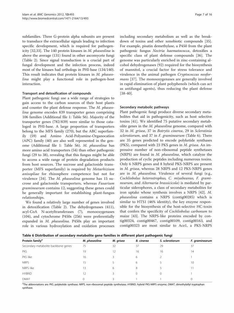
Table 6 Distribution of secondary metabolite gene families in
Protein familya M. phaseolina M. gr
Secondary metabolite backbone genes 75 32
PKS 19 12
PKS like 16 3
NRPS 15 5
NRPS like 13 6
HYBRID 12 3
DMAT 0 3aThe abbreviations are PKS, polyketide synthase; NRPS, non-ribosomal peptide synthsynthase.
including secondary metabolism as well as the break-down of toxins and other xenobiotic compounds [35].For example, pisatin demethylase, a P450 from the plantpathogenic fungus Nectria haematococca, detoxifies aspecific class of plant defense compounds [36]. Thegenome was particularly enriched in zinc-containing al-cohol dehydrogenases (92) required for the biosynthesisof mannitol, a crucial factor for stress tolerance andvirulence in the animal pathogen Cryptococcus neofor-mans [37]. The monooxygenases are generally involvedin rapid elimination of plant polyphenols (which can actas antifungal agents), thus reducing the plant defense[38-40].
Secondary metabolic pathwaysPlant pathogenic fungi produce diverse secondary meta-bolites that aid in pathogenicity, such as host selectivetoxins [41]. We identified 75 putative secondary metab-olite genes in the M. phaseolina genome, compared with32 in M. grisea, 37 in Botrytis cinerea, 29 in Sclerotiniasclerotiorum, and 37 in F. graminearum (Table 6). Thereare 35 genes predicted to encode polyketide synthases(PKS), compared with 23 PKS genes in M. grisea. An im-pressive number of non-ribosomal peptide synthetases(NRPS) are found in M. phaseolina, which catalyze theproduction of cyclic peptides including numerous toxins.Only 6 NRPS genes and 8 hybrid PKS-NRPS are presentin M. grisea, whereas 28 NRPS and 12 PKS-NRPS genesare in M. phaseolina. Virulence of several fungi (e.g.,Cochliobolus heterostrophus, C. miyabeanus, F. grami-nearum, and Alternaria brassicicola) is mediated by par-ticular siderophores, a class of secondary metabolites foriron uptake whose synthesis involves a NRPS [42]. M.phaseolina contains a NRPS (contig00285) which issimilar to HTS1 (46% identity), the key enzyme respon-sible for the biosynthesis of the host-selective HC-toxinthat confers the specificity of Cochliobolus carbonum tomaize [43]. The NRPS-like proteins encoded by con-tig00324, contig00467, contig00109, contig00163, andcontig00323 are most similar to Ace1, a PKS-NRPS
different plant pathogenic fungi
isea B. cinerea S. sclerotiorum F. graminearum
37 29 37
16 16 14
6 2 1
6 5 10
8 5 11
0 0 1
1 1 0
etase; HYBRID, hybrid PKS-NRPS enzyme; DMAT, dimethylallyl tryptophan

Islam et al. BMC Genomics 2012, 13:493 Page 8 of 16http://www.biomedcentral.com/1471-2164/13/493
hybrid that confers avirulence to M. grisea during riceinfection [44].
Phenotypic response in relation to differentenvironmental stimuliWe used Phenotype Microarray (PM) analysis (BiologInc.) to evaluate M. phaseolina against ~960 differentcarbon, nitrogen, phosphorus, sulfur, nutrient supple-ment, peptide nitrogen, osmolytes, and pH sources(Figure 3; Additional file 2: Figures S6-S11). Of particu-lar interest, we found that the adaptability to wide os-motic and pH ranges could be a contributing factor tothis organism’s pervasive nature.PM analysis revealed that M. phaseolina is capable of
growing in sodium salt concentrations of 1-8%, withmaximal growth at 2-4% (Figure 3a). In comparison, 3%NaCl is toxic for Saccharomyces cerevisiae but is close tothe optimum for growth of the halophilic black yeastHortaea werneckii, one of the most salt-toleranteukaryotic organisms so far described [45]. The effi-cient utilization of various osmolytes provides clues toM. phaseolina’s osmoadaptation strategy. It was re-cently shown that salinity increases the disease severitycaused by M. phaseolina on Phaseolus vulgaris (commonbean), by enhancing the growth rate of the pathogen aswell as weakening the plant due to ion (K+ and Na+) im-balance [46]. This indicates that M. phaseolina could be agreater threat in areas with saline soils.pH is one of the major environmental factors affecting
pathogenicity. The M. phaseolina genome contains 2 pu-tative PalH and 5 PalI proteins which are responsible forsensing ambient pH [47]. Moreover, there are severalpH-regulated proteins including 19 acid phosphatases, 2α-L-arabinofuranosidases, and 7 alkaline phosphatases.The presence of acid and alkaline phosphatases indicatesthat M. phaseolina has an extraordinary capability toneutralize both acidic and alkaline environments for itsgrowth. This is clearly evident from our PM analysis.The results revealed that M. phaseolina can grow in pHranging from strongly acidic to alkaline (pH 3.5 to 10),with maximum growth between pH 5 to 7 (Figure 3b).Therefore, M. phaseolina has a robust pH sensing sys-tem which enables it to adapt to adverse conditions.
Pathogenesis of M. phaseolinaThe array of metabolic genes within the M. phaseolina gen-ome reflects its pathogenic lifestyle (Figure 4; Additional file1: Table S7).The production and dispersal of conidia is important
for fungal survival and infection of new hosts. Hyperos-motic stress is one of the environmental stimuli thatoften trigger conidiation [48]. The M. phaseolina gen-ome encodes MPH_01444, a homolog of the MAP kin-ase OSM1, which regulates the osmotic stress response
(to maintain cellular turgor) along with MPH_10325 andMPH_03305 (Figure 5; Additional file 1: Table S8). In M.grisea, deletion of OSM1 has pleiotropic effects includ-ing osmotic sensitivity, reduced conidiation, and over-production of appressoria [49].Adhesion of fungal propagules to the plant surface is
the prerequisite to establish disease. We identified 8homologs of Cellulose-Binding Elicitor Lectin (CBEL), acell surface glycoprotein that plays a role in adhesion tohost wall components [50], as well as a Class II hydro-phobic protein which mediates contact and communica-tion between the fungus and its environment [51]. M.phaseolina also has three transglutaminase-like proteinscontaining a 13-amino acid motif (Pep-13) that is able tostimulate the plant defense response. Presence of thesecell surface proteins suggests that M. phaseolina pro-duces PAMPs, which can be efficiently perceived by awide range of plant species. Activation of the plant im-mune system results in the synthesis of antifungal pep-tides, inhibitors of cell wall degrading enzymes, andphytoalexins. At the same time, the fungus responds tosurface inductive cues via a cAMP-dependent pathwayin order to initiate the infection process and combat theplant defense response (e.g., production of salicylate-1-monooxygenase).Invasion begins with the emergence of a germ tube
from the conidium, followed by appressorium formation.The developing appressorium swells as the cytoskeletonand Golgi vesicles accumulate in the tip. In bean rustpathogen Uromyces appendiculatus, the cytoskeletonand vesicles in the apex of the hypha are reorganizedalong the cell wall within 4 minutes of signal perception[52]. Enzymes for fungal cell wall synthesis (e.g., chitinsynthase and β-1,3-glucan synthase) as well as cell walldegradation (e.g., cutinase and β-1,4-endoglucanase) arecontained within different vesicles. M. phaseolina alsosynthesizes phytosphingosine and phytoceramide to pro-tect the cell membrane from mechanical damage duringpenetration of the host cell. High turgor pressure is gen-erated within the appressorium, allowing the penetrationpeg to break through the plant epidermis. Secretion of avariety of cell wall degrading enzymes and other toxinsaid in the invasion of the host. These hyphae penetrateepidermal walls directly and subsequently colonize thetissue by intra- and intercellular growth (Figure 1c).Regulation of these invasion processes involves both
cAMP-dependent and MAP kinase pathways (Figure 5).Surface recognition and appressoria initiation isdependent on the adenylate cyclase MAC1 (MPH_00975).Early research revealed that deletion of the MAC1 geneblocked appressorium formation in M. grisea [53]. Thecatalytic subunit of cAMP-dependent protein kinase A(MPH_07566 and MPH_00397) is also required forappressoria formation and penetration. Furthermore, the
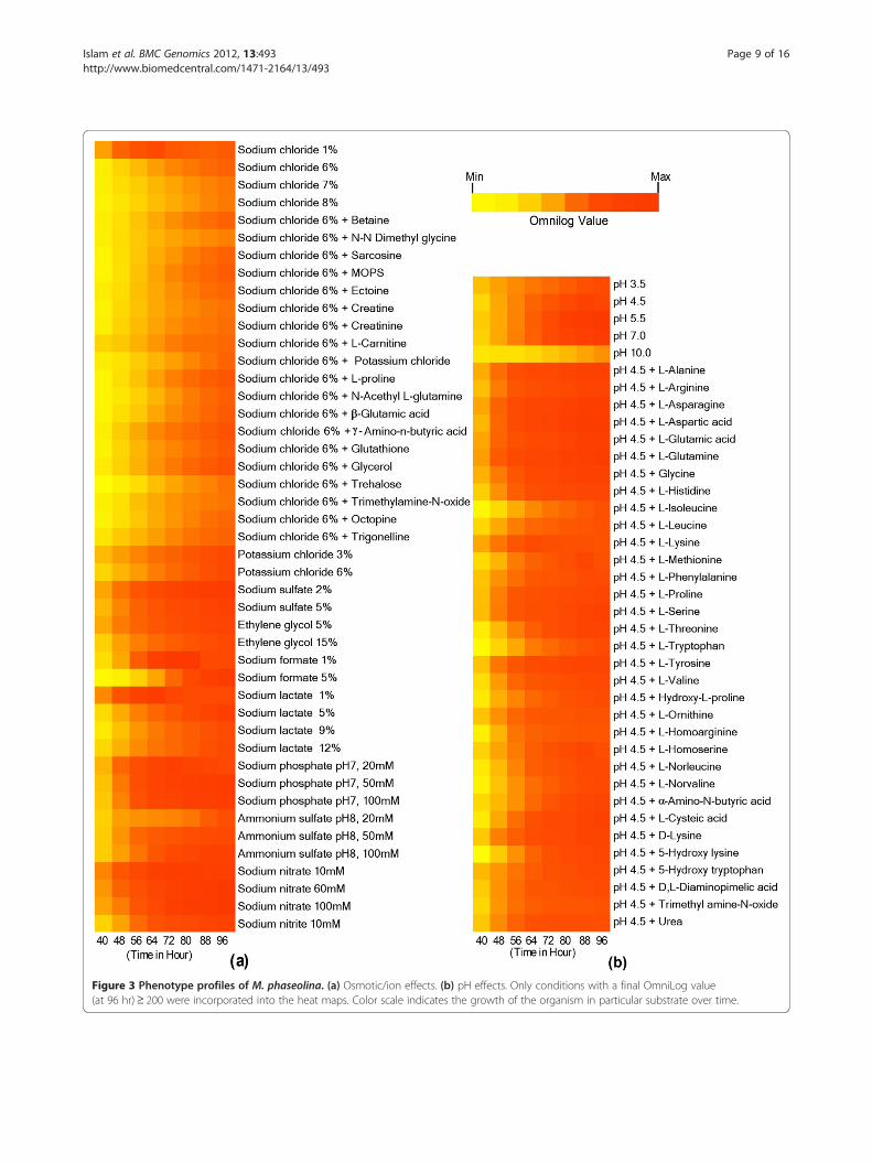
Figure 3 Phenotype profiles of M. phaseolina. (a) Osmotic/ion effects. (b) pH effects. Only conditions with a final OmniLog value(at 96 hr)≥ 200 were incorporated into the heat maps. Color scale indicates the growth of the organism in particular substrate over time.
Islam et al. BMC Genomics 2012, 13:493 Page 9 of 16http://www.biomedcentral.com/1471-2164/13/493
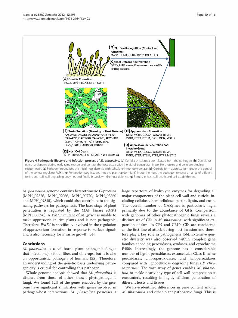
Figure 4 Pathogenic lifestyle and infection process of M. phaseolina. (a) Conidia or sclerotia are released from the pathogen. (b) Conidia orsclerotia disperse during early rainy season and contact the host tissue with the aid of transglutaminase-like proteins and cellulose-bindingelicitor lectin. (c) Pathogen neutralizes the initial host defense with salicylate-1-monooxygenase. (d) Conidia form appressorium under the controlof the central regulator PMK1. (e) Penetration peg invades into the plant epidermis. (f) Inside the host, the pathogen releases an array of differenttoxins and cell wall degrading enzymes and finally breakdown the host defense. (g) Results in host cell death and self-establishment.
Islam et al. BMC Genomics 2012, 13:493 Page 10 of 16http://www.biomedcentral.com/1471-2164/13/493
M. phaseolina genome contains heterotrimeric G-proteins(MPH_02326, MPH_07066, MPH_00770, MPH_05860and MPH_09815), which could also contribute to the sig-naling pathways for pathogenesis. The later stage of plantpenetration is regulated by the MAP kinase PMK1(MPH_06596). A PMK1 mutant of M. grisea is unable tomake appressoria in rice plants and is non-pathogenic.Therefore, PMK1 is specifically involved in the regulationof appressorium formation in response to surface signalsand is also necessary for invasive growth [54].
ConclusionsM. phaseolina is a soil-borne plant pathogenic fungusthat infects major food, fiber, and oil crops, but it is alsoan opportunistic pathogen of humans [55]. Therefore,an understanding of the genetic basis underlying patho-genicity is crucial for controlling this pathogen.Whole genome analysis showed that M. phaseolina is
distinct from those of other known phytopathogenicfungi. We found 12% of the genes encoded by the gen-ome have significant similarities with genes involved inpathogen-host interactions. M. phaseolina possesses a
large repertoire of hydrolytic enzymes for degrading allmajor components of the plant cell wall and cuticle, in-cluding cellulose, hemicellulose, pectin, lignin, and cutin.The overall number of CAZymes is particularly high,primarily due to the abundance of GHs. Comparisonwith genomes of other phytopathogenic fungi reveals adistinct set of CEs in M. phaseolina, with significant ex-pansion of families CE9 and CE10. CEs are consideredas the first line of attack during host invasion and there-fore play a key role in pathogenesis [56]. Extensive gen-etic diversity was also observed within complex genefamilies encoding peroxidases, oxidases, and cytochromeP450s. Interestingly, the genome has a considerablenumber of lignin peroxidases, extracellular Class II hemeperoxidases, chloroperoxidases, and haloperoxidasescompared with lignocellulose degrading fungus P. chry-sosporium. The vast array of genes enables M. phaseo-lina to tackle nearly any type of cell wall composition itencounters, resulting in highly efficient penetration ofdifferent hosts and tissues.We have identified differences in gene content among
M. phaseolina and other plant pathogenic fungi. This is

Figure 5 Pathogenicity regulatory pathway of M. phaseolina. Dotted line, Regulation; Solid line (gray), Molecular transport; Solid line (yellow-green), Protein modification.
Islam et al. BMC Genomics 2012, 13:493 Page 11 of 16http://www.biomedcentral.com/1471-2164/13/493
the first analysis of the genome of a plant pathogenicfungus that contains a large number of enzymes for thedegradation of cell wall polysaccharides and lignocellu-lose. The M. phaseolina genome laid the foundation toelucidate its specialized mechanism to infect more than500 plant hosts. Furthermore, it would decipher in-depth understanding of pathogenesis to resistancestrategies.
MethodsStrain, growth condition, and nucleic acid isolationM. phaseolina strain MS6 was isolated from an infectedjute plant at Bangladesh Jute Research Institute (BJRI),Dhaka. Strain MS6 is the most virulent among the 19isolates so far isolated in BJRI. This strain has myceliumthat is grey-white at the initial stage and turns darkgreen at the mature stage. It is coarse with featherystrand. The sclerotia are embedded in the strand. Thefungus was cultured at 30°C in liquid potato dextrose
medium. DNA and RNA were extracted as previouslydescribed [57,58], respectively.
Genome sequencing and assemblyWhole-genome shotgun sequencing of the M. phaseo-lina MS6 strain was performed using the 454 and Illu-mina sequencing platforms. We generated a total of 6.92Gb raw data, having 40.56 millions of raw reads. In the454 sequencing strategy, both single-end (SE) andpaired-end (PE) genomic libraries were constructed, ofwhich 2.38 Gb of shotgun sequences provided 48.29xcoverage and 1.57 Gb of 8 kb, 15 kb, and 20 kb PEsequences provided 31.95x coverage of the M. phaseo-lina genome. In the Illumina sequencing strategy, 2.97Gb of PE libraries were generated, of which 1.73 Gb of500 bp PE sequences provided 35.11x coverage and 1.24Gb of 3 kb mate-paired sequences provided 25.14xcoverage of the M. phaseolina genome.We produced a high quality assembly of the M. pha-
seolina genome using Newbler assembly program version

Islam et al. BMC Genomics 2012, 13:493 Page 12 of 16http://www.biomedcentral.com/1471-2164/13/493
2.5.3 (http://my454.com/products/analysis-software/index.asp). For both de novo assembly and reference mapping ofthe M. phaseolina genome, we only used raw data gener-ated from 454 pyrosequencing. For de novo assembly, wefed the Newbler GS de novo assembler first with the shot-gun sequences in one-step form and later with paired-endsequences incrementally in order to get better contiggingand scaffolding. About 96.50% raw reads were assembledinto 3,036 contigs and 94 scaffolds having 98.92% baseswith Q40 plus bases. For reference mapping, we usedNewbler GS reference mapper to map the raw sequencefiles onto the all contigs file generated that gave 98.89%reads and 99.11% bases mapped to the reference.We also used Illumina PE sequences with GapCloser
version 1.10, a tool from SOAP de novo (http://soap.-genomics.org.cn/soapdenovo.html), in order to close thegaps inside the scaffolds that were generated from theNewbler scaffolding process. A total of 785 gaps weredetected by GapCloser that cover 1.5 Mb residues ofwhich 197 gaps were completely filled up, leaving only1.47% (0.73 Mb) gaps inside the 94 scaffolds.We checked the relative completeness of the M. pha-
seolina MS6 draft assembly version 1.0 by performingcore gene annotation using the CEGMA pipeline [59].The resulting contigs as well as scaffolds from the M.phaseolina MS6 assembly were independently analyzedthrough this pipeline. In both cases we have found 245(98.79%) complete gene models out of 248 ultra-conserved core eukaryotic genes (CEGs) present in theM. phaseolina genome.This Whole Genome Shotgun project has been depos-
ited at GenBank under the accession AHHD00000000.The version described in this paper is the first version,AHHD01000000.
Genome annotationWe used Program to Assemble Spliced Alignments(PASA), a eukaryotic genome annotation pipeline [60],to generate potential training gene sets that were used totrain other ab initio gene prediction software like Au-gustus v. 2.5.5 [61] and Glimmer HMM v. 3.0.1 [62] forpredicting M. phaseolina genes.A total of 13,481 gene assemblies under 11,414 gene
clusters predicted by PASA, along with cDNA of M.phaseolina and other 4 closely related species (Aspergil-lus nidulans, M. grisea, P. marneffei, and S. cerevisiae)were used for this training purpose. Augustus and Glim-mer independently predicted 12,231 and 11,432 ORFs,respectively, which were then subjected to correct genestructure annotation by EVidenceModeler (EVM) [63].EVM, when combined with PASA, yields a comprehen-sive, configurable annotation system that predictsprotein-coding genes and alternatively spliced isoforms.
We also used Analysis and Annotation Tool (AAT)[64] as a pipeline for transcript and protein alignments.To align the M. phaseolina genome independently, weused cDNA of its own with cDNA of other 4 relatedfungal species (described earlier) and fungal proteindatabases downloaded from Fungal Genome Research(http://fungalgenome.org/data/PEP/). These two alongwith PASA transcript alignments were used as transcriptand protein evidences for EVM. Finally, a total of 14,249ab initios were predicted of which 11,975 were correctedgene structures from EVM along with 2,274 genes fromAugustus and Glimmer underlying the intergenic regionof EVM predictions.The ab initios were then subjected to InterProScan
[65] and nr BLAST (threshold value of E < 10-5) [66] forsearching functional domains and homologies. Resultsrevealed a total of 10,250 genes with either potentialdomains or homologies with other fungal proteins and3,999 novel genes out of 14,249 predicted protein codinggenes of M. phaseolina.Transfer RNA-coding regions were searched using
tRNAscan-SE [67] and rRNA was searched usingRNAmmer [68]. Repetitive elements were predicted byusing RepeatMasker (http://www.repeatmasker.org/) andPutative Transposon elements were identified byTransposon-PSI (http://transposonpsi.sourceforge.net), aprogram that performs tBLASTn searches using a set ofposition specific scoring matrices (PSSMs) specific fordifferent TE families.The genomes of M. phaseolina and F. oxysporum were
compared using MUMmer [69] tools to identify regionsof synteny, with Aspergillus fumigatus used as a refer-ence genome. For visualizing multiple genome compari-sons, SyntenyMiner (http://syntenyminer.sourceforge.net/) was also used to visualize orthologous gene clus-ters among the organisms.To identify proteins involved in carbohydrate metabol-
ism, we used the Carbohydrate Active Enzymes (CAZy)database (http://www.cazy.org/). All CAZy related Gen-Bank accession numbers were first downloaded from theCAZy website and then sequences were downloadedfrom NCBI using a custom python script. Thesesequences were searched by RPS-BLAST against thePfam database to reveal protein domain architecturesand compared against the Pfam domains identified in M.phaseolina proteins. The sequences were also comparedby BLASTp against all M. phaseolina proteins to con-firm the Pfam database matches.Putative secondary metabolites (PKS and NRPS) were
identified by using antiSMASH [70]. Pathogenicity andvirulence associated genes were identified using thePHI-base database (http://www.phibase.org/), a databasethat catalogs experimentally verified pathogenicity, viru-lence, and effector genes from fungal, Oomycete, and
Islam et al. BMC Genomics 2012, 13:493 Page 13 of 16http://www.biomedcentral.com/1471-2164/13/493
bacterial pathogens which infect animal, plant, fungaland insect hosts. Briefly, all sequences were first down-loaded from the PHI-base database and then comparedby BLASTp against all M. phaseolina proteins to con-firm the presence of homologous genes in the M. pha-seolina genome.The in silico predictions were manually curated and
tested experimentally with several larger gene familiessuch as CAZymes and lignin degrading protein codinggenes by PCR.
Construction of phylogenetic treeOrthologous relationships were determined for a 14-wayclustering from the complete genomes of the 14 fungaltaxa: Aspergillus nidulans, Fusarium oxysporum, Penicil-lium chrysogenum, Grosmannia clavigera, Magnaporthegrisea, Podospora anserina, M. phaseolina, Botryosphaeriadothidea, Laccaria bicolor, Phanerochaete chrysosporium,Postia placenta, Yarrowia lipolytica, Saccharomyces cerevi-siae, and Trichoderma reesei. All predicted proteinsequences for the genomes of these fungi were searchedagainst each other using BLASTp and clustered intoorthologous groups using MCL-10-201. Single-copyorthologs were identified as the clusters with exactly onemember per species. Phylogenetic relationships weredetermined from these single-copy orthologs and werealigned with MAFFT [71]. Alignments were pruned withGblocks [72].The evolutionary history was inferred by using the
Maximum Likelihood method based on the JTT matrix-based model [73] and bootstrap value was 1,000. Evolu-tionary analyses were conducted in MEGA5 [74].
Metabolic pathway reconstructionThe Yeast-5 Pathway Studio database (Ariadne Genom-ics) contains molecular interactions extracted by MedS-can natural processing technology for all fungal speciesfrom over 1,000,000 Pubmed abstracts annotated withMedical Subject Headings (MeSH) term “Yeast ORFungi” and from more than 100,000 full-length open ac-cess articles. Proteins in the Yeast-5 database are anno-tated with Entrez Gene and GenBank identifiers from sixfungal genomes: S. cerevisiae, Schizosaccharomycespombe, Cryptococcus neoformans var. neoformans JEC21,A. fumigatus Af293, A. nidulans FGSC A4, and Aspergil-lus niger CBS 513.88. To facilitate analysis of the M. pha-seolina genome, we have added to the Yeast-5 databaseannotated proteins from recently sequenced genomes forUstilago maydis 52 [75], Metarhizium anisopliae ARSEF23, and Metarhizium acridum CQMa 102 [76].The Yeast-5 database contains a collection of 303
metabolic pathways copied from MetaCyc. Pathways arerepresented as a collection of complex database entitiescalled functional classes (enzymes) and a set of
corresponding Chemical reactions. Every functional classin the database can contain an unlimited number of pro-tein members performing corresponding enzymatic ac-tivity. Usually a set of members includes paralogs ofcatalytic and regulatory subunits necessary to performenzymatic activity. The original Yeast-5 database has 444functional classes with members out of a total of 769pathways. We augmented the Yeast-5 database with anadditional 168 metabolic pathways from RiceCyc andPoplarCyc to increase the pool of candidate enzymaticreactions for metabolic reconstruction of M. phaseolina.The database of M. phaseolina interologs (predicted
interactions) and reconstructed pathways was created byannotating proteins in the Yeast-5 database with M. pha-seolina ortholog identifiers. Orthologs for M. phaseolinaproteins in other fungal organisms were calculated usingthe best reciprocal hit method from full length proteinsequence similarities calculated from BLAST alignmentsas described previously [77]. First, orthologs were calcu-lated between M. phaseolina and each of the nine fungalgenomes supported in the Yeast-5 database. The bestortholog was then chosen for each M. phaseolina proteinamong the nine possible ortholog pairs. All interactionsextracted for M. phaseolina orthologs were exportedfrom the Yeast-5 database along with pathways contain-ing M. phaseolina orthologs. M. phaseolina interologsand predicted pathways were imported into a new Path-way Studio database for manual pathway reconstructionand genome analysis. Pathways that contained at leastone functional class with no M. phaseolina orthologswere manually curated to achieve one of the followingthree outcomes: a) close the gap by finding members inthe M. phaseolina genome and adding them to emptyfunctional classes, b) dismiss entire pathway if gap can-not be closed, or c) remove enzymatic step if empty func-tional class represents redundant path in the pathway.To identify paralog families in the M. phaseolina gen-
ome, we used BLASTP to calculate all possible proteinhomologs in the M. phaseolina genome and thenselected only homologs that have 30% shared amino acidsimilarity calculated as the average sequence similaritybetween two homologs. Paralog pairs were importedinto the M. phaseolina database as a new type of inter-action called “Paralog”. Protein functional families wereidentified as clusters in the global Paralog network usingthe direct force layout algorithm. To assign biologicalfunction to each Paralog cluster we found Gene Ontol-ogy groups enriched by the proteins in the cluster orsimply inspected available functional annotation for pro-teins in the cluster.
Phenotype microarray analysisPhenotype microarray (PM) is a high-throughput tech-nique for screening the response of an organism against
Islam et al. BMC Genomics 2012, 13:493 Page 14 of 16http://www.biomedcentral.com/1471-2164/13/493
various substrates. M. phaseolina was evaluated usingpanels PM1 to PM10 (Biolog Inc.). The PM plates aredenoted as PM1 and PM2A MicroPlates for Carbonsources; PM3B MicroPlate for Nitrogen sources; PM4AMicroPlate for Phosphorus and Sulfur sources; PM5MicroPlate for Nutrient supplements; PM6, PM7, andPM8 MicroPlates for Peptide nitrogen sources; PM9MicroPlate for Osmolytes; and PM10 MicroPlate for pH.There are 96 wells in each plate, so the substrateutilization patterns by the fungus were evaluated againsta total of 960 substrates including 9 negative and 4 posi-tive controls (see Additional file 3 for list of substrates).M. phaseolina was grown on potato dextrose agar
(PDA) at 30°C for 72 hr. Active hyphae was inoculatedinto 30 ml of liquid potato dextrose medium and incu-bated at 30°C for 60 hr. The mycelia from the liquid cul-ture (~2 g) were washed with physiological buffer solution(10 mM sodium phosphate, pH 7.0, filter sterilized) atleast 7 times to remove nutrient contamination. Afterwashing, the mycelia were aliquoted into two 1.5 mlmicrocentrifuge tubes and macerated by a pellet pestlemotor for 15 minutes. One ml of filamentous fungi inocu-lating fluid (FF-IF, Biolog) was added into each tube. Thesolution was transferred into a 15 ml falcon tube and 6 mlof FF-IF was added. The macerated mycelia were centri-fuged at 3500 rpm for 5 minutes and the supernatant wasdiscarded. Six to 8 ml of FF-IF was added to the tube andlet stand for at least 40 minutes to settle down the biggermycelial clumps. Approximately 2 ml from the clear upperportion was harvested for measuring the transmittance atOD590nm. The transmittance was adjusted to 62% for us-able concentration of inoculums.The inoculum suspensions along with different stock
solutions were prepared as per the protocol standardizedby Biolog (“PM Procedures for Filamentous Fungi”, 25-Aug-07). All the wells of PM 1–10 were inoculated with100 μl of the inoculum suspensions and incubated in theOmniLog machine at 30°C for 96 hr. The instrumentwas programmed for recording data from each well in15 minute intervals. After completion of incubation, therecorded data were extracted and analyzed using TIBCOSpotfire v23.0.0.320. The experiment was replicatedthree times.It was observed that there was almost no growth in all
the plates up to 40 hr of incubation. We therefore con-sidered the 40 hr incubation readings as the baseline inour analysis. The data for each 8 hrs from three replica-tions were averaged for analysis. Based on the 96 hr read-ing, all the figures (Figure 3; Additional file 2: FiguresS6-S11) were constructed using OmniLog value ≥ 200.
Verification of lignin degradationWe measured the ability of M. phaseolina to degrade lig-nin on modified Boyd and Kohlmeyer (B&K) agar
medium containing 4 mM guaiacol along with 0.001%azure B dye [78]. The medium was inoculated with thisfungus and incubated at 30°C in the dark. After 4 daysof incubation, a halo of intense brownish white colorwas formed under and around the fungal colony, andthe azure B dye turned from blue to white (Additionalfile 2: Figure S5). The growth of intense brownish whitecolor fungal colony indicates a positive reaction resultingfrom guaiacol oxidation [79]. The disappearance of theblue colored medium is also evidence of peroxidase pro-duction (Additional file 2: Figure S5). The discolorationof azure B dye has been positively correlated with theproduction of lignin peroxidase and Mn dependent per-oxidase, but it does not indicate the presence of laccase[80].
Additional files
Additional file 1: Supplemental Table S1 to Table S8. Table S1provides the raw data generation statistics. Table S2 provides theassembly statistics of M. phaseolina genome. Table S3 provides thetranscriptome assembly statistics. Table S4 lists the InterPro codescorresponding to the protein families in Table 2. Table S5 lists thevirulence associated genes in M. phaseolina. Table S6 lists the transporterfamilies in M. phaseolina. Table S7 details the M. phaseolina gene modelscorresponding to Figure 4. Table S8 details the M. phaseolina genemodels corresponding to Figure 5.
Additional file 2: Supplemental Figure S1 to Figure S11, supportingdata analyses. Figure S1 indicates the number of unique and sharedgenes between M. phaseolina and F. oxysporum. Figure S2 shows theparalog network clusters of M. phaseolina. Figure S3 indicates thedistribution of transposable elements over the genome. Figure S4indicates the distribution of CAZymes against the 15 largest supercontigsof M. phaseolina. Figure S5 shows the positive guaiacol oxidation by M.phaseolina after 4 days of inoculation. Figures S6 to S11 show the PMprofiles for various substrates.
Additional file 3: Supplemental methods. This file provides the full listof substrates assayed in the PM experiments.
AbbreviationsAAT: Analysis and Annotation Tool; ABC: ATP-binding cassette; APC: Aminoacid-polyamine-organocation; CAZy: Carbohydrate active enzyme;CBEL: Cellulose-binding elicitor lectin; CBM: Carbohydrate binding module;CE: Carbohydrate esterase; CEG: Core eukaryotic gene; DMAT: Dimethylallyltryptophan synthase; EST: Expressed sequence tag; EVM: EVidenceModeler;GH: Glycoside hydrolase; GPCR: G-protein coupled receptor;GT: Glycosyltransferase; HYBRID: Hybrid PKS-NRPS enzyme; MeSH: MedicalSubject Heading; MFS: Major facilitator superfamily; NCBI: National Center forBiotechnology Information; NRPS: Non-ribosomal peptide synthetase;ORF: Open reading frame; PAMP: Pathogen-associated molecular pattern;PASA: Program to Assemble Spliced Alignments; PDA: Potato dextrose agar;PE: Paired-end; PHI: Pathogen-host interaction; PKS: Polyketide synthase;PL: Polysaccharide lyase; PM: Phenotype microarray; PSSMs: Position specificscoring matrices; RIP: Repeat-induced point mutation.
Competing interestsThe authors declare that they have no competing interests.
Authors’ contributionsMA designed the study; MSI, MSH, MMI, EME, AH, QMMH, MZH, BA, SR, MSR,MMA, SH, and XW performed the experiments; MSI, MSH, MMI, EME, AH,QMMH, MZH, BA, SR, MSR, MMA, and MA analyzed the data; MSI, MSH, JAS,and MA wrote the manuscript. All authors read and approved the finalmanuscript.

Islam et al. BMC Genomics 2012, 13:493 Page 15 of 16http://www.biomedcentral.com/1471-2164/13/493
AcknowledgmentsWe thank Rasel Ahmed, Md. Amzad Hossain, and Md. Mursalin Khan for theirinitial help and Anton Yuryev from Ariadne Genomics, Elsevier Company forinitial pathway reconstruction. We also thank Project Director, Basic andApplied Research on Jute project and Director General, Bangladesh JuteResearch Institute Dr. Md. Kamal Uddin for his continuous support for theprogram. This research was funded by the Government of Bangladesh.
Author details1Basic and Applied Research on Jute Project, Bangladesh Jute ResearchInstitute, Manik Mia Avenue, Dhaka 1207, Bangladesh. 2Advanced Studies inGenomics, Proteomics and Bioinformatics, University of Hawaii, 2565McCarthy Mall, Keller 319, Honolulu, Hawaii 96822, USA. 3Centre for ChemicalBiology, Universiti Sains Malaysia, Penang 11800, Malaysia.
Received: 17 January 2012 Accepted: 13 September 2012Published: 19 September 2012
References1. Wyllie TD: Charcoal rot of soybean-current status. In Soybean Diseases of
the North Central Region. Edited by Wyllie TD, Scott DH. St. Paul: APS;1988:106–113.
2. Su G, Suh SO, Schneider RW, Russin JS: Host specialization in the charcoalrot fungus, Macrophomina phaseolina. Phytopathology 2001, 91:120–126.
3. Mayek-Pérez N, López-Castañeda C, López-Salinas E, Cumpián-Gutiérrez J,Acosta-Gallegos JA: Macrophomina phaseolina resistance in commonbean under field conditions in Mexico. Agrociencia 2001, 35:649–661.
4. Raguchander T, Samiyappan R, Arjunan G: Biocontrol of Macrophominaroot rot of mungbean. Indian Phytopathol 1993, 46:379–382.
5. De BK, Chattopadhya SB: Effect of potash on stem rot diseases of jutecaused by Macrophomicna phaseolina. J Mycopathol Res 1992, 30:51–55.
6. Aly AA, Abdel-Sattar MA, Omar MR, Abd-Elsalam KA: Differentialantagonism of Trichoderma sp. against Macrophomina phaseolina. J PlantProtection Res 2007, 47:91–102.
7. Khan SK: M. phaseolina as causal agent for charcoal rot of sunflower.Mycopath 2007, 5:111–118.
8. Dinakaran D, Mohammed N: Identification of resistant sources to root rotof sesame caused by M. phaseolina (Tassi.) Goid. Sesame and SafflowerNewsletter 2001, 16:68–71.
9. Wyllie TD: Charcoal rot. In Compendium of Soybean Diseases. 3rd edition.Edited by Sinclair JB, Backman PA. St. Paul: APS; 1993:30–33.
10. Sandhu A, Singh RD, Sandhu A: Factors influencing susceptibility ofcowpea to M. phaseolina. J Mycol Plant Pathol 1999, 29:421–424.
11. Short GE, Wyllie TD, Bristow PR: Survival of M. phaseolina in soil andresidue of soybeans. Phytopathology 1980, 70:13–17.
12. Mahmoud A, Budak H: First report of charcoal rot caused by M.phaseolina in sunflower in Turkey. Plant Dis 2011, 95:223.
13. Avilés M, Castillo S, Bascon J, Zea-Bonilla T, Martín-Sánchez PM, Pérez-Jiménez RM: First report of M. phaseolina causing crown and root rot ofstrawberry in Spain. Plant Pathol 2008, 57:382.
14. Khangura R, Aberra M: First reports of charcoal rot on canola caused byM. phaseolina in Western Australia. Plant Dis 2009, 93:666.
15. Gray FA, Kolp BJ, Mohamed MA: A disease survey of crops grown in theBay Region of Somalia, East Africa. FAO Plant Prot Bull 1990, 38:39–47.
16. Diourte M, Starr JL, Jeger MJ, Stack JP, Rosenow DT: Charcoal rot(Macrophomina phaseolina) resistance and the effects of water stress ondisease development in sorghum. Plant Pathol 1995, l44:196–202.
17. Wrather JA, Anderson TR, Arsyad DM, Tan Y, Ploper LD, Porta-Puglia A, RamHH, Yorinori JT: Soybean disease loss estimates for the top 10 soybeanproducing countries in 1998. Can J Plant Pathol 2001, 23:115–121.
18. Wrather JA, Koenning SR, Anderson TR: Effect of diseases on soybeanyields in the United States and Ontario (1999–2002). Online. Plant HealthProgress 2003, doi:10.1094/PHP-2003-0325-01-RV.
19. Ammon V, Wyllie TD, Brown MF: An ultrastructural investigation ofpathological alterations induced by M. phaseolina (Tassi) Goid. inseedlings of soybean, Glycine max (L.) Merril. Physiol. Plant Pathol 1974,4:1–4.
20. Cuomo CA, Güldener U, Xu JR, Trail F, Turgeon BG, Di Pietro A, Walton JD,Ma LJ, Baker SE, Rep M, Adam G, Antoniw J, Baldwin T, Calvo S, Chang YL,Decaprio D, Gale LR, Gnerre S, Goswami RS, Hammond-Kosack K, Harris LJ,Hilburn K, Kennell JC, Kroken S, Magnuson JK, Mannhaupt G, Mauceli E,
Mewes HW, Mitterbauer R, Muehlbauer G, et al: The Fusarium graminearumgenome reveals a link between localized polymorphism and pathogenspecialization. Science 2007, 317:1400–1402.
21. Lowe RGT, Howlett BJ: Indifferent, affectionate, or deceitful: Lifestyles andsecretomes of fungi. PLoS Pathog 2012, 8:e1002515. doi:10.1371/journal.ppat.1002515.
22. Gough J, Karplus K, Hughey R, Chothia C: Assignment of homology togenome sequences using a library of hidden markov models thatrepresent all proteins of known structure. J Mol Biol 2001,313:903–919.
23. Kang S, Lebrun MH, Farrall L, Valent B: Gain of virulence caused byinsertion of a Pot3 transposon in a Magnaporthe grisea avirulence gene.Mol Plant Microbe Interact 2001, 14:671–674.
24. Hane JK, Oliver RP: RIPCAL: a tool for alignment-based analysis of repeat-induced point mutations in fungal genomic sequences. BMC Bioinforma2008, 9:478.
25. Knogge W: Fungal Infection of Plants. Plant Cell 1996, 8:1711–1722.26. Kaur S, Dhillon GS, Brar SK, Chauhan VB: Carbohydrate degrading enzyme
production by plant pathogenic mycelia and microsclerotia isolates ofMacrophomina phaseolina through koji fermentation. Ind Crop Prod 2012,36:140–148.
27. Martinez D, Larrondo LF, Putnam N, Gelpke MD, Huang K, Chapman J,Helfenbein KG, Ramaiya P, Detter JC, Larimer F, Coutinho PM, Henrissat B,Berka R, Cullen D, Rokhsar D: Genome sequence of the lignocellulosedegrading fungus Phanerochaete chrysosporium strain RP78. NatBiotechnol 2004, 22:695–700.
28. Winnenburg R, Urban M, Beacham A, Baldwin TK, Holland S, Lindeberg M,Hansen H, Rawlings C, Hammond-Kosack KE, Kohler J: PHI-base update:additions to the pathogen host interaction database. Nucleic Acids Res2008, 36:572–576.
29. Morschhauser J: Regulation of multidrug resistance in pathogenic fungi.Fungal Genet Biol 2010, 47:94–106.
30. Beck J, Ripka S, Siegner A, Schiltz E, Schweizer E: The multifunctional 6-methylsalicylic acid synthase gene of Penicillium patulum. Its genestructure relative to that of other polyketide synthases. Eur J Biochem1990, 192:487–498.
31. Kulkarni RD, Thon MR, Pan H, Dean RA: Novel G-protein-coupled receptor-like proteins in the plant pathogenic fungus Magnaporthe grisea.Genome Biol 2005, 6:R24.
32. Solomon PS, Tan KC, Sanchez P, Cooper RM, Oliver RP: The disruption of aGalpha subunit sheds new light on the pathogenicity of Stagonosporanodorum on wheat. Mol Plant Microbe Interact 2004, 17:456–466.
33. Li L, Wright SJ, Krystofova S, Park G, Borkovich KA: Heterotrimeric G proteinsignaling in filamentous fungi. Annu Rev Microbiol 2007, 61:423–452.
34. Fang W, St Leger RJ: Mrt, a gene unique to fungi, encodes anoligosaccharide transporter and facilitates rhizosphere competency inMetarhizium robertsii. Plant Physiol 2010, 154:1549–1557.
35. Nelson DR: Cytochrome P450 and the individuality of species. ArchBiochem Biophys 1999, 369:1–10.
36. Maloney AP, VanEtten HD: A gene from the fungal plant pathogen Nectriahaematococca that encodes the phytoalexin detoxifying enzyme pisatindemethylase defines a new cytochrome P450 family. Mol Gen Genet 1994,243:506–514.
37. Suvarna K, Bartiss A, Wong B: Mannitol-1-phosphate dehydrogenase fromCryptococcus neoformans is a zinc-containing long-chain alcohol/polyoldehydrogenase. Microbiology 2000, 146:2705–2713.
38. Duke SO, Vaughn KC: Lack of involvement of polyphenol oxidase inortho-hydroxylation of phenolic compounds in mung bean seedlings.Physiol Plant 1982, 54:381–385.
39. Okoro1 IO, Osagie A, Asibor EO: Antioxidant and antimicrobial activities ofpolyphenols from ethnomedicinal plants of Nigeria. African J Biotechnol2010, 9:2989–2993.
40. John P, Morrissey, Osbourn AE: Fungal resistance to plant antibiotics as amechanism of pathogenesis. Microbiol Mol Biol Rev 1999, 63:708–724.
41. Wolpert TJ, Dunkle LD, Ciuffetti LM: Host-selective toxins and avirulencedeterminants: what’s in a name? Annu Rev Phytopathol 2002, 40:251–285.
42. Oide S, Moeder W, Krasnoff S, Gibson D, Haas H, Yoshioka K, Turgeon BG:NPS6, encoding a nonribosomal peptide synthetase involved insiderophore-mediated iron metabolism, is a conserved virulencedeterminant of plant pathogenic ascomycetes. Plant Cell 2006,18:2836–2853.
Islam et al. BMC Genomics 2012, 13:493 Page 16 of 16http://www.biomedcentral.com/1471-2164/13/493
43. Panaccione DG, Scott-Craig JS, Pocard JA, Walton JD: A cyclic peptidesynthetase gene required for pathogenicity of the fungus Cochlioboluscarbonum on maize. Proc Natl Acad Sci USA 1992, 89:6590–6594.
44. Böhnert HU, Fudal I, Dioh W, Tharreau D, Notteghem JL, Lebrun MH: Aputative polyketide synthase/peptide synthetase from Magnaporthegrisea signals pathogen attack to resistant rice. Plant Cell 2004,16:2499–2513.
45. Gunde-Cimernam N, Zalar P, Hoog SD, Plemenitas A: Hypersaline waters insalterns – natural ecological niches for halophilic black yeasts. FEMSMicrobiol Ecol 2000, 32:235–240.
46. You MP, Colmer TD, Barbetti MJ: Salinity drives host reaction in Phaseolusvulgaris (common bean) to Macrophomina phaseolina. Funct Plant Biol2011, 38:984–992.
47. Herranz S, Rodríguez JM, Bussink HJ, Sánchez-Ferrero JC, Arst HN Jr, PeñalvaMA, Vincent O: Arrestin-related proteins mediate pH signaling in fungi.Proc Natl Acad Sci USA 2005, 102:12141–12146.
48. Duran R, Cary JW, Calvo AM: Role of the osmotic stress regulatorypathway in morphogenesis and secondary metabolism in filamentousfungi. Toxins 2010, 2:367–381.
49. Dixon KP, Xu JR, Smirnoff N, Talbot NJ: Independent signaling pathwaysregulate cellular turgor during hyperosmotic stress and appressoriummediated plant infection by the rice blast fungus Magnaporthe grisea.Plant Cell 1999, 11:2045–2058.
50. Gaulin E, Jauneau A, Villalba F, Rickauer M, Esquerré-Tugayé MT, Bottin A:The CBEL glycoprotein of Phytophthora parasitica var. nicotianae isinvolved in cell wall deposition and adhesion to cellulosic substrates. JCell Sci 2002, 115:4565–4575.
51. Whiteford JR, Spanu PD: The hydrophobin HCf-1 of Cladosporium fulvumis required for efficient water-mediated dispersal of conidia. Fungal GenetBiol 2001, 32:159–168.
52. Kwon YH, Hoch HC, Aist JR: Initiation of appressorium formation inUromyces appendiculatus: organization of the apex and the responsesinvolving microtubules and apical vesicles. Can J Bot 1991, 69:2560–2573.
53. Choi W, Dean RA: The adenylate cyclase gene MAC1 of Magnaporthegrisea controls appressorium formation and other aspects of growth anddevelopment. Plant Cell 1997, 9:1973–1983.
54. Xu J-R, Hamer JE: MAP kinase and cAMP signaling regulate infectionstructure formation and pathogenic growth in the rice blast fungusMagnaporthe grisea. Genes Dev 1996, 10:2696–706.
55. Tan DHS, Sigler L, Gibas CFC, Fong IW: Disseminated fungal infection in arenal transplant recipient involving M. phaseolina and Scytalidiumdimidiatum: case report and review of taxonomic changes amongmedically important members of the Botryosphaeriaceae. Med Mycol2008, 46:285–292.
56. Ospina-Giraldo MD, Griffith JG, Laird EW, Mingora C: The CAZyome ofPhytophthora spp.: A comprehensive analysis of the gene complementcoding for carbohydrate-active enzymes in species of the genusPhytophthora. BMC Genomics 2010, 11:525–540.
57. Benčina M, Jakopič M, Friedrich J: High-molecular-weight genomic DNAisolation from Doratomyces microsporus and synthesis of a genomic DNAlibrary. Acta Chim Slov 2007, 54:893–899.
58. Li Y, Wang W, Du X, Yuan Q: An improved RNA isolation method forfilamentous fungus Blakeslea trispora rich in polysaccharides. ApplBiochem Biotechnol 2010, 160:322–327.
59. Parra G, Bradnam K, Korf I: CEGMA: a pipeline to accurately annotate coregenes in eukaryotic genomes. Bioinformatics 2007, 23:1061–1067.
60. Haas BJ, Delcher AL, Mount SM, Wortman JR, Smith RK Jr, Hannick LI, MaitiR, Ronning CM, Rusch DB, Town CD, Salzberg SL, White O: Improving theArabidopsis genome annotation using maximal transcript alignmentassemblies. Nucleic Acids Res 2003, 31:5654–5666.
61. Stanke M, Waack S: Gene prediction with a hidden Markov model and anew intron submodel. Bioinformatics 2003, 19:215–225.
62. Majoros WH, Pertea M, Salzberg SL: TigrScan and GlimmerHMM: two opensource ab initio eukaryotic gene-finders. Bioinformatics 2004,20:2878–2879.
63. Haas BJ, Salzberg SL, Zhu W, Pertea M, Allen JE, Orvis J, White O, Buell CR,Wortman JR: Automated eukaryotic gene structure annotation usingEVidenceModeler and the Program to Assemble Spliced Alignments.Genome Biol 2008, 9:R7.
64. Huang X, Adams MD, Zhou H, Kerlavage AR: A tool for analyzing andannotating genomic sequences. Genomics 1997, 46:37–45.
65. Zdobnov EM, Apweiler R: InterProScan - an integration platform for thesignature-recognition methods in InterPro. Bioinformatics 2001,17:847–848.
66. Altschul SF, Gish W, Miller W, Myers EW, Lipman DJ: Basic local alignmentsearch tool. J Mol Biol 1990, 215:403–410.
67. Lowe TM, Eddy SR: tRNAscan-SE: a program for improved detection oftransfer RNA genes in genomic sequence. Nucleic Acids Res 1997,25:955–964.
68. Lagesen K, Hallin PF, Rødland E, Stærfeldt HH, Rognes T, Ussery DW:RNAmmer: consistent and rapid annotation of ribosomal RNA genes.Nucleic Acids Res 2007, 35:3100–3108.
69. Kurtz S, Phillippy A, Delcher AL, Smoot M, Shumway M, Antonescu C,Salzberg SL: Versatile and open software for comparing large genomes.Genome Biol 2004, 5:R12.
70. Medema MH, Blin K, Cimermanci P, de Jager V, Zakrzewski P, Fischbach MA,Weber T, Takano E, Breitling R: antiSMASH: rapid identification, annotationand analysis of secondary metabolite biosynthesis gene clusters inbacterial and fungal genome sequences. Nucleic Acid Res 2011, 39:1–8.
71. Katoh K, Toh H: Improved accuracy of multiple ncRNA alignment byincorporating structural information into a MAFFT-based framework.BMC Bioinforma 2008, 9:212.
72. Castresana J: Selection of conserved blocks from multiple alignments fortheir use in phylogenetic analysis. Mol Biol Evol 2000, 17:540–552.
73. Jones DT, Taylor WR, Thornton JM: The rapid generation of mutation datamatrices from protein sequences. Comput Appl Biosci 1992, 8:275–282.
74. Tamura K, Peterson D, Peterson N, Stecher G, Nei M, Kumar S: MEGA5:Molecular Evolutionary Genetics Analysis Using Maximum Likelihood,Evolutionary Distance, and Maximum Parsimony Methods. Mol Biol Evol2011, 28:2731–2739.
75. Kämper J, Kahmann R, Bölker M, Ma LJ, Brefort T, Saville BJ, Banuett F,Kronstad JW, Gold SE, Müller O, Perlin MH, Wösten HA, de Vries R, Ruiz-Herrera J, Reynaga-Peña CG, Snetselaar K, McCann M, Pérez-Martín J,Feldbrügge M, Basse CW, Steinberg G, Ibeas JI, Holloman W, Guzman P,Farman M, Stajich JE, Sentandreu R, González-Prieto JM, Kennell JC, MolinaL, Schirawski J, Mendoza-Mendoza A, Greilinger D, Münch K, Rössel N,Scherer M, Vrane M, Ladendorf O, Vincon VV, Fuchs U, Sandrock B, Meng S,Ho ECH, Cahill MJ, Boyce KJ, Klose J, Klosterman SJ, Deelstra HJ, Ortiz-Castellanos L, Li W, Sanchez-Alonso P, Schreier PH, Häuser-Hahn I, Vaupel M,Koopmann E, Friedrich G, Voss H, Schlüter T, Margolis J, Platt D, Swimmer C,Gnirke A, Chen F, Vysotskaia V, Mannhaupt G, Güldener U, Münsterkötter M,Haase D, Oesterheld M, Mewes HW, Mauceli EW, DeCaprio D, Wade CM,Butler J, Young S, Jaffe DB, Calvo S, Nusbaum C, Galagan J, Birren BW:Insights from the genome of the biotrophic fungal plant pathogenUstilago maydis. Nature 2006, 444:97–101.
76. Gao Q, Jin K, Ying SH, Zhang Y, Xiao G, Shang Y, Duan Z, Hu X, Xie XQ,Zhou G, Peng G, Luo Z, Huang W, Wang B, Fang W, Wang S, Zhong Y, MaLJ, St Leger RJ, Zhao GP, Pei Y, Feng MG, Xia Y, Wang C: GenomeSequencing and Comparative Transcriptomics of the ModelEntomopathogenic Fungi Metarhizium anisopliae and M. acridum. PLoSGenet 2011, 7:e1001264.
77. Ispolatov I, Yuryev A, Mazo I, Maslov S: Binding properties and evolutionof homodimers in protein–protein interaction networks. Nucleic Acid Res2005, 33:3629–3635.
78. D’Souza DT, Tiwari R, Sah AK, Raghukumar C: Enhanced production oflaccase by a marine fungus during treatment of colored effluent andsynthetic dyes. Enzyme Microb Technol 2006, 38:504–511.
79. Okino LK, Machado KMG, Fabric C, Bonomi VLR: Ligninolytic activity oftropical rainforest basidiomycetes. World J Microbiol Biotech 2000,16:889–893.
80. Archibald FS: A new assay for lignin-type peroxidases employing the dyeazure B. Appl Environ Microbiol 1992, 58:3110–3116.
doi:10.1186/1471-2164-13-493Cite this article as: Islam et al.: Tools to kill: Genome of one of the mostdestructive plant pathogenic fungi Macrophomina phaseolina. BMCGenomics 2012 13:493.