tonotopic map of potassium currents in chick auditory hair...
TRANSCRIPT

Tonotopic map of potassium currents in chick auditory hair cells usingan intact basilar papilla
Anastasia A. Pantelias, Pablo Monsivais, Edwin W Rubel *Virginia Merrill Bloedel Hearing Research Center and Department of Otolaryngology-Head and Neck Surgery, P.O. Box 357923 CHDD CD 176,
University of Washington, Seattle, WA 98195, USA
Received 3 July 1999; accepted 26 February 2001
Abstract
The avian basilar papilla is tonotopically organized such that hair cells along the sensory epithelium respond best to acousticstimulation at differing frequencies. This specificity arises due to the mechanics of the cochlea itself and intrinsic electrical propertiesof the hair cells. Tall hair cells show membrane voltage oscillations in response to step current injection that may allow cells to act aselectrical resonators, boosting the response at the resonant frequency. These oscillations and the underlying currents have beenstudied in enzymatically isolated cells. This study uses a whole chick (Gallus domesticus) basilar papilla preparation where the entireepithelium and its afferent connections are intact. With this preparation, a map of changes in potassium currents of tall hair cells wasproduced. All cells recorded from expressed two K� currents, a calcium-activated K� current, IK�Ca�, and a voltage-activated K�
current, IK. Also, apical cells expressed an inward rectifier K� current, IIR. The amplitude of total outward current increases in agradient along the tonotopic axis. Pharmacological blockers were used to separate the outward K� currents. These experimentsshowed that both currents individually increase in magnitude along a gradient from apex to base. Finally, measurements ofoscillation frequency in response to current steps suggest a discontinuous change in the electrical resonances at about 33% from theapex. This study demonstrates a new preparation to study the electrical properties of hair cells in more detail along the tonotopic axisof the chick basilar papilla. ß 2001 Elsevier Science B.V. All rights reserved.
Key words: Potassium current; Basilar papilla; Hair cell physiology; Patch clamp
1. Introduction
An important function of the cochlea is to breakdown complex sounds into their constituent frequen-cies. To accomplish this task, hair cells at di¡erent re-gions of the cochlea are most sensitive to speci¢c fre-quencies of acoustic stimulation. In birds, mammalsand some reptiles, hair cells located at the apex of thecochlea best transduce low frequencies while cells lo-cated at the base best transduce high frequencies (vonBekesy, 1960). This gradient in frequency selectivity ispartly achieved through changes in the mechanicalproperties such as variation in the passive mechanical
properties of the basilar membrane (von Bekesy, 1960)and length of hair cell bundles (Tilney and Saunders,1983). In addition to mechanical variations along thecochlea, the hair cells of many amphibians (Lewis andHudspeth, 1983; Pitchford and Ashmore, 1987), reptiles(Crawford and Fettiplace, 1981; Art and Fettiplace,1987; Fuchs and Evans, 1988) and birds (Fuchs et al.,1988) are thought to be tuned as a result of intrinsicelectrical properties which allow cells to act as electricalresonators. When depolarized by current injection, thehair cells of these animals exhibit damped, sinusoidalmembrane voltage oscillations that arise from the in-trinsic membrane conductances. This property of haircells may act to boost responses to acoustic stimuliwhen the resonant frequency of the cell and the stim-ulus frequency are matched.
Electrical tuning in avian auditory hair cells has beenstudied previously in tall hair cells, which receive the
0378-5955 / 01 / $ ^ see front matter ß 2001 Elsevier Science B.V. All rights reserved.PII: S 0 3 7 8 - 5 9 5 5 ( 0 1 ) 0 0 2 6 9 - 6
* Corresponding author. Tel. : +1 (206) 543-8360;Fax: +1 (206) 616-1828; E-mail: [email protected]
HEARES 3684 17-5-01
Hearing Research 156 (2001) 81^94
www.elsevier.com/locate/heares
majority of a¡erent endings. In contrast, short hair cellshave a larger proportion of e¡erent endings (Tanakaand Smith, 1978; Fischer, 1992). In the basilar papilla,tall and short hair cells can be distinguished based onmorphological criteria (Takasaka and Smith, 1971,1978) and location within the papilla (Hirokawa,1978; Fischer, 1992).
Tall hair cells show membrane properties and ionicconductances that di¡er depending on their locationwithin the papilla. Three identi¢ed currents that con-tribute to the tonotopic variation in electrical resonancefound in tall hair cells (Fuchs et al., 1988) are: a de-layed recti¢er (IK); a Ca2�-activated K� current(IK�Ca�) ; and an inward Ca2� current (Fuchs and Evans,1990; Fuchs et al., 1990). It has been shown that whilethe kinetics of the Ca2� current do not change, there isa clear gradient in the magnitude of the current alongthe length of the papilla (Martinez-Dunst et al., 1997).This may, in turn, a¡ect the magnitude of IK�Ca�. Pre-vious work has also shown that there are basic phar-macological and kinetic di¡erences between IK�Ca� andIK. IK�Ca� has faster kinetics and can be blocked by 10mM tetraethylammonium (TEA). In contrast, IK hasslower kinetics and can be blocked by 0.8 mM 4-amino-pyridine (4-AP) (Fuchs and Evans, 1990). It can bepostulated that it is the di¡erential distribution andmagnitude of the K� currents that determine the reso-nant frequency of a hair cell.
The purpose of this study was to create a preparationuseful for further investigating the physiological expres-sion and tonotopic map of the outward K� currents intall hair cells along the chick basilar papilla. While pre-vious studies by Fuchs and colleagues were conductedon hair cells dissociated from identi¢ed regions of thepapilla, our study used an intact basilar papilla prepa-ration in which the entire sheet of sensory epitheliumand a¡erent connections remain intact. This approachallowed greater resolution in the mapping of ionic cur-rents along the papilla while avoiding the possible alter-ation of ion channel properties due to enzymatic disso-ciation. We found that across cells, the peak magnitudeof the total outward K� currents follows an exponentialrise, with as much as a 10-fold increase in total currentfrom apex to base. In addition, it was found that bothIK and IK�Ca� individually increase in amplitude fromapex to base. Recordings performed in current-clampmode suggested that despite the gradients in outwardcurrent magnitude, the hair cells recorded from fell intoone of two categories depending on their location.More apical cells showed voltage oscillations below 30Hz while more basal cells showed oscillations above 120Hz, and a gradient of increasing oscillation frequencytoward the base. Preliminary results were presented pre-viously (Pantelias et al., 1998).
2. Materials and methods
2.1. Preparation
1^10 day old post-hatch White Leghorn chicks (Gal-lus domesticus) were used for these studies. By hatching,the response properties of the chick auditory system arenearly mature in every respect that has been studiedexcept for the sensitivity to the highest frequencies(Saunders et al., 1973; Rebillard and Rubel, 1981;Tucci and Rubel, 1990). The majority of data weretaken from 1^5 day old chicks and there were no ap-parent di¡erences in cellular response properties amongthe ages. Fertilized eggs were obtained from HpN In-ternational (Redmond, WA, USA) and kept in a hu-midity- and temperature-controlled incubator untilhatched when they were transferred to warm broodingcages with a 12:12 h light:dark cycle with food andwater ad libitum. Useful data were collected from 36animals. The University of Washington Animal CareCommittee approved animal maintenance proceduresand experimental protocols.
Animals were sacri¢ced by rapid decapitation. Thetympanic membrane was removed followed by the col-umella and the oval window to expose the basilar pa-pilla. Bone and cartilage around the entire cochlea wascleared to allow access. The basilar papilla was pinchedwith ¢ne forceps at the extreme basal tip and lifted outof its bony casing. Following removal, the basilar pa-pilla was placed in Hanks' bu¡ered saline solution(HBSS), pH 7.4 (Life Technologies, Grand Island,NY, USA). The overlying tegmentum vasculosum wasremoved and the basilar papilla was transferred to asolution of HBSS and 0.01% collagenase type I (Sigma).After approximately 3 min, the tectorial membranecould be lifted o¡ of the sensory epithelium and thepapilla was transferred back to HBSS and stored at4³C until the recording chamber was prepared, 30min^1 h later.
Individual basilar papilla were placed in the record-ing chamber with the apical surfaces of the hair cellsfacing up. The tissue was held in place by a set of nylonthreads secured to a horse-shoe-shaped platinumweight, and viewed through a 40U immersion objectiveon an Axioskop (Zeiss, Germany) upright microscopeequipped with infra-red illumination and Nomarski op-tics. The chamber was continuously superfused withoxygenated (95:5 O2/CO2) arti¢cial perilymph (inmM: 130 NaCl, 26 NaHCO3, 1.3 NaH2PO4, 3 KCl,2 CaCl2, 1 MgCl2, 10 dextrose) through a gravity feedsystem with the excess siphoned o¡ by vacuum. In ex-periments calling for more than one solution, all solu-tions were kept in separate lines until converging at aWarner Inst. Corp. (Hamden, CT, USA) P6 manifold
HEARES 3684 17-5-01
A.A. Pantelias et al. / Hearing Research 156 (2001) 81^9482

placed about 2 cm from the recording chamber to allowfor quick solution changes. A Ca2�-free solution wasused in which the Ca2� of the standard arti¢cial peri-lymph external solution was replaced with an equimolaramount of Mg2�. In the text and ¢gures, this solutionwill be referred to as the low-Ca2� external solution.TEA solutions were made by adding 0.1, 0.5, 1, 5 or10 mM TEA (Sigma) to the standard external solution.4-AP solutions were made by adding 0.8 mM 4-AP tothe standard external solution, and the Cs� solutionwas made by adding 5 mM CsOH to the standard ex-ternal and correcting the pH to 7.2. Experiments wereperformed at a room temperature of 22.4³C.
Only cells that had a smooth, luminescent appear-ance under the microscope were used for recordings.Cells that looked swollen, granular or had begun tobleb were avoided. Only tall hair cells were selectedfor this study. To be sure that only tall hair cells wereused, cells were only recorded from along the superioredge of the papilla. Cells were recorded with the pipetattached to their baso-lateral surface. In many casesthis surface was accessible for recording due to thecurve of the papilla along the superior edge. The patchelectrodes were lowered from above at an angle toreach between the apical surfaces of the hair cells andnestle up against the baso-lateral surface. However, ifcells in areas of interest along the tonotopic axis for aparticular experiment could not be reached, a glass mi-cropipet pulled as ¢ne as possible was used to create atrough. The micropipet was drawn gently across theapical surface of the basilar papilla such that the tipjust barely touched the papilla. The trough would ex-pose a pathway for the recording pipet to the baso-lateral surface of the hair cells. The positions of allhair cells studied were recorded from the infra-red videoimage onto a map of the basilar papilla, and expressedas percent distance from apical tip of the sensory epi-thelium.
2.2. Recording techniques
Whole-cell, tight-seal recordings (Hamill et al., 1981)were made on individual tall hair cells using a patch-clamp ampli¢er with a capacitive headstage (Axopatch200, Axon Instruments, Burlingame, CA, USA). Patchpipets were fabricated from hematocrit tubing (VWRScienti¢c, San Francisco, CA, USA) and pulled on avertical puller (David Kopf Instruments, Tujunga, CA,USA). Electrodes were ¢lled with an internal solutioncontaining (in mM): 1 MgCl2, 35 KCl, 105 Kgluconate,5 EGTA, 10 HEPES and bu¡ered to pH 7.2 with KOH.Open-tip resistance measured in the grounded bath sa-line ranged between 5 and 6 M6 and the junction po-tential of the internal solution was 37 mV. All mem-brane potentials indicated have been corrected for
junction potential. During each recording, a series re-sistance was monitored regularly and compensated 85%using the ampli¢er's analog controls. Recordings inwhich series resistance exceeded 14 M6 were discardedfrom the analysis. All recordings were correctedfor uncompensated series resistance. Voltage-clamptraces are shown without leak subtraction because ofthe presence of a variable inward recti¢er current insome cells.
Recordings were ¢ltered at 2 kHz (8-pole Bessel) andsampled at 20 kHz. Data were digitized at 16-bit reso-lution (ITC-16, Instrutech Corp., Great Neck, NY,USA) and stored by computer (Power Macintosh6500/250) for o¡-line analysis. Synapse 2.2 software(Synergy Research Corp., Bethesda, MD, USA) wasused to write stimulus protocols and record hair cellresponses. Data were analyzed using either CricketGraph III (Cricket Software, Malvern, PA, USA) orMicrosoft Excel 5 (Microsoft Corp., Redmond, WA,USA) and ¢gures were constructed using either IgorPro 3.03 (WaveMetrics, Lake Oswego, OR, USA) orCanvas 3.5 (Deneba Software, Miami, FL, USA). Atotal of 45 cells yielded recordings with low series resis-tance and very small leak currents, as determined bycurrent versus voltage (I/V) analysis, and were subse-quently used in this study.
3. Results
Using the intact basilar papilla preparation, we inves-tigated the physiology of chick auditory hair cells. Thispreparation allowed the results of the recordings to bemapped in detail along the epithelium. Whole-cell re-cordings using this preparation showed that there was acontinuous tonotopic increase in the total amount ofthe cells' outward currents from apex to about 60%towards the base where cells have a characteristic fre-quency of about 2 kHz (Chen et al., 1994). Hair cellslocated more than 60% towards the base were not re-corded from in the study because their health was ques-tionable due to either a granular appearance or to bleb-bing. The main outward current of tall hair cellsconsisted of two distinct types of potassium currents,a voltage-activated K� current, IK, and a calcium-acti-vated K� current, IK�Ca� (Fuchs and Evans, 1990). Inthis study, all tall hair cells recorded displayed bothoutward K� currents. Apical tall hair cells also showedan inward recti¢er K� current (IIR). In addition, bothoutward K� currents showed an increase in magnitudefrom apex to base. The combination of these two K�
currents created the tonotopic gradient in amplitude ofoutward current seen in this study.
Fig. 1 shows the increase in total outward currentfrom apex to base of the chick basilar papilla. For all
HEARES 3684 17-5-01
A.A. Pantelias et al. / Hearing Research 156 (2001) 81^94 83

voltage-clamp traces shown, the holding membrane po-tential (VH) was 367 mV and step commands are 150ms in duration presented in 10 mV increments. The cellsin Fig. 1 were located at the apical tip (A), 9% (B), 22%(C) and 40% (D) from the apex. There was a continu-ous increase in outward current recorded in voltage-clamp mode as distance from the apex increases. Noticethe change in scale in C and again in D. During hyper-polarizing steps to 3117 mV, an inward recti¢er current(IIR) was activated in the more apical cells (A, B and C)and was not present in the most basal cell (D). Theposition of each cell is shown in the schematic of asurface view of the papilla. In this and subsequent ¢g-ures, the basal end is towards the right.
I/V graphs for three representative cells along the
tonotopic axis of the basilar papilla are shown in Fig.2. This ¢gure compares the magnitude of the outwardcurrents for a cell located at the apical tip (Fig. 2A), acell located 15% from apex to base (Fig. 2B) and a celllocated 45% from apex to base (Fig. 2C). Note thedi¡erences in scales for the currents in each of the I/Vcurves. The graphs in Fig. 2 were generated by measur-ing steady-state current at the end of 150 ms voltagesteps delivered at 10 mV increments from VH =377 forFig. 2A and VH =367 for Fig. 2B,C. These plots showthat the outward current activated in all three cells be-tween 355 and 345 mV. The I/V plots also show thatIIR was absent in hyperpolarizing steps in the mostbasal cell. In the majority of basal cells, IIR was notseen.
Fig. 1. Voltage-clamp recordings of tall hair cells along the basilar papilla shown with a schematic of the papilla depicting each cell's location.A: Voltage clamp of a cell at the apical-most tip. B: Cell 9% from apex. C: Cell 22% from apex. D: Cell 40% from apex. All cells wereclamped to a holding potential of 367 mV and voltage steps (corrected for residual series resistance here and in all subsequent ¢gures) were150 ms in duration. Notice the change in scales in C and D. Inset at the bottom shows the schematic of the basilar papilla (apex to left) withposition of hair cells recorded for this and all subsequent ¢gures indicated by dots.
HEARES 3684 17-5-01
A.A. Pantelias et al. / Hearing Research 156 (2001) 81^9484

3.1. Outward currents
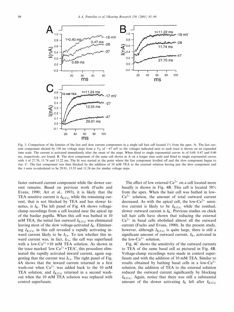
Two distinct outward currents were resolved basedon their kinetics and pharmacology. Recordings shownin Fig. 3 exemplify this. These recordings were from acell located 1% from the apex. Two components of out-ward current with di¡erent activation time constants (d)can be seen in Fig. 3A,B. The traces are from the samevoltage-clamp series of the same cell presented on dif-ferent time scales. The two current components wereseparately ¢tted to single exponentials in order to cal-culate the d. The initial outward component was quitefast, which is shown in Fig. 3A where voltage stepsfrom VH =367 mV to the voltages indicated besideeach trace yield ds of 0.69, 0.47 and 0.40 ms. The sec-ond component was much slower, as shown in Fig. 3B,where single exponential ¢ts give d of 27.70, 11.74 and11.22 ms. Fig. 3C shows recordings from the same cellwhen bathed in 10 mM TEA. The fast component wasreduced, allowing the remaining current to be ¢ttedmore cleanly to a single exponential curve from onset.
Voltage steps similar to those in Fig. 3B were appliedgiving d of 29.91, 13.55 and 11.24 ms, which are verysimilar to the d calculated for the slow component inthe absence of TEA (as shown in Fig. 3B).
3.2. Ca2+ and TEA sensitivity of the outward currents
The calcium dependence of the outward currents wastested by bathing the basilar papilla in a solution wherean equimolar amount of Mg2� replaced Ca2� (n = 16).Generally, the low-Ca2� external solution did not a¡ectthe resting membrane potential or it caused a slighthyperpolarization of less than 5 mV. The sensitivity ofthe outward currents to TEA was tested by bathing thebasilar papilla in solutions with various molarities ofTEA ranging from 0.1 mM to 10 mM (n = 6). The ad-dition of TEA to the bath did not a¡ect the restingmembrane potentials of the cells. The separation ofthe outward currents using these pharmacological ma-nipulations is demonstrated in Fig. 4. Fig. 4 shows thatboth of these manipulations substantially reduced the
Fig. 2. I/V curves for three tall hair cells at various positions along the basilar papilla. There is an increase in total outward current from acell at the apical tip (A), 15% from the apex (B) and 45% from the apex (C). Notice the change in scales for each curve. The inward recti¢er(IIR) was present in the two more apical cells (A and B) and not present in the more basal cell (C). The I/V curves were made by measuringthe current 140 ms into 150 ms voltage steps given in 10 mV increments from VH =377 (A) or 367 (B and C).
HEARES 3684 17-5-01
A.A. Pantelias et al. / Hearing Research 156 (2001) 81^94 85
faster outward current component while the slower cur-rent remains. Based on previous work (Fuchs andEvans, 1990; Art et al., 1993), it is likely that theTEA sensitive current is IK�Ca� while the remaining cur-rent, that is not blocked by TEA and has slower ki-netics, is IK. The left panel of Fig. 4A shows voltage-clamp recordings from a cell located near the apical tipof the basilar papilla. When this cell was bathed in 10mM TEA, the initial fast outward IK�Ca� was eliminatedleaving most of the slow voltage-activated IK. Eliminat-ing IK�Ca� in this cell revealed a rapidly activating in-ward current likely to be ICa. To test whether this in-ward current was, in fact, ICa, the cell was superfusedwith a low-Ca2�+10 mM TEA solution. As shown inthe trace marked `low Ca2�+TEA', this procedure elim-inated the rapidly activated inward current, again sug-gesting that the current was ICa. The right panel of Fig.4A shows that the inward current returned in a ¢rstwash-out when Ca2� was added back to the 10 mMTEA solution, and IK�Ca� returned in a second wash-out when the 10 mM TEA solution was replaced withcontrol superfusate.
The e¡ect of low external Ca2� on a cell located morebasally is shown in Fig. 4B. This cell is located 58%from the apex. When the hair cell was bathed in low-Ca2� solution, the amount of total outward currentdecreased. As with the apical cell, the low-Ca2� sensi-tive current is likely to be IK�Ca� while the residual,slower outward current is IK. Previous studies on chicktall hair cells have shown that reducing the externalCa2� in basal cells abolished almost all the outwardcurrent (Fuchs and Evans, 1990). In the present study,however, although IK�Ca� is quite large, there is still asigni¢cant amount of outward current, IK, activated inthe low-Ca2� solution.
Fig. 4C shows the sensitivity of the outward currentsto TEA of the same basal cell as pictured in Fig. 4B.Voltage-clamp recordings were made in control super-fusate and with the addition of 10 mM TEA. Similar toresults obtained by bathing basal cells in a low-Ca2�
solution, the addition of TEA to the external solutionreduced the outward current signi¢cantly by blockingIK�Ca�. Again, notice that there was still a substantialamount of the slower activating IK left after IK�Ca�
Fig. 3. Comparison of the kinetics of the fast and slow current components in a single tall hair cell located 1% from the apex. A: The fast cur-rent component elicited by 150 ms voltage steps from a VH of 367 mV to the voltages indicated next to each trace is shown on an expandedtime scale. The current is activated immediately after the onset of the steps. When ¢tted to single exponential curves ds of 0.69, 0.47 and 0.40ms, respectively, are found. B: The slow component of the same cell shown in A on a longer time scale and ¢tted to single exponential curveswith d of 27.70, 11.74 and 11.22 ms. The ¢t was started at the point where the fast component levelled o¡ and the slow component began torise. C: The fast component was then blocked by the addition of 10 mM TEA to the external solution leaving just the slow component andthe d were re-calculated to be 29.91, 13.55 and 11.24 ms for similar voltage steps.
HEARES 3684 17-5-01
A.A. Pantelias et al. / Hearing Research 156 (2001) 81^9486

is blocked. The current began to recover in the wash-out.
3.3. 4-AP sensitivity of the outward currents
In order to separate the two outward currents fur-ther, 0.8 mM 4-AP was added to the external solution(n = 16). Previous work has shown that this concentra-
tion of 4-AP e¡ectively blocks the voltage-activated IK
in turtle hair cells while not signi¢cantly a¡ecting IK�Ca�(Goodman and Art, 1996b). Fig. 5A shows the e¡ect of4-AP on the outward current of a representative apicaltall hair cell. The cell was held at 367 mV and wasgiven 150 ms voltage steps. In the presence of 0.8mM 4-AP in the external solution, the outward currentwas substantially reduced. In addition, the activation
Fig. 4. E¡ect of low-Ca2� external solution and TEA on the outward currents of tall hair cells. The locations of the cells are depicted on theschematic. A: This cell is located 2% from the apex. In voltage clamp, 10 mM TEA (trace marked `10 mM TEA') reduced the fast componentof the outward current and revealed an inward current that is blocked by superfusing the cell in a 10 mM TEA, low-Ca2� external solution(trace marked `low Ca2� and TEA'). The right panel shows that the inward current returned when the cell was bathed again in just 10 mMTEA. The fast component recovered during the wash with control superfusate. Holding potential for all recordings was 367 mV and all volt-age steps were 150 ms in duration. B: E¡ect of low-Ca2� external solution on a hair cell, 58% from the apex. Replacing external Ca2� withMg2� reduced the magnitude of the outward current, which almost fully recovered in the wash. C: E¡ect of TEA in the external solution onthe same cell located 58% from the apex. The addition of TEA reduced the outward current most notable at onset. The current partially recov-ered in wash. In all voltage-clamp traces for this cell, the cell was held at 367 mV and stepped for 150 ms to 38 mV.
HEARES 3684 17-5-01
A.A. Pantelias et al. / Hearing Research 156 (2001) 81^94 87
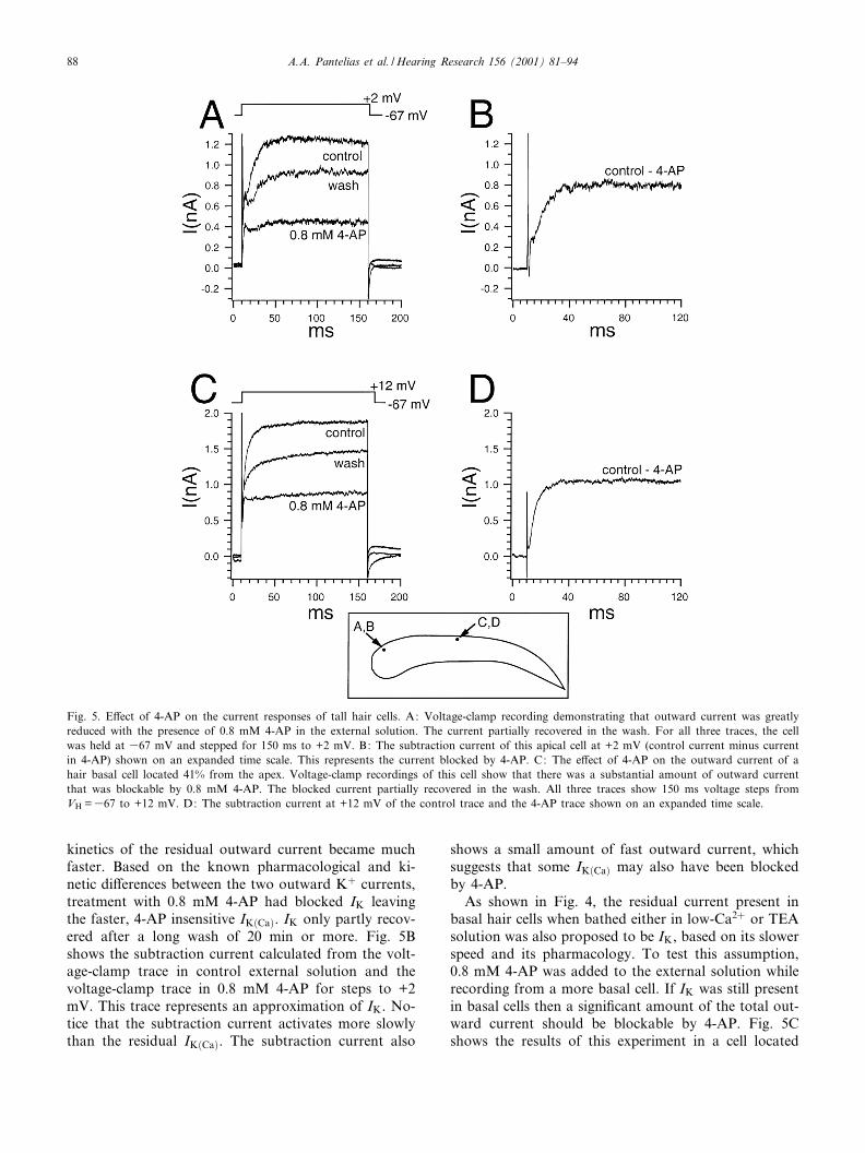
kinetics of the residual outward current became muchfaster. Based on the known pharmacological and ki-netic di¡erences between the two outward K� currents,treatment with 0.8 mM 4-AP had blocked IK leavingthe faster, 4-AP insensitive IK�Ca�. IK only partly recov-ered after a long wash of 20 min or more. Fig. 5Bshows the subtraction current calculated from the volt-age-clamp trace in control external solution and thevoltage-clamp trace in 0.8 mM 4-AP for steps to +2mV. This trace represents an approximation of IK. No-tice that the subtraction current activates more slowlythan the residual IK�Ca�. The subtraction current also
shows a small amount of fast outward current, whichsuggests that some IK�Ca� may also have been blockedby 4-AP.
As shown in Fig. 4, the residual current present inbasal hair cells when bathed either in low-Ca2� or TEAsolution was also proposed to be IK, based on its slowerspeed and its pharmacology. To test this assumption,0.8 mM 4-AP was added to the external solution whilerecording from a more basal cell. If IK was still presentin basal cells then a signi¢cant amount of the total out-ward current should be blockable by 4-AP. Fig. 5Cshows the results of this experiment in a cell located
Fig. 5. E¡ect of 4-AP on the current responses of tall hair cells. A: Voltage-clamp recording demonstrating that outward current was greatlyreduced with the presence of 0.8 mM 4-AP in the external solution. The current partially recovered in the wash. For all three traces, the cellwas held at 367 mV and stepped for 150 ms to +2 mV. B: The subtraction current of this apical cell at +2 mV (control current minus currentin 4-AP) shown on an expanded time scale. This represents the current blocked by 4-AP. C: The e¡ect of 4-AP on the outward current of ahair basal cell located 41% from the apex. Voltage-clamp recordings of this cell show that there was a substantial amount of outward currentthat was blockable by 0.8 mM 4-AP. The blocked current partially recovered in the wash. All three traces show 150 ms voltage steps fromVH =367 to +12 mV. D: The subtraction current at +12 mV of the control trace and the 4-AP trace shown on an expanded time scale.
HEARES 3684 17-5-01
A.A. Pantelias et al. / Hearing Research 156 (2001) 81^9488

41.3% from the apex. The cell was held at 367 mV andstepped to +12 mV for 150 ms. The addition of 0.8 mM4-AP to the external solution did reduce the amount ofoutward current by eliminating the slower current com-ponent. As a result, the remaining outward current wasthe faster current component. The 4-AP sensitive cur-rent is expressed in Fig. 5D as the subtraction currentof the control voltage-clamp trace and the voltage-clamp trace in 4-AP for steps to +12 mV. As in theapical cell, the subtraction current had slower activationkinetics than the unblocked current. The subtractioncurrent is, again, an approximation of the blocked IK.Therefore, both apical and more basal cells have signi¢-cant amounts of both IK and IK�Ca�, which are at leastpartially separable using these pharmacological manip-ulations.
3.4. E¡ect of external Cs+ on IIR
Low-frequency apical hair cells, in addition to thetwo outward potassium currents, also have an inwardrecti¢er potassium current, IIR, that is activated at hy-perpolarized potentials. Our observations and previousstudies of isolated chick hair cells (Fuchs and Evans,1990) indicate that more basal, high-frequency hair cellsdo not have this current. Likewise, turtle hair cellstuned to the lower frequencies have IIR, and as reso-nance frequency increases, IIR decreases. Blocking IIR inturtle hair cells resulted in a distinct decrease in thequality of resonance (Goodman and Art, 1996a). Inaddition, it has been shown in both turtle hair cells ofthe lower frequencies (Goodman and Art, 1996a) andchick isolated apical hair cells (Fuchs and Evans, 1990)that 5 mM Cs� in the external solution can block IIR.Therefore, this concentration of Cs� was used in ourexperiments to block this current (n = 6).
Fig. 6 shows the e¡ect of 5 mM Cs� in the externalsolution on IIR in a cell at the apical tip. Fig. 6A, in thetrace marked `control', shows that IIR was activated bya 150 ms voltage step from VH =367 to 3112 mV. Thesame voltage step with the addition of Cs� to the ex-ternal solution shows that IIR was blocked, as can beseen in the trace marked, `5 mM Cs�'. The currentreturned to near normal as the Cs� was washed out.The e¡ect of blocking IIR on the voltage/current rela-tionship of the steady-state outward current of an ap-ical tall hair cell is shown in Fig. 6B. Steady-state cur-rent was measured at the end of 150 ms voltage stepsgiven in 10 mV increments from VH =367. In Cs�, theresponse to hyperpolarizing steps £attens re£ecting theblock of IIR. For all apical cells recorded in Cs� con-taining external solution (n = 6), there was no signi¢cante¡ect on the outward currents measured at more depo-larized voltage steps.
3.5. Mapping of the papilla
Since the entire intact basilar papilla was explanted ineach of our preparations, we could determine the rela-tionships between various electrophysiological charac-teristics and the positions of the hair cells along thelength (tonotopic axis) of the papilla. Fig. 7 presentsthese data for steady-state current (Fig. 7A), currentin low external Ca2� (Fig. 7B) and the subtraction cur-rent in low external Ca2�, which approximates IK�Ca�(Fig. 7C). In Fig. 7, the position of each cell alongthe papilla is indicated on the abscissa and the param-eter value is indicated on the ordinate. In Fig. 7A, thesolid line describes the best ¢t exponential regressionwith the r2 value indicated at the bottom right. Samplesize (n) is indicated in all three graphs at the bottomright. For the three graphs shown in Fig. 7, holding
Fig. 6. E¡ect of external Cs� on IIR of apical hair cells. A: Voltage-clamp recording of a hair cell located near the apex of the papilla showingthe a¡ect of 5 mM Cs� in the external solution on IIR. A 150 ms voltage step from VH =367 mV to 3112 mV demonstrates that IIR is acti-vated in control external solution, reduced in the presence of 5 mM Cs� and partially recovers in the wash. B: Plot of current versus mem-brane voltage for control (closed circles) and 5 mM Cs� (open squares) external solution.
HEARES 3684 17-5-01
A.A. Pantelias et al. / Hearing Research 156 (2001) 81^94 89

potentials for the cells were near their resting membranepotential of near 367 mV. Cells with series resistancesof 14 M6 or more were eliminated as well as cells thatshowed a signi¢cant (s 0.2 nA) Ohmic conductance atpotentials around the holding potential determined dur-ing I/V analysis. In this way, cells used for these graphshad little leak current. The locations of the cells weremeasured as a percentage of total papilla length with0% denoting the apical tip. It is apparent in Fig. 7 thatwe were unable to achieve adequate recordings fromhair cells in the basal one-third of the papilla. Thisproblem also plagued previous studies (e.g. Fuchs andSokolowski, 1990). In our preparations, the basal haircells would become unhealthy and bleb. It is unknownwhy these hair cells do not remain healthy in vitro.
Fig. 7A shows the relationship between total, steady-state outward current and location on papilla. Totalsteady-state current for all cells plotted in Fig. 7Awas measured at the end of a 150 ms voltage step. Allvoltages were corrected for residual series resistance re-sulting in a variability in command potentials. There-fore, for each cell, the amount of current at 37 mV wasinterpolated from the amount of current recorded atvoltage steps to potentials less than and greater than37 mV. When the results were mapped against placeon papilla, it is best ¢t by an exponential rise in totalcurrent amplitude as a function of position from apexto base (r2 = 0.73).
This current measurement, therefore, is the sum ofboth outward K� currents, IK�Ca� and IK. The measure-ment of the outward current also will be in£uenced bythe inward Ca2� current, which will lead to an under-estimation of the outward current all along this map.However, these measurements cannot be done in theabsence of the Ca2� current since one of the currentsof interest, IK�Ca�, is Ca2� dependent. Despite the pres-ence of ICa which increases in amplitude from apex tobase (Martinez-Dunst et al., 1997), the opposing out-ward current clearly shows a dramatic increase fromapex to base, demonstrating that the trend in the mag-nitude of outward current is still observable. Further-more, ICa was measured with Ba2� as the current carrierand it was found that this current increases only over arange of about 100 pA over 1.7 mm of basilar papillalength (Martinez-Dunst et al., 1997). Another possibleconcern, for apical cells in particular, is IIR. However,as can be seen in Fig. 6B, the presence of IIR does nota¡ect the measurement of total outward steady-statecurrent at the depolarized membrane potentials usedhere.
Fig. 7B shows the relationship between the voltage-activated IK and place on the papilla. IK was measuredat steady-state, at the end of a 150 ms voltage stepunder low-Ca2� conditions. Therefore, the majority ofcurrent measured would be IK. Current at 37 mV is
Fig. 7. Maps of total outward steady-state current (A), outwardcurrent in low Ca2� (B), and the subtraction current from voltage-clamp traces in control external and in low-Ca2� external solution(C), plotted against location on papilla expressed as a percentagefrom apex to base with 0% denoting the apical tip. Holding poten-tials were near resting membrane potentials for all cells (between367 and 377 mV). Sample sizes indicated in each graph by n.A: Total outward steady-state current was measured at the end ofvoltage steps. Current at 37 mV was interpolated from values forvoltage steps above and below 37 mV. The data were ¢tted to anexponential with an r2 value of 0.73. B: Steady-state outward cur-rent in low-Ca2� external solution was used to estimate IK, sinceIK�Ca� was largely blocked. Current at 37 mV was calculated as wasdescribed in A. C: The subtraction current was calculated to esti-mate the magnitude of IK�Ca�.
HEARES 3684 17-5-01
A.A. Pantelias et al. / Hearing Research 156 (2001) 81^9490

plotted and was measured as described for Fig. 7A.When these results are plotted against place on papilla,an increase in current magnitude is seen from apex tobase.
Fig. 7C shows the relationship between the subtrac-tion current and place on the basilar papilla. The mag-nitude of this current was measured by subtracting theamount of outward current activated at 37 mV in con-trol external and the amount of outward current acti-vated at 37 mV in low-Ca2� external solution. Thissubtraction approximates the magnitude of IK�Ca�,which is present in control external but is not presentin low external Ca2�. When these values are plottedagainst place on papilla, an increase in magnitude isseen from apex to base. For all three graphs, note thechanges in scales.
3.6. Current-clamp recording of membrane voltageoscillations
We recorded from 20 cells in current-clamp mode inwhich we observed the membrane voltage responses ofcells during steps of depolarizing current injections. Allcells displayed a damped, sinusoidal membrane voltageoscillation similar to responses reported by Fuchs andEvans (1990). However, the frequency of these oscilla-tions was either on the order of 12^23 Hz or above 120Hz. Cells located in the apical 35% of the basilar papillashowed the slower oscillations while cells more basalabruptly began to display the faster oscillations. Nocells were recorded from that oscillated between 25and 120 Hz despite that fact there was a smooth gra-
dient in the locations of the cells along the tonotopicaxis. Fig. 8 shows typical examples of these oscillationsfor both frequency groups. Fig. 8A shows a cell located14.8% from the apex which, when depolarized with +30pA current injection, oscillated at 16 Hz. Fig. 8B, how-ever, shows a more basal cell, located 45% from the
Fig. 9. Map of oscillation frequency versus location on papilla ex-pressed as a percentage from apex to base with 0% denoting the ap-ical tip. Oscillation frequency was measured at the best resolved fre-quency. Notice the sharp increase in frequency that occurs between30 and 35% from the apex. While oscillation frequency was not re-lated to position in the apex, cells more than 30% from the apex ex-pressed higher frequency oscillations as the base was approached.
Fig. 8. Current-clamp recordings of two tall hair cells from di¡erent areas of the basilar papilla. A: A cell 14.8% from the apex was depolar-ized with a 150 ms, +30 pA step of injected current. Oscillation frequency was 16 Hz. B: A cell 45% from the apex was depolarized with a20 ms, +50 pA current step to reveal oscillations of 190.5 Hz.
HEARES 3684 17-5-01
A.A. Pantelias et al. / Hearing Research 156 (2001) 81^94 91

apex which when depolarized with +50 pA of injectedcurrent, oscillated at 190.5 Hz.
Fig. 9 shows an analysis of oscillation frequency ver-sus location on the papilla (n = 20). For apical cells theamplitude of injected current which yielded the bestresolved oscillations (highest peak to trough ratio)used to measure frequency was between +10 and +30and for basal cells it was between +20 and +90 pA. Thegraph shows that cells located in the apical 30% allshow very slow oscillations ranging from about 12 to23 Hz and there is no apparent tonotopic organization.On the other hand, cells 35% or further from the apexshow oscillations above 120 Hz and are organized alongthe tonotopic axis with respect to oscillation frequency.
4. Discussion
Anatomical studies of the chick cochlea have shownvariations in hair cell morphology and innervation pat-terns. For example, there are gradients in the morphol-ogy of stereocilia bundles (Tilney and Saunders, 1983)and gradients in the morphology of cell bodies betweenthe two extreme forms, tall hair cells and short hair cells(Hirokawa, 1978; Fischer, 1992). In addition, the pat-tern of innervation of the basilar papilla changes fromthe tall hair cells being much more heavily innervatedby a¡erent than e¡erent ¢bers, to basal short hair cellsthat have mostly or only e¡erent endings (Tanaka andSmith, 1978; Hirokawa, 1978; Manley et al., 1989;Fischer, 1992). Observations of these changes have ledto the question of functional variation. For example,the morphological di¡erences of very apical hair cellsfrom the hair cells along the rest of the epithelium havebeen postulated to re£ect a di¡erence in function (La-vigne-Rebillard et al., 1985). Similarly, observations ofanatomical variation raise the question of whether thehair cells' phenotypes play a role in auditory frequencyselectivity (Hirokawa, 1978).
The issue of peripheral frequency tuning in birdsgained indirect support from physiological recordingsof the cochlear ganglion, VIIIth nerve ¢bers, and cen-tral nervous system (Rubel and Parks, 1975; Manley,1979; Manley et al., 1985; Temchin, 1988). These stud-ies demonstrated that the nerve, cochlear ganglion andcentral terminations are tonotopically organized; aproperty that could have originated in the cochlea. Itwas already established that there was mechanical tun-ing in the chick cochlea with von Bekesy's (1960) ob-servations that the basilar membrane optimally vibratedat di¡erent frequencies along the length of the papilla.The question remained, however, as to whether avianhair cells are capable of sharpening this tuning throughelectrical resonance as demonstrated in hair cells ofreptiles and amphibians (Crawford and Fettiplace,
1981; Lewis and Hudspeth, 1983; Art and Fettiplace,1987; Pitchford and Ashmore, 1987; Fuchs and Evans,1988). Direct evidence for electrical resonance in thechick cochlea was provided by studies of isolated tallhair cells (Fuchs et al., 1988). This resonance is theresult of the interaction of the cell's membrane currents.The tonotopy of resonance is thought to be determinedby the tonotopy of the magnitudes of these currents.The goal of our studies was to map changes in theoutward K� conductances, which are essential to gen-erating electrical resonance in chick tall hair cells(Fuchs and Evans, 1990), in order to get a clearerunderstanding of how the physiology of tall hair cellschanges along the length of the basilar papilla of nor-mal chicks. These data can then be compared with ani-mals in which genetic or environmental manipulationshave been made. We think it will be of particular inter-est to compare the results presented here with similarobservations on regenerating hair cells.
4.1. K+ currents
The initial fast activating outward current found inavian tall hair cells is blocked by Ca2�-free solutions,and shows sensitivity to the potassium channel blockerTEA. This initial fast outward current displays kineticsand pharmacology very similar to the calcium-activatedpotassium current (IK�Ca�) as described in dissociatedchick tall hair cells (Fuchs et al., 1988; Fuchs andEvans, 1990), isolated chick vestibular hair cells (Oh-mori, 1984), turtle auditory (Art et al., 1993) and frogsaccular hair cells (Lewis and Hudspeth, 1983).
The slow outward current component, in contrast, isuna¡ected by Ca2�-free solution, insensitive to TEAbut quite sensitive to 4-AP. It is voltage-activated atabout 350 mV. The voltage activation, kinetics andpharmacology seem to categorize this current as a de-layed recti¢er potassium current (IK�DR�). Although ki-netics and pharmacology vary, IK�DR� can display slowkinetics (Hille, 1992; Johnston and Wu, 1995) and rel-ative insensitivity to TEA (Tasaki and Hagiwara, 1957;Stan¢eld, 1970; Wong and Binstock, 1980). A currentbearing properties very similar to IK�DR� has been re-ported in dissociated chick hair cells (Fuchs and Evans,1990) as well as turtle cochlear hair cells (Art et al.,1993).
4.2. Basilar papilla maps
When the results of these experiments are mappedalong the length of the papilla, several interesting ob-servations are evident. First, the amount of steady-stateoutward current of tall hair cells increases exponentiallyfrom apex to base. There is also a tonotopic increase inthe magnitude of IK ; however, the rise is not as dra-
HEARES 3684 17-5-01
A.A. Pantelias et al. / Hearing Research 156 (2001) 81^9492
matic. In addition, the subtraction current, which is anapproximation of IK�Ca�, also increases in magnitudefrom apex to base. These results re£ect the gradientsin the two individual outward K� currents as one trav-els along the length of the basilar papilla. It can be seenthat IK and IK�Ca� increase in magnitude as the base ofthe papilla is approached, resulting in basal hair cellsdisplaying much larger outward currents. This resultbrings to mind frequency^place maps described in thechick cochlea by a variety of investigators (Manley etal., 1987; Chen et al., 1994; Jones and Jones, 1995;Ryals and Rubel, 1982, 1985a,b). The increase in speedand amplitude of tall hair cells' currents may re£ect theincrease in frequency that the cells are tuned to fromapex to base. Our results, in addition, show that tallhair cells of the apex, which show low-frequency oscil-lations, as well as hair cells 50^60% from the apex,which have much higher frequency oscillations, haveboth IK and IK�Ca�. In studies of turtle cochlear haircells it was found that the tonotopy of resonant fre-quency resulted from variations in the amounts ofboth IK�Ca� and IK (Goodman and Art, 1996b). Theseresults are also consistent with those of Navaratnam etal. (1997) and Rosenblatt et al. (1997) who showedsystematic variation in the expression of splice variantsof a gene encoding one IK�Ca� along the length of thepapilla.
An unexpected result of this study was the abruptchange in oscillation frequency when mapped againstplace on papilla. Although the outward currents forma gradient in magnitude, the membrane voltage oscilla-tions were either slow (below 25 Hz) or faster (above120 Hz). Cells located in the apical 30% of the papilladisplayed the slow oscillations and the frequency ofoscillation did not appear to change tonotopically. Cellslocated more basal than 35% from the apex, however,displayed the faster oscillations and these cells weretonotopically organized with respect to frequency.This is a curious result because it raises the questionof if and how the membrane voltage oscillations help inelectrical tuning and resonance in chick tall hair cells. Italso raises the question of what changes at about 35%along the papilla from the apex to underlie such anabrupt and obvious change in oscillation frequency.One obvious caveat is that these experiments weredone at room temperature and therefore, the frequen-cies recorded would be slower than what would exist invivo. In addition, an explanted basilar papilla lacks theadded tuning a¡orded to it by the e¡ects of the me-chanical tuning of the basilar membrane or stereociliabundles. Also, the method of stimulation may not bephysiological for apical hair cells. High-frequencysounds may produce a DC current in the high-fre-
quency tuned basal hair cells while lower frequencysounds may produce a more AC response in the low-frequency apical hair cells. Therefore, injection ofsquare current pulses may not be a biologically relevantmethod of stimulating apical hair cells (Eisen et al.,1998). Lastly, it is possible that the currents recordedin this in vitro preparation still may not be of the mag-nitude of hair cells in vivo. Although the currents re-corded here are greater than the currents measured inisolated hair cells and may more accurately depict haircell physiology, it is still an in vitro preparation and anyloss of current will a¡ect the frequency of membranevoltage oscillations.
This study also demonstrates a new preparationwhere the entire chick basilar papilla is kept intact.The bene¢ts of this preparation are the ability to mapthe physiology of hair cells along the epithelium to amore detailed degree, as well as minimizing enzymatictreatments. Only collagenase was used to remove thetectorial membrane in these experiments, eliminatingthe use of isolation enzymes which have been shownto quite dramatically a¡ect recorded currents and mem-brane voltage oscillations in the frog sacculus (Arm-strong and Roberts, 1998). As a result, the currentsthat were recorded here are considerably greater thanthe currents that have been reported previously fromisolated hair cells (Fuchs and Evans, 1990). Addition-ally, the magnitude of the individual currents whenpharmacologically separated is signi¢cantly larger.Therefore, this method of recording from chick haircells may give a more accurate physiological re£ectionof the magnitude and distribution of the two outwardK� currents. Lastly, this preparation allows for the to-notopic changes in other physiological properties ofchick auditory hair cells to be investigated in more de-tail. For example, the preparation a¡ords one the abil-ity to track di¡erences in currents and voltage responsesof hair cells along the superior to inferior axis. Thiswould allow one to study the physiological changesthat occur alongside morphological changes as thehair cells move from tall to intermediate to short haircells. In addition, it also may allow for the study of haircells during various manipulations of the cochlea wherethe basilar papilla needs to be intact, such as hair celldamage and regeneration.
Acknowledgements
The authors are grateful to Dr. Paul Fuchs and Dr.Cecilia Armstrong for critically reading and comment-ing on the manuscript. The research was supported byNIH/NIDCD Grant DC00395.
HEARES 3684 17-5-01
A.A. Pantelias et al. / Hearing Research 156 (2001) 81^94 93

References
Armstrong, C.E., Roberts, W.M., 1998. The electrical properties offrog saccular hair cells: Distortion by enzymatic dissociation.J. Neurosci. 18, 2962^2973.
Art, J.J., Fettiplace, R., 1987. Variation of membrane properties inhair cells isolated from the turtle cochlea. J. Physiol. 385, 207^242.
Art, J.J., Fettiplace, R., Wu, Y.C., 1993. The e¡ects of low calcium onthe voltage-dependent conductances involved in tuning of turtlehair cells. J. Physiol. 470, 109^126.
Chen, L., Salvi, R., Shero, M., 1994. Cochlear frequency^place mapin adult chickens: intracellular biocytin labeling. Hear. Res. 81,130^136.
Crawford, A.C., Fettiplace, R., 1981. An electrical tuning mechanismin turtle cochlear hair cells. J. Physiol. 306, 79^125.
Eisen, M.D., Saunders, J.C., Parsons, T.D., 1998. High frequencymechanoelectric transduction by individual chick cochlear tallhair cells. Assoc. Res. Otolaryngol. 21, 1998.
Fischer, F.P., 1992. Quantitative analysis of the innervation of thechicken basilar papilla. Hear. Res. 61, 167^178.
Fuchs, P.A., Evans, M.G., 1988. Voltage oscillations and ionic con-ductances in hair cells isolated from the alligator cochlea. J. Comp.Physiol. A 164, 151^163.
Fuchs, P.A., Evans, M.G., 1990. Potassium currents in hair cells iso-lated from the cochlea of the chick. J. Physiol. 429, 529^552.
Fuchs, P.A., Evans, M.G., Murrow, B.W., 1990. Calcium currents inhair cells isolated from the cochlea of the chick. J. Physiol. 429,553^568.
Fuchs, P.A., Nagai, T., Evans, M.G., 1988. Electrical tuning in haircells isolated from the chick cochlea. J. Neurosci. 8, 2460^2467.
Fuchs, P.A., Sokolowski, B.H., 1990. The acquisition during develop-ment of Ca-activated potassium currents by cochlear hair cells ofthe chick. Proc. R. Soc. Lond. Biol. Sci. 241, 122^126.
Goodman, M.B., Art, J.J., 1996a. Positive feedback by a potassium-selective inward recti¢er enhances tuning in vertebrate hair cells.Biophys. J. 71, 430^442.
Goodman, M.B., Art, J.J., 1996b. Variations in the ensemble of po-tassium currents underlying resonance in turtle hair cells. J. Phys-iol. 497, 395^412.
Hamill, O.P., Marty, A., Neher, E., Sakmann, B., Sigworth, F.J.,1981. Improved patch-clamp techniques for high-resolution cur-rent recordings from cells and cell-free membrane patches. P£ug.Arch. 391, 85^100.
Hille, B., 1992. Ionic Channels of Excitable Membranes. Sinauer,Sunderland, pp. 74^78.
Hirokawa, N., 1978. The ultrastructure of the basilar papilla of thechick. J. Comp. Neurol. 181, 361^374.
Johnston, D., Wu, S.M.-S., 1995. Foundations of Cellular Neuro-physiology. The MIT, Cambridge, MA, p. 198.
Jones, S.M., Jones, T.A., 1995. The tonotopic map in the embryonicchicken cochlea. Hear. Res. 82, 149^157.
Lavigne-Rebillard, M., Cousillas, H., Pujol, R., 1985. The very distalpart of the basilar papilla in the chicken: A morphological ap-proach. J. Comp. Neurol. 238, 340^347.
Lewis, R.S., Hudspeth, A.J., 1983. Voltage- and ion-dependent con-ductances in solitary vertebrate hair cells. Nature 304, 538^540.
Manley, G.A., 1979. Preferred intervals in the spontaneous activity ofprimary auditory neurons. Naturwissenshaften 66, 582^584.
Manley, G.A., Gleich, O., Kaiser, A., Brix, J., 1989. Functional di¡er-entiation of sensory cells in avian auditory periphery. J. Comp.Physiol. A 164, 289^296.
Manley, G.A., Gleich, O., Leppelsack, H.-J., Oekinghaus, H., 1985.Activity patterns of cochlear ganglion neurons in the starling.J. Comp. Physiol. A 157, 161^181.
Manley, G.A., Brix, J., Kaiser, A., 1987. Developmental stability ofthe tonotopic organization of the chick's basilar papilla. Science237, 655^656.
Martinez-Dunst, C., Micheals, R.L., Fuchs, P.A., 1997. Release sitesand calcium channels in hair cells of the chick's cochlea. J. Neuro-sci. 17, 9133^9144.
Navaratnam, D.S., Bell, T.J., Tu, T.D., Cohen, E.L., Oberholtzer,J.C., 1997. Di¡erential distribution of Ca2�-activated K� channelsplice variants among hair cells along the tonotopic axis of thechick cochlea. Neuron 19, 1077^1085.
Ohmori, H., 1984. Studies of ionic currents in the isolated vestibularhair cell of the chick. J. Physiol. 350, 561^581.
Pantelias, A.A., Monsivais, P., Rubel, E.W., 1998. Tonotopic map ofcurrent and voltage responses of chick auditory hair cells using anintact basilar papilla preparation. Assoc. Res. Otolaryngol. 20, 21.
Pitchford, S., Ashmore, J.F., 1987. An electrical resonance in haircells of the amphibian papilla of the frog Rana temporaria.Hear. Res. 27, 75^83.
Rebillard, G., Rubel, E.W., 1981. Electrophysiological study of thematuration of auditory responses from the inner ear of the chick.Brain Res. 229, 15^23.
Rosenblatt, K.P., Sun, Z.P., Helly, S., Hudspeth, A.J., 1997. Distri-bution of Ca2� activated K� channel isoforms along the tonotopicgradient of the chicken cochlea. Neuron 19, 1061^1075.
Rubel, E.W., Parks, T.N., 1975. Organization and development ofbrain stem auditory nuclei of the chicken: tonotopic organizationof n. magnocellularis and n. laminaris. Comp. Neurol. 164, 411^433.
Ryals, B.M., Rubel, E.W., 1982. Patterns of hair cell loss in chickbasilar papilla after intense auditory stimulation. Frequency orga-nization. Acta Otolaryngol. 93, 205^210.
Ryals, B.M., Rubel, E.W., 1985a. Di¡erential susceptibility of avianhair cells to acoustic trauma. Hear. Res. 19, 73^84.
Ryals, B.M., Rubel, E.W., 1985b. Ontogenetic changes in the positionof hair cell loss after acoustic overstimulation in avian basilarpapilla. Hear. Res. 19, 135^142.
Saunders, J.C., Coles, R.B., Gates, G.R., 1973. The development ofauditory evoked responses in the cochlea and cochlear nuclei ofthe chick. Brain Res. 63, 59^74.
Stan¢eld, P.R., 1970. The e¡ect of the tetraethylammonium ion on thedelayed currents of frog skeletal muscle. J. Physiol. 209, 209^229.
Takasaka, T., Smith, C.A., 1971. The structure and innervation of thepigeon's basilar papilla. J. Ultrastruct. Res. 35, 20^65.
Tanaka, K., Smith, C.A., 1978. Structure of the chicken's inner ear:SEM and TEM study. Am. J. Anat. 153, 251^272.
Tasaki, I., Hagiwara, S., 1957. Demonstration of two stable potentialstates in the squid giant axon under tetraethylammonium chloride.J. Gen. Physiol. 40, 859^885.
Temchin, A.N., 1988. Unusual discharge patterns of single ¢bers inthe pigeon's auditory nerve. J. Comp. Physiol. A 163, 99^115.
Tilney, L.G., Saunders, J.C., 1983. Actin ¢laments, stereocilia, andhair cells of the bird cochlea. I. Length, number, width, and dis-tribution of stereocilia of each hair cell are related to the positionof the hair cell on the cochlea. J. Cell Biol. 96, 807^821.
Tucci, D.L., Rubel, E.W., 1990. Physiological status of regeneratedhair cells in the avian inner ear following aminoglycoside ototox-icity. Otolaryngol. Head Neck Surg. 103, 443^450.
von Bekesy, G., 1960. Experiments in Hearing. McGraw-Hill, NewYork.
Wong, B.S., Binstock, L., 1980. Inhibition of potassium conductancewith external tetraethylammonium ion in Myxicola giant axons.Biophys. J. 32, 1037^1042.
HEARES 3684 17-5-01
A.A. Pantelias et al. / Hearing Research 156 (2001) 81^9494