tolerance of heavy metals by gram positive soil bacteria€”with the intention of screening for...
TRANSCRIPT

Abstract—With the intention of screening for heavy metal
tolerance, a number of bacteria were isolated and characterized from a pristine soil. Two Gram positive isolates were identified as Paenibacillus sp. and Bacillus thuringeinsis. Tolerance of Cd2+, Cu2+ and Zn2+ by these bacteria was studied and found that both bacteria were highly sensitive to Cu2+ compared to other two metals. Both bacteria showed the same pattern of metal tolerance in the order Zn+
> Cd2+ > Cu2+. When the metal tolerance in both bacteria was compared, Paenibacillus sp. showed the highest sensitivity to Cu2+ where as B. thuringiensis showed highest sensitivity to Cd2+ and Zn2+
.These findings revealed the potential of Paenibacillus sp. in developing a biosensor to detect Cu2+ in environmental samples.
Keywords—Heavy metals, bacteria, soil, tolerance.
I. INTRODUCTION EAVY metals are often defined as a group of metals whose atomic density is greater than 5 g/cm3 [1],[2].
Metals play a vital role in the metabolic processes of the biota. Some of the heavy metals are essential and are required by the organisms as micro nutrients (cobalt, chromium, nickel, iron manganese and zinc etc.) and are known as ‘trace elements’ [3]. They are involved in redox processes, in order to stabilize molecules through electrostatic interactions, as catalysts in enzymatic reactions, and regulating the osmotic balance [2],[4]. On the other hand some other heavy metals have no biological role and are detrimental to the organisms even at very low concentration (cadmium, mercury, lead etc.). However, at high levels both of the essential and non essential metals become toxic to the organisms.
These heavy metals influence the microbial population by affecting their growth, morphology, biochemical activities and ultimately resulting in decreased biomass and diversity [5]. Heavy metals can damage the cell membranes, alter enzymes specificity, disrupt cellular functions and damage the structure of the DNA. Toxicity of these heavy metals occurs through the displacement of essential metals from their native binding sites or through ligand interactions [3]. Also, toxicity can occur as a result of alterations in the conformational structure of the nucleic acids and proteins and interference with oxidative phosphorylation and osmotic balance [6],[3].
Authors are with the Centre for Environmental Risk Assessment and
Remediation, University of South Australia, Mawson Lakes, SA 5095, Australia, CRC for the Contaminant Assessment and Remediation of the Environment Mawson Lakes, SA 5095, Australia. (I.V.N.Rathnayake, corresponding author, phone: +61883026254; fax: +61883023057; e-mail: [email protected]).
Due to the selective pressure from the metal in the growth environment, microorganisms have evolved various mechanisms to resist the heavy metal stress. Several metal resistance mechanisms have been identified: exclusion by permeability barrier, intra and extra cellular sequestration, active transport, efflux pumps, enzymatic detoxification, and reduction in the sensitivity of the cellular targets to metal ions [6],[3].
Heavy metal contamination in the environment has become a serious problem due to the increase in the addition of these metals to the environment. Natural sources as well as the anthropogenic sources account for this contamination, which has become a threat to public health. Cadmium, copper and zinc are among those heavy metals that are being released to the environment [7].
In this perspective many approaches have been used to assess the risk posed by the contaminating metals in soil, water bodies etc. At present the tolerance of soil bacteria to heavy metals has been proposed as an indicator of the potential toxicity of heavy metals to other forms of biota [9],[10]. Therefore, there is a dramatic increase in the interest on studying the interactions of heavy metals with microorganisms. The favoured approach now is selecting the organisms that can be used to develop tools to assess the metal levels in the environment. The objective of this study was to isolate and identify the bacteria from uncontaminated soil to determine their tolerance to cadmium, copper, and zinc.
II. PROCEDURE A. Test Chemicals and Media Stock solutions of cadmium, copper and zinc were prepared
by dissolving the respective nitrate salt (Sigma) in MilliQ water. Working test metal solutions were prepared by diluting the concentrated stock solutions as required, and were sterilized by filtration. All glassware was acid washed before use to avoid binding of metal. All the media used in the experiments were dissolved in MilliQ water and sterilized by autoclaving.
B. Isolation of the Bacteria Bacterial cultures were isolated from a pristine soil sampled
from Mt. Lofty, South Australia using the standard dilution plate technique. The soil was characterised in our previous study which was found to contain the metals levels below the National Environment Protection (Assessment of site contamination) Measure (NEPM) ecological investigation levels (EILs) for the metals in soil [11]. A 10-fold dilutions of fresh soil (1 g) were made in phosphate buffered saline and
Tolerance of Heavy Metals by Gram Positive Soil Bacteria
I. V. N. Rathnayake, Mallavarapu Megharaj, Nanthi Bolan, and Ravi Naidu
H
International Journal of Civil and Environmental Engineering 2:4 2010
191

0.1 ml from each of these dilutions were spread on triplicate trypticase soy agar plates. Plates were incubated at 25о C for 2-3 days. Colonies with different morphological appearance were selected from these culture plates and purified by further subculturing in the same media. All the cultures were stored at -80ºC in the trypticase soy broth with 20% glycerol.
B. Identification of Bacteria Two isolates were selected based on the Gram’s reaction
and further characterized using morphological (shape, motility, presence of endospores), biochemical (catalase activity, oxidase activity, acid production from glucose, oxidation fermentation reaction (OF) characteristics) properties [12].
Based on preliminary screening 2 isolates were selected for further taxonomic identification using molecular techniques. The crude DNA extracts were prepared from bacterial cultures grown in Luria Bertani medium. The cultures were harvested by centrifugation at 2000 x g for 10 minutes and washed twice with 1 ml of sterile TE buffer (10 mM Tris, 1m M EDTA). After the final wash the pellet was re-suspended in 50 uL of sterile TE buffer containing 1% Triton X-100. The tubes were then incubated at 70oC for 30 mins, vortexed and placed on ice. To collect cellular debris the tube was centrifuged at 10 000 x g for 5 min., and the supernatant was collected [13]. The crude DNA extract was diluted in sterile water just prior to carrying out PCR under the following conditions. A 25 uL PCR mixture contained 1 x concentration of Taq DNA polymerase buffer, 2.5 mM MgCl2, 200 uM betaine, 0.2 mM of each deocynucleoside trisphosphate, 25 pmol of each forward and reverse primers, 1 U of DNA polymerase (Promega), and 1 uL of the diluted DNA extract as template. Almost complete 16S rRNA genes were amplified with the forward primer E8f (5’-AGAGTTTGATCCTGGCTCAG-3’) and the reverse primer 1541r (5’-AAGGAGGTGATCCANCCRCA-3’) [14]. The DNA was amplified with a iCycler thermocycler (BioRad, Sydney) with the following program: 5 min of preheating at 95°C, 30 cycles of 30 s of denaturation at 95°C, 30 s of primer annealing at 55°C, and 2 min of elongation at 72°C. A final extension step of 10 min at 72°C was included. Successful amplification of a ~1525 bp DNA fragment was confirmed by running 5uL of the PCR reaction on a 1% agarose gel. PCR reactions were purified using MoBio UltraClean PCR Purification Kit (Geneworks, Adelaide), before being submitted for sequencing at the Flinders DNA sequencing facility (Adelaide).
All 16S rRNA gene sequences from the clone libraries were aligned with the “align” tool as available on greengenes website [15]. Most similar 16S rRNA genes sequences from the greengenes database [16] were also included in the alignment, and a phylogenetic tree constructed with MEGA, version 4.0 [17].
C. Determination of the Effect of Metals on Bacterial Growth
Toxicity of the selected metals to the bacterial isolates was determined using seven concentrations of each metal. These concentrations ranged from 0 to 8.0 mg/L medium. Several 48
well sterile polystyrene microplates (Iwaki polystyrene, sterile, non treated, flat bottom with lid) were used in this study as growth vessels. Sterile MES buffered minimal medium (developed in our previous study) was amended with each heavy metal and inoculated with exponentially growing cultures (24 h old, optical density of 0.090 at 600 nm) of bacterial isolates prepared in the same medium. Medium without metal but the bacterial inoculum (bacterial growth control) and medium with metal but without bacteria (abiotic control) served as controls. All the experiments were conducted in triplicate. Microplates were then closed with their lids with condensation rings and sealed using additional laboratory film (Parafilm® M). The test microplates were incubated at 25ºC on an orbital shaker at 100 rpm. Bacterial growth was measured in terms of optical density at 600 nm for 4 days at 24 hour intervals using the Bio-Tek® SynergyTM HT Multi-Detection Microplate Reader with equipped with KC4 software.
D. Statistical Analysis Non linear regression analysis was performed with the
statistical program Grapher 7, to fit the data obtained for the heavy metal toxicity experiments to the logistic model [18]. EC50 values (statistically derived estimate of a concentration of a substance resulting in 50% reduction of the growth within a specified time) were estimated and presented as mean EC50± standard deviation based on the dose-response data obtained from 3 replicate samples.
III. RESULTS AND DISCUSSION A. Identification of the Isolated Bacteria The results of the morphological and biochemical
identification experiments are shown in the Table I. Both of these bacterial isolates are rod shaped, spore formers. They showed almost same response to the biochemical reactions tested during the study. Based on the molecular analysis data, a phylogenetic tree was constructed by comparing nucleotide sequences with available 16S rRNA sequences. The two bacterial isolates were identified as Paenobacillus sp. and Bacillus thuringiensis. Constructed phylogenic tree was presented in the Fig. 1. Paenobacillus sp. was originally grouped within the genus Bacillus. Both of these bacterial genera represent the common soil bacteria and have been reported as soil inhabitants [5],[19],[20].
International Journal of Civil and Environmental Engineering 2:4 2010
192
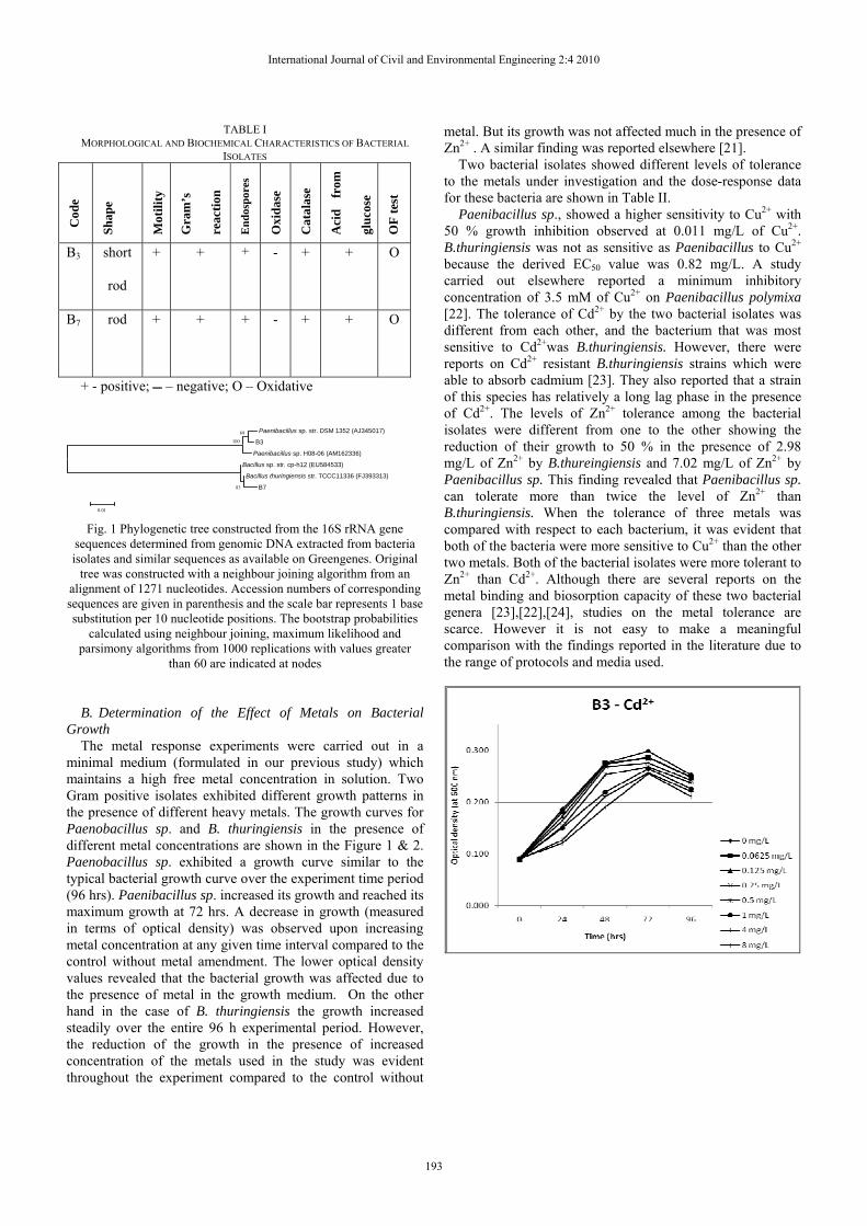
TABLE I MORPHOLOGICAL AND BIOCHEMICAL CHARACTERISTICS OF BACTERIAL
ISOLATES
Cod
e
Shap
e
Mot
ility
Gra
m’s
reac
tion
End
ospo
res
Oxi
dase
Cat
alas
e
Aci
d fr
om
gluc
ose
OF
test
B3 short
rod
+ + + - + + O
B7 rod + + + - + + O
+ - positive; – negative; O – Oxidative
Fig. 1 Phylogenetic tree constructed from the 16S rRNA gene
sequences determined from genomic DNA extracted from bacteria isolates and similar sequences as available on Greengenes. Original
tree was constructed with a neighbour joining algorithm from an alignment of 1271 nucleotides. Accession numbers of corresponding sequences are given in parenthesis and the scale bar represents 1 base substitution per 10 nucleotide positions. The bootstrap probabilities
calculated using neighbour joining, maximum likelihood and parsimony algorithms from 1000 replications with values greater
than 60 are indicated at nodes
B. Determination of the Effect of Metals on Bacterial
Growth The metal response experiments were carried out in a
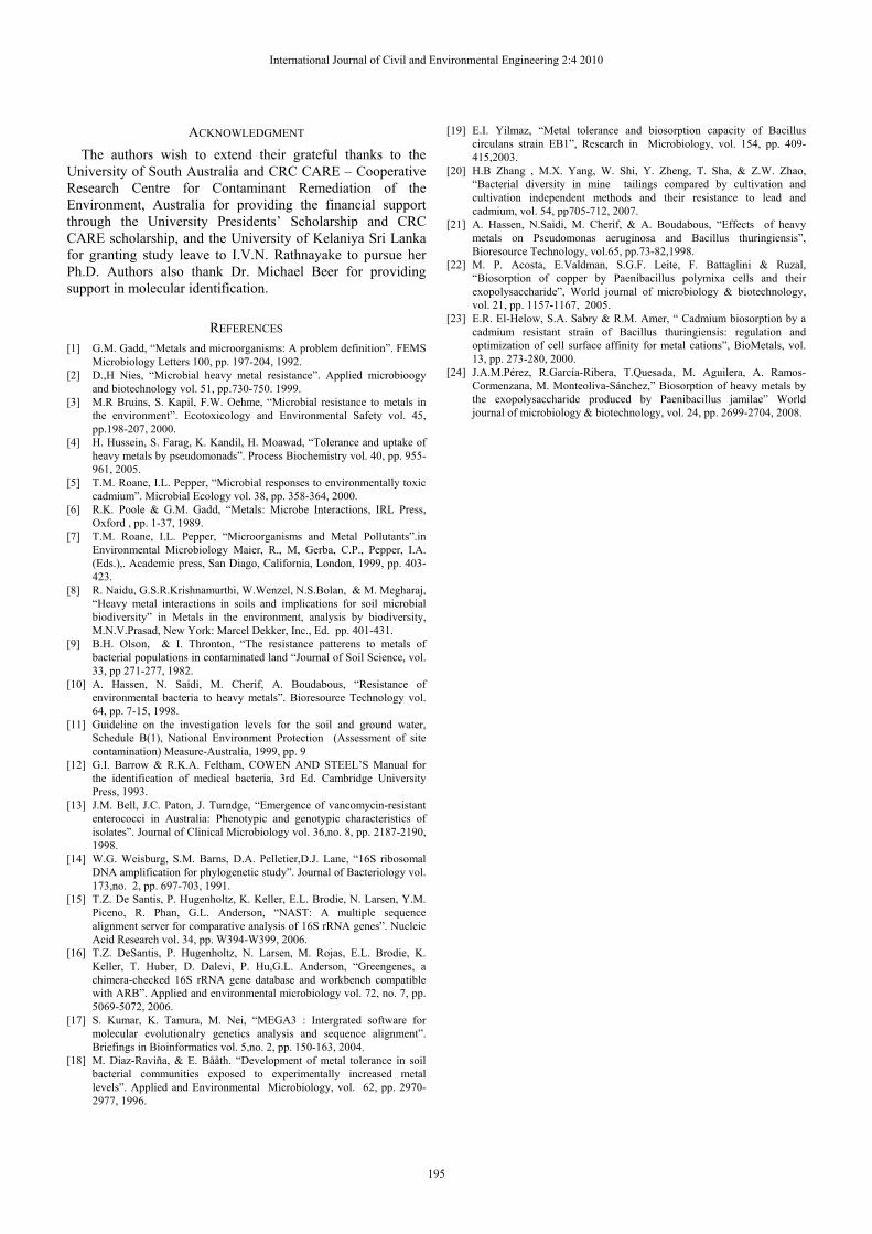
minimal medium (formulated in our previous study) which maintains a high free metal concentration in solution. Two Gram positive isolates exhibited different growth patterns in the presence of different heavy metals. The growth curves for Paenobacillus sp. and B. thuringiensis in the presence of different metal concentrations are shown in the Figure 1 & 2. Paenobacillus sp. exhibited a growth curve similar to the typical bacterial growth curve over the experiment time period (96 hrs). Paenibacillus sp. increased its growth and reached its maximum growth at 72 hrs. A decrease in growth (measured in terms of optical density) was observed upon increasing metal concentration at any given time interval compared to the control without metal amendment. The lower optical density values revealed that the bacterial growth was affected due to the presence of metal in the growth medium. On the other hand in the case of B. thuringiensis the growth increased steadily over the entire 96 h experimental period. However, the reduction of the growth in the presence of increased concentration of the metals used in the study was evident throughout the experiment compared to the control without
metal. But its growth was not affected much in the presence of Zn2+ . A similar finding was reported elsewhere [21].
Two bacterial isolates showed different levels of tolerance to the metals under investigation and the dose-response data for these bacteria are shown in Table II.
Paenibacillus sp., showed a higher sensitivity to Cu2+ with 50 % growth inhibition observed at 0.011 mg/L of Cu2+. B.thuringiensis was not as sensitive as Paenibacillus to Cu2+ because the derived EC50 value was 0.82 mg/L. A study carried out elsewhere reported a minimum inhibitory concentration of 3.5 mM of Cu2+ on Paenibacillus polymixa [22]. The tolerance of Cd2+ by the two bacterial isolates was different from each other, and the bacterium that was most sensitive to Cd2+was B.thuringiensis. However, there were reports on Cd2+ resistant B.thuringiensis strains which were able to absorb cadmium [23]. They also reported that a strain of this species has relatively a long lag phase in the presence of Cd2+. The levels of Zn2+ tolerance among the bacterial isolates were different from one to the other showing the reduction of their growth to 50 % in the presence of 2.98 mg/L of Zn2+ by B.thureingiensis and 7.02 mg/L of Zn2+ by Paenibacillus sp. This finding revealed that Paenibacillus sp. can tolerate more than twice the level of Zn2+ than B.thuringiensis. When the tolerance of three metals was compared with respect to each bacterium, it was evident that both of the bacteria were more sensitive to Cu2+ than the other two metals. Both of the bacterial isolates were more tolerant to Zn2+ than Cd2+. Although there are several reports on the metal binding and biosorption capacity of these two bacterial genera [23],[22],[24], studies on the metal tolerance are scarce. However it is not easy to make a meaningful comparison with the findings reported in the literature due to the range of protocols and media used.
Paenibacillus sp. str. DSM 1352 (AJ345017)
B3
Paenibacillus sp. H08-06 (AM162336)
Bacillus sp. str. cp-h12 (EU584533) Bacillus thuringiensis str. TCCC11336 (FJ393313)
B7
68 100
83
0.01
International Journal of Civil and Environmental Engineering 2:4 2010
193

Fig. 2 Growth curves of Paenibacillus sp. in the presence of different
heavy metals
Fig. 3 Growth curves of B. thuringiensis in the presence of different
heavy metals
TABLE II ESTIMATED EC50 VALUES FOR PAENIBACILLUS SP., AND B.THURINGIENSIS
Bacterium Cd2+ (mg/L) Cu2+ (mg/L)
Zn2+ (mg/L)
Paenibacillus sp. 1.77± 0.16* 0.011±0.003 7.02±0.44 B.thuringiensis 1.53±0.01 0.82±0.04 2.98±0.2 *Mean ±standard deviation
IV. CONCLUSION The current work demonstrated that the tolerance of heavy
metals varied between bacteria even though they were isolated from the same soil. Both the Gram positive bacteria were highly sensitive to Cu2+ than the other two metals. Paenibacillus sp. has great potential to be used as a biosensor to assess the Cu2+ toxicity in the environment due to its high sensitivity to Cu.
International Journal of Civil and Environmental Engineering 2:4 2010
194
ACKNOWLEDGMENT The authors wish to extend their grateful thanks to the
University of South Australia and CRC CARE – Cooperative Research Centre for Contaminant Remediation of the Environment, Australia for providing the financial support through the University Presidents’ Scholarship and CRC CARE scholarship, and the University of Kelaniya Sri Lanka for granting study leave to I.V.N. Rathnayake to pursue her Ph.D. Authors also thank Dr. Michael Beer for providing support in molecular identification.
REFERENCES [1] G.M. Gadd, “Metals and microorganisms: A problem definition”. FEMS
Microbiology Letters 100, pp. 197-204, 1992. [2] D.,H Nies, “Microbial heavy metal resistance”. Applied microbioogy
and biotechnology vol. 51, pp.730-750. 1999. [3] M.R Bruins, S. Kapil, F.W. Oehme, “Microbial resistance to metals in
the environment”. Ecotoxicology and Environmental Safety vol. 45, pp.198-207, 2000.
[4] H. Hussein, S. Farag, K. Kandil, H. Moawad, “Tolerance and uptake of heavy metals by pseudomonads”. Process Biochemistry vol. 40, pp. 955-961, 2005.
[5] T.M. Roane, I.L. Pepper, “Microbial responses to environmentally toxic cadmium”. Microbial Ecology vol. 38, pp. 358-364, 2000.
[6] R.K. Poole & G.M. Gadd, “Metals: Microbe Interactions, IRL Press, Oxford , pp. 1-37, 1989.
[7] T.M. Roane, I.L. Pepper, “Microorganisms and Metal Pollutants”.in Environmental Microbiology Maier, R., M, Gerba, C.P., Pepper, I.A. (Eds.),. Academic press, San Diago, California, London, 1999, pp. 403-423.
[8] R. Naidu, G.S.R.Krishnamurthi, W.Wenzel, N.S.Bolan, & M. Megharaj, “Heavy metal interactions in soils and implications for soil microbial biodiversity” in Metals in the environment, analysis by biodiversity, M.N.V.Prasad, New York: Marcel Dekker, Inc., Ed. pp. 401-431.
[9] B.H. Olson, & I. Thronton, “The resistance patterens to metals of bacterial populations in contaminated land “Journal of Soil Science, vol. 33, pp 271-277, 1982.
[10] A. Hassen, N. Saidi, M. Cherif, A. Boudabous, “Resistance of environmental bacteria to heavy metals”. Bioresource Technology vol. 64, pp. 7-15, 1998.
[11] Guideline on the investigation levels for the soil and ground water, Schedule B(1), National Environment Protection (Assessment of site contamination) Measure-Australia, 1999, pp. 9
[12] G.I. Barrow & R.K.A. Feltham, COWEN AND STEEL’S Manual for the identification of medical bacteria, 3rd Ed. Cambridge University Press, 1993.
[13] J.M. Bell, J.C. Paton, J. Turndge, “Emergence of vancomycin-resistant enterococci in Australia: Phenotypic and genotypic characteristics of isolates”. Journal of Clinical Microbiology vol. 36,no. 8, pp. 2187-2190, 1998.
[14] W.G. Weisburg, S.M. Barns, D.A. Pelletier,D.J. Lane, “16S ribosomal DNA amplification for phylogenetic study”. Journal of Bacteriology vol. 173,no. 2, pp. 697-703, 1991.
[15] T.Z. De Santis, P. Hugenholtz, K. Keller, E.L. Brodie, N. Larsen, Y.M. Piceno, R. Phan, G.L. Anderson, “NAST: A multiple sequence alignment server for comparative analysis of 16S rRNA genes”. Nucleic Acid Research vol. 34, pp. W394-W399, 2006.
[16] T.Z. DeSantis, P. Hugenholtz, N. Larsen, M. Rojas, E.L. Brodie, K. Keller, T. Huber, D. Dalevi, P. Hu,G.L. Anderson, “Greengenes, a chimera-checked 16S rRNA gene database and workbench compatible with ARB”. Applied and environmental microbiology vol. 72, no. 7, pp. 5069-5072, 2006.
[17] S. Kumar, K. Tamura, M. Nei, “MEGA3 : Intergrated software for molecular evolutionalry genetics analysis and sequence alignment”. Briefings in Bioinformatics vol. 5,no. 2, pp. 150-163, 2004.
[18] M. Diaz-Raviña, & E. Bååth. “Development of metal tolerance in soil bacterial communities exposed to experimentally increased metal levels”. Applied and Environmental Microbiology, vol. 62, pp. 2970-2977, 1996.
[19] E.I. Yilmaz, “Metal tolerance and biosorption capacity of Bacillus circulans strain EB1”, Research in Microbiology, vol. 154, pp. 409-415,2003.
[20] H.B Zhang , M.X. Yang, W. Shi, Y. Zheng, T. Sha, & Z.W. Zhao, “Bacterial diversity in mine tailings compared by cultivation and cultivation independent methods and their resistance to lead and cadmium, vol. 54, pp705-712, 2007.
[21] A. Hassen, N.Saidi, M. Cherif, & A. Boudabous, “Effects of heavy metals on Pseudomonas aeruginosa and Bacillus thuringiensis”, Bioresource Technology, vol.65, pp.73-82,1998.
[22] M. P. Acosta, E.Valdman, S.G.F. Leite, F. Battaglini & Ruzal, “Biosorption of copper by Paenibacillus polymixa cells and their exopolysaccharide”, World journal of microbiology & biotechnology, vol. 21, pp. 1157-1167, 2005.
[23] E.R. El-Helow, S.A. Sabry & R.M. Amer, “ Cadmium biosorption by a cadmium resistant strain of Bacillus thuringiensis: regulation and optimization of cell surface affinity for metal cations”, BioMetals, vol. 13, pp. 273-280, 2000.
[24] J.A.M.Pérez, R.García-Ribera, T.Quesada, M. Aguilera, A. Ramos-Cormenzana, M. Monteoliva-Sánchez,” Biosorption of heavy metals by the exopolysaccharide produced by Paenibacillus jamilae” World journal of microbiology & biotechnology, vol. 24, pp. 2699-2704, 2008.
International Journal of Civil and Environmental Engineering 2:4 2010
195