tolerance, autoimmunity, immunodeficiences. tolerance is broadly defined as a state of...
TRANSCRIPT

Tolerance, Autoimmunity,Immunodeficiences
Tolerance is broadly defined as a state of unresponsiveness to an antigen, be it self or foreign
Antigen-specific cell receives signals that eitheractivate OR inactivate the cell
Central tolerance to self antigens is acquired during development through the elimination or silencing of lymphocytes capable of binding self antigens
Peripheral tolerance is induced in mature lymphocytesin the periphery
Tolerance
Mechanisms to induce tolerance
Elimination of self-reactive lymphocytes = clonal deletion
(negative selective of T and B cells during development)
Silencing of self-reactive lymphocytes = clonal anergy
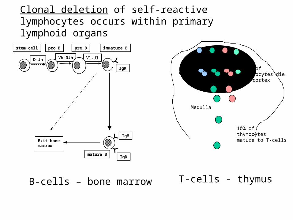
Clonal deletion of self-reactive lymphocytes occurs within primary lymphoid organs
Vh-DJh Vl-Jl
stem cell pro B pre B
D-Jh
IgM
immature B
IgD
IgM
mature B
Exit bone marrow
Cortex
Medulla
90% ofthymocytes diein cortex
10% ofthymocytes mature to T-cells
B-cells – bone marrow T-cells - thymus
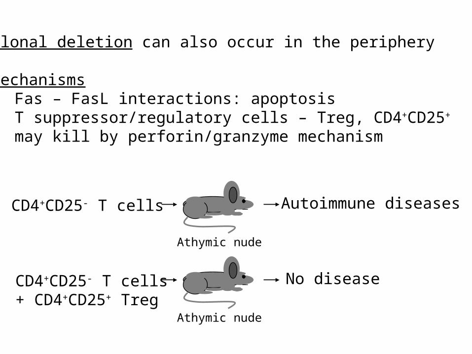
Clonal deletion can also occur in the periphery
Mechanisms• Fas – FasL interactions: apoptosis• T suppressor/regulatory cells – Treg, CD4+CD25+
may kill by perforin/granzyme mechanism
Athymic nude
CD4+CD25- T cells Autoimmune diseases
Athymic nude
CD4+CD25- T cells+ CD4+CD25+ Treg
No disease
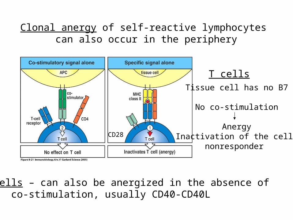
Clonal anergy of self-reactive lymphocytes can also occur in the periphery
CD28
Tissue cell has no B7
No co-stimulation
Anergy Inactivation of the cell –
nonresponder
T cells
B cells – can also be anergized in the absence of co-stimulation, usually CD40-CD40L
Other factors involved in Tolerance
-Dose/Route of Antigen
-Inappropriate cytokine responses
-anti-idiotypic responses
-Psychogenic factors
(poorly defined but could include the immunosuppressive effect of steroid hormones)
Tolerance to a fetus
The fetus is really an allograft with nonself MHC proteins & RBCs of the father so why is it not rejected by the mother?We know mothers’ makes antibodies against fathers’ MHC & RBCPotential mechanisms
Placenta – outer layer does not express classical MHC proteins expresses a molecule that inhibits NK cell killing
depletion of tryptophan – necessary T cell nutrientT cell tolerance to paternal ags, suppressed T cell responsesSecretion of cytokines that suppress TH1 cells – IL4, IL10, TGFRole for Treg cells?
“Immune privileged” sites
EyeTestisBrainOvary Placenta
Potential reasonsThe presence of FasL expressing cells that kill
infiltrating inflammatory T cells (Fas)Immunosuppressive cytokines
AutoimmunityAutoimmunity constitutes immune response against self
antigen. Autoimmunity may be benign or may be damaging to host
An immune response against self antigen(s) that results in the destruction of host tissue or damage to the function of an organ or tissue constitutes autoimmune disease
Autoimmunity can be thought of as a breakdown of tolerance, which is multi-layered, consisting of both central and peripheral mechanisms
Occasionally, self-reactive cells escape, resulting in autoimmune diseases (approximately 5% U.S. population)
Autoimmune diseases are multifactorial – genetic & environment
• Genetics. Presentation of self-antigens by MHC molecules: Linkage to certain MHC alleles in many autoimmune diseases
•Autoreactive lymphocytes
•Initiating Event: Environmental: Chemical exposureInfection: Viral and bacterial infection
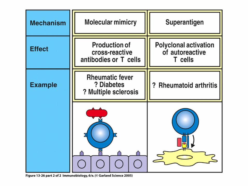
molecular mimicry-cross reactivity between a microbial antigen with a self-antigen
•Gender: Females more frequently affected
•‘Handedness’: a tenuous but statistically-significant higher frequency in left-handed people
Contributing factors
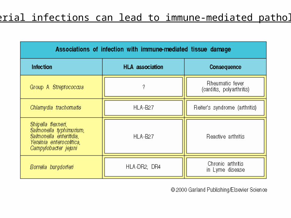
Bacterial infections can lead to immune-mediated pathology
Classification of autoimmune disease
Historically – organ or systemic
Effector mechanism – antibody, complement, T cells
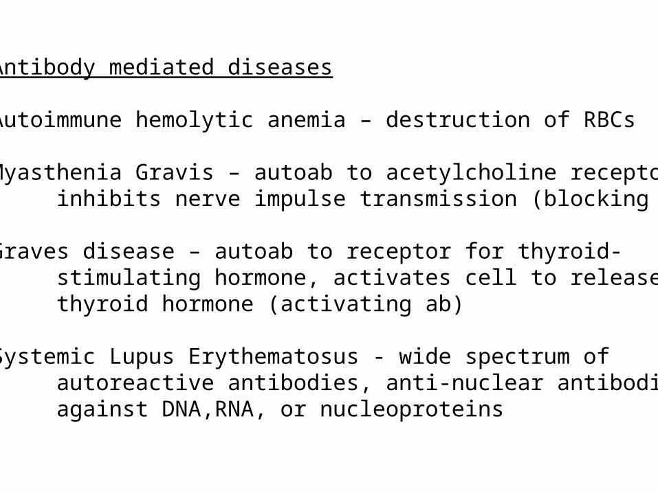
Antibody mediated diseases
Autoimmune hemolytic anemia – destruction of RBCs
Myasthenia Gravis – autoab to acetylcholine receptor, inhibits nerve impulse transmission (blocking ab)
Graves disease – autoab to receptor for thyroid-stimulating hormone, activates cell to release thyroid hormone (activating ab)
Systemic Lupus Erythematosus - wide spectrum of autoreactive antibodies, anti-nuclear antibodies against DNA,RNA, or nucleoproteins
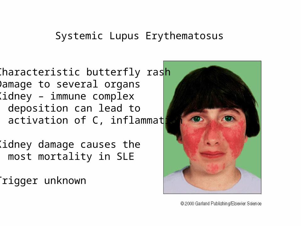
Systemic Lupus Erythematosus
Characteristic butterfly rashDamage to several organsKidney – immune complex deposition can lead to activation of C, inflammation
Kidney damage causes the most mortality in SLE
Trigger unknown
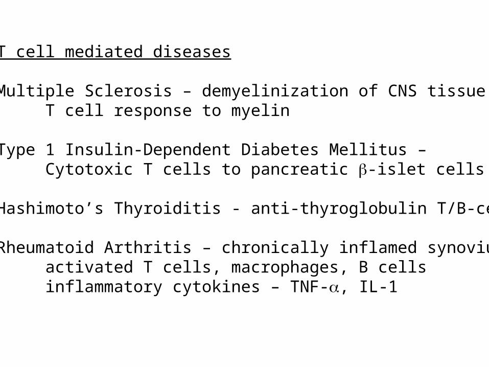
T cell mediated diseases
Multiple Sclerosis – demyelinization of CNS tissueT cell response to myelin
Type 1 Insulin-Dependent Diabetes Mellitus – Cytotoxic T cells to pancreatic -islet cells
Hashimoto’s Thyroiditis - anti-thyroglobulin T/B-cells
Rheumatoid Arthritis – chronically inflamed synoviumactivated T cells, macrophages, B cellsinflammatory cytokines – TNF-, IL-1
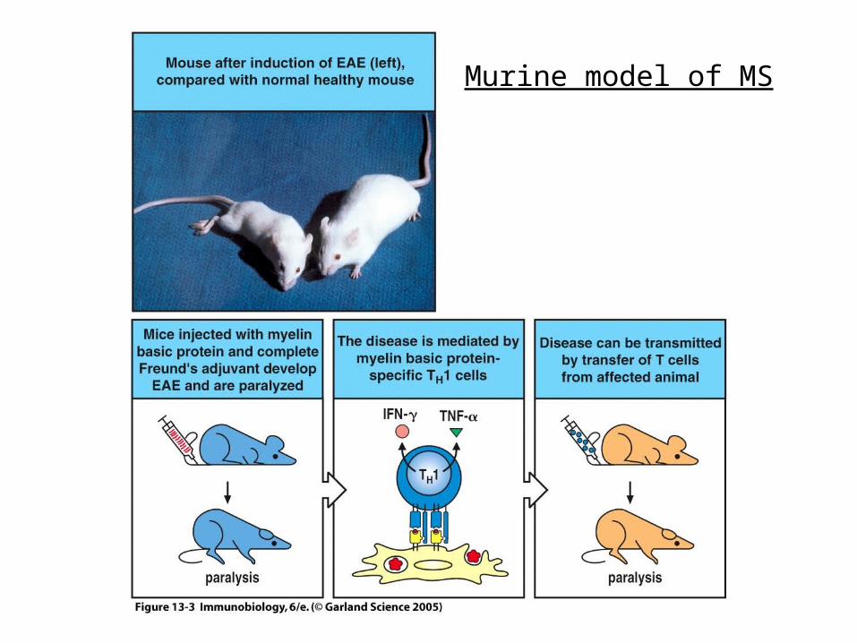
Murine model of MS
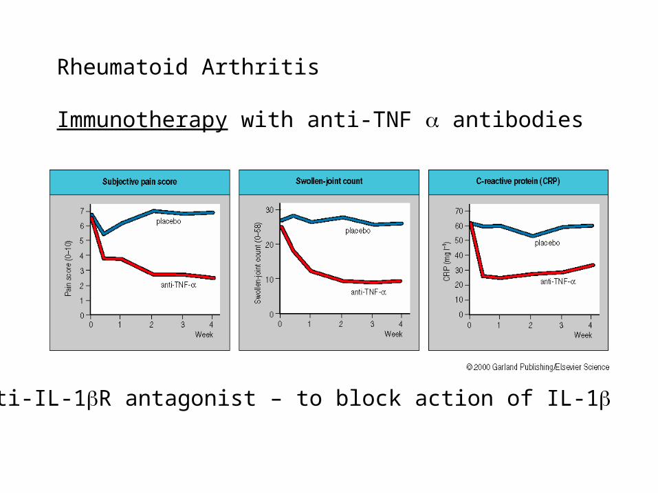
Rheumatoid Arthritis
Immunotherapy with anti-TNF antibodies
Anti-IL-1R antagonist – to block action of IL-1
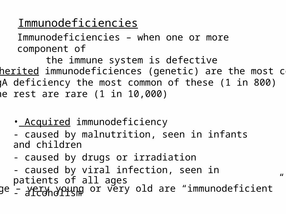
Immunodeficiencies
• Inherited immunodeficiences (genetic) are the most commonIgA deficiency the most common of these (1 in 800)The rest are rare (1 in 10,000)
• Acquired immunodeficiency- caused by malnutrition, seen in infants and children- caused by drugs or irradiation- caused by viral infection, seen in patients of all ages- alcoholism• age – very young or very old are “immunodeficient”
Immunodeficiencies – when one or more component of
the immune system is defective
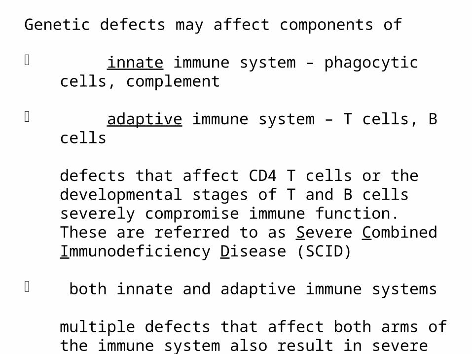
Genetic defects may affect components of
innate immune system – phagocytic cells, complement
adaptive immune system – T cells, B cells
defects that affect CD4 T cells or the developmental stages of T and B cells severely compromise immune function. These are referred to as Severe Combined Immunodeficiency Disease (SCID)
both innate and adaptive immune systems
multiple defects that affect both arms of the immune system also result in severe compromise of immune functions. These are rare.
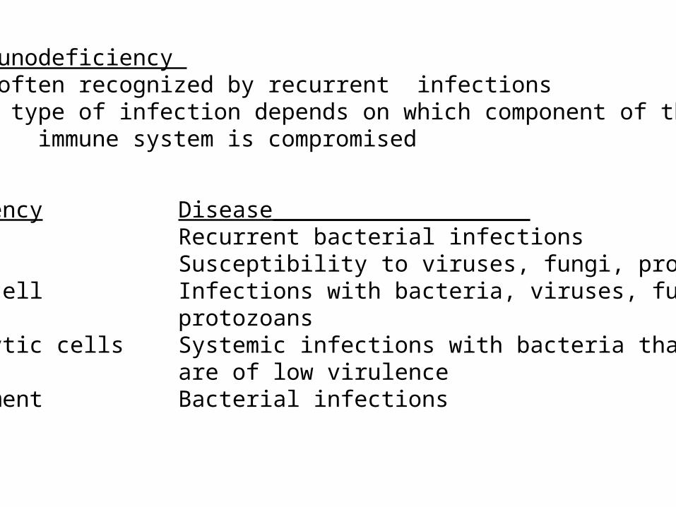
Immunodeficiency Is often recognized by recurrent infectionsThe type of infection depends on which component of the
immune system is compromised
Deficiency Disease___________________B cell Recurrent bacterial infectionsT cell Susceptibility to viruses, fungi, protozoansT & B cell Infections with bacteria, viruses, fungi,
protozoansPhagocytic cells Systemic infections with bacteria that
are of low virulenceComplement Bacterial infections
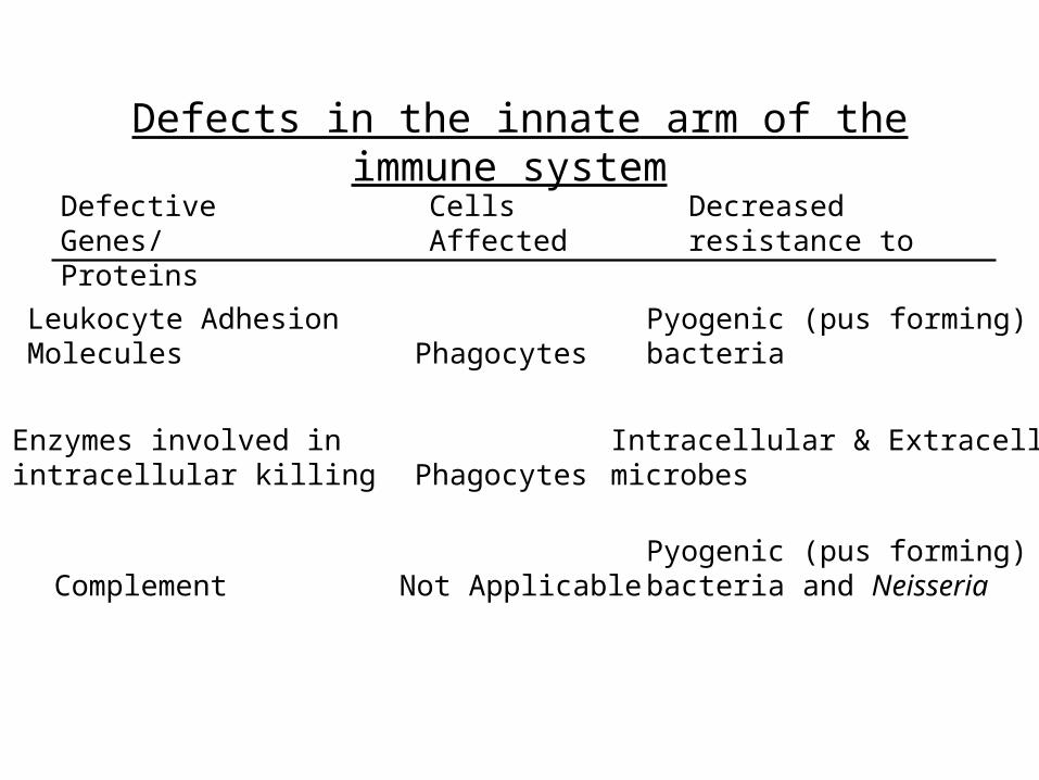
Defects in the innate arm of the immune system
Defective Genes/Proteins
Cells Affected
Decreased resistance to
Leukocyte Adhesion Molecules
Enzymes involved in intracellular killing
Complement Not Applicable
Phagocytes
PhagocytesPyogenic (pus forming)bacteria
Intracellular & Extracellularmicrobes
Pyogenic (pus forming)bacteria and Neisseria
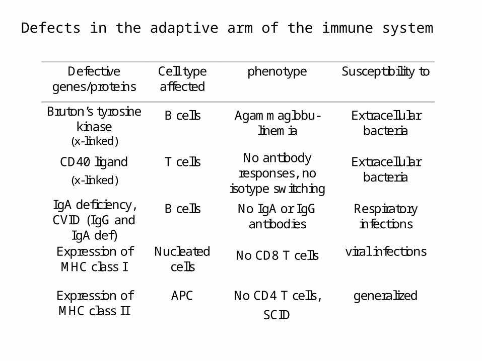
Defective genes/proteins
Cell type affected
phenotype Susceptibility to
Bruton’s tyrosine kinase
(x-linked)
B cells Agammaglobu-linemia
Extracellular bacteria
CD40 ligand (x-linked)
T cells No antibody responses, no
isotype switching
Extracellular bacteria
IgA deficiency, CVID (IgG and
IgA def)
B cells No IgA or IgG antibodies
Respiratory infections
Expression of MHC class I
Nucleated cells
No CD8 T cells viral infections
Expression of MHC class II
APC No CD4 T cells,
SCID
generalized
Defects in the adaptive arm of the immune system
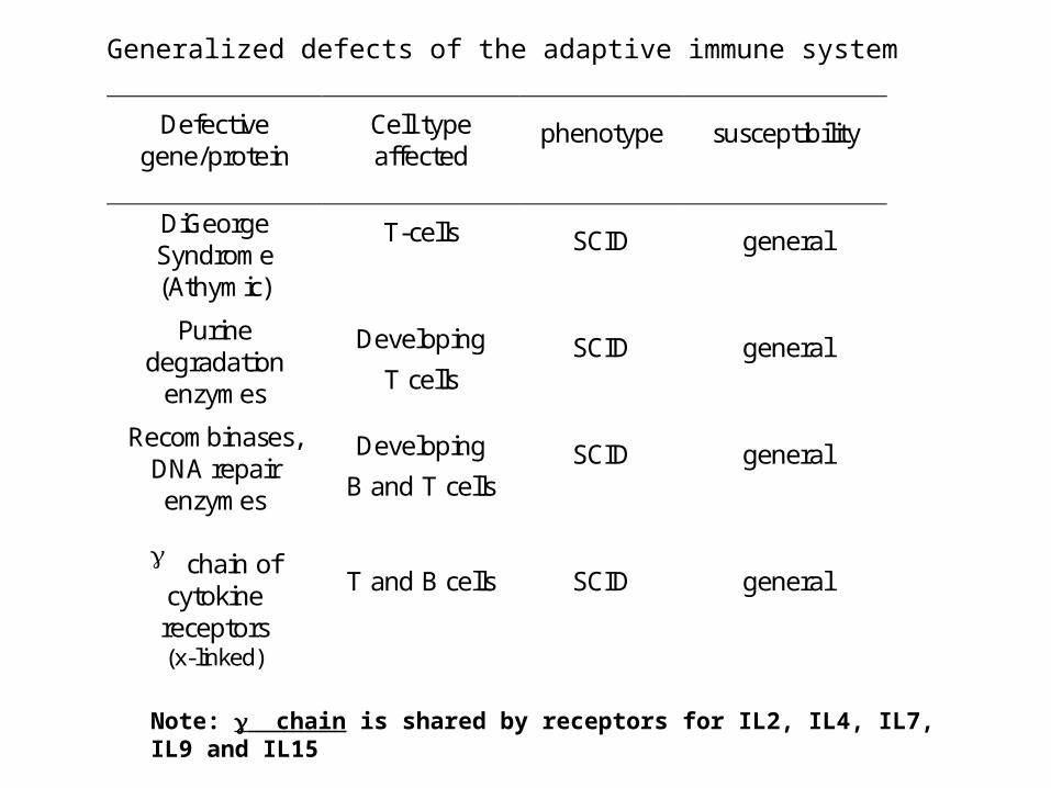
Generalized defects of the adaptive immune system
Defective gene/protein
Cell type affected
phenotype susceptibility
DiGeorge Syndrome (Athymic)
T-cells SCID general
Purine degradation
enzymes
Developing
T cells SCID general
Recombinases, DNA repair enzymes
Developing
B and T cells SCID general
chain of cytokine receptors (x-linked)
T and B cells SCID general
Note: chain is shared by receptors for IL2, IL4, IL7, IL9 and IL15

Acquired Immunodeficiencies
Severe immunodeficiency caused by HIV (AIDS) generalized immunosuppression due to loss of CD4 T cells.
Immune suppression induced by Epstein-Barr Virus (EBV) following infectious mononucleosis.
Radiation or Cytotoxic drugs
Malnutrition
Alcoholism
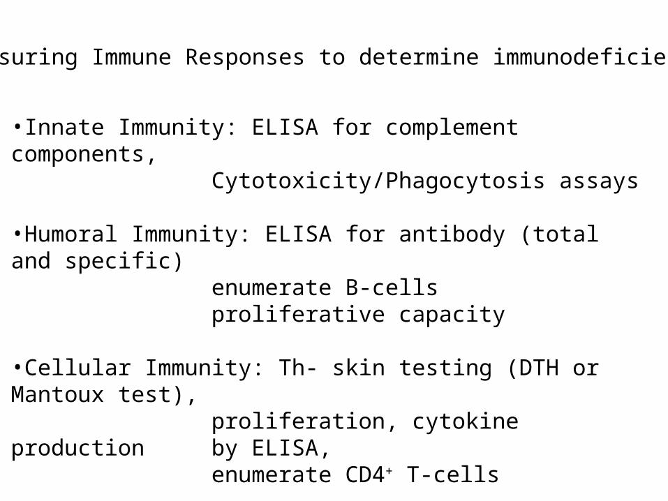
•Innate Immunity: ELISA for complement components,Cytotoxicity/Phagocytosis assays
•Humoral Immunity: ELISA for antibody (total and specific)
enumerate B-cellsproliferative capacity
•Cellular Immunity: Th- skin testing (DTH or Mantoux test),
proliferation, cytokine production by ELISA,
enumerate CD4+ T-cells
Tc- Cytotoxicity testing, IFN-production,
enumerate CD8+ T-cells
Measuring Immune Responses to determine immunodeficiency