tocotrienol rich fraction supplementation improved lipid profile and
TRANSCRIPT

RESEARCH Open Access
Tocotrienol rich fraction supplementationimproved lipid profile and oxidative status inhealthy older adults: A randomized controlledstudySiok-Fong Chin1, Johari Ibahim1, Suzana Makpol1, Noor Aini Abdul Hamid1, Azian Abdul Latiff2, Zaiton Zakaria3,Musalmah Mazlan1, Yasmin Anum Mohd Yusof1, Aminuddin Abdul Karim3 and Wan Zurinah Wan Ngah1*
Abstract
Background: Vitamin E supplements containing tocotrienols are now being recommended for optimum healthbut its effects are scarcely known. The objective was to determine the effects of Tocotrienol Rich Fraction (TRF)supplementation on lipid profile and oxidative status in healthy older individuals at a dose of 160 mg/day for 6months.
Methods: Sixty-two subjects were recruited from two age groups: 35-49 years (n = 31) and above 50 years (n =31), and randomly assigned to receive either TRF or placebo capsules for six months. Blood samples were obtainedat 0, 3rd and 6th months.
Results: HDL-cholesterol in the TRF-supplemented group was elevated after 6 months (p < 0.01). Protein carbonylcontents were markedly decreased (p < 0.001), whereas AGE levels were lowered in the > 50 year-old group (p <0.05). Plasma levels of total vitamin E particularly tocopherols were significantly increased in the TRF-supplementedgroup after 3 months (p < 0.01). Plasma total tocotrienols were only increased in the > 50 year-old group afterreceiving 6 months of TRF supplementation. Changes in enzyme activities were only observed in the > 50 year-oldgroup. SOD activity was decreased after 3 (p < 0.05) and 6 (p < 0.05) months of TRF supplementation whereasCAT activity was decreased after 3 (p < 0.01) and 6 (p < 0.05) months in the placebo group. GPx activity wasincreased at 6 months for both treatment and placebo groups (p < 0.05).
Conclusion: The observed improvement of plasma cholesterol, AGE and antioxidant vitamin levels as well as thereduced protein damage may indicate a restoration of redox balance after TRF supplementation, particularly inindividuals over 50 years of age.
BackgroundDietary and supplemental sources of vitamin E isoformshave been demonstrated to possess unique propertiesthat can influence critical pathways involved in cancer[1,2], cardiovascular [3,4] and neurodegenerative disease[5,6] development. Recent studies have identified toco-trienol, the lesser known isomer of vitamin E as themore effective compound in providing such protection
in comparison to the well-established tocopherol [7-14].The most promising function of tocotrienol has beenreported in neuroprotection [14,15] and stroke preven-tion with the latter being attributed to the lipid-correc-tive properties of tocotrienol [11,13]. However, mostdata were generated from in vitro studies or by usinganimal models. To date, very limited data available fromhuman intervention study particularly involving supple-mental vitamin E and its fundamental effects on diseasesand ageing.Recent interest has focussed on finding compounds
that could intervened the chemical processes underlying
* Correspondence: [email protected] of Biochemistry, Faculty of Medicine, Universiti KebangsaanMalaysia, Kuala Lumpur, MalaysiaFull list of author information is available at the end of the article
Chin et al. Nutrition & Metabolism 2011, 8:42http://www.nutritionandmetabolism.com/content/8/1/42
© 2011 Chin et al; licensee BioMed Central Ltd. This is an Open Access article distributed under the terms of the Creative CommonsAttribution License (http://creativecommons.org/licenses/by/2.0), which permits unrestricted use, distribution, and reproduction inany medium, provided the original work is properly cited.
age-related degenerative diseases in which has beendemonstrated to be also responsible for the ageing phe-nomena [16,17]. Although vitamin E supplementationhas yielded equivocal results in human intervention stu-dies largely on a short term basis [18-20], supplementa-tion studies measuring benefits to cardiovascular endpoints are still controversial with some showing no ben-efits and some even detrimental effects [21-23]. Studiesare warranted to elucidate the underlying mechanism onthe effects of vitamin E in preventing or treating thesediseases that may explain the observed effects.Most vitamin E supplements available in the market
usually contain only alpha tocopherol. However, recentlywith the availability of tocotrienols commercially, theuses of tocotrienol-containing supplement are morewidespread. There has been a paradigm shift to vitaminE supplementation where all isomers of vitamin E arerecommended rather than alpha tocopherol alone [24].However, it has yet to be determined whether supple-mentation with a mixture of vitamin E isomers contain-ing high tocotrienol fractions will affect biochemicalparameters and other blood indices and the possibleunderlying mechanism. We are interested in investigat-ing age-associated biochemical changes with tocotrienolenriched vitamin E supplementation and identifying thespecific oxidation pathways involved.Different aged individuals respond differently to var-
ious food and vitamin supplementation [25]. Needs forsupplement may be different with the different agewhere older people may benefit from supplementationas they were reported to have lower antioxidant levels[26].We previously reported that TRF supplementation
decreased DNA damage in healthy older adults, mostlyin those over 50 years of age, and that the levels ofdamage were associated with age [27]. In the presentwork, changes in lipoprotein-lipid profile, protein carbo-nyl content, advanced glycosylation end products(AGEs), malondialdehyde (MDA) levels, levels of theantioxidant vitamins E and C, and antioxidant activitiesof superoxide dismutase (SOD), catalase (CAT) and glu-tathione peroxidase (GPx) were measured in a rando-mized, double-blinded, placebo-controlled interventionstudy of TRF supplementation. We also studied theassociation between oxidative biomarkers and antioxi-dant levels to further elucidate the oxidant-antioxidantbalance with increasing age.
MethodsStudy designHealthy volunteers were recruited through screening ofa population study on oxidative stress and ageing.Selected individuals were aged 35 years and older, non-smokers, not pregnant, not taking any vitamin
supplements or minerals, alcohol, or drugs, and free ofcardiac, hepatic, renal or any other chronic diseases.Adult females were recruited as the compliance fromthis gender is higher and mostly are non-smokers.Results of a pre-study consisting a full physical examina-tion, previous medical history, blood chemistry and hae-matology were used to confirm suitability. Sixty-twosubjects were recruited from two age groups, 35-49years (n = 31) and over 50 years (n = 31), and randomlyassigned to receive either Tocotrienol Rich Fraction(TRF) capsules (160 mg/day) daily in a single eveningdose or an identical placebo for a period of six months.All subjects were requested to consume the capsulesafter dinner to ensure proper absorption [28,29] andencouraged to maintain their usual lifestyle throughoutthe study period. The commercially prepared TRF (TriE® Tocotrienol) is a palm-based vitamin E consisted ofapproximately 74% tocotrienols and 26% tocopherol insoft gelatine capsules containing palm superolein oil,and was supplied by Sime Darby Bioganic Sdn. Bhd.(previously known as Golden Hope Bioganic, Selangor,Malaysia). Each capsules contained approximately 70.4mg a-tocotrienol, 4.8 mg b-tocotrienol, 57.6 mg g-toco-trienol, 33.6 mg δ-tocotrienol and 48 mg a-tocopherol.The capsules given throughout the study were from thesame lot and provided loosely in plastic containers. Theplacebo capsules contained only palm superolein oil. Inaddition to plasma samples for vitamin E levels, compli-ance was assessed by capsule counts at each 3-monthinterval. The amount of allotted and returned capsulesfor each participant is recorded during the interval visits.The treatment was double blinded throughout the studyperiod until all data were collected, after which the ran-domisation code was broken. The protocol of the studywas approved by the Research and Ethics Committee ofthe Faculty of Medicine, Universiti Kebangsaan Malay-sia. Written informed consent was obtained. Subjectdemographics are summarised in Table 1.
Sample collectionBlood sampling was performed at baseline (month 0), 3months and 6 months of supplementation. Venousblood samples were drawn from fasting subjects intolithium heparin-coated and K2EDTA-containing tubes(BD Vacutainer, Becton, Dickinson and Company,Franklin Lakes, NJ, USA) for plasma extraction whileinto a plain tube for serum extraction. Plasma andserum were immediately separated by centrifugation at3000 g for 10 minutes. The obtained packed erythro-cytes were washed three times with 0.9% sodium chlor-ide solution. Heparinized plasma aliquots wereseparated for lipid profiling as well as vitamin C andvitamin E determination. Plasma-EDTA aliquots wereused for protein carbonyl and MDA quantification
Chin et al. Nutrition & Metabolism 2011, 8:42http://www.nutritionandmetabolism.com/content/8/1/42
Page 2 of 14

whereas serum aliquots were used for AGE producttesting. Washed erythrocytes were used for determina-tion of antioxidant activities of SOD, CAT and GPx.Plasma samples for vitamin C determination weredeproteinised with 5% perchloric acid and centrifuged at1000 g for 2 minutes, and the resulting clear superna-tant was transferred into new tubes. Lipid profile testingwas done immediately whereas other samples were fro-zen at -80°C until further analysis.
Lipid profile determinationPlasma cholesterol and triglyceride concentrations weredetermined using enzymatic colorimetric kits (Rochediagnostics GmbH, Indianapolis, USA) on a Hitachi 747Automatic Analyzer (Boehringer Mannheim, Germany).High density lipoprotein-cholesterol (HDL-C) was mea-sured using the precipitation method whereas low den-sity lipoprotein-cholesterol (LDL-C) was calculatedusing Friedewald’s formula.
Plasma vitamin E determinationPlasma tocopherol and tocotrienol were determined byHPLC. Briefly, stored plasma samples were thawed andaliquots of 200 µl plasma and 50 µl 95% ethanol
containing 10 µg/ml butylated hydroxytoluene (SigmaChemical Co., St. Louis, MO, USA) were pipetted intotubes, covered and mixed vigorously for 5 seconds. 1 mlof absolute ethanol (Merck KGaA, Darmstadt, Germany)was then added to each tube. The tubes were coveredand mixed again before being centrifuged at 1500 g for15 minutes at 18°C. The bottom pellet was carefullyremoved with a spatula. Hexane, 3 ml (Merck KGaA,Darmstadt, Germany) were then added to each tube andmixed vigorously for 5 minutes. The samples were thencentrifuged at 1500 g for 15 minutes at 18°C. After cen-trifugation, a portion of the upper layer (2.5 ml) wascarefully removed, placed in new tubes and vacuum-eva-porated for 40 minutes. The dried sample residue wasthen reconstituted in 100 µl HPLC grade hexane (MerckKGaA, Darmstadt, Germany), vortexed, and passedthrough a 0.45 µm filter to remove any non-dissolvedparticles. The samples were then transferred to ambervial inserts with care taken to avoid air bubbles and ana-lysed by HPLC using a Shimadzu RF-10A XL fluores-cence detector (Shimadzu Corporation, Kyoto, Japan) atan excitation wavelength of 294 nm and an emissionwavelength of 330 nm. Isomers a-tocopherol, g-toco-pherol, a-tocotrienol, g-tocotrienol and δ-tocotrienol
Table 1 Baseline and intervention characteristics of the study groups
Placebo (n = 30) TRF (n = 32)
35-49 years > 50 years 35-49 years > 50 years
Age (years) 44.7 ± 0.9 a 59.1 ± 1.7 a 44.5 ± 1.0 b 56.1 ± 1.2 b
Blood pressure (mmHg)
Systolic
Baseline (0 month) 121 ± 2.8 c 137 ± 3.6 c 121 ± 3.5 d 131 ± 3.3 d
3 months 123 ± 2.4 f 137 ± 5.1 f 121 ± 3.4 131 ± 3.5
6 months 120 ± 2.5 g 135 ± 5.0 g 122 ± 3.6 132 ± 3.3
Diastolic
Baseline (0 month) 75.4 ± 1.9 e 82.0 ± 2.7 e 80.6 ± 2.8 81.3 ± 2.6
3 months 73.7 ± 1.7 79.2 ± 2.9 79.2 ± 2.6 82.3 ± 2.6
6 months 75.7 ± 2.8 79.2 ± 3.3 79.0 ± 3.1 82.8 ± 1.9
Pulse (beat/minute)
Baseline (0 month) 73 ± 2 73 ± 3 70 ± 3 70 ± 2
3 months 70 ± 2 72 ± 2 71 ± 3 71 ± 2
6 months 71 ± 2 71 ± 3 69 ± 3 72 ± 2
Body mass index (kg/m2)
Baseline (0 month) 27.5 ± 1.2 26.3 ± 0.6 26.3 ± 0.7 27.7 ± 1.3
3 months 27.5 ± 1.2 25.0 ± 1.0 25.6 ± 0.6 27.4 ± 1.2
6 months 26.8 ± 1.2 25.2 ± 1.0 25.7 ± 0.7 27.1 ± 1.1
Fasting blood glucose (mmol/L)
Baseline (0 month) 5.53 ± 0.33 5.34 ± 0.13 5.74 ± 0.30 5.49 ± 0.10
3 months 5.40 ± 0.44 5.37 ± 0.10 5.59 ± 0.22 5.24 ± 0.15
6 months 5.55 ± 0.49 5.28 ± 0.14 5.42 ± 0.19 5.47 ± 0.11
Values are expressed as mean ± SEM.
Same letters indicate significant difference between 35-49 and > 50 year-old groups.
a: p < 0.001, b: p < 0.001, c: p < 0.001, d: p < 0.05, e: p < 0.05, f: p < 0.001, g: p < 0.01
Chin et al. Nutrition & Metabolism 2011, 8:42http://www.nutritionandmetabolism.com/content/8/1/42
Page 3 of 14

were separated on a 250 mm × 4.6 mm, 5 µm AllsphereSilica column (Alltech Associated, Inc, IL, USA) andeluted with a mobile phase of 99:1 (v/v) hexane-isopro-panol at a flow rate of 1.5 ml per minute. The identityof each compound was confirmed by co-elution withspiked standard. All external standards used wereobtained from Malaysian Palm Oil Board (MPOB),Malaysia. The peaks were quantified and integrated withShimadzu Class-VP™ version 6.1 LC Workstation soft-ware (Shimadzu Corporation, Kyoto, Japan).
Plasma vitamin C determinationThe measurement of plasma vitamin C (ascorbic acid)was performed using HPLC as described by Pachla andKissinger [30]. Plasma ascorbic acid was chromatogra-phically separated on a 25 cm VYDAC® Genesis C-18 4µm column (Grace Davison, USA) using a mobile phasecontaining 0.04 M sodium acetate, 0.48 mM disodiumEDTA, 2.9 mM tetrabutylammonium hydroxide and1.4% methanol at pH 4.75. Detection was performed onan electrochemical detector (Gilson model 142, GilsonMedical Electronics S.A., Villiers-le-Bel, France) set atan applied potential of +700 mV and referenced to anAg/AgCl electrode, with a flow rate of 1 ml/min andsensitivity of 100 nA/V on a Gilson HPLC set (GilsonMedical Electronics S.A., Villiers-le-Bel, France). Assaycalibration was performed for each run using six con-centrations of the calibrator (0, 2, 4, 6, 8, 10 and 12 mg/L). Stored deproteinised plasma samples were thawed,added with 5 mg/L 3,4-dihydroxybenzylamine (SigmaChemical Co., USA) and spiked with 5 mg/L ascorbicacid (Sigma Chemical Co., St. Louis, MO, USA). 3,4-dihydroxybenzylamine was added to all samples andcalibrators as an internal standard whereas uric acid Rai-chem™ (Reagents Applications Inc., USA) was injectedseparately for peak identification. All samples were fil-tered through a 0.45 µm membrane before being sub-jected to HPLC and all peaks were baseline separated.Ascorbic acid, 3,4-dihydroxybenzylamine and uric acidpeaks were resolved and detected within 11 minutes ofrun time and were identified using the retention timesof calibrators. Peaks were integrated using peak-arearatios of ascorbic acid to 3,4-dihydroxybenzylamine.
Plasma protein carbonyl determinationCarbonyl content in oxidatively-modified proteins wasassayed using the Cayman Chemical protein carbonylassay kit (Cayman Chemical Company, Ann Arbor, MI,USA) based on the method of Levine et al. [31] by fol-lowing the manufacturer instructions. Briefly, 2,4-dini-trophenyl-hydrazine (DNPH) reacts with proteincarbonyls, forming a Schiff base to produce the corre-sponding hydrazone. The protein-hydrazone producedwas quantified at an absorbance of 370 nm with an
extinction coefficient of 22,000 M-1cm-1. Each samplewas assayed with a parallel control and the concentra-tion of carbonyls was determined after correction withthe respective control. The carbonyl content was thenstandardized against the protein concentration in thesample and expressed as nmol carbonyl per mg protein.The amount of protein was calculated from a bovineserum albumin (Sigma Chemical Co., St. Louis, USA)standard curve (0.25-2.0 mg/ml) read at 280 nm.
Serum AGE determinationSerum advanced glycosylation end products (AGEs) wasmeasured with an in-house competitive enzyme immu-noassay technique developed by Wan Nazaimoon andKhalid [32]. Briefly, microtitre wells were coated withAGE-BSA at 8 µg/ml followed by an overnight incuba-tion with 20 µl of the prediluted sample (1:6) and 80 µlanti-AGE-KLH (1:8000). HRP-labelled goat anti-rabbit(1:3000) was used as the secondary antibody and3,5’,5,5’-tetramethylbenzidine dihydrochloride (SigmaChemical Co., St. Louis, USA) as the substrate. The col-our reaction was stopped with 1.25 M sulphuric acidand the absorbance was read at 450 nm with referenceat 620 nm. All samples were assayed in triplicate andassay performance was monitored using a set of in-house quality control sera containing three differentlevels of AGE.
Plasma MDA determinationPlasma malondialdehyde (MDA) was determined byHPLC based on the derivatisation of MDA with 2,4-dinitrophenylhydrazine (DNPH) (Sigma-Aldrich, St.Louis, MO, USA) as described by Pilz et al. [33] withsome modifications. Briefly, stored plasma samples werethawed and aliquots of 250 µl plasma were mixed with50 µl 6 M sodium hydroxide (Merck, Germany). Thesample mixture was incubated at 60°C for 30 minutes ina water bath. After cooling to room temperature, thehydrolysed sample was then acidified with 125 µl 35%(v/v) perchloric acid to precipitate proteins and centri-fuged at 6000 g for 10 minutes at 18°C. The supernatantwas transferred into fresh tubes and the sample wasthen derivatised with 50 µl 5 mM DNPH for 30 minutesat room temperature. Derivatised samples were used forHPLC analysis and protected from light from this steponwards.Analytical HPLC separations were performed on a
Shimadzu Chromatographic system (Shimadzu Class-VP™ version 6.1 LC Workstation software, ShimadzuCorporation, Kyoto, Japan) with a diode array detectorequipped with an auto injector and operated at 310 nmon a 150 mm × 3.9 mm, 5 µm alphaBond C18 column(Alltech Associated, Inc. IL, USA). Samples and stan-dards were eluted with a mobile phase consisting of 380
Chin et al. Nutrition & Metabolism 2011, 8:42http://www.nutritionandmetabolism.com/content/8/1/42
Page 4 of 14

ml acetonitrile with 620 ml of distilled water, acidifiedwith 0.2% (v/v) acetic acid and degassed at a flow rateof 0.6 ml/min. The plasma MDA level was calculatedfrom a calibration curve prepared by acidic hydrolysis of1,1,3,3-Tetraethoxypropane (TEP) (Sigma-Aldrich, St.Louis, MO, USA).
Determination of erythrocyte antioxidant enzymesSOD, CAT and GPx activities in the hemolysates wereexpressed as U/mg Hb. Erythrocyte Cu, Zn-SOD activitywas assayed using the spectrophotometric indirect inhi-bition technique of Beyer and Fridovich, which is basedon the ability of SOD to inhibit the photoreduction ofnitro blue tetrazolium [34]. CAT activity was measuredin erythrocytes using the method of Aebi [35] withhydrogen peroxide as the substrate. This method isbased on the decomposition of hydrogen peroxide andmeasured by decreased absorbance at 240 nm. GPxactivity was measured in erythrocytes using the coupledmethod of Paglia and Valentine with t-butyl hydroper-oxide as the substrate [36]. Hemoglobin was assayedusing the cyanmethemoglobin procedure (Eagle Diag-nostics, Desoto, Texas, USA) based on the determina-tion of cyanmethemoglobin at 540 nm.
Statistical AnalysisStatistical analysis was performed using Statistical Pack-age for Social Sciences (SPSS) Version 11.5 (Chicago,Illinois, USA). Mixed model analysis of variance(ANOVA) was used to compare changes from baselineto 3 and 6 months for all variables to ascertain theeffects of treatment. One way analysis of variance wasused to identify significant differences between agegroups. The associations of all parameters were assessedby partial correlations and analyses of covariance toaccount for the increased proportion of women in thestudy group. Pearson’s correlation coefficient was deter-mined for baseline variables to determine their relation-ships. Mauchly’s test of sphericity was used to assess thehomogeneity of variance and Dunnett’s test was per-formed to compare the means of the treatment group tothe means of the control group. Bonferroni’s adjustmentwas applied to control the inflation of Type 1 erroracross multiple tests. All data are presented as mean +standard error of the mean (SEM). The null hypothesiswas tested using a 2-tailed a < 0.05 criterion. PS Pro-gram version 2.1.31 was used to determine the power ofstudy in which the probability of rejecting the nullhypothesis with Type I error was set at a = 0.05, giventhe specified sample size n = 30, a standard deviation s= 0.104 and when the true difference in populationmeans was δ = 0.097. The statistical power obtainedfrom our approach was 0.9977.
ResultsSubject characteristicsThe baseline characteristics of subject age, gender, bloodpressure, pulse, body mass index and fasting blood glu-cose in the TRF groups were similar to those in the pla-cebo groups (Table 1). Systolic and diastolic bloodpressure in the younger group (35-49 years old) weresignificantly lower than those in the older group (over50 years old) while no significant differences wasobserved between groups regarding to pulse, body massindex (BMI) or fasting blood sugar (FBS). None of theseclinical parameters showed statistical difference uponsupplementation except for BMI (Table 1) where theyounger group who received TRF demonstrated reduc-tions in BMI at 3 months (p = 0.024) and 6 months (p< 0.001).
Lipid profilePlasma total cholesterol was within the normal range inall subjects before supplementation. There was no sig-nificant difference in lipids level between the youngerand older groups at baseline (Table 2). A statisticallysignificant effect for duration of treatment was observedin HDL-C in TRF group (F = 4.196, p = 0.020, effectsize = 0.139, power = 0.713). The HDL-C level in theyounger group increased significantly after 6 months ofTocotrienol supplementation (p = 0.014) as comparedto the baseline, while in the > 50 year-old group, a non-significant elevation of 8% was observed. However, theplasma ratio of HDL-C to total cholesterol improved inboth younger (p = 0.007) and older groups (p = 0.029)with supplementation. Values obtained for all lipid vari-ables were within the normal range.
Antioxidant vitaminsPlasma vitamin E levels were corrected for cholesterollevels. Increased plasma total vitamin E concentrationswere evident after 3 months in the treatment group (F =9.491, p = 0.006, effect size = 0.311, power = 0.836) butnot in placebo group (Table 3). Further grouping of sub-jects by age (Table 4) revealed significant increases inlipid-corrected total vitamin E in both 35-49 year-oldand > 50 year-old groups after 6 months (F = 10.233, p= 0.011, effect size = 0.532, power = 0.812 and F =43.707, p < 0.001, effect size = 0.785, power = 1 respec-tively), but in the > 50 year-old group, levels had signifi-cantly increased as early as 3 months after startingtreatment (F = 6.933, p = 0.022, effect size = 0.366,power = 0.677).Similar results were observed for plasma tocopherol
concentration. Changes in plasma level of tocopherolwere statistically significant with duration of treatment(F = 12.896, p < 0.001, effect size = 0.380, power =
Chin et al. Nutrition & Metabolism 2011, 8:42http://www.nutritionandmetabolism.com/content/8/1/42
Page 5 of 14
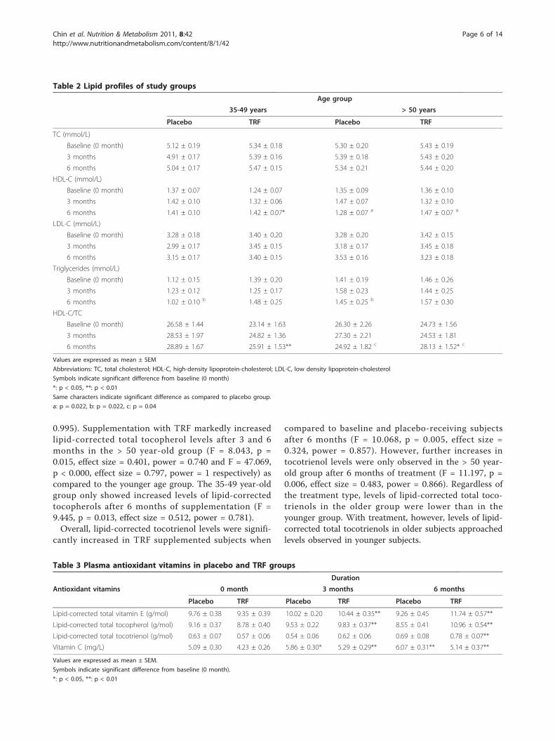
0.995). Supplementation with TRF markedly increasedlipid-corrected total tocopherol levels after 3 and 6months in the > 50 year-old group (F = 8.043, p =0.015, effect size = 0.401, power = 0.740 and F = 47.069,p < 0.000, effect size = 0.797, power = 1 respectively) ascompared to the younger age group. The 35-49 year-oldgroup only showed increased levels of lipid-correctedtocopherols after 6 months of supplementation (F =9.445, p = 0.013, effect size = 0.512, power = 0.781).Overall, lipid-corrected tocotrienol levels were signifi-
cantly increased in TRF supplemented subjects when
compared to baseline and placebo-receiving subjectsafter 6 months (F = 10.068, p = 0.005, effect size =0.324, power = 0.857). However, further increases intocotrienol levels were only observed in the > 50 year-old group after 6 months of treatment (F = 11.197, p =0.006, effect size = 0.483, power = 0.866). Regardless ofthe treatment type, levels of lipid-corrected total toco-trienols in the older group were lower than in theyounger group. With treatment, however, levels of lipid-corrected total tocotrienols in older subjects approachedlevels observed in younger subjects.
Table 2 Lipid profiles of study groups
Age group
35-49 years > 50 years
Placebo TRF Placebo TRF
TC (mmol/L)
Baseline (0 month) 5.12 ± 0.19 5.34 ± 0.18 5.30 ± 0.20 5.43 ± 0.19
3 months 4.91 ± 0.17 5.39 ± 0.16 5.39 ± 0.18 5.43 ± 0.20
6 months 5.04 ± 0.17 5.47 ± 0.15 5.34 ± 0.21 5.44 ± 0.20
HDL-C (mmol/L)
Baseline (0 month) 1.37 ± 0.07 1.24 ± 0.07 1.35 ± 0.09 1.36 ± 0.10
3 months 1.42 ± 0.10 1.32 ± 0.06 1.47 ± 0.07 1.32 ± 0.10
6 months 1.41 ± 0.10 1.42 ± 0.07* 1.28 ± 0.07 a 1.47 ± 0.07 a
LDL-C (mmol/L)
Baseline (0 month) 3.28 ± 0.18 3.40 ± 0.20 3.28 ± 0.20 3.42 ± 0.15
3 months 2.99 ± 0.17 3.45 ± 0.15 3.18 ± 0.17 3.45 ± 0.18
6 months 3.15 ± 0.17 3.40 ± 0.15 3.53 ± 0.16 3.23 ± 0.18
Triglycerides (mmol/L)
Baseline (0 month) 1.12 ± 0.15 1.39 ± 0.20 1.41 ± 0.19 1.46 ± 0.26
3 months 1.23 ± 0.12 1.25 ± 0.17 1.58 ± 0.23 1.44 ± 0.25
6 months 1.02 ± 0.10 b 1.48 ± 0.25 1.45 ± 0.25 b 1.57 ± 0.30
HDL-C/TC
Baseline (0 month) 26.58 ± 1.44 23.14 ± 1.63 26.30 ± 2.26 24.73 ± 1.56
3 months 28.53 ± 1.97 24.82 ± 1.36 27.30 ± 2.21 24.53 ± 1.81
6 months 28.89 ± 1.67 25.91 ± 1.53** 24.92 ± 1.82 c 28.13 ± 1.52* c
Values are expressed as mean ± SEM
Abbreviations: TC, total cholesterol; HDL-C, high-density lipoprotein-cholesterol; LDL-C, low density lipoprotein-cholesterol
Symbols indicate significant difference from baseline (0 month)
*: p < 0.05, **: p < 0.01
Same characters indicate significant difference as compared to placebo group.
a: p = 0.022, b: p = 0.022, c: p = 0.04
Table 3 Plasma antioxidant vitamins in placebo and TRF groups
Duration
Antioxidant vitamins 0 month 3 months 6 months
Placebo TRF Placebo TRF Placebo TRF
Lipid-corrected total vitamin E (g/mol) 9.76 ± 0.38 9.35 ± 0.39 10.02 ± 0.20 10.44 ± 0.35** 9.26 ± 0.45 11.74 ± 0.57**
Lipid-corrected total tocopherol (g/mol) 9.16 ± 0.37 8.78 ± 0.40 9.53 ± 0.22 9.83 ± 0.37** 8.55 ± 0.41 10.96 ± 0.54**
Lipid-corrected total tocotrienol (g/mol) 0.63 ± 0.07 0.57 ± 0.06 0.54 ± 0.06 0.62 ± 0.06 0.69 ± 0.08 0.78 ± 0.07**
Vitamin C (mg/L) 5.09 ± 0.30 4.23 ± 0.26 5.86 ± 0.30* 5.29 ± 0.29** 6.07 ± 0.31** 5.14 ± 0.37**
Values are expressed as mean ± SEM.
Symbols indicate significant difference from baseline (0 month).
*: p < 0.05, **: p < 0.01
Chin et al. Nutrition & Metabolism 2011, 8:42http://www.nutritionandmetabolism.com/content/8/1/42
Page 6 of 14

Plasma vitamin C concentrations increased in bothgroups regardless of the treatment received. Furtheranalysis of age groups showed similar increases for allgroups to varying degrees of significance.
Antioxidant enzymesA significant effect for duration was observed in SODactivity with TRF treatment (F = 6.838, p = 0.006, effectsize = 0.229, power = 0.826) as well as in CAT and GPxactivity with placebo (F = 4.185, p = 0.002, effect size =0.160, power = 0.707 and F = 5.271, p = 0.009, effectsize = 0.193, power = 0.809 respectively). SOD activitydecreased after 3 and 6 months of TRF treatment(Table 5); these effects were seen in both younger andolder groups, although only the > 50 year-old groupreached statistical significance (Table 6). CAT activitywas decreased in the placebo group after 3 months; thischange was also only observed in the > 50 year-old
group. Similar effects were observed for GPx, whereonly subjects in the > 50 year-old group showedincreased activity after 6 months regardless of treatment.There was nearly no association between SOD activity
and age (Figure 1), whereas GPx activity slightlydeclined with age (Figure 2), while CAT activity slightlyincreased with age (Figure 3); however, none of thesecorrelations was significant. Supplementation with TRFstrengthened the relationship between SOD activity andage as well as for CAT activity. GPx activity wasreversed in the older individuals in association with age.
Oxidative markersChanges in protein carbonyl levels were statistically sig-nificant with duration of treatment (F = 6.193, p =0.008, effect size = 0.212, power = 0.810). Protein carbo-nyl content was significantly decreased after 6 monthsof TRF treatment (p = 0.002) as compared to baseline
Table 4 Plasma antioxidant vitamins according to age group
Age group
Antioxidant vitamins 35-49 years > 50 years
Placebo TRF Placebo TRF
Lipid-corrected total vitamin E (g/mol)
Baseline (0 month) 8.71 ± 0.37 8.57 ± 0.43 10.41 ± 0.50 9.89 ± 0.56
3 months 9.49 ± 0.24 9.59 ± 0.46 10.35 ± 0.25 11.03 ± 0.45*
6 months 9.04 ± 0.67 10.39 ± 0.53** 9.52 ± 0.61 12.89 ± 0.85**
Lipid-corrected total tocopherol (g/mol)
Baseline (0 month) 8.04 ± 0.33 7.89 ± 0.45 9.84 ± 0.47 9.39 ± 0.57
3 months 8.92 ± 0.35 8.86 ± 0.46 9.92 ± 0.29 10.49 ± 0.48*
6 months 8.20 ± 0.62 9.66 ± 0.52** 8.97 ± 0.52 12.06 ± 0.79**
Lipid-corrected total tocotrienol (g/mol)
Baseline (0 month) 0.69 ± 0.09 0.66 ± 0.08 0.58 ± 0.11 0.50 ± 0.07
3 months 0.61 ± 0.07 0.73 ± 0.11 0.46 ± 0.09 0.54 ± 0.07
6 months 0.80 ± 0.09 0.73 ± 0.09 0.55 ± 0.14 0.82 ± 0.11**
Vitamin C (mg/L)
Baseline (0 month) 4.61 ± 0.35 3.95 ± 0.19 5.57 ± 0.47 4.50 ± 0.48
3 months 5.26 ± 0.42 5.16 ± 0.43* 6.48 ± 0.36 5.42 ± 0.39*
6 months 5.66 ± 0.32** 4.98 ± 0.63 6.66 ± 0.56 5.26 ± 0.46**
Values are expressed as mean ± SEM.
Symbols indicate significant difference from baseline (0 month).
*: p < 0.05, **: p < 0.01
Table 5 Erythrocyte antioxidant enzymes activity in placebo and TRF groups
Duration
Antioxidant enzymes 0 month 3 months 6 months
Placebo TRF Placebo TRF Placebo TRF
Superoxide dismutase (U/mg Hb) 1.90 ± 0.09 2.29 ± 0.14 1.90 ± 0.16 1.83 ± 0.09** 1.91 ± 0.13 1.77 ± 0.09**
Catalase (U/mg Hb) 0.30 ± 0.01 0.28 ± 0.01 0.24 ± 0.01** 0.27 ± 0.01 0.27 ± 0.01 0.27 ± 0.01
Glutathione peroxidase (U/mg Hb) 0.82 ± 0.05 0.87 ± 0.04 0.87 ± 0.05 1.04 ± 0.08 1.04 ± 0.05** 1.09 ± 0.06*
Values are expressed as mean ± SEM.
Symbols indicate significant difference from baseline (0 month).
*: p < 0.05, **: p < 0.01
Chin et al. Nutrition & Metabolism 2011, 8:42http://www.nutritionandmetabolism.com/content/8/1/42
Page 7 of 14

(0 month) (Table 7). Further grouping by age (Table 8)revealed a marked reduction (p < 0.001) in the > 50year-old group after 6 months of treatment. Nonethe-less, no significant effect for duration was obtained inAGE and MDA levels despite both levels were reducedin the older TRF-treated group after 3 months andremained low thereafter; however, this tendency did notreach significance.The relationships between age and oxidative stress
marker levels are shown in Figures 4, 5 and 6. All mea-sured biomarkers (protein carbonyl, AGE and MDA)were weakly correlated with age. TRF treatment reversed
these relationships, particularly for MDA, which showeda correlation coefficient of close to 0.4 (p < 0.05).
DiscussionHuman ageing is affected by both genetic factors andlifestyle-related factors such as diet. Dietary interventionis feasible, as nutrients can affect the rate of ageing byaltering the type and quantity of proteins synthesized[37] by modulating gene expression [38], thereby alter-ing the oxidative status of individuals [39].Our results indicate that daily supplementation for up
to 6 months with TRF raised plasma HDL cholesterol
Table 6 Erythrocyte antioxidant enzymes activity according to age group
Age group
Antioxidant enzymes 35-49 years > 50 years
Placebo TRF Placebo TRF
Superoxide dismutase (U/mg Hb)
Baseline (0 month) 1.85 ± 0.14 2.23 ± 0.22 1.89 ± 0.12 2.35 ± 0.20
3 months 1.95 ± 0.16 1.81 ± 0.15 1.85 ± 0.28 1.85 ± 0.12*
6 months 1.98 ± 0.24 1.74 ± 0.14 1.85 ± 0.10 1.79 ± 0.12*
Catalase (U/mg Hb)
Baseline (0 month) 0.29 ± 0.02 0.28 ± 0.02 0.32 ± 0.02 0.28 ± 0.01
3 months 0.25 ± 0.01 0.25 ± 0.02 0.23 ± 0.01** 0.29 ± 0.02
6 months 0.29 ± 0.02 0.23 ± 0.02 0.24 ± 0.02* 0.30 ± 0.02
Glutathione peroxidase (U/mg Hb)
Baseline (0 month) 0.74 ± 0.07 0.89 ± 0.06 0.90 ± 0.06 0.86 ± 0.06
3 months 0.87 ± 0.10 1.02 ± 0.15 0.88 ± 0.06 1.07 ± 0.08
6 months 0.88 ± 0.06 1.05 ± 0.13 1.19 ± 0.06* 1.12 ± 0.06*
Values are expressed as mean ± SEM.
Symbols indicate significant difference from baseline (0 month).
*: p < 0.05, **: p < 0.01
Figure 1 Correlation between superoxide dismutase activityand age with TRF treatment for 6 months. TRF supplementationstrengthened the association after 6 months. 0 month: r = 0.04, p =0.81; 3 months: r = 0.06, p = 0.73; 6 months: r = 0.26, p = 0.21.
Figure 2 Correlation between glutathione peroxidase activityand age with TRF treatment for 6 months. TRF supplementationreversed the relationship after 6 months. 0 month: r = -0.17, p =0.40; 3 months: r = -0.16, p = 0.41; 6 months: r = 0.05, p = 0.82.
Chin et al. Nutrition & Metabolism 2011, 8:42http://www.nutritionandmetabolism.com/content/8/1/42
Page 8 of 14

levels as early as 3 months, thereby increasing the HDL-cholesterol/total cholesterol ratio. This ratio reflects theproportion of anti-atherogenic to atherogenic lipids andhas been suggested as a better predictor of cardiovascu-lar disease risk than the individual lipoprotein values[40]. TRF might thus help to reduce the risk of coronaryheart disease (CHD) in healthy older adults. In fact,HDL cholesterol increases of the magnitude observed inthis study have been associated with a 22.5% reducedrisk of cardiovascular events [41]. Raising plasma HDLcholesterol and thus the HDL-cholesterol/total choles-terol is recommended by the American Diabetes Asso-ciation (ADA) guidelines together with lowering plasmatriglycerides for high-risk individuals particularly olderadults as major mortality cases of CHD were 65 yearsold or older [42].Conflicting data have been reported regarding effects
of vitamin E supplementation which were mainly a-tocopherol on HDL cholesterol. Increased HDL choles-terol after a-tocopherol supplementation has been
reported by some investigators [43-45] and disputed byothers [46,47]. It should be noted that the supplementused in this study was high in tocotrienols, which hasbeen reported to have different effect from a-tocopherol[48]. Tocotrienol but not tocopherol increases HDLcholesterol by inhibiting HMG-CoA reductase throughsignalling, thereby regulating cholesterol biosynthesis[49]. Tocotrienol may increase HDL in this study bymodulating signal transduction and gene expression;specifically, and may normalize any aberrant geneexpression incurred by aging [50]. Increases in HDLcould be attained by increasing physical exercise [51,52]but similar effects by supplementation of vitamins havenot been reported in human.Compliance of the subjects was indicated by the
observed increase in plasma lipid-corrected total toco-trienol and tocopherol concentration. Standardization ofplasma vitamin E levels to total cholesterol was neces-sary to control for age-related changes in baseline cho-lesterol levels as vitamin E is transported by thelipoproteins. The finding that tocotrienol levels wereincreased is of particular interest, as tocotrienol is nowreported to have functions distinct from a-tocopherol asreviewed by Sen et al. [53]. The marked increase intocotrienol in the > 50 age group is interesting and sug-gests an increased bioavailability and possibly the needfor tocotrienol with aging. The level of total tocotrienolwas slightly lower in the older adults as opposed to theyounger group. Supplementation of older subjects withTRF restored plasma vitamin E availability to near thelevels of in the controls of the younger group. We spec-ulate that a steady state plasma vitamin E concentrationwas achieved after 6 months of supplementation, asplasma concentrations were similar to those in theyounger group were seen at that time point. Some stu-dies have reported the achievement of steady-stateplasma vitamin E levels after 10 to 15 days of supple-mentation with either natural or synthetic forms of a-tocopherol at much higher dosages [54-56]. Consideringthe well-documented preferential absorption and trans-portation of different vitamin E isomers in the body and
Figure 3 Correlation between catalase activity and age withTRF treatment for 6 months. TRF supplementation strengthenedthe association after 3 and 6 months. 0 month: r = 0.16, p = 0.41; 3months: r = 0.36, p = 0.06; 6 months: r = 0.23, p = 0.26.
Table 7 Oxidative markers in placebo and TRF groups
Duration
Oxidative Markers 0 month 3 months 6 months
Placebo TRF Placebo TRF Placebo TRF
Protein carbonyl (nmol/mg) 0.52 ± 0.05 0.63 ± 0.04 0.51 ± 0.05 0.53 ± 0.04 0.56 ± 0.05 0.45 ± 0.04**
Advanced glycosylation end-product (units/ml) 2.37 ± 0.11 2.38 ± 0.19 2.00 ± 0.15 2.40 ± 0.18 1.93 ± 0.17 1.92 ± 0.14
Malondialdehyde (nmol/ml) 4.21 ± 0.19 4.10 ± 0.30 4.00 ± 0.24 3.90 ± 0.25 3.91 ± 0.23 4.14 ± 0.22
Values are expressed as mean ± SEM.
Symbols indicate significant difference from baseline (0 month).
**: p < 0.01
Chin et al. Nutrition & Metabolism 2011, 8:42http://www.nutritionandmetabolism.com/content/8/1/42
Page 9 of 14

by taking into account the tocotrienol-rich compositionin the TRF, such a slow but steady increment isreasonable.The tocotrienols are found in a wide variety of foods
and it has been suggested recently that all 8 isomers ofvitamin E may be necessary for optimum health [24].This requirement maybe more crucial for the older indi-viduals where digestion and absorption may not be asefficient resulting in the potential benefits of supplemen-tation. Differences noted in the plasma levels of toco-trienols detected in various human studies are mainly
due to the different daily fat diet. Asians consumehigher levels of palm oil rich in tocotrienols. Therefore,a comparatively higher absorption of tocotrienol andthus better level found in circulation.The elevated plasma vitamin C level detected in the
present study was most likely absorbed from the diet, asthe supplement is not a source of vitamin C. Indeed, wefound a positive correlation between vitamin C intakeand plasma vitamin C levels (r = 0.308, p = 0.048). Thisis in accordance with findings by Padayatty et al. [57]who had reported that small changes in oral intake of
Table 8 Oxidative markers according to age group
Age group
Oxidative markers 35-49 years > 50 years
Placebo TRF Placebo TRF
Protein carbonyl (nmol/mg)
Baseline (0 month) 0.54 ± 0.08 0.58 ± 0.08 0.50 ± 0.06 0.63 ± 0.03
3 months 0.56 ± 0.13 0.54 ± 0.05 0.48 ± 0.05 0.52 ± 0.06
6 months 0.60 ± 0.08 0.51 ± 0.06 0.47 ± 0.04 0.40 ± 0.03**
Advanced glycosylation end-product (units/ml)
Baseline (0 month) 2.39 ± 0.15 2.10 ± 0.22 2.34 ± 0.22 2.73 ± 0.26
3 months 2.01 ± 0.18 2.38 ± 0.21 2.30 ± 0.26 2.24 ± 0.35
6 months 2.12 ± 0.22 2.02 ± 0.17 1.98 ± 0.25 1.71 ± 0.24*
Malondialdehyde (nmol/ml)
Baseline (0 month) 4.25 ± 0.30 3.45 ± 0.37 4.18 ± 0.23 4.71 ± 0.41
3 months 4.82 ± 0.34 3.95 ± 0.39 3.22 ± 0.22 3.86 ± 0.34
6 months 3.95 ± 0.31 4.48 ± 0.27 3.87 ± 0.36 3.87 ± 0.32
Values are expressed as mean ± SEM.
Symbols indicate significant difference from baseline (0 month).
*: p < 0.05, **: p < 0.001
Figure 4 Correlation between plasma protein carbonyl levelsand age with TRF treatment for 6 months. TRF supplementationweakened the association after 3 and 6 months. 0 month: r = 0.21,p = 0.31; 3 months: r = 0.004, p = 0.98; 6 months: r = -0.15, p =0.47.
Figure 5 Correlation between serum advanced glycosylationend-products and age with TRF treatment for 6 months. TRFsupplementation reversed the association after 3 and 6 months. 0month: r = 0.18, p = 0.21; 3 months: r = -0.13, p = 0.37; 6 months: r= -0.12, p = 0.47.
Chin et al. Nutrition & Metabolism 2011, 8:42http://www.nutritionandmetabolism.com/content/8/1/42
Page 10 of 14

vitamin C resulted in large changes in plasma vitamin Cconcentration. The increase in plasma vitamin C mightalso result from a complementary effect by vitamin E inan interlinking antioxidant network. The involvement ofvitamin C in regenerating vitamin E directly from itstocotrienoxyl or tocopheroxyl radical back to tocotrienoland tocopherol respectively has been well documented[58]. Increased levels of vitamin E might reflectincreased reactions with reactive free radicals, additionalformation of tocotrienoxyl or tocopheroxyl radical, anda further increased need for vitamin C.The variation observed in enzyme activity might have
been due to the different roles of the analysed antioxi-dant enzymes. The functional roles of these enzymes arewell established, with SOD acting upstream by dismutat-ing reactive superoxide anion radicals into more stablehydrogen peroxide (H2O2) whereas catalase and GPxfunction downstream by converting H2O2 into waterand oxygen in apparently parallel pathways. Changes inantioxidant enzymes activity observed were clearly infavour of the > 50 years old group. Reduction in SODactivity with TRF supplementation after 3 and 6 monthsin the older group possibly due to lesser formation ofradicals as a result of radical scavenging effect by toco-trienol and tocopherol. On the other hand, increase inGPx activity in both placebo and TRF group after 6months possibly due to higher need for H2O2 removal.Increased level of tocotrienol and tocopherol attained byTRF supplementation might reflect more radical scaven-ging activity, followed by increased H2O2 formation andtherefore increased requirement for its removal. As forthe placebo group, increased intake of dietary vitamin C
might results in a similar increase in radical scavengingactivity and the subsequent reactions involving forma-tion and detoxification of H2O2. Given a decline in thecatalase activity in the placebo group, the action ofremoving H2O2 was predominantly done by GPx. In thecurrent study, the enzymes activity measured was evi-dently influenced by TRF supplementation as shown bythe shifted correlation patterns with age.Long term supplementation of TRF for 6 months
caused the increase in plasma vitamin E availability(both tocopherol and tocotrienol) observed in the cur-rent study, accompanied by changes in the oxidativestress biomarkers measured. Protein carbonyls havebeen described as oxidized amino acids resulting fromdirect oxidation of protein by reactive oxygen species[59]. Proteins are also modified indirectly by glycationor glycoxidation of amino groups with the eventual for-mation of the advanced glycosylation end products(AGEs) [60]. Consistent with the fall in plasma concen-trations of carbonylated protein with TRF supplementa-tion, a sharp decrease in serum AGE was observed.When the effect of age was factored out of the statisticalmodel, it was found that the interaction between supple-mentation and duration was significant for the olderindividuals, indicating a favourable gain in the oldergroup. Figures 4, 5 and 6 give individual presentationsof the changes in these oxidative markers with age.Ascending trends of protein damage and lipid peroxida-tion accumulation during ageing as shown by the corre-lative data were reduced, even reversed by TRFsupplementation. These findings confirm that nutritionalintervention can exert cumulative effects on oxidativestress in healthy individuals in the long term [27]. Theunique combination of vitamin E isomers used in thestudy might have acted synergistically to provide thebeneficial effect.The reduced levels of oxidative markers were mainly
observed in the older group, for whom the lower cut-offpoint was 50 years of age. This is of interest as previousstudies typically evaluated older subjects, mostly 60years old and over. It is also noteworthy that in the pre-sent study the treatment was administered to healthyindividuals for a lengthy period of 6 months and studiesinvolving supplementation of this duration are fairlylimited. This may then results in the observed changesin oxidative status as measured by protein carbonyl andAGE. Although some of the younger subjects in thestudy also showed increased levels of antioxidant vita-mins, the magnitude of changes was less evident ascompared to the older individuals. The absence or lackof response by the younger age group might reflect awell-maintained antioxidant level, more effective mainte-nance of oxidative balance, and better defence againstspontaneous oxidative injury. It is thus possible that
Figure 6 Correlation between plasma malondialdehyde levelsand age with TRF treatment for 6 months. TRF supplementationreversed the association after 3 and 6 months. 0 month: r = 0.17, p= 0.25; 3 months: r = -0.19, p = 0.22; 6 months: r = -0.37, p = 0.02.
Chin et al. Nutrition & Metabolism 2011, 8:42http://www.nutritionandmetabolism.com/content/8/1/42
Page 11 of 14

TRF supplementation did not provide any furtherimprovement. Although baseline antioxidant levels inthe > 50 year-old group were similar to those in the <50 year-old group, baseline oxidative marker levels werehigher in the older group, suggesting a higher level ofoxidative damage. However, the amount of damagedlipids and proteins in this group was reduced by supple-mentation, probably due to the increased requirementfor antioxidants in older individuals. It is possible thatan antioxidant threshold for optimum performanceexists and that this threshold (and therefore the require-ment for antioxidants) could increase with ageing, thusallowing supplementation to generate an effect in thepresent study. A long-term prospective study will berequired to test this hypothesis, particularly at the mole-cular level.Compelling evidence suggests a new level of action for
vitamin E under the non-antioxidative control in protec-tion against disease [50]. Therefore, TRF might not onlyact directly or solely as an antioxidant, but it may actuallyalso act through signalling pathways and specific signal-regulated protein reaction as suggested by Sen et al.[14,61]. Tocotrienol was shown to provide complete neu-roprotection via antioxidant-independent mechanism withthe protective property reported not only limited inresponse to non-oxidative challenges but also to oxidativeinsults [14,15]. Further studies of the effects of tocotrienolsin a cell model are currently underway in our laboratory.
ConclusionOur data revealed an age-related increase in oxidativedamage. We established a role of nutritional supplemen-tation in oxidative damage and antioxidant levels inolder individuals. To our best knowledge, tocotrienol-rich vitamin E supplementation has not yet been studiedin relation to oxidative stress in healthy older indivi-duals. Consistent with increased concentrations ofplasma antioxidants (vitamins E and C), we observedsignificant decreases in protein carbonyl and AGE levels,as well as improvement of plasma cholesterol levels.The protective effects of TRF supplementation observedin this study might represent a restoration of redox bal-ance, particularly in the > 50-year old group. Increasedtocotrienol level might be an important mechanism bywhich TRF supplementation confers its protective bene-fits via protection against oxidative stress, involvementin oxidized protein repair, besides contributing to theregulation of redox homeostasis through signalling.
AcknowledgementsThe authors thank Ms. Zalina Hamid from Sime Darby Bioganic Sdn. Bhd.(Selangor, Malaysia) for supplying the TRF and placebo capsules used in thestudy, and Dr. Wan Nazaimoon Wan Mohamud (Division of Endocrinology,Institute for Medical Research, Kuala Lumpur, Malaysia) for kindly providing
us with techniques and antibodies to measure the serum AGEconcentration. Financial support for this study was provided by grantsawarded from the Malaysian Government, the IRPA 06-02-02-0016-PR0008/09 and UKM-GUP-SK-07-21-199.
Author details1Department of Biochemistry, Faculty of Medicine, Universiti KebangsaanMalaysia, Kuala Lumpur, Malaysia. 2Department of Anatomy, Faculty ofMedicine, Universiti Kebangsaan Malaysia, Kuala Lumpur, Malaysia.3Department of Physiology, Faculty of Medicine, Universiti KebangsaanMalaysia, Kuala Lumpur, Malaysia.
Authors’ contributionsSFC was involved in the acquisition, analysis and interpretation of data inaddition to drafting the manuscript; JI, NAAH, AAL, ZZ and AAK madesignificant contributions to the acquisition of the data, SM was involved ininterpretation of data and critical analysis of intellectual content of themanuscript, MM and YAMY contributed to the design, acquisition andinterpretation of the data; and WZWN was instrumental in the study’sinception, design and approval while providing critical analysis of datainterpretation and manuscript review. The final manuscript have been readand approved by all authors.
Competing interestsThe authors declare that they have no competing interests.
Received: 22 February 2011 Accepted: 24 June 2011Published: 24 June 2011
References1. Shay JW, Wright WE: Telomerase therapeutics for cancer: challenges and
new directions. Nat Rev Drug Discov 2006, 5(7):577-584.2. Yap WN, Chang PN, Han HY, Lee DTW, Ling MT, Wong YC, Yap YL: γ-
Tocotrienol suppresses prostate cancer cell proliferation and invasionthrough multiple-signalling pathways. British Journal of Cancer 2008,99(11):1832-1841.
3. Meydani M: Vitamin E modulation of cardiovascular disease. Ann NY AcadSci 2004, 1031:271-279.
4. Koga T, Kwan P, Zubik L, Ameho C, Smith D, Meydani M: Vitamin Esupplementation suppresses macrophage accumulation and endothelialcell expression of adhesion molecules in the aorta ofhypercholesterolemic rabbits. Atherosclerosis 2004, 176(2):265-272.
5. Montiel T, Quiroz-Baez R, Massieu L, Arias C: Role of oxidative stress onbeta-amyloid neurotoxicity elicited during impairment of energymetabolism in the hippocampus: protection by antioxidants. Exp Neurol2006, 200:496-508.
6. Butterfield DA, Castegna A, Drake J, Scapagnini G, Calabrese V: Vitamin Eand neurodegenerative disorders associated with oxidative stress. NutrNeurosci 2002, 5:229-239.
7. Inokuchi H, Hirokane H, Tsuzuki T, Nakagawa K, Igarashi M, Miyazawa T:Antiangiogenic activity of tocotrienol. Biosci Biotechnol Biochem 2003,67(7):1623-1627.
8. Mizushina Y, Nakagawa K, Shibata A, Awata Y, Kuriyama I, Shimazaki N,Koiwai O, Uchiyama Y, Sakaguchi K, Miyazawa T, Yoshida H: Inhibitoryeffect of tocotrienol on eukaryotic DNA polymerase lambda andangiogenesis. Biochem Biophys Res Commun 2006, 339(3):949-955.
9. Nesaretnam K, Dorasamy S, Darbre PD: Tocotrienols inhibit growth of ZR-75-1 breast cancer cells. Int J Food Sci Nutr 2000, , Suppl 51: S95-S103.
10. Khor HT, Ng TT: Effects of administration of alpha-tocopherol andtocotrienols on serum lipids and liver HMG CoA reductase activity. Int JFood Sci Nutr 2000, , Suppl 51: S3-S11.
11. Qureshi AA, Qureshi N, Wright JJ, Shen Z, Kramer G, Gapor A, Chong YH,DeWitt G, Ong A, Peterson DM: Lowering of serum cholesterol inhypercholesterolemic humans by tocotrienols (palmvitee). Am J Clin Nutr1991, 53(Suppl 4):1021S-1026S.
12. Qureshi AA, Bradlow BA, Brace L, Manganello J, Peterson DM, Pearce BC,Wright JJ, Gapor A, Elson CE: Response of hypercholesterolemic subjectsto administration of tocotrienols. Lipids 1995, 30(12):1171-1177.
13. Qureshi AA, Sami SA, Salser WA, Khan FA: Dose-dependent suppression ofserum cholesterol by tocotrienol-rich fraction (TRF25) of rice bran inhypercholesterolemic humans. Atherosclerosis 2002, 161(1):199-207.
Chin et al. Nutrition & Metabolism 2011, 8:42http://www.nutritionandmetabolism.com/content/8/1/42
Page 12 of 14

14. Sen CK, Khanna S, Roy S, Packer L: Molecular basis of vitamin E action.Tocotrienol potently inhibits glutamate-induced pp60(c-Src) kinaseactivation and death of HT4 neuronal cells. J Biol Chem 2000,275(17):13049-13055.
15. Khanna S, Roy S, Parinandi NL, Maurer M, Sen CK: Characterization of thepotent neuroprotective properties of the natural vitamin E alpha-tocotrienol. J Neurochem 2006, 98(5):1474-1486.
16. Halliwell B: Role of free radicals in the neurodegenerative diseases:therapeutic implications for antioxidant treatment. Drugs Aging 2001,18:685-716.
17. Holliday R: Aging is no longer an unsolved problem in biology. Ann NYAcad Sci 2006, 1067:1-9.
18. Duthie SJ, Ma A, Ross MA, Collins AR: Antioxidant supplementationdecreases oxidative DNA damage in human lymphocytes. Cancer Res1996, 56(6):1291-1295.
19. Lee BM, Lee SK, Kim HS: Inhibition of oxidative DNA damage, 8-OHdGand carbonyl contents in smokers treated with antioxidants (vitamin E,vitamin C, beta-carotene and red ginseng). Cancer Lett 1998, 132(1/2):219-227.
20. Jenkinson AM, Collins AR, Duthie SJ, Wahle KW, Duthie GG: The effect ofincreased intakes of polyunsaturated fatty acids and vitamin E on DNAdamage in human lymphocytes. FASEB J 1999, 13(15):2138-2142.
21. Dereska NH, McLemore EC, Tessier DJ, Bash DS, Brophy CM: Short-term,moderate dosage vitamin E supplementation may have no effect onplatelet aggregation, coagulation profile and bleeding time in healthyindividuals. J Surg Res 2006, 132:121-129.
22. The HOPE Study Investigators: Vitamin E supplementation andcardiovascular events in high-risk patients. N Engl J Med 2000,342:154-160.
23. Rapola JM, Virtamo J, Haukka JK, Heinonen OP, Albanes D, Taylor PR,Huttunen JK: Effect of vitamin E and beta carotene on the incidence ofangina pectoris: A randomized, double-blind, controlled trial. JAMA 1996,275:693-698.
24. Krishnan K, Stone W, Campbell S: More optimal forms of vitamin E.Letters to the editor. J Am Diet Assoc 2005, 105(2):204-205.
25. Winklhofer-Roob BM, Roob JM, Maritschnegg M, Sprinz G, Hiller D,Marktfelder E, Preinsberger M, Wuga S, Sundl I, Tiran B, Cardinault N,Ribalta J, Rock E, Vitage Study Group: Does aging affect the response ofvitamin E status to vitamin E depletion and supplementation? Ann NYAcad Sci 2004, 1031:381-384.
26. Andriollo-Sanchez M, Hininger-Favier I, Meunier N, Venneria E, O’Connor JM,Maiani G, Coudray C, Roussel AM: Age-related oxidative stress andantioxidant parameters in middle-aged and older European subjects: theZENITH study. Eur J Clin Nutr 2005, 59(Suppl 2):S58-S62.
27. Chin SF, Noor Aini AH, Azian AL, Zaiton Z, Musalmah M, Yasmin AMY,Aminuddin AK, Johari I, Zalina H, Wan Zurinah WN: Reduction of DNAdamage in older healthy adults by Tri E ® Tocotrienol supplementation.Nutrition 2008, 24:1-10.
28. Mastaloudis A, Leonard SW, Traber MJ: Oxidative stress in athletes duringextreme endurance exercise. Free Radic Biol Med 2001, 31(7):911-922.
29. O’Byrne D, Grundy S, Packer L, Devaraj S, Baldenius K, Hoppe PP, Kraemer K,Jialal I, Traber MG: Studies of LDL oxidation following α-, γ-, or δ-tocotrienyl acetate supplementation of hypercholesterolemic humans.Free Radic Biol Med 2000, 29(9):834-845.
30. Pachla LA, Kissinger PT: Analysis of ascorbate by liquid chromatographywith amperometric detection. Methods Enzymol 1979, 62:15-24.
31. Levine RL, Williams JA, Stadtman EP, Shacter E: Carbonyl assays fordetermination of oxidatively modified proteins. Methods Enzymol 1994,233:346-357.
32. Wan Nazaimoon WM, Khalid BAK: An enzyme immunoassay for advancedglycosylation end-products in serum. Malaysian J Pathol 1998, 20(2):83-89.
33. Pilz J, Meineke I, Gleiter CH: Measurement of free and boundmalondialdehyde in plasma by high-performance liquidchromatography as the 2, 4-dinitrophenylhydrazine derivative. J Chrom B2000, 742:315-325.
34. Beyer WF Jr, Fridovich I: Assaying for superoxide dismutase activity: somelarge consequences of minor changes in conditions. Anal Biochem 1987,161(2):559-566.
35. Aebi H: Catalase in vitro. Methods Enzymol 1984, 105:121-126.
36. Paglia DE, Valentine WN: Studies on the quantitative and qualitativecharacterization of erythrocyte glutathione peroxidase. J Clin Med 1967,70:158-169.
37. Papet I, Dardevet D, Sornet C, Béchereau F, Prugnaud J, Pouyet C, Obled C:Acute phase protein levels and thymus, spleen and plasma proteinsynthesis rates differ in adult and old rats. J Nutr 2003, 133:215-219.
38. Pletcher SD, Libert S, Skorupa D: Flies and their golden apples: The effectof dietary restriction on Drosophila aging and age-dependent geneexpression. Ageing Res Rev 2005, 4:451-480.
39. Friel JK, Widness JA, Jiang T, Belkhode SL, Rebouche CJ, Ziegler EE:Antioxidant status and oxidant stress may be associated with vitamin Eintakes in very low birth weight infants during the first month of life.Nutr Res 2002, 22:55-64.
40. Natarajan S, Glick H, Criqui M, Horowitz D, Lipsitz SR, Kinosian B:Cholesterol measures to identify and treat individuals at risk forcoronary heart disease. Am J Prev Med 2003, 25:50-57.
41. Shai I, Rimm EB, Hankinson SE, Curhan G, Manson JAE, Rifai N, Stampfer MJ,Ma J: Multivariate assessment of lipid parameters as predictors ofcoronary heart disease among postmenopausal women: Potentialimplications for clinical guidelines. Circulation 2004, 110:2824-2830.
42. Haffner S: Rationale for new American Diabetes Association Guidelines:are national cholesterol education program goals adequate for thepatient with Diabetes Mellitus? Am J Cardiol 2005, 96(4A):33E-36E.
43. da Costa VAV, Vianna LM: Effect of α -tocopherol supplementation onblood pressure and lipidic profile in streptozotocin-induced diabetesmellitus in spontaneously hypertensive rats. Clin Chim Acta 2005,351:101-104.
44. Komaratat P, Chupukcharoen N, Wilairat P: Effect of vitamin E oncholesterol plasma lipoprotein distribution and metabolism in rabbit. IntJ Vitam Nutr Res 1985, 55:167-171.
45. Hermann WJ Jr, Ward K, Faucett J: The effect of tocopherol on high-density lipoprotein cholesterol. A clinical observation. Am J Clin Pathol1979, 72:848-852.
46. van Tits LJH, de Waart F, Hak-Lemmers HLM, van Heijst P, de Graaf J,Demacker PNM, Stalenhoef AFH: Effects of α -tocopherol on superoxideproduction and plasma intercellular adhesion molecule-1 and antibodiesto oxidized LDL in chronic smokers. Free Radic Biol Med 2001,30(10):1122-1129.
47. Perugini C, Bagnati M, Cau C, Bordone R, Paffoni P, Re R, Zoppis E,Albano E, Bellomo G: Distribution of lipid-soluble antioxidants inlipoproteins from healthy subjects. II. Effects of in vivo supplementationwith α-tocopherol. Pharmacol Res 2000, 41(1):67-74.
48. Packer L, Weber SU, Rimbach G: Molecular aspects of α-tocotrienolantioxidant action and cell signalling. J Nutr 2001, 369S-373S.
49. Qureshi AA, Sami SA, Salser WA, Khan FA: Dose-dependent suppression ofserum cholesterol by tocotrienol-rich fraction (TRF25) of rice bran inhypercholesterolemic humans. Atherosclerosis 2002, 161:199-207.
50. Azzi A, Gysin R, Kempna P, Ricciarelli R, Villacorta L, Visarius T, Zingg JM:Regulation of gene and protein expression by Vitamin E. Free Radic Res2002, 36(1):30-35.
51. Nicklas BJ, Katzel LI, Busby-Whitehead J, Goldberg AP: Increases in high-density lipoprotein cholesterol with endurance exercise training areblunted in obese compared with lean men. Metabolism 1997,46(5):556-561.
52. Delgado M, González-Gross M, Cano MD, Gutiérrez A, Castillo MJ: Physicalexercise reverses diet-induced increases in LDL-cholesterol and apo Blevels in healthy ovo-lactovegetarian subjects. Nutr Res 2000,20(12):1707-1714.
53. Sen CK, Khanna S, Roy S: Tocotrienols in health and disease: the otherhalf of the natural vitamin E family. Mol Aspects Med 2007, 28:692-728.
54. Hoppe PP, Krennrich G: Bioavailability and potency of natural-source andall-racemic alpha-tocopherol in the human: a dispute. Eur J Nutr 2000,39:183-193.
55. Burton GW, Traber MG, Acuff RV, Walters DN, Kayden H, Hughes L,Ingold KU: Human plasma and tissue alphatocopherol concentrations inresponse to supplementation with deuterated natural and syntheticvitamin E. Am J Clin Nutr 1998, 67:669-684.
56. Lodge JK: Vitamin E bioavailability in humans. J Plant Physiol 2005,162:790-796.
Chin et al. Nutrition & Metabolism 2011, 8:42http://www.nutritionandmetabolism.com/content/8/1/42
Page 13 of 14

57. Padayatty SJ, Katz A, Wang Y, Eck P, Kwon O, Lee JH, Chen S, Corpe C,Dutta A, Dutta SK, Levine M: Vitamin C as an antioxidant: Evaluation of itsrole in disease prevention. J Am Coll Nutr 2003, 22(1):18-35.
58. Neuzil J, Thomas SR, Stocker R: Requirement for promotion or inhibitionby alpha-tocopherol of radical-induced initiation of plasma lipoproteinlipid peroxidation. Free Radic Biol Med 1997, 22:57-71.
59. Stadtman ER, Levine RL: Protein oxidation. Ann NY Acad Sci 2000,899:191-208.
60. Inagi R, Miyata T: Oxidative protein damage with carbohydrates andlipids in uremia: ‘Carbonyl stress’. Blood Purif 1999, 17:95-98.
61. 61 Sen CK, Khanna S, Roy S: Tocotrienol: the natural vitamin E to defendthe nervous system? Ann NY Acad Sci 2004, 1031:127-142.
doi:10.1186/1743-7075-8-42Cite this article as: Chin et al.: Tocotrienol rich fraction supplementationimproved lipid profile and oxidative status in healthy older adults: Arandomized controlled study. Nutrition & Metabolism 2011 8:42.
Submit your next manuscript to BioMed Centraland take full advantage of:
• Convenient online submission
• Thorough peer review
• No space constraints or color figure charges
• Immediate publication on acceptance
• Inclusion in PubMed, CAS, Scopus and Google Scholar
• Research which is freely available for redistribution
Submit your manuscript at www.biomedcentral.com/submit
Chin et al. Nutrition & Metabolism 2011, 8:42http://www.nutritionandmetabolism.com/content/8/1/42
Page 14 of 14