tnt coupled reticulocyte lysate systems - promega · revised 6/15 tb126 technical bulletin tnt®...
TRANSCRIPT

Revised 6/15 TB126
T E C H N I C A L B U L L E T I N
TnT® Coupled Reticulocyte Lysate SystemsInstructions for Use of Products L4600, L4610, L4950, L5010, L5020, L4601 and L4611

Promega Corporation · 2800 Woods Hollow Road · Madison, WI 53711-5399 USA · Toll Free in USA 800-356-9526 · 608-274-4330 · Fax 608-277-2516 1www.promega.com TB126 · Revised 6/15
All technical literature is available at: www.promega.com/protocols/ Visit the web site to verify that you are using the most current version of this Technical Manual.
E-mail Promega Technical Services if you have questions on use of this system: [email protected]
TnT® Coupled Reticulocyte Lysate Systems
1. Description .........................................................................................................................................2
2. Product Components and Storage Conditions ........................................................................................3
3. General Considerations .......................................................................................................................53.A. DNA Template Considerations .....................................................................................................53.B. Creating a Ribonuclease-Free Environment ..................................................................................6
4. Translation Procedure .........................................................................................................................64.A. General Protocol for TnT® Lysate Coupled Transcription/Translation Reactions .............................6
5. Positive Control Translation Reactions Using Luciferase ........................................................................95.A. Radioactive Luciferase Control Reaction ......................................................................................95.B. Non-Radioactive Luciferase Control Reaction ............................................................................. 10
6. Cotranslational Processing Using Canine Pancreatic Microsomal Membranes in TnT® Lysate Systems ..... 106.A. General Protocol for Translation with Microsomal Membranes .................................................... 10
7. Post-Translational Analysis ................................................................................................................ 127.A. Determination of Percent Incorporation of Radioactive Label ...................................................... 127.B. Denaturing Gel Analysis of Translation Products ......................................................................... 13
8. Positive Control Luciferase Assays ...................................................................................................... 148.A. Using a Luminometer ................................................................................................................ 158.B. Using a Scintillation Counter ..................................................................................................... 158.C. Photographic Luciferase Assay ................................................................................................... 158.D. Qualitative Visual Detection of Luciferase Activity ....................................................................... 16
9. Troubleshooting................................................................................................................................ 17
10. References ........................................................................................................................................ 18
11. Appendix .......................................................................................................................................... 2011.A. Composition of Buffers and Solutions ......................................................................................... 2011.B. Luciferase SP6/T7/T3 Control DNAs ......................................................................................... 2011.C. Related Products ...................................................................................................................... 24
12. Summary of Changes .......................................................................................................................... 28
2 Promega Corporation · 2800 Woods Hollow Road · Madison, WI 53711-5399 USA · Toll Free in USA 800-356-9526 · 608-274-4330 · Fax 608-277-2516TB126 · Revised 6/15 www.promega.com
1. Description
The TnT® Coupled Reticulocyte Lysate System(a) offers researchers an alternative for eukaryotic in vitro translations: a single-tube, coupled transcription/translation system. The TnT® Lysate Systems greatly simplify the process and reduce the time required to obtain in vitro translation results (Figure 1). Standard rabbit reticulocyte lysate translations (1) commonly use RNA synthesized in vitro (2) from SP6, T3 or T7 RNA polymerase promoters and require three separate reactions with several steps between each reaction. The TnT® Systems bypass many of these steps by incorporating transcription directly in the translation mix.
In most cases, the TnT® Lysate reactions produce significantly more protein (two- to sixfold) in a 1.5-hour reaction than standard in vitro rabbit reticulocyte lysate translations using RNA templates. In comparisons of 35S incorporation, the TnT® Lysate reactions incorporate significantly more radiolabel than do standard in vitro translation reactions. Published applications of these systems include:
• Truncation mutation analysis [e.g., the Protein Truncation Test (PTT)]• Drug screening (affecting translation rates)• Mutation and detection analysis (i.e., enzyme kinetics)• Protein-protein interactions (using GST fusion proteins)• Immunoprecipitation of protein complexes• Protein dimerization assays• Ligand-binding region determination/confirmation/competition assays• Protein structure analysis• Electrophoretic mobility shift assays (EMSAs) for DNA:protein interactions• DNA footprinting and protein cross-linking studies• Protein-RNA binding assays• Post-translational modification tests• In vitro expression cloning (3; functional genomics)• Verification/characterization of cloned gene products
For a complete list of references for these and other applications, please visit our citations database at: www.promega.com/citations/
The TnT® Lysate Systems are available in a variety of configurations for transcription and translation of genes cloned downstream from the SP6, T3 or T7 RNA polymerase promoter. To use these systems, 0.2–2.0µg of circular plasmid DNA (or linear DNA for the T3 and T7 Systems, see Note 3, Section 3.A) are added directly to TnT® Lysate and incubated in a 50µl reaction for 1.5 hours at 30°C. Included with the TnT® Lysate Systems are luciferase-encoding control plasmids and Luciferase Assay Reagent, which can be used in a non-radioactive assay for functionally active luciferase protein. Starting with circular plasmid DNA, in vitro translation results (autoradiograms) are obtained easily in 8 hours. Alternatively, the Transcend™ Non-Radioactive Translation Detection Systems allow colorimetric or chemiluminescent detection of proteins synthesized using the TnT® Coupled Reticulocyte Lysate Systems.

Promega Corporation · 2800 Woods Hollow Road · Madison, WI 53711-5399 USA · Toll Free in USA 800-356-9526 · 608-274-4330 · Fax 608-277-2516 3www.promega.com TB126 · Revised 6/15
2. Product Components and Storage Conditions
P R O D U C T S I Z E C AT. #
TnT® SP6 Coupled Reticulocyte Lysate System 40 reactions L4600*
TnT® T7 Coupled Reticulocyte Lysate System 40 reactions L4610
TnT® T3 Coupled Reticulocyte Lysate System 40 reactions L4950
TnT® T7/T3 Coupled Reticulocyte Lysate System 40 reactions L5010
TnT® T7/SP6 Coupled Reticulocyte Lysate System 40 reactions L5020
TnT® Rabbit Reticulocyte Lysate is supplied in 200µl aliquots. Each system contains sufficient reagents to perform approximately 40 × 50µl translation reactions. Includes:
• 1ml TnT® Rabbit Reticulocyte Lysate• 90µl TnT® Reaction Buffer• 60µl TnT® T3, T7 or SP6 RNA Polymerase (2 × 30µl in dual systems)• 5µg Luciferase T3, T7 or SP6 Control DNA, 0.5mg/ml• 50µl Amino Acid Mixture, Minus Methionine, 1mM• 50µl Amino Acid Mixture, Minus Leucine, 1mM• 50µl Amino Acid Mixture, Minus Cysteine, 1mM• 250µl Luciferase Assay Reagent• 1 Luciferase Assay Wells (set of 3)• 100µl Magnesium Acetate*
P R O D U C T S I Z E C AT. #
TnT® SP6 Coupled Reticulocyte Lysate System, Trial Size 8 reactions L4601*
TnT® T7 Coupled Reticulocyte Lysate System, Trial Size 8 reactions L4611
TnT® Rabbit Reticulocyte Lysate is supplied in 200µl aliquots. Each trial size system contains sufficient reagents to perform approximately 8 × 50µl translation reactions. Includes:
• 200µl TnT® Rabbit Reticulocyte Lysate• 20µl TnT® Reaction Buffer• 20µl TnT® T7 or SP6 RNA Polymerase• 5µg Luciferase T7 or SP6 Control DNA, 0.5mg/ml• 25µl Amino Acid Mixture, Minus Methionine, 1mM• 25µl Amino Acid Mixture, Minus Leucine, 1mM• 250µl Luciferase Assay Reagent• 1 Luciferase Assay Wells (set of 3)• 100µl Magnesium Acetate*
*Magnesium Acetate is supplied only with Cat.# L4600 and L4601.

4 Promega Corporation · 2800 Woods Hollow Road · Madison, WI 53711-5399 USA · Toll Free in USA 800-356-9526 · 608-274-4330 · Fax 608-277-2516TB126 · Revised 6/15 www.promega.com
2. Product Components and Storage Conditions (continued)
Storage and Stability: Store all components at –70°C (except Luciferase Assay Wells, which should be stored at room temperature). Product is sensitive to CO2 (avoid prolonged exposure) and multiple freeze-thaw cycles, which may have an adverse effect on activity/performance. LAR is stable for at least 12 months if stored and handled properly.
Do not store the lysate at any temperature other than –70°C. Storage at other temperatures (e.g., –20°C) for even a short time will dramatically reduce activity.
Do not freeze-thaw the lysate more than 2 times.
Do not store the lysate in the presence of dry ice. Prolonged exposure to dry ice can cause significant loss of lysate activity.
Note: All components of the TnT® Systems are quality tested and verified for coupled transcription/translation in the lot-specific combinations represented in each prepackaged system. Use of reagents or reagent combinations other than those verified for the TnT® Reticulocyte Lysate Systems may result in suboptimal coupled transcription/translation. To ensure performance, TnT® RNA polymerases are not available as separate products.
Do not use lysates, polymerases and buffers from other lots or systems.
2924
MA
04_0
A
Linearize DNA templateby restriction digestion.
Extract and precipitate DNA.
Perform in vitro transcription.
Remove template with RQ1 DNase.
Extract and precipitate RNA.
Perform in vitro translation usingRabbit Reticulocyte Lysate.
Separate translation products by SDS-PAGE.
Fix and dry gel.(Fluorography optional)
Autoradiography.
(1 hour) Add DNA template and perform transcription/translation
using TNT® Lysate.
Separate translation products by SDS-PAGE.
Fix and dry gel.(Fluorography optional)
Autoradiography.
(30 minutes)
(1 hour)
(1 hour)
(30 minutes)
(1.5 hours at 30°C)
(1.5 hours)
(2 hours)
(3 hours at –70°C)
(1.5 hours at 30°C)
(1.5 hours)
(2 hours)
(3 hours at –70°C)
Standard in vitro Transcription and Translation
TNT® Lysate Coupled Transcription/Translation
Time required = 12 hours
Time required = 8 hours
Figure 1. Comparison of standard in vitro transcription and translation procedures to the TnT® Lysate coupled transcription/translation protocol.
!
!
!
!
Promega Corporation · 2800 Woods Hollow Road · Madison, WI 53711-5399 USA · Toll Free in USA 800-356-9526 · 608-274-4330 · Fax 608-277-2516 5www.promega.com TB126 · Revised 6/15
3. General Considerations
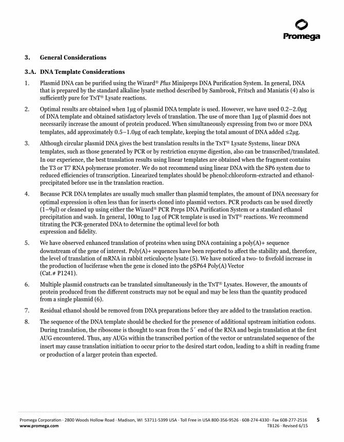
3.A. DNA Template Considerations
1. Plasmid DNA can be purified using the Wizard® Plus Minipreps DNA Purification System. In general, DNA that is prepared by the standard alkaline lysate method described by Sambrook, Fritsch and Maniatis (4) also is sufficiently pure for TnT® Lysate reactions.
2. Optimal results are obtained when 1µg of plasmid DNA template is used. However, we have used 0.2–2.0µg of DNA template and obtained satisfactory levels of translation. The use of more than 1µg of plasmid does not necessarily increase the amount of protein produced. When simultaneously expressing from two or more DNA templates, add approximately 0.5–1.0µg of each template, keeping the total amount of DNA added ≤2µg.
3. Although circular plasmid DNA gives the best translation results in the TnT® Lysate Systems, linear DNA templates, such as those generated by PCR or by restriction enzyme digestion, also can be transcribed/translated. In our experience, the best translation results using linear templates are obtained when the fragment contains the T3 or T7 RNA polymerase promoter. We do not recommend using linear DNA with the SP6 system due to reduced efficiencies of transcription. Linearized templates should be phenol:chloroform-extracted and ethanol-precipitated before use in the translation reaction.
4. Because PCR DNA templates are usually much smaller than plasmid templates, the amount of DNA necessary for optimal expression is often less than for inserts cloned into plasmid vectors. PCR products can be used directly (1–9µl) or cleaned up using either the Wizard® PCR Preps DNA Purification System or a standard ethanol precipitation and wash. In general, 100ng to 1µg of PCR template is used in TnT® reactions. We recommend titrating the PCR-generated DNA to determine the optimal level for both expression and fidelity.
5. We have observed enhanced translation of proteins when using DNA containing a poly(A)+ sequence downstream of the gene of interest. Poly(A)+ sequences have been reported to affect the stability and, therefore, the level of translation of mRNA in rabbit reticulocyte lysate (5). We have noticed a two- to fivefold increase in the production of luciferase when the gene is cloned into the pSP64 Poly(A) Vector (Cat.# P1241).
6. Multiple plasmid constructs can be translated simultaneously in the TnT® Lysates. However, the amounts of protein produced from the different constructs may not be equal and may be less than the quantity produced from a single plasmid (6).
7. Residual ethanol should be removed from DNA preparations before they are added to the translation reaction.
8. The sequence of the DNA template should be checked for the presence of additional upstream initiation codons. During translation, the ribosome is thought to scan from the 5´ end of the RNA and begin translation at the first AUG encountered. Thus, any AUGs within the transcribed portion of the vector or untranslated sequence of the insert may cause translation initiation to occur prior to the desired start codon, leading to a shift in reading frame or production of a larger protein than expected.

6 Promega Corporation · 2800 Woods Hollow Road · Madison, WI 53711-5399 USA · Toll Free in USA 800-356-9526 · 608-274-4330 · Fax 608-277-2516TB126 · Revised 6/15 www.promega.com
3.B. Creating a Ribonuclease-Free Environment
Two of the most common sources of RNase contamination are the user’s hands and bacteria or mold that may be present on airborne dust particles. We recommend the addition of RNasin® Ribonuclease Inhibitor to all TnT® Lysate reactions to prevent degradation of RNA.
Important: To reduce the chance of RNase contamination, gloves should be worn when setting up experiments, and microcentrifuge tubes and pipette tips should be RNase-free.
4. Translation Procedure
Materials to Be Supplied by the User• RNasin® Ribonuclease Inhibitor• Nuclease-Free Water (Cat.# P1193)• radiolabeled amino acid (for radioactive detection)• Transcend™ Colorimetric (Cat.# L5070) or Chemiluminescent (Cat.# L5080)
Translation Detection System (for non-radioactive detection)• Transcend™ tRNA (Cat.# L5061; for non-radioactive detection)
4.A. General Protocol for TnT® Lysate Coupled Transcription/Translation Reactions
The following is a general guideline for setting up a translation reaction. Also provided are examples of standard reactions using [35S]methionine (radioactive) or Transcend™ Non-Radioactive Detection Systems. Using the Transcend™ Systems, biotinylated lysine residues are incorporated into nascent proteins during translation. This biotinylated lysine is added to the translation reaction as a precharged ε-labeled, biotinylated lysine-tRNA complex (Transcend™ tRNA) rather than a free amino acid. For more information on the Transcend™ Systems, request Technical Bulletin #TB182.
Radioactive and non-radioactive control reactions for the production of luciferase are described in Section 5. We also recommend including a control reaction containing no added DNA. This reaction allows measurement of any background incorporation of labeled amino acids.
1. Remove the reagents from storage at –70°C. Immediately place the TnT® RNA Polymerase on ice. Rapidly thaw the TnT® Reticulocyte Lysate by hand warming and immediately place on ice. The other components can be thawed at 25°C and stored on ice.
2. Following the example below, assemble the reaction components in a 0.5ml or 1.5ml microcentrifuge tube. After addition of all the components, gently mix the lysate by pipetting the reaction. If necessary, centrifuge briefly to return the reaction to the bottom of the tube.
Note: Except for the actual translation incubation, all handling of the lysate components should be done at 4°C or on ice.
!
Promega Corporation · 2800 Woods Hollow Road · Madison, WI 53711-5399 USA · Toll Free in USA 800-356-9526 · 608-274-4330 · Fax 608-277-2516 7www.promega.com TB126 · Revised 6/15
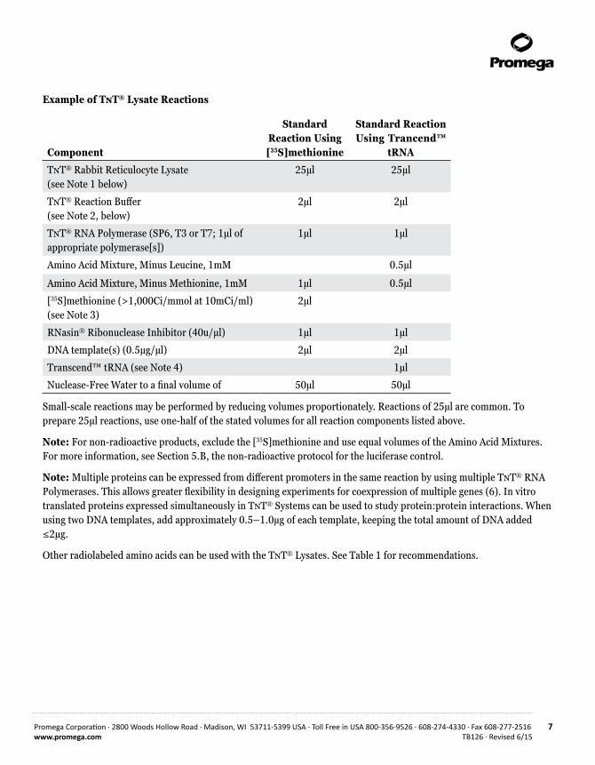
Example of TnT® Lysate Reactions
Component
Standard Reaction Using [35S]methionine
Standard Reaction Using Trancend™
tRNA
TnT® Rabbit Reticulocyte Lysate (see Note 1 below)
25µl 25µl
TnT® Reaction Buffer (see Note 2, below)
2µl 2µl
TnT® RNA Polymerase (SP6, T3 or T7; 1µl of appropriate polymerase[s])
1µl 1µl
Amino Acid Mixture, Minus Leucine, 1mM 0.5µl
Amino Acid Mixture, Minus Methionine, 1mM 1µl 0.5µl
[35S]methionine (>1,000Ci/mmol at 10mCi/ml) (see Note 3)
2µl
RNasin® Ribonuclease Inhibitor (40u/µl) 1µl 1µl
DNA template(s) (0.5µg/µl) 2µl 2µl
Transcend™ tRNA (see Note 4) 1µl
Nuclease-Free Water to a final volume of 50µl 50µl
Small-scale reactions may be performed by reducing volumes proportionately. Reactions of 25µl are common. To prepare 25µl reactions, use one-half of the stated volumes for all reaction components listed above.
Note: For non-radioactive products, exclude the [35S]methionine and use equal volumes of the Amino Acid Mixtures. For more information, see Section 5.B, the non-radioactive protocol for the luciferase control.
Note: Multiple proteins can be expressed from different promoters in the same reaction by using multiple TnT® RNA Polymerases. This allows greater flexibility in designing experiments for coexpression of multiple genes (6). In vitro translated proteins expressed simultaneously in TnT® Systems can be used to study protein:protein interactions. When using two DNA templates, add approximately 0.5–1.0µg of each template, keeping the total amount of DNA added ≤2µg.
Other radiolabeled amino acids can be used with the TnT® Lysates. See Table 1 for recommendations.

8 Promega Corporation · 2800 Woods Hollow Road · Madison, WI 53711-5399 USA · Toll Free in USA 800-356-9526 · 608-274-4330 · Fax 608-277-2516TB126 · Revised 6/15 www.promega.com
Table 1. Recommended Volumes of Alternative Radiolabeled Amino Acids.
Component Standard
[3H]leucine (100–200Ci/mmol) 5µl
[14C]leucine (300mCi/mmol) 5µl
[35S]cysteine (1,200Ci/mmol) 5µl
3. Incubate the reaction at 30°C for 90 minutes (Note 5).
4. Analyze the results of translation. Procedures are provided for incorporation assays (Section 7.A), gel analysis of translation products (Section 7.B) and an assay for luciferase production in the control reactions (Section 5). For analysis of reactions using Transcend™ tRNA, refer to the Promega Transcend™ Non-Radioactive Detection Systems Technical Bulletin #TB182.
Notes:
1. We have found that a 50% lysate concentration is optimal for most TnT® Lysate reactions. In some cases, a lysate concentration of 55% (27.5µl) will enhance translation.
For optimal protein expression using the TnT® SP6 RNA polymerase, we recommend titrating magnesium acetate in 0.1mM increments between 0.1mM and 0.5mM. In some instances the addition of 0.2mM magnesium acetate has been shown to increase protein expression by 40%. Magnesium acetate is supplied only with Cat.# L4600 and L4601.
2. The TnT® Reaction Buffer may contain a precipitate after thawing and sitting on ice. Redissolve the precipitate by vortexing at room temperature for 30 seconds.
3. We recommend using a grade of [35S]methionine, PerkinElmer EasyTag™ L-[35S]methionine (PerkinElmer Cat.# NEG709A), which does not cause the background labeling of the rabbit reticulocyte lysate 42kDa protein. Background labeling of the 42kDa protein can occur using other grades of label (7).
4. The level of added Transcend™ tRNA can be increased (1–4µl) to allow more sensitive detection of proteins that contain few lysines or are poorly expressed.
5. Using the T7 or T3 promoter, optimal coupled transcription/translation will occur in 60–90 minutes at 30°C.
6. Except for the actual translation incubation, all handling of the lysate components should be done at 4°C or on ice. Any unused lysate should be refrozen in a dry ice/ethanol bath as soon as possible after thawing to minimize loss of translational activity.
Do not freeze-thaw the lysate more than two times.
7. The lysate contains roughly 100–200mg/ml of endogenous protein.
!
!

Promega Corporation · 2800 Woods Hollow Road · Madison, WI 53711-5399 USA · Toll Free in USA 800-356-9526 · 608-274-4330 · Fax 608-277-2516 9www.promega.com TB126 · Revised 6/15
8. Avoid adding calcium to the translation reaction. Calcium may reactivate the micrococcal nuclease used to destroy endogenous RNA in the lysate and result in degradation of DNA or RNA templates.
9. The Luciferase Control reaction usually produces 50 to 500ng of protein per 50µl reaction, as deduced from luciferase activity.
Do not use more than one polymerase per control reaction.
5. Positive Control Translation Reactions Using Luciferase
The assay for firefly luciferase activity is extremely sensitive, rapid and easy to perform. It is an excellent control for in vitro translations because only full-length luciferase is active. Additionally, luciferase is a monomeric protein (approximately 61kDa) that does not require post-translational processing or modification for enzymatic activity. Promega Luciferase Assay System is a substantial improvement over conventional methods in both sensitivity and simplicity (8).
5.A. Radioactive Luciferase Control Reaction
This section provides information on how to perform a radioactive luciferase control reaction. For use of radiolabeled amino acids other than [35S]methionine, see Section 4.A, Step 2. For maps of and information on the Luciferase Control DNAs, please see Section 11.B.
1. Assemble the following reaction:
TnT® Lysate (see Note 1, Section 4.A) 25µl
TnT® Reaction Buffer (see Note 2, Section 4.A) 2µl
TnT® RNA Polymerase (SP6, T3 or T7; 1µl of appropriate polymerase)
1µl
Amino Acid Mixture, Minus Methionine, 1mM 1µl
[35S]methionine (>1,000Ci/mmol) at 10mCi/ml (see Note 3, Section 4.A)
2µl
RNasin® Ribonuclease Inhibitor, 40u/µl 1µl
Luciferase Control DNA, 0.5µg/µl 2µl
Nuclease-Free Water to a final volume of 50µl
Note: The control reaction can be performed with or without the addition of radiolabeled amino acids.
2. Incubate the reaction at 30°C for 90 minutes (see Note 5, Section 4.A).
3. Analyze the results of translation by measuring direct incorporation of radiolabel (Section 7.A) and/or gel analy-sis of translation products (Section 7.B).
4. The Luciferase Control reactions can be stored at –20°C for up to 2 months.
!

10 Promega Corporation · 2800 Woods Hollow Road · Madison, WI 53711-5399 USA · Toll Free in USA 800-356-9526 · 608-274-4330 · Fax 608-277-2516TB126 · Revised 6/15 www.promega.com
5.B. Non-Radioactive Luciferase Control Reaction
Note: Both Amino Acid Mixture Minus Leucine and Amino Acid Mixture Minus Methionine are used in this reaction. By using both incomplete mixes, a sufficient concentration of all amino acids is provided. As an alternative to assaying luciferase activity, this reaction can be performed using the Transcend™ tRNA and Non-Radioactive Detection Systems. For more information on these systems, request the Transcend™ Non-Radioactive Detection Systems Technical Bulletin #TB182.
1. Assemble the following reaction:
TnT® Lysate (see Note 1, Section 4.A) 25µl
TnT® Reaction Buffer (see Note 2, Section 4.A) 2µl
TnT® RNA Polymerase (SP6, T3 or T7; 1µl of appropriate polymerase)
1µl
Amino Acid Mixture, Minus Leucine, 1mM 0.5µl
Amino Acid Mixture, Minus Methionine, 1mM 0.5µl
RNasin® Ribonuclease Inhibitor, 40u/µl 1µl
Luciferase Control DNA, 0.5µg/µl 2µl
Nuclease-Free Water to a final volume of 50µl
2. Follow Steps 2 through 4, Section 5.A.
6. Cotranslational Processing Using Canine Pancreatic Microsomal Membranes in TnT® Lysate Systems
Microsomal vesicles are used to study cotranslational and initial post-translational processing of proteins. Processing events such as signal peptide cleavage and core glycosylation can be examined by the translation of the appropriate gene in vitro in the presence of these membranes (9). To ensure consistent performance with minimal translational inhibition and background, Promega Canine Pancreatic Microsomal Membranes (Cat.# Y4041) have been isolated free from contaminating membrane fractions and stripped of endogenous membrane-bound ribosomes and mRNA.
Materials to Be Supplied by the User• double-distilled, RNase-free water• RNasin® Ribonuclease Inhibitor• isotopically labeled amino acids, typically [35S]methionine, [35S]cysteine, [3H]leucine or [14C]leucine
6.A. General Protocol for Translation with Microsomal Membranes
1. Remove the reagents from the freezer and allow them to thaw on ice.
Promega Corporation · 2800 Woods Hollow Road · Madison, WI 53711-5399 USA · Toll Free in USA 800-356-9526 · 608-274-4330 · Fax 608-277-2516 11www.promega.com TB126 · Revised 6/15
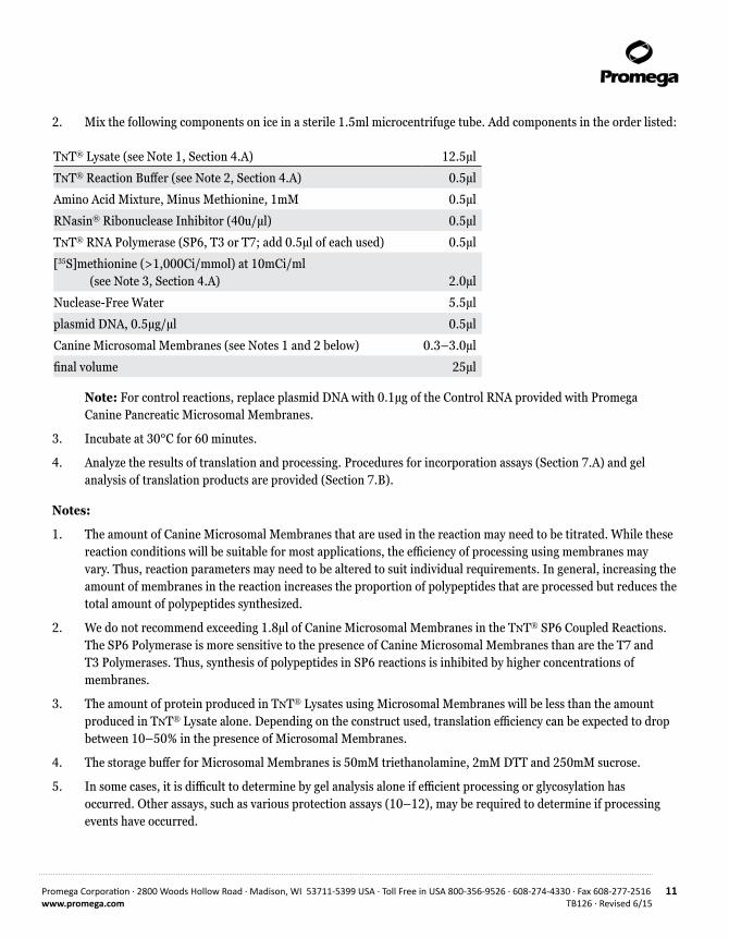
2. Mix the following components on ice in a sterile 1.5ml microcentrifuge tube. Add components in the order listed:
TnT® Lysate (see Note 1, Section 4.A) 12.5µl
TnT® Reaction Buffer (see Note 2, Section 4.A) 0.5µl
Amino Acid Mixture, Minus Methionine, 1mM 0.5µl
RNasin® Ribonuclease Inhibitor (40u/µl) 0.5µl
TnT® RNA Polymerase (SP6, T3 or T7; add 0.5µl of each used) 0.5µl
[35S]methionine (>1,000Ci/mmol) at 10mCi/ml (see Note 3, Section 4.A)
2.0µl
Nuclease-Free Water 5.5µl
plasmid DNA, 0.5µg/µl 0.5µl
Canine Microsomal Membranes (see Notes 1 and 2 below) 0.3–3.0µl
final volume 25µl
Note: For control reactions, replace plasmid DNA with 0.1µg of the Control RNA provided with Promega Canine Pancreatic Microsomal Membranes.
3. Incubate at 30°C for 60 minutes.
4. Analyze the results of translation and processing. Procedures for incorporation assays (Section 7.A) and gel analysis of translation products are provided (Section 7.B).
Notes:
1. The amount of Canine Microsomal Membranes that are used in the reaction may need to be titrated. While these reaction conditions will be suitable for most applications, the efficiency of processing using membranes may vary. Thus, reaction parameters may need to be altered to suit individual requirements. In general, increasing the amount of membranes in the reaction increases the proportion of polypeptides that are processed but reduces the total amount of polypeptides synthesized.
2. We do not recommend exceeding 1.8µl of Canine Microsomal Membranes in the TnT® SP6 Coupled Reactions. The SP6 Polymerase is more sensitive to the presence of Canine Microsomal Membranes than are the T7 and T3 Polymerases. Thus, synthesis of polypeptides in SP6 reactions is inhibited by higher concentrations of membranes.
3. The amount of protein produced in TnT® Lysates using Microsomal Membranes will be less than the amount produced in TnT® Lysate alone. Depending on the construct used, translation efficiency can be expected to drop between 10–50% in the presence of Microsomal Membranes.
4. The storage buffer for Microsomal Membranes is 50mM triethanolamine, 2mM DTT and 250mM sucrose.
5. In some cases, it is difficult to determine by gel analysis alone if efficient processing or glycosylation has occurred. Other assays, such as various protection assays (10–12), may be required to determine if processing events have occurred.

12 Promega Corporation · 2800 Woods Hollow Road · Madison, WI 53711-5399 USA · Toll Free in USA 800-356-9526 · 608-274-4330 · Fax 608-277-2516TB126 · Revised 6/15 www.promega.com
7. Post-Translational Analysis
Materials to Be Supplied by the User (Solution compositions are provided in Section 11.A.)• 1M NaOH/2% H2O2• 25% TCA/2% casamino acids (Difco® brand, Vitamin Assay Grade)• 5% TCA• Whatman® GF/C glass fiber filter (Whatman® Cat.# 1822A021)• acetone• 30% acrylamide solution• 1X SDS gel-loading buffer• separating gel 4X buffer• stacking gel 4X buffer• SDS polyacrylamide 10X running buffer• Optional: precast polyacrylamide gels• fixing solution• Whatman® 3MM filter paper
7.A. Determination of Percent Incorporation of Radioactive Label
1. After the 50µl translation reaction is completed, remove 2µl from the reaction and add it to 98µl of 1M NaOH/2% H2O2.
2. Vortex briefly and incubate at 37°C for 10 minutes.
3. At the end of the incubation, add 900µl of ice-cold 25% TCA/2% casamino acids to precipitate the translation product. Incubate on ice for 30 minutes.
4. Wet a Whatman® GF/C glass fiber filter with a small amount of cold 5% TCA. Collect the precipitated translation product by vacuum filtering 250µl of the TCA reaction mix. Rinse the filter 3 times with 1–3ml of ice-cold 5% TCA. Rinse once with 1–3ml of acetone. Allow the filter to dry at room-temperature or under a heat lamp for at least 10 minutes.
5. For determination of 35S incorporation, put the filter in 1–3ml of appropriate scintillation mixture, invert to mix and count in a liquid scintillation counter.
6. To determine total counts present in the reaction, spot a 5µl aliquot of the TCA reaction mix directly onto a filter. Dry the filter for 10 minutes. Count in a liquid scintillation counter as in Step 5. The measured counts per minute (cpm) are the “cpm of unwashed filter”.
7. To determine background counts, remove 2µl from a 50µl translation reaction containing no DNA and proceed as described in Steps 1–5.
8. Perform the following calculation to determine percent incorporation:
cpm of unwashed filter (Step 4)× 100 = percent incorporation
cpm of unwashed filter (Step 6) × 50
Promega Corporation · 2800 Woods Hollow Road · Madison, WI 53711-5399 USA · Toll Free in USA 800-356-9526 · 608-274-4330 · Fax 608-277-2516 13www.promega.com TB126 · Revised 6/15
9. Perform the following calculation to determine the amount of stimulation above background levels:
cpm of washed filter (Step 4) = fold stilmulation
cpm of “no DNA control reaction” filter (Step 7)
7.B. Denaturing Gel Analysis of Translation Products
For protein analysis, Invitrogen NOVEX® and Bio-Rad® Laboratories, Inc., offer a variety of precast mini-gels, which are compatible with their vertical electrophoresis and blotter systems. These companies offer Tris-Glycine, Tricine and Bis-Tris gels for resolution of proteins under different conditions and over a broad spectrum of protein sizes. The NOVEX® 4-20% Tris-Glycine gradient gels (NOVEX® Invitrogen Cat.# EC6025BOX or EC60355BOX) and the Bio-Rad® Ready Gel 4-20% Tris-HCl (Bio-Rad® Cat.# 161-1105EDU) are convenient for resolving proteins over a wide range of molecular weights. In addition to convenience and safety, precast gels provide consistent results.
1. Once the 50µl translation reaction is complete (or at any desired timepoint), remove a 5µl aliquot and add it to 20µl of 1X SDS gel-loading buffer. The remainder of the reaction may be stored at –20°C.
2. Cap the tube and heat at 100°C for 2 minutes to denature the proteins.
Note: In some cases, high molecular weight complexes are formed at 100°C, and denaturation may need to be performed at lower temperatures (e.g., 20 minutes at 60°C or 3–4 minutes at 80–85°C).
3. Load a small aliquot (5–10µl) of the denatured sample onto an SDS-polyacrylamide gel or store at –20°C. It is not necessary to separate labeled polypeptides from free amino acids by acetone precipitation.
4. Typically, electrophoresis is carried out at a constant current of 15mA in the stacking gel and 30mA in the separating gel (or 30mA for a gradient gel). Electrophoresis is usually performed until the bromophenol blue dye has run off the bottom of the gel. Because the dye front also contains the free labeled amino acids, disposal of unincorporated label may be easier if the gel is stopped while the dye front remains in the gel. Proceed to Step 7 for Western blotting.
5. Place the polyacrylamide gel in a plastic box and cover the gel with fixing solution (as prepared in Section 11.A). Agitate slowly on an orbital shaker for 30 minutes. Pour off the fixing solution.
Optional: Labeled protein bands in gels may be visualized by autoradiography or fluorography. Fluorography dramatically increases the sensitivity of detection of 35S-, 14C- and 3H-labeled proteins and is recommended for the analysis of in vitro translation products. The increased detection sensitivity of fluorography is obtained by infusing an organic scintillant into the gel. The scintillant converts the emitted energy of the isotope to visible light and increases the proportion of energy that may be detected by X-ray film. Commercial reagents, such as Amplify® Reagent (GE Healthcare Bio-sciences), can be used for fluorographic enhancement of signal. Alternatively, the fixed gel can be exposed to a phosphorimaging screen. These systems provide greater sensitivity, greater speed and the ability to quantitate the radioactive bands.
14 Promega Corporation · 2800 Woods Hollow Road · Madison, WI 53711-5399 USA · Toll Free in USA 800-356-9526 · 608-274-4330 · Fax 608-277-2516TB126 · Revised 6/15 www.promega.com
7.B. Denaturing Gel Analysis of Translation Products (continued)
6. Dry the gel for exposure to film as follows: Soak the gel in 7% acetic acid, 7% methanol, 1% glycerol for 5 minutes to prevent the gel from cracking during drying. Place the gel on a sheet of Whatman® 3MM filter paper, cover with plastic wrap and dry at 80°C for 30–90 minutes under a vacuum using a conventional gel dryer; dry completely. The gel also may be dried overnight using the Promega Gel Drying Kit (Cat.# V7120). To decrease the likelihood of cracking gradient gels, dry them with the wells pointing down. Expose the gel on Kodak® X-ray film for 1–6 hours at –70°C (with fluorography) or for 6–15 hours at room-temperature (with autoradiography).
7. For Western blot analysis of proteins, transfer (immobilize) the protein from the gel onto nitrocellulose or PVDF membrane (14,15). Usually Western blots are made by electrophoretic transfer of proteins from SDS-polyacrylamide gels. Detailed procedures for electrophoretic blotting usually are included with |commercial devices and can be found in references 14 and 16–18. A general discussion of Western blotting with PVDF membranes is found in reference 19. PVDF membranes must be prewet in methanol or ethanol before equilibrating in transfer buffer. The blot then may be analyzed by immunodetection.
8. Positive Control Luciferase Assays
Light intensity is a measure of the rate of catalysis by luciferase and is therefore dependent upon temperature. The optimum temperature for luciferase activity is approximately room temperature (20–25°C). It is important that the Luciferase Assay Reagent be fully equilibrated to room temperature before beginning measurements. To ensure temperature equilibration, place a thawed aliquot of the Luciferase Assay Reagent in a sealed tube into a water bath maintained at ambient temperature, and equilibrate for at least 30 minutes. The sample to be assayed should also be at ambient temperature.
Either a luminometer or a scintillation counter can be used for quantitation. (There is usually insufficient light output for qualitative visual detection.) A luminometer can measure as little as 10–20 moles (0.001pg) of luciferase, whereas a scintillation counter typically has a less sensitive detection limit. However, the limits of sensitivity may vary depending upon the particular instrument used. The assay should be linear in some portion of the detection range of the instrument. Please consult your instrument operator’s manual for general operating instructions.
Promega Corporation · 2800 Woods Hollow Road · Madison, WI 53711-5399 USA · Toll Free in USA 800-356-9526 · 608-274-4330 · Fax 608-277-2516 15www.promega.com TB126 · Revised 6/15
8.A. Using a Luminometer
The Luciferase Assay Reagent and samples should be at ambient temperature prior to performing a luciferase assay.
1. Dispense 50µl of the Luciferase Assay Reagent into luminometer tubes, one tube per sample.
2. Program the luminometer to perform a 2-second measurement delay followed by a 10-second measurement read for luciferase activity. The read time may be shortened if sufficient light is produced.
3. Add 2.5µl of cell lysate to a luminometer tube containing the Luciferase Assay Reagent. Mix by pipetting 2–3 times or vortex briefly.
4. Place the tube in the luminometer and initiate reading.
5. If the luminometer is not connected to a printer or computer, record the reading.
8.B. Using a Scintillation Counter
Ideally, the coincidence circuit of the scintillation counter should be turned off. Usually, this is achieved through an option of the programming menu or by a switch within the instrument. If the circuit cannot be turned off, a linear relationship between luciferase concentration and cpm still can be produced by calculating the square root of measured counts per minute (cpm) minus background cpm (i.e., [sample – background]1/2). To measure background cpm, use water or Luciferase Assay Reagent as a blank.
Use the same protocol as luciferase assays using a luminometer (Section 8.A). The sample may be placed directly in the scintillation vial if it completely covers the bottom of the vial (clear or translucent vials are acceptable).
Do not add scintillant, because it will inactivate luciferase. Alternatively, place the sample in a microcentrifuge tube, and then place the tube in the scintillation vial. To ensure consistency when working with multiple samples, place each microcentrifuge tube at the same relative position within the scintillation vial.
For consistency in measuring luciferase activity, use the scintillation counter in manual mode. Initiate each sample reaction immediately before measurement, and read the samples one at a time. Because the enzymatic reaction produces light at all wavelengths, read the samples with all channels open (open window). To reduce background counts, it may be necessary to wait 10–30 seconds before counting. Read individual samples for 1–5 minutes.
8.C. Photographic Luciferase Assay
1. Prepare a Polaroid® camera for an 8-minute exposure. Either a hand-held (IBI Quickshooter model QSP) or overhead positioned Polaroid® camera is acceptable. Position the camera over the provided Luciferase Assay Wells and focus on the top rim of the well. Open the aperture as wide as possible (e.g., f4.5), and set the shutter speed to the bulb, or B, setting. Make sure the camera is loaded with Polaroid® 667 (ISO 3,000) or 612 film (ISO 20,000).
Note: We have observed that the 667 film “negative” (not a true negative though, as in 665 type film) is more sensitive in recording the luminescence-produced image than the 612 high speed positive film. The “negative” can be dried and saved or, alternatively, a picture can be taken of the “negative” for a permanent record.
!
16 Promega Corporation · 2800 Woods Hollow Road · Madison, WI 53711-5399 USA · Toll Free in USA 800-356-9526 · 608-274-4330 · Fax 608-277-2516TB126 · Revised 6/15 www.promega.com
2. Add 50µl of room-temperature Luciferase Assay Reagent (see Section 8.A, Step 1) to one of the white wells of the provided Luciferase Assay Wells.
3. Add 5µl of the 50µl luciferase control translation reaction (either radioactive or non-radioactive) and mix quickly by pipetting.
4. Immediately turn off all lights (including red darkroom lights) and set the camera for an 8–10 minute exposure (for 667 film). For 612 film, set the exposure time to approximately 4–5 minutes.
5. The photographic assay is sensitive in the 2–50ng luciferase range using these conditions.
8.D. Qualitative Visual Detection of Luciferase Activity
For qualitative determination of luciferase activity, the reactions may be visualized by eye in a dark room after acclimation to the dark. Most individuals should be able to see the reaction after a minute or two of acclimation, although individuals may differ in their ability to detect these low light levels.
Promega Corporation · 2800 Woods Hollow Road · Madison, WI 53711-5399 USA · Toll Free in USA 800-356-9526 · 608-274-4330 · Fax 608-277-2516 17www.promega.com TB126 · Revised 6/15
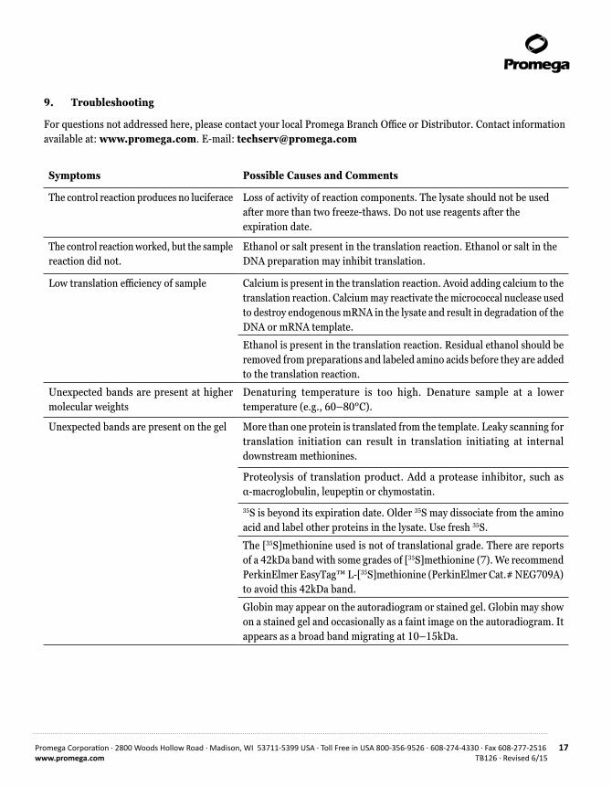
9. Troubleshooting
For questions not addressed here, please contact your local Promega Branch Office or Distributor. Contact information available at: www.promega.com. E-mail: [email protected]
Symptoms Possible Causes and Comments
The control reaction produces no luciferace Loss of activity of reaction components. The lysate should not be used after more than two freeze-thaws. Do not use reagents after the expiration date.
The control reaction worked, but the sample reaction did not.
Ethanol or salt present in the translation reaction. Ethanol or salt in the DNA preparation may inhibit translation.
Low translation efficiency of sample Calcium is present in the translation reaction. Avoid adding calcium to the translation reaction. Calcium may reactivate the micrococcal nuclease used to destroy endogenous mRNA in the lysate and result in degradation of the DNA or mRNA template.
Ethanol is present in the translation reaction. Residual ethanol should be removed from preparations and labeled amino acids before they are added to the translation reaction.
Unexpected bands are present at higher molecular weights
Denaturing temperature is too high. Denature sample at a lower temperature (e.g., 60–80°C).
Unexpected bands are present on the gel More than one protein is translated from the template. Leaky scanning for translation initiation can result in translation initiating at internal downstream methionines.
Proteolysis of translation product. Add a protease inhibitor, such as α-macroglobulin, leupeptin or chymostatin.
35S is beyond its expiration date. Older 35S may dissociate from the amino acid and label other proteins in the lysate. Use fresh 35S.
The [35S]methionine used is not of translational grade. There are reports of a 42kDa band with some grades of [35S]methionine (7). We recommend PerkinElmer EasyTag™ L-[35S]methionine (PerkinElmer Cat.# NEG709A) to avoid this 42kDa band.
Globin may appear on the autoradiogram or stained gel. Globin may show on a stained gel and occasionally as a faint image on the autoradiogram. It appears as a broad band migrating at 10–15kDa.
18 Promega Corporation · 2800 Woods Hollow Road · Madison, WI 53711-5399 USA · Toll Free in USA 800-356-9526 · 608-274-4330 · Fax 608-277-2516TB126 · Revised 6/15 www.promega.com
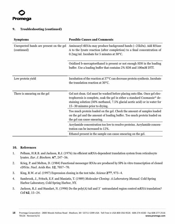
9. Troubleshooting (continued)
Symptoms Possible Causes and Comments
Unexpected bands are present on the gel (continued)
Aminoacyl tRNAs may produce background bands (~25kDa). Add RNase A to the lysate reaction (after completion) to a final concentration of 0.2mg/ml. Incubate for 5 minutes at 30°C.
Oxidized b-mercaptoethanol is present or not enough SDS in the loading buffer. Use a loading buffer that contains 2% SDS and 100mM DTT.
Low protein yield Incubation of the reaction at 37°C can decrease protein synthesis. Incubate the translation reaction at 30°C.
There is smearing on the gel Gel not clean. Gel must be washed before placing onto film. Once gel elec-trophoresis is complete, soak the gel in either a standard Coomassie® de-staining solution (50% methanol, 7.5% glacial acetic acid) or in water for 15–30 minutes prior to drying.
Too much protein loaded on the gel. Check the amount of samples loaded on the gel and the amount of loading buffer. Too much protein loaded on the gel can cause smearing.
Acrylamide concentration too low to resolve proteins. Acrylamide concen-tration can be increased to 12%.
Ethanol present in the sample can cause smearing on the gel.
10. References
1. Pelham, H.R.B. and Jackson, R.J. (1976) An efficient mRNA-dependent translation system from reticulocyte lysates. Eur. J. Biochem. 67, 247–56.
2. Krieg, P. and Melton, D. (1984) Functional messenger RNAs are produced by SP6 in vitro transcription of cloned cDNAs. Nucl. Acids Res. 12, 7057–70.
3. King, R.W. et al. (1997) Expression cloning in the test tube. Science 277, 973–4.
4. Sambrook, J., Fritsch, E.F. and Maniatis, T. (1989) Molecular Cloning: A Laboratory Manual. Cold Spring Harbor Laboratory, Cold Spring Harbor, NY.
5. Jackson, R.J. and Standart, N. (1990) Do the poly(A) tail and 3´ untranslated region control mRNA translation? Cell 62, 15–24.

Promega Corporation · 2800 Woods Hollow Road · Madison, WI 53711-5399 USA · Toll Free in USA 800-356-9526 · 608-274-4330 · Fax 608-277-2516 19www.promega.com TB126 · Revised 6/15
6. DiDonato, J.A. and Karin, M. (1993) Co-expression of multiple NF-ĸB subunits using the TnT® System. Pro-mega Notes 42, 18–22.
7. Jackson, R.J. and Hunt, T. (1983) Preparation and use of nuclease-treated rabbit reticulocyte lysates for the translation of eukaryotic messenger RNA. Meth. Enzymol. 96, 50–74.
8. Wood, K.V. (1991) Recent advances and prospects for use of beetle luciferases as genetic reporters. In: Biolu-minescence and Chemiluminescence: Current Status, Stanley, P.E. and Kricka, J., eds., John Wiley and Sons, Chichester.
9. Walter, P. and Blobel, G. (1983) Preparation of microsomal membranes for cotranslational protein translocation. Meth. Enzymol. 96, 84–93.
10. Schmidt-Rose, T. and Jentsch, T.J. (1997) Transmembrane topology of a CLC chloride channel. Proc. Natl. Acad. Sci. USA 94, 7633–8.
11. Hackman, A.S. et al. (1997) The N-terminal domain of human GABA Receptor r1 subunits contains signals for homooligomeric and heterooligomeric interaction. J. Biol. Chem. 272, 13750–7.
12. Alperin, E.S. and Shapiro, L.J. (1997) Characterizatioin of point mutations in patients with X-linked ichthyosis: Effects on the structure and function of the steroid sulfatase protein. J. Biol. Chem. 272, 20758–63.
13. Protocols and Applications Guide, Third Edition (1996) Promega Corporation.
14. Towbin, H., Staehelin, T. and Gordon, J. (1979) Electrophoretic transfer of proteins from polyacrylamide gels to nitrocellulose sheets: procedure and some applications. Proc. Natl. Acad. Sci. USA 76, 4350–4.
15. Burnette, W.N. (1981) “Western blotting”: electrophoretic transfer of proteins from sodium dodecyl sulfate—polyacrylamide gels to unmodified nitrocellulose and radiographic detection with antibody and radioiodinated protein A. Anal. Biochem. 112, 195–201.
16. Bittner, M., Kupferer, P. and Morris, C.F. (1980) Electrophoretic transfer of proteins and nucleic acids from slab gels to diazobenzyloxymethyl cellulose or nitrocellulose sheets. Anal. Biochem. 102, 459–71.
17. Towbin, H. and Gordon, J. (1984) Immunoblotting and dot immunobinding—current status and outlook. J. Immunol. Meth. 72, 313–40.
18. Bers, G. and Garfin, D. (1985) Protein and nucleic acid blotting and immunological detection. BioTechniques 3, 276–86.
19. Hicks, D. et al. (1986) Immobilon™ PVDF Transfer Membrane: A new membrane substrate for Western blotting of proteins. BioTechniques 4, 272–282.
20. Kozak, M. (1986) Point mutations define a sequence flanking the AUG initiator codon that modulates translation by eukaryotic ribosomes. Cell 44, 283–92.

20 Promega Corporation · 2800 Woods Hollow Road · Madison, WI 53711-5399 USA · Toll Free in USA 800-356-9526 · 608-274-4330 · Fax 608-277-2516TB126 · Revised 6/15 www.promega.com
11. Appendix
11.A. Composition of Buffers and Solutions
11.B. Luciferase SP6/T7/T3 Control DNAs
The Luciferase SP6/T7/T3 Control DNAs are used as functional controls in the TnT® Coupled Transcription/Transltion Systems. The Control DNAs contain the gene for luciferase under transcriptional control of a phage RNA polymerase promoter. All constructs carry a 30-base-pair poly[d(A)/d(T)] tail following the luciferase gene. The maps of the Luciferase SP6 Control DNA, the Luciferase T7 Control DNA and the Luciferase T3 Control DNA are shown in Figure 2, Figure 3 and Figure 4, respectively.
acrylamide solution, 30% 30g acrylamide 0.8g bisacrylamide
Add water to a final volume of 100ml. Store at 4°C.
fixing solution 50% methanol 10% glacial acetic acid 40% water
1X SDS gel-loading buffer 50mM Tris-HCl (pH 6.8) 2% SDS 0.1% bromophenol blue 10% glycerol 100mM dithiothreitol
1X SDS gel-loading buffer lacking dithiothretol can be stored at room temperature. Dithioth-reitol should be added from a 1M stock just before the buffer is used.
SDS polyacrylamide running 10X buffer 30g Tris base 144g glycine 100ml 10% SDS
Add water to a final volume of 1L.
separating gel 4X buffer 18.17g Tris base 4ml 10% SDS
Bring the volume to approximately 80ml with deionized water. Adjust to pH 8.8 with 12N HCl and add deionized water to a final volume of 100ml. Store at room temperature.
stacking gel 4X buffer 6.06g Tris-base 4ml 10% SDS
Bring the volume to approximately 80ml with deionized water. Adjust to pH 6.8 with 12N HCl and add deionized water to a final volume of 100ml. Store at room temperature.

Promega Corporation · 2800 Woods Hollow Road · Madison, WI 53711-5399 USA · Toll Free in USA 800-356-9526 · 608-274-4330 · Fax 608-277-2516 21www.promega.com TB126 · Revised 6/15
ori
Luciferase SP6Control DNA(4747bp)Ampr
lucXmnI
(3651)
SP6 Initiation (1)SP6 Promoter
HindIII (8)NotI (21)BamHI (41)
SacI(1764)
XmnI(1804)
(dA:dT)30
1917
VA
04_6
A
Figure 2. Luciferase SP6 Control DNA circle map and sequence reference points. Additional description: Ampr, β-lactamase gene (resistant to ampicillin); ori, origin of plasmid replication.
Sequence reference points:
SP6 RNA polymerase initiation 1
GLprimer2 49–71
Luciferase gene 48–1697
Poly(A) (dA)30 1767–1796
pUC/M13 reverse primer (17mer) 1833–1817
pUC/M13 reverse primer (22mer) 1838–1817
β-lactamase gene (Ampr) 3838–2975
SP6 RNA polymerase promoter primer 4731–1
SP6 RNA polymerase promoter 4731–3
Note: There is a single base mismatch at the 5´ end of the SP6 RNA polymerase promoter primer.

22 Promega Corporation · 2800 Woods Hollow Road · Madison, WI 53711-5399 USA · Toll Free in USA 800-356-9526 · 608-274-4330 · Fax 608-277-2516TB126 · Revised 6/15 www.promega.com
ori
Luciferase T7 Control DNA(4331bp)
Ampr
XmnI(2632)
luc
(dA:dT)30
SacI (1767)
T7 Initiation (1)T7 Promoter
BamHI (44)NotI (22)HindIII (11)
1916
VA
04_6
A
Figure 3. Luciferase T7 Control DNA circle map and sequence reference points. Additional description: Ampr, β-lactamase gene (resistant to ampicillin); ori, origin of plasmid replication.
Sequence reference points:
T7 RNA polymerase initiation 1
GLprimer2 52–74
Luciferase gene 51–1700
Poly(A) (dA)30 1770–1799
β-lactamase gene (Ampr) 2444–3301
T7 RNA polymerase promoter 4315–3
T7 RNA polymerase promoter primer 4315–3

Promega Corporation · 2800 Woods Hollow Road · Madison, WI 53711-5399 USA · Toll Free in USA 800-356-9526 · 608-274-4330 · Fax 608-277-2516 23www.promega.com TB126 · Revised 6/15
fl ori
Luciferase T3Control DNA(4646bp)
Ampr
luc
XmnI(3688)
T3 Initiation (1) HindIII (8)NotI (21)BamHI (41)
SacI (1764)KpnI (1810)SacI (1816)
(dA:dT)30
T3 Promoter
pBR ori
1918
VA
04_6
A
Figure 4. Luciferase T3 Control DNA circle map and sequence reference points. Additional description: Ampr, β-lactamase gene (resistant to ampicillin); f1 ori, origin of replication; pBR ori, origin of plasmid replication.
Sequence reference points:
T3 RNA polymerase initiation 1
GLprimer2 49–71
Luciferase gene 48–1697
Poly(A) (dA)30 1767–1796
β-lactamase gene (Ampr) 3875–3012
T7 RNA polymerase promoter (–17 to +2) 1840–1822
pUC/M13 reverse primer (17mer) 1870–1854
pUC/M13 reverse primer (22mer) 1875–1854
f1 origin 4006–4461
pUC/M13 forward primer (24mer) 4576–4599
pUC/M13 forward primer (17mer) 4583–4599
T3 RNA polymerase promoter 4631–4
T3 RNA polymerase promoter primer 4631–4
Note: The T7 sequencing primer has a 3´ mismatch and will not bind.

24 Promega Corporation · 2800 Woods Hollow Road · Madison, WI 53711-5399 USA · Toll Free in USA 800-356-9526 · 608-274-4330 · Fax 608-277-2516TB126 · Revised 6/15 www.promega.com
11.C. Related Products
The in vitro synthesis of proteins is a popular method in biological research. Among other applications, translation systems are used to characterize plasmid clones, study structural mutations and examine translational signals.
Two basic approaches to in vitro protein synthesis are available: 1) in vitro systems programmed with RNA (translation systems), or 2) those programmed with DNA (coupled transcription/translation systems). Several general considerations to assist you in selecting the appropriate Promega product(s) are given below.
Translation Systems
A number of cell-free protein synthesizing systems have been developed for the translation of mRNA isolated from tissue or generated in vitro. Promega offers several Rabbit Reticulocyte Lysate and Wheat Germ Extract Systems. All are reliable, convenient and easy-to-use systems to initiate translation and produce full-size polypeptide products. Rabbit Reticulocyte Lysate is appropriate for the translation of larger mRNA species, and is generally recommended when microsomal membranes are to be added for cotranslational processing of translation products. The Flexi® Rabbit Reticulocyte Lysate System is recommended where optimization of translation of particular RNAs through adjustments to salt and DTT concentrations is required. Wheat Germ Extract readily translates a variety of RNA preparations, including those containing low concentrations of double-stranded RNA (dsRNA) or oxidized thiols, which are inhibitory to reticulocyte lysate.
Coupled Transcription/Translation Systems
DNA sequences cloned in plasmid vectors also may be expressed directly using either the TnT® Coupled Wheat Germ Extract Systems or E. coli S30 Coupled Transcription/Translation Systems. The TnT® Systems are used to direct eukaryotic translation, whereas the S30 Systems are under prokaryotic translational controls. The TnT® Systems require plasmid constructs containing a prokaryotic phage RNA polymerase promoter (SP6, T3 or T7) for the initiation of transcription, but translation in this system is under eukaryotic controls. Optimal translation will occur if the AUG initiation codon is in a “Kozak consensus” context (A/GCCAUGG) (20) in the absence of inhibiting secondary structure. The template DNA to be expressed in the S30 Systems must contain E. coli promoter sequences and prokaryotic ribosome binding sites (GGAGG) for translation. The TnT® and E. coli S30 Systems can use either circular or linear DNA templates.

Promega Corporation · 2800 Woods Hollow Road · Madison, WI 53711-5399 USA · Toll Free in USA 800-356-9526 · 608-274-4330 · Fax 608-277-2516 25www.promega.com TB126 · Revised 6/15
Vectors
Product Size Cat.#pTnT™ Vector 20µg L5610
pCMVTnT™ Vector 20µg L5620
TnT® Quick Coupled Transcription/Translation Systems
Product Size Cat.#TnT® T7 Quick Coupled Transcription/Translation System 40 × 50µl reactions L1170
TnT® T7 Quick Coupled Transcription/Translation System Trial Size 5 × 50µl reactions L1171
TnT® SP6 Quick Coupled Transcription/Translation System 40 × 50µl reactions L2080
TnT® SP6 Quick Coupled Transcription/Translation System Trial Size 5 × 50µl reactions L2081
TnT® Coupled Wheat Germ Extract Systems
Product Size Cat.#TnT® T3 Coupled Wheat Germ Extract System 40 × 50µl reactions L4120
TnT® SP6 Coupled Wheat Germ Extract System 40 × 50µl reactions L4130
TnT® T7 Coupled Wheat Germ Extract System 40 × 50µl reactions L4140
TnT® T7/SP6 Coupled Wheat Germ Extract System 40 × 50µl reactions L5030
TnT® T7/T3 Coupled Wheat Germ Extract System 40 × 50µl reactions L5040
Rabbit Reticulocyte Lysate Systems
Product Size Cat.#Rabbit Reticulocyte Lysate, Nuclease Treated 5 × 200µl L4960
Rabbit Reticulocyte Lysate, Untreated 1ml L4151
Bulk Rabbit Reticulocyte Lysate is available from Promega.

26 Promega Corporation · 2800 Woods Hollow Road · Madison, WI 53711-5399 USA · Toll Free in USA 800-356-9526 · 608-274-4330 · Fax 608-277-2516TB126 · Revised 6/15 www.promega.com
11.C. Related Products (continued)
Flexi® Rabbit Reticulocyte Lysate System
Product Size Cat.#Flexi® Rabbit Reticulocyte Lysate System 5 × 200µl L4540
Bulk Flexi® Rabbit Reticulocyte Lysate is available from Promega.
Wheat Germ Extract
Product Size Cat.#Wheat Germ Extract 5 × 200µl L4380
Rabbit Reticulocyte Lysate/Wheat Germ Extract Combination Systems
Product Size Cat.#Rabbit Reticulocyte Lysate/Wheat Germ Extract Combination System 24 reactions L4330
E. coli S30 Extracts
Product Size Cat.#E. coli S30 Extract System for Circular DNA 30 × 50µl reactions L1020
E. coli S30 Extract System for Linear Templates 30 × 50µl reactions L1030
E. coli T7 S30 Extract System for Circular DNA 30 × 50µl reactions L1130
Amino Acid Mixtures
Product Size Cat.#Amino Acid Mixture Minus Leucine 175µl L9951
Amino Acid Mixture Minus Methionine 175µl L9961
Amino Acid Mixture Minus Cysteine 175µl L4471
Amino Acid Mixture, Complete 175µl L4461
Amino Acid Mixture Minus Methionine and Cysteine 175µl L5511
Luciferase Assay Systems and Control DNA
Product Size Cat.#Luciferase Assay System 100 assays E1500
Luciferase SP6 Control DNA 20µg L4741
Luciferase T7 Control DNA 20µg L4821
pGEM®-luc Vector 20µg E1541
Promega Corporation · 2800 Woods Hollow Road · Madison, WI 53711-5399 USA · Toll Free in USA 800-356-9526 · 608-274-4330 · Fax 608-277-2516 27www.promega.com TB126 · Revised 6/15
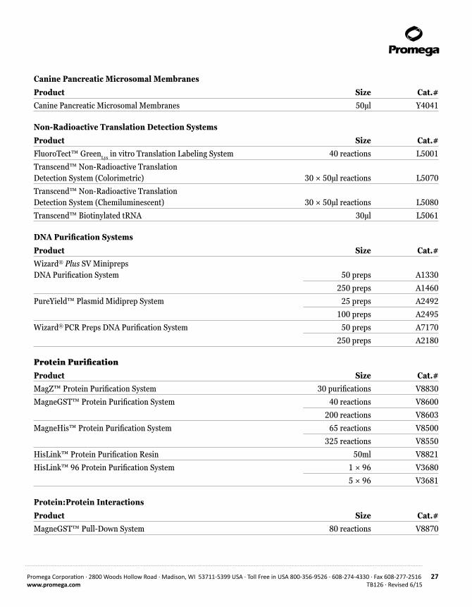
Canine Pancreatic Microsomal Membranes
Product Size Cat.#Canine Pancreatic Microsomal Membranes 50µl Y4041
Non-Radioactive Translation Detection Systems
Product Size Cat.#FluoroTect™ GreenLys in vitro Translation Labeling System 40 reactions L5001
Transcend™ Non-Radioactive Translation Detection System (Colorimetric) 30 × 50µl reactions L5070
Transcend™ Non-Radioactive Translation Detection System (Chemiluminescent) 30 × 50µl reactions L5080
Transcend™ Biotinylated tRNA 30µl L5061
DNA Purification Systems
Product Size Cat.#Wizard® Plus SV Minipreps DNA Purification System 50 preps A1330
250 preps A1460
PureYield™ Plasmid Midiprep System 25 preps A2492
100 preps A2495
Wizard® PCR Preps DNA Purification System 50 preps A7170
250 preps A2180
Protein Purification
Product Size Cat.#MagZ™ Protein Purification System 30 purifications V8830
MagneGST™ Protein Purification System 40 reactions V8600
200 reactions V8603
MagneHis™ Protein Purification System 65 reactions V8500
325 reactions V8550
HisLink™ Protein Purification Resin 50ml V8821
HisLink™ 96 Protein Purification System 1 × 96 V3680
5 × 96 V3681
Protein:Protein Interactions
Product Size Cat.#MagneGST™ Pull-Down System 80 reactions V8870
28 Promega Corporation · 2800 Woods Hollow Road · Madison, WI 53711-5399 USA · Toll Free in USA 800-356-9526 · 608-274-4330 · Fax 608-277-2516TB126 · Revised 6/15 www.promega.com
12. Summary of Changes
The following changes were made to the 6/15 revision of this document:
1. The patent information was updated to remove expired statements.
2. The document design was updated.
(a)U.S. Pat. Nos. 5,641,641 and 5,650,289.
© 1998-2015 Promega Corporation. All Rights Reserved.
Flexi, pGEM, RNasin, TnT and Wizard are registered trademarks of Promega Corporation. FluoroTect, HisLink, MagneGST, MagneHis, MagZ, pCMVTnT, pTnT, PureYield and Transcend are trademarks of Promega Corporation.
Amplify is a registered trademark of GE Healthcare Bio-sciences. Bio-Rad is a registered trademark of Bio-Rad Laboratories, Inc. Coomassie is a registered trademark of Imperial Chemical Industries, Ltd. Difco is a registered trademark of Difco Laboratories. EasyTag is a trademark of PerkinElmer. Immobilon is a trademark of Millipore Corporation. Kodak is a registered trademark of Eastman Kodak Co. Novex is a registered trademark of Novel Experimental Technology. Polaroid is a registered trademark of Polaroid Corporation. Whatman is a registered trademark of Whatman Paper Company, Ltd.
Products may be covered by pending or issued patents or may have certain limitations. Please visit our Web site for more information.
All prices and specifications are subject to change without prior notice.
Product claims are subject to change. Please contact Promega Technical Services or access the Promega online catalog for the most up-to-date information on Promega products.