export of honeybee prepromelittin in escherichia coli ... · tin. import of honeybee...
TRANSCRIPT
THE JOURNAL OF BIOLOGICAL CHEMISTRY 0 1989 by The American Society for Biochemistry and Molecular Biology, Inc.
Vol . 264, No. 17, Issue of June 15, pp. 10169-10176 1989 Printed in d. S. A.
Export of Honeybee Prepromelittin in Escherichia coli Depends on the Membrane Potential but Does Not Depend on Proteins secA and secY*
(Received for publication, July 11, 1988, and in revised form, February 7, 1989)
Werner W. E. Cobet, Christa MollayS, Gunter Muller, and Richard Zimmermannp From the Znstitut fur Physiologische, Chemie der Universitat Miinchen, Munchen, Federal Republic of Germany and the 4Znstitut fur Molekulurbiologie der Osterreichischen Akademie der Wissenschaften, Salzburg, Austria
Honeybee prepromelittin (70 amino acid residues), the precursor of an eukaryotic secretory protein, and a hybrid protein between prepromelittin and mouse dihydrofolate reductase (257 amino acid residues) were expressed in Escherichia coli and characterized with respect to their requirements for transport across the plasma membrane. Both precursor proteins are posttranslationally processed and exported into the periplasm, and they both depend on the membrane potential for this to occur. With respect to dependence on components of the export machinery, however, the two precursor proteins show striking differences: the small precursor protein prepromelittin does not re- quire the function of proteins secA and secY; the large precursor protein prepromelittin-dihydrofolate reduc- tase, on the other hand, depends on both components. The implications of these observations with respect to the mechanisms of protein export in E. coli and of protein import into the endoplasmic reticulum are dis- cussed.
Recent results regarding the mechanisms of transport of proteins across bacterial plasma membranes and membranes of the endoplasmic reticulum have emphasized homologies between the two systems (Wickner and Lodish, 1985; Zim- mermann and Meyer, 1986). (i) In general, there seems to be a need for a signal or leader peptide on the respective precur- sor protein and some type of receptor on the cis side of the target membrane as well as some type of endopeptidase (signal or leader peptidase) on the trans side of the target membrane. The signals for protein export in Escherichia coli and protein import into microsomes are quite similar when compared to each other (von Heijne, 1984). Furthermore, the processing enzymes of the bacterial plasma membrane and of the micro- somal membrane accept the same precursor proteins as sub- strates and process them correctly (Talmadge et al., 1980a; Watts et al., 1983). On the other hand, protein export in E. coli (Date et al., 1980a, 1980b; Enequist et al., 1981; Daniels et al., 1981; Zimmermann et al., 1982) depends on a membrane potential, but protein import into microsomes does not show a membrane potential effect, at least in uitro (Rothblatt and
* This work was supported by Grant B10 from the Sonderforschun- gobereich 184: Molekulare Grundlagen der Biogenese von Zellorga- nellen, by the Fonds der Chemischen Industrie, and by Grants 4555 and S29T4 from the Osterreichischer Fonds zur Forderung der wis- senscbaftlichen Forschung. The costs of publication of this article were defrayed in part by the payment of page charges. This article must therefore be hereby marked ‘‘advertisement” in accordance with 18 U.S.C. Section 1734 solely to indicate this fact.
8 To whom correspondence should be addressed Institut fur Physiologische Chemie, Goethestrasse 33, D-8000 Miinchen 2, Fed- eral Republic of Germany. Tel.: 89-5996265.
Meyer, 1986; Hansen et al., 1986; Waters and Blobel, 1986; Schlenstedt and Zimmermann, 1987). (ii) Apparently, there is no mechanistic coupling between translation and transport, but the folding of the mature part within the precursor protein into a stable tertiary structure has to be prevented or reversed in order to allow transport (Randall and Hardy, 1986; Muller and Zimmermann, 1988). In the case of import of large pre- cursor proteins into microsomes, this may be accomplished by the cooperation of SRP’ and ribosome and thus results in an apparent coupling of translation and transport (Mueckler and Lodish, 1986a, 1986b; Perara et al., 1986; Garcia and Walter, 1988). Of more ubiquitous importance in this respect may be the action of an ATP-dependent system which has been described for protein export in E. coli (Chen and Tai, 1985; Geller et al., 1986; Crooke and Wickner, 1987) as well as for protein import into microsomes (Hansen et al., 1986; Rothblatt and Meyer, 1986; Rothblatt et al., 1987; Waters and Blobel, 1986; Waters et al., 1986; Schlenstedt and Zimmer- mann, 1987; Wiech et al., 1987; Muller and Zimmermann, 1988).
We are investigating these comparable mechanisms by means of precursor proteins related to honeybee prepromelit- tin. Import of honeybee prepromelittin, synthesized in a rab- bit reticulocyte lysate, into dog pancreas microsomes is effi- cient under posttranslational conditions and does not involve the ribosome/ribosome receptor and signal recognition parti- cle (SRP)/docking protein systems (Zimmermann and Mol- lay, 1986). Related precursor proteins having a content of more than approximately 80 amino acids, like a hybrid protein between prepromelittin and mouse dihydrofolate reductase (prepromelittin-DHFR), however, behave like typical secre- tory proteins; they depend on the ribonucleoparticles and their receptors and are imported efficiently only under co- translational conditions (Muller and Zimmermann, 1987, 1988a, 1988b).
Here we report on the characteristics of the two types of precursor proteins (prepromelittin and prepromelittin- DHFR) for their export in E. coli. Both precursor proteins are posttranslationally processed and exported into the peri- plasm. In both cases these events depend on the membrane potential. With respect to dependence on proteins secA and secY, two components of the bacterial protein export machin- ery (Benson et al., 1985; Oliver, 1985; Michaelis and Beckwith, 1982), the two precursor proteins behave in a strikingly dif- ferent manner. The data presented here allow a correlation to be made between protein export in E. coli and protein
‘ The abbreviations used are: SRP, signal recognition particle; DHFR, dihydrofolate reductase; IPTG, isopropyl thiogalactoside; CCCP, carbonyl cyanide p-chlorophenylhydrazone; Hepes, 4-(2-hy- droxyethy1)-l-piperazineethanesulfonic acid; SDS, sodium dodecyl sulfate.
10169
at UB
M B
ibliothek Grosshadern on D
ecember 18, 2008
ww
w.jbc.org
Dow
nloaded from
10170 Protein Export in E. coli
import into the endoplasmic reticulum with respect to the role of components involved in the different systems.
EXPERIMENTAL PROCEDURES
Materials-Isopropyl thiogalactoside (IPTG), proteinase K, and enzymes used for cloning were obtained from Boehringer Mannheim. Plasmid pKK223-3 was from Pharmacia LKB Biotechnology Inc. [3H]Proline (100 Ci/mmol) was purchased from Amersham Corp.; x- ray films were from Kodak (Kodak X-Omat AR). Carbonyl cyanide chlorophenylhydrazone (CCCP) was obtained from Sigma; phenyl- methylsulfonyl fluoride and all other chemicals were from Merck.
Bacterial Strains and Growth Conditions-E. coli strains HJM 114 (K 12), IQ 292 (secY ts), and MM 52 (secA ts) were obtained from Drs. W. Wickner, K. Ito, and J. Beckwith, respectively. Cells were grown in M9 minimal medium (Miller, 1972), supplemented with thiamin (1 pg/ml), glucose (0.2%), and ampicillin (25 pg/ml) at 37 "C (HJM 114) or 30 "C (IQ 292 and MM 52).
Construction of Plasmids-Construction of a vector which codes for prepromelittin or prepromelittin-DHFR and allows the inducible expression of the respective precursor proteins in the various E. coli strains was carried out according to standard procedures (Maniatis et al., 1982). Specifically, an EcoRI/HindIII fragment, containing the coding region of interest, was excised from the respective pSP 65 derivatives (Muller and Zimmermann, 1987) and was inserted into the polylinker region behind the tac-promoter of plasmid pKK 223- 3. In order to avoid any constitutive expression of the respective precursor proteins, the gene coding for the kc-repressor and contain- ing a strong constitutive promoter was included into the vector. For this, a PuuI/SalI fragment, derived from the respective vector, was combined with a PuuIISalI fragment which was derived from plasmid PAP 5* and contains the gene coding for lac'Q.
Pulse-Chase Experiments-Overnight cultures were diluted to give an Asw of 0.05 in M9 minimal medium with all supplements and incubated under the same conditions as before until they reached an A m of 0.40. To induce the expression of prepromelittin or prepro- melittin-DHFR, isopropyl thiogalactoside (2.5 mM) was added, and the incubation was continued for 2 min. [3H]Proline (50 pCi/ml, 0.5 PM) was added to the cultures for pulse labeling; unlabeled proline (5 mM) was added as a chase. [3H]Proline was employed because it is absent from prolipoprotein and lipoprotein.
Analytical Techniques-Analysis of the labeled proteins by (i) precipitation with trichloroacetic acid and immunoprecipitation (Zimmermann and Wickner, 1983) with antisera raised against mel- ittin, OmpA, or dihydrofolate reductase, and (ii) gel electrophoresis and fluorography (Zimmermann and Mollay, 1986) was carried out as described previously. Densitometric analysis of the x-ray films was performed with a LKB Ultrascan XL laser densitometer.
RESULTS
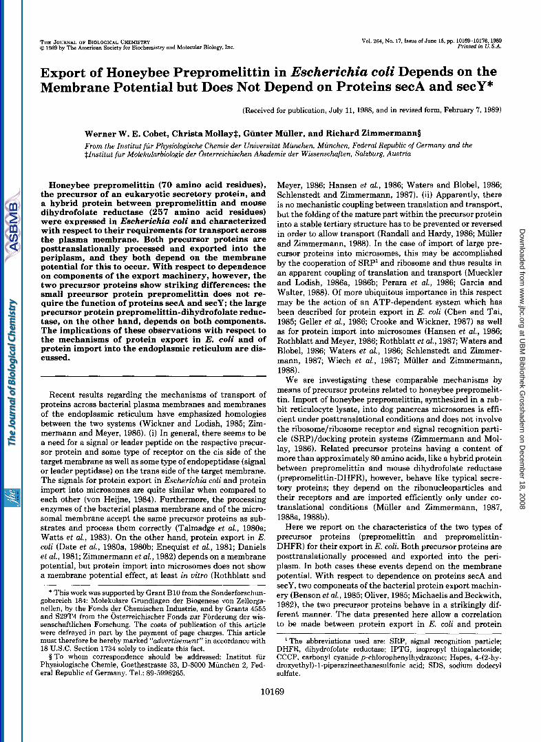
Kinetics of Prepromelittin and Prepromelittin-DHFR Proc- essing in E. coli-In order to facilitate the inducible expression of honeybee prepromelittin and a hybrid protein between prepromelittin and mouse dihydrofolate reductase in E. coli, corresponding DNA fragments were excised from plasmid pSP 65 derivatives, which contain a SP 6-promoter and have previously been used for in vitro experiments (prepromelittin and prepromelittin-DHFR/l, Muller and Zimmermann, 1987), and ligated into a derivative of plasmid pKK 223-3 which contains a trpllac-promoter (tac-promoter) and the gene coding for the lac-repressor (lac"). When wild-type cells were transformed with these plasmids and pulse-labeled with [3H]proline in the presence of isopropyl thiogalactoside, expression of prepromelittin or prepromelittin-DHFR was observed (Fig. 1, A and C). No expression was detected in the absence of the inducer (Fig. 1, A , lanes 1 versus 3, and C, lanes 9 versus 1 ). The identity of the induced translation products was confirmed by immunoprecipitation with antiserum di- rected against melittin and dihydrofolate reductase, respec- tively (Fig. 1, A and C, lanes 11 ), and coelectrophoresis with in vitro synthesized produces (data not shown).
* A. Pluckthun, personal communication.
A " . . , . . + . + IPTG """ * . + . 0 18 0 6 12 18 0 6 I2 18 0
CCCP 18 T l m e
1 2 3 4 5 6 7 8 9 1 0 I 1 12
qII)rrE*ioL,.rrI*.r.II,
"1 B
total Drotein 1 ppm+pm ' rn 10 h
2 3 4 5
lane number
c . . . . * . . . 0 14 28 42 0 14 28 42 0 42
1 2 3 4 5 6 7 8 9 1 0
" " . . . I . * I P T G
0 28 Tlrne - CCCP
I I 12
"
FIG. 1. Kinetics and CCCP sensitivity of prepromelittin (pprn) and prepromelittin-DHFR processing in E. coli HJM 114 cells (wt). Two min after incubation in the presence (+) or absence (-) of IPTG (final concentration: 2.5 mM), cultures of E. coli HJM 114 (0.4 ml), transformed with a plasmid coding for prepro- melittin ( A ) or prepromelittin-DHFR (C), were pulse-labeled for 90
at UB
M B
ibliothek Grosshadern on D
ecember 18, 2008
ww
w.jbc.org
Dow
nloaded from

Protein Export in E. coli 10171
Processing of the eukaryotic precursor proteins took place in the transformed cells, after induction and pulse labeling, when the incubation was continued in the presence of an excess of nonradioactive proline (chase) (Fig. 1, A, lanes 3 through 6, and C, lanes 1 through 4 ) . The processing of prepromelittin was slow (time required to obtain 50% proc- essing = tH = 15 min) compared to processing of the precursor of the outer membrane protein OmpA, pro-OmpA (tIh = 15 5).
Processing of prepromelittin-DHFR was even slower (tlh = 45 min). The identity of the processing products was confirmed by immunoprecipitation (Fig. 1, A , and C, lanes 12) and coelectrophoresis with in vitro processed products (data not shown). In the case of prepromelittin, radiosequencing of the product obtained after processing of prepromelittin by puri- fied leader peptidase in mixed micellar solutions (Fig. 2, A and B ) and in vivo (Fig. 2C) was carried out. The fact that authentic processing was observed under both conditions shows that the processing observed in vivo was correct and was carried out by leader peptidase. It therefore seems safe to conclude that both precursor proteins are correctly processed by bacterial leader peptidase but that processing is slow as compared to authentic precursor proteins. We conclude that processing of prepromelittin and prepromelittin-DHFR were posttranslational events, since there was no synthesis of la- beled proteins under the chase conditions (e.g. no increase in the acid-precipitable radioactivity and the sum of preprome- littin plus promelittin (Fig. 123)).
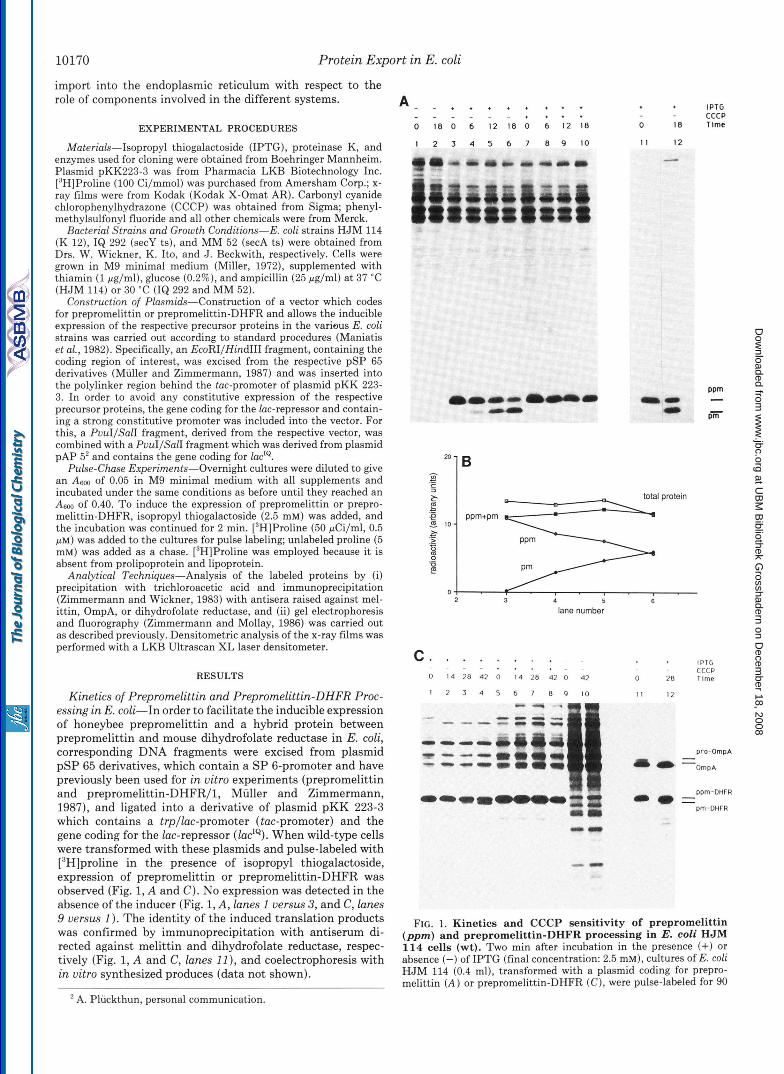
Processing also reflects translocation across the plasma membrane since the mature forms were located in the peri- plasmic space according to two independent approaches: (i) When cells were fractionated according to established proce- dures (Neu and Heppel, 1965) following a pulse-chase incu- bation, the mature proteins, promelittin and promelittin- DHFR, respectively, were recovered in a shock fluid (i.e. the periplasmic fraction), whereas the precursor forms were dis- tributed between the cytoplasm and membrane fractions to a different degree (Fig. 3, A and B, lanes 3 versus 4 and 5). There was no apparent secretion of either mature protein into the medium (lanes 2) . The validity of the fractionation pro- cedure was controlled by marker proteins for the cytoplasm (P-galactosidase), the periplasm (p-lactamase), and a mem- brane fraction which contains inner and outer membrane (OmpA) (Fig. 3C). In order to get good recovery of the mature hybrid protein, salt (0.2 M KCl) had to be added after the osmotic shock, which did not, however, change the distribu- tion of the marker proteins (data not shown). (ii) When the outer membrane of the cells was permeabilized after pulse- chase incubation and the permeabilized cells were exposed to externally added protease (Zimmermann and Wickner, 1983), the mature proteins were protease-sensitive, whereas the cor- responding precursor proteins were protease-resistant (Fig. 4, lanes 1 through 4). The fact that the precursor proteins were protease sensitive when detergent was included (lunes 6), in order to breach the permeability barrier made up by the
s at 37 “C with 20 pCi of [3H]proline and then chased for 30 s with unlabeled proline (10,000-fold molar excess). The cultures were then supplemented with either ethanol (final concentration: 0.2%, v/v) or 10 mM CCCP (final concentration: 20 pM) in ethanol. Following incubation for the indicated times, aliquots (0.1 ml) were removed and mixed with cold trichloroacetic acid (0.4 ml, lo%, w/v). The resulting precipitates were analyzed immediately (lanes 1-10) or following immunoprecipitation (lanes 11 and 12) with antisera di- rected against melittin ( A ) or dihydrofolate reductase and OmpA ( C ) by SDS-polyacrylamide gel electrophoresis and fluorography as de- scribed under “Experimental Procedures.” Densitometric analysis of the x-ray films ( B ) related to part A yielded arbitrary units for single proteins (prepromelittin, promelittin ( p m ) or the sum of all acid- precipitable material (total protein).
4 0 4 e 12 0 4
distance from origin (cm)
0 0 2 4 6 8 1 0 1 2
degradation cycle
FIG. 2. Processing of prepromelittin in E. coli and by puri- fied E. coli leader peptidase in detergent. A and B, total RNA from bee venom glands was translated in a wheat germ system in the presence of [3H]alanine (Mollay et al., 1982; Zimmermann and Mol- lay, 1986). The translation products (100 pl) were adjusted to 0.2 M Hepes-KOH, pH 7.6, and 0.01% (v/v) Triton X-100 and incubated with E. coli leader peptidase (1 pg) for 60 min at 37 “C. The yield of promelittin under these conditions was approximately 50% (as judged by electrophoretic analysis). The processing products were isolated by extraction with 1-butanol (Mollay et al., 1982; Zimmermann and Mollay, 1986). Promelittin, contained in the aqueous phase and purified by paper chromatography, as well as prepromelittin, present in the interface, were digested with chymotrypsin. The resulting characteristic acidic fragments then were isolated by high voltage electrophoresis at pH 4.7 ( A and B; the positions of marker amino acids, Leu and Glu, and the origin, arrow, are indicated). Subsequent manual Edman degradation (data not shown) revealed that alanine was present in positions 1,2, and 8 in the case of the peptide derived from prepromelittin ( A ) , whereas the peptide derived from promelit- tin ( B ) had alanine in positions 1 and 7 (for comparison, authentic promelittin contains alanine in positions 1 and 7 ) . Thus it was shown that the product of leader peptidase is authentic promelittin with a correct amino terminus. Likewise, the second product of the reaction, signal peptide, could be identified as described previously (Mollay et al., 1982) and proved to have the correct carboxyl terminus (data not shown). C, two min after incubation in the presence of IPTG, a culture of E. coli HJM 114 (0.4 ml), transformed with a plasmid coding for prepromelittin, was pulse-labeled for 90 s at 37 “C with 20 pCi of [3H]proline and then chased for 18.5 min with unlabeled proline. Then the culture was subjected to precipitation with cold trichloroacetic acid (1.6 ml, lo%, w/v). The resulting precipitate was dissolved in sample buffer and subjected to SDS-polyacrylamide gel electrophoresis. Promelittin was eluted from the gel and subjected to 12 steps of Edman degradation (for comparison, authentic promelittin contains proline in positions 2, 4, 6, 8, 10, 20, and 36).
plasma membrane, allows us to conclude that the protease sensitivity of the mature proteins was due to their location in the periplasm.
Expression of prepromelittin and prepromelittin-DHFR was lethal for the cells; they survived for about 2 h after
at UB
M B
ibliothek Grosshadern on D
ecember 18, 2008
ww
w.jbc.org
Dow
nloaded from
10172 Protein Export i n E. coli
L 3 4 5
S P C M
B T S P C ~ 1 2 3 4 5
Medlum Periplasm Cytoplasm Membranes
") 1
I 2 3 4
nedlum Perlplasrn Cytoplasm Membranes
FIG. 3. Localization of prepromelittin (ppm)/promelittin (pm) and prepromelittin-DHFR/promelittin-DHFR by cell fractionation. Two min after incubation in the presence of IPTG, cultures of E. coli HJM 114 (0.75 ml), transformed with a plasmid coding for prepromelittin ( A and C ) or prepromelittin-DHFR ( B ) ,
addition of IPTG, as monitored by their ability to synthesize proteins (data not shown). No further cell growth, however, was detected during this time period as deduced from optical density measurements. Cell death was not a result of melittin action since related hybrid proteins between prepromelittin and dihydrofolate reductase (prepromelittinA-DHFR/l, ppd- DHFR/l, pa-DHFR/l) which have deletions in the promelit- tin part and are competent for import into microsomes (Muller and Zimmermann, 1987) were also toxic. Rather, interference with the export of authentic E. coli proteins might account for this toxicity.
CCCP Sensitivity of Prepromelittin and Prepromelittin- DHFR Processing-In the following experiments we ad- dressed the question of whether export of prepromelittin and prepromelittin-DHFR shows the same requirements as the export of authentic precursor proteins. As a first step we asked whether export of the two eukaryotic precursor proteins in E. coli depends on a membrane potential. Cells transformed with the respective plasmids were pulse-labeled and the un- coupler carbonyl cyanide chlorophenylhydrazone (CCCP) was added during the chase (Fig. lA, lanes 7 through 10, and C, lanes 5 through 8). Processing of both precursor proteins (prepromelittin and prepromelittin-DHFR) was inhibited un- der these conditions. The accumulated precursor proteins were recovered in cytoplasmic and membrane fractions with the same distribution as in the absence of CCCP (data not shown). The export of eukaryotic secretory proteins in E. coli therefore depends on a membrane potential. Since it has been suggested that the membrane potential acts directly on the precursor proteins (Daniels et al., 1981; Zimmermann et al., 1982) we conclude that even a eukaryotic precursor protein, which does not seem to depend on a membrane potential for import into the endoplasmic reticulum, has the ability to sense a membrane potential in E. coli.
Kinetics of Prepromelittin and Prepromelittin-DHFR Proc- essing in IQ 292 (secY ts) and MM 52 (secA ts)-We next studied the role of proteins secA and secY, which have been described as components of the export machinery, during the
were pulse-labeled for 90 s at 37 "C with 37.5 pCi of [3H]proline and chased for 13 min (prepromelittin) and 43 min (prepromelittin- DHFR), respectively, with unlabeled proline. The cultures were then chilled on ice and divided into two parts (0.1 and 0.65 ml, respec- tively). The one part (0.1 ml) was kept on ice (T), the other part (0.65 ml) was subjected to centrifugation (10 min at 4 "C and at 10,000 rpm, rotor JA 20, Beckman 52-21). The supernatant contain- ing the medium was removed and kept on ice (S), the pellet was resuspended in 0.35 ml of cold 20% (w/v) sucrose, 30 mM Tris-C1, pH 8.0, adjusted to 1 mM EDTA and subjected to centrifugation as above. The supernatant was discarded; the pellet was resuspended in 0.35 ml of cold water and again subjected to centrifugation. (Alternatively, the pellet was resuspended in water, subsequently adjusted to 0.2 M KCl, and then subjected to centrifugation (B).) The supernatant which contained the periplasmic fraction was removed and kept on ice (P) ; the pellet was resuspended in 0.35 ml of cold water and subjected to sonication (2 min at 0 "C and at output 3, 50% duty, Branson B15 sonifier with microtip). The subsequent centrifugation (10 min at 4 "C and at 20,000 rpm) yielded a supernatant which contained the cytoplasmic fraction ( C ) and a pellet which was resus- pended in 0.35 ml of cold water and contained the membranes (M). All samples were subjected to acid precipitation and analyzed imme- diately (prepromelittin, promelittin) or following immunoprecipita- tion (prepromelittin-DHFR, promelittin-DHFR, and OmpA) by SDS- polyacrylamide gel electrophoresis and fluorography as described under "Experimental Procedures." Densitometric analysis of the x- ray films yielded arbitrary units for OmpA. In a parallel experiment without radiolabeling, fractions were collected for analysis of the distribution of marker enzymes between the various fractions. Activ- ities for @-galactosidase and @-lactamase were determined according to established procedures (Miller, 1972; O'Callaghan et al., 1972) and are given as percentage of total activities.
at UB
M B
ibliothek Grosshadern on D
ecember 18, 2008
ww
w.jbc.org
Dow
nloaded from

Protein Export in E. coli 10173
21 A
c
B 1 pm-DHFR
1 2 3 4 5 6 PK 0 100 200 300 400 250
Triton - - - - - + FIG. 4. Localization of prepromelittin (pprn)/promelittin
(pm) and prepromelittin-DHFR/promelittin-DHFR by sus- ceptibility to protease. Two min after incubation in the presence of IPTG, cultures of E. coli HJM 114 (1.20 ml), transformed with a plasmid coding for prepromelittin ( A ) or prepromelittin-DHFR ( B ) , were pulse-labeled for 90 s a t 37 "C with 60 pCi of [3H]proline and chased for 13 min (prepromelittin and 43 min (prepromelittin- DHFR), respectively, with unlabeled proline. The cultures were then mixed with 1.2 ml of cold 40% (w/v) sucrose, 20 mM EDTA, 60 mM Tris-CI, pH 8.1. Each sample was divided into six aliquots and incubated for 1 h a t 0 "C without further addition or in the presence of proteinase K (PK) , a t the final concentrations as indicated, or with proteinase K plus Triton X-100 (0.5%, v/v). After addition of phenylmethylsulfonyl fluoride (2 mM), the samples were subjected to acid precipitation and analyzed immediately (prepromelittin) or fol- lowing immunoprecipitation (prepromelittin-DHFR) by SDS-poly- acrylamide gel electrophoresis and fluorography as described under "Experimental Procedures." Densitometric analysis of the x-ray films yielded arbitrary units.
export of prepromelittin and prepromelittin-DHFR. In order to do so E. coli conditional lethal mutants MM 52 (secA ts) and IQ 292 (secY ts) were transformed with the plasmids of interest. The expression of prepromelittin andprepromelittin- DHFR, respectively, was then induced and pulse-chase exper- iments were performed a t various temperatures and at various times after shift from the permissive (30 "C) to the nonper- missive (42 "C) temperature. Pro-OmpA, which has been shown to depend on both components (Wolfe et al., 1985; Bacallao et al., 1986; Fandl and Tai, 1987; MacIntyre et al., 1987; Kuhn et al., 1987), was used as an internal control for the expression of the mutant phenotype. The processing of pro-OmpA to OmpA was progressively slowed in both mutant
" lllF
Temperature Time
- pro-0mpA OmpA
- ppm-DHFR - pm-DHFR
'"1 0-
0 20 40 60 80 shift time (mid
FIG. 5. Kinetics of prepromelittin-DHFR processing in I& 292 cells (secY ts) and in MM 52 cells (secA ts). Cultures of E. coli HJM 114, IQ 292, or MM 52 (0.4 ml), each transformed with a plasmid coding for prepromelittin-DHFR, were shifted from 30 "C to either 37 "C, 40 "C, or 42 "C for the times indicated ( B ) . Two min after addition of IPTG (2.5 mM), the cultures were pulse-labeled for 90 s with 20 pCi of [3H]proline and chased for 30 s with unlabeled proline (10,000-fold molar excess). Following further incubation for the indicated times ( A ) , aliquots (0.1 ml) were removed and mixed with cold trichloroacetic acid (0.4 ml, 10% w/v). The precipitates were analyzed immediately by SDS-polyacrylamide gel electrophore- sis and fluorgraphy as described under "Experimental Procedures." The data from an experiment with IQ 292 shifted from 30 "C to the temperatures indicated for 40 min are shown ( A ) . Densitometric analysis of the x-ray films from a parallel experiment, carried out at 40 "C and chase times up to 35 min, yielded arbitrary units; these were used to determine the times required to yield 50% processing ( b h ) which are plotted against the shift times ( B ) . e, prepromelittin- DHFR in MM 52; A, prepromelittin-DHFR in IQ 292; B, prepro- melittin-DHFR in HJM 114. ppm, prepromelittin; pm, promelittin.
strains with higher temperatures and longer shift times (Figs. 5A and 6). Processing of prepromelittin-DHFR was likewise affected by both mutations (Fig. 5A, lanes 112 versus 314 and 5/6, and B) . Within the same time periods of shift to the nonpermissive temperature, however, no alteration in the kinetics of prepromelittin processing was observed in either mutant (Fig. 6, A and B ) . We conclude that the small precur- sor protein prepromelittin does not depend on either secA or secY, while the related but extended precursor protein pre- promelittin-DHFR behaves like pro-OmpA.
DISCUSSION
Export of Foreign Proteins in E. coli-For various reasons there have been attempts to accomplish export of otherwise not exported proteins from E. coli cells. These include (i) the expression of precursors of eukaryotic secretory proteins, like rat preproinsulin (Villa-Komaroff et al., 1978; Talmadge et al., 1980b) or chicken ovalbumin (Fraser and Bruce, 1978);
at UB
M B
ibliothek Grosshadern on D
ecember 18, 2008
ww
w.jbc.org
Dow
nloaded from

10174 Prote in Expor t in E. coli
A 0 rnln 20 rnln 40 rnin 42.C 0 6 12 18 0 6 12 18 0 6 12 18 Time
1 2 3 4 5 6 7 8 9 1 0 1 1 1 2
0 rnin 20 rnln 0 6 12 I8 0 6 12 18 0
1 2 3 4 5 6 7 8 9 - 40 rnln 420c 6 12 18 Tlrne
IO 1 1 12
B 4 1 pro-0mpA
3 -I h c I /
P
0 20 40 60 80
0 20 40 60 80 shift time ( m i d
FIG. 6. Kinetics of prepromelittin processing in IQ 292 cells (secY ts) and in MM 52 cells (secA ts). Cultures of E. coli HJM 114, IQ 292 or MM 52 (0.4 ml), each transformed with a plasmid coding for prepromelittin, were shifted from 30 to 42 “C for the times indicated. Two min after addition of IPTG (2.5 mM) the cultures were pulse-labeled for 90 s with 20 pCi of [3H]proline and chased for 30 s with unlabeled proline (10,000-fold molar excess). Following further incubation for the indicated times, aliquots (0.1 ml) were removed and mixed with cold trichloroacetic acid (0.4 ml, lo%, w/v). The precipitates were analyzed immediately (prepromelittin ( p p m ) and promelittin ( p m ) ) or following immunoprecipitation (pro-OmpA and OmpA) by SDS-polyacrylamide gel electrophoresis and fluorog- raphy as described under “Experimental Procedures.” The data from an experiment with IQ 292 are shown (A). Densitometric analysis of the x-ray films yielded arbitrary units; these were used to determine the times required to yield 50% processing (t8,+) which are plotted against the shift times ( B ) . 6, prepromelittin in MM 52; A, prepro- melittin in IQ 292; W, prepromelittin in HJM 114; 0, pro-OmpA in MM 52; A, pro-OmpA in IQ 292; 0, pro-OmpA in HJM 114.
(ii) the construction of hybrid proteins between prokaryotic signal or leader peptides and eukaryotic proteins, like rat proinsulin (Talmadge et al., 1980a) or chicken triosephosphate isomerase (Kadonaga et al., 1984; Pluckthun and Knowles, 1987); and (iii) the construction of hybrid proteins between prokaryotic signal or leader peptides and prokaryotic proteins which normally are not exported, such as p-galactosidase (Moreno et al., 1980; Benson et al., 1984; Tomassen et al., 1984; Bassford et al., 1979; Ito et al., 1981) or a part of a tail fiber protein of phage T4 (MacIntyre et al., 1987). The general conclusions from these studies were that eukaryotic signal peptides can function in E. coli but do so rather inefficiently, and that no general rule can be established with respect to the effectiveness of a certain signal or leader peptide in directing the export of passenger proteins. The data presented here support the view that the eukaryotic and prokaryotic signal peptides, as well as their respective processing enzymes, are interchangeable. The authentic eukaryotic precursor pro- tein prepromelittin is exported across the plasma membrane and is correctly processed by bacterial leader peptidase. Fur- thermore, mouse dihydrofolate reductase, which has been previously used in hybrid proteins directed to microsomes, is also a suitable passenger protein for export in E. coli.
Implications for the Mechanism of Export of Proteins in E. coli-There appear to be two classes of precursor proteins with respect to their mechanism of import into mammalian microsomes (Muller and Zimmermann, 1987; Schlenstedt and Zimmermann, 1987). One class consists of precursor proteins with a content of more than approximately 80 amino acid residues (including the signal peptide); the other class consists of precursor proteins comprising less than about 80 amino acid residues. I t is important to note in this context that approximately 40 amino acid residues of a nascent polypeptide are buried within the ribosome and that a typical signal peptide contains 20-30 amino acid residues. The two mecha- nisms differ in several aspects from each other; these various aspects, however, are related to each other. The transport of large precursor proteins involves the ribosome and SRP and their respective receptors on the microsomal surface (Mueck- ler and Lodish, 1986a, 1986b; Perara et al., 1986; Garcia and Walter, 1988). The transport of small precursor proteins does not involve the ribosome or SRP nor the respective receptors (Mollay and Zimmermann, 1986; Muller and Zimmermann, l987,1988a, 1988b; Schlenstedt and Zimmermann, 1987). On the other hand, the small precursor proteins show constraints with respect to their primary structures (specifically, the mature parts), while the large precursor proteins do not show any such constraints (Muller and Zimmermann, 1987). Small precursor proteins can be imported both in a cotranslational or in a posttranslational experimental set-up, whereas large precursor proteins can only be imported in a cotranslational set-up (Muller and Zimmermann, 1988a, 1988b; Schlenstedt and Zimmermann, 1987). The explanation for the various differences seems to come from the following facts. SRP typically binds to signal peptides of nascent polypeptides as soon as they emerge from the ribosome (Walter and Blobel, 1981; Ainger and Meyer, 1986; Wiedmann et al., 1987a). This interaction is proposed to lead to a subsequent SRP-ribosome interaction and to slow down or even block elongation (Walter and Blobel, 1981); this effect on elongation is released by interaction of SRP with its receptor on the microsomal sur- face, the docking protein (Meyer et al., 1982). At this point the signal peptide is believed to be handed over to a putative signal peptide receptor on the microsomal surface, the so- called signal sequence receptor (SSR), and the ribosome is thought to bind to a putative ribosome receptor on the micro-
at UB
M B
ibliothek Grosshadern on D
ecember 18, 2008
ww
w.jbc.org
Dow
nloaded from
Protein Export in E. coli 10175
soma1 surface (Wiedmann et al., 1987b). Since the SRP-signal peptide interaction can occur only as long as the signal peptide is presented to SRP by the ribosome (Ainger and Meyer, 1986; Wiedmann et al., 1987a), the import process appears to be coupled to translation (cotranslational transport). On the other hand, since translation of a small precursor protein is terminated and the polypeptide released from the ribosome before any of these interactions can physically occur, the result is posttranslational import. Because the small precursor proteins cannot make use of this complex system, they have apparently evolved with constraints on the primary structure of their mature part.
Both precursor proteins (prepromelittin and prepromelit- tin-DHFR) are posttranslationally processed in E. coli and exported into the periplasm. Prepromelittin dose not require the function of proteins secA and secY, two components of the bacterial protein export machinery (Benson et al., 1985; Oliver, 1985; Michaelis and Beckwith, 1982; Oliver and Beck- with, 1981; Liss and Oliver, 1986; Ito et al., 1983; Shiba et al., 1984); the prepromelittin-dihydrofolate reductase hybrid pro- tein, however, depends on both components. Other data have to be taken into account in this context. Membrane assembly of M13 procoat protein (73 amino acid residues) also does not depend on secA and secY in E. coli (Wolfe et al., 1985; Kuhn et al., 1987) or SRP and docking protein in the microsomal system (Watts et al., 1983; Wiech et al., 1987), and a procoat protein derivative with an insertion in the mature amino terminus (246 amino acid residues) was secA- and secY- dependent (Kuhn, 1988). Furthermore, shortening of a bac- terial precursor protein (pro-OmpA) or eukaryotic precursor proteins (pre-lysozyme, preprolactin) to about 80 amino acid residues led to an incompetent molecule in the respective transport systems (Freudl et al., 1988; Ibrahimi et al., 1986; Siege1 and Walter, 1988).
There is apparently a direct analogy between the prokar- yotic and the eukaryotic transport systems. There appear to be two mechanisms in the bacterial system, too. Again, the size of the precursor is the decisive feature with respect to which of the mechanisms is used by a given precursor protein. Although we did not test the full set of prepromelittin-related precursor proteins (Muller and Zimmermann, 1987) in E. coli, we suggest that the critical size is again around 80 amino acid residues. By comparison to the eukaryotic system, we fur- thermore conclude that in E. coli there is a role for the ribosome (the only place where this size effect makes any sense) in the export of large precursor proteins as well. There seems to be one or more component(s) involved in the export of large precursor proteins which bind to the precursor protein when it is presented by the ribosome. In this case, however, there does not seem to be an effect of this interaction on translation; therefore, there is no coupling of the export process and translation. It is quite possible that secA, a soluble cytoplasmic component (Oliver and Beckwith, 1982), and secY, a membrane component (Akiyama and Ito, 1985), func- tionally make up the prokaryotic equivalent to SRP and docking protein of the eukaryotic system. Other candidates for a prokaryotic SRP analog are proteins secB (Collier et al., 1988) and trigger factor (Crooke and Wickner, 1987; Crooke et al., 1988). The latter has most recently been described as cycling between cytoplasm, ribosome, and a receptor on the membrane surface, and possibly makes the best candidate for a SRP analog (Lill et al., 1988). If so, secA and secY would have to act subsequently.
Implications for the Mechanism of Import of Proteins into the Endoplasmic Reticulum-Both eukaryotic precursor pro- teins (prepromelittin and prepromelittin-DHFR) are proc-
essed and exported in E. coli in a membrane potential-de- pendent fashion. A striking difference between the two sys- tems, therefore, resides in the involvement of a membrane potential in the prokaryotic cell and the lack of such an effect in the eukaryotic in vitro system. It is possible that a mem- brane potential effect could have been overlooked in the eukaryotic system since only cell-free systems have been used. If this is not the case, it should be interesting to determine the functional substitute of a membrane potential in the microsomal system.
Acknowledgments-We are grateful to Dr. Giinter Kreil for provid- ing the cDNA coding for prepromelittin, to Dr. Dietrich Stiiber for providing both a cDNA coding for dihydrofolate reductase and an antiserum directed toward dihydrofolate reductase, to Dr. Andreas Pliickthun for the DNA coding for lacla, to Dr. William Wickner for E. coli leader peptidase, and to Birgitta Kasseckert, Ulrike Vilas, and Maria Sagstetter for expert technical assistance. Furthermore, we are thankful to Drs. Ulf Henning, Roland Freudl, and Andreas Kuhn for making available to us their results prior to publication, to Dr. Donald W. Nicholson for critical comments on the manuscript, and to Axel Laminet for his support in performing the 8-lactamase assay.
REFERENCES
Ainger, K. J., and Meyer, D. I. (1986) EMBO J. 5, 951-955 Akiyama, Y., and Ito, K. (1985) EMBO J. 4, 3351-3356 Bacallao, R., Crooke, E., Shiha, K., Wickner, W., and Ito, K. (1986)
Bassford, P. J., Jr., Silhavy, T. J., and Beckwith, J. R. (1979) J.
Benson, S. A., Bremer, E., and Silhavy, T. J. (1984) Proc. Natl. Acad.
Benson, S. A., Hall, M. N., and Silhavy, T. J. (1985) Annu. Reu.
Chen, L., and Tai, P. C. (1985) Proc. Natl. Acad. Sci. U. S. A . 82,
Collier, D. N., Bankaitis, V. A., Weiss, J. B., and Bassford, P. J.
Crooke, E., and Wickner, W. (1987) Proc. Natl. Acad. Sci. U. S. A.
Crooke, E., Guthrie, B., Lill, R., Lecker, S., and Wickner, W. (1988)
Daniels, C. J., Bole, D. G., Quay, S. C., and Oxender, D. L. (1981)
J. Biol. Chem. 261, 12907-12910
Bacteriol. 139, 19-31
Sci. U. S. A. 81, 3830-3834
Biochem. 54, 101-134
4384-4388
(1988) Cell 53, 273-283
84,5216-5220
Cell 54, 1003-1011
Proc. Natl. Acad. Sci. U. S. A. 78, 5396-5400 Date, T., Goodman, J. M., and Wickner, W. (1980a) Proc. Natl. Acad.
Sci. U. S. A. 77, 4669-4673 Date, T., Zwizinski, C., Ludmerer, S., and Wickner, W. (1980b) Proc.
Enequist, H. G., Hirst, T. R., Harayama, S., Hardy, S. J. S., and
Fandl, J. P., and Tai, P. C. (1987) Proc. Natl. Acad. Sci. U. S. A. 84,
Fraser, T., and Bruce, G. (1978) Proc. Natl. Acad. Sci. U. S. A. 75,
Freudl, R., Schwarz, H., Degen, M., and Henning U. (1989) J. Mol.
Garcia, P. D., and Walter, P. (1988) J. Cell Biol. 106, 1043-1048 Geller, B., Mowa, N. R., and Wickner, W. (1986) Proc. Natl. Acad.
Hansen, W., Garcia, P. D., and Walter, P. (1986) Cell 45, 397-406 Ibrahimi, I. M., Cutler, D., Stueber, D., and Bujard, H. (1986) Eur.
Ito, K., Bassford, P. J., and Beckwith, J. (1981) Cell 24, 707-717 Ito, K., Wittekind, M., Nomura, M., Shiba, K., Yura, T., Miura, A.,
and Nashimoto, H. (1983) Cell 32, 789-797 Kadonaga, J. T., Gautier, A. E., Strauss, D. R., Charles, A. D., Edge,
M. D., and Knowles, J. R. (1984) J. Biol. Chem. 259, 2149-2154 Kuhn, A. (1988) Eur. J. Biochem. 177, 267-271 Kuhn, A., Kreil, G., and Wickner, W. (1987) EMBO J. 6, 501-505 Lill, R., Crooke, E., Guthrie, B., and Wickner, W. (1988) Cell 54,
Liss, L. R., and Oliver, D. B. (1986) J. Biol. Chem. 261, 2299-2303 MacIntyre, S., Freudl, R., Degen, M., Hindennach, I., and Henning,
Maniatis, T., Fritsch, E. F., and Sambrook, J. (1982) Molecular
Natl. Acad. Sci. U. S. A. 77,827-831
Randall, L. L. (1981) Eur. J. Biochem. 116, 227-233
7448-7452
5936-5940
Biol.,
Sci. U. S. A. 83,4219-4222
J. Biochem. 155,571-576
1013-1018
U. (1987) J. Biol. Chem. 262,8416-8422
at UB
M B
ibliothek Grosshadern on D
ecember 18, 2008
ww
w.jbc.org
Dow
nloaded from
10176 Protein Export in E. coli Cloning: A Laboratory Manual, Cold Spring Harbor Laboratory, Cold Spring Harbor, NY
Meyer, D. I., Krause, E., and Dobberstein, B. (1982) Nature 297, 647-650
Michaelis, S., and Beckwith, J. (1982) Annu. Rev. Microbiol. 36,435- 465
Miller, J. H. (1972) Experiments in Molecular Genetics, Cold Spring Harbor Laboratory, Cold Spring Harbor, NY
Mollay, C., Vilas, U., and Kreil, G. (1982) Proc. Natl. Acad. Sci.
Moreno, F., Fowler, A. V., Hall, M., Silhavy, T. J., Zabin, I., and
Mueckler, M., and Lodish, H. F. (1986a) Cell 44 , 629-637 Mueckler, M., and Lodish, H. F. (1986b) Nature 322,549-552 Muller, G., and Zimmermann, R. (1987) EMBO J. 6 , 2099-2107 Muller, G., and Zimmermann, R. (1988a) EMBO J . 7,639-648 Miiller, G., and Zimmermann, R. (1988b) in Gene Expression and
Regulation (Bissel, M., Deho, G., Sironi, G., and Torriani, A., eds) pp. 199-208, Elsevier Science Publishers B. V., Amsterdam
Neu, H. C., and Heppel, L. A. (1965) J. Biol. Chem. 240,3685-3692 OCallaghan, C. H., Morris, A., Kirby, S. M., and Shingler, A. H.
(1972) Antimicrob. Agents Chemother. 1, 283-288 Oliver, D. (1985) Annu. Reu. Microbiol. 39,615-648 Oliver, D. B., and Beckwith, J. (1981) Cell 25, 765-772 Oliver, D. B., and Beckwith, J. (1982) Cell 30, 311-314 Perara, E., Rothman, R. E., and Lingappa, V. R. (1986) Science 2 3 2 ,
Pluckthun, A., and Knowles, J. R. (1987) J. Biol. Chem. 262 , 3951-
Randall, L. L., and Hardy, S. J. S. (1986) Cell 46,921-928 Rothblatt, J. A., and Meyer, D. I. (1986) EMBO J. 5, 1031-1036 Rothblatt, J. A., Webb, J. R., Ammerer, G., and Meyer, D. I. (1987)
Schlenstedt, G., and Zimmermann, R. (1987) EMBO J. 6,699-703
U. S. A. 79,2260-2263
Schwartz, M. (1980) Nature 286, 356-359
348-352
3957
EMBO J. 6,3455-3464
Shiba, K., Ito, K., Yura, T., and Ceretti, D. P. (1984) EMBO J. 3 ,
Siegel, V., and Walter, P. (1988) EMBO J. 7, 1769-1775 Talmadge, K., Kaufman, J., and Gilbert, W. (1980a) Proc. Natl. Acad.
Sci. U. S. A. 7 7 , 3988-3992 Talmadge, K., Stahl, S., and Gilbert, W. (1980b) Proc. Natl. Acud.
Sci. U. S. A. 7 7 , 3369-3373 Tommassen, J., Leunissen, J., van Damme-Jongsten, M., and Over-
duin, P. (1985) EMBO J. 4 , 1041-1047 Villa-Komaroff, L., Efstratiatis, A., Broome, S., Lomedico, P., Tizard,
R., Naber, S. P., Chick, W. L., and Gilbert, W. (1978) Proc. Natl. Acud. Sci. U. S. A. 75,3727-3731
631-635
von Heijne, G. (1984) EMBO J. 3 , 2315-2318 Walter, P., and Blobel, G. (1981) J. Cell Biol. 9 1 , 557-561 Waters, M. G., and Blobel, G. (1986) J. Cell Biol. 102 , 1543-1550 Waters, M. G., Chirico, W. J., and Blobel, G. (1986) J. Cell Biol. 103,
Watts, C., Wickner, W., and Zimmermann, R. (1983) Proc. Natl.
Wickner, W., and Lodish, H. (1985) Science 230,400-407 Wiech. H.. Saestetter. M.. Muller. G.. and Zimmermann. R. (1987)
2629-2636
Acad. Sci. U. S. A. 80,2809-2813
, . EMbO 2. 6,-1011-1016’
(1987a) J. Cell Biol. 104 , 201-208
A. (198713) Nature 328,830-833
Wiedmann, M., Kurzchalia, T. V., Bielka, H., and Rapoport, T. A.
Wiedmann, M., Kurzchalia, T. V., Hartmann, E., and Rapoport, T.
Wolfe, P. B., Rice, M., and Wickner, W. (1985) J. Biol. Chem. 2 6 0 ,
Zimmermann, R., and Meyer, D. I. (1986) Trends Biochem. Sci. 11 ,
Zimmermann. R., and Mollas, C. (1986) J. Biol. Chem. 2 6 1 , 12889-
1836-1841
512-515 . . - .
12895
3925 Zimmermann, R., and Wickner, W. (1983) J . BioZ. Chem. 268,3920-
Zimmermann, R., Watts, C., and Wickner, W. (1982) J. Biol. Chem. 257,6529-6536
at UB
M B
ibliothek Grosshadern on D
ecember 18, 2008
ww
w.jbc.org
Dow
nloaded from